Article

An In Silico Feasibility Study of Dose-Escalated Hypofractionated Proton Therapy for Rectal Cancer

1
Department of Nuclear Medicine and Medical Physics, Karolinska University Hospital, 14186 Stockholm, Sweden
2
Department of Oncology-Pathology, Karolinska Institute, 17177 Stockholm, Sweden
3
Danish Centre for Particle Therapy, Aarhus University Hospital, 8200 Aarhus, Denmark
4
Department of Clinical Medicine, Aarhus University, 8200 Aarhus, Denmark
5
Department of Oncology, Oslo University Hospital, 0424 Oslo, Norway
6
Institute of Clinical Medicine, University of Oslo, 0424 Oslo, Norway
7
Cancer Clinic, Haukeland University Hospital, 5009 Bergen, Norway
8
Institute of Physics and Technology, University of Bergen, 5007 Bergen, Norway
9
Department of Radiation Oncology, Karolinska University Hospital, 17176 Stockholm, Sweden
*
Author to whom correspondence should be addressed.
Cancers 2025, 17(16), 2627; https://doi.org/10.3390/cancers17162627
Submission received: 19 June 2025 / Revised: 25 July 2025 / Accepted: 4 August 2025 / Published: 11 August 2025
(This article belongs to the Special Issue The Advance of Pencil Beam Scanning Proton Beam Therapy in Cancers)

Simple Summary

The incidence of colorectal cancer is increasing in young adults. Treatment often includes extensive surgery with burdensome late side effects. Radiation therapy along with chemotherapy is often used in combination before surgery and, in some cases, renders surgery unnecessary. The purpose of this study is to investigate the possibility of delivering larger radiation doses to patients without elevating the dose to healthy organs and tissue in the vicinity of the rectum. We considered two modalities of radiation: protons and photons. Our results indicate that it should be safe to give these higher doses with both radiation modalities. With protons the dose given to healthy tissue can be approximately reduced by half, compared to photons. A clinical trial would be required to verify the efficacy of larger doses to the tumor.

Abstract

Background/Objectives: The current standard treatment for locally advanced rectal cancer (LARC) is neoadjuvant chemoradiotherapy, or total neoadjuvant therapy (TNT), followed by total mesorectal excision (TME). If the neoadjuvant treatment results in a clinical complete response (cCR), non-operative management of LARC might be possible. It is hypothesized that cCR rates will increase with increasing radiotherapy doses. By using proton therapy, doses to organs at risk (OAR) may be decreased. In preparation for a clinical trial on dose-escalated proton therapy for LARC, the purpose of this study is to establish the feasibility of proton therapy for dose-escalated hypofractionated radiotherapy of LARC. Methods: Ten patients, having previously received short course radiotherapy (SCRT) for LARC, were included in this planning study. Two photon plans and two proton plans were created for each patient: one with a standard 5 × 5 Gy fractionation and one dose-escalated up to 5 × 7 Gy. Proton plans were robustly optimized. For all plans the integral dose (ID) was computed, and for the proton plans relative biological effectiveness (RBE) distributions were calculated. Feasibility was assessed in terms of target coverage and OAR doses. Results: All treatment plans satisfied target coverage criteria. Three of the photon and two of the proton dose-escalated plans exceeded recommended OAR objectives. Proton IDs were on average lower by a factor of 1.97 compared to photon IDs. Mean doses to OAR were, in general, lower for protons. All proton RBE values in the escalated target volumes were between 1.09 and 1.16. Conclusions: The proposed dose escalation was found to be feasible. Protons can reduce the integral dose and mean doses to OARs compared to photons in both the dose-escalated and non-escalated cases. Differences in RBE between escalated and standard fractionation were small.

1. Introduction

Colorectal cancer is the third most common form of cancer globally, with increasing rates among young adults [1,2,3]. The standard of care for locally advanced rectal cancer (LARC) is neoadjuvant chemoradiotherapy (CRT) or total neoadjuvant therapy (TNT) followed by total mesorectal excision (TME) [4,5,6,7]. TME may come with significant sequelae [8], with recent advances in surgical techniques showing no significant improvement in post-surgery quality of life [9].
The radiotherapy (RT) doses typically given in long-course radiotherapy (LCRT) are 45–50 Gy in 1.8–2 Gy fractions [10,11]. Increasing the RT dose may increase clinical complete response (cCR) rates, with one study suggesting elevated tumor dose response in the 50.4–70 Gy interval [12]. In addition, increasing the dose may be particularly important in unresectable cases [13]. If cCR is achieved, it may be followed by a watch and wait approach [14,15] or a local excision with reduced sequelae compared to TME [16,17,18]. Although increasing RT doses may lead to increased toxicity, a meta-analysis found no increase in grade 3 toxicities with dose-escalated RT as compared to conventionally fractionated RT [19].
Proton therapy (PT) is a modality that may significantly reduce the integral dose (ID) compared to conventional photon radiotherapy (xRT) [20]. This is particularly important for younger patients with longer expected remnant lifespans, where a lower ID may reduce the risk of radiation-induced cancers [21,22]. Planning studies have identified reduced doses to the bladder, femoral heads, and bowel bag as possibly significant advantages of PT over xRT [23,24,25] for rectal cancer. These advantages may enable a more aggressive chemotherapy treatment [26]. Reduced organ at risk (OAR) doses may allow for even further dose escalation with PT, and thus increased cCR, with tolerable toxicity levels [27]. Clinical trials confirming these findings are lacking; however, one ongoing study investigates dose-escalated proton reirradiation for rectal cancer (RE-RAD II).
The increased costs and more limited access to PT as compared to xRT may make the former unfeasible, especially with LCRT; hypofractionated alternatives may be more cost-effective [28]. A short course radiotherapy (SCRT) approach, giving 25 Gy in 5 fractions, has been found to be a viable alternative to LCRT, both alone and as part of TNT, in terms of tumor control and OAR toxicity [29,30]. The optimal fractionation scheme is yet to be determined. However, SCRT is the preferred RT approach as part of the total neoadjuvant treatment in Sweden. A moderately low α/β ratio of 5 [31] indicates improved tumor control by using hypofractionation, but the same study found increased loco-regional control rates for hyperfractionation. A current phase I study is ongoing [32], comparing 5 × 6 Gy, 5 × 7 Gy, and 5 × 8 Gy schemes using xRT.
The ongoing PRORECT (NCT04525989) phase II trial [33] compares PT and xRT SCRT. The purpose of this article is to assess the feasibility of dose-escalated PT SCRT compared to xRT SCRT in preparation for a Nordic follow up study, PRORECT II. In the PRORECT II study, the primary tumor will be prescribed a dose of 5 × 6 Gy, and pathological lateral lymph nodes (LLN) will be prescribed a dose of 5 × 7 Gy in a simultaneous integrated boost (SIB), with the remaining part of the CTV receiving 5 × 5 Gy. In this we are following the approach of the NORMAL-R study [34,35]. The feasibility will be assessed in terms of achievable OAR doses and integral dose, given that target coverage is compliant with a set of criteria based on local practice at Karolinska University Hospital and the PRORECT study protocol.

2. Materials and Methods

2.1. Patient Data

Ten patients, already treated as part of the PRORECT study, were included in this study. The original planning computed tomography (CT) and structure sets were used, which include the CTV25Gy structure. The structures were delineated in accordance with the PRORECT study, described previously [33] and in the PRORECT study protocol radiotherapy appendix [36]. All planning CTs were taken in the head-first supine position; slice thickness was 3 mm, with pixel sizes of 1 × 1 mm2. To facilitate dose planning, a physician delineated a gross tumor volume (GTV) for pathological lateral lymph nodes, around which a CTV35Gy structure was delineated using a 10 mm margin. Around the primary tumor GTV, a CTV30Gy structure was defined using a 15 mm margin. Both CTV30Gy and CTV35Gy are fully enclosed by CTV25Gy. Details on the patient cohort are found in Table 1.

2.2. Treatment Planning

All treatment planning was performed in the Eclipse (Varian Medical Systems, Palo Alto, CA, USA) treatment planning system (TPS), v16.1. Two plans were created for each treatment modality: a standard PRORECT plan and a dose-escalated PRORECT II plan. For PRORECT plans, the CTV25Gy, including the primary tumor and all lymph nodes, was prescribed a dose of 5 × 5 Gy RBE. For PRORECT II plans, in addition to the 5 × 5 Gy RBE prescription to the CTV25Gy, the CTV30Gy was prescribed a dose of 5 × 6 Gy RBE, and the CTV35Gy was prescribed a dose of 5 × 7 Gy RBE.
For xRT, volumetric modulated arc therapy (VMAT) plans were created using two arcs with a single isocenter. Proton plans were created using two posterior oblique fields to keep the integral dose low whilst allowing for some rectal distension [37], optimized with a single field uniform dose (SFUD) approach for all cases except one, in which multifield optimization (MFO) was used. Gaseous areas in the vicinity of the target volumes were delineated, and their Hounsfield unit (HU) values were overridden and set to water equivalent.
For the VMAT plans, planning target volumes (PTV) with an isotropic CTV-to-PTV margin of 6 mm were created. Proton plans were robustly optimized using a robustness margin of 6 mm by displacing the isocenter parallel and anti-parallel to the cardinal axes and a HU value uncertainty of ±3.5% by increasing and decreasing the HU values of the planning CT accordingly. This results in a set of 14 robustness scenarios (12 with combined isocenter displacements and HU value changes, and 2 with HU value changes exclusively). Figure 1 illustrates dose distributions of an xRT and a PT PRORECT II plan.

2.3. RBE and Integral Dose

Relative biological effectiveness (RBE) was assumed to be a constant 1.1 for all proton plans during treatment planning, in line with AAPM recommendations [38]. All proton doses referred to in this article are RBE-weighted by a constant factor of 1.1.
There is some evidence of RBE decreasing with increasing fraction doses [38,39,40]. To investigate whether this may lead to underdosage due to RBE decreasing below 1.1, RBE distributions were calculated. Since tools for linear energy transfer (LET) and RBE calculations are not available in our version of Eclipse, we exported all treatment plans to a research version of RayStation, v14.0.100 (Raysearch Laboratories, Stockholm, Sweden). Furthermore, RayStation was used to compute a dose-weighted LET distribution. RBE was calculated using
RBE = α / β + α / β 2 + 4 D · RBE max · α / β + 4 D 2 · RBE min 2 2 D
where D is the proton physical fraction dose, α/β is the tissue-dependent LQ-parameter ratio, and RBE max and RBE min are LET and tissue-dependent parameters calculated by using the McNamara model [41]. We used α/β = 5 [31] for the GTV, while for all other tissue α/β = 3. No LET optimization of the treatment plans was performed.
As a metric for integral dose (ID), we used
ID = i R B E i · D i · V i
where D i is the dose to the i:th voxel, R B E i is the RBE of the i:th voxel, and V i is the volume of the i:th voxel. For xRT plans, RBE = 1.0, whereas for PT plans, RBE = 1.1.

2.4. Plan and Feasibility Evaluation

To compare plans with differing fractionation, EQD2 calculations were carried out, with α/β = 5 for the GTV and α/β = 3 for all other tissues. All EQD2-dose evaluation was carried out in RayStation. To evaluate the feasibility of the proposed PRORECT II study dose prescriptions, target coverage was ensured to satisfy the local Karolinska criteria, supplemented by PRORECT study protocol criteria. The appropriateness of these follows from preliminary results of the ongoing PRORECT trial, indicating the absence of any inferior clinical effects of PT compared to xRT (unpublished data). For the PT plans, robust evaluation of the CTV was used with the exact same scenarios as during robust optimization. With plans satisfying these criteria, OAR doses were compared to objectives in the PRORECT study protocol radiotherapy appendix, supplemented by OAR objectives used locally at the Karolinska University Hospital. Target coverage criteria and OAR objectives are found in Table 2. None of the OAR objectives are to be interpreted as hard constraints; target coverage takes precedence. To assess the statistical significance of the differences between xRT and PT plans in the PRORECT and PRORECT II plans, Wilcoxon signed-rank tests were carried out.

3. Results

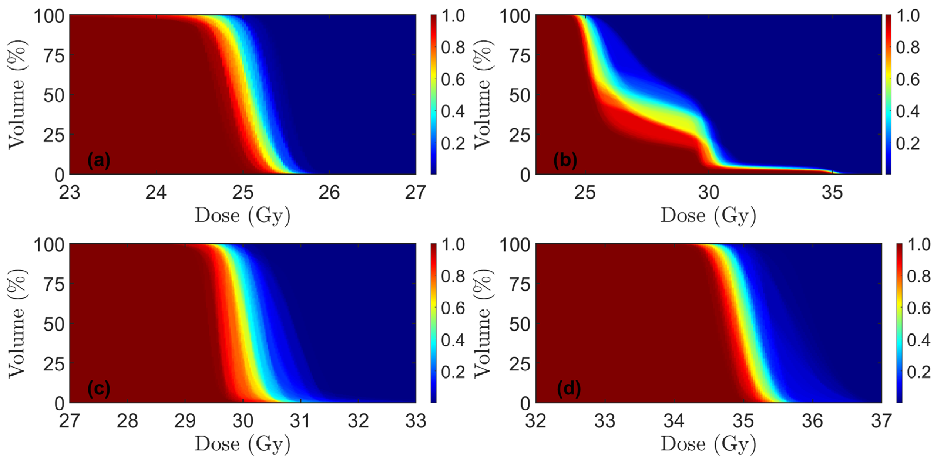
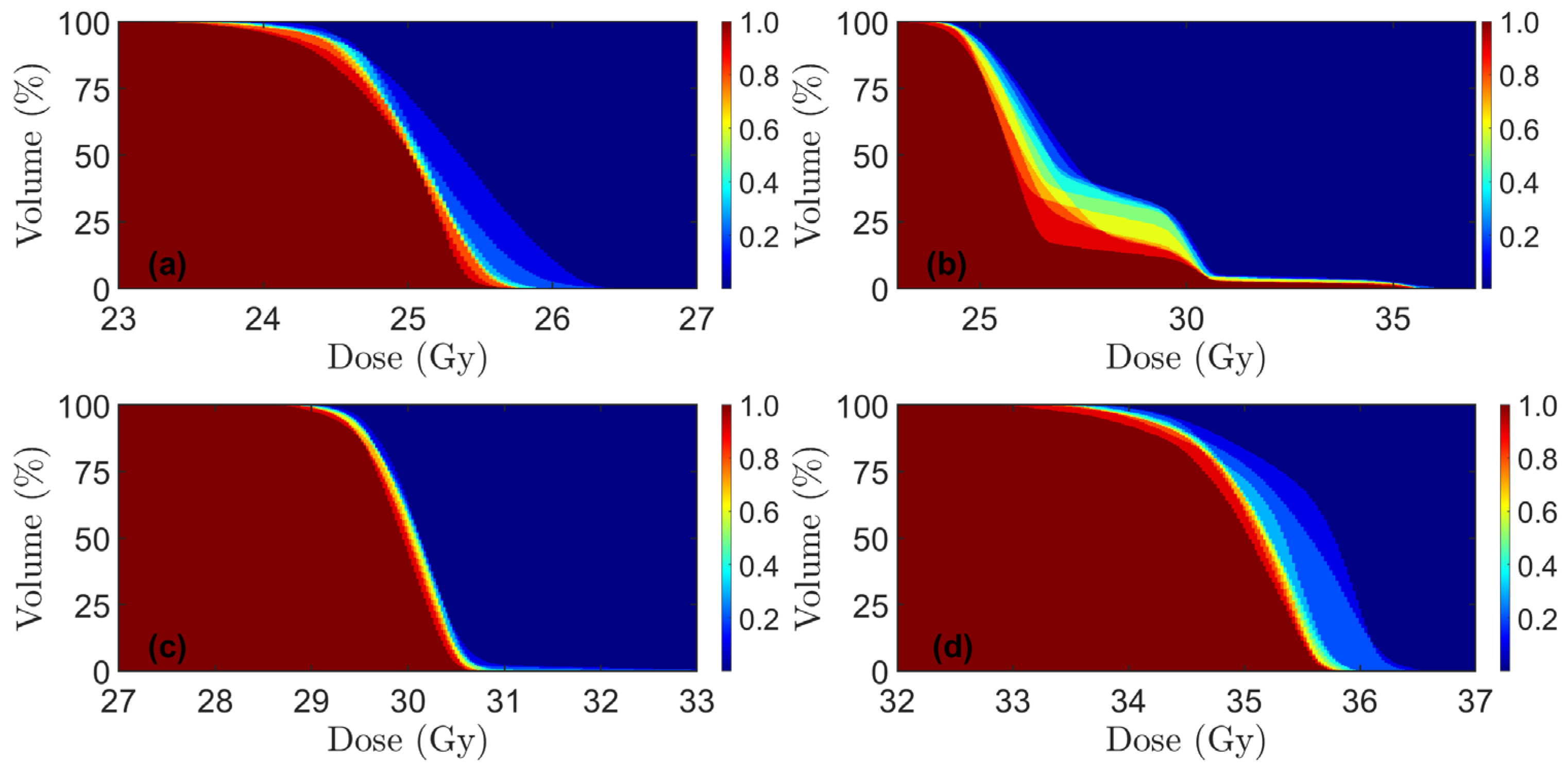
Dose volume coverage maps for PT and xRT plans are shown in Figure 2 and Figure 3, respectively. These show the fraction of the number of DVH curves above a certain point. For the PRORECT PT plans in patient 9, two robustness scenarios had D98% = 94.6% for CTV25Gy. All other robustness scenarios for all patients, CTV volumes, and both PRORECT and PRORECT II PT plans satisfied D98% > 95%. All robustness scenarios satisfied D2% < 105% for the PRORECT PT plans and CTV25Gy. For CTV25Gy in the PRORECT II PT plans, the enclosing of CTV30Gy and CTV35Gy led to significantly higher doses to parts of the target, as visible in Figure 2, such that the D2% < 105% failed for all PRORECT II PT plans. This is not the case for CTV30Gy and CTV35Gy, which do not overlap. For patients 5 and 7 the close proximity of CTV30Gy and CTV35Gy led to the failure of the D2% < 105% objective for 9 and 4 robustness scenarios for patients 5 and 7, respectively, for CTV30Gy.
All PTV criteria in Table 1 for all xRT plans are satisfied, with the exception of the D 2 % criteria for PTV25Gy in the PRORECT II xRT plans, which failed for the same reasons as for the PT cases.
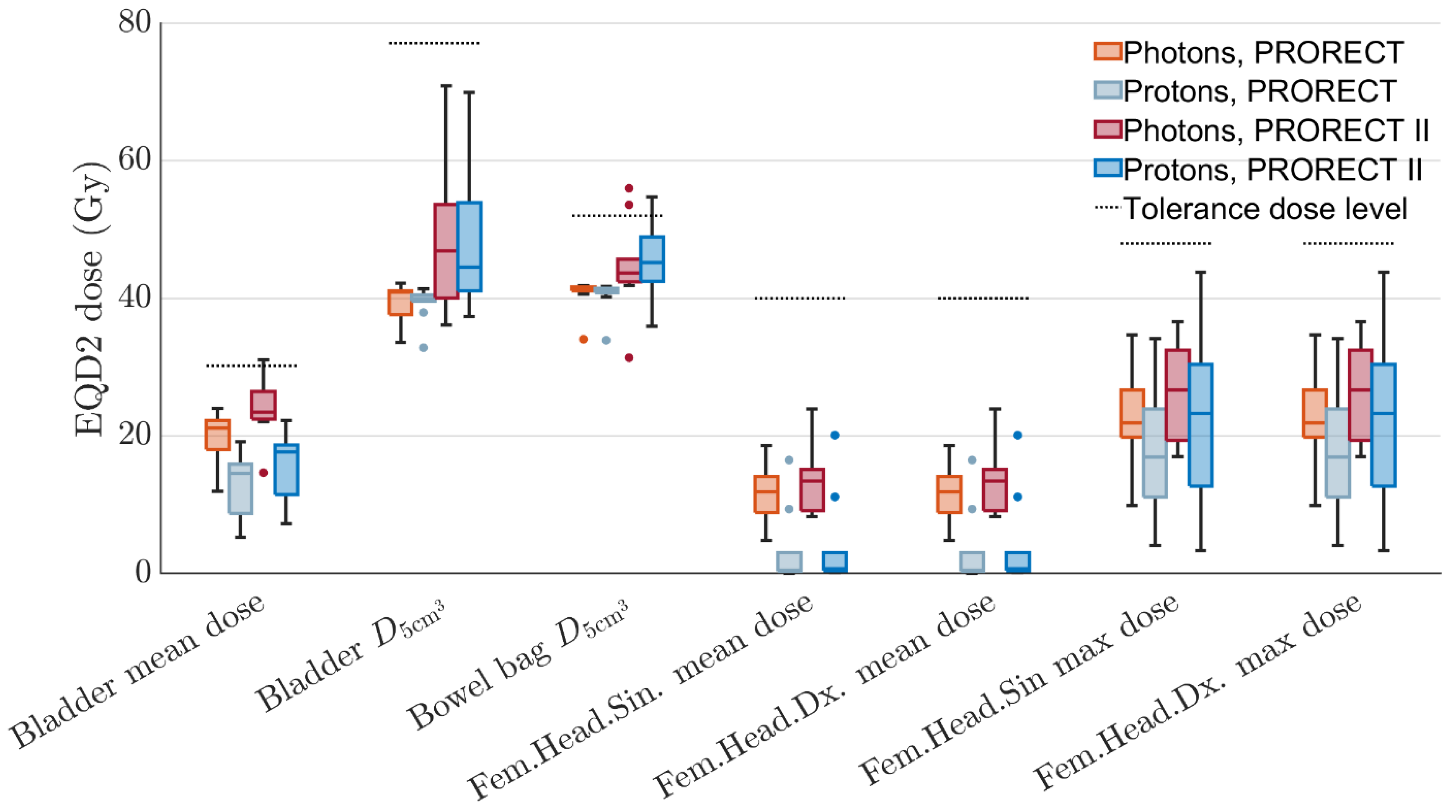
Distributions of OAR EQD2 doses for all ten patients are visualized in a set of boxplots in Figure 4. In general, PT achieves lower mean doses compared to xRT. For two patients, the PRORECT II plans for both modalities exceeded the bowel bag D 5 cm 3 objective. The PT median bowel bag D 5 cm 3 dose is higher than for xRT in the PRORECT II plans, although the difference is not statistically significant (see Table 3) at the 5% level. Exceeding this dose may lead to grade 3+ enteritis/obstruction [42]. In general, results are more similar between xRT and PT plans for the doses to small volumes/maximum doses. This is due to the similar or slightly worse lateral penumbra for protons compared to photons.
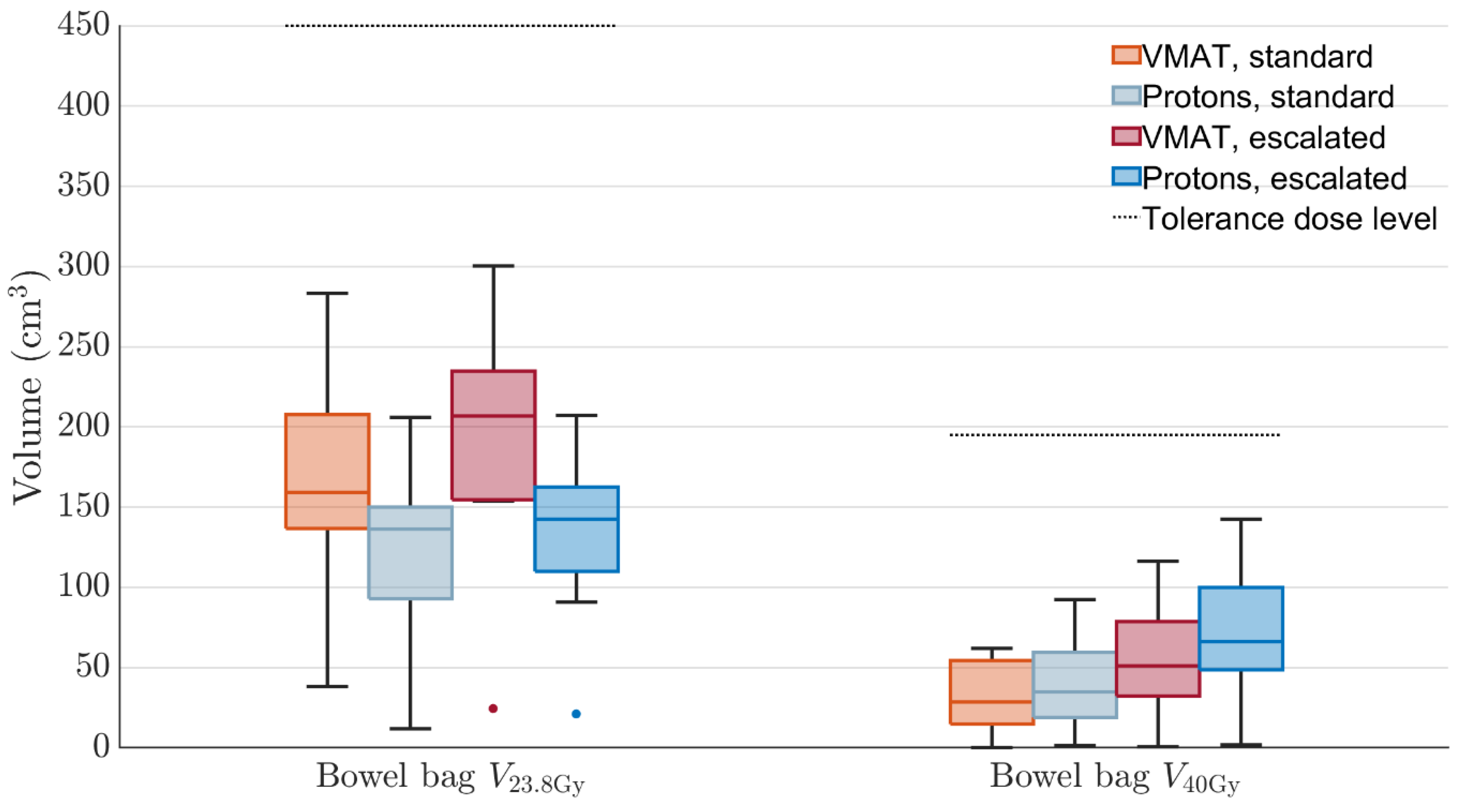
For one patient, the PRORECT II xRT plan exceeded the bladder mean dose objective. This objective is relatively strict; data from the PACE B study showed no statistically significant association with D40% < 18.1 Gy in five fractions and acute/late genitourinary toxicity [43]. We would not expect a large increase in toxicity due to the meager exceedance of the dose objective for the one PRORECT II plan. The volumetric DVH criteria for the bowel bag, with dose distributions shown in Figure 5, are not exceeded in any plan. The PT plans have slightly higher median bowel bag V 40 Gy doses, while the xRT plans have higher median V 23.8 Gy doses.
Table 3 shows the Wilcoxon signed-rank test p-values. These result from a paired data test, where for each OAR dose objective the ten xRT and ten PT doses were compared, once for the PRORECT plans and once for the PRORECT II plans. Lower p-values were seen for the mean doses where the ID-reducing properties of PT yield larger differences. For the maximum/near maximum doses, the p-values are generally higher. This is in line with results seen in Figure 4 and Figure 5.
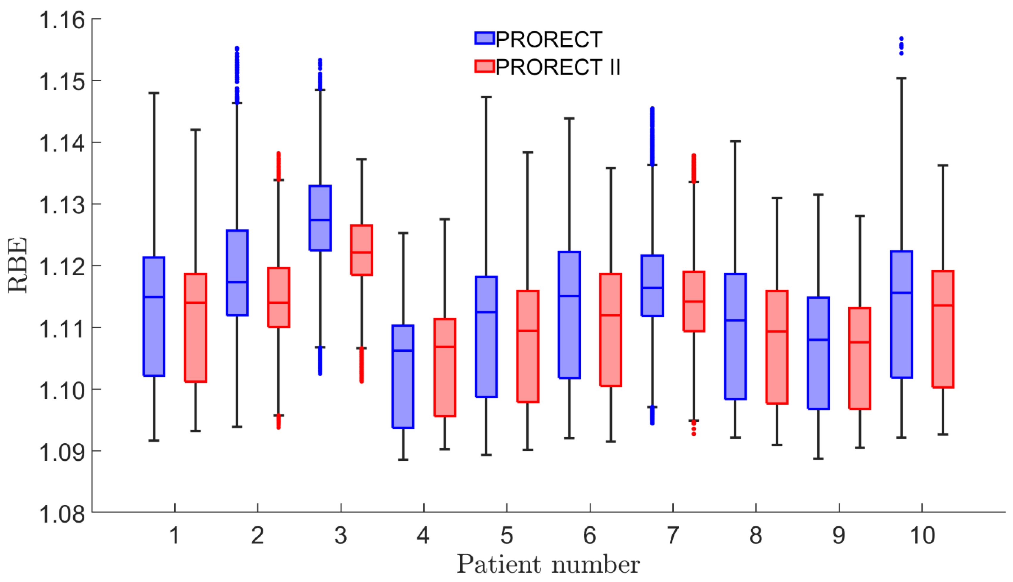
Figure 6 shows the distribution of RBE values for the union of CTV30Gy and CTV35Gy for each of the ten patients. The range for both PRORECT and PRORECT II plans, rounded to two decimals, is [1.09–1.16]. For the PRORECT plans, 78.7% of the number of voxels has RBE 1.1; for the PRORECT II plans, this number is 76.3%. Utilizing a Wilcoxon signed-rank test of the voxel pairs of the PRORECT and PRORECT II plans yields a p-value < 0.0001.
The ID for each patient, modality, and plan type is shown in Figure 7. The mean xRT/PT-ID ratio for both PRORECT and PRORECT II plans is 1.97, with a standard deviation of 0.26. The Wilcoxon signed-rank test resulted in p-values of 0.002 when comparing both PRORECT and PRORECT II plans, respectively, indicative of a statistically significant difference. For patient 1, the PRORECT II PT plan was MFO-optimized, leading to a slightly lower ID than the SFUD-optimized PRORECT PT plan. Due to the PT potential for modulation along the beam central axis, the IDs for PT plans are sensitive to increases in CTV size both in the superior–inferior direction as well as the anterior–posterior direction; for xRT plans this sensitivity is greater in the superior–inferior direction than the anterior–posterior direction.

4. Discussion

It is our clinical experience that it is relatively easy to create PRORECT treatment plans that achieve all target coverage and OAR objectives. This is suggestive of the potential for dose escalation. For the PRORECT II plans, although sufficient levels of target coverage were achieved, in this case some OAR objective doses were exceeded. We believe this indicates the appropriateness of the magnitude of the PRORECT II dose escalation.
One study found that for PT, two posterior oblique fields is the most robust against rectal distension [37], which is what we used in our PT plans. We employed robust optimization and an SFUD planning approach in the majority of our PT plans to increase robustness [44]. Furthermore, by overriding any gaseous areas and forcing the optimizer to consider those areas as water equivalent, we mitigate the risk of undershooting, i.e., protons stopping short of the distal target edge, but increase the risk of overshooting, which may increase the dose to distal OARs such as the bladder and bowel bag.
There are no PT dose escalation clinical trials for primary rectal cancer known to us at this time. Although this planning study and others [23] indicate the favorable dosimetric properties of PT over xRT for rectal cancer in some cases, PT-specific uncertainties may lead to in-patient dose distribution degradation. Due to bowel motion and rectal distention and the breakdown of the static dose cloud approximation for PT, dosimetric uncertainties are present with PT that planning studies such as the present one may fail to account for. In an LCRT setting the averaging effect over many fractions may alleviate the effect of these uncertainties. However, in an SCRT setting as considered here, the magnitude of the PT uncertainties may be larger. The small size of the CTV35Gy structure and the small number of fractions may in unfortunate circumstances lead to lower cumulative doses than those prescribed. A dose accumulation study based on daily imaging may shed light on this issue.
Although there is some evidence for a dose escalation [12], the RECTAL-BOOST phase II trial found no evidence of increased cCR/pCR rates with dose escalation up to 65 Gy [45]. However, the trial did find an increase in near-complete response rates. It differs from our approach here since an LCRT approach was used with 25 × 2 Gy fractions up to 50 Gy and then 5 × 3 Gy fractions up to 65 Gy to the GTV; furthermore, target coverage was compromised to ensure OAR dose criteria were fulfilled. In our feasibility study, we generally only boost a small volume up to a dose of 7 × 5 Gy RBE, leading to a dose of 60 Gy EQD2 (α/β = 5). OAR dose objectives were exceeded in only a few cases, and even when they were, target coverage criteria were not compromised.
The reduction in ID for PT may be significant for younger patients with a longer life expectancy, as there is evidence of a reduced risk of secondary cancer induction when using PT as compared to xRT [22,46]. The clinical effects of the lower mean doses for PT compared to xRT remain unclear, but the limiting of a dose bath effect may be beneficial for reirradiation [47,48,49,50]; one study found a local recurrence rate for LARC of 5% [51]. Although not used in this feasibility study, LET optimization in PT could potentially increase the biological efficiency of PT in the target compared to xRT, even if posterior oblique fields are used exclusively [52]. With LET optimization, MFO would be necessary, although the previously cited study indicated acceptable levels of robustness even with MFO.
Per Equation (1), RBE is a function of fraction dose, LET, and α/β. In following the AAPM recommendation to assume RBE = 1.1 [38] during treatment planning, there is the possibility of delivering a lower biologically weighted dose to the target due to the high fraction doses. In general, the PRORECT plans yield higher RBE values in the escalated CTV voxels compared to PRORECT II plans. This is in line with in vitro data [40], as well as with Equation (1) for which lim D RBE = RBE min [53]. However, the McNamara model suggests that deviations from RBE = 1.1 in the escalated CTV voxels are relatively small, as are the RBE differences between the PRORECT and PRORECT II plans. With preliminary results from the PRORECT trial not indicating inferiority of PT compared to xRT, to the extent RBE = 1.1 is appropriate for the 5 Gy fractions of PRORECT, the McNamara model would suggest it is appropriate also for the 6 and 7 Gy fractions of PRORECT II. However, RBE models in PT are associated with large uncertainties, and the inter-model variation in predicted RBE is large [54].
The establishing of clinical benefits of PT with its lower ID and mean doses compared to xRT, especially in light of the previously mentioned uncertainties, necessitates a clinical trial, which is the object of the upcoming PRORECT II study. In an SCRT approach, dose escalation will invariably lead to relatively large fraction doses. An interesting and important secondary endpoint for a clinical trial would be any differences in tumor control relative to xRT due to in vivo RBE deviating significantly from 1.1.
A limitation of this study is the size of our patient cohort. Using ten patients may limit the generalizability of our results. However, we believe ten patients is sufficient for our purposes with this article. As shown in Table 1, there is substantial variation in tumor heights, volumes, and staging in the cohort. Furthermore, the clinical NORMAL-R study [35], which we used as a basis for our proposed dose escalation, only used twenty patients in its cohort. Furthermore, since this is an in silico feasibility study ahead of the actual clinical trial, no clinical outcome data can be provided to assess toxicity levels that would result from the PRORECT II plans.

5. Conclusions

The achievable target coverage for the PRORECT II treatment plans, in combination with the acceptable doses to the OAR, shows the feasibility of the proposed PRORECT II dose escalation. PT can reduce the ID by a factor of two compared to xRT, which may be especially important for the growing numbers of young patients to reduce the risk of late radiation-induced malignancies. RBE calculations suggest that deviations from RBE = 1.1 inside the target are small, both for the PRORECT and PRORECT II plans. A clinical trial, PRORECT II, would be necessary to establish conclusively the standard RT modality and fractionation for treatment of LARC.

Author Contributions

Conceptualization, E.A. and A.V.; data curation, E.A. and A.V.; formal analysis, E.A.; funding acquisition, E.A. and A.V.; investigation, E.A., A.A., B.S., F.A., and A.V.; methodology, E.A., A.A., B.S., F.A. and A.V.; project administration, E.A. and A.V.; resources, E.A., A.A., B.S., F.A. and A.V.; software, E.A.; supervision, A.V.; validation, E.A. and A.V.; visualization, E.A.; writing—original draft, E.A.; writing—review and editing, E.A., A.A., B.S., F.A., C.J.S.K., H.S.R., M.G.G., S.P. and A.V. All authors have read and agreed to the published version of the manuscript.

Funding

This research was funded by Cancerfonden (grant# 21 1474 Pj), Radiumhemmets Forskningsfonder (grant# 231372), Region Stockholm (ALF# 540415) and The Skandion Clinic Research Council.

Institutional Review Board Statement

The PRORECT trial (NCT04525989) is conducted in accord-ance with the Declaration of Helsinki, and in accordance with research ethical approval (Dnr 2020–02192)) granted by the Swedish Research Ethics Committee on 15 June 2020. Written informed con-sent was obtained from all trial participants.

Informed Consent Statement

Written informed consent was obtained from all trial participants.

Data Availability Statement

The patient data cannot be made available due to privacy and ethical concerns. Post-processed data, such as DVHs, based on the patient data are available from the corresponding author upon reasonable request.

Conflicts of Interest

The authors declare no conflicts of interest.

Abbreviations

The following abbreviations are used in this manuscript:
cCRClinical complete response
CTComputed tomography
CTVClinical target volume
DVHDose volume histogram
GTVGross tumor volume
HUHounsfield unit
IDIntegral dose
LARCLocally advanced rectal cancer
LCRTLong-course radiotherapy
LETLinear energy transfer
MFOMultifield optimization
OAROrgan at risk
pCRPathological complete response
PTProton therapy
PTVPlanning target volume
RBERelative biological effectiveness
RTRadiotherapy
SCRTShort course radiotherapy
SFUDSingle field uniform dose
TMETotal mesorectal excision
xRTPhoton radiotherapy

References

  1. Ystgaard, M.F.; Myklebust, T.Å.; Smeby, J.; Larsen, I.K.; Guren, T.K.; Kure, E.H.; Tveit, K.M.; Glimelius, B.; Guren, M.G.; Hamfjord, J. Early-onset colorectal cancer incidence in Norway: A national registry-based study (1993-2022) analyzing subsite and morphology trends. ESMO Gastrointest. Oncol. 2025, 7, 100065. [Google Scholar] [CrossRef]
  2. Suresh, R.S.; Garcia, L.E.; Gearhart, S.L. Young-Onset Rectal Cancer: Is It for Real? Adv. Surg. 2024, 58, 275–291. [Google Scholar] [CrossRef]
  3. Ilic, M.; Ilic, I. Cancer of colon, rectum and anus: The rising burden of disease worldwide from 1990 to 2019. J. Public Health 2024, 46, 20–29. [Google Scholar] [CrossRef]
  4. Hofheinz, R.D.; Fokas, E.; Benhaim, L.; Price, T.J.; Arnold, D.; Beets-Tan, R.; Guren, M.G.; Hospers, G.A.P.; Lonardi, S.; Nagtegaal, I.D.; et al. Localised rectal cancer: ESMO Clinical Practice Guideline for diagnosis, treatment and follow-up. Ann. Oncol. 2025; in press. [Google Scholar] [CrossRef] [PubMed]
  5. Glimelius, B. Recent advances in rectal cancer treatment—Are we on the right track? Ups. J. Med. Sci. 2024, 129, e10537. [Google Scholar] [CrossRef] [PubMed]
  6. Garcia-Aguilar, J.; Patil, S.; Gollub, M.J.; Kim, J.K.; Yuval, J.B.; Thompson, H.M.; Verheij, F.S.; Omer, D.M.; Lee, M.; Dunne, R.F.; et al. Organ Preservation in Patients With Rectal Adenocarcinoma Treated With Total Neoadjuvant Therapy. J. Clin. Oncol. 2022, 40, 2546–2556. [Google Scholar] [CrossRef] [PubMed]
  7. Bahadoer, R.R.; Dijkstra, E.A.; van Etten, B.; Marijnen, C.A.M.; Putter, H.; Kranenbarg, E.M.; Roodvoets, A.G.H.; Nagtegaal, I.D.; Beets-Tan, R.G.H.; Blomqvist, L.K.; et al. Short-course radiotherapy followed by chemotherapy before total mesorectal excision (TME) versus preoperative chemoradiotherapy, TME, and optional adjuvant chemotherapy in locally advanced rectal cancer (RAPIDO): A randomised, open-label, phase 3 trial. Lancet Oncol. 2021, 22, 29–42. [Google Scholar] [CrossRef]
  8. van der Heijden, J.A.G.; Koëter, T.; Smits, L.J.H.; Sietses, C.; Tuynman, J.B.; Maaskant-Braat, A.J.G.; Klarenbeek, B.R.; de Wilt, J.H.W. Functional complaints and quality of life after transanal total mesorectal excision: A meta-analysis. Br. J. Surg. 2020, 107, 489–498. [Google Scholar] [CrossRef]
  9. Lauricella, S.; Brucchi, F.; Carrano, F.M.; Cassini, D.; Cirocchi, R.; Sylla, P. Quality of life and functional outcomes after laparoscopic total mesorectal excision (LaTME) and transanal total mesorectal excision (taTME) for rectal cancer. an updated meta-analysis. Int. J. Color. Dis. 2024, 39, 129. [Google Scholar] [CrossRef]
  10. Feeney, G.; Sehgal, R.; Sheehan, M.; Hogan, A.; Regan, M.; Joyce, M.; Kerin, M. Neoadjuvant radiotherapy for rectal cancer management. World J. Gastroenterol. 2019, 25, 4850–4869. [Google Scholar] [CrossRef]
  11. Vendrely, V.; Rivin Del Campo, E.; Modesto, A.; Jolnerowski, M.; Meillan, N.; Chiavassa, S.; Serre, A.A.; Gérard, J.P.; Créhanges, G.; Huguet, F.; et al. Rectal cancer radiotherapy. Cancer Radiother. 2022, 26, 272–278. [Google Scholar] [CrossRef]
  12. Appelt, A.L.; Pløen, J.; Vogelius, I.R.; Bentzen, S.M.; Jakobsen, A. Radiation dose-response model for locally advanced rectal cancer after preoperative chemoradiation therapy. Int. J. Radiat. Oncol. Biol. Phys. 2013, 85, 74–80. [Google Scholar] [CrossRef] [PubMed]
  13. Braendengen, M.; Tveit, K.M.; Berglund, A.; Birkemeyer, E.; Frykholm, G.; Påhlman, L.; Wiig, J.N.; Byström, P.; Bujko, K.; Glimelius, B. Randomized phase III study comparing preoperative radiotherapy with chemoradiotherapy in nonresectable rectal cancer. J. Clin. Oncol. 2008, 26, 3687–3694. [Google Scholar] [CrossRef] [PubMed]
  14. Habr-Gama, A.; Gama-Rodrigues, J.; São Julião, G.P.; Proscurshim, I.; Sabbagh, C.; Lynn, P.B.; Perez, R.O. Local recurrence after complete clinical response and watch and wait in rectal cancer after neoadjuvant chemoradiation: Impact of salvage therapy on local disease control. Int. J. Radiat. Oncol. Biol. Phys. 2014, 88, 822–828. [Google Scholar] [CrossRef] [PubMed]
  15. Smith, J.D.; Ruby, J.A.; Goodman, K.A.; Saltz, L.B.; Guillem, J.G.; Weiser, M.R.; Temple, L.K.; Nash, G.M.; Paty, P.B. Nonoperative management of rectal cancer with complete clinical response after neoadjuvant therapy. Ann. Surg. 2012, 256, 965–972. [Google Scholar] [CrossRef]
  16. Teste, B.; Rouanet, P.; Tuech, J.J.; Valverde, A.; Lelong, B.; Rivoire, M.; Faucheron, J.L.; Jafari, M.; Portier, G.; Meunier, B.; et al. Early and late morbidity of local excision after chemoradiotherapy for rectal cancer. BJS Open 2021, 5, zrab043. [Google Scholar] [CrossRef]
  17. Wo, J.Y.; Anker, C.J.; Ashman, J.B.; Bhadkamkar, N.A.; Bradfield, L.; Chang, D.T.; Dorth, J.; Garcia-Aguilar, J.; Goff, D.; Jacqmin, D.; et al. Radiation Therapy for Rectal Cancer: Executive Summary of an ASTRO Clinical Practice Guideline. Pract. Radiat. Oncol. 2021, 11, 13–25. [Google Scholar] [CrossRef]
  18. Bach, S.P.; Gilbert, A.; Brock, K.; Korsgen, S.; Geh, I.; Hill, J.; Gill, T.; Hainsworth, P.; Tutton, M.G.; Khan, J.; et al. Radical surgery versus organ preservation via short-course radiotherapy followed by transanal endoscopic microsurgery for early-stage rectal cancer (TREC): A randomised, open-label feasibility study. Lancet Gastroenterol. Hepatol. 2021, 6, 92–105. [Google Scholar] [CrossRef]
  19. Hearn, N.; Atwell, D.; Cahill, K.; Elks, J.; Vignarajah, D.; Lagopoulos, J.; Min, M. Neoadjuvant Radiotherapy Dose Escalation in Locally Advanced Rectal Cancer: A Systematic Review and Meta-analysis of Modern Treatment Approaches and Outcomes. Clin. Oncol. (R. Coll. Radiol.) 2021, 33, e1–e14. [Google Scholar] [CrossRef]
  20. Lomax, A.J.; Bortfeld, T.; Goitein, G.; Debus, J.; Dykstra, C.; Tercier, P.A.; Coucke, P.A.; Mirimanoff, R.O. A treatment planning inter-comparison of proton and intensity modulated photon radiotherapy. Radiother. Oncol. 1999, 51, 257–271. [Google Scholar] [CrossRef]
  21. Dracham, C.B.; Shankar, A.; Madan, R. Radiation induced secondary malignancies: A review article. Radiat. Oncol. J. 2018, 36, 85–94. [Google Scholar] [CrossRef] [PubMed]
  22. Eaton, B.R.; MacDonald, S.M.; Yock, T.I.; Tarbell, N.J. Secondary Malignancy Risk Following Proton Radiation Therapy. Front. Oncol. 2015, 5, 261. [Google Scholar] [CrossRef] [PubMed]
  23. Fok, M.; Toh, S.; Easow, J.; Fowler, H.; Clifford, R.; Parsons, J.; Vimalachandran, D. Proton beam therapy in rectal cancer: A systematic review and meta-analysis. Surg. Oncol. 2021, 38, 101638. [Google Scholar] [CrossRef] [PubMed]
  24. Vaios, E.J.; Wo, J.Y. Proton beam radiotherapy for anal and rectal cancers. J. Gastrointest. Oncol. 2020, 11, 176–186. [Google Scholar] [CrossRef]
  25. Radu, C.; Norrlid, O.; Brændengen, M.; Hansson, K.; Isacsson, U.; Glimelius, B. Integrated peripheral boost in preoperative radiotherapy for the locally most advanced non-resectable rectal cancer patients. Acta Oncol. 2013, 52, 528–537. [Google Scholar] [CrossRef]
  26. Colaco, R.J.; Nichols, R.C.; Huh, S.; Getman, N.; Ho, M.W.; Li, Z.; Morris, C.G.; Mendenhall, W.M.; Mendenhall, N.P.; Hoppe, B.S. Protons offer reduced bone marrow, small bowel, and urinary bladder exposure for patients receiving neoadjuvant radiotherapy for resectable rectal cancer. J. Gastrointest. Oncol. 2014, 5, 3–8. [Google Scholar] [CrossRef]
  27. Ling, T.C.; Kang, J.I.; Slater, J.D.; Yang, G.Y. Proton therapy for gastrointestinal cancers. Transl. Cancer Res. 2012, 1, 150–158. [Google Scholar] [CrossRef]
  28. Santos, A.; Penfold, S.; Gorayski, P.; Le, H. The Role of Hypofractionation in Proton Therapy. Cancers 2022, 14, 2271. [Google Scholar] [CrossRef]
  29. Glimelius, B.; Khan, T.; Adolfsson, K.; Angenete, E.; Berglund, Å.; Bonde, K.; Elander, N.; Fokstuen, T.; Haux, J.; Imam, I.; et al. Total neoadjuvant treatment using short-course radiotherapy and four CAPOX cycles in locally advanced rectal cancer with high-risk criteria for recurrence: A Swedish nationwide cohort study (LARCT-US). EClinicalMedicine 2024, 75, 102771. [Google Scholar] [CrossRef]
  30. Erlandsson, J.; Fuentes, S.; Radu, C.; Frödin, J.E.; Johansson, H.; Brandberg, Y.; Holm, T.; Glimelius, B.; Martling, A. Radiotherapy regimens for rectal cancer: Long-term outcomes and health-related quality of life in the Stockholm III trial. BJS Open 2021, 5, zrab137. [Google Scholar] [CrossRef]
  31. Suwinski, R.; Wzietek, I.; Tarnawski, R.; Namysl-Kaletka, A.; Kryj, M.; Chmielarz, A.; Wydmanski, J. Moderately low alpha/beta ratio for rectal cancer may best explain the outcome of three fractionation schedules of preoperative radiotherapy. Int. J. Radiat. Oncol. Biol. Phys. 2007, 69, 793–799. [Google Scholar] [CrossRef]
  32. Zhang, M.X.; Li, X.B.; Guan, B.J.; Guan, G.X.; Lin, X.Y.; Wu, X.D.; Chi, P.; Xu, B.H. Dose escalation of preoperative short-course radiotherapy followed by neoadjuvant chemotherapy in locally advanced rectal cancer: Protocol for an open-label, single-centre, phase I clinical trial. BMJ Open 2019, 9, e025944. [Google Scholar] [CrossRef]
  33. Pedone, C.; Sorcini, B.; Staff, C.; Färlin, J.; Fokstuen, T.; Frödin, J.E.; Nilsson, P.J.; Martling, A.; Valdman, A. Preoperative short-course radiation therapy with PROtons compared to photons in high-risk RECTal cancer (PRORECT): Initial dosimetric experience. Clin. Transl. Radiat. Oncol. 2023, 39, 100562. [Google Scholar] [CrossRef] [PubMed]
  34. Chin, R.I.; Roy, A.; Pedersen, K.S.; Huang, Y.; Hunt, S.R.; Glasgow, S.C.; Tan, B.R.; Wise, P.E.; Silviera, M.L.; Smith, R.K.; et al. Clinical Complete Response in Patients With Rectal Adenocarcinoma Treated With Short-Course Radiation Therapy and Nonoperative Management. Int. J. Radiat. Oncol. Biol. Phys. 2022, 112, 715–725. [Google Scholar] [CrossRef] [PubMed]
  35. Kim, H.; Pedersen, K.; Olsen, J.R.; Mutch, M.G.; Chin, R.I.; Glasgow, S.C.; Wise, P.E.; Silviera, M.L.; Tan, B.R.; Wang-Gillam, A.; et al. Nonoperative Rectal Cancer Management With Short-Course Radiation Followed by Chemotherapy: A Nonrandomized Control Trial. Clin. Color. Cancer 2021, 20, e185–e193. [Google Scholar] [CrossRef] [PubMed]
  36. PRORECT Radiotherapy Appendix, Version 3 (September). Available online: https://prorect.se/ws/media-library/f577f9c145804282bd319e893ef2cf46/appendix-1_radiotherapy_appendix_v3_sep2024.pdf (accessed on 3 June 2025).
  37. Parzen, J.S.; Zheng, W.; Li, X.; Ding, X.; Kabolizadeh, P. Optimization of Field Design in the Treatment of Rectal Cancer with Intensity Modulated Proton Beam Radiation Therapy: How Many Fields Are Needed to Account for Rectal Distension Uncertainty? Adv. Radiat. Oncol. 2021, 6, 100749. [Google Scholar] [CrossRef]
  38. Paganetti, H.; Blakely, E.; Carabe-Fernandez, A.; Carlson, D.J.; Das, I.J.; Dong, L.; Grosshans, D.; Held, K.D.; Mohan, R.; Moiseenko, V.; et al. Report of the AAPM TG-256 on the relative biological effectiveness of proton beams in radiation therapy. Med. Phys. 2019, 46, e53–e78. [Google Scholar] [CrossRef]
  39. Friedrich, T. Proton RBE dependence on dose in the setting of hypofractionation. Br. J. Radiol. 2020, 93, 20190291. [Google Scholar] [CrossRef]
  40. Paganetti, H. Relative biological effectiveness (RBE) values for proton beam therapy. Variations as a function of biological endpoint, dose, and linear energy transfer. Phys. Med. Biol. 2014, 59, R419–R472. [Google Scholar] [CrossRef]
  41. McNamara, A.L.; Schuemann, J.; Paganetti, H. A phenomenological relative biological effectiveness (RBE) model for proton therapy based on all published in vitro cell survival data. Phys. Med. Biol. 2015, 60, 8399–8416. [Google Scholar] [CrossRef]
  42. Hanna, G.G.; Murray, L.; Patel, R.; Jain, S.; Aitken, K.L.; Franks, K.N.; van As, N.; Tree, A.; Hatfield, P.; Harrow, S.; et al. UK Consensus on Normal Tissue Dose Constraints for Stereotactic Radiotherapy. Clin. Oncol. (R. Coll. Radiol.) 2018, 30, 5–14. [Google Scholar] [CrossRef]
  43. Ratnakumaran, R.; Hinder, V.; Brand, D.; Staffurth, J.; Hall, E.; van As, N.; Tree, A. The Association between Acute and Late Genitourinary and Gastrointestinal Toxicities: An Analysis of the PACE B Study. Cancers 2023, 15, 1288. [Google Scholar] [CrossRef] [PubMed]
  44. Zhu, X.R.; Poenisch, F.; Li, H.; Zhang, X.; Sahoo, N.; Wu, R.Y.; Li, X.; Lee, A.K.; Chang, E.L.; Choi, S.; et al. A single-field integrated boost treatment planning technique for spot scanning proton therapy. Radiat. Oncol. 2014, 9, 202. [Google Scholar] [CrossRef] [PubMed]
  45. Couwenberg, A.M.; Burbach, J.P.M.; Berbee, M.; Lacle, M.M.; Arensman, R.; Raicu, M.G.; Wessels, F.J.; Verdult, J.; Roodhart, J.; Reerink, O.; et al. Efficacy of Dose-Escalated Chemoradiation on Complete Tumor Response in Patients with Locally Advanced Rectal Cancer (RECTAL-BOOST): A Phase 2 Randomized Controlled Trial. Int. J. Radiat. Oncol. Biol. Phys. 2020, 108, 1008–1018. [Google Scholar] [CrossRef]
  46. Xiang, M.; Chang, D.T.; Pollom, E.L. Second cancer risk after primary cancer treatment with three-dimensional conformal, intensity-modulated, or proton beam radiation therapy. Cancer 2020, 126, 3560–3568. [Google Scholar] [CrossRef]
  47. Berman, A.T.; Both, B.; Sharkoski, T.; Goldrath, K.; Tochner, Z.; Apisamthanarax, S.; Metz, J.M.; Plastaras, J.P. Proton Reirradiation of Recurrent Rectal Cancer: Dosimetric Comparison, Toxicities, and Preliminary Outcomes. Int. J. Part. Ther. 2014, 1, 2–13. [Google Scholar] [CrossRef]
  48. Hiroshima, Y.; Ishikawa, H.; Murakami, M.; Nakamura, M.; Shimizu, S.; Enomoto, T.; Oda, T.; Mizumoto, M.; Nakai, K.; Okumura, T.; et al. Proton Beam Therapy for Local Recurrence of Rectal Cancer. Anticancer Res. 2021, 41, 3589–3595. [Google Scholar] [CrossRef]
  49. Koroulakis, A.; Molitoris, J.; Kaiser, A.; Hanna, N.; Bafford, A.; Jiang, Y.; Bentzen, S.; Regine, W.F. Reirradiation for Rectal Cancer Using Pencil Beam Scanning Proton Therapy: A Single Institutional Experience. Adv. Radiat. Oncol. 2021, 6, 100595. [Google Scholar] [CrossRef]
  50. Rønde, H.S.; Kallehauge, J.F.; Kronborg, C.J.S.; Nyvang, L.; Rekstad, B.L.; Hanekamp, B.A.; Appelt, A.L.; Guren, M.G.; Spindler, K.L.S. Intensity modulated proton therapy planning study for organ at risk sparing in rectal cancer re-irradiation. Acta Oncol. 2021, 60, 1436–1439. [Google Scholar] [CrossRef]
  51. van Gijn, W.; Marijnen, C.A.; Nagtegaal, I.D.; Kranenbarg, E.M.; Putter, H.; Wiggers, T.; Rutten, H.J.; Påhlman, L.; Glimelius, B.; van de Velde, C.J. Preoperative radiotherapy combined with total mesorectal excision for resectable rectal cancer: 12-year follow-up of the multicentre, randomised controlled TME trial. Lancet Oncol. 2011, 12, 575–582. [Google Scholar] [CrossRef]
  52. Ma, J.; Dragojevic, S.; Remmes, N.B.; Mendelson, N.L.; Kloeber, J.A.; Ebner, D.K.; Wu, Z.; Gunn, H.J.; Merrell, K.W.; Hallemeier, C.L.; et al. Linear energy transfer optimized proton therapy for rectal cancer. Radiother. Oncol. 2025, 207, 110850. [Google Scholar] [CrossRef]
  53. Carabe-Fernandez, A.; Dale, R.G.; Jones, B. The incorporation of the concept of minimum RBE (RbEmin) into the linear-quadratic model and the potential for improved radiobiological analysis of high-LET treatments. Int. J. Radiat. Biol. 2007, 83, 27–39. [Google Scholar] [CrossRef] [PubMed]
  54. Gardner, L.L.; O’Connor, J.D.; McMahon, S.J. Benchmarking proton RBE models. Phys. Med. Biol. 2024, 69, 085022. [Google Scholar] [CrossRef]
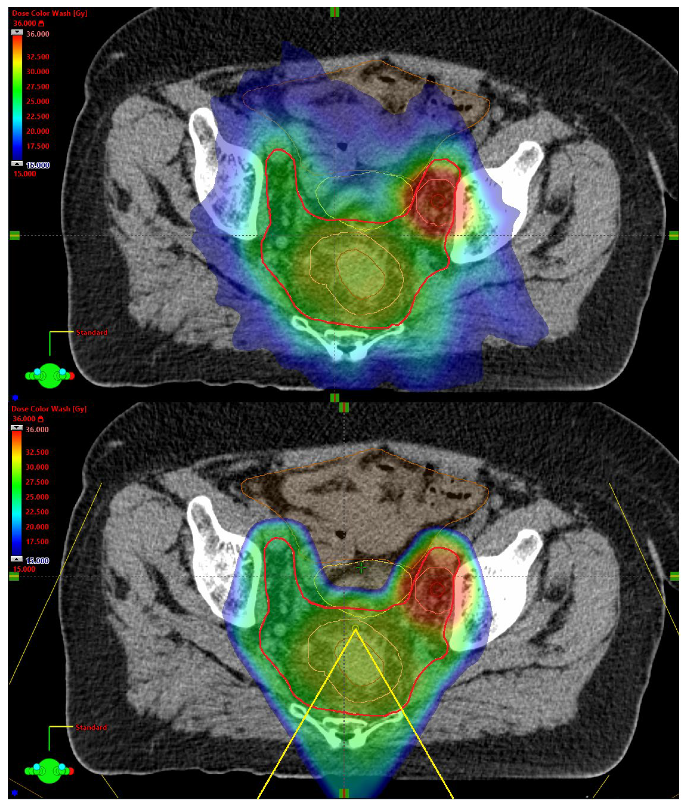
Figure 1. Examples of in-patient dose distributions for PRORECT II plans. The large bright red structure is the CTV25Gy. The central pink structure is the CTV30Gy, encompassing the primary tumor GTV. The pink structure on the right is the CTV35Gy, encompassing the GTV of the pathological lateral lymph node. The dose distribution shown ranges from 15 Gy in dark blue to 36 Gy in dark red. (upper) The xRT dose distribution. (lower) The PT dose distribution, with the field angles shown in yellow.
Cancers 17 02627 g001
Figure 2. Dose volume coverage maps for PT, showing the fraction of robustness scenarios pooled for all ten patients (N = 140) in which the DVH curve lies above a certain point. Note that x-axis scales differ among the plots. (a) PRORECT plan, CTV25Gy; (b) PRORECT II plan, CTV25Gy (c) PRORECT II plan, CTV30Gy; (d) PRORECT II plan, CTV35Gy.
Cancers 17 02627 g002
Figure 3. Dose volume coverage maps for xRT, showing the fraction of treatment plans pooled for all ten patients (N = 10), in which the DVH curve lies above a certain point in dose-volume space. Note that x-axis scales differ among the plots. (a) PRORECT plan, PTV25Gy; (b) PRORECT II plan, PTV25Gy; (c) PRORECT II plan, PTV30Gy; (d) PRORECT II plan, PTV35Gy.
Cancers 17 02627 g003
Figure 4. Distribution of EQD2 doses for all ten patients, for different OARs. Dotted horizontal lines show the tolerance EQD2 dose for each OAR and DVH metric.
Cancers 17 02627 g004
Figure 5. Distribution of volumes receiving DVH criteria EQD2 doses for all ten patients, for the bowel bag. Dotted horizontal lines show the tolerance volume.
Cancers 17 02627 g005
Figure 6. Boxplots of the distribution of RBE values, in the voxels of the union of escalated CTVs (i.e., CTV30Gy and CTV35Gy), for each patient.
Cancers 17 02627 g006
Figure 7. The integral dose for each patient, plan type and modality, calculated using Equation (2).
Cancers 17 02627 g007
Table 1. Baseline clinical characteristics of the patients in the study.
PatientSexAgeTumor Height, mmLateral LN+mr T-Stagemr N-StageMRF+EMVI+LN +
Compartment *
Inguinal Lymphatic Nodes in CTVIliaca
Externa
Subsite in CTV
1Male47900T4aN210100
2Female51850T3bN101200
3Male62501T2N100200
4Male48300T4bN211110
5Female73600T4bN211201
6Male611000T3cN211200
7Male511200T3bN101100
8Male60701T3aN200100
9Female58200T4bN111111
10Male68600T3cN200200
* 1 = Iliaca interna, 2 = obturator.
Table 2. DVH criteria for all plans. All absolute doses are given in EQD2. Target coverage criteria take precedence over OAR objective criteria.
ModalityStructureDVH Criteria
PTAll CTV volumes D 90 % > 95 % for all robustness scenarios
PTAll CTV volumes D 98 % > 95 % for 12/14 robustness scenarios
PTAll CTV volumes D 2 % < 105 % for 12/14 robustness scenarios
xRTAll PTVs D 98 % > 95 %
xRTAll PTVs D 2 % < 105 %
BothBowel bag V 23.8 Gy < 450   cm 3
BothBowel bag V 40 Gy < 195   cm 3
BothBowel bag D 5 cm 3 < 52   Gy
BothBladder D mean < 30.2   Gy
BothBladder D 5 cm 3 < 77.1   Gy
BothFemoral head D mean < 40   Gy
BothFemoral head D max < 48   Gy
Table 3. p-values resulting from comparing proton and photon plans in the PRORECT and PRORECT II arms, respectively. p-values that are below 5% significance levels are in bold.
MetricPRORECT (xRT vs. PT)PRORECT II (xRT vs. PT)
Bladder mean dose0.0020.004
Bladder   D 5 cm 3 0.492 0.625
Bowel   bag   D 5 cm 3 0.193 0.375
Fem. head left, mean dose0.0020.002
Fem. head right, mean dose0.0020.002
Fem. head left, max dose0.037 0.106
Fem. head right, max dose0.037 0.106
Bowel   bag   V 23.8 Gy 0.0020.002
Bowel   bag   V 40 Gy 0.3220.014
Disclaimer/Publisher’s Note: The statements, opinions and data contained in all publications are solely those of the individual author(s) and contributor(s) and not of MDPI and/or the editor(s). MDPI and/or the editor(s) disclaim responsibility for any injury to people or property resulting from any ideas, methods, instructions or products referred to in the content.