Review

Cell-Free DNA as a Prognostic Biomarker in Oral Carcinogenesis and Oral Squamous Cell Carcinoma: A Translational Perspective

1
Department of General Surgery and Surgical-Medical Specialties, School of Dentistry, University of Catania, 95124 Catania, Italy
2
Department of Life, Health & Environmental Sciences, Postgraduate School of Orthodontics, University of L’Aquila, 67100 L’Aquila, Italy
3
Department of Neurosciences, Reproductive and Odontostomatological Sciences, University of Naples Federico II, 80138 Naples, Italy
4
Department of Health Sciences, Magna Græcia University, 88100 Catanzaro, Italy
5
Department of Clinical and Experimental Medicine, University of Foggia, Via Rovelli 50, 71122 Foggia, Italy
6
International Research Center on Periodontal and Systemic Health “PerioHealth”, University of Catania, 95124 Catania, Italy
*
Author to whom correspondence should be addressed.
These authors contributed equally to the work.
Cancers 2025, 17(14), 2366; https://doi.org/10.3390/cancers17142366
Submission received: 30 May 2025 / Revised: 11 July 2025 / Accepted: 14 July 2025 / Published: 16 July 2025

Simple Summary

Oral cancer is a serious and often aggressive disease that can develop from earlier changes in the mouth, known as oral potentially malignant disorders (OPMDs). Detecting which OPMDs will progress to cancer remains a major clinical challenge. In recent years, scientists have discovered that small fragments of DNA released by cancer cells—called cell-free DNA (cfDNA)—can be found in the blood or saliva of patients with solid tumors. This type of “liquid biopsy” offers a non-invasive way to monitor cancer development and response to treatment. In this article, we explore how cfDNA could be used as a biomarker to detect early oral cancer, predict disease progression, and guide personalized treatment strategies. Understanding how cfDNA changes during oral carcinogenesis could help clinicians better identify high-risk patients and improve survival outcomes through earlier, targeted interventions.

Abstract

Oral squamous cell carcinoma (OSCC) remains one of the most common malignancies in the head and neck region, often preceded by a spectrum of oral potentially malignant disorders (OPMDs). Despite advances in diagnostic methods, reliable and non-invasive biomarkers for early detection and prognostic stratification are still lacking. In recent years, circulating cell-free DNA (cfDNA) has emerged as a promising liquid biopsy tool in several solid tumors, offering insights into tumor burden, heterogeneity, and molecular dynamics. However, its application in oral oncology remains underexplored. This study aims to review and discuss the current evidence on cfDNA quantification and mutation analysis (including TP53, NOTCH1, and EGFR) in patients with OPMDs and OSCC. Particular attention is given to cfDNA fragmentation patterns, methylation signatures, and tumor-specific mutations as prognostic and predictive biomarkers. Moreover, we highlight the challenges in standardizing pre-analytical and analytical workflows in oral cancer patients and explore the potential role of cfDNA in monitoring oral carcinogenesis. Understanding cfDNA dynamics in the oral cavity might offer a novel, minimally invasive strategy to improve early diagnosis, risk assessment, and treatment decision-making in oral oncology.

1. Introduction

Oral squamous cell carcinoma (OSCC) is the most common malignant neoplasm of the oral cavity [1], accounting for over 90% of all oral cancers [2,3,4,5]. Globally, oral cancer represents a significant public health issue and it is ranked among the most prevalent cancers worldwide [6,7], affecting approximately 600,000 individuals annually [8]. According to recent data, including GLOBOCAN 2020, the global incidence of lip and oral cavity cancer is estimated at around 377,000 new cases per year [9], while broader estimates from 2021 report approximately 890,000 newly diagnosed head and neck cancers and 450,000 cancer-related deaths each year [10]. Specifically for oral cancer, GLOBOCAN 2018 recorded approximately 500,550 cases and 177,384 deaths worldwide [6], with 178,000 OSCC-related deaths reported in 2020 [11]. OSCC can affect various anatomical subsites, including the gingiva, tongue, lips, and floor of the mouth [7], and is significantly more common in individuals over the age of 45, with a higher incidence observed in women [12].
Despite advances in molecular alterations [13], diagnostic techniques, and therapeutic strategies, the five-year survival rate for OSCC remains relatively low, estimated at around 50–60% [8] or approximately 62% [3], with only marginal improvements over the past years, increasing by less than 1% annually between 2005 and 2017 [5]. This high morbidity and mortality are primarily attributed to late-stage diagnosis, locoregional recurrence, and treatment resistance [8]. In contrast, early-stage detection of OSCC significantly improves prognosis, with stage I patients showing survival rates over 85% [5], compared to approximately 20% for those diagnosed at stage III or IV [7]. These figures underscore the critical importance of early diagnosis in improving clinical outcomes for OSCC patients [8,14].
The development of OSCC may be preceded by a group of clinical conditions known as oral potentially malignant disorders (OPMDs), which carry an increased risk of progression to cancer [2,15]. Common examples of OPMDs include oral leukoplakia (OLK), oral lichen planus (OLP), erythroplakia, and oral submucous fibrosis (OSF). The overall malignant transformation rate of OPMDs is estimated to be around 7.9% [9]. Moreover, although oral dysplasia is graded histologically, histopathological assessment alone does not reliably predict the clinical behavior or malignant potential of these lesions [5].
While tissue biopsy provides the definitive diagnosis for OSCC and OPMDs [6], these conditions are often diagnosed at advanced stages due to asymptomatic early disease [16]. Therefore, the identification of reliable biomarkers is essential for early diagnosis, prognosis, and treatment response prediction [8]. Biomarkers derived from various biological fields, such as genomics, proteomics, and metabolomics, play a critical role and may help to forecast the probability of OSCC development and/or progression [5]. The discovery of dependable biomarkers obtainable through non-invasive methods, for instance, from saliva, could significantly enhance OSCC management [3,5]. Although many potential biomarkers, both non-invasive (saliva and blood) and invasive (biopsy), have been identified over the years, few have been incorporated into standard clinical practice [7]. The discovery of novel biomarkers holds the promise to improve early OSCC diagnosis and patients’ prognosis [17]. For this reason, there is an urgent need for cost-effective diagnostic methods with high sensitivity and specificity and for more accurate biomarkers capable of predicting the progression of oral dysplasia to cancer, ultimately enabling better clinical management of these lesions [5,6].
In this context, liquid biopsy is emerging as a novel tool [18], offering promising prospects for the early diagnosis of oral cancer [19,20]. Liquid biopsy represents a minimally invasive and repeatable approach that enables the analysis of biomarkers released into body fluids, such as blood and saliva. Among the various components analyzed through liquid biopsy, circulating cell-free DNA (ccfDNA) has garnered particular interest due to its potential clinical relevance [18].
cfDNA consists of extracellular DNA fragments that freely circulate in the bloodstream and other body fluids [21]. While a significant portion of cfDNA is released by apoptotic cells, a crucial fraction, known as circulating tumor DNA (ctDNA), originates from malignant cells and carries tumor-specific genetic and epigenetic alterations, including somatic mutations and DNA methylation patterns [22]. The analysis of cfDNA, particularly ctDNA, through liquid biopsy offers several advantages: early tumor detection, assessment of tumor heterogeneity, prognostic evaluation, monitoring of treatment response, and real-time detection of recurrence [19].
In oral cancer, liquid biopsy using samples, such as peripheral blood and saliva, may significantly enhance current diagnostic strategies by enabling earlier detection and dynamic disease monitoring [18]. Despite its huge potential, the full integration of cfDNA-based liquid biopsy into clinical practice in oral oncology still requires further investigation to address challenges related to sensitivity, specificity, and methodological standardization (Figure 1) [23].
In this regard, this review aims to provide a translational perspective on the role of cfDNA as a prognostic biomarker in oral carcinogenesis and OSCC, highlighting current evidence and future directions in this evolving field.

2. Overview of Cell-Free DNA

Cell-free DNA (cfDNA) refers to extracellular DNA molecules circulating within various bodily fluids [25]. These molecules represent a highly dense and multidimensional information repository, acting as a “biological mirror” reflecting the state and dynamics of genomes within the body [26]. The analysis of cfDNA has emerged as a non-invasive or minimally invasive approach, commonly referred to as “liquid biopsy,” offering significant potential in various clinical fields, including oncology [25,27].

2.1. Origin, Biological Features, and Mechanism Release in Cancer of cfDNA

The cfDNA molecules found in circulation are actively and/or passively released from cells [25]. In healthy individuals, the primary source of cfDNA is the hematopoietic system. However, in various physiological and pathological conditions, such as cancer, affected tissues significantly contribute to the cfDNA pool [28]. This fraction of cfDNA originating from tumors is specifically known as circulating tumor DNA (ctDNA) [29].
CfDNA fragments are generally short, non-randomly fragmented molecules [30], and the release of cfDNA into circulation is believed to occur mainly through apoptosis, necrosis, and active secretion [31]. A prevalent size of cfDNA is approximately 166–175 base pairs (bp), reflecting the length of DNA wrapped around nucleosomes [32]. Fragments released from apoptotic normal cells and blood cells are typically at around 160–180 bp in length, reflecting nucleosomal packaging [29,33]. However, cfDNA fragment length can vary, ranging from 40 bp up to more than 10 kilobases (kb), with longer fragments often derived from necrotic cells [32]. The pattern of apoptotic fragmentation is usually reflected in the size of cfDNA, yielding fragments of approximately 167 bp, corresponding to nucleosomal DNA plus a linker segment [29]. The ratio of longer to shorter cfDNA fragments has been proposed as a measure of cfDNA integrity (cfDI) [33]. Beyond passive release via cell death, active cellular secretion is another proposed mechanism for cfDNA generation [32]. Research, particularly in breast cancer cell lines, indicates that cells can actively release cfDNA, not exclusively through exosomes [34]. The exact mechanisms underlying cfDNA production and release are still being studied [32].
Importantly, cfDNA in circulation carries genetic information from its originating cells, including mutations, copy number alterations, chromosomal rearrangements, and epigenetic status like hyper/hypo-methylation. Carrying both genetic and epigenetic information, cfDNA represents a valuable source for molecular profiling [25].

2.2. cfDNA Detection in Blood vs. Saliva: Advantages and Disadvantages

CfDNA is present in various body fluids, including blood, saliva, urine, cerebrospinal fluid (CSF), and others [25,35]. For cancer diagnostics, blood plasma or serum has been the most widely studied source of cfDNA [27]. It is generally considered convenient for sampling, and cfDNA in plasma is relatively stable and straightforward to extract [36].
However, saliva is emerging as a promising alternative for liquid biopsy, particularly relevant for oral carcinogenesis and head and neck cancers. The collection of saliva is convenient, non-invasive, and painless, allowing for repetitive sampling [29,37]. Saliva contains biomarkers that diffuse from the circulation and are also shed directly from tumors or affected tissues in the oral cavity [38]; thus, thanks to this dual origin, saliva has the potential for the early detection of oral diseases [29].
While saliva offers ease of collection, a study has shown that salivary cfDNA concentration can be significantly lower than plasma cfDNA concentration. For some cancers, like non-small cell lung cancer, the concentration of salivary cfDNA may not be suitable for quantitative diagnostic analysis. However, salivary cfDNA can still be valuable for qualitative analysis, such as detecting specific mutations like EGFR mutations [39]. The yield of ctDNA can impact assay sensitivity, highlighting the importance of optimizing pre-analytical and analytical procedures for both blood and saliva samples [29].
However, this lower yield of salivary cfDNA can reduce analytical sensitivity, particularly when screening for early-stage or low-burden lesions. Studies have demonstrated that standard quantitative assays may fail to detect tumor-derived cfDNA in saliva when total cfDNA levels fall below assay limits of detection, potentially leading to false negatives in early disease. To overcome this, highly sensitive methods—such as digital PCR or targeted ultra-deep sequencing—are often required for salivary cfDNA analysis in early-stage OSCC and OPMDs, and assay performance should be validated with spiked-in controls to determine the minimal detectable tumor fraction [40].
Despite the potential challenges, salivary biomarkers, including cfDNA and circulating tumor DNA, show promise for the early detection of head and neck cancers, although large prospective clinical trials are still needed [29], and cfDNA analyses are increasingly seen as valuable tools for monitoring patients and offering personalized care [27].

3. CfDNA in Oral Squamous Cell Carcinoma

3.1. cfDNA Levels and Tumor Burden Correlation

Two studies of Lin et al. have investigated the presence and concentration of cfDNA in the plasma and saliva of OSCC patients as a potential indicator of tumor burden and disease progression. In patients with OSCC, plasma cfDNA concentrations have been reported to be significantly elevated compared to healthy controls [41,42]. For instance, one of these two studies observed a mean cfDNA concentration of 53.1 ± 6.69 ng/mL in OSCC patients compared to 24.0 ± 3.33 ng/mL in controls. This elevated plasma cfDNA level has shown correlation with larger tumor size (TNM T stage), cervical lymph node metastasis (N status), and advanced OSCC stage. While total cfDNA levels may not always be significantly elevated in early lesions or after surgery, cfDNA has been reported as a highly sensitive genetic biomarker reflecting tumor burden and genetic dynamics in various cancers, including OSCC. The accessible nature of plasma cfDNA makes it a potentially valuable tool for both diagnosis and prognosis in OSCC [42].

3.2. Gene Mutations of cfDNA

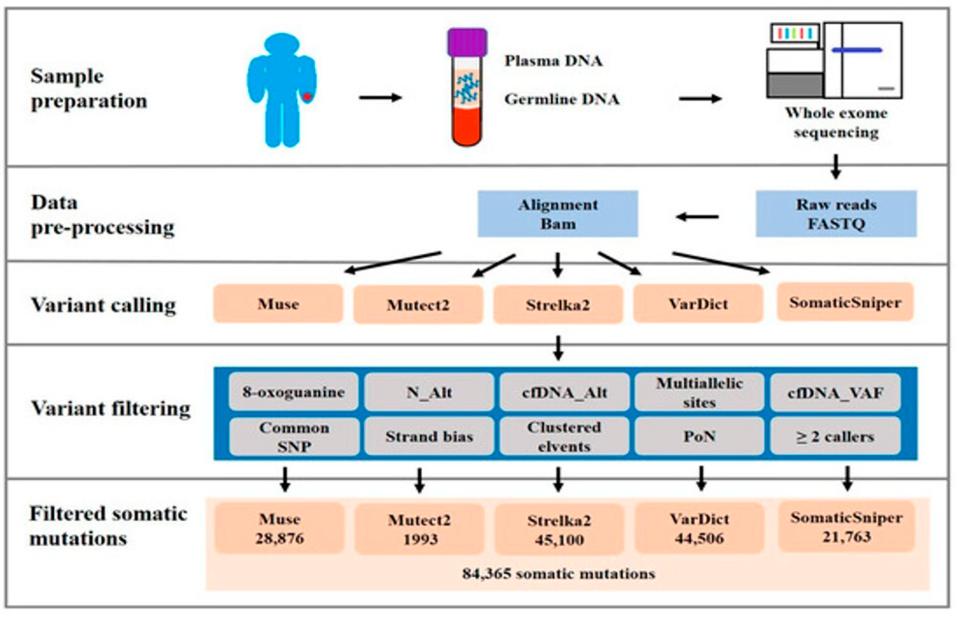
Analyzing gene mutations in cfDNA provides a non-invasive way to profile the genomic alterations present in a tumor [41]. TP53 mutation is recognized as a driver mutation in oral carcinogenesis and is one of the most frequently explored gene mutations in ctDNA from Head and Neck Squamous Cell Carcinoma (HNSCC) patients [43,44]. While OSCC can have relatively low cfDNA shedding and mutations at low allele frequencies, detection of TP53 target hotspot somatic mutations in cfDNA from OSCC patients has been shown to be technically feasible, for instance, using techniques like droplet digital polymerase chain reaction (ddPCR). Moreover target somatic mutations detected in cancerous DNA and cfDNA can be related to cervical lymph node metastasis [44]. Beyond TP53, comprehensive genomic profiling of plasma cfDNA in patients with metastatic OSCC has identified other frequently mutated genes, including TTN, PLEC, SYNE1, and USH2A. Known driver genes such as KMT2D, LRP1B, TRRAP, and FLNA were also found to be significantly and frequently mutated. Additionally, novel mutated genes like CCDC168, HMCN2, STARD9, and CRAMP1 were significantly present in metastatic OSCC patients. Genes like RORC, SLC49A3, and NUMBL were most frequently mutated in patients with distant metastatic OSCC. Whole-exome sequencing (WES) of plasma cfDNA and paired whole blood samples allows for the identification of somatic mutations and analysis of mutation burden, which has been significantly associated with clinical staging and distant metastasis status in OSCC (Figure 2) [41]. Analysis of public databases and specific OSCC cohorts revealed frequently mutated genes like TP53, FAT1, CASP8, CDKN2A, NOTCH1, PIK3CA, and HRAS [45]. This molecular profiling of cfDNA provides insights into the genomic landscape of OSCC and may help in designing targeted therapies and patient stratification [41].

3.3. CfDNA Fragmentation and Integrity as Prognostic Markers

The process by which cfDNA is released, particularly from malignant cells undergoing necrosis, often results in incompletely digested DNA fragments that are longer than those typically produced by apoptosis in healthy individuals. This difference in fragment size distribution can be assessed by measuring cfDNA integrity, commonly calculated as the ratio of longer DNA fragments to shorter fragments. Specific short interspersed elements, such as ALU115 and ALU247 fragments, are frequently used to assess cfDNA integrity [46,47]. A higher cfDNA integrity index is often associated with malignancy; indeed, in oropharyngeal squamous cell carcinoma (OPSCC), a subtype of HNSCC, significantly higher cfDNA integrity has been observed compared to normal controls. This higher integrity in OPSCC was found to be associated with nodal status, and the combination of ALU115 quantity with cfDNA integrity enhanced diagnostic sensitivity [48]. Similarly, in laryngeal squamous cell carcinoma (LSCC), significantly higher levels of plasma cfDNA (ALU115 and ALU247) and cfDNA integrity index were detected in patients compared to non-tumor individuals. These findings suggest that cfDNA integrity can differentiate cancer patients from healthy controls [47]. While studies directly linking cfDNA integrity in saliva to OSCC prognosis are preliminary, salivary cfDNA integrity indexes (ALU115/ALU60 and ALU247/ALU60) have shown potential as non-invasive diagnostic biomarkers for OSCC. For instance, the median salivary DNA integrity, calculated as ALU115/ALU60, was higher in patients affected by OSCC [40]. These results indicate that analyzing cfDNA integrity, in addition to concentration, can provide valuable diagnostic and potentially prognostic information for OSCC and related head and neck cancers [40,47].

3.4. DNA Methylation Patterns and Epigenetic Signatures in cfDNA

Abnormal epigenetic modifications, particularly DNA methylation, are hallmarks of cancer and play a significant role in the development and progression of OSCC. Aberrant DNA methylation, especially hypermethylation of tumor suppressor genes (TSGs), is a well-documented event in OSCC tissues [49,50]. Detecting these methylation patterns in cfDNA from easily accessible body fluids like saliva and plasma offers a promising non-invasive approach for diagnosis and prognosis [51].
Several tumor suppressor genes have been found to be aberrantly hypermethylated in OSCC, including CDKN2A, p16, DAPK, RASSF1A, MGMT, E-cadherin, SFRP1, and TIMP3 [48,51,52,53]. Hypermethylation of these genes has been identified in OSCC tumor tissue and detected in corresponding cfDNA samples from saliva and plasma. For instance, promoter methylation of P16, DAPK, and RASSF1A has been found to be significantly hypermethylated in serum of OPSCC patients compared to normal controls, showing high concordance with tissue samples. Methylation of P16 was associated with smoking, while RASSF1A methylation correlated with stage [48,51]. TIMP3 hypermethylation in post-treatment salivary rinse samples has shown prognostic significance in HNSCC patients [53], and ZNF582 methylation in saliva has also been investigated as a potential biomarker for OSCC [54].
Beyond TSGs, the methylation status of genes involved in DNA methylation processes themselves, like DNMT3A and TET2, has been correlated with OSCC. Varying methylation degrees of DNMT3A and TET2 were revealed in OSCC tissues, and high DNMT3A methylation levels were associated with higher survival rates [49]. The detection and analysis of these specific DNA methylation patterns and broader epigenetic signatures in cfDNA hold significant potential as non-invasive biomarkers for early detection, diagnosis, prognosis, and perhaps even predicting treatment response in OSCC [50].

4. Clinical Applications and Translational Relevance

The emergence of liquid biopsy, particularly the analysis of cfDNA and ctDNA, represents a significant advancement in oncology, offering non-invasive approaches to cancer management [55]. This technology holds substantial translational relevance for oral carcinogenesis and OSCC, with potential applications spanning early detection, prognostic assessment, treatment monitoring, and guiding personalized therapy. Liquid biopsies can be performed on various bodily fluids, including blood plasma, saliva, and serum, providing a real-time window into tumor dynamics that traditional tissue biopsies cannot fully capture due to factors like intra- and inter-tumor heterogeneity [56].

4.1. CfDNA for Early Detection and Prognostic Stratification

Early detection remains critical for improving outcomes in HNSCC, which is often identified at advanced stages [57]. CfDNA has been explored in blood and saliva samples from HNSCC patients, and specific molecular alterations detectable in cfDNA, such as DNA methylation patterns, are being investigated as potential biomarkers for early disease identification [58,59]. For instance, methylation of tumor suppressor genes like PAX5 has been assessed in oral rinses of HNSCC patients for early detection and monitoring, and PAX5 has demonstrated encouraging specificity and sensitivity in oral rinses [59]. DNA methylation is considered an early event in cancer development and progression, and its analysis in cfDNA holds promise for diagnostics, although further highlights and investigations are needed [58,60]. Research also suggests that cfDNA coverage patterns at transcription start sites (TSSs) can reflect gene expression and have potential in early cancer screening [61].
Beyond early detection, cfDNA and ctDNA show promise for prognostic stratification in OSCC and HNSCC [57,62], and elevated cfDNA levels have been correlated with poor prognosis in various cancers, including OSCC [62]. Higher total cfDNA concentrations have been linked to worse overall survival in pancreatic ductal adenocarcinoma and prostate cancer, and the baseline concentration of cfDNA has been identified as an independent prognostic factor [63,64]. Moreover, increased plasma cfDNA has been suggested as a potential marker for oral cancer [42]. A study in OSCC patients has shown that a rapid increase in total cfDNA concentration can predict systemic metastases, and analysis of total cfDNA concentrations may be useful in predicting patient prognosis. The concentration of total cfDNA has also been reported to correlate directly with the extent of disease progression in several cancer types [65]. Furthermore, specific characteristics of cfDNA, such as fragmentation patterns and size distributions, are being investigated for their potential diagnostic and prognostic value [66].

4.2. Monitoring Therapeutic Response and Recurrence

Circulating tumor DNA analysis allows for real-time monitoring of tumor dynamics and treatment response, which is particularly valuable in HNSCC given the challenges in discriminating residual or recurrent disease post-treatment [56,67]. Longitudinal monitoring of ctDNA can assess minimal residual disease (MRD) after curative-intent therapy, and the presence of MRD detected by ctDNA assays after treatment has been shown to predict progression-free survival and overall survival in locally advanced HNSCC, often without the need for tumor sequencing [68]. Patients who remain negative for ctDNA after initial curative treatment tend to have a significantly better prognosis than those who test positive again [69]. Changes in ctDNA levels throughout treatment correlate with therapeutic efficacy and can precede clinical detection of recurrence or progression [62]. For instance, monitoring the methylation levels of specific markers in oral rinses has been suggested as an effective way to predict recurrence in HNSCC [59]. Similarly, rapid clearance profiles of plasma circulating tumor HPV DNA during chemoradiotherapy correlate with disease control in HPV-associated oropharyngeal cancer, and detectable postoperative circulating tumor HPV DNA is associated with recurrence [70]. CtDNA kinetics during treatment with immune checkpoint blockade (ICB) can potentially offer real-time assessment of disease burden [71]. Individualized ctDNA monitoring using specific gene panels and techniques like digital PCR (dPCR) is being explored for its clinical validity in monitoring treatment response and relapse in HNSCC [69]. Analyses of fluctuations in total cfDNA concentration can also help predict recurrence or metastasis in OSCC [65].

4.3. CfDNA as a Companion Diagnostic Tool in Personalized Oncology

Liquid biopsy facilitates the identification of tumor-specific genetic alterations, including mutations and epigenetic changes like methylation, which are crucial for guiding personalized treatment strategies [72,73,74]. Unlike traditional tissue biopsies, liquid biopsies can better capture the genetic heterogeneity of the tumor, including primary sites and metastases [57]. Detecting actionable mutations through ctDNA analysis offers the opportunity to select patients who may benefit from targeted therapies [75]. Comprehensive genomic profiling assays using cfDNA can identify a wide range of genetic alterations [76], while TP53 is a frequently mutated gene in HNSCC, including OSCC, and its mutations can be detected in ctDNA, in which panels for HNSCC also include other frequently altered genes [44,69,77]. The ability to identify specific genetic alterations in ctDNA without the need for repeated tissue biopsies is particularly advantageous in the context of treatment decisions and managing resistance mechanisms [75]. Although the clinical application of cfDNA and ctDNA in oral cancer is still developing and requires further research for standardization, their potential as companion diagnostic tools in personalized oncology is significant [62]. In this regard, even if integrating cfDNA analysis into clinical practice has challenges, it presents opportunities for enhancing precision medicine in cancer care [65].

5. Challenges, Limitations, and Future Directions

Despite the promising potential of cfDNA as a prognostic biomarker in oral carcinogenesis and OSCC, several challenges and limitations must be addressed to facilitate its clinical translation, shaping the future directions of research in this field. A significant hurdle lies in technical and pre-analytical variables. Saliva, while easily collected non-invasively [38], presents challenges in sample collection, handling, and processing, impacting the yield and quality of cfDNA and ctDNA [78]. The concentration of cfDNA in saliva is relatively low, and its stability during storage and transportation without proper preservatives is a concern, risking degradation and contamination from cellular genomic DNA. Although technologies like ddPCR have improved sensitivity for low analyte volumes, optimizing preservation methods remains crucial for maximizing detectable cfDNA [38].
Standardization and validation in oral oncology are also critical needs. Current studies utilize varied techniques for sample collection, extraction, and analysis of biomarkers, leading to variability in results and hindering comparison and widespread clinical adoption [78,79,80,81]. In particular, significant analytical variability remains a critical barrier to the clinical adoption of cfDNA analysis. Differences in pre-analytical handling (e.g., blood vs. saliva samples, processing time, and storage conditions), cfDNA isolation techniques, and quantification methods can lead to inconsistent results. Additionally, variability in detection thresholds and assay sensitivity across platforms further complicates clinical interpretation and reproducibility. Standardization of protocols across laboratories and development of validated reference materials will be crucial for ensuring consistency and comparability in cfDNA-based diagnostics. Furthermore, establishing standardized protocols for collection, processing, analysis, and reporting of liquid biopsy findings is essential [20].
The biological complexity of the oral tumor microenvironment (TME) poses another challenge. HNSCC is characterized by a complex and heterogeneous mutational landscape and a dysregulated TME [82,83], which includes various cellular and non-cellular components that interact with tumor cells [84]. Lymph node metastasis, a major determinant of poor prognosis in OSCC, is influenced by the stromal cues within the lymph node microenvironment [85].
Integrating cfDNA with other biomarkers offers a promising future direction to assess new therapeutic strategies’ effectiveness and improve diagnostic and prognostic accuracy [86]. Analyzing multiple components from liquid biopsies, such as ctDNA, for genetic mutations and copy number alterations [87], cell-free RNA, including microRNAs, which play a crucial regulatory role in oncogenesis [20], and proteins or metabolites can provide a more comprehensive molecular profile of the tumor [88,89], potentially identifying multi-marker signatures with higher sensitivity and specificity than single markers [20,90].
Finally, longitudinal studies and prospective clinical trials are indispensable for evaluating the clinical utility of cfDNA and other liquid biopsy biomarkers. Monitoring changes in biomarker levels over time can provide insights into disease progression, response to therapy, and early detection of recurrence, which is often missed by conventional imaging methods at early stages [89,91]. Prospective trials comparing liquid biopsy approaches to existing standard methods, such as tissue biopsy and imaging, are needed to generate the evidence required for widespread clinical implementation and to validate their impact on patient outcomes [92,93]. Furthermore, the development of integrated platforms that combine cfDNA with other analytes, such as exosomal RNA or proteomic panels, could overcome current limitations in sensitivity and specificity. By capturing complementary molecular signals across different biological layers, these multi-analyte approaches may enhance early detection, improve disease stratification, and offer a more robust and comprehensive tool for personalized management of OSCC.

Techniques and Challenges for cfDNA-Containing Sample Handling in Liquid Biopsy

The successful integration of cfDNA into clinical practice for liquid biopsy, particularly for HNSCC and OSCC, hinges on standardized protocols for sample collection, storage, and transport.
Regarding sample collection and preservation, blood samples are commonly used for liquid biopsies in solid malignancies and are typically collected in K2EDTA or specialized Streck Cell-Free DNA Blood Collection Tubes (cfDNA BCTs). Streck cfDNA BCTs can maintain cfDNA stability for at least 3 days, at 18–22 °C, making them suitable for transport. K2EDTA tubes, while common, generally require processing within 6 h at room temperature to prevent contamination from lysed blood cells [38]. Saliva is also a promising, non-invasive alternative, due to its association with cancer progression [94]. Collection methods include passive spitting into sterile tubes or using devices like Salivette or Oragene OG-600 receptacle, which can yield high concentrations of total salivary DNA [80,94].
cfDNA stability in saliva is a significant challenge, as unpreserved salivary cfDNA can degrade rapidly, losing over half within 24 h, with reported half-lives around 13–14 h at room temperature. In contrast, cfDNA in blood typically has a shorter half-life of 15 min to 2.5 h. Research indicates that UAS preservative can stabilize salivary cfDNA at room temperature for up to one week. Additionally, 20 mM EDTA can stabilize salivary supernatant cfDNA for up to one week if processed immediately after collection [38]. Salivary miRNAs have shown stability for up to 48 h at 4 °C in saliva supernatant, though significant alteration occurs after 96 h. Mid-term storage at −20 °C can decrease miRNA stability compared to the standard −80 °C, suggesting immediate processing and long-term storage at −80 °C is generally recommended for optimal preservation [78].
Beyond technical limitations, real-world barriers hinder the clinical implementation of cfDNA and ctDNA analysis in OSCC. High assay costs, the need for specialized equipment and trained personnel, and limited availability of next-generation sequencing platforms pose challenges, especially in low- and middle-income countries. Furthermore, regulatory frameworks for the approval and standardization of liquid biopsy tests in oral oncology remain underdeveloped. Addressing these practical and systemic hurdles will be essential for translating molecular diagnostics into widespread clinical practice.

6. The Emerging Role of Proteomics and Metabolomics in Oral Cancer Biomarker Discovery

OSCC is a prevalent malignancy of the head and neck region, often diagnosed at advanced stages, leading to an unfavorable prognosis. The absence of precise and efficient early diagnostic techniques remains a key challenge in OSCC management. In recent years, “omics” technologies, such as genomics, proteomics, and metabolomics, have been leveraged to identify clinically relevant biomarkers in various biological fluids and tissues of OSCC patients. These approaches offer promising non-invasive strategies for early detection, prognostication, and monitoring of the disease (Figure 3) [95,96].

6.1. Proteomics in Oral Cancer Biomarker Discovery

Proteomics involves the large-scale study of proteins, providing insights into cellular functions and physiological states. Mass spectrometry (MS)-based proteomics has become a powerful analytical tool for identifying molecular markers. Among the various biofluids, saliva is extensively used as a preferred biofluid for identifying novel biomarkers for OSCC. This preference is due to its non-invasive collection method, its proximity to oral cancer lesions, and its relative simplicity compared to tissue biopsies or blood [96].
Several studies have applied MS-based proteomics (e.g., iTRAQ, LC-MRM/MS, MALDI-MSI, and SELDI-TOF/MS) to profile saliva from OSCC, OPMD, and healthy subjects [95,96,97,98,99]. For instance, a study found 67 proteins up- and 18 down-regulated in OSCC versus non-cancerous samples; three candidates (CFH, FGA, and SERPINA1) were confirmed by targeted MRM-MS and immunoassays and correlated with advanced disease stage [99].
Beyond saliva, proteomic analysis has also been applied to other samples related to oral cancer, such as serum, plasma, tumor tissue, and the TME [100,101]. A systematic review of proteomic studies in the OSCC TME identified 570 proteins detected by MS-based proteomics, with 28 being cited by two or more studies. These frequently identified proteins participate in extracellular matrix remodeling and/or energy metabolism, and most share the feature of being modulated by hypoxia in the proliferating OSCC mass [101].
In this regard, the analysis of proteomics in saliva and blood, often focusing on exosomal proteins, is considered helpful in the assistance of diagnosis and personalized treatments of OSCC [102] and, overall, analysis of salivary proteome and proteomic studies of the TME demonstrate feasibility and significant potential for discovering biomarkers for oral cancer [99,101].

6.2. Metabolomics in Oral Cancer Biomarker Discovery

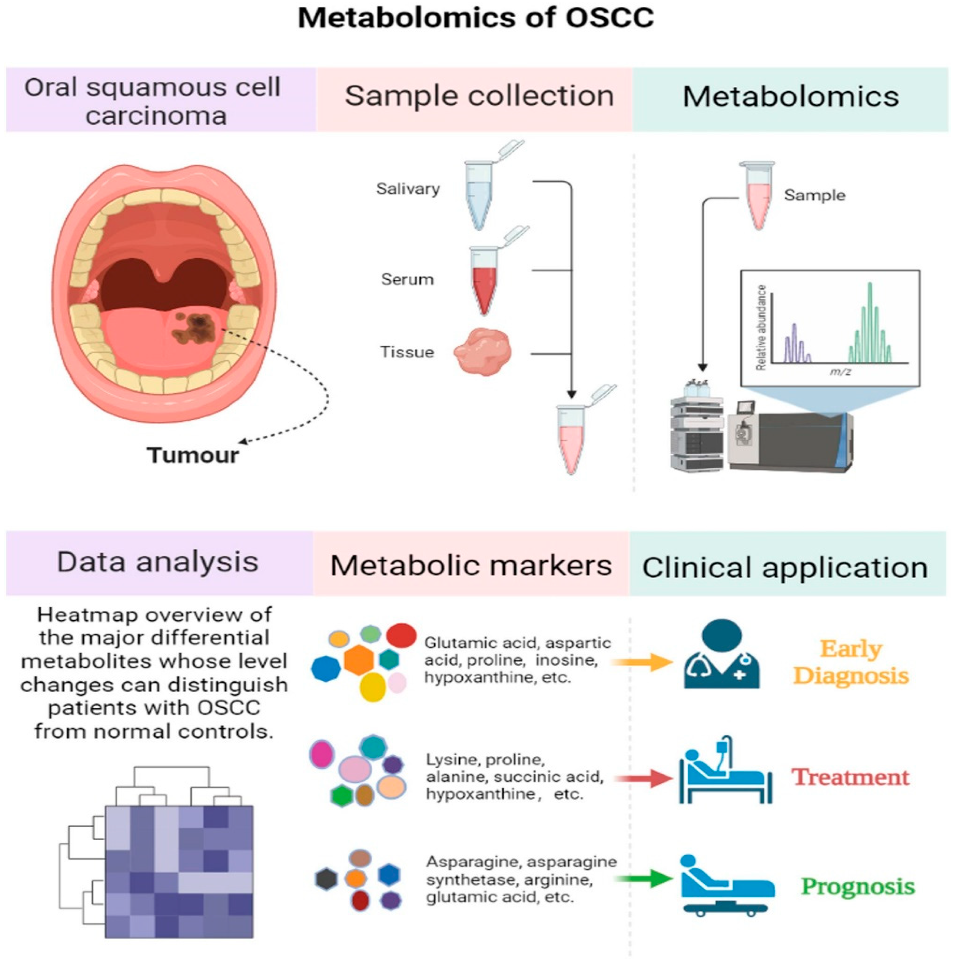
Metabolomics involves the comprehensive study of small molecule metabolites within biological systems [103]. Similar to proteomics, salivary metabolomics has emerged as a promising non-invasive approach for discovering biomarkers for oral cancer and OPMDs [102]. Techniques such as gas chromatography–mass spectrometry (GC-MS), ultra-high-performance liquid chromatography–tandem mass spectrometry (UHPLC-MS/MS), capillary electrophoresis–mass spectrometry (CE-MS), and nuclear magnetic resonance (NMR) spectroscopy are commonly employed to identify and quantify salivary metabolites [95,104,105]. Metabolomics detection technology can efficiently identify differential metabolites (Figure 4) [95]. Studies have shown significant alterations in salivary metabolites in OSCC patients compared to healthy individuals and, importantly, differences can also be observed between OSCC and OPMD patients and even between OSCC and other oral conditions like oral lichen planus [106,107,108].
These metabolic changes often reflect impaired metabolic pathways associated with cancer development and progression, such as choline metabolism, amino acid pathways, polyamine metabolism, the urea cycle, creatine metabolism, glycolysis, and glycerolipid metabolism [106]. Specific metabolites frequently reported in salivary metabolomics studies of oral diseases include glycine, leucine, phenylalanine, dipeptides, linoleic acid, arachidonic acid, tyrosine, choline, taurine, lactate, valine, and proline [103]. Studies on OSCC tissue metabolomics have also provided insights, revealing changes like decreased levels of certain amino acids (glycine, serine, glutamine, lysine, proline, alanine, glutamic acid, leucine, and threonine) from healthy tissue to precancerous lesions and then to OSCC. The enhanced consumption of glucose and lactate production (the Warburg effect) has also been observed in OSCC tissues through metabolomic analysis [95]. Salivary metabolomics, especially when combining analyses of different metabolites, shows feasibility for the diagnosis and monitoring of OSCC [102].
While some salivary metabolites exhibit significant changes in OPMDs and oral cancer compared to healthy controls, further high-quality studies are needed to confirm their reliability and establish their diagnostic potential [106]. Integrating metabolomics with statistical data analyses and artificial intelligence technology holds promise for rapid screening of characteristic biomarkers for early diagnosis, treatment, and prognosis evaluations (Table 1) [95].

7. Conclusions

This review highlights the growing relevance of liquid biopsy approaches, particularly cfDNA and ctDNA analysis, in the early detection, prognosis, monitoring, and personalized treatment of OSCC. CfDNA, especially when analyzed for specific methylation patterns or concentration changes, shows potential as a non-invasive biomarker for early diagnosis and risk stratification. CtDNA offers real-time insights into tumor dynamics, enabling the detection of minimal residual disease and recurrence monitoring. The integration of these biomarkers with digital PCR and gene panels enhances their clinical utility in tracking disease burden and guiding therapy decisions. Moreover, the expanding roles of proteomics and metabolomics, particularly in saliva, have led to the identification of promising protein and metabolite signatures that may improve diagnostic precision and prognostic accuracy in OSCC and OPMDs.
The application of cfDNA and ctDNA in OSCC represents a shift towards precision oncology, with the potential to complement or even replace traditional invasive diagnostic tools. However, challenges remain in standardizing sample processing, ensuring analyte stability, especially in saliva, and addressing variability in analytic platforms. Further, the biological complexity of the TME necessitates a multimodal biomarker approach. Combining cfDNA with other biomolecules such as microRNAs, exosomal proteins, and metabolites may provide a more comprehensive tumor profile. The utility of proteomic and metabolomic analyses in salivary samples reinforces the feasibility of non-invasive diagnostics and offers valuable insights into tumor biology and progression. Nonetheless, high-quality longitudinal studies and prospective clinical trials are essential to validate these biomarkers and translate them into standardized clinical workflows. Future research developing integrated and multiplexed platforms is needed to develop reliable and accurate interpretation and early intervention in oral carcinogenesis.

Author Contributions

Conceptualization, A.P. and G.I.; methodology, P.R. and A.P.; validation, T.L., A.B., V.Q. and G.I.; formal analysis, T.L., V.Q. and A.B.; investigation, P.R. and A.P.; resources, G.I.; data curation, P.R., A.P., E.L.M. and G.I.; writing—original draft preparation, P.R. and A.P.; writing—review and editing, P.R., A.P., E.L.M. and G.I.; visualization, T.L. and A.B.; supervision, G.I. All authors have read and agreed to the published version of the manuscript.

Funding

This research received no external funding.

Institutional Review Board Statement

Not applicable.

Informed Consent Statement

Not applicable.

Data Availability Statement

Not applicable.

Conflicts of Interest

The authors declare no conflicts of interest.

Abbreviations

OPMDsOral potentially malignant disorders
OSCCOral squamous cell carcinoma
Cf-DNACell-free DNA
Ct-DNACirculating tumor DNA
OLKOral leukoplakia
OLPOral lichen planus
OSFOral submucous fibrosis
CSFCerebrospinal fluid
HNSCCHead and Neck Squamous Cell Carcinoma
ddPCRDroplet digital polymerase chain reaction
WESWhole-exome sequencing
MRDMinimal residual disease
dPCRDigital PCR
TMETumor microenvironment
MSMass spectrometry
iTRAQIsobaric tags for relative and absolute quantitation
LC-MRM/MSLiquid Chromatography Multiple-Reaction-Monitoring Mass Spectrometry
MALDI-MSIMatrix-assisted laser desorption/ionization mass spectrometry imaging
SELDI-TOF/MSSurface-enhanced laser desorption/ionization-time-of-flight/mass spectrometry
CFHComplement factor H
FGAFibrinogen alpha chain
SERPINA1Alpha-1-antitrypsin
GC-MSGas chromatography–mass spectrometry
UHPLC-MS/MSUltra-high-performance liquid chromatography–tandem mass spectrometry
CE-MSCapillary electrophoresis-mass spectrometry
NMRNuclear magnetic resonance

References

  1. Gupta, A.; Devi, A.; Kamboj, M.; Narwal, A.; Kumar, A.; Singh, S. Immunohistochemical Expression of PARK2 and YAP in Oral Epithelial Dysplasia and Oral Squamous Cell Carcinoma. Head Neck Pathol. 2025, 19, 50. [Google Scholar] [CrossRef] [PubMed]
  2. Tarrad, N.A.F.; Hassan, S.; Shaker, O.G.; AbdelKawy, M. Salivary LINC00657 and miRNA-106a as Diagnostic Biomarkers for Oral Squamous Cell Carcinoma, an Observational Diagnostic Study. BMC Oral Health 2023, 23, 994. [Google Scholar] [CrossRef] [PubMed]
  3. Garg, A.; Urs, A.B.; Koner, B.C.; Augustine, J.; Guru, S.A. Evaluation of Diagnostic Significance of Salivary miRNA-184 and miRNA-21 in Oral Squamous Cell Carcinoma and Oral Potentially Malignant Disorders. Head Neck Pathol. 2023, 17, 961–968. [Google Scholar] [CrossRef] [PubMed]
  4. Farshbaf, A.; Mohtasham, N.; Shamshiri, A.K.; Khazaeni, K.; Aghaee-Bakhtiari, S.H.; Arab, F.; Cheshomi, M.; Hassanabadi, M.; Mohajertehran, F. Clinical Value and Potential Circulating of miR-99a as Tumor Suppressor Biomarker in Serum of Oral Squamous Cell Carcinoma and Erosive Atrophic Lichen Planus. J. Stomatol. Oral Maxillofac. Surg. 2024, 125, 101806. [Google Scholar] [CrossRef] [PubMed]
  5. Radaic, A.; Kamarajan, P.; Cho, A.; Wang, S.; Hung, G.; Najarzadegan, F.; Wong, D.T.; Ton-That, H.; Wang, C.; Kapila, Y.L. Biological Biomarkers of Oral Cancer. Periodontology 2000 2024, 96, 250–280. [Google Scholar] [CrossRef] [PubMed]
  6. Chen, L.; Luo, T.; Yang, J.; Wang, K.; Liu, S.; Wei, Y.; Liu, H.; Xu, J.; Zheng, J.; Zeng, Y. Assessment of Serum Synuclein-γ and Squamous Cell Carcinoma Antigen as Diagnostic Biomarkers in Patients with Oral Squamous Cell Carcinoma and Oral Potentially Malignant Disorders. J. Oral Pathol. Med. 2021, 50, 165–174. [Google Scholar] [CrossRef] [PubMed]
  7. Prasad, M.; Sekar, R.; Priya, M.D.L.; Varma, S.R.; Karobari, M.I. A New Perspective on Diagnostic Strategies Concerning the Potential of Saliva-Based miRNA Signatures in Oral Cancer. Diagn. Pathol. 2024, 19, 147. [Google Scholar] [CrossRef] [PubMed]
  8. Pekarek, L.; Garrido-Gil, M.; Sánchez-Cendra, A.; Cassinello, J.; Pekarek, T.; Fraile-Martinez, O.; García-Montero, C.; Lopez-Gonzalez, L.; Rios-Parra, A.; Álvarez-Mon, M.; et al. Emerging Histological and Serological Biomarkers in Oral Squamous Cell Carcinoma: Applications in Diagnosis, Prognosis Evaluation and Personalized Therapeutics (Review). Oncol. Rep. 2023, 50, 213. [Google Scholar] [CrossRef] [PubMed]
  9. Qian, L.; Qian, B.; Xu, J.; Yang, J.; Wu, G.; Zhao, Y.; Liu, Q.; Yuan, Z.; Fan, Y.; Li, H. Clinical Relevance of Serum Lipids in the Carcinogenesis of Oral Squamous Cell Carcinoma. BMC Oral Health 2023, 23, 200. [Google Scholar] [CrossRef] [PubMed]
  10. Rahman, R.; Shaikh, M.H.; Gopinath, D.; Idris, A.; Johnson, N.W. Human Papillomavirus and Epstein-Barr Virus Co-infection in Oral and Oropharyngeal Squamous Cell Carcinomas: A Systematic Review and Meta-analysis. Mol. Oral Microbiol. 2023, 38, 259–274. [Google Scholar] [CrossRef] [PubMed]
  11. Gonçalves, F.A.; Da Silva Bittencourt, L.; Barbosa, S.; Diel, L.F.; Bernardi, L.; Matte, C.; Lamers, M.L. Energy Metabolic Profile in Oral Potentially Malignant Disorders and Oral Squamous Cell Carcinoma: A Preliminary Landscape of Warburg Effect in Oral Cancer. Mol. Carcinog. 2025, 64, 126–137. [Google Scholar] [CrossRef] [PubMed]
  12. López-Pintor, R.M.; González-Serrano, J.; Vallina, C.; Ivaylova Serkedzhieva, K.; Virto, L.; Nuevo, P.; Caponio, V.C.A.; Iniesta, M.; Rodríguez Santamarta, T.; Lequerica Fernández, P.; et al. Factors Influencing Salivary Lactate Dehydrogenase Levels in Oral Squamous Cell Carcinoma and Oral Potentially Malignant Disorders. Front. Oral Health 2025, 5, 1525936. [Google Scholar] [CrossRef] [PubMed]
  13. Soparlo, J.; McLean, L.; McCord, C.; Jackson-Boeters, L.; Shimizu, M.; Robinson, M.; Tilakaratne, W.; Darling, M. S100A7 as a Predictive Biomarker in Malignant Transformation of Oral Epithelial Dysplastic Lesions. Discov. Oncol. 2025, 16, 610. [Google Scholar] [CrossRef] [PubMed]
  14. Khijmatgar, S.; Yong, J.; Rübsamen, N.; Lorusso, F.; Rai, P.; Cenzato, N.; Gaffuri, F.; Del Fabbro, M.; Tartaglia, G.M. Salivary Biomarkers for Early Detection of Oral Squamous Cell Carcinoma (OSCC) and Head/Neck Squamous Cell Carcinoma (HNSCC): A Systematic Review and Network Meta-Analysis. Jpn. Dent. Sci. Rev. 2024, 60, 32–39. [Google Scholar] [CrossRef] [PubMed]
  15. Contaldo, M.; Di Spirito, F.; Di Palo, M.P.; Amato, A.; Fiori, F.; Serpico, R. Fractal Analysis Applied to the Diagnosis of Oral Cancer and Oral Potentially Malignant Disorders: A Comprehensive Review. Appl. Sci. 2024, 14, 777. [Google Scholar] [CrossRef]
  16. Smriti, K.; Ray, M.; Chatterjee, T.; Shenoy, R.-P.; Gadicherla, S.; Pentapati, K.-C.; Rustaqi, N. Salivary MMP-9 as a Biomarker for the Diagnosis of Oral Potentially Malignant Disorders and Oral Squamous Cell Carcinoma. Asian Pac. J. Cancer Prev. 2020, 21, 233–238. [Google Scholar] [CrossRef] [PubMed]
  17. Campagna, R.; Pozzi, V.; Salvucci, A.; Togni, L.; Mascitti, M.; Sartini, D.; Salvolini, E.; Santarelli, A.; Lo Muzio, L.; Emanuelli, M. Paraoxonase-2 Expression in Oral Squamous Cell Carcinoma. Hum. Cell 2023, 36, 1211–1213. [Google Scholar] [CrossRef] [PubMed]
  18. Gattuso, G.; Crimi, S.; Lavoro, A.; Rizzo, R.; Musumarra, G.; Gallo, S.; Facciponte, F.; Paratore, S.; Russo, A.; Bordonaro, R.; et al. Liquid Biopsy and Circulating Biomarkers for the Diagnosis of Precancerous and Cancerous Oral Lesions. ncRNA 2022, 8, 60. [Google Scholar] [CrossRef] [PubMed]
  19. Kinane, D.F.; Gabert, J.; Xynopoulos, G.; Guzeldemir-Akcakanat, E. Strategic Approaches in Oral Squamous Cell Carcinoma Diagnostics Using Liquid Biopsy. Periodontology 2000 2024, 96, 316–328. [Google Scholar] [CrossRef] [PubMed]
  20. Roi, A.; Boia, S.; Rusu, L.-C.; Roi, C.I.; Boia, E.R.; Riviș, M. Circulating miRNA as a Biomarker in Oral Cancer Liquid Biopsy. Biomedicines 2023, 11, 965. [Google Scholar] [CrossRef] [PubMed]
  21. Liu, Y.; Cheng, L.; Wang, G.; Lv, J.; He, Y.; Shao, P.-L.; Hu, R.; Xiao, H.; Tang, J.; Niu, D.; et al. A Nano-Magnetic Size Selective cfDNA Extraction Platform for Liquid Biopsy with Enhanced Precision. J. Chromatogr. B 2022, 1199, 123236. [Google Scholar] [CrossRef] [PubMed]
  22. Loy, C.; Ahmann, L.; De Vlaminck, I.; Gu, W. Liquid Biopsy Based on Cell-Free DNA and RNA. Annu. Rev. Biomed. Eng. 2024, 26, 169–195. [Google Scholar] [CrossRef] [PubMed]
  23. Anitha, K.; Posinasetty, B.; Naveen Kumari, K.; Chenchula, S.; Padmavathi, R.; Prakash, S.; Radhika, C. Liquid Biopsy for Precision Diagnostics and Therapeutics. Clin. Chim. Acta 2024, 554, 117746. [Google Scholar] [CrossRef] [PubMed]
  24. Rapado-González, Ó.; Rodríguez-Ces, A.M.; López-López, R.; Suárez-Cunqueiro, M.M. Liquid Biopsies Based on Cell-Free DNA as a Potential Biomarker in Head and Neck Cancer. Jpn. Dent. Sci. Rev. 2023, 59, 289–302. [Google Scholar] [CrossRef] [PubMed]
  25. Das, D.; Avssn, R.; Chittela, R.K. A Phenol-Chloroform Free Method for cfDNA Isolation from Cell Conditioned Media: Development, Optimization and Comparative Analysis. Anal. Biochem. 2024, 687, 115454. [Google Scholar] [CrossRef] [PubMed]
  26. Bronkhorst, A.J.; Holdenrieder, S. A Pocket Companion to Cell-Free DNA (cfDNA) Preanalytics. Tumour Biol. 2024, 46, S297–S308. [Google Scholar] [CrossRef] [PubMed]
  27. Sorbini, M.; Carradori, T.; Togliatto, G.M.; Vaisitti, T.; Deaglio, S. Technical Advances in Circulating Cell-Free DNA Detection and Analysis for Personalized Medicine in Patients’ Care. Biomolecules 2024, 14, 498. [Google Scholar] [CrossRef] [PubMed]
  28. Ju, J.; Zhao, X.; An, Y.; Yang, M.; Zhang, Z.; Liu, X.; Hu, D.; Wang, W.; Pan, Y.; Xia, Z.; et al. Cell-Free DNA End Characteristics Enable Accurate and Sensitive Cancer Diagnosis. Cell Rep. Methods 2024, 4, 100877. [Google Scholar] [CrossRef] [PubMed]
  29. Birknerova, N.; Mancikova, V.; Paul, E.D.; Matyasovsky, J.; Cekan, P.; Palicka, V.; Parova, H. Circulating Cell-Free DNA-Based Methylation Pattern in Saliva for Early Diagnosis of Head and Neck Cancer. Cancers 2022, 14, 4882. [Google Scholar] [CrossRef] [PubMed]
  30. Luo, Y.; Zhang, H.; Li, L.; Lin, Y.; Wang, X.; Chen, W.; Tao, Y.; Ou, R.; Zhou, W.; Zheng, F.; et al. Heat Inactivation Does Not Alter Host Plasma Cell-Free DNA Characteristics in Infectious Disease Research. Clin. Chim. Acta 2024, 553, 117751. [Google Scholar] [CrossRef] [PubMed]
  31. Hu, Z.; Chen, H.; Long, Y.; Li, P.; Gu, Y. The Main Sources of Circulating Cell-Free DNA: Apoptosis, Necrosis and Active Secretion. Crit. Rev. Oncol./Hematol. 2021, 157, 103166. [Google Scholar] [CrossRef] [PubMed]
  32. Nafar, S.; Hosseini, K.; Shokrgozar, N.; Farahmandi, A.Y.; Alamdari-Palangi, V.; Saber Sichani, A.; Fallahi, J. An Investigation into Cell-Free DNA in Different Common Cancers. Mol. Biotechnol. 2024, 66, 3462–3474. [Google Scholar] [CrossRef] [PubMed]
  33. Liu, M.; Zhang, Z.; Zhang, W.; Liu, S. Advances in Biomarker Discovery Using Circulating Cell-free DNA for Early Detection of Hepatocellular Carcinoma. WIREs Mech. Dis. 2023, 15, e1598. [Google Scholar] [CrossRef] [PubMed]
  34. Wang, W.; Kong, P.; Ma, G.; Li, L.; Zhu, J.; Xia, T.; Xie, H.; Zhou, W.; Wang, S. Characterization of the Release and Biological Significance of Cell-Free DNA from Breast Cancer Cell Lines. Oncotarget 2017, 8, 43180–43191. [Google Scholar] [CrossRef] [PubMed]
  35. Viglianisi, G.; Santonocito, S.; Polizzi, A.; Troiano, G.; Amato, M.; Zhurakivska, K.; Pesce, P.; Isola, G. Impact of Circulating Cell-Free DNA (cfDNA) as a Biomarker of the Development and Evolution of Periodontitis. Int. J. Mol. Sci. 2023, 24, 9981. [Google Scholar] [CrossRef] [PubMed]
  36. Zhang, Y.; Liu, Z.; Ji, K.; Li, X.; Wang, C.; Ren, Z.; Liu, Y.; Chen, X.; Han, X.; Meng, L.; et al. Clinical Application Value of Circulating Cell-Free DNA in Hepatocellular Carcinoma. Front. Mol. Biosci. 2021, 8, 736330. [Google Scholar] [CrossRef] [PubMed]
  37. Brooks, P.J.; Malkin, E.Z.; De Michino, S.; Bratman, S.V. Isolation of Salivary Cell-Free DNA for Cancer Detection. PLoS ONE 2023, 18, e0285214. [Google Scholar] [CrossRef] [PubMed]
  38. Salfer, B.; Havo, D.; Kuppinger, S.; Wong, D.; Li, F.; Zhang, L. Evaluating Pre-Analytical Variables for Saliva Cell-Free DNA Liquid Biopsy. Diagnostics 2023, 13, 1665. [Google Scholar] [CrossRef] [PubMed]
  39. Ding, S.; Song, X.; Geng, X.; Liu, L.; Ma, H.; Wang, X.; Wei, L.; Xie, L.; Song, X. Saliva-derived cfDNA Is Applicable for EGFR Mutation Detection but Not for Quantitation Analysis in Non-small Cell Lung Cancer. Thorac. Cancer 2019, 10, 1973–1983. [Google Scholar] [CrossRef] [PubMed]
  40. Rapado-González, Ó.; López-Cedrún, J.L.; Lago-Lestón, R.M.; Abalo, A.; Rubin-Roger, G.; Salgado-Barreira, Á.; López-López, R.; Muinelo-Romay, L.; Suárez-Cunqueiro, M.M. Integrity and Quantity of Salivary Cell-free DNA as a Potential Molecular Biomarker in Oral Cancer: A Preliminary Study. J. Oral Pathol. Med. 2022, 51, 429–435. [Google Scholar] [CrossRef] [PubMed]
  41. Lin, L.-H.; Chang, K.-W.; Cheng, H.-W.; Liu, C.-J. Identification of Somatic Mutations in Plasma Cell-Free DNA from Patients with Metastatic Oral Squamous Cell Carcinoma. Int. J. Mol. Sci. 2023, 24, 10408. [Google Scholar] [CrossRef] [PubMed]
  42. Lin, L.-H.; Chang, K.-W.; Kao, S.-Y.; Cheng, H.-W.; Liu, C.-J. Increased Plasma Circulating Cell-Free DNA Could Be a Potential Marker for Oral Cancer. Int. J. Mol. Sci. 2018, 19, 3303. [Google Scholar] [CrossRef] [PubMed]
  43. Hudečková, M.; Koucký, V.; Rottenberg, J.; Gál, B. Gene Mutations in Circulating Tumour DNA as a Diagnostic and Prognostic Marker in Head and Neck Cancer—A Systematic Review. Biomedicines 2021, 9, 1548. [Google Scholar] [CrossRef] [PubMed]
  44. Lin, L.-H.; Cheng, H.-W.; Liu, C.-J. Droplet Digital Polymerase Chain Reaction for Detection and Quantification of Cell-Free DNA TP53 Target Somatic Mutations in Oral Cancer. CBM 2022, 33, 29–41. [Google Scholar] [CrossRef] [PubMed]
  45. Shanmugam, A.; Hariharan, A.K.; Hasina, R.; Nair, J.R.; Katragadda, S.; Irusappan, S.; Ravichandran, A.; Veeramachaneni, V.; Bettadapura, R.; Bhati, M.; et al. Ultrasensitive Detection of Tumor-specific Mutations in Saliva of Patients with Oral Cavity Squamous Cell Carcinoma. Cancer 2021, 127, 1576–1589. [Google Scholar] [CrossRef] [PubMed]
  46. Azab, N.; Zahran, F.; Amin, A.; Rady, N. DNA Integrity in Diagnosis of Premalignant Lesions. Med. Oral 2021, 26, e445–e450. [Google Scholar] [CrossRef] [PubMed]
  47. Huang, Q.; Ji, M.; Li, F.; Li, Y.; Zhou, X.; Hsueh, C.; Zhou, L. Diagnostic and Prognostic Value of Plasma Cell-Free DNA Combined with VEGF-C in Laryngeal Squamous Cell Carcinoma. Mol. Cell. Probes 2023, 67, 101895. [Google Scholar] [CrossRef] [PubMed]
  48. Kumari, S.; Mishra, S.; Anand, N.; Hadi, R.; Rastogi, M.; Husain, N. Circulating Free DNA Integrity Index and Promoter Methylation of Tumor Suppressor Gene P16, DAPK and RASSF1A as a Biomarker for Oropharyngeal Squamous Cell Carcinoma. Pathol.—Res. Pract. 2023, 246, 154489. [Google Scholar] [CrossRef] [PubMed]
  49. Li, X.; Li, Z.; Gao, Q.; Peng, Y.; Yu, Y.; Hu, T.; Wang, W. Correlation of DNA Methylation of DNMT3A and TET2 with Oral Squamous Cell Carcinoma. Discov. Oncol. 2024, 15, 15. [Google Scholar] [CrossRef] [PubMed]
  50. Mesgari, H.; Esmaelian, S.; Nasiri, K.; Ghasemzadeh, S.; Doroudgar, P.; Payandeh, Z. Epigenetic Regulation in Oral Squamous Cell Carcinoma Microenvironment: A Comprehensive Review. Cancers 2023, 15, 5600. [Google Scholar] [CrossRef] [PubMed]
  51. Rapado-González, Ó.; Salta, S.; López-López, R.; Henrique, R.; Suárez-Cunqueiro, M.M.; Jerónimo, C. DNA Methylation Markers for Oral Cancer Detection in Non- and Minimally Invasive Samples: A Systematic Review. Clin. Epigenetics 2024, 16, 105. [Google Scholar] [CrossRef] [PubMed]
  52. Burassakarn, A.; Pientong, C.; Sunthamala, N.; Chuerduangphui, J.; Vatanasapt, P.; Patarapadungkit, N.; Kongyingyoes, B.; Ekalaksananan, T. Aberrant Gene Promoter Methylation of E-Cadherin, P16 INK4a, P14 ARF, and MGMT in Epstein–Barr Virus-Associated Oral Squamous Cell Carcinomas. Med. Oncol. 2017, 34, 128. [Google Scholar] [CrossRef] [PubMed]
  53. Rettori, M.M.; De Carvalho, A.C.; Bomfim Longo, A.L.; De Oliveira, C.Z.; Kowalski, L.P.; Carvalho, A.L.; Vettore, A.L. Prognostic Significance of TIMP3 Hypermethylation in Post-Treatment Salivary Rinse from Head and Neck Squamous Cell Carcinoma Patients. Carcinogenesis 2013, 34, 20–27. [Google Scholar] [CrossRef] [PubMed]
  54. Cheng, S.; Chang, C.; Ko, H.; Lee, J.; Chen, H.; Wang, H.; Lin, H.; Chiang, C. Hypermethylated ZNF582 and PAX1 Genes in Mouth Rinse Samples as Biomarkers for Oral Dysplasia and Oral Cancer Detection. Head Neck 2018, 40, 355–368. [Google Scholar] [CrossRef] [PubMed]
  55. Liu, S.-C. Circulating Tumor DNA in Liquid Biopsy: Current Diagnostic Limitation. World J. Gastroenterol. 2024, 30, 2175–2178. [Google Scholar] [CrossRef] [PubMed]
  56. Ghiyasimoghaddam, N.; Shayan, N.; Mirkatuli, H.A.; Baghbani, M.; Ameli, N.; Ashari, Z.; Mohtasham, N. Does Circulating Tumor DNA Apply as a Reliable Biomarker for the Diagnosis and Prognosis of Head and Neck Squamous Cell Carcinoma? Discov. Oncol. 2024, 15, 427. [Google Scholar] [CrossRef] [PubMed]
  57. Aulakh, S.S.; Silverman, D.A.; Young, K.; Dennis, S.K.; Birkeland, A.C. The Promise of Circulating Tumor DNA in Head and Neck Cancer. Cancers 2022, 14, 2968. [Google Scholar] [CrossRef] [PubMed]
  58. Rendek, T.; Pos, O.; Duranova, T.; Saade, R.; Budis, J.; Repiska, V.; Szemes, T. Current Challenges of Methylation-Based Liquid Biopsies in Cancer Diagnostics. Cancers 2024, 16, 2001. [Google Scholar] [CrossRef] [PubMed]
  59. Mishra, M.; Ahmed, R.; Das, D.K.; Pramanik, D.D.; Dash, S.K.; Pramanik, A. Recent Advancements in the Application of Circulating Tumor DNA as Biomarkers for Early Detection of Cancers. ACS Biomater. Sci. Eng. 2024, 10, 4740–4756. [Google Scholar] [CrossRef] [PubMed]
  60. Markou, A.; Londra, D.; Tserpeli, V.; Kollias, Ι.; Tsaroucha, E.; Vamvakaris, I.; Potaris, K.; Pateras, I.; Kotsakis, A.; Georgoulias, V.; et al. DNA Methylation Analysis of Tumor Suppressor Genes in Liquid Biopsy Components of Early Stage NSCLC: A Promising Tool for Early Detection. Clin. Epigenetics 2022, 14, 427. [Google Scholar] [CrossRef] [PubMed]
  61. Bie, Z.; Ping, Y.; Li, X.; Lan, X.; Wang, L. Accurate Early Detection and EGFR Mutation Status Prediction of Lung Cancer Using Plasma cfDNA Coverage Patterns: A Proof-of-Concept Study. Biomolecules 2024, 14, 716. [Google Scholar] [CrossRef] [PubMed]
  62. Mohammad, S.; Ullah, I.; Ali, A.; Jan, Z.; Aleem, B.; Khan, M.; Naseem, W. Role of Circulating Tumor DNA and Cell-Free DNA Biomarkers in Diagnosis and Prognosis of Oral Cancer—A Systematic Review. BMC Oral Health 2025, 25, 522. [Google Scholar] [CrossRef] [PubMed]
  63. Varzaru, B.; Iacob, R.A.; Bunduc, S.; Manea, I.; Sorop, A.; Spiridon, A.; Chelaru, R.; Croitoru, A.; Topala, M.; Becheanu, G.; et al. Prognostic Value of Circulating Cell-Free DNA Concentration and Neutrophil-to-Lymphocyte Ratio in Patients with Pancreatic Ductal Adenocarcinoma: A Prospective Cohort Study. Int. J. Mol. Sci. 2024, 25, 2854. [Google Scholar] [CrossRef] [PubMed]
  64. Liu, H.; Gao, Y.; Vafaei, S.; Gu, X.; Zhong, X. The Prognostic Value of Plasma Cell-Free DNA Concentration in the Prostate Cancer: A Systematic Review and Meta-Analysis. Front. Oncol. 2021, 11, 599602. [Google Scholar] [CrossRef] [PubMed]
  65. Uchibori, M.; Hosomichi, K.; Hoshimoto, Y.; Sasaki, M.; Aoki, T.; Tajima, A.; Ota, Y.; Kimura, M. The Efficacy of Liquid Biopsy of Total cfDNA for Predicting Systemic Metastasis in Japanese Patients With Oral Squamous Cell Carcinoma. Head Neck 2025, 47, 1411–1420. [Google Scholar] [CrossRef] [PubMed]
  66. Ivanov, M.; Chernenko, P.; Breder, V.; Laktionov, K.; Rozhavskaya, E.; Musienko, S.; Baranova, A.; Mileyko, V. Utility of cfDNA Fragmentation Patterns in Designing the Liquid Biopsy Profiling Panels to Improve Their Sensitivity. Front. Genet. 2019, 10, 194. [Google Scholar] [CrossRef] [PubMed]
  67. Bittla, P.; Kaur, S.; Sojitra, V.; Zahra, A.; Hutchinson, J.; Folawemi, O.; Khan, S. Exploring Circulating Tumor DNA (CtDNA) and Its Role in Early Detection of Cancer: A Systematic Review. Cureus 2023, 15, e45784. [Google Scholar] [CrossRef] [PubMed]
  68. Honoré, N.; Van Marcke, C.; Galot, R.; Helaers, R.; Ambroise, J.; Van Maanen, A.; Mendola, A.; Dahou, H.; Marbaix, E.; Van Eeckhout, P.; et al. Tumor-Agnostic Plasma Assay for Circulating Tumor DNA Detects Minimal Residual Disease and Predicts Outcome in Locally Advanced Squamous Cell Carcinoma of the Head and Neck. Ann. Oncol. 2023, 34, 1175–1186. [Google Scholar] [CrossRef] [PubMed]
  69. Kogo, R.; Manako, T.; Iwaya, T.; Nishizuka, S.; Hiraki, H.; Sasaki, Y.; Idogawa, M.; Tokino, T.; Koide, A.; Komune, N.; et al. Individualized Circulating Tumor DNA Monitoring in Head and Neck Squamous Cell Carcinoma. Cancer Med. 2022, 11, 3960–3968. [Google Scholar] [CrossRef] [PubMed]
  70. Chera, B.S.; Kumar, S.; Beaty, B.T.; Marron, D.; Jefferys, S.; Green, R.; Goldman, E.C.; Amdur, R.; Sheets, N.; Dagan, R.; et al. Rapid Clearance Profile of Plasma Circulating Tumor HPV Type 16 DNA during Chemoradiotherapy Correlates with Disease Control in HPV-Associated Oropharyngeal Cancer. Clin. Cancer Res. 2019, 25, 4682–4690. [Google Scholar] [CrossRef] [PubMed]
  71. Taylor, K.; Zou, J.; Magalhaes, M.; Oliva, M.; Spreafico, A.; Hansen, A.R.; McDade, S.S.; Coyle, V.M.; Lawler, M.; Elimova, E.; et al. Circulating Tumour DNA Kinetics in Recurrent/Metastatic Head and Neck Squamous Cell Cancer Patients. Eur. J. Cancer 2023, 188, 29–38. [Google Scholar] [CrossRef] [PubMed]
  72. Lianidou, E. Detection and Relevance of Epigenetic Markers on ctDNA: Recent Advances and Future Outlook. Mol. Oncol. 2021, 15, 1683–1700. [Google Scholar] [CrossRef] [PubMed]
  73. Maansson, C.T.; Thomsen, L.S.; Meldgaard, P.; Nielsen, A.L.; Sorensen, B.S. Integration of Cell-Free DNA End Motifs and Fragment Lengths Can Identify Active Genes in Liquid Biopsies. Int. J. Mol. Sci. 2024, 25, 1243. [Google Scholar] [CrossRef] [PubMed]
  74. Markou, A.; Tzanikou, E.; Lianidou, E. The Potential of Liquid Biopsy in the Management of Cancer Patients. Semin. Cancer Biol. 2022, 84, 69–79. [Google Scholar] [CrossRef] [PubMed]
  75. Torresan, S.; De Scordilli, M.; Bortolot, M.; Di Nardo, P.; Foltran, L.; Fumagalli, A.; Guardascione, M.; Ongaro, E.; Puglisi, F. Liquid Biopsy in Colorectal Cancer: Onward and Upward. Crit. Rev. Oncol./Hematol. 2024, 194, 104242. [Google Scholar] [CrossRef] [PubMed]
  76. Ignatiadis, M.; Sledge, G.W.; Jeffrey, S.S. Liquid Biopsy Enters the Clinic—Implementation Issues and Future Challenges. Nat. Rev. Clin. Oncol. 2021, 18, 297–312. [Google Scholar] [CrossRef] [PubMed]
  77. Wilson, H.L.; D’Agostino, R.B.; Meegalla, N.; Petro, R.; Commander, S.; Topaloglu, U.; Zhang, W.; Porosnicu, M. The Prognostic and Therapeutic Value of the Mutational Profile of Blood and Tumor Tissue in Head and Neck Squamous Cell Carcinoma. Oncologist 2021, 26, e279–e289. [Google Scholar] [CrossRef] [PubMed]
  78. Romani, C.; Baronchelli, M.; Assoni, C.; Mattavelli, D.; Calza, S.; Piazza, C.; Bossi, P. Stability of Circulating miRNA in Saliva: The Influence of Sample Associated Pre-Analytical Variables. Clin. Chim. Acta 2024, 553, 117702. [Google Scholar] [CrossRef] [PubMed]
  79. Bartolomucci, A.; Nobrega, M.; Ferrier, T.; Dickinson, K.; Kaorey, N.; Nadeau, A.; Castillo, A.; Burnier, J.V. Circulating Tumor DNA to Monitor Treatment Response in Solid Tumors and Advance Precision Oncology. NPJ Precis. Onc. 2025, 9, 84. [Google Scholar] [CrossRef] [PubMed]
  80. Sanesi, L.; Mori, G.; Troiano, G.; Ballini, A.; Valzano, F.; Dioguardi, M.; Muzio, L.L.; Magalhaes, M.; Caponio, V.C.A. Salivary Exosomal microRNA Profile as Biomonitoring Tool for Diagnosis and Prognosis of Patients with Head and Neck Squamous Cell Carcinoma: A Systematic Review. Arch. Oral Biol. 2024, 165, 106012. [Google Scholar] [CrossRef] [PubMed]
  81. Togni, L.; Mascitti, M.; Sartini, D.; Campagna, R.; Pozzi, V.; Salvolini, E.; Offidani, A.; Santarelli, A.; Emanuelli, M. Nicotinamide N-Methyltransferase in Head and Neck Tumors: A Comprehensive Review. Biomolecules 2021, 11, 1594. [Google Scholar] [CrossRef] [PubMed]
  82. Tan, Y.; Wang, Z.; Xu, M.; Li, B.; Huang, Z.; Qin, S.; Nice, E.C.; Tang, J.; Huang, C. Oral Squamous Cell Carcinomas: State of the Field and Emerging Directions. Int. J. Oral Sci. 2023, 15, 44. [Google Scholar] [CrossRef] [PubMed]
  83. Silvoniemi, A.; Laine, J.; Aro, K.; Nissi, L.; Bäck, L.; Schildt, J.; Hirvonen, J.; Hagström, J.; Irjala, H.; Aaltonen, L.-M.; et al. Circulating Tumor DNA in Head and Neck Squamous Cell Carcinoma: Association with Metabolic Tumor Burden Determined with FDG-PET/CT. Cancers 2023, 15, 3970. [Google Scholar] [CrossRef] [PubMed]
  84. Yuan, J.; Yang, L.; Zhang, H.; Beeraka, N.M.; Zhang, D.; Wang, Q.; Wang, M.; Pr, H.V.; Sethi, G.; Wang, G. Decoding Tumor Microenvironment: EMT Modulation in Breast Cancer Metastasis and Therapeutic Resistance, and Implications of Novel Immune Checkpoint Blockers. Biomed. Pharmacother. 2024, 181, 117714. [Google Scholar] [CrossRef] [PubMed]
  85. Dong, L.; Hu, S.; Li, X.; Pei, S.; Jin, L.; Zhang, L.; Chen, X.; Min, A.; Yin, M. SPP1+ TAM Regulates the Metastatic Colonization of CXCR4+ Metastasis-Associated Tumor Cells by Remodeling the Lymph Node Microenvironment. Adv. Sci. 2024, 11, 2400524. [Google Scholar] [CrossRef] [PubMed]
  86. Schiavoni, V.; Emanuelli, M.; Sartini, D.; Salvolini, E.; Pozzi, V.; Campagna, R. Curcumin and Its Analogues in Oral Squamous Cell Carcinoma: State-of-the-Art and Therapeutic Potential. Anti-Cancer Agents Med. Chem. 2025, 25, 313–329. [Google Scholar] [CrossRef] [PubMed]
  87. Huang, X.; Leo, P.; Jones, L.; Duijf, P.H.G.; Hartel, G.; Kenny, L.; Vasani, S.; Punyadeera, C. A Comparison between Mutational Profiles in Tumour Tissue DNA and Circulating Tumour DNA in Head and Neck Squamous Cell Carcinoma—A Systematic Review. Mutat. Res.—Rev. Mutat. Res. 2024, 793, 108477. [Google Scholar] [CrossRef] [PubMed]
  88. Liu, Y.; Yang, Z.; Pu, J.J.; Zhong, J.; Khoo, U.; Su, Y.; Zhang, G. Proteogenomic Characterisation of Primary Oral Cancer Unveils Extracellular Matrix Remodelling and Immunosuppressive Microenvironment Linked to Lymph Node Metastasis. Clin. Transl. Med. 2025, 15, e70261. [Google Scholar] [CrossRef] [PubMed]
  89. Kong, L.; Birkeland, A.C. Liquid Biopsies in Head and Neck Cancer: Current State and Future Challenges. Cancers 2021, 13, 1874. [Google Scholar] [CrossRef] [PubMed]
  90. Moon, G.Y.; Dalkiran, B.; Park, H.S.; Shin, D.; Son, C.; Choi, J.H.; Bang, S.; Lee, H.; Doh, I.; Kim, D.H.; et al. Dual Biomarker Strategies for Liquid Biopsy: Integrating Circulating Tumor Cells and Circulating Tumor DNA for Enhanced Tumor Monitoring. Biosensors 2025, 15, 74. [Google Scholar] [CrossRef] [PubMed]
  91. Ahmed, A.A.; Sborchia, M.; Bye, H.; Roman-Escorza, M.; Amar, A.; Henley-Smith, R.; Odell, E.; McGurk, M.; Simpson, M.; Ng, T.; et al. Mutation Detection in Saliva from Oral Cancer Patients. Oral Oncol. 2024, 151, 106717. [Google Scholar] [CrossRef] [PubMed]
  92. Hitchen, N.; Shahnam, A.; Tie, J. Circulating Tumor DNA: A Pan-Cancer Biomarker in Solid Tumors with Prognostic and Predictive Value. Annu. Rev. Med. 2025, 76, 207–223. [Google Scholar] [CrossRef] [PubMed]
  93. Kramer, A.; Rubio-Alarcón, C.; Van Den Broek, D.; Vessies, D.C.L.; Van’T Erve, I.; Meijer, G.A.; Vink, G.R.; Schuuring, E.; Fijneman, R.J.A.; Coupé, V.M.H.; et al. A Scenario-drafting Study to Explore Potential Future Implementation Pathways of Circulating Tumor DNA Testing in Oncology. Mol. Oncol. 2024, 18, 2730–2742. [Google Scholar] [CrossRef] [PubMed]
  94. Rashid, S.; Puttagunta, P.; Pamulapati, S.; Yang, J.; Pocha, S.; Saba, N.F.; Teng, Y. Leveraging Saliva for Insights into Head and Neck Cancer. Int. J. Mol. Sci. 2024, 25, 13514. [Google Scholar] [CrossRef] [PubMed]
  95. Luo, G.; Wang, S.; Lu, W.; Ju, W.; Li, J.; Tan, X.; Zhao, H.; Han, W.; Yang, X. Application of Metabolomics in Oral Squamous Cell Carcinoma. Oral Dis. 2024, 30, 3719–3731. [Google Scholar] [CrossRef] [PubMed]
  96. Gautam, S.S.; Singh, R.P.; Karsauliya, K.; Sonker, A.K.; Reddy, P.J.; Mehrotra, D.; Gupta, S.; Singh, S.; Kumar, R.; Singh, S.P. Label-Free Plasma Proteomics for the Identification of the Putative Biomarkers of Oral Squamous Cell Carcinoma. J. Proteom. 2022, 259, 104541. [Google Scholar] [CrossRef] [PubMed]
  97. Chen, Y.-T.; Chen, H.-W.; Wu, C.-F.; Chu, L.J.; Chiang, W.-F.; Wu, C.-C.; Yu, J.-S.; Tsai, C.-H.; Liang, K.-H.; Chang, Y.-S.; et al. Development of a Multiplexed Liquid Chromatography Multiple-Reaction-Monitoring Mass Spectrometry (LC-MRM/MS) Method for Evaluation of Salivary Proteins as Oral Cancer Biomarkers. Mol. Cell Proteom. 2017, 16, 799–811. [Google Scholar] [CrossRef] [PubMed]
  98. Ardito, F.; Perrone, D.; Cocchi, R.; Lo Russo, L.; DE Lillo, A.; Giannatempo, G.; Lo Muzio, L. Novel Possibilities in the Study of the Salivary Proteomic Profile Using SELDI-TOF/MS Technology. Oncol. Lett. 2016, 11, 1967–1972. [Google Scholar] [CrossRef] [PubMed]
  99. Chu, H.-W.; Chang, K.-P.; Hsu, C.-W.; Chang, I.Y.-F.; Liu, H.-P.; Chen, Y.-T.; Wu, C.-C. Identification of Salivary Biomarkers for Oral Cancer Detection with Untargeted and Targeted Quantitative Proteomics Approaches. Mol. Cell. Proteom. 2019, 18, 1796–1806. [Google Scholar] [CrossRef] [PubMed]
  100. Pillai, J.; Chincholkar, T.; Dixit, R.; Pandey, M. A Systematic Review of Proteomic Biomarkers in Oral Squamous Cell Cancer. World J. Surg. Oncol. 2021, 19, 315. [Google Scholar] [CrossRef] [PubMed]
  101. Pomella, S.; Melaiu, O.; Cifaldi, L.; Bei, R.; Gargari, M.; Campanella, V.; Barillari, G. Biomarkers Identification in the Microenvironment of Oral Squamous Cell Carcinoma: A Systematic Review of Proteomic Studies. Int. J. Mol. Sci. 2024, 25, 8929. [Google Scholar] [CrossRef] [PubMed]
  102. Zhang, X.; Li, B. Updates of Liquid Biopsy in Oral Cancer and Multiomics Analysis. Oral Dis. 2023, 29, 51–61. [Google Scholar] [CrossRef] [PubMed]
  103. Garcia, P.N.; De Souza, M.M.; Izidoro, M.A.; Juliano, L.; Lourenço, S.V.; Camillo, C.M.C. Saliva Metabolomics: Concepts and Applications in Oral Disorders. Clin. Oral Investig. 2024, 28, 579. [Google Scholar] [CrossRef] [PubMed]
  104. Drouin, N.; Ramautar, R. Capillary Electrophoresis-Mass Spectrometry for Metabolomics: Possibilities and Perspectives. Adv. Exp. Med. Biol. 2021, 1336, 159–178. [Google Scholar] [CrossRef] [PubMed]
  105. Nagana Gowda, G.A.; Raftery, D. NMR-Based Metabolomics. Adv. Exp. Med. Biol. 2021, 1280, 19–37. [Google Scholar] [CrossRef] [PubMed]
  106. Nijakowski, K.; Gruszczyński, D.; Kopała, D.; Surdacka, A. Salivary Metabolomics for Oral Squamous Cell Carcinoma Diagnosis: A Systematic Review. Metabolites 2022, 12, 294. [Google Scholar] [CrossRef] [PubMed]
  107. Nazar, N.S.B.M.; Ramanathan, A.; Ghani, W.M.N.; Rokhani, F.B.; Jacob, P.S.; Sabri, N.E.B.; Hassan, M.S.; Kadir, K.; Dharmarajan, L. Salivary Metabolomics in Oral Potentially Malignant Disorders and Oral Cancer Patients—A Systematic Review with Meta-Analysis. Clin. Oral Investig. 2024, 28, 98. [Google Scholar] [CrossRef] [PubMed]
  108. Ishikawa, S.; Sugimoto, M.; Edamatsu, K.; Sugano, A.; Kitabatake, K.; Iino, M. Discrimination of Oral Squamous Cell Carcinoma from Oral Lichen Planus by Salivary Metabolomics. Oral Dis. 2020, 26, 35–42. [Google Scholar] [CrossRef] [PubMed]
  109. Xue, J.-Y.; Zhu, X.-Y.; Wei, C.-L.; Sun, P.; Zhang, H.; Deng, J. Study of salivary metabonomics in patients with oral squamous cell carcinoma and leukoplakia. Shanghai Kou Qiang Yi Xue 2021, 30, 201–205. [Google Scholar] [PubMed]
  110. Ferrarini, A.; Di Poto, C.; He, S.; Tu, C.; Varghese, R.S.; Kara Balla, A.; Jayatilake, M.; Li, Z.; Ghaffari, K.; Fan, Z.; et al. Metabolomic Analysis of Liver Tissues for Characterization of Hepatocellular Carcinoma. J. Proteome Res. 2019, 18, 3067–3076. [Google Scholar] [CrossRef] [PubMed]
  111. Tantray, S.; Sharma, S.; Prabhat, K.; Nasrullah, N.; Gupta, M. Salivary Metabolite Signatures of Oral Cancer and Leukoplakia through Gas Chromatography-Mass Spectrometry. J. Oral Maxillofac. Pathol. 2022, 26, 31–37. [Google Scholar] [CrossRef] [PubMed]
  112. Song, X.; Yang, X.; Narayanan, R.; Shankar, V.; Ethiraj, S.; Wang, X.; Duan, N.; Ni, Y.-H.; Hu, Q.; Zare, R.N. Oral Squamous Cell Carcinoma Diagnosed from Saliva Metabolic Profiling. Proc. Natl. Acad. Sci. USA 2020, 117, 16167–16173. [Google Scholar] [CrossRef] [PubMed]
  113. Li, X.B.; Liu, L.W.; Li, N.; Jia, Q.Q.; Wang, X.S.; Long, J.L.; Xue, P.; Sun, Z.; Zhao, H.Y. Identification of serum biomarkers and evaluation of metabolism disorders in patients with oral squamous cell carcinoma. Zhonghua Kou Qiang Yi Xue Za Zhi 2021, 56, 926–932. [Google Scholar] [CrossRef] [PubMed]
  114. Yang, X.; Song, X.; Yang, X.; Han, W.; Fu, Y.; Wang, S.; Zhang, X.; Sun, G.; Lu, Y.; Wang, Z.; et al. Big Cohort Metabolomic Profiling of Serum for Oral Squamous Cell Carcinoma Screening and Diagnosis. Nat. Sci. 2022, 2, e20210071. [Google Scholar] [CrossRef]
  115. Zuo, L.; Chen, Z.; Chen, L.; Kang, J.; Shi, Y.; Liu, L.; Zhang, S.; Jia, Q.; Huang, Y.; Sun, Z. Integrative Analysis of Metabolomics and Transcriptomics Data Identifies Prognostic Biomarkers Associated With Oral Squamous Cell Carcinoma. Front. Oncol. 2021, 11, 750794. [Google Scholar] [CrossRef] [PubMed]
  116. Yang, X.-H.; Jing, Y.; Wang, S.; Ding, F.; Zhang, X.-X.; Chen, S.; Zhang, L.; Hu, Q.-G.; Ni, Y.-H. Integrated Non-Targeted and Targeted Metabolomics Uncovers Amino Acid Markers of Oral Squamous Cell Carcinoma. Front. Oncol. 2020, 10, 426. [Google Scholar] [CrossRef] [PubMed]
  117. Musharraf, S.G.; Shahid, N.; Naqvi, S.M.A.; Saleem, M.; Siddiqui, A.J.; Ali, A. Metabolite Profiling of Preneoplastic and Neoplastic Lesions of Oral Cavity Tissue Samples Revealed a Biomarker Pattern. Sci. Rep. 2016, 6, 38985. [Google Scholar] [CrossRef] [PubMed]
  118. Ogawa, T.; Washio, J.; Takahashi, T.; Echigo, S.; Takahashi, N. Glucose and Glutamine Metabolism in Oral Squamous Cell Carcinoma: Insight from a Quantitative Metabolomic Approach. Oral Surg. Oral Med. Oral Pathol. Oral Radiol. 2014, 118, 218–225. [Google Scholar] [CrossRef] [PubMed]
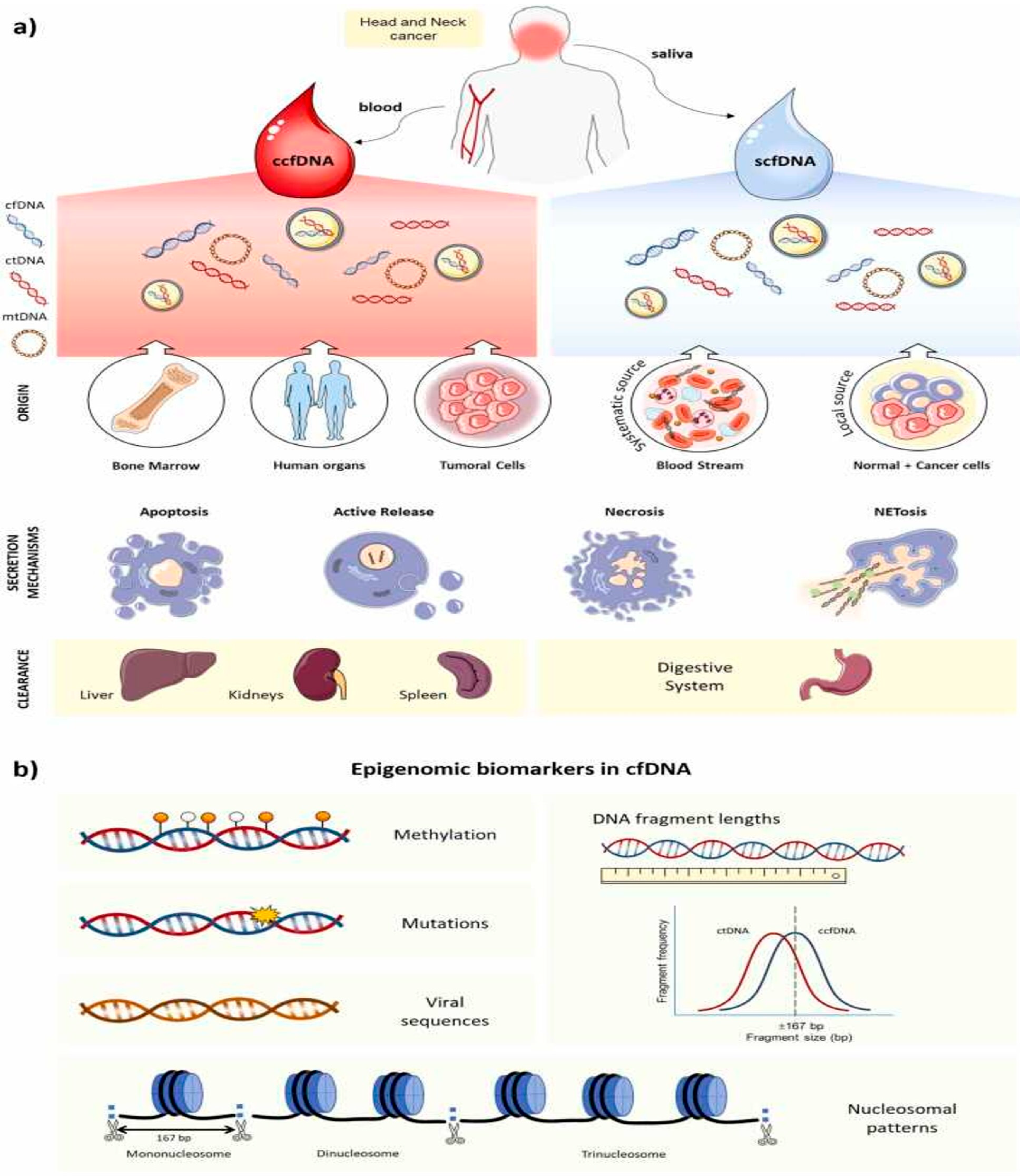
Figure 1. Overview of cell-free DNA (cfDNA) in head and neck cancer. (a) Origin, release, and clearance: cfDNA derives from normal and tumor cells via apoptosis, necrosis, active secretion, or NETosis. Circulating cfDNA (including ctDNA) originates largely from bone marrow, various organs, and tumor sites and is cleared by the liver, kidneys, and spleen. In saliva, cfDNA arises both locally—from shed oral epithelial cells—and systemically—via salivary-gland transudation—and is ultimately excreted through the digestive tract. (b) Epigenomic biomarkers: cfDNA carries tumor-specific alterations (mutations, methylation changes, and viral sequences) and bears fragmentomic and nucleosome patterns, providing insights into gene regulation and disease processes. From [24] with permission.
Cancers 17 02366 g001
Figure 2. Flowchart illustrating the whole-exome sequencing (WES) analysis of cfDNA. From [41], reused under CC-BY 4.0.
Cancers 17 02366 g002
Figure 3. Plasma immunodepletion and label-free MS quantitation workflow: (a) Immunodepletion: Remove the top 12 abundant plasma proteins. (b) Proteolysis: Digest the depleted sample with Trypsin and Lys-C; then, wash and elute peptides. (c) Untargeted LC-MS/MS: Analyze peptides via nano-LC coupled to tandem MS to identify all detectable sequences. (d) Label-Free Quantitation: Process raw spectra in MaxQuant, Proteome Discoverer, and the Trans-Proteomic Pipeline; filter and statistically pinpoint differentially abundant proteins. (e) Functional Analysis: Subject those proteins to pathway and network analysis to elucidate their roles in disease biology. From [96] with permission.
Cancers 17 02366 g003
Figure 4. The application of metabolomics in oral squamous cell carcinoma research. This figure was created using biorender.com. From [95] with permission.
Cancers 17 02366 g004
Table 1. Metabolic markers for early diagnosis of OSCC. From [95] with permission.
SampleComparisonMetabolite Analysis TechniqueDifferential MetabolitesReferences
SalivaOSCC versus OLKGC-TOF-MSTyrosine, malic acid, tyrosine, lactic acid, 2-hydroxybutyric acid, prostaglandin E2Xue et al. [109]
OSCC versus HCGC-TOF-MSProstaglandin E2, lactic acid, tyrosine, lactic acid, 2-hydroxybutyric acid, prostaglandin E2Ferrarini et al. [110]
OLK versus HCGC-TOF-MSTyrosine, lactic acid, 2-hydroxybutanedioic acid, prostaglandin E2Xue et al. [109]
OSCC versus OLKGC-MSDecanedioic acid, 2-methyloctadecane, eicosanoic acid, octane, 3,5-dimethylTantray et al. [111]
OSCC versus HCCPSI-MSPolyamines (e.g., spermidine, spermine) and amino acids (e.g., arginine, lysine, histidine, glutamine, leucine) Metabolites related to energy metabolism (e.g., glucose, creatine, creatinine) and purine metabolites (e.g., inosine, hypoxanthine)Song et al. [112]
HC versus PMLCPSI-MSInosine, hypoxanthine, adenosine, thymidine, uridine, guanosine, cytosine, choline, sphingolipids, etc.Song et al. [112]
SerumOSCC versus HCUHPLC-Q-Orbitrap HRMSLysoPC, taurine, and glutamateLi et al. [113]
OSCC versus HCCPSI-MSGPC, lyso-GPC, acylcarnitine, DG, sphingolipids, etc.Yang, Song, Yang, et al. [114]
OSCC preoperative versus postoperativeUHPLC-Q-Orbitrap HRMSSuccinic acid, arginine, L-carnitine, NAcetyl-L-tyrosine, glutamine, xanthine, sphingosine, palmitoyl ethanolamide, hexanoyl carnitine, whey acid, uric acid, vanillylmandelic acid, ethyl acetate, thromboxane B2Zuo et al. [115]
TissueOSCC versus HCGC–MS UHPLC–MS/MSGlutamic acid, aspartic acid, prolineYang et al. [116]
OSCC versus PML versus HCGC-MS AnalysesGlycine, threonine, glutamine, lysine, proline, alanine, glutamic acid, leucine, serineMusharraf et al. [117]
OSCC versus HCCE-TOF-MSGlucose, glycerol triphosphate, glycerol diphosphate, lactic acidOgawa et al. [118]
Disclaimer/Publisher’s Note: The statements, opinions and data contained in all publications are solely those of the individual author(s) and contributor(s) and not of MDPI and/or the editor(s). MDPI and/or the editor(s) disclaim responsibility for any injury to people or property resulting from any ideas, methods, instructions or products referred to in the content.