Article

MicroRNA Signatures in Lung Adenocarcinoma Metastases: Exploring the Oncogenic Targets of Tumor-Suppressive miR-195-5p and miR-195-3p

1
Department of Pulmonary Medicine, Graduate School of Medical and Dental Sciences, Kagoshima University, Kagoshima 890-8520, Japan
2
Department of Functional Genomics, Graduate School of Medicine, Chiba University Chuo-ku, Chiba 260-8670, Japan
3
Department of Neurosurgery, Graduate School of Medical and Dental Sciences, Kagoshima University, Kagoshima 890-8520, Japan
*
Author to whom correspondence should be addressed.
Cancers 2025, 17(14), 2348; https://doi.org/10.3390/cancers17142348
Submission received: 11 June 2025 / Revised: 12 July 2025 / Accepted: 13 July 2025 / Published: 15 July 2025

Simple Summary

To identify genes involved in lung cancer brain metastasis, we generated microRNA signatures using lung cancer brain metastasis tissues. Based on the microRNA signatures in lung adenocarcinoma metastases, both strands of pre-miR-195 (miR-195-5p and miR-195-3p) were significantly downregulated in metastatic tissues. A total of 12 genes (ANLN, CDC6, CDCA2, CDK1, CEP55, CHEK1, CLSPN, GINS1, KIF23, MAD2L1, OIP5, and TIMELESS) were identified as pre-miR-195-controlled genes, and these genes greatly contributed to the molecular pathogenesis of lung adenocarcinoma cells. Knockdown assays using siRNAs revealed that ANLN and MAD2L1 facilitated the malignant transformation of lung adenocarcinoma cells. Analysis of the microRNA signature generated in this study will accelerate the identification of genes involved in lung cancer brain metastasis.

Abstract

Background: To improve the prognosis of patients with lung adenocarcinoma (LUAD), revolutionary treatments for metastatic lesions are essential. Methods: To identify genes closely involved in LUAD-cell-derived metastasis, we used RNA sequencing to generate microRNA (miRNA) expression signatures of brain metastatic lesions. Once tumor-suppressive miRNAs are identified, it will be possible to explore the numerous tumor-promoting genes that are regulated by miRNAs. Results: By comparison with a previously created LUAD signature, we identified several miRNAs whose expression was significantly suppressed in brain metastases. We focused on both strands of pre-miR-195 (miR-195-5p and miR-195-3p), which were significantly downregulated in brain metastatic tissues, and confirmed by ectopic expression assays that both strands of pre-miR-195 attenuated the aggressive phenotypes (cell proliferation, migration, and invasion) of LUAD cells. These data suggest that both strands of pre-miR-195 have tumor-suppressive functions in LUAD cells. Next, we explored the target molecules that each miRNA strand regulates in LUAD cells. We identified 159 target genes regulated by miR-195-5p and miR-195-3p, of which 12 genes (ANLN, CDC6, CDCA2, CDK1, CEP55, CHEK1, CLSPN, GINS1, KIF23, MAD2L1, OIP5, and TIMELESS) affect cell cycle/cell division and the prognosis of LUAD patients. Finally, we focused on two genes, ANLN (miR-195-5p target) and MAD2L1 (miR-195-3p target), and demonstrated their oncogenic functions and the molecular pathways they regulate in LUAD cells. Conclusions: The miRNA signature derived from lung cancer brain metastasis will be a landmark in the field, and analysis of this miRNA signature will accelerate the identification of genes involved in lung cancer brain metastasis.

1. Introduction

Lung cancer is the most commonly diagnosed malignant tumor worldwide, with approximately 2.5 million new cases reported in 2022. It is also the leading cause of cancer-related deaths, accounting for 18.7% of cancer-related deaths worldwide [1]. Histologically, lung cancer is classified into non-small-cell lung cancer (NSCLC; which accounts for 85% of cases) and small-cell lung cancer (SCLC; which accounts for 15% of cases). Most NSCLC cases are adenocarcinoma (LUAD) [2]. In recent years, the prognosis of patients with advanced LUAD has improved with the advent of targeted therapies against specific genetic mutations and the introduction of immune checkpoint inhibitors [3,4]. However, despite these advances, distant metastasis remains the primary cause of lung-cancer-related deaths, with the brain being the most common site of dissemination [5,6]. Almost 10% of patients with newly diagnosed NSCLC present with metastasis, and 25–40% develop brain metastases during the disease course [6,7]. The prognosis for patients with brain metastases remains extremely poor, with the median survival ranging from only 3 to 6 months, even after treatment [8]. Therefore, our challenge is to elucidate the molecular mechanisms involved in the brain metastasis of lung cancer cells.
MicroRNAs (miRNAs) are small non-coding RNAs that act as post-transcriptional gene controllers and are essential for cell maintenance [9,10]. Numerous studies have demonstrated that aberrantly expressed miRNAs play important roles in the development, progression, metastasis, and drug resistance of cancer cells [11,12]. miRNA expression signatures provide a wealth of information about the miRNAs dysregulated in cancer cells [13]. Recent advances in RNA-sequencing technology have significantly contributed to the generation of miRNA expression signatures for various types of cancer cells [14]. Our research group has created miRNA expression signatures for various cancer tissues, including LUAD and SCLC, and identified tumor-suppressive miRNAs based on these signatures [15,16].
Our miRNA signatures demonstrated that some passenger strands of miRNAs were actually downregulated or upregulated in brain metastases.. Furthermore, we have demonstrated that some passenger strands, whose functions had previously been unclear, function as tumor-suppressive miRNAs in cancer cells and directly control multiple genes [15,16,17,18,19].
The aim of this study was to identify genes that are essential for lung cancer cells to metastasize to the brain. In this study, we generated a new miRNA expression signature from brain metastatic lesions and compared it with a signature from LUAD tissue that we had previously generated. Our signature showed that multiple passenger strands were downregulated in brain metastatic tissues. In particular, both the guide and passenger strands of the following eight pre-miRNAs were downregulated in metastatic tissues: miR-10a, miR-34b, miR-34c, miR-195, miR-199a, miR-199b, and miR-497. Exploring novel molecular pathways controlled by these miRNAs will provide new insights into the metastasis of LUAD cells. Interestingly, among these downregulated miRNAs, miR-195 and miR-497 are part of a cluster of miRNAs on human chromosome 17p13.1. The target genes of these clustered miRNAs may be involved in malignant transformation of LUAD cells.
To verify the validity of the new signature we created in this study, we focused on both strands of pre-miR-195 (miR-195-5p and miR-195-3p), which were significantly downregulated in brain metastatic tissues, and attempted to search for their target molecules. Our functional assays confirmed that both strands of pre-miR-195 acted as tumor suppressors in LUAD cells. A total of 159 genes were identified as target genes controlled by pre-miR-195 in LUAD cells. We found that 12 of these genes (ANLN, CDC6, CDCA2, CDK1, CEP55, CHEK1, CLSPN, GINS1, KIF23, MAD2L1, OIP5, and TIMELESS) are involved in cell cycle/cell division and affect the prognosis of LUAD patients. Finally, we focused on two genes, ANLN (miR-195-5p target) and MAD2L1 (miR-195-3p target), and demonstrated their oncogenic functions and the molecular pathways regulated by them in LUAD cells.

2. Materials and Methods

2.1. LUAD Patients and LUAD Cell Lines

We obtained surgical specimens from the primary tumor and brain metastatic tissues of patients with LUAD. The background and clinical characteristics of the patients are described in Table S1.
We also employed two LUAD cell lines, A549 and H1299 (American Type Culture Collection, Manassas, VA, USA). The procedures for cell maintenance have been described in our previous publications [15,18].

2.2. Construction of an miRNA Expression Signature for LUAD Based on RNA Sequencing

To assess miRNA expression, LUAD and brain metastatic tissue samples were subjected to sequencing using the Illumina NextSeq 500 system (Illumina, Inc., San Diego, CA, USA). The raw sequencing data have been deposited in the Gene Expression Omnibus (GEO; GEO accession number: GSE230229).Sequence reads matched to the human genome were classified into different types of small RNAs based on their biological features, and the corresponding read counts for each category are presented in Table S2.

2.3. Identification of Oncogenic Targets of miR-195-5p and miR-195-3p in LUAD Cells

We analyzed gene expression data from A549 cells transfected with miR-195-5p or miR-195-3p (GEO accession number: GSE281258) in combination with predictions with TargetScanHuman ver. 8.0 (https://www.targetscan.org/vert_80/; accessed on 25 September 2024) to identify oncogenic targets regulated by miR-195-5p and miR-195-3p. This expression profile represents a comparison of the gene expression between LUAD and normal lung tissues. The molecular functions of the miR-195-5p and miR-195-3p target genes were inferred using GeneCodis4 software (https://genecodis.genyo.es/; accessed on 25 September 2024) [20].

2.4. Analysis of miRNA and Gene Expression and Their Clinical Significance in Patients with LUAD Using an In Silico Database

The expression of miRNAs and genes in LUAD clinical tissues was analyzed using the following publicly available databases: The Cancer Genome Atlas (TCGA) (https://www.cancer.gov/tcga; accessed on 20 September 2024), Genomic Data Commons Data Portal (https://portal.gdc.cancer.gov/; accessed on 20 September 2024), and FIREBROWSE (http://firebrowse.org/; accessed on 20 September 2024). Data related to overall survival were retrieved from OncoLnc (http://www.oncolnc.org/; downloaded on 20 September 2024) and cBioPortal (https://www.cbioportal.org/; accessed on 20 September 2024).

2.5. Functional Assays of miRNAs and miRNA Target Genes in LUAD Cells

miRNAs and small interfering RNAs (siRNAs) were transfected into LUAD cell lines. After transfection, cell proliferation, migration, and invasion were assessed and compared with those of untreated cells. The transfection procedures for the miRNAs and siRNAs were described in our previous studies [15,16,18]. The mock group comprised cells transfected without miRNAs or siRNAs. The cell cycle and apoptotic cells were analyzed using the BD FACSCelestaTM Flow Cytometer (BD Biosciences, Franklin Lakes, NJ, USA). Details of the cell functional assays are described in our previous papers [15,16,18]. The reagents used for these analyses are listed in Table S3.

2.6. Dual-Luciferase Reporter Assay

The binding sequences of miR-195-5p and miR-195-3p, which were cloned into the psiCHEK2 vector (C8021; Promega, Madison, WI, USA), are shown in Figures S1 and S2. The procedures for the transfection and dual-luciferase reporter assays have been described in our previous studies [15,16,18]. We performed the dual-luciferase reporter assays at 72 h after transfection using the Dual-Luciferase Reporter Assay System (catalog no. E1910, Promega). The reagents used for these analyses are listed in Table S3.

2.7. Western Blotting

Western blotting was conducted following protocols described in our previous publications [15,16,18]. The reagents and antibodies used in the present study are listed in Table S3.

2.8. Statistical Analysis

Statistical analyses were performed using R ver. 4.4.0 (R Core Team, Vienna, Austria; https://www.R-project.org/; accessed on 25 September 2024) and GraphPad Prism 8 (GraphPad Software, La Jolla, CA, USA). Differences between two groups were assessed by the Student’s t-test. Multiple-group comparisons were performed using one-way analysis of variance and Tukey’s test for post hoc analysis. Survival analysis was carried out using Kaplan–Meier survival curves and the log-rank test.

3. Results

3.1. The RNA-Sequencing-Based miRNA Expression Signature Using Clinical Specimens Obtained from LUAD Brain Metastases

In this study, we created a new miRNA expression signature using LUAD brain metastatic tissues. We explored miRNAs whose expression was downregulated in brain metastasis tissues by comparing them with the LUAD signature we had previously created [15]. A total of 48 downregulated miRNAs were identified (Table 1), and 14 miRNAs were annotated as passenger strands based on the miRBase database (Release 22; http://www.mirbase.org/) [21]. Interestingly, both the guide and the passenger strands derived from the following pre-miRNAs were significantly downregulated: miR-10a, miR-34b, miR-34c, miR-195, miR-199a, miR-199b, and miR-497 (Table 1). Exploring the genes controlled by these miRNAs will provide novel information regarding brain metastasis of LUAD cells.
In the human genome, miR-195 and miR-497 are part of a cluster of miRNAs on human chromosome 17p13.1 (Figure 1A). Both miR-195 and miR-497 were significantly downregulated in brain metastatic tissues compared with LUAD and normal lung tissues (Figure 1B). Analysis of the functional RNA network controlled by this miRNA cluster could potentially elucidate the molecular mechanism of lung cancer cell metastasis. Here, we first focused on miR-195-5p and miR-195-3p to identify their regulated RNA networks in LUAD cells.

3.2. Expression and Clinical Significance of miR-195-5p and miR-195-3p in LUAD Clinical Specimens

We validated the expression of miR-195-5p and miR-195-3p in LUAD clinical specimens using the large clinical cohort database TCGA. The expression levels of miR-195-5p and miR-195-3p were significantly reduced in LUAD tissues compared with normal tissues (Figure 1C). Using the TCGA dataset, we analyzed whether the expression levels of these miRNAs can serve as prognostic markers for LUAD. Low expression of miR-195-3p was associated with a significantly poor prognosis compared with high expression of this miRNA (Figure 1D). A similar analysis showed no significant difference in the expression of miR-195-5p among the tissue types (Figure 1D).

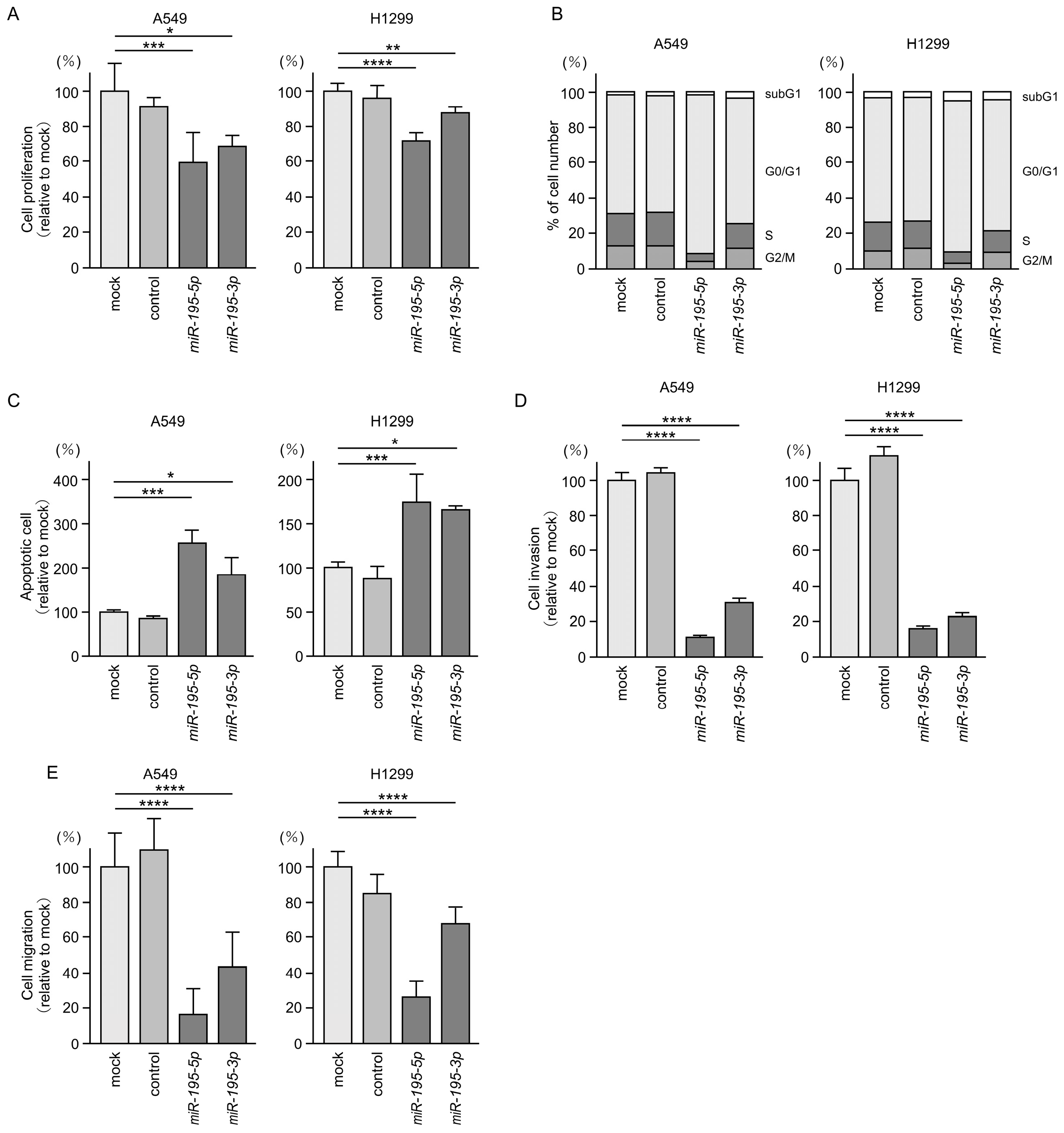
3.3. Tumor-Suppressive Functions of miR-195-5p and miR-195-3p in LUAD Cells

To demonstrate the antitumor functions of both strands of pre-miR-195 in LUAD cells, we ectopically expressed these miRNAs in A549 and H1299 cell lines and examined their effects on cancer cell behavior. Ectopic expression of miR-195-5p or miR-195-3p significantly suppressed the proliferation of LUAD cells (Figure 2A). Cell cycle analysis revealed the typical G0/G1 phase arrest following transfection with either miRNA (Figure 2B). Furthermore, expression of miR-195-5p or miR-195-3p led to a marked increase in the apoptotic cell population (Figure 2C). In addition, the invasive and migratory capacities of LUAD cells were significantly inhibited upon ectopic expression of miR-195-5p or miR-195-3p (Figure 2D,E). Flow cytometry images demonstrating this apoptosis are presented in Figure S3, and representative images of the invasion and migration assays are shown in Figure S4. Based on these results, we conclude that miR-195-5p and miR-195-3p function as tumor-suppressive miRNAs in LUAD cells.

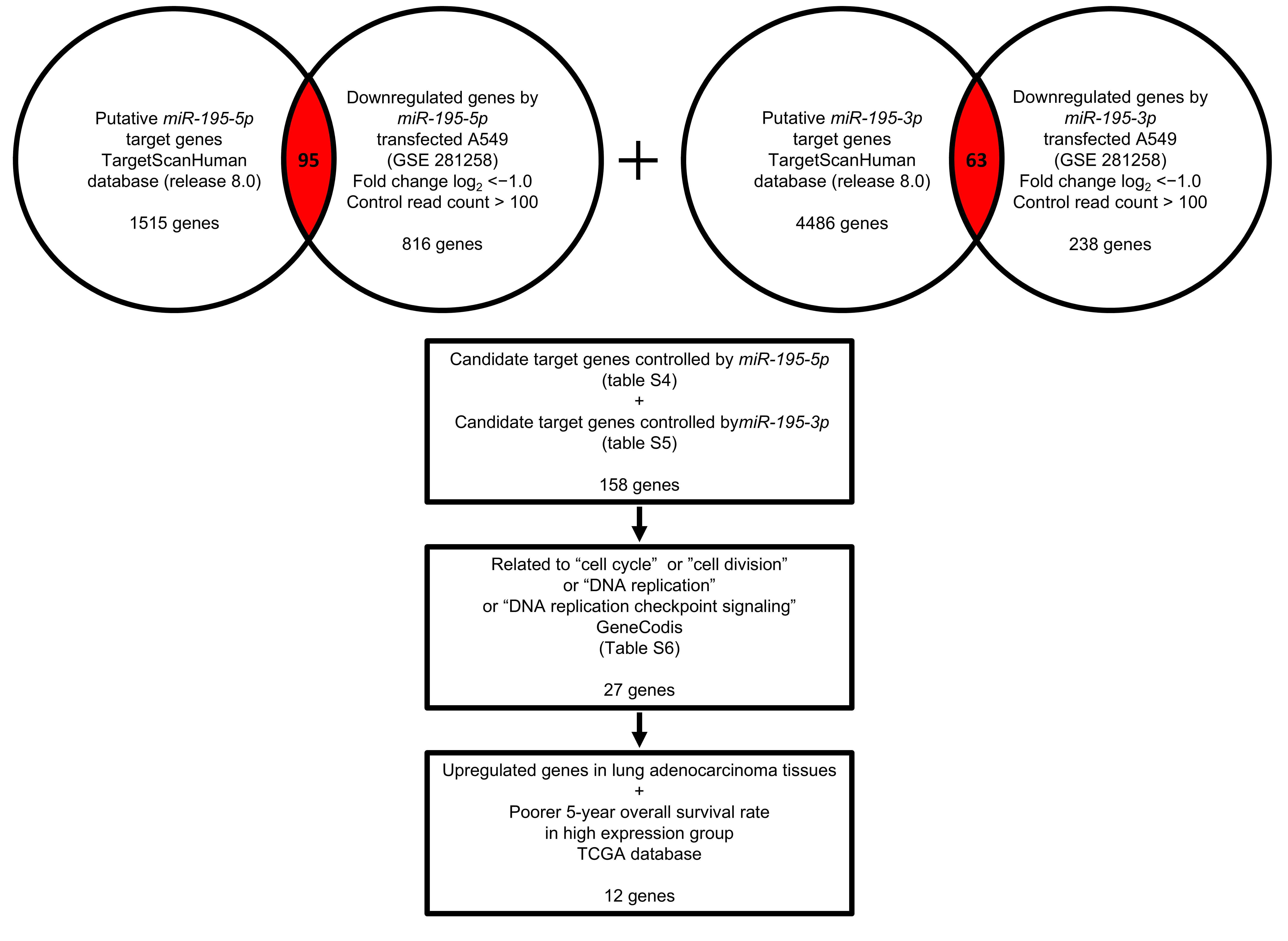
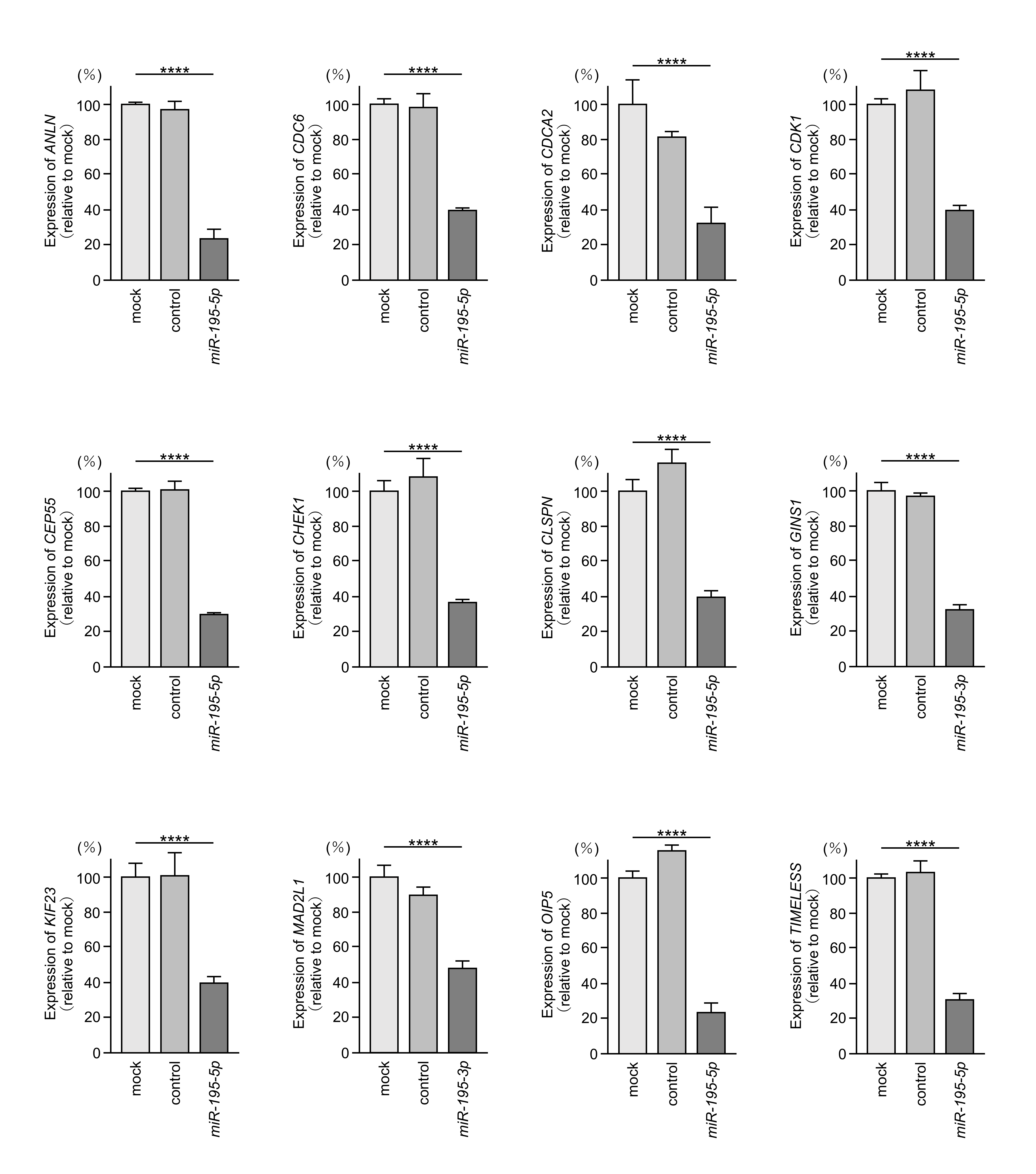
3.4. Identification of miR-195-5p- and miR-195-3p-Regulated Genes in LUAD Cells

As reported previously, we confirmed that both strands of pre-miR-195 function as antitumor miRNAs. Identifying the oncogenic targets regulated by these miRNAs in LUAD cells will aid in the search for genes involved in the malignant progression of lung cancer. Our strategy for the microRNA target search is shown in Figure 3. In this study, we performed a genome-wide gene expression analysis in A549 cells transfected with miR-195-5p or miR-195-3p. The expression data obtained have been deposited in the GEO database and are accessible (accession number GSE281258).
Using a combination of the TargetScanHuman database (release 8.0) and the gene expression profiles obtained from miRNA-transfected LUAD cells, we identified putative target genes regulated by miR-195-5p (95 genes) and miR-195-3p (63 genes). These candidate targets are listed in Tables S4 and S5. To further characterize their potential roles, we analyzed the molecular functions of all 158 genes using the GeneCodis4 database (Table S6). Notably, 27 of these genes were associated with cell cycle regulation (Table 2).

3.5. Clinical Significance of Genes Regulated by miR-195-5p or miR-195-3p in LUAD

To assess the clinical relevance of the 27 genes potentially regulated by miR-195-5p or miR-195-3p, we analyzed the TCGA-LUAD dataset. Among the 27 target genes, 12 (Table 2, bold) were significantly upregulated in LUAD tissues (n = 516) compared with normal lung tissues (n = 59) (Figure 4A). Furthermore, elevated expression of these genes was significantly associated with a poor prognosis (5-year overall survival rate, p < 0.05) in LUAD patients (Figure 4B). Ectopic expression of miR-195-5p or miR-195-3p in LUAD cells significantly reduced the mRNA levels of these 12 target genes (Figure 5). Among the 12 target genes, we focused on anillin (ANLN), the top prognostic candidate regulated by miR-195-5p, and mitotic arrest deficient 2-like 1 (MAD2L1), the top prognostic candidate regulated by miR-195-3p, for further functional analyses.

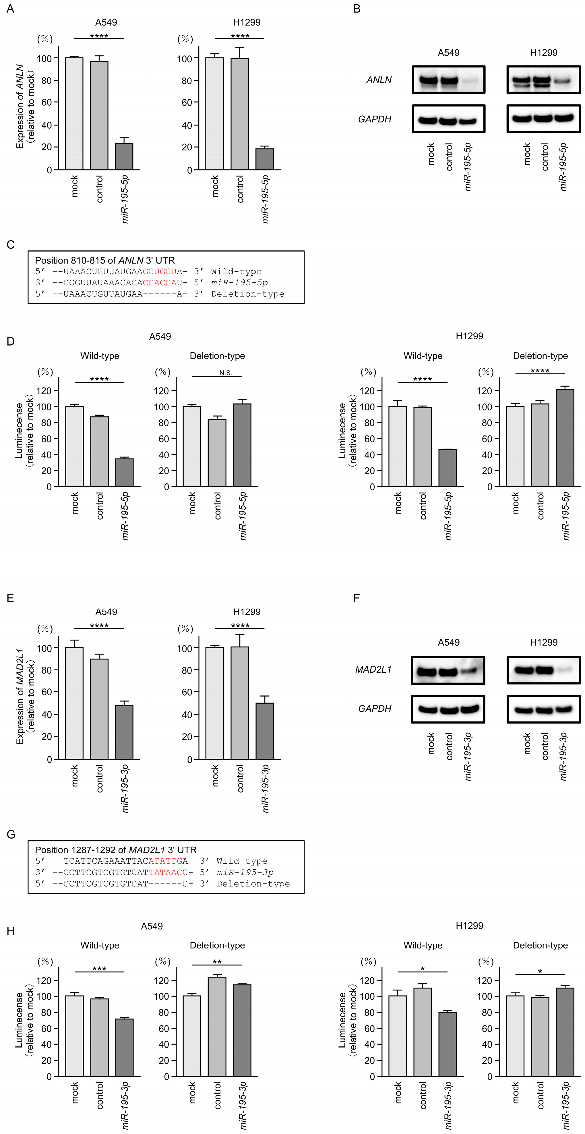
3.6. Direct Regulation of ANLN by miR-195-5p and MAD2L1 by miR-195-3p in LUAD Cells

Ectopic expression of miR-195-5p and miR-195-3p in LUAD cells significantly reduced the mRNA and protein expression levels of ANLN and MAD2L1, respectively (Figure 6A,B,E,F). Full-length Western blot images are provided in Figures S5 and S6. Luciferase reporter assays further demonstrated that both miRNAs directly bind to the 3′ untranslated regions (3′-UTRs) of their respective target genes. The predicted miR-195-5p-binding site in the ANLN 3′-UTR and the miR-195-3p-binding site in the MAD2L1 3′-UTR are shown in Figure 6C and G, respectively. Co-transfection of miR-195-5p or miR-195-3p with the corresponding reporter construct led to a marked reduction in luciferase activity, whereas no such reduction was observed after transfection with constructs lacking the respective binding sites (Figure 6D,H). These findings indicate that miR-195-5p and miR-195-3p directly target ANLN and MAD2L1, respectively, and post-transcriptionally suppress their expression in LUAD cells.

3.7. Functional Significance of ANLN in LUAD Cells

To investigate the oncogenic role of ANLN in LUAD cells, we performed siRNA-mediated knockdown assays. Transfection with two independent siRNAs targeting ANLN (siANLN-1 and siANLN-2) resulted in significant reductions in both ANLN mRNA and protein levels in LUAD cells (Figures S7 and S8). Cell proliferation assays revealed that ANLN knockdown inhibited the proliferation of LUAD cells (Figure 7A). Furthermore, flow cytometric analysis showed that ANLN knockdown in LUAD cells induced G0/G1 cell cycle arrest and increased the proportion of apoptotic cells (Figure 7B,C). Notably, ANLN knockdown markedly suppressed both the invasive and migratory capacities of LUAD cells (Figure 7D,E). Flow cytometry images showing the proportion of cells in apoptosis are presented in Figure S9, and representative images of the invasion and migration assays are shown in Figure S10.

3.8. Functional Significance of MAD2L1 in LUAD Cells

Similarly, to investigate the oncogenic role of MAD2L1 in LUAD cells, we performed siRNA-mediated knockdown assays. Transfection with two independent siRNAs targeting MAD2L1 (siMAD2L1-1 and siMAD2L1-2) resulted in significant reductions in both MAD2L1 mRNA and protein expression levels in LUAD cells (Figures S11 and S12). Cell proliferation assays showed that MAD2L1 knockdown slightly inhibited the proliferation of LUAD cells (Figure 8A). Additionally, flow cytometric analysis demonstrated that MAD2L1 knockdown induced G0/G1 cell cycle arrest and increased the proportion of apoptotic cells in LUAD cells (Figure 8B,C). However, in H1299 cells, no increase in the proportion of G0/G1 phase cells was observed; instead, there was a notable increase in the proportion of subG1 cells, indicating enhanced apoptotic activity. Furthermore, MAD2L1 knockdown markedly suppressed the invasive and migratory abilities of LUAD cells (Figure 8D,E). Flow cytometry images showing the proportion of cells in apoptosis are presented in Figure S13, and representative images of the invasion and migration assays are shown in Figure S14.

3.9. ANLN- and MAD2L1-Mediated Cancer Pathways in LUAD Cells

To investigate the signaling pathways influenced by ANLN and MAD2L1 in cancer, we generated genome-wide gene expression profiles using A549 cells transfected with siANLN or siMAD2L1. The resultant expression data have been deposited in the GEO database under accession numbers GSE281585 and GSE281905.
Interestingly, we also observed that siANLN transfection suppressed MAD2L1 expression, while siMAD2L1 transfection suppressed ANLN expression, suggesting the existence of reciprocal regulatory interactions between these two genes (Figure 9). A total of 39 genes were commonly downregulated in both siANLN- and siMAD2L1-transfected cells (Table 3). These genes were analyzed using GeneCodis4, which revealed that a substantial proportion of the genes are involved in cell-cycle-related pathways (Table 4). Among these genes, the expression of 26 genes had a negative effect on the prognosis of LUAD patients (Figure 10).

4. Discussion

In this study, we focused on both strands of pre-miR-195 (miR-195-5p and miR-195-3p), which were significantly downregulated in brain metastatic tissues. Furthermore, we explored the target molecules that each miRNA controls in LUAD cells. A total of 159 genes were identified as target genes controlled by pre-miR-195. We found that 12 of these genes (ANLN, CDC6, CDCA2, CDK1, CEP55, CHEK1, CLSPN, GINS1, KIF23, MAD2L1, OIP5, and TIMELESS) are involved in cell cycle/cell division and affect the prognosis of LUAD patients. Using in vitro functional assays, we revealed that ANLN and MAD2L1 actually play an oncogenic role.
Our group has previously identified tumor-suppressive miRNAs and their novel regulatory RNA networks in lung cancer cells [15,18,19]. RNA-sequencing-based miRNA signatures provide important information that suggests which miRNAs should be analyzed. Our recent studies revealed that passenger strands derived from pre-miRNAs function as oncogenes or tumor suppressors and actually regulate intracellular target molecules in LUAD cells [18,19]. Similarly to previous results, this study also showed that both the passenger strand and guide strand of miR-195 equally and essentially affect the function of cancer cells (Figure 2). Comprehensive analysis of the passenger strands as well as the guide strands of miRNAs will bring new information to cancer research.
An interesting feature of this signature was that it contained several miRNAs (e.g., miR-10a, miR-34b, miR-34c, miR-195, miR-199a, miR-199b, and miR-497) in which both strands (guide and passenger strands derived from the pre-miRNAs) were significantly downregulated in brain metastatic tissues. Analysis of these miRNAs, including their passenger strands, will shed light on the RNA networks associated with lung cancer metastasis. Among these miRNAs, we focused on the miR-195/miR-497 miRNA cluster on human chromosome 17p13.1. We first investigated the tumor-suppressive functions of miR-195 and its regulated RNA networks in LUAD cells.
Previous reports have shown that the expression of miR-195-5p (the guide strand) is suppressed in various types of cancers. Furthermore, ectopic expression assays demonstrated that miR-195-5p acts as a tumor-suppressive miRNA across cancer types by targeting several oncogenes [22,23,24,25]. Expression of miR-195-5p enhanced the cisplatin or gemcitabine sensitivity of LUAD cells by targeting E2F transcription factor 7 [26]. Recent studies have demonstrated that aberrantly expressed long non-coding RNAs or circulating RNAs may be involved in promoting cancer cell malignant transformation by adsorbing tumor-suppressive miR-195-5p in LUAD cells [27]. Compared with the numerous studies on miR-195-5p, functional assays of miR-195-3p (the passenger strand) are scarce. Our present analysis demonstrates that miR-195-3p is a tumor-suppressive miRNA that regulates many cancer genes in LUAD cells. Combining our data with previous reports, it is clear that both strands of pre-miR-195 (miR-195-5p and miR-195-3p) behave as tumor suppressors in LUAD cells.
Next, we identified cancer-promoting genes among those regulated by pre-miR-195 (miR-195-5p and miR-195-3p) in LUAD cells. Our strategy successfully identified 159 genes as candidate pre-miR-195 targets in LUAD cells. Of these, 12 genes (ANLN, CDC6, CDCA2, CDK1, CEP55, CHEK1, CLSPN, GINS1, KIF23, MAD2L1, OIP5, and TIMELESS) are involved in the cell cycle, cell division, and DNA replication checkpoint-related pathways, and their overexpression has a significant impact on the prognosis of patients with LUAD; therefore, these genes are potential therapeutic targets in LUAD. Among these therapeutic targets, we focused on ANLN (miR-195-5p target) and MAD2L1 (miR-195-3p target) and revealed their cancer-promoting functions in LUAD cells.
ANLN is an actin-binding protein that interacts with actin and cytoskeletal filaments to control the cell cycle [28]. Recent studies show that aberrant expression of ANLN is observed in multiple types of cancer, and that overexpression of ANLN is a useful biomarker predicting cancer cell metastasis and patient prognosis [29]. Knockdown assays targeting ANLN expression revealed dramatically reduced aggressive phenotypes (cell growth, migration, and invasion) of cancer cells, including pancreatic, breast, and lung cancers [30,31,32]. Aberrant expression of ANLN is intricately involved in the molecular pathogenesis of various human cancers and is a potential therapeutic target.
MAD2L1 is an essential component of the spindle assembly checkpoint [33]. In normal cells, MAD2 accumulates at kinetochores before cell division to ensure that spindle microtubules are properly aligned with the kinetochores of each chromosome [33]. Thus, MAD2 is a gatekeeper of cell cycle checkpoints and is essential for maintaining faithful replicative cell division and preventing cancer development [34]. In multiple cancers, overexpression of MAD2L1 is involved in the malignant transformation of cancer cells, and patients overexpressing MAD2L1 have a poor prognosis [35,36,37,38]. More recently, it has been reported that MAD2L1 expression is involved in carboplatin resistance in lung cancer cells [39]. MAD2L1 serves as a therapeutic target molecule in various cancers, including LUAD.
Examination of the interaction between ANLN and MAD2L1 showed that these two genes affect each other. In addition, the existence of genes whose expression is commonly regulated downstream of ANLN and MAD2L1 was revealed. The expression of 26 of these genes had a negative effect on the prognosis of LUAD patients. Our study has shown that suppression of miR-195-5p/-3p expression and overexpression of the ANLN/MAD2L1 genes are among the factors that accelerate the progression of lung cancer cells. Future studies involving in vivo validation or analyses using larger clinical datasets, including independent patient cohorts, will be essential to confirm the biological and therapeutic significance of the identified targets.
Analyses based on the miRNA signature created in this study will facilitate the identification of genes involved in the malignant progression and brain metastasis of LUAD cells. This study is exploratory and based on a limited number of LUAD brain metastasis specimens, which are rare and difficult to obtain. While the findings offer important insights, they should be interpreted with caution and require further validation in larger patient cohorts to confirm their broader applicability.

5. Conclusions

We generated a novel RNA-sequencing-based miRNA signature using clinical specimens obtained from lung cancer brain metastases. Both strands of pre-miR-195 (miR-195-5p and miR-195-3p) were significantly downregulated in brain tissues. Ectopic expression assays demonstrated that the two miRNAs derived from pre-miR-195 attenuated LUAD cell aggressiveness by targeting several oncogenes, particularly ANLN (miR-195-5p target) and MAD2L1 (miR-195-3p target). ANLN and MAD2L1 may be novel therapeutic targets in LUAD cells.

Supplementary Materials

The following supporting information can be downloaded at: https://www.mdpi.com/article/10.3390/cancers17142348/s1, Figure S1: Predicted miR-195-5p binding site in ANLN and inserted sequences in the luciferase vectors; Figure S2: Predicted miR-195-3p binding site in MAD2L1 and inserted sequences in the luciferase vectors; Figure S3: Flow cytometry plots of Annexin V/PI-stained cells showing apoptosis after miR-195-5p or miR-195-3p transfection in A549 and H1299 cells; Figure S4: Representative images of invasion and migration assays after miR-195-5p or miR-195-3p transfection in A549 and H1299 cells; Figure S5: Full-sized Western blot images of ANLN expression after ectopic miR-195-5p expression in A549 and H1299 cells; Figure S6: Full-sized Western blot images of MAD2L1 expression after ectopic miR-195-3p expression in A549 and H1299 cells; Figure S7: Suppression of ANLN mRNA levels by siANLN-1 or siANLN-2 transfection in A549 and H1299 cells; Figure S8: Full-sized Western blot images showing ANLN expression after siANLN-1 or siANLN-2 transfection in A549 and H1299 cells; Figure S9: Flow cytometry plots of Annexin V/PI-stained cells showing apoptosis after siANLN-1 or siANLN-2 transfection in A549 and H1299 cells; Figure S10: Representative images of invasion and migration assays after siANLN-1 or siANLN-2 transfection in A549 and H1299 cells; Figure S11: Suppression of MAD2L1 mRNA levels by siMAD2L1-1 or siMAD2L1-2 transfection in A549 and H1299 cells; Figure S12: Full-sized Western blot images showing MAD2L1 expression after siMAD2L1-1 or siMAD2L1-2 transfection in A549 and H1299 cells; Figure S13: Flow cytometry plots of Annexin V/PI-stained cells showing apoptosis after siMAD2L1-1 or siMAD2L1-2 transfection in A549 and H1299 cells; Figure S14: Representative images of invasion and migration assays after siMAD2L1-1 or siMAD2L1-2 transfection in A549 and H1299 cells; Table S1: Clinical features of the LUAD patients who provided tissues at the time of initial diagnosis; Table S2: Annotations of reads aligned to small RNAs; Table S3: Reagents used in this study; Table S4: Putative target genes regulated by miR-195-5p in A549 cells; Table S5: Putative target genes regulated by miR-195-3p in A549 cells, Table S6: Significantly enriched annotations of target genes regulated by miR-195-5p or miR-195-3p.

Author Contributions

Conceptualization, N.S., K.M., and K.T. (Kentaro Tanaka); methodology, N.S.; validation, N.S., K.M., K.T. (Kentaro Tanaka), and H.I.; formal analysis, Y.T., T.S., K.T. (Kentaro Tsuruzono), Y.H., M.K., and C.M.; investigation, Y.T., T.S., K.T. (Kentaro Tsuruzono), Y.H., M.K., and C.M.; resources, H.Y.; data curation, Y.T., T.S., K.T. (Kentaro Tsuruzono), and Y.H.; writing—original draft preparation, N.S.; writing—review and editing, Y.T., T.S., K.T. (Kentaro Tsuruzono), and Y.H.; visualization, Y.T., N.S., T.S., K.T. (Kentaro Tsuruzono), and Y.H.; supervision, N.S.; project administration, N.S., K.M., K.T. (Kentaro Tanaka), and H.I.; funding acquisition, N.S., T.S., Y.H., M.K., and C.M. All authors have read and agreed to the published version of the manuscript.

Funding

This research was funded by KAKENHI; grant numbers 23K09346, 24K11347, 24K11370, 24K12457, and 24K12641.

Institutional Review Board Statement

The study was conducted in accordance with the Declaration of Helsinki and approved by the Ethics Committee on Epidemiological and its related Studies, Sakuragaoka Campus, Kagoshima University (approval no. 210101 eki-kai 2; 31 August 2023).

Informed Consent Statement

Informed consent was obtained from all subjects involved in the study.

Data Availability Statement

Publicly available datasets were analyzed in this study. These data can be accessed here: https://www.ncbi.nlm.nih.gov/geo/query/acc.cgi?acc=GSE230229 (accessed on 25 September 2024), https://www.ncbi.nlm.nih.gov/geo/query/acc.cgi?acc=GSE281258 (accessed on 24 November 2024) and https://www.ncbi.nlm.nih.gov/geo/query/acc.cgi?acc=GSE281905 (accessed on 24 November 2024).

Conflicts of Interest

The authors declare no conflicts of interest.

References

  1. Bray, F.; Laversanne, M.; Sung, H.; Ferlay, J.; Siegel, R.L.; Soerjomataram, I.; Jemal, A. Global cancer statistics 2022: GLOBOCAN estimates of incidence and mortality worldwide for 36 cancers in 185 countries. CA Cancer J. Clin. 2024, 74, 229–263. [Google Scholar] [CrossRef] [PubMed]
  2. Schabath, M.B.; Cote, M.L. Cancer Progress and Priorities: Lung Cancer. Cancer Epidemiol. Biomarkers Prev. 2019, 28, 1563–1579. [Google Scholar] [CrossRef] [PubMed]
  3. Tan, A.C.; Tan, D.S.W. Targeted Therapies for Lung Cancer Patients With Oncogenic Driver Molecular Alterations. J. Clin. Oncol. 2022, 40, 611–625. [Google Scholar] [CrossRef] [PubMed]
  4. Reck, M.; Remon, J.; Hellmann, M.D. First-Line Immunotherapy for Non-Small-Cell Lung Cancer. J. Clin. Oncol. 2022, 40, 586–597. [Google Scholar] [CrossRef]
  5. Duma, N.; Santana-Davila, R.; Molina, J.R. Non-Small Cell Lung Cancer: Epidemiology, Screening, Diagnosis, and Treatment. Mayo Clin. Proc. 2019, 94, 1623–1640. [Google Scholar] [CrossRef]
  6. Karimpour, M.; Ravanbakhsh, R.; Maydanchi, M.; Rajabi, A.; Azizi, F.; Saber, A. Cancer driver gene and non-coding RNA alterations as biomarkers of brain metastasis in lung cancer: A review of the literature. Biomed. Pharmacother. 2021, 143, 112190. [Google Scholar] [CrossRef]
  7. Martínez-Espinosa, I.; Serrato, J.A.; Ortiz-Quintero, B. MicroRNAs in Lung Cancer Brain Metastasis. Int. J. Mol. Sci. 2024, 25, 10325. [Google Scholar] [CrossRef]
  8. Liu, W.; Powell, C.A.; Wang, Q. Tumor microenvironment in lung cancer-derived brain metastasis. Chin. Med. J. 2022, 135, 1781–1791. [Google Scholar] [CrossRef]
  9. Bartel, D.P. MicroRNAs: Genomics, biogenesis, mechanism, and function. Cell 2004, 116, 281–297. [Google Scholar] [CrossRef]
  10. Bartel, D.P. MicroRNAs: Target recognition and regulatory functions. Cell 2009, 136, 215–233. [Google Scholar] [CrossRef]
  11. Smolarz, B.; Durczyński, A.; Romanowicz, H.; Szyłło, K.; Hogendorf, P. miRNAs in Cancer (Review of Literature). Int. J. Mol. Sci. 2022, 23, 2805. [Google Scholar] [CrossRef] [PubMed]
  12. Hussen, B.M.; Hidayat, H.J.; Salihi, A.; Sabir, D.K.; Taheri, M.; Ghafouri-Fard, S. MicroRNA: A signature for cancer progression. Biomed. Pharmacother. 2021, 138, 111528. [Google Scholar] [CrossRef] [PubMed]
  13. Calin, G.A.; Croce, C.M. MicroRNA signatures in human cancers. Nat. Rev. Cancer. 2006, 6, 857–866. [Google Scholar] [CrossRef]
  14. Li, M.H.; Fu, S.B.; Xiao, H.S. Genome-wide analysis of microRNA and mRNA expression signatures in cancer. Acta Pharmacol. Sin. 2015, 36, 1200–1211. [Google Scholar] [CrossRef]
  15. Tomioka, Y.; Suetsugu, T.; Seki, N.; Tanigawa, K.; Hagihara, Y.; Shinmura, M.; Asai, S.; Kikkawa, N.; Inoue, H.; Mizuno, K. The Molecular Pathogenesis of Tumor-Suppressive miR-486-5p and miR-486-3p Target Genes: GINS4 Facilitates Aggressiveness in Lung Adenocarcinoma. Cells 2023, 12, 1885. [Google Scholar] [CrossRef]
  16. Tanigawa, K.; Misono, S.; Mizuno, K.; Asai, S.; Suetsugu, T.; Uchida, A.; Kawano, M.; Inoue, H.; Seki, N. MicroRNA signature of small-cell lung cancer after treatment failure: Impact on oncogenic targets by miR-30a-3p control. Mol. Oncol. 2023, 17, 328–343. [Google Scholar] [CrossRef]
  17. Matranga, C.; Tomari, Y.; Shin, C.; Bartel, D.P.; Zamore, P.D. Passenger-strand cleavage facilitates assembly of siRNA into Ago2-containing RNAi enzyme complexes. Cell 2005, 123, 607–620. [Google Scholar] [CrossRef]
  18. Tomioka, Y.; Seki, N.; Suetsugu, T.; Hagihara, Y.; Sanada, H.; Goto, Y.; Kikkawa, N.; Mizuno, K.; Tanaka, K.; Inoue, H. Identification of Tumor Suppressive miR-144-5p Targets: FAM111B Expression Accelerates the Malignant Phenotypes of Lung Adenocarcinoma. Int. J. Mol. Sci. 2024, 25, 9974. [Google Scholar] [CrossRef]
  19. Hagihara, Y.; Tomioka, Y.; Suetsugu, T.; Shinmura, M.; Misono, S.; Goto, Y.; Kikkawa, N.; Kato, M.; Inoue, H.; Mizuno, K.; et al. Identification of Tumor-Suppressive miR-139-3p-Regulated Genes: TRIP13 as a Therapeutic Target in Lung Adenocarcinoma. Cancers 2023, 15, 5571. [Google Scholar] [CrossRef]
  20. Garcia-Moreno, A.; López-Domínguez, R.; Villatoro-García, J.A.; Ramirez-Mena, A.; Aparicio-Puerta, E.; Hackenberg, M.; Pascual-Montano, A.; Carmona-Saez, P. Functional Enrichment Analysis of Regulatory Elements. Biomedicines 2022, 10, 590. [Google Scholar] [CrossRef]
  21. Kozomara, A.; Birgaoanu, M.; Griffiths-Jones, S. miRBase: From microRNA sequences to function. Nucleic Acids Res. 2019, 47, D155–D162. [Google Scholar] [CrossRef] [PubMed]
  22. Bu, L.; Tian, Y.; Wen, H.; Jia, W.; Yang, S. miR-195-5p exerts tumor-suppressive functions in human lung cancer cells through targeting TrxR2. Acta Biochim. Biophys. Sin. 2021, 53, 189–200. [Google Scholar] [CrossRef]
  23. Liu, X.; Zhou, Y.; Ning, Y.E.; Gu, H.; Tong, Y.; Wang, N. MiR-195-5p Inhibits Malignant Progression of Cervical Cancer by Targeting YAP1. Onco Targets Ther. 2020, 13, 931–944. [Google Scholar] [CrossRef]
  24. Piccinno, E.; Scalavino, V.; Labarile, N.; Armentano, R.; Giannelli, G.; Serino, G. miR-195-5p Inhibits Colon Cancer Progression via KRT23 Regulation. Pharmaceutics 2024, 16, 1554. [Google Scholar] [CrossRef] [PubMed]
  25. Zhu, H.; Chen, Z.; Yu, J.; Wu, J.; Zhuo, X.; Chen, Q.; Liang, Y.; Li, G.; Wan, Y. MiR-195-5p suppresses the proliferation, migration, and invasion of gallbladder cancer cells by targeting FOSL1 and regulating the Wnt/β-catenin pathway. Ann. Transl. Med. 2022, 10, 893. [Google Scholar] [CrossRef] [PubMed]
  26. Wu, H.; Zheng, B. miR-195-5p inhibits cisplatin resistance in lung adenocarcinoma by regulating DNA damage via targeting E2F7. J. Biochem. Mol. Toxicol. 2024, 38, e70015. [Google Scholar] [CrossRef]
  27. Xi, J.; Xi, Y.; Zhang, Z.; Hao, Y.; Wu, F.; Bian, B.; Hao, G.; Li, W.; Zhang, S. Hsa_circ_0060937 accelerates non-small cell lung cancer progression via modulating miR-195-5p/HMGB3 pathway. Cell Cycle. 2021, 20, 2040–2052. [Google Scholar] [CrossRef] [PubMed]
  28. Piekny, A.J.; Maddox, A.S. The myriad roles of Anillin during cytokinesis. Semin. Cell Dev. Biol. 2010, 21, 881–891. [Google Scholar] [CrossRef]
  29. Zhang, X.; Li, L.; Huang, S.; Liao, W.; Li, J.; Huang, Z.; Huang, Y.; Lian, Y. Comprehensive Analysis of ANLN in Human Tumors: A Prognostic Biomarker Associated with Cancer Immunity. Oxid. Med. Cell Longev. 2022, 2022, 5322929. [Google Scholar] [CrossRef]
  30. Idichi, T.; Seki, N.; Kurahara, H.; Yonemori, K.; Osako, Y.; Arai, T.; Okato, A.; Kita, Y.; Arigami, T.; Mataki, Y.; et al. Regulation of actin-binding protein ANLN by antitumor miR-217 inhibits cancer cell aggressiveness in pancreatic ductal adenocarcinoma. Oncotarget 2017, 8, 53180–53193. [Google Scholar] [CrossRef]
  31. Wang, Z.; Hu, S.; Li, X.; Liu, Z.; Han, D.; Wang, Y.; Wei, L.; Zhang, G.; Wang, X. MiR-16-5p suppresses breast cancer proliferation by targeting ANLN. BMC Cancer. 2021, 21, 1188. [Google Scholar] [CrossRef] [PubMed]
  32. Xu, J.; Zheng, H.; Yuan, S.; Zhou, B.; Zhao, W.; Pan, Y.; Qi, D. Overexpression of ANLN in lung adenocarcinoma is associated with metastasis. Thorac. Cancer. 2019, 10, 1702–1709. [Google Scholar] [CrossRef] [PubMed]
  33. Mossaid, I.; Fahrenkrog, B. Complex Commingling: Nucleoporins and the Spindle Assembly Checkpoint. Cells 2015, 4, 706–725. [Google Scholar] [CrossRef] [PubMed]
  34. Michel, L.; Benezra, R.; Diaz-Rodriguez, E. MAD2 dependent mitotic checkpoint defects in tumorigenesis and tumor cell death: A double edged sword. Cell Cycle 2004, 3, 990–992. [Google Scholar] [CrossRef]
  35. Chen, Q.; Yang, S.; Zhang, Y.; Li, B.; Xu, H.; Zuo, S. Identification of MAD2L1 as a Potential Biomarker in Hepatocellular Carcinoma via Comprehensive Bioinformatics Analysis. Biomed. Res. Int. 2022, 2022, 9868022. [Google Scholar] [CrossRef]
  36. Zhu, X.F.; Yi, M.; He, J.; Tang, W.; Lu, M.Y.; Li, T.; Feng, Z.B. Pathological significance of MAD2L1 in breast cancer: An immunohistochemical study and meta analysis. Int. J. Clin. Exp. Pathol. 2017, 10, 9190–9201. [Google Scholar]
  37. Li, Q.; Tong, D.; Jing, X.; Ma, P.; Li, F.; Jiang, Q.; Zhang, J.; Wen, H.; Cui, M.; Huang, C.; et al. MAD2L1 is transcriptionally regulated by TEAD4 and promotes cell proliferation and migration in colorectal cancer. Cancer Gene Ther. 2023, 30, 727–737. [Google Scholar] [CrossRef]
  38. Wei, R.; Wang, Z.; Zhang, Y.; Wang, B.; Shen, N.; E, L.; Li, X.; Shang, L.; Shang, Y.; Yan, W.; et al. Bioinformatic analysis revealing mitotic spindle assembly regulated NDC80 and MAD2L1 as prognostic biomarkers in non-small cell lung cancer development. BMC Med. Genomics 2020, 13, 112. [Google Scholar] [CrossRef]
  39. Zhao, H.; Liu, Y.; Zhu, L.; Cheng, J.; Li, Y. MAD2L1-mediated NANOG nuclear translocation: A critical factor in lung cancer chemoresistance. Cell Signal. 2025, 132, 111811. [Google Scholar] [CrossRef]
Figure 1. Expression patterns of miR-195-5p and miR-195-3p in LUAD clinical specimens and their association with 5-year overall survival rates. (A) Genomic location of pre-miR-195 on the human chromosome. The mature sequences of miR-195-5p (guide strand) and miR-195-3p (passenger strand) are indicated. (B) Heatmap of the expression levels of miRNAs downregulated in brain metastases across normal lung tissue, LUAD tissue, and brain metastases, based on the LUAD miRNA signature obtained by RNA sequencing. (C) Expression analysis of miR-195-5p and miR-195-3p using the TCGA-LUAD dataset. (D) Kaplan–Meier survival curves showing the 5-year overall survival of LUAD patients (n = 485), stratified by the median expression level of miR-195-5p or miR-195-3p. High-and low-expression groups are shown in red and blue, respectively.
Cancers 17 02348 g001
Figure 2. Antitumor effects of miR-195-5p and miR-195-3p in LUAD cells (A549 and H1299). (A) Cell proliferation evaluated using XTT assays 72 h after transient transfection with miR-195-5p or miR-195-3p. (B) Cell cycle status analyzed by flow cytometry 72 h after transient transfection with miR-195-5p or miR-195-3p. (C) Apoptotic cells evaluated by flow cytometry following Annexin V-FITC and PI-PerCP-Cy5-5-A staining 72 h after transient transfection with miR-195-5p or miR-195-3p. (D) Cell invasion assessed using Matrigel invasion assays. Cells were transfected with miR-195-5p or miR-195-3p for 72 h prior to seeding. (E) Cell migration assessed using a membrane culture system. Cells were transfected with miR-195-5p or miR-195-3p for 72 h prior to seeding. *, p < 0.05; **, p < 0.01; ***, p < 0.001; ****, p < 0.0001.
Cancers 17 02348 g002
Figure 3. Strategy for identifying oncogenic targets regulated by miR-195-5p or miR-195-3p in LUAD cells. Two datasets were utilized to identify the target genes: the TargetScanHuman database (release 8.0) and our original mRNA expression dataset (GEO accession number: GSE281258). Associations with LUAD patient prognosis were evaluated using two databases: OncoLnc and cBioPortal.
Cancers 17 02348 g003
Figure 4. Expression levels of the 12 target genes regulated by miR-195-5p or miR-195-3p in LUAD cells, along with their associated 5-year overall survival rates. (A) The expression levels of the 12 target genes of miR-195-5p or miR-195-3p (ANLN, CDC6, CDCA2, CDK1, CEP55, CHEK1, CLSPN, GINS1, KIF23, MAD2L1, OIP5, and TIMELESS) in LUAD clinical specimens assessed using the TCGA-LUAD dataset. (B) Kaplan–Meier curves for the 5-year overall survival rate based on the expression of the 12 target genes (ANLN, CDC6, CDCA2, CDK1, CEP55, CHEK1, CLSPN, GINS1, KIF23, MAD2L1, OIP5, and TIMELESS). The patients (n = 487) were divided into high- and low-expression groups based on the median expression level of each gene. The red and blue lines denote the high- and low-expression groups, respectively.
Cancers 17 02348 g004
Figure 5. Regulation of the 12 target genes by miR-195-5p or miR-195-3p in A549 cells. Total RNA was extracted 72 h after miRNA transfection and analyzed by quantitative real-time PCR. GAPDH was used as an internal control. ****, p < 0.0001.
Cancers 17 02348 g005
Figure 6. Direct regulation of ANLN and MAD2L1 by miR-195 expression in LUAD cells. (A,E) Marked reduction in ANLN or MAD2L1 mRNA expression by ectopic expression of miR-195-5p or miR-195-3p in LUAD cells (A549 and H1299). Total RNA was isolated 72 h after miRNA transfection and quantified by real-time PCR. GAPDH was used as an internal control. (B,F) Significant reduction in ANLN or MAD2L1 protein levels by ectopic expression of miR-195-5p or miR-195-3p in LUAD cells (A549 and H1299). Protein samples were isolated 72 h after miR-195-5p or miR-195-3p transfection and quantified by Western blotting. GAPDH was used as an internal control. (C,G) Putative miR-195-5p- or miR-195-3p-binding sites in the 3′-UTR of ANLN or MAD2L1, detected using the TargetScanHuman database (release 8.0). (D,H) Dual-luciferase reporter assays revealing the reduced luminescence activity after co-transfection of miR-195-5p or miR-195-3p with a vector containing the miR-195-5p- or miR-195-3p-binding site (wild-type) in LUAD cells (A549 and H1299). *, p < 0.05; **, p < 0.01; ***, p < 0.001; ****, p < 0.0001; N.S., not significant.
Cancers 17 02348 g006
Figure 7. Effects of ANLN knockdown by siRNAs in LUAD cells (A549 and H1299). (A) Cell proliferation evaluated using XTT assays after transient transfection with siANLN-1 or siANLN-2. (B) Cell cycle status analyzed by flow cytometry 72 h after transient transfection with siANLN-1 or siANLN-2. (C) Apoptotic cells evaluated by flow cytometry following Annexin V-FITC and PI-PerCP-Cy5-5-A staining 72 h after transient transfection with siANLN-1 or siANLN-2. (D) Cell invasion assessed using Matrigel invasion assays. Cells were transfected with siANLN-1 or siANLN-2 for 72 h prior to seeding. (E) Cell migration assessed using a membrane culture system. Cells were transfected with siANLN-1 or siANLN-2 for 72 h prior to seeding. **, p < 0.01; ***, p < 0.001; ****, p < 0.0001.
Cancers 17 02348 g007
Figure 8. Effects of MAD2L1 knockdown by siRNAs in LUAD cells (A549 and H1299). (A) Cell proliferation evaluated using XTT assays after transient transfection with siMAD2L1-1 or siMAD2L1-2. (B) Cell cycle status analyzed by flow cytometry 72 h after transient transfection with siMAD2L1-1 or siMAD2L1-2. (C) Apoptotic cells evaluated by flow cytometry following Annexin V-FITC and PI-PerCP-Cy5-5-A staining 72 h after transient transfection with siMAD2L1-1 or siMAD2L1-2. (D) Cell invasion assessed using Matrigel invasion assays. Cells were transfected with siMAD2L1-1 or siMAD2L1-2 for 72 h prior to seeding. (E) Cell migration assessed using a membrane culture system. Cells were transfected with siMAD2L1-1 or siMAD2L1-2 for 72 h prior to seeding. ***, p < 0.001; ****, p < 0.0001.
Cancers 17 02348 g008
Figure 9. Reciprocal suppression of ANLN and MAD2L1 mRNA levels by siRNA transfection in A549 and H1299 cells. Total RNA was extracted 72 h after siRNA transfection and analyzed by quantitative real-time PCR. GAPDH was used as an internal control. *, p < 0.05; **, p < 0.01; ***, p < 0.001; ****, p < 0.0001.
Cancers 17 02348 g009
Figure 10. Kaplan–Meier survival curves showing the 5-year overall survival based on the expression of the 26 genes negatively associated with the LUAD patient prognosis. The patients (n = 487) were stratified by the median expression levels of each target gene. High- and low-expression groups are shown in red and blue, respectively.
Cancers 17 02348 g010
Table 1. Downregulated miRNAs in brain metastasis compared with those in lung adenocarcinoma, identified by RNA sequencing.
MicroRNAmiRBase
Accession No.
Guide or Passenger StrandChromosomal LocationLog2 FCp-ValueFDR
hsa-miR-30d-5pMIMAT0000245Guide strand8q24.22−2.510.0040.018
hsa-miR-34b-5pMIMAT0000685Passenger strand11q23.1−2.560.0470.117
hsa-miR-4536-5pMIMAT0019078Guide strandXp11.21−2.560.0010.005
hsa-miR-6785-3pMIMAT0027471Passenger strand17q25.1−2.590.0000.000
hsa-miR-3141MIMAT0015010Guide strand5q33.2−2.610.0080.031
hsa-miR-30b-5pMIMAT0000420Guide strand8q24.22−2.630.0030.016
hsa-miR-497-3pMIMAT0004768Passenger strand17p13.1−2.630.0000.003
hsa-miR-4322MIMAT0016873Guide strand19p13.2−2.640.0160.053
hsa-miR-195-3pMIMAT0004615Passenger strand17p13.1−2.660.0000.001
hsa-miR-6743-3pMIMAT0027388Guide strand11p15.5−2.660.0000.002
hsa-miR-148a-5pMIMAT0004549Passenger strand7p15.2−2.790.0000.000
hsa-miR-195-5pMIMAT0000461Guide strand17p13.1−2.790.0000.002
hsa-miR-548iMIMAT0005935Guide strand3q21.2−2.810.0080.032
hsa-miR-6788-3pMIMAT0027477Guide strand18p11.22−2.820.0000.000
hsa-miR-363-5pMIMAT0003385Passenger strandXq26.2−2.850.0010.005
hsa-miR-4536-3pMIMAT0020959Passenger strandXp11.21−2.910.0000.000
hsa-miR-497-5pMIMAT0002820Guide strand17p13.1−2.910.0000.002
hsa-miR-214-5pMIMAT0004564Passenger strand1q24.3−2.920.0000.001
hsa-miR-95-3pMIMAT0000094Guide strand4p16.1−2.920.0080.033
hsa-miR-4662a-5pMIMAT0019731Guide strand8q24.13−2.940.0080.031
hsa-miR-145-5pMIMAT0000437Guide strand5q32−2.950.0000.002
hsa-miR-130a-3pMIMAT0000425Guide strand11q12.1−3.020.0000.002
hsa-miR-504-5pMIMAT0002875Guide strandXq26.3−3.020.0120.042
hsa-miR-199a-3pMIMAT0000232Guide strand19p13.2−3.030.0000.002
hsa-miR-34c-3pMIMAT0004677Passenger strand11q23.1−3.060.0090.036
hsa-miR-6763-5pMIMAT0027426Guide strand12q24.33−3.070.0000.000
hsa-miR-214-3pMIMAT0000271Guide strand1q24.3−3.140.0000.002
hsa-miR-199b-3pMIMAT0004563Guide strand9q34.11−3.150.0000.002
hsa-miR-218-5pMIMAT0000275Guide strand4p15.31−3.170.0010.006
hsa-miR-34c-5pMIMAT0000686Guide strand11q23.1−3.220.0420.107
hsa-miR-4470MIMAT0018997Guide strand8q12.3−3.300.0000.001
hsa-miR-1281MIMAT0005939Guide strand22q13.2−3.320.0000.000
hsa-miR-4259MIMAT0016880Guide strand1q23.2−3.330.0000.000
hsa-miR-3064-3pMIMAT0019865Passenger strand17q23.3−3.410.0000.000
hsa-miR-3926MIMAT0018201Guide strand8p23.1−3.440.0000.000
hsa-miR-5091MIMAT0021083Guide strand4p15.33−3.460.0000.001
hsa-miR-199b-5pMIMAT0000263Passenger strand9q34.11−3.560.0010.007
hsa-miR-602MIMAT0003270Guide strand9q34.3−3.610.0000.003
hsa-miR-548ar-3pMIMAT0022266Passenger strand13q34−3.620.0000.000
hsa-miR-10a-3pMIMAT0004555Passenger strand17q21.32−3.820.0010.004
hsa-miR-10a-5pMIMAT0000253Guide strand17q21.32−3.820.0000.002
hsa-miR-548wMIMAT0015060Guide strand16p12.1−3.850.0000.000
hsa-miR-199a-5pMIMAT0000231Passenger strand19p13.2−3.910.0000.000
hsa-miR-150-5pMIMAT0000451Guide strand19q13.33−4.000.0000.001
hsa-miR-548aMIMAT0003251Guide strand6p22.3−4.190.0000.000
hsa-miR-34b-3pMIMAT0004676Guide strand11q23.1−4.320.0050.023
Table 2. Putative target genes regulated by miR-195-5p or miR-195-3p in A549 cells.
Gene IDGene SymbolGene NameDownregulated
by miR-195-5p
or miR-195-3p
miR-195-5p
Transfectant
Log2 Fold Change
miR-195-3p
Transfectant
Log2 Fold Change
402ARL2ADP ribosylation factor-like GTPase 2miR-195-5p−3.310.11
896CCND3cyclin D3miR-195-5p−3.000.74
11339OIP5Opa-interacting protein 5miR-195-5p−2.49−0.62
54443ANLNanillin, actin-binding proteinmiR-195-5p−2.35−0.36
55165CEP55centrosomal protein 55miR-195-5p−2.27−0.67
983CDK1cyclin-dependent kinase 1miR-195-5p−1.86−0.63
63967CLSPNclaspinmiR-195-5p−1.85−0.70
993CDC25Acell division cycle 25AmiR-195-5p−1.800.18
990CDC6cell division cycle 6miR-195-5p−1.78−0.54
157313CDCA2cell division cycle-
associated 2
miR-195-5p−1.78−0.53
55038CDCA4cell division cycle-
associated 4
miR-195-5p−1.75−0.28
1111CHEK1checkpoint kinase 1 miR-195-5p−1.56−0.31
6867TACC1transforming acidic coiled-coil-containing protein 1miR-195-5p−1.520.27
9493KIF23kinesin family member 23 miR-195-5p−1.52−0.11
80010RMI1RecQ-mediated genome
instability 1
miR-195-3p−1.52−1.07
144455E2F7E2F transcription factor 7miR-195-5p−1.46−0.01
4085MAD2L1mitotic arrest
deficient 2-like 1
miR-195-3p−1.44−2.13
8914TIMELESStimeless circadian clock miR-195-5p−1.39−0.23
79187FSD1fibronectin type III and SPRY domain-containing 1 miR-195-5p−1.26−0.72
6197RPS6KA3ribosomal protein S6 kinase A3 miR-195-5p−1.240.62
27183VPS4Avacuolar protein sorting 4 homolog AmiR-195-5p−1.16−0.07
9874TLK1tousled-like kinase 1 miR-195-5p−1.120.13
9837GINS1GINS complex subunit 1miR-195-3p−1.08−1.85
996CDC27cell division cycle 27miR-195-5p−1.06−0.11
8243SMC1Astructural maintenance of chromosomes 1AmiR-195-3p−0.47−1.32
7329UBE2Iubiquitin-conjugating
enzyme E2 I
miR-195-3p−0.03−1.00
57804POLD4DNA polymerase delta 4miR-195-3p0.19−1.35
Table 3. Downregulated genes after siANLN-1 or siMAD2L1-1 transfection in A549 cells.
Entrez
Gene ID
Gene SymbolGene NamesiANLN
Transfectant
Log2 Fold Change
siMAD2L1
Transfectant
Log2 Fold Change
54443ANLNanillin actin-binding protein−6.14−2.32
84904ARHGEF39Rho guanine nucleotide exchange factor 39−3.61−2.28
55723ASF1Banti-silencing function 1B histone chaperone−4.52−2.71
890CCNA2cyclin A2−3.64−2.09
991CDC20cell division cycle 20−4.26−2.67
157313CDCA2cell division cycle-associated 2−3.01−2.03
55536CDCA7Lcell division cycle-associated 7-like−1.55−2.61
983CDK1cyclin-dependent kinase 1−4.60−1.96
1033CDKN3cyclin-dependent kinase inhibitor 3−2.92−2.43
2491CENPIcentromere protein I−3.73−2.60
55165CEP55centrosomal protein 55−3.59−2.03
1164CKS2CDC28 protein kinase regulatory subunit 2−2.68−1.55
1719DHFRdihydrofolate reductase−4.01−2.18
79075DSCC1DNA replication and sister chromatid cohesion 1−2.76−2.39
374393FAM111Bfamily with sequence similarity 111 member B−4.26−3.13
54478FAM64Afamily with sequence similarity 64 member A−3.89−1.92
9837GINS1GINS complex subunit 1−2.36−3.05
51512GTSE1G2 and S-phase-expressed 1−4.21−2.36
283120H19H19, imprinted maternally expressed transcript−2.58−2.98
8479HIRIP3HIRA-interacting protein 3−2.21−1.93
3161HMMRhyaluronan-mediated motility receptor −3.78−2.60
55806HRhair growth-associated −1.94−1.91
56992KIF15kinesin family member 15−4.13−2.51
10112KIF20Akinesin family member 20A−3.86−2.05
90417KNSTRNkinetochore localized astrin/SPAG5-binding protein−1.65−1.25
4085MAD2L1MAD2 mitotic arrest deficient-like 1−2.91−5.58
4288MKI67marker of proliferation Ki-67 −4.47−3.08
727897MUC5Bmucin 5B, oligomeric mucus/gel-forming−1.82−1.00
23397NCAPHnon-SMC condensin I complex subunit H −3.76−2.26
9768PCLAFPCNA clamp-associated factor−4.43−2.75
5933RBL1RB transcriptional corepressor like 1 −1.87−2.54
10535RNASEH2Aribonuclease H2 subunit A −2.93−2.08
200916RPL22L1ribosomal protein L22 like 1 −1.07−2.64
9123SLC16A3solute carrier family 16 member 3 −1.13−1.57
147841SPC24SPC24, NDC80 kinetochore complex component−3.75−2.40
57405SPC25SPC25, NDC80 kinetochore complex component−4.89−2.89
7083TK1thymidine kinase 1−4.42−2.45
8458TTF2transcription termination factor 2 −1.57−1.46
89891WDR34WD repeat domain 34 −1.21−1.80
Table 4. Significantly enriched annotations of downregulated genes after siANLN-1 or siMAD2L1-1 transfection.
Descriptionp-ValueGenes
cell division<0.001MAD2L1, CDC20, NCAPH, CCNA2, ANLN, SPC25, FAM64A, KNSTRN, CDCA2, CEP55, CDK1, SPC24, CDCA7L, CKS2
DNA replication<0.001FAM111B, RNASEH2A, KIAA0101, GINS1, DSCC1, CDK1
mitotic spindle assembly checkpoint signaling<0.001MAD2L1, CDC20, SPC25, SPC24
chromosome segregation<0.001SPC25, KNSTRN, CDCA2, SPC24, CENPI
mitotic cell cycle phase transition<0.001CCNA2, CDK1, CKS2
mitotic sister chromatid segregation<0.001MAD2L1, KNSTRN, CENPI
regulation of chromosome segregation<0.001MKI67, CDCA2
mitotic cytokinesis<0.001KIF20A, ANLN, CEP55
G1/S transition in the mitotic cell cycle<0.001CCNA2, CDK1, CDKN3
mitotic sister chromatid cohesion<0.001CDC20, DSCC1
Disclaimer/Publisher’s Note: The statements, opinions and data contained in all publications are solely those of the individual author(s) and contributor(s) and not of MDPI and/or the editor(s). MDPI and/or the editor(s) disclaim responsibility for any injury to people or property resulting from any ideas, methods, instructions or products referred to in the content.

