Simple Summary
KDM6A (lysine demethylase 6A) is an epigenetic modulator involved in chromatin remodeling and gene expression. However, the impact of KDM6A variants on the cumulative incidence of relapse in adults with AML in histological remission is untested. This study validates KDM6A mutations as rare but recurrent in AML, particularly in RUNX1::RUNX1T1 AML, where they predict poor outcomes and high relapse risk. Early molecular relapse and resistance to conventional therapies highlight the need for frequent monitoring and targeted interventions. These findings underscore the potential of epigenetic therapies and emphasize the importance of KDM6A as a prognostic biomarker, paving the way for improved management of AML patients.
Abstract
Background/Objectives: The role of KDM6A gene mutations in acute myeloid leukemia (AML) remains poorly understood. This study aimed to evaluate the impact of KDM6A mutations on relapse risk, cumulative incidence of relapse (CIR), relapse-free survival (RFS), and overall survival (OS) in adult AML patients, with a particular focus on those with RUNX1::RUNX1T1 fusion. Methods: the retrospective analysis was conducted on 1970 adult AML patients treated at Peking University People’s Hospital. Of these, 1676 patients who achieved complete remission (CR) were included. Among them, 27 harbored KDM6A mutations. Propensity score matching (PSM) was used (1:10 ratio) to compare outcomes between patients with and without KDM6A mutations. Further analysis focused on 207 patients with RUNX1::RUNX1T1 fusion, among whom 13 had KDM6A mutations (PSM 1:5). Results: In the overall cohort, KDM6A variants (n = 27) had a higher 2-year CIR (45.7% vs. 28.6%, p = 0.04). Fine–Gray analysis showed KDM6A variants independently increased relapse risk (HR = 1.98 [1.08–3.63], p = 0.03). KDM6A mutations were associated with inferior 2-year RFS (36.3% vs. 60.9%, p = 0.044). Multivariable analysis confirmed KDM6A mutations as independent predictors of poor RFS (HR = 3.08 [1.56–6.08], p = 0.001). Among RUNX1::RUNX1T1 patients, KDM6A mutations significantly increased relapse risk (75.0% vs. 21.7%, p < 0.001), raised 2-year CIR (46.9% vs. 24.0%, p = 0.05), worsened 2-year RFS (31.3% vs. 71.9%, p < 0.001), and lowered 2-year OS (63.3% vs. 86.4%, p = 0.002). They were also independent predictors of CIR (HR = 2.46 [1.11–5.47], p = 0.03), RFS (HR = 5.1, [2.5–10.5], p < 0.001) and OS (HR = 12.9, [4.3–38.7], p < 0.001). Conclusions: KDM6A mutations are significantly associated with increased relapse risk and poor prognosis in AML, especially in patients with RUNX1::RUNX1T1 fusion, and may serve as a valuable prognostic biomarker.
1. Introduction
Acute myeloid leukemia (AML) is a heterogeneous group of aggressive hematologic malignancies driven by different genetic and epigenetic aberrations. These mutations may promote leukemic cell proliferation, impair differentiation, and enhance cell survival, making AML a particularly challenging disease to treat [1]. Although extensive research has identified numerous genetic drivers, ongoing studies continue to uncover the role of less common mutations and their interactions, which may offer therapeutic potential.
KDM6A (lysine demethylase 6A) is an epigenetic modulator involved in chromatin remodeling and gene expression [2]. Variants of KDM6A result in global epigenetic dysregulation and are associated with diverse cancers, including acute myeloid leukemia (AML). Pre-clinical data indicate that KDM6A variants are associated with a worse survival in AML, possibly because of repressive H3K27me3 marks, which silence genes critical for hematopoietic differentiation [3,4,5]. However, the impact of KDM6A variants on the cumulative incidence of relapse in adults with AML in histological remission is untested.
This study aimed to investigate the correlation between KDM6A mutations and relapse risk in AML. Our preliminary research found that KDM6A mutations predicted poor outcomes in patients with RUNX1::RUNX1T1 [6], so we conducted a subgroup analysis on the RUNX1::RUNX1T1 subtype. By integrating genomic data, measurable residual disease (MRD) information, and clinical outcomes, we assessed the impact of KDM6A variants on the cumulative incidence of relapse in 1676 adults with AML in histological remission, including a subgroup of 207 patients with RUNX1::RUNX1T1 fusion.
2. Materials and Methods
2.1. Participants
Data from 1970 consecutive adult patients with AML diagnosed and treated between January 2017 and July 2024 at Peking University People’s Hospital were reviewed. AML was diagnosed by histology, immunology, cytogenetics, and genetic abnormalities as described. The study enrolled participants who met the following inclusion criteria: (1) age ≥ 16 years; (2) histological complete remission (CR) achieved after induction therapy. Among 1970 AML patients, 1676 (85.1%) achieved final CR/CRi after induction chemotherapy and were enrolled in this study. A total of 207 participants with the RUNX1::RUNX1T1 fusion gene were eligible, along with 178 participants with the RUNX1::RUNX1T1 quantitation and MRD reduction record. This study was approved by Peking University People’s Hospital Ethics Committee (2025PHB305-001); conducted as per the Declaration of Helsinki.
2.2. Diagnosis, Monitoring, and Therapy Responses
Diagnosis, monitoring, and treatment responses adhered to the 2022 European Leukemia Net (ELN) recommendations [7]. Immune phenotyping was performed using multi-parameter flow cytometry with CD45/side scatter (SSC) gating. Cytogenetic analyses were conducted using standard G-banding techniques. Molecular screening for leukemia-associated fusion genes and high-depth targeted regional sequencing (TRS) was performed for all patients. Demographic and clinical variables, including complete blood count (CBC) and results of initial hematological, cytogenetic, and molecular analyses, were extracted from medical records. Measurable residual disease was identified using real-time quantitative polymerase chain reaction (RT-qPCR) and multi-parameter flow cytometry measured minimal residual disease (MRD). RT-qPCR was performed on BM samples using DNA extracted with DNAzol kits (Invitrogen, Carlsbad, CA, USA) following standard protocols as previously described [8]. Primers and TaqMan® probes (Foster City, CA, USA) for the target gene and internal control (ALB) were designed with Primer Express 2.0. Reactions were run on an ABI PRISM® 7500 (Foster City, CA, USA) using TaqMan® Universal PCR Master Mix (Foster City, CA, USA), with specific primer/probe concentrations and 150–250 ng DNA. Cycling conditions included an initial step at 50 °C for 2 min and 95 °C for 10 min, followed by 50 cycles of 95 °C for 15 s and 60 °C for 1 min. Gene expression was calculated by normalizing target gene copies to ALB. Detection sensitivity ranged from 10−4 to 10−5 [8]. In RUNX1::RUNX1T1 AML, molecular response (MR)2.5 (>2.5 log reduction) was determined after treatment cycle 1, while MR3.0 (>3.0 log reduction) was identified after treatment cycle 2, according to the MRD detection guideline in previous study [9].
The induction regimens, as detailed in previous studies [6], comprised intensive therapy options including the homoharringtonine-cytarabine-aclarubicin (HAA) regimen and the idarubicin-cytarabine (IA) regimen, and the DA regimen. For patients unsuitable for intensive therapy, less intensive approaches such as hypomethylating agents (HMAs) combined with venetoclax (VEN) or the CAG regimen were employed. For favorable risk AML, patients who achieved CR or CR with incomplete hematologic recovery (CRi) received high-dose cytarabine-based consolidation therapy for 3–4 cycles or less intensive continued therapy. If patients did not achieve CR/CRi after 2 cycles of induction or relapsed, intermediate or high-dose cytarabine-based regimens, such as revised CLAG (cladribine, cytarabine, and granulocyte colony-stimulating factor [G-CSF]) or FLAG (fludarabine, cytarabine and G-CSF) regimens, were considered as salvage therapy. For intermediate and adverse risk AML, patients eligible for allogeneic hematopoietic stem cell transplantation (allo-HSCT) underwent ≥ 2 cycles of consolidation chemotherapy. If transplantation is not eligible, the chemotherapy regimen will be determined by the physician [10]. Donor selection included human leukocyte antigen (HLA)-matched siblings, HLA-matched unrelated donors, or HLA haploidentical-related donors. Allo-HSCT was performed following previously reported methodologies. After the second consolidation course, eligible participants with a potential donor and physicians discussed the risks and benefits of transplant versus continuing consolidation chemotherapy, considering covariates, such as risk stratification, measurable residual disease test results, economics, and patient preference. Based on these discussions, 735 of 1676 participants (43.8%) and 78 of 207 RUNX1::RUNX1T1 fusion-positive participants (37%) received a transplant after a median of 4 (2–6) courses of consolidation chemotherapy, as described.
2.3. High-Depth Targeted Regional Sequencing (TRS)
TRS was performed on bone marrow samples from patients initially diagnosed with AML, and sequencing was conducted by Kingmed Diagnostics in Guangzhou. The deep-targeted sequencing panel initially comprised 175 genes for patients diagnosed between 2018 and 2020, and it was expanded to 290 genes for patients diagnosed from 2021 onwards. All genes included in the targeted sequencing panel were associated with hematological myeloid malignancies (Table A1 in Appendix A). DNA sequencing was executed using the Illumina NovaSeq6000 system (Illumina, San Diego, CA, USA) in accordance with the manufacturer’s recommendations. Variant curation adhered to the Standards and Guidelines for the Interpretation and Reporting of Sequence Variants in Cancer. This study primarily focused on variants categorized as having strong clinical significance (Tier I) and those presenting potential clinical significance (Tier II) [11].
2.4. Statistical Analyses
Propensity score matching (PSM) was employed to estimate the causal effect of KDM6A mutations on clinical outcomes. Among the 1677 participants, covariates, such as age, gender, ELN 2022 risk category [7], Eastern Cooperative Oncology Group (ECOG) performance status, and whether allo-HSCT was performed after CR1 were included in the analysis. The matching procedure followed a 1:10 strategy, where 27 participants with KDM6A mutations were matched with 270 participants with the wild-type genotype. The most frequent chromosomal abnormality co-occurring with KDM6A mutations was RUNX1::RUNX1T1 fusion, observed in 48.1% of cases (n = 13). For the RUNX1::RUNX1T1 fusion AML, additional covariates, including age, sex, MR2.5, MR3.0, and KIT mutation, and allo-HSCT status, were incorporated [12]. A similar 1:5 matching strategy was applied, with each treated participant matched to 10 control participants using nearest-neighbor matching with a 0.2 caliper. The balance of covariates before and after matching was assessed using standardized mean differences (SMDs) to ensure comparability between groups.
Descriptive statistics were employed to summarize covariates, with categorical variables presented as counts and percentages and continuous variables expressed as medians and interquartile ranges (IQRs). The Pearson chi-square test was applied to analyze categorical covariates. The correlation analysis was performed using the Pearson correlation coefficient to assess the linear relationship between genes. In contrast, the Student’s t-test (for normal distributed data) or the Mann–Whitney U test (for non-normally distributed data) was used to assess continuous covariates. Cox regression models were used to conduct multivariable analyses to identify covariates associated with overall survival (OS) and relapse-free survival (RFS). Variance inflation factor (VIF) was estimated to assess multicollinearity among covariates in the Cox model. OS and RFS were calculated using the Kaplan–Meier method with the log-rank test. The CIR was assessed using competing risk analysis, and Gray’s test compared differences between groups. A two-sided p-value < 0.05 was considered significant. For analysis and graphing, SPSS 27.0 (SPSS, Chicago, IL), R version 4.0.2 (R Core Team, Vienna, Austria), and GraphPad Prism 10 (GraphPad Software Inc., Boston, MA, USA) were employed.
2.5. Bioinformatics Analysis
Bioinformatics analyses were performed using R. Mutation annotation and visualization were conducted with the maftools package, including lollipop plots for KDM6A variants. Gene co-mutation patterns were assessed using corrplot and visualized with circlize and ComplexHeatmap. Baseline clinical characteristics were summarized using the tableone package. Data processing and figure generation were supported by the tidyverse suite.
3. Results
3.1. Study Participant Demographics
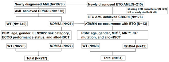
A total of 1676 consecutive patients with AML were enrolled in this study. The study flowchart is shown in Figure 1. After performing a (1:10) PSM for KDM6A mutations, 297 participants were selected for analysis (mutation: wild-type, 27:270), with key demographical and clinical data summarized in Table 1. Among the 297 participants, 204 (68.7%) were female. The median age was 45 years (IQR 33–57 years). The risk stratification based on the 2022 ELN classification identified 126 patients (42.4%) in the favorable risk category, 80 (26.9%) in the intermediate risk category, and 91 (30.6%) in the adverse risk category. All 297 participants received induction therapy, with the following distribution: 86 (29%) received HAA; 95 (32%) received IA; 19 (6.4%) received DA; 48 (18.2%) were treated with venetoclax and azacitidine; 29 (9.8%) received CAG; and 14 (4.7%) received unclassifiable therapy. Of these participants, 231 (77.8%) achieved CR/CRi following the first induction chemotherapy. Among them, 88 (29.6%) underwent allo-HSCT during CR1. For the 297 participants, the median follow-up time for survival was 1.5 years (IQR, 0.7–2.7). During this period, 26 patients (14.6%) died. The median OS and RFS were 1.5 years (IQR, 0.7–2.7) and 1.3 years (IQR, 0.5–2.4), respectively. A total of 104 patients (35.0%) relapsed, with the median relapse time being 0.68 years (IQR, 0.3–1.4).

Figure 1.
Flowchart.

Table 1.
Baseline characteristics of the 297-propensity score-matched patients who achieved CR/CRi.
3.2. Mutation Analysis Overview
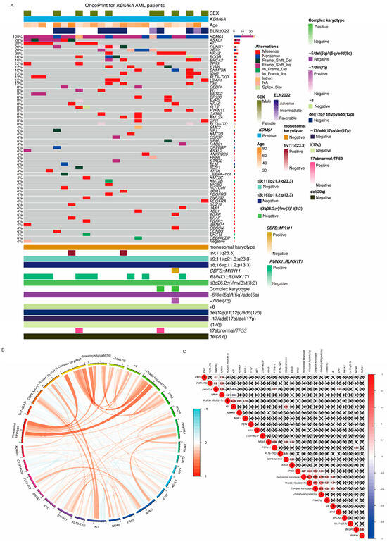
KDM6A mutations occurred in 27 (1.6%) patients across all AML subtypes. The OncoPrint visualization showed the mutation landscape, co-occurrence patterns, chromosomal abnormalities, and clinical information in KDM6A-mutated AML patients in Figure 2A. As shown by Figure 2A,B, the most frequent chromosomal abnormality co-occurring with KDM6A mutations was the RUNX1::RUNX1T1 fusion genes mutations, found in 48.1% of cases (n = 13). The most co-occurring mutations were the KIT gene (8 patients, 29.6%) and ASXL1 (7 patients, 25.9%), followed by RUNX1 (5 patients, 18.5%), TET2 (5 patients, 18.5%), and NRAS (5 patients, 18.5%), which may have synergistic effects on disease progression. Based on our further investigation into the co-occurrence relationship between KDM6A and other genes, of 27 KDM6A mutations, 13 (48.1%) co-occurred with the RUNX1::RUNX1T1 fusion (r = 0.25, p < 0.001), revealing a significant association between KDM6A and RUNX1::RUNX1T1 fusion genes (Figure 2C). KDM6A mutations did not show significant co-occurrence with FLT3-ITD, FLT3-TKD, CEBPA, or any other mutations in our cohort.

Figure 2.
KDM6A OncoPrint visualization. (A) OncoPrint of KDM6A-mutated AML patients. Each row represents an individual patient, and colored blocks indicate different genetic alterations. The top panel shows patient characteristics. The leftmost column lists mutated genes, with mutation types annotated by different colors. The right panel summarizes key cytogenetic abnormalities and their presence (positive) or absence (negative) in these patients. CEBPA-noif: CEBPA bZIP zone mutation but not in frame mutation; CEBPA bZIP: bZIP zone in frame mutation; CEBPA: CEBPA mutation out of bZIP zone. Mutation (B) Circos plot of co-occurring genetic alterations. This circular diagram visualizes the co-occurrence and mutual exclusivity of genetic alterations in KDM6A-mutated AML patients. Orange lines represent co-occurring alterations, while blue lines indicate mutually exclusive events. Different genetic events, including chromosomal abnormalities and gene mutations, are labeled on the outer circle. (C) Correlation matrix of genetic alterations. A heatmap illustrating the correlation between different genetic alterations. Red circles indicate positive correlations, while blue circles denote negative correlations. The size and intensity of the circles correspond to the strength of the correlation. Black “X” marks indicate non-significant correlations.
3.3. Clinical Impact of KDM6A Mutations
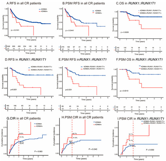
With respect to clinical outcomes, the KDM6A mutation was associated with shorter relapse-free survival, with a 2-year RFS of 60.1% for the wild-type group versus 34.7% for the mutation group (p = 0.016) in univariable analyses of 1676 consecutive patients with AML achieving CR/CRi (Figure 3A). The KDM6A mutation had a significantly higher 2-year CIR compared with subjects with wild-type (50.9% [54.6%, 47.3%] versus 33.8% [33.7%, 33.8%], p = 0.06 (Figure 3G). In the Fine–Gray regression, the KDM6A mutation was independently associated with an increased relapse risk (HR = 1.72 [1.01–2.94], p = 0.05). After PSM in 297 participants, the KDM6A mutation was also associated with a shorter RFS, with a 2-year RFS of 60.9% v.s. 36.3% (p = 0.044) (Figure 3B). The KDM6A mutation had a significantly higher 2-year CIR compared with subjects with wild-type (45.7% [41.6%, 49.7%] versus 28.6% [28.2%, 29.0%], p = 0.04) (Figure 3H). In the Fine–Gray regression, the KDM6A mutation was independently associated with an increased relapse risk (HR = 1.98 [1.08–3.63], p = 0.03). Likewise, the KDM6A mutation was an independent prognostic factor in multivariable analysis for RFS (3.078 [1.56–6.08]; p = 0.001) (Table 2). No significant difference was observed in OS, with 2-year OS rates of 65.9% v.s. 72.1% (p = 0.249). Multivariable analyses confirmed that this variable was not an independent prognostic factor for OS (HR = 1.82, [0.84–3.95]; p = 0.131).

Figure 3.
Outcomes of KDM6A mutations for AML. (A) RFS in cohorts with wild-type or KDM6A mutated group in all AML subgroups. (B) RFS after PSM in cohorts with wild-type or KDM6A mutations across all AML risk categories. (C) OS in cohorts with wild-type or KDM6A mutations in RUNX1::RUNX1T1 fusion gene–AML. (D) RFS in cohorts with wild-type or KDM6A mutated group in RUNX1::RUNX1T1 fusion gene–AML. (E) RFS after PSM in cohorts with wild-type or KDM6A mutated group in RUNX1::RUNX1T1 fusion gene–AML. (F) OS after PSM in cohorts with wild-type or KDM6A mutated group in RUNX1::RUNX1T1 fusion gene–AML. CIR for KDM6A mutations. (G–I) according to the all-risk category AML, PSM in all-risk category, and in RUNX1::RUNX1T1 fusion gene–AML.

Table 2.
Multivariate Cox analyses determining the prognostic significance of KDM6A after PSM in all AML subgroups.
3.4. KDM6A Mutations in RUNX1::RUNX1T1 AML
Since approximately half of the KDM6A mutations co-occurred with the RUNX1::RUNX1T1 fusion gene, we further analyzed the RUNX1::RUNX1T1 AML in patients harboring the KDM6A mutation. A total of 207 participants with the RUNX1::RUNX1T1 fusion gene were deemed eligible for analysis, of whom 106 (51%) were men. The median age was 37 years (IQR: 27–48.5 years). According to the 2022 ELN risk classification, all participants were classified as low risk. Following PSM (1:5.8) for KDM6A mutation, 81 participants from the RUNX1::RUNX1T1 fusion gene–AML cohort were selected for analysis. Baseline characteristics are summarized in Table 3.

Table 3.
Baseline characteristics of the 81 propensity-score-matched patients who achieved CR/CRi with the RUNX1::RUNX1T1 fusion gene–AML.
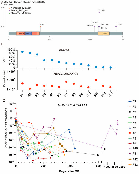
We further detected the mutation landscape of RUNX1::RUNX1T1 AML in patients harboring the KDM6A mutation, with a median of six mutations per participant (0–13). In RUNX1::RUNX1T1 AML patients, KIT was the most frequently mutated gene (81 patients, 46%), followed by ASXL2 (32 patients, 17%) and ASXL1 (28 patients, 16%). KDM6A mutations were detected in 13 (6%) patients with the RUNX1::RUNX1T1 fusion gene–AML, compared with 27 (1.6%) across all AML subtypes. Most identified mutations were frameshift insertions, with a total of nine such instances observed (Figure A1A). The median variant allele frequency (VAF) for these mutations was 36% (2.3–91.1%). Notably, 10 mutations exhibited a VAF exceeding the 10% threshold.
Referring to clinical outcomes, similarly, in the RUNX1::RUNX1T1 fusion gene subgroup, the KDM6A mutation was also associated with a shorter RFS, with a 2-year RFS of 27.7% vs. 75.8% (p < 0.001) and OS, and with a 2-year OS of 66.7% vs. 86.2% (p = 0.003) in univariable analyses (Figure 3C,D). Likewise, the KDM6A mutation was an independent prognostic factor in multivariable analyses for RFS (5.1 [2.5–10.5]; p < 0.001) and OS (12.6 [4.3–38.7]; p < 0.001) (Table 4).

Table 4.
Multivariate analyses determining the prognostic significance of KDM6A.
In the RUNX1::RUNX1T1 fusion gene PSM subgroup, the KDM6A mutation was also associated with a shorter RFS, with a 2-year cumulative RFS of 31.3% vs. 71.9% (p < 0.001), and OS with a 2-year cumulative OS of 63.6% vs. 86.4% (p = 0.002) in univariable analyses (Figure 3E,F). The KDM6A mutation had a significantly higher 2-year CIR compared with subjects with wild-type (46.9% [41.7, 52.1%] versus 24.0% [22.8, 25.1%], p = 0.05) (Figure 3I). In the Fine–Gray regression, the KDM6A mutation was independently associated with an increased relapse risk (HR = 2.46 [1.11–5.47], p = 0.03). Likewise, the KDM6A mutation was an independent prognostic factor in multivariable analyses for RFS (5.7 [1.1–10.3]; p = 0.039), and OS (8.7 [1.2–16.2]; p = 0.026) (Table 5). These findings suggest that KDM6A mutations are associated with poor prognosis in AML patients, especially the RUNX1::RUNX1T1 fusion gene subgroup.

Table 5.
Multivariate Cox analyses determining the prognostic significance of KDM6A after PSM.
We conducted a retrospective analysis of MRD monitoring in 13 AML patients with KDM6A mutations and the RUNX1::RUNX1T1 fusion gene. We observed that these patients experienced varying degrees of molecular relapse after achieving remission. The average time to molecular relapse, determined using RT-PCR detection of RUNX1::RUNX1T1 transcripts, was 43 d (IQR: 53–60) after the deepest molecular remission. Figure A1C depicts the molecular monitoring results, showing the time course of molecular relapse for each patient. Most patients exhibited a detectable rise in RUNX1::RUNX1T1 fusion gene transcripts within the first two months after achieving remission.
4. Discussion
Based on the large scale analyzed for this alteration, we validated KDM6A mutations as a rare but recurrent genetic lesion in AML (1.6%), and RUNX1::RUNX1T1 AML (6.3%). KDM6A mutations are an independent prognostic marker for poor clinical outcomes, associated with a specific co-occurrence profile with the RUNX1::RUNX1T1 fusion gene.
To our knowledge, this study represents one of the first comprehensive investigations examining the clinical impact of KDM6A mutations in patients with AML. Despite emerging evidence implicating KDM6A alterations in hematologic malignancies [13], prior research has predominantly focused on its transcriptional downregulation or epigenetic dysregulation rather than somatic mutations. Notably, only one previous study from our institute reported an association between KDM6A mutations and adverse prognosis in the subset of RUNX1::RUNX1T1-positive CBF-AML, partially aligning with our findings [6]. However, that study did not explore the broader implications of KDM6A mutations across unselected AML cohorts, leaving a critical gap in understanding their all subtype of AML relevance.
While several groups have linked reduced KDM6A expression to chemotherapy resistance and poor outcomes [2,14,15,16], these observations operate at a distinct mechanistic level compared to genomic KDM6A mutations. KDM6A promotes AML progression and drug resistance through its tumor suppressor functions and involvement in DNA repair mechanisms [17]. KDM6A is frequently mutated in AML, leading to a loss of function and increased drug resistance [5]. For example, KDM6A mutations are associated with higher IC50 values for cytarabine, indicating reduced sensitivity to this common AML treatment. Additionally, KDM6A regulates the expression of DNA repair genes. Its deficiency impairs the DNA damage response (DDR), leading to increased sensitivity to PARP and BCL2 inhibition [4]. In AML cells, KDM6A is recruited to the transcriptional start sites of key homologous recombination genes upon DNA damage, facilitating their transcription. Loss of KDM6A activity, either through genetic or pharmacological inhibition, results in elevated H3K27me3 levels at these regulatory elements, preventing gene transcription and compromising DNA repair. This highlights KDM6A’s essential role in maintaining genomic stability and its potential as a therapeutic target in AML. Expression level-based studies reflect regulatory or epigenetic perturbations, whereas truncating or loss-of-function mutations directly impair KDM6A’s function, potentially exacerbating genomic instability and altering transcriptional programs in a mutation-specific manner [3,4,5]. Previous research has primarily focused on the impact of the KDM6A expression level on tumor recurrence and treatment resistance, with limited direct data on complete remission rates [18]. KDM6A mutations may enhance tumor cell drug resistance or promote immune evasion, thereby affecting treatment outcomes and recurrence rates [19].
Our data extend these prior observations by demonstrating that KDM6A mutations, independent of expression changes, confer a high-risk phenotype characterized by shortened survival, even after adjusting for established prognostic factors such as age, cytogenetics, MRD, and ELN guidelines for AML risk stratification. The uniqueness of our study lies in its systematic integration of clinical, molecular, and functional data to establish KDM6A mutations as independent biomarkers of adverse prognosis. In AML, there are few studies on the coexistence of KDM6A mutations with other gene mutations or chromosomal abnormalities. To date, no large-scale AML cohorts have specifically interrogated the genetic signature, prognostic, or therapeutic relevance of KDM6A mutations, underscoring the novelty of our findings.
Our study had some limitations. First, it was a retrospective single-center clinical study, which inherently involves therapy selection biases. Second, we lacked functional studies to validate the clinical findings. Despite these limitations, the findings represent an important step towards understanding the KDM6A mutations influencing the prognosis of AML and could inform future therapeutic strategies.
5. Conclusions
In conclusion, our study validates KDM6A mutations as rare but recurrent in AML, particularly in RUNX1::RUNX1T1 AML, where they predict poor outcomes and a high relapse risk. Early molecular relapse and resistance to conventional therapies highlight the need for frequent monitoring and targeted interventions. These findings underscore the potential of epigenetic therapies and emphasize the importance of KDM6A as a prognostic biomarker, paving the way for improved management of AML patients.
Author Contributions
X.H., F.T. and L.X. conceived and designed the research study. Y.Z. analyzed and interpreted the data. L.N., S.Y., L.Y., T.Z., H.J., Y.W., X.H., X.Z. and Q.J. provided samples and clinical data. Y.Z., X.H. and F.T. wrote the manuscript. All authors have read and agreed to the published version of the manuscript.
Funding
This study was supported by Beijing Nova Program of Science and Technology (grant number Z211100002121058), the Beijing Research Ward Excellence Program (BRWEP, BRWEP2024W134080106), the National Natural Science Foundation of China (Grant No. 82100168), the Beijing Nova Program Interdisciplinary Cooperation Project (grant number 0107050), Peking University People’s Hospital Research and Development Funds (grant numbers RS2020-03 and RDY2020-29), Peking University Medicine Fund of Fostering Young Scholars’ Scientific & Technological Innovation, and the Fundamental Research Funds for the Central Universities (grant number BMU2021PYB005).
Institutional Review Board Statement
This study was approved by the Ethics Committee of Peking University People’s Hospital (approval number: 2025PHB305-001, approval date 24 May 2025) and conducted in accordance with the principles of the Declaration of Helsinki.
Informed Consent Statement
Since this is a retrospective study, we were granted an exemption from obtaining informed consent from the patients, as our research does not disclose any personal information of the patients and does not cause any direct or indirect harm to them.
Data Availability Statement
Data are contained within the article.
Acknowledgments
We appreciated the timely help offered by Xiaojun Huang and Robert Peter Gale (Department of Hematology, Peking University People’s Hospital) with this study.
Conflicts of Interest
The authors declare no conflict of interest.
Abbreviations
The following abbreviations are used in this manuscript:
| Allo-HSCT | Allogeneic Hematopoietic Stem Cell Transplantation |
| BM | Bone Marrow |
| CI | Confidence Interval |
| CR/CRi | Complete Response/Complete Response with Incomplete Hematologic Recovery |
| CIR | cumulative incidence of relapse |
| ECOG | Eastern Cooperative Oncology Group |
| ELN2022 | European LeukemiaNet 2022 |
| HB | Hemoglobin |
| HR | Hazard Ratio |
| IQR | Interquartile Range |
| MR | Molecular Response |
| MRD | Minimal Residual Disease |
| OS | Overall Survival |
| PB | Peripheral Blood |
| PLT | Platelets |
| PSM | Propensity Score Matching |
| RFS | Relapse-Free Survival |
| VAF | Variant Allele Frequency |
| WBC | White Blood Cells |
| WT | Wild-type |
Appendix A

Table A1.
List of the Deep-Targeted Sequencing Panel: From 175 Genes (2018–2020) to 290 Genes (2021 Onwards).
Table A1.
List of the Deep-Targeted Sequencing Panel: From 175 Genes (2018–2020) to 290 Genes (2021 Onwards).
| 175 Genes List (2018–2020) | Expanded to 290 Genes List (2021 Onwards) | |||||||||||||
|---|---|---|---|---|---|---|---|---|---|---|---|---|---|---|
| ABCB1 | BRAF | CEBPA | EPHA7 | HRAS | KRAS | PAX5 | SBDS | U2AF1 | AKT3 | CHD2 | GATA3 | MSH2 | PRKD2 | TYK2 |
| ABL1 | BRCA1 | CHD8 | EPOR | ID3 | KRT20 | PDGFRA | SETBP1 | VHL | ALK | CHEK2 | HFE | MSH6 | PTCH1 | U2AF2 |
| ANKRD26 | BRCA2 | CIITA | ETV6 | IDH1 | LMO2 | PDGFRB | SETD2 | WHSC1 | ARID5B | CIC | HIST1H1B | NBN | PTPN2 | UBR5 |
| APC | BRIP1 | CREBBP | EZH2 | IDH2 | LYN | PHF6 | SETDB1 | WT1 | ASXL2 | CYLD | HIST1H1C | NFKBIA | PTPN6 | WAS |
| ARID1A | BTG1 | CRLF2 | FAM46C | IKZF1 | MAP2K1 | PIGA | SF3B1 | XPO1 | ATP6V1B2 | DDX3X | HIST1H1D | NFKBIE | PTPRD | ZFHX4 |
| ARID1B | BTK | CSF1R | FAS | IKZF2 | MCL1 | PIK3CA | SGK1 | ZAP70 | AXIN1 | DHFR | HIST1H1E | NOTCH3 | PTPRT | ZMYM3 |
| ARID2 | CALR | CSF3R | FAT1 | IKZF3 | MEF2B | PIK3CD | SH2B3 | ZRSR2 | BAX | DNAH10 | HUWE1 | NOTCH4 | RAD50 | SPEN |
| ASXL1 | CARD11 | CTCF | FBXO11 | IL7R | MFHAS1 | PIM1 | SMC1A | BCL11B | DOT1L | IGLL5 | NTRK1 | RARA | TBL1XR1 | |
| ATG2B | CBL | CUX1 | FBXW7 | IRF4 | MPL | PLCG2 | SMC3 | BCL7A | DTX1 | IKBKB | NTRK2 | RB1 | TET1 | |
| ATM | CBLB | CXCR4 | FLT3 | IRF8 | MTOR | PPM1D | SOCS1 | BLNK | DUSP2 | ITK | NTRK3 | RPL10 | TMEM30A | |
| ATRX | CBLC | DDX41 | FOXO1 | ITPKB | MYC | PRDM1 | SRP72 | BRD4 | EBF1 | JUNB | NUDT15 | RPS15 | TRAF5 | |
| B2M | CCND1 | DIS3 | GATA1 | JAK1 | MYD88 | PRF1 | SRSF2 | BTG2 | EGR1 | KAT6A | P2RY8 | RRAGC | ||
| BCL10 | CCND3 | DKC1 | GATA2 | JAK2 | MYOM2 | PRKDC | STAG2 | CACNA1H | EGR2 | KLF2 | PIK3R1 | RTEL1 | ||
| BCL2 | CD28 | DNM2 | GFI1 | JAK3 | NF1 | PRPF8 | STAT3 | CCR4 | ERBB3 | KLHL6 | PIM2 | SETD1B | ||
| BCL6 | CD58 | DNMT3A | GNA13 | KDM6A | NOTCH1 | PTEN | STAT5B | CD22 | ERG | LTB | PLCG1 | SF1 | ||
| BCOR | CD79A | EED | GNAI2 | KIT | NOTCH2 | PTPN11 | STAT6 | CD70 | ETNK1 | MAX | PML | SF3A1 | ||
| BCORL1 | CD79B | EGFR | GNAS | KMT2A | NPM1 | RAD21 | SUZ12 | CDC25C | FAT3 | MDM2 | PMS2 | SH2D1A | ||
| BIRC3 | CDKN1A | EGLN1 | GNB1 | KMT2B | NRAS | RELN | SYK | CDK6 | FAT4 | MED12 | POT1 | SLC29A1 | ||
| BLM | CDKN2A | ELANE | GSKIP | KMT2C | NT5C2 | RHOA | TAL1 | CDKN1B | FGFR3 | MGA | POU2AF1 | SMARCA4 | ||
| BPGM | CDKN2B | EP300 | HAX1 | KMT2D | PALB2 | RUNX1 | TCF3 | CEBPE | G6PC3 | MLH1 | PRKCB | SMARCB1 | ||
| TERT | TERC | TET2 | TNFAIP3 | TNFRSF14 | TP53 | TPMT | TRAF3 | SP140 | STAT5A | TCF3 | TINF2 | TRAF2 | ||

Figure A1.
Topography of KDM6A mutation. (A) lollipop plot illustrating KDM6A mutation. (B) VAF of KDM6A in initially diagnosed RUNX1::RUNX1T1 fusion gene–AML. (C) MRD monitoring in participants with KDM6A mutation. * Stop codon.
References
- Papaemmanuil, E.; Gerstung, M.; Bullinger, L.; Gaidzik, V.I.; Paschka, P.; Roberts, N.D.; Potter, N.E.; Heuser, M.; Thol, F.; Bolli, N.; et al. Genomic Classification and Prognosis in Acute Myeloid Leukemia. N. Engl. J. Med. 2016, 374, 2209–2221. [Google Scholar] [CrossRef] [PubMed]
- Shi, B.; Li, W.; Song, Y.; Wang, Z.; Ju, R.; Ulman, A.; Hu, J.; Palomba, F.; Zhao, Y.; Le, J.P.; et al. UTX condensation underlies its tumour-suppressive activity. Nature 2021, 597, 726–731. [Google Scholar] [CrossRef] [PubMed]
- Greif, P.A.; Hartmann, L.; Vosberg, S.; Stief, S.M.; Mattes, R.; Hellmann, I.; Metzeler, K.H.; Herold, T.; Bamopoulos, S.A.; Kerbs, P.; et al. Evolution of Cytogenetically Normal Acute Myeloid Leukemia During Therapy and Relapse: An Exome Sequencing Study of 50 Patients. Clin. Cancer Res. 2018, 24, 1716–1726. [Google Scholar] [CrossRef] [PubMed]
- Boila, L.D.; Ghosh, S.; Bandyopadhyay, S.K.; Jin, L.; Murison, A.; Zeng, A.G.X.; Shaikh, W.; Bhowmik, S.; Muddineni, S.; Biswas, M.; et al. KDM6 demethylases integrate DNA repair gene regulation and loss of KDM6A sensitizes human acute myeloid leukemia to PARP and BCL2 inhibition. Leukemia 2023, 37, 751–764. [Google Scholar] [CrossRef] [PubMed]
- Stief, S.M.; Hanneforth, A.L.; Weser, S.; Mattes, R.; Carlet, M.; Liu, W.H.; Bartoschek, M.D.; Dominguez Moreno, H.; Oettle, M.; Kempf, J.; et al. Loss of KDM6A confers drug resistance in acute myeloid leukemia. Leukemia 2020, 34, 50–62. [Google Scholar] [CrossRef] [PubMed]
- Yu, S.; Yang, S.; Hu, L.; Duan, W.; Zhao, T.; Qin, Y.; Wang, Y.; Lai, Y.; Shi, H.; Tang, F.; et al. Genetic abnormalities predict outcomes in patients with core binding factor acute myeloid leukemia. Ann. Hematol. 2025, 104, 997–1006. [Google Scholar] [CrossRef] [PubMed]
- Dohner, H.; Wei, A.H.; Appelbaum, F.R.; Craddock, C.; DiNardo, C.D.; Dombret, H.; Ebert, B.L.; Fenaux, P.; Godley, L.A.; Hasserjian, R.P.; et al. Diagnosis and management of AML in adults: 2022 recommendations from an international expert panel on behalf of the ELN. Blood 2022, 140, 1345–1377. [Google Scholar] [CrossRef] [PubMed]
- Gao, M.G.; Ruan, G.R.; Chang, Y.J.; Liu, Y.R.; Qin, Y.Z.; Jiang, Q.; Jiang, H.; Huang, X.J.; Zhao, X.S. The predictive value of minimal residual disease when facing the inconsistent results detected by real-time quantitative PCR and flow cytometry in NPM1-mutated acute myeloid leukemia. Ann. Hematol. 2020, 99, 73–82. [Google Scholar] [CrossRef] [PubMed]
- Rücker, F.G.; Agrawal, M.; Corbacioglu, A.; Weber, D.; Kapp-Schwoerer, S.; Gaidzik, V.I.; Jahn, N.; Schroeder, T.; Wattad, M.; Lübbert, M. Measurable Residual Disease (MRD) Monitoring in Acute Myeloid Leukemia (AML) with t (8; 21)(q22; q22. 1) RUNX1-RUNX1T1 Identifies Patients at High Risk of Relapse: Results of the AML Study Group (AMLSG). Blood 2019, 134, 2740. [Google Scholar] [CrossRef]
- Wang, L.; Gao, H.; Fu, Q.; Jiang, Q.; Jiang, H.; Wang, Y.; Xu, L.; Zhang, X.; Huang, X.; Tang, F. Clinical and Molecular Predictors of Response and Survival Following Venetoclax Plus Hypomethylating Agents in Relapsed/Refractory Acute Myeloid Leukemia: A Single-Center Study in Chinese Patients. Cancers 2025, 17, 586. [Google Scholar] [CrossRef] [PubMed]
- Li, M.M.; Datto, M.; Duncavage, E.J.; Kulkarni, S.; Lindeman, N.I.; Roy, S.; Tsimberidou, A.M.; Vnencak-Jones, C.L.; Wolff, D.J.; Younes, A.; et al. Standards and Guidelines for the Interpretation and Reporting of Sequence Variants in Cancer: A Joint Consensus Recommendation of the Association for Molecular Pathology, American Society of Clinical Oncology, and College of American Pathologists. J. Mol. Diagn. 2017, 19, 4–23. [Google Scholar] [CrossRef] [PubMed]
- Xu, D.; Yang, Y.; Yin, Z.; Tu, S.; Nie, D.; Li, Y.; Huang, Z.; Sun, Q.; Huang, C.; Nie, X.; et al. Risk-directed therapy based on genetics and MRD improves the outcomes of AML1-ETO-positive AML patients, a multi-center prospective cohort study. Blood Cancer J. 2023, 13, 168. [Google Scholar] [CrossRef] [PubMed]
- Chen, H.; Wang, S.; Dong, R.; Yu, P.; Li, T.; Hu, L.; Wang, M.; Qian, Z.; Zhou, H.; Yue, X. KDM6A Deficiency Induces Myeloid Bias and Promotes CMML-Like Disease Through JAK/STAT3 Activation by Repressing SOCS3. Adv. Sci. 2025, 12, 2413091. [Google Scholar] [CrossRef] [PubMed]
- Duplaquet, L.; Li, Y.; Booker, M.A.; Xie, Y.; Olsen, S.N.; Patel, R.A.; Hong, D.; Hatton, C.; Denize, T.; Walton, E.; et al. KDM6A epigenetically regulates subtype plasticity in small cell lung cancer. Nat. Cell Biol. 2023, 25, 1346–1358. [Google Scholar] [CrossRef] [PubMed]
- Feng, M.; Chai, C.; Hao, X.; Lai, X.; Luo, Y.; Zhang, H.; Tang, W.; Gao, N.; Pan, G.; Liu, X.; et al. Inherited KDM6A(A649T) facilitates tumor-immune escape and exacerbates colorectal signet-ring cell carcinoma outcomes. Oncogene 2024, 43, 1757–1768. [Google Scholar] [CrossRef] [PubMed]
- Seehawer, M.; Li, Z.; Nishida, J.; Foidart, P.; Reiter, A.H.; Rojas-Jimenez, E.; Goyette, M.A.; Yan, P.; Raval, S.; Munoz Gomez, M.; et al. Loss of Kmt2c or Kmt2d drives brain metastasis via KDM6A-dependent upregulation of MMP3. Nat. Cell Biol. 2024, 26, 1165–1175. [Google Scholar] [CrossRef] [PubMed]
- Tian, L.; Wartman, L.D. The Role of Kdm6a in Malignant Hematopoiesis. Blood 2017, 130, 1227. [Google Scholar]
- Chen, Z.; Qi, Y.; Shen, J.; Chen, Z. Histone demethylase KDM6A coordinating with KMT2B regulates self-renewal and chemoresistance of non-small cell lung cancer stem cells. Transl. Oncol. 2023, 37, 101778. [Google Scholar] [CrossRef] [PubMed]
- Chen, X.; Lin, X.; Pang, G.; Deng, J.; Xie, Q.; Zhang, Z. Significance of KDM6A mutation in bladder cancer immune escape. BMC Cancer 2021, 21, 635. [Google Scholar] [CrossRef] [PubMed]
Disclaimer/Publisher’s Note: The statements, opinions and data contained in all publications are solely those of the individual author(s) and contributor(s) and not of MDPI and/or the editor(s). MDPI and/or the editor(s) disclaim responsibility for any injury to people or property resulting from any ideas, methods, instructions or products referred to in the content. |
© 2025 by the authors. Licensee MDPI, Basel, Switzerland. This article is an open access article distributed under the terms and conditions of the Creative Commons Attribution (CC BY) license (https://creativecommons.org/licenses/by/4.0/).