Article

Advancing the Landscape of Clinical Actionability in Von Hippel–Lindau Syndrome: An Evidence-Based Framework from the INT2GRATE Oncology Consortium

by
Diane R. Koeller
1,†,
McKenzie Walker
2,†,
Busra Unal
2,
Anu Chittenden
1,
Alison Schwartz Levine
1,
Connor P. Hayes
2,
Paul C. Oramasionwu
2,
Monica D. Manam
3,
Ryan M. Buehler
1,
Israel Gomy
4,
Wilson Araujo Silva, Jr.
4,
Jordan Lerner-Ellis
5,
Selina Casalino
5,
Radhika Mahajan
5,
Nicholas Watkins
5,
Nihat Bugra Agaoglu
6,
Danielle K. Manning
7,
Justine A. Barletta
7,8,
Jason L. Hornick
7,8,
Neal I. Lindeman
9,
Lynette M. Sholl
7,8,
Huma Q. Rana
1,8,10,
Judy E. Garber
1,8,10 and
Arezou A. Ghazani
2,7,8,11,*
add Show full author list remove Hide full author list
1
Division of Cancer Genetics and Prevention, Dana-Farber Cancer Institute, Boston, MA 02215, USA
2
Division of Genetics, Brigham and Women’s Hospital, Boston, MA 02115, USA
3
Boston Children’s Hospital, Boston, MA 02115, USA
4
Department of Genetics, Ribeirão Preto Medical School, University of São Paulo, Ribeirão Preto 14040-902, Brazil
5
Pathology and Laboratory Medicine, Mount Sinai Hospital, Toronto, ON M5G 1X5, Canada
6
Division of Cancer Genetics, Umraniye Training and Research Hospital, 34764 Istanbul, Turkey
7
Department of Pathology, Brigham and Women’s Hospital, Boston, MA 02115, USA
8
Harvard Medical School, Boston, MA 02114, USA
9
Pathology and Laboratory Medicine, Weill Cornell Medical College, New York, NY 10065, USA
10
Division of Population Sciences, Dana-Farber Cancer Institute, Boston, MA 02115, USA
11
Department of Medicine, Brigham and Women’s Hospital, Boston, MA 02115, USA
*
Author to whom correspondence should be addressed.
These authors contributed equally to this work.
Cancers 2025, 17(13), 2173; https://doi.org/10.3390/cancers17132173
Submission received: 6 May 2025 / Revised: 2 June 2025 / Accepted: 25 June 2025 / Published: 27 June 2025

Simple Summary

In cancer syndromes, a precise assessment of variant actionability requires a comprehensive set of evidence to delineate the role of a variant in disease while effectively distinguishing it from potential differential diagnoses with similar cancer presentations and from sporadic occurrences. The INT2GRATE programs and the INT2GRATE Oncology Consortium address this challenge through an integrated analysis of constitutional and tumor data. Here, we present a novel variant evidence framework (VEF) for precision variant assessment in Von Hippel–Lindau Syndrome (VHL). The INT2GRATE VEF, applied to 2672 VHL variants, distinguishes constitutional, sporadic, VHL differential, and VHL allelic conditions. We also present the open-access INT2GRATE Variant Portal, a novel resource that provides a repository of the germline VHL variants and evidence, promoting data sharing, advancing precision oncology, and improving patient care.

Abstract

Background/Objectives: An accurate evaluation of variant actionability is essential in cancer management. In Von Hippel–Lindau Syndrome (VHL), the interpretation of the germline variants is confounded by the presence of non-syndromic component tumors, such as clear cell renal cell carcinoma (ccRCC), hemangioblastoma, pheochromocytoma, and neuroendocrine tumors. These tumors frequently occur sporadically, without any association with VHL syndrome. The presence of these tumors in a patient with a germline VHL variant could lead to inaccurate attribution of these tumors to the germline variant and VHL syndrome. In our previous INT2GRATE (INTegrated INTerpretation of GeRmline And Tumor gEnomes) programs, we demonstrated that integrating tumor-derived and germline evidence offers a comprehensive approach for the accurate assessment of the germline variants in cancer syndromes. Methods/Results: Here, we present a novel INT2GRATE variant evidence framework (VEF) for evaluating the clinical actionability of the germline variants in VHL syndrome, offering an integrated approach that incorporates both constitutional and tumor data. We analyzed 2672 variants in the VHL gene and their associated tumors and clinical evidence to effectively distinguish between constitutional, sporadic, VHL differentials, and VHL allelic genetic conditions. The germline INT2GRATE variants, along with their comprehensive associated evidence, were made accessible in the first open-access INT2GRATE Variant data Portal. Conclusions: This novel and integrated approach to variant assessment and data sharing in hereditary cancer syndromes is essential in the clinical evaluation of genomic variants, advancing precision oncology, and improving patient care.

1. Introduction

Von Hippel–Lindau syndrome (VHL, OMIM # 193300) is a rare, autosomal dominant hereditary cancer syndrome caused by the heterozygous germline pathogenic variants in the VHL gene. With an estimated prevalence of 1 in 35,000 [1], VHL syndrome is characterized by the presence of multiple tumor types, specifically clear cell renal cell carcinoma (ccRCC), hemangioblastomas of the central nervous system and retina, pheochromocytomas, pancreatic cysts, and neuroendocrine tumors [2,3]. The mechanism of disease in VHL syndrome is the loss of function of the VHL tumor suppressor gene, leading to the accumulation of hypoxia-inducible factors (HIFs) and the formation of highly vascularized tumors [4,5,6]. Nearly 100% of the individuals with VHL syndrome are expected to be symptomatic by the age of 65 years, but some germline VHL variants may be associated with lower penetrance [1,7,8].
The interpretation of the germline VHL variants and the assessment of their clinical significance in VHL syndrome can be complex. Often, patients suspected of having VHL syndrome present with a single VHL component tumor, such as isolated RCC, hemangioblastoma, pheochromocytoma, or retinal angioma. Non-syndromic component tumors may be common sporadic occurrences that do not involve germline VHL alterations. These sporadic tumors have distinct tumorigenic drivers, often involving somatic alterations in the VHL or other genes. The allelic autosomal recessive disorder familial erythrocytosis type 2 (ECYT2, OMIM # 263400) adds further complexity, as the VHL germline variants related to ECYT2 are generally not associated with VHL syndrome. Alternatively, component tumors may be constitutional, caused by germline alterations in genes other than the VHL. Genes such as FH, FLCN, MET, BAP1, SDHA, SDHB, SDHC, SDHD, SDHAF2, TMEM127, MAX, and RET are associated with cancer susceptibility syndromes that present overlapping tumors commonly seen in VHL syndrome [9]. Finally, a single VHL-component tumor could be attributed to age-related penetrance or the low penetrance of certain VHL alleles in adults (Figure 1). These observations highlight the confounding factors in the clinical interpretation of the VHL variants and underscore the need for an integrated and comprehensive evidence-based assessment of the VHL variants.
The INT2GRATE|Oncology Consortium has addressed this challenge by developing an integrated evidence framework to assess the actionability of the germline variants using germline data, clinical genetic information, tumor-derived data, and somatic genetic evidence (INT2GRATEoncology.org). We have previously presented the application of INT2GRATE in the assessment of variant actionability in multiple hereditary cancer syndromes, and further demonstrated that this comprehensive approach can differentiate between non-syndromic sporadic tumors or syndromic cancers [8,10,11,12,13]. Here, we present the novel INT2GRATE variant evidence framework (VEF) for evaluating the germline variants in VHL syndrome. We describe the parameters and rationale behind the VEF, discuss the collation and programmatic processing of 2672 variants in the INT2GRATE|VHL program, and unveil the launch of the first open INT2GRATE|Variant Portal for public access. This novel framework integrates evidence derived from constitutional and sporadic variants, VHL differential conditions, and allelic conditions. Processing the INT2GRATE|Variants and sharing comprehensive constitutional and somatic evidence is pivotal in advancing precision oncology.

2. Methods

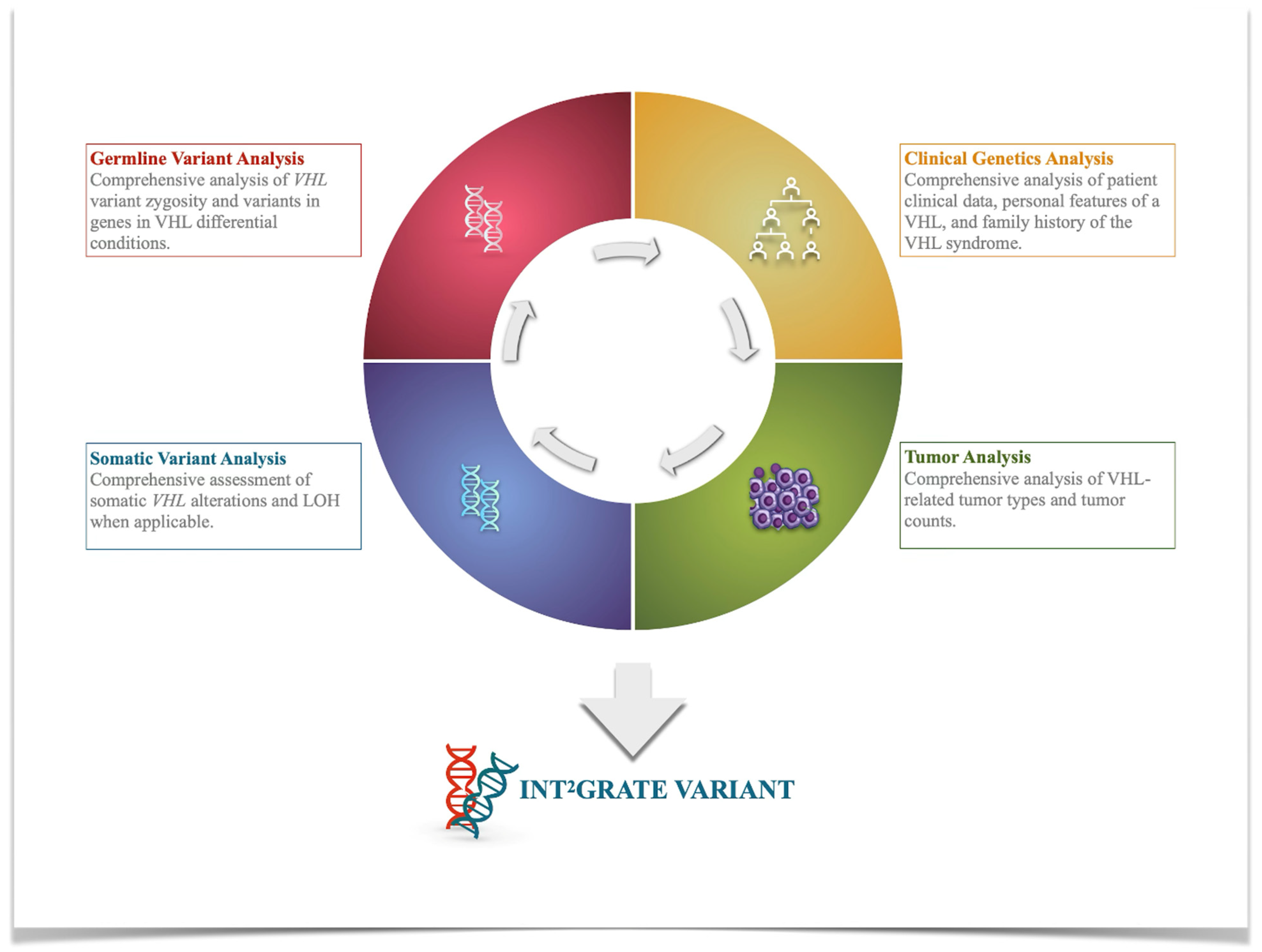
2.1. Development of the INT2GRATE|VHL Platform

INT2GRATE|VHL Variant Evidence Framework (VEF)

The variant evidence framework (VEF) for the INT2GRATE|VHL has four main components, including (1) germline variants in the VHL gene, (2) pertinent patient clinical genetics data related to personal and family history of VHL syndrome, (3) VHL tumor-derived data, and (4) somatic genetic variants in the VHL gene (Figure 2). The VEF was established by cataloging different clinical scenarios, marking the presence or absence of each variant parameter, and assessing the combination of the evidence in each scenario (Supplementary Materials Table S1). The details of the VEF and clinical scenarios were reviewed by the INT2GRATE expert group, comprising board-certified medical geneticists with experience in cancer genetics and tumor profiling, board-certified clinicians, and genetic counselors with expertise in the diagnosis and management of hereditary cancer syndromes, including VHL, and board-certified pathologists with expertise in tumor pathology. The description and rationale for the VEF components are described in detail in the Results (Section 3).

2.2. INT2GRATE Data Processing and Analysis

2.2.1. Development of the Digital INT2GRATE VEF

Once the INT2GRATE expert committee signed off on the VEF, a digital version was developed at the INT2GRATE Data Coordinating Center to support large-scale data analysis. The digital VEF was developed in Python (https://www.python.org/psf-landing/ Python Software Foundation, Wilmington, DE, USA) through a series of logical combinations for each scenario.

2.2.2. INT2GRATE Encoder

The INT2GRATE Encoder was developed in Python to pragmatically analyze the data provided by the INT2GRATE institute members by parsing them through the digital VEF. The Encoder integrates all VEF parameters in each case, identifies the matching INT2GRATE scenario, and assigns the case to the relevant unique INT2GRATE code. In the resulting output file, all variants were labeled with the appropriate unique INT2GRATE code as determined by the VEF. Each patient was assigned a unique Subject ID.

2.2.3. INT2GRATE Pattern Quantifier

The INT2GRATE Pattern Quantifier was developed in Python to assess, identify, and quantify unique patterns among the VHL variants in the dataset. First, it computed the frequency of each variant, which was termed variant recurrence. For each variant, it further assessed and quantified the distinct patterns of the supporting evidence (e.g., INT2GRATE Evidence 1, INT2GRATE Evidence 2). Then, it enumerated the number of times that an identical pattern of evidence was observed for a given variant (e.g., n = 3). The INT2GRATE Pattern Quantifier facilitates a large-scale investigation of the landscape and the distribution of variants, along with their associated clinical evidence.

2.2.4. INT2GRATE|Data Portal and Data Sharing

The INT2GRATE Data Portal is a novel digital platform for sharing the de-identified INT2GRATE variants and their associated and comprehensive clinical evidence. After formatting the variants through the INT2GRATE Encoder and Pattern Quantifier, the aggregate dataset was uploaded to the portal through a web-based user interface available at INT2GRATEdata.bwh.harvard.edu.

2.3. Patient Data Query and Cohorts

In total, data from 2672 patients were retrospectively collected and analyzed in the INT2GRATE VHL program. An agnostic approach included all VHL variant-positive cases (at least one germline or somatic VHL variant) without prior knowledge of VHL syndrome evaluation to minimize ascertainment bias. The INT2GRATE VEF was provided to the consortium members to ensure a uniform process for querying relevant data. Member institutes include Mass General Brigham (MGB, Boston, MA, USA), Dana Farber Cancer Institute Center for Cancer Genetics and Prevention (DFCI, Boston, MA, USA), Mount Sinai Hospital (MSH, Toronto, ON, Canada), University of São Paulo (USP, Ribeirão Preto, São Paulo, Brazil), and Umraniye Training and Research Hospital (UTRH, Istanbul, Turkey).
Data were divided into two cohorts. Cohort 1 consisted of 133 patients, 122 of whom had a germline VHL variant and detailed tumor data and were evaluated at a genetics clinic. The remaining 11 patients all had the same data plus somatic VHL genetic information. Cohort 2 included 2539 patients, all of whom had at least one somatic VHL variant and detailed tumor data. A subset of these patients (n = 638) was evaluated in the genetics clinic and had no germline VHL variants, while the remaining 1901 patients had no germline testing and no record of clinical genetic evaluation for any constitutional cancer. About 94% of the somatic alterations were copy numbers (154 SNVs and 2394 copy numbers that included 1817 deletions). In the absence of germline sequencing data, it was unclear whether the VHL alterations identified through tumor sequencing were true somatic events. However, given that germline VHL deletions occur in only ~10% of VHL syndrome cases, and these cases did not have VHL-associated tumors, the alterations identified through tumor sequencing were presumed to be somatic events. In cases with VHL-related component tumors (RCC, hemangioblastoma, paraganglioma, and pancreatic neuroendocrine tumor), the VHL alterations from tumor sequencing were extrapolated to be presumed somatic due to the absence of clinical suspicion for VHL syndrome and the variant allelic fractions (VAF) outside of the expected heterozygous range (median VAF < 50 = 27%; median VAF > 50 = 68%). Nonetheless, because germline sequencing was not performed for these patients, these variants were only used for landscape analysis.
For germline variant collection, each institution collected all types of VHL variants, including SNVs, indels, or copy numbers (CNs) reported as pathogenic, likely pathogenic, VUS, or favor polymorphism in their routine clinical care. Favor polymorphism classification was assigned by the reporting laboratory to variants that were downgraded from VUS after their initial report. These variants were entered as benign/likely benign (B/LB) in this study. Patients with the germline VHL variants were also investigated for germline findings in the genes associated with syndromes that are in the differential diagnosis for VHL syndrome. These genes included MAX, SDHA, SDHAF2, SDHB, SDHC, SDHD, TMEM127, NF1, RET, FH, and FLCN. Comprehensive clinical information was collected and evaluated, which included personal and familial cancer history, the component tumor types, sex, and age at diagnosis.

2.4. Germline Genetic Laboratory Testing

Germline genetic testing was performed at multiple sites, including MSH, UTRH, USP, or commercial laboratories (Invitae Corp., San Francisco, CA, USA; Ambry Genetics, Aliso Viejo, CA, USA). Briefly, DNA was extracted from peripheral blood, and targeted regions were enriched by a hybridization-based protocol according to standard protocols [10]. Sequencing was carried out on a next-generation sequencing (NGS) platform. In the exonic regions, copy number variations were assessed by analyzing each target sequence read depth, along with the mean read depth and distribution of the read depth, based on the parameters established in the validation experiments.

2.5. Tumor Genetic Laboratory Testing

Somatic variants were obtained from routine tumor profiling by the OncoPanel assay at the Center for Advanced Molecular Diagnostics (CAMD) at BWH (Boston, MA, USA). Data were retrospectively collected through a custom search for all cases between 2018 and 2022 with the reported VHL variants as previously described [10]. Briefly, single nucleotide variants, copy numbers, and structural alterations were processed as below. Somatic loss of heterozygosity (LOH) was assessed by detecting allelic loss at the VHL locus.

2.5.1. Single-Nucleotide Variant (SNV)/Indel Analysis

Per the previously described protocols, MuTect (Broad Institute, Cambridge, MA, USA) and GATK Indelocator (Broad Institute, Cambridge, MA, USA) were used to detect somatic SNVs and indels, respectively. The variant filtration was performed based on the annotated allele frequencies in the Exome Sequencing Project (ESP) and/or gnomAD, excluding the variants with an allelic frequency higher than 0.1% in these databases. The variants that were found in the plate normal control were excluded from further analysis. Any variant filtered based on the previous criteria but reported in the COSMIC database (COSMIC, Wellcome Sanger, London, UK) at least two times was rescued. The following information was used to annotate each variant: the gene, genome coordinates, reference and alternate alleles, coverage, allele fraction, cDNA, and protein change. According to the somatic OncoPanel validation data, 50× coverage and 10% variant allele fraction were set as the limit of detection. The variants failing these quality criteria for coverage and/or allele fraction or read support (at least five unique reads) were not further analyzed.

2.5.2. OncoPanel Copy Number Analysis

As previously described, the somatic copy number (CN) variants were identified using RobustCNV, a tool developed at the Dana–Farber Cancer Institute (DFCI, Boston, MA, USA). The normalization of each baited genomic segment was performed by comparing it with a panel of normal samples. Genomic segments with a Log2 ratio of zero were classified as having a neutral copy number. Prior to the technical evaluation, copy number variations, including low and high amplifications, as well as one- and two-copy deletions, were recorded. A Log2 ratio of 0.43 or higher was used to identify low-level amplifications, and a Log2 ratio of −0.32 or lower indicated a copy number loss.

2.5.3. OncoPanel Structural Variant Analysis

BreaKmer, created at DFCI (DFCI, Boston, MA, USA), was utilized in the assessment of somatic structural variants, including chromosomal rearrangements, inversions, and large indels, as previously described [14]. BreaKmer detects sequence segments that align to non-contiguous regions of the reference. For each structural variant (SV), the involved gene(s), genomic coordinates, and a corresponding IGV snapshot were provided for visual confirmation. The pipeline identifies split reads, single reads that align to two non-contiguous locations of the genome, and discordant read pairs in which the paired ends map to different locations in the genome. Variants with equal or less than 2% of total split and discordant reads/total coverage along the detected breakpoints were carefully assessed. SVs in repetitive regions were not included in the analysis.

3. Results

3.1. Development of INT2GRATE|Variant Evidence Framework (VEF) for VHL

The INT2GRATE|VEF incorporates four key types of evidence to assess the clinical relevance of the VHL variants (Figure 2), including germline genetics, somatic genetics, clinical genetics, and tumor-derived data. The INT2GRATE|VEF is designed to analyze the details within each category in different realistic scenarios routinely used in clinical practice. The details and rationale for each type of evidence are described below.

3.1.1. Germline Variants and Rationale

The INT2GRATE|VEF requires a comprehensive assessment of the VHL zygosity, allelic disorder, and differential conditions to ensure a conservative evaluation of the germline variant associated with VHL syndrome.
VHL allelic disorder: While deleterious heterozygous VHL variants can be diagnostic for VHL syndrome, certain variants in the VHL gene are associated only with an increased risk for autosomal recessive familial erythrocytosis type 2 (ECYT 2) (also known as VHL-associated polycythemia, congenital erythrocytosis type 2, or Chuvash polycythemia). Individuals who are heterozygous for these variants are considered to be carriers of ECYT2 and are not expected to be at risk for VHL syndrome. Additionally, if an individual had bi-allelic VHL variants, it would be challenging to assess each variant separately in relation to any associated phenotypic features. Thus, zygosity is the first criterion in the assessment of the VHL variants, with the INT2GRATE|VEF for VHL only considering monoallelic (i.e., heterozygous) variants.
VHL differential disorders: Alterations in several genes can result in genetic conditions with clinical features resembling those in VHL syndrome. To accurately assess the potential association between a germline VHL variant and VHL syndrome, the INT2GRATE|VEF requires the absence of germline variants in genes related to differential disorders (Supplementary Materials Table S2). This conservative approach minimizes any potential confounding factors when evaluating the actionability of germline variants, especially in cases with unusual disease presentations where only non-syndromic tumors are present. The genes related to VHL differential disorders include FH, FLCN, MET, BAP1, SDHA, SDHB, SDHC, SDHD, SDHAF2, TMEM127, MAX, RET, and NF1, which are collectively associated with an increased risk of pheochromocytoma, paraganglioma, and renal cell carcinoma (RCC) including ccRCC. Table S2 in the Supplementary Materials lists each gene along with its associated VHL-differential condition(s).

3.1.2. Clinical Genetics Criteria and Rationale

In the design of the INT2GRATE|VEF, the parameters for personal and family history of VHL are intentionally structured with different criteria. Personal history has minimal requirements, while family history has stringent criteria. This approach ensures a broader inclusion of cases while maintaining a more conservative analysis.
Personal history: While the presence of two or more VHL tumors is strongly suggestive of VHL syndrome, isolated VHL component tumors [15,16] (e.g., clear cell renal cell carcinoma, hemangioblastoma) can be either constitutional or sporadic. The VEF requires at least one VHL-related tumor or feature in the personal history. This low threshold allows for the inclusion and assessment of a broader range of cases with various tumor types (Supplementary Materials Table S1). The VEF captures the following VHL-related tumors: retinal hemangioblastoma, CNS hemangioblastoma, renal cell carcinoma, pheochromocytoma, pancreatic neuroendocrine tumor, endolymphatic sac tumor, and paraganglioma. VHL-related features include multiple kidney cysts, multiple pancreatic cysts, and epididymal and broad ligament cysts.
Family history: The family history requirement is designed to be very conservative. To meet the VEF criteria, family history is considered when a first or second-degree family member is diagnosed with VHL syndrome.

3.1.3. Tumor-Derived Information and Rationale

A key feature of the INT2GRATE|VEF is its ability to distinguish between the various molecular etiologies of VHL tumors. An isolated VHL tumor may be (i) sporadic, where somatic VHL variants are putative drivers and the tumor is not related to VHL syndrome, (ii) caused by a germline variant in a gene related to VHL differential conditions, (iii) the result of a VHL germline variant in a young patient, or (iv) a VHL variant with low penetrance, which may result in an incomplete VHL phenotype (Figure 1). The INT2GRATE|VEF evaluates VHL tumor characteristics in the context of the patient’s clinical details to determine the potential tumor etiology and the association of the germline variant with VHL syndrome.
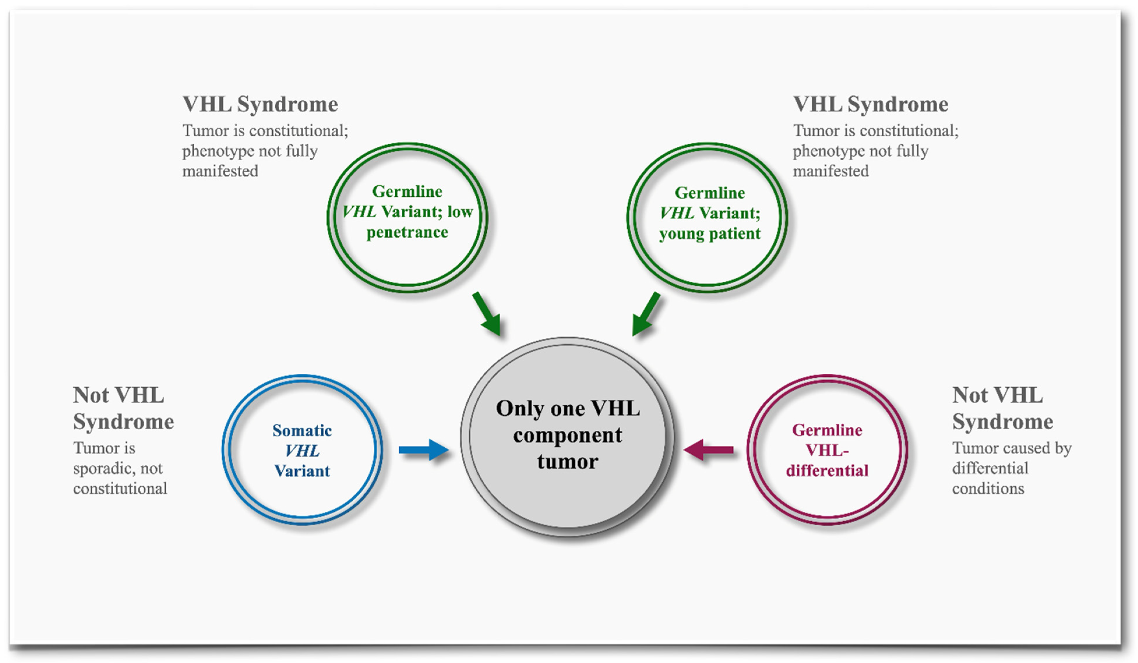
Only One VHL Component Tumor: Renal Cell Carcinoma
RCC, particularly clear cell histology, is common in VHL syndrome, occurring in 70% of the affected individuals. However, only 5–8% of RCCs are expected to be hereditary [17]. Because of the common occurrence of sporadic RCC, the VEF requires somatic genetic evidence to ensure an accurate evaluation of the germline VHL variants.
Only One VHL Component Tumor: Except Renal Cell Carcinoma
Non-RCC tumors are common in VHL syndrome, though they occur less frequently in the general population compared to RCC. With the exception of retinal hemangioblastomas, most non-RCC tumors associated with the VHL are sporadic. Therefore, when only one tumor other than RCC is present, the VEF requires additional evidence to support an association between a VHL variant and VHL syndrome.
Two or More VHL Component Tumors
Multiple VHL-related tumors can be highly suggestive of VHL syndrome, but the type of tumor (RCC vs non-RCC) impacts the assessment. Due to the common occurrence of sporadic RCC, including bilateral RCC tumors, the INT2GRATE|VEF considers scenarios in this category where one, two, or no RCC tumors are present (Supplementary Materials Table S1, Codes I–I, I–II, I–IV, II–I, II–IV, II–VII scenarios). The presence of multiple non-RCC VHL-associated tumors is more suggestive of VHL syndrome.
Non-VHL Tumor(s)
The INT2GRATE|VEF captures scenarios where the patient has neither VHL tumors nor any tumors at all. The absence of a VHL tumor, combined with the absence of relevant VHL-specific evidence, suggests that the germline VHL variant is not associated with VHL syndrome.

3.1.4. Somatic Variants and Rationale

The INT2GRATE|VEF can distinguish between sporadic VHL tumors and those arising constitutionally as part of VHL syndrome. When a tumor is suspected to be sporadic, the INT2GRATE|VEF requires additional steps to assess somatic VHL alleles. Biallelic somatic VHL inactivating variants may indicate sporadic tumors, while a monoallelic somatic inactivating variant could represent the second hit in a constitutional tumor, where the first hit is the germline VHL variant.

3.2. Assignment of INT2GRATE Categories

INT2GRATE evaluates the combination of evidence across different scenarios to determine the relationship between the VHL variant and VHL disease. Each scenario is characterized by an INT2GRATE code along with a detailed comment that explains the reasoning behind the association (or absence of association) between the germline variants and VHL disease (Supplementary Materials Table S1).
INT2GRATE Positive designates scenarios in which all evidence parameters align conservatively with VHL syndrome (Supplementary Materials Table S1, Codes I–I to I–IV). Conversely, INT2GRATE Negative refers to cases where the evidence pattern does not support VHL syndrome (Supplementary Materials Table S1, Codes III–I to III–III). INT2GRATE Neutral represents scenarios where the evidence is strong, but not sufficient for a complete VHL variant assessment (Supplementary Materials Table S1, Codes II–I to II–VII). Lastly, INT2GRATE NOS denotes cases where a key piece of evidence is absent that prevents any assessment (Supplementary Materials Table S1, Codes IV–I to IV–III).

3.3. Patient Cohorts and Clinical Presentations

An agnostic approach was used in this study to select patients based on their VHL variant status rather than their personal history of VHL syndrome, thereby reducing potential ascertainment bias. Data from 2672 patients were collected from five institutions and analyzed at the INT2GRATE Coordinating Center (Table 1). Data were categorized based on the availability of germline sequencing data, clinical genetics evaluation, tumor details, and somatic sequencing data.
The INT2GRATE variant analysis was performed on the germline VHL variants from 133 patients evaluated in a genetics clinic for cancer predisposition, all of whom had tumor data, with a subset also having somatic sequencing data (Cohort 1; Table 1). Cohort 1 comprised 98 females (74%) and 35 males (26%), with the median age of cancer diagnosis at 54 years old (Table 2) presented with various VHL tumors, including hemangioblastomas and renal lesions (Table 3, Supplementary Materials Table S3).
A tumor landscape analysis was conducted on the remaining 2539 patients who were positive for the somatic VHL variant(s), had comprehensive tumor data, and a subset of whom had been evaluated in genetics but did not have the germline VHL variants (Cohort 2; Table 1).

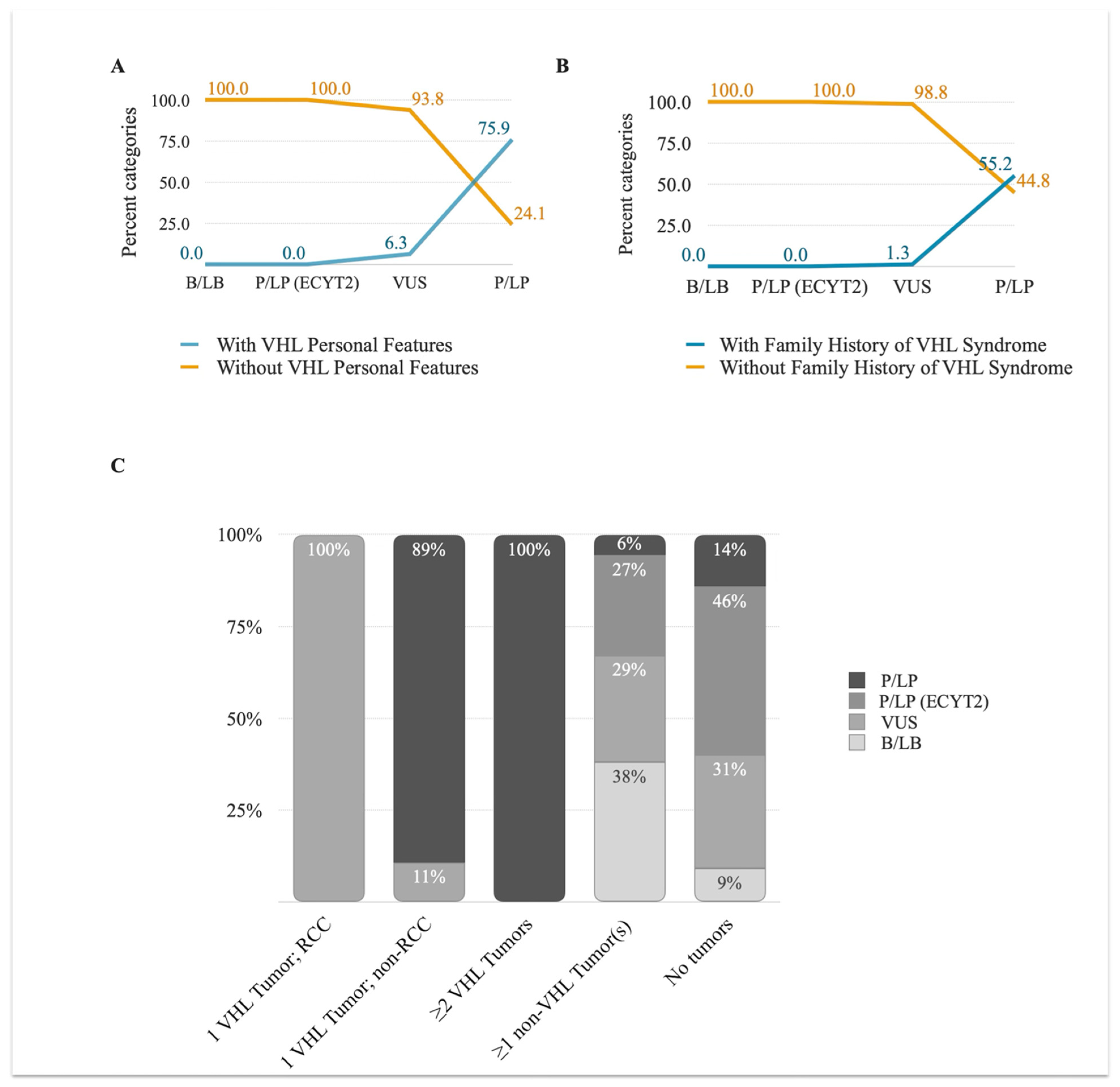
3.3.1. VHL Personal and Family History

Prior to performing the INT2GRATE analysis, the patients’ clinical data and the germline variant pathogenicity (as reported by the clinical laboratory) from Cohort 1 were evaluated. Among the germline VHL variants from 133 patients, 60% (80/133) were VUS. A small proportion of patients with germline VUS showed personal features and/or tumors and a family history of VHL diagnosis (6.3% and 1.3%, respectively) (Figure 3A). Among the 29 patients with pathogenic/likely pathogenic VHL variants, 26% did not exhibit VHL personal features and/or tumors, and 45% were negative for a family history of VHL syndrome (Figure 3B). In the remaining patients, none with polymorphic benign variants (n = 15) or monoallelic pathogenic variants associated with familial erythrocytosis (n = 9) exhibited personal features and/or tumors or a family history of VHL syndrome.

3.3.2. VHL Tumor Information

The distribution of VHL component tumors among the patients in this study displayed a broad spectrum (Figure 3C). Among the patients with P/LP variants, only 65.5% (19/29) had ≥2 VHL component tumors, 10% (3/29) had only one component tumor, and another 14% (4/29) had only non-VHL tumors. The remaining 10% (3/29) had no tumors. The difference was even wider among the patients with VUS variants: 71% (57/80) had only non-VHL component tumors, 23% (18/80) had no tumors, and 5% (4/80) and 1% (1/80) had isolated RCC and isolated paraganglioma, respectively. All patients with B/LB or P/LP-ECYT2 monoallelic variants either had no VHL tumors or no tumors at all. The broad distribution of the VHL variants in the context of personal and family history or VHL tumor status highlights the inefficiency of assessing the evidence in isolation and the need for an integrated evaluation of evidence using INT2GRATE.

3.4. INT2GRATE|VHL Variant Analysis

The INT2GRATE|VHL analysis was performed on 133 germline VHL variants. An input file containing all detailed data points was processed programmatically through the digital VEF to generate the INT2GRATE variants and associated codes. Overall, 94% of variants had informative INT2GRATE categories. This includes 79% (105/133) INT2GRATE Negative variants and 15% (20/133) INT2GRATE Positive variants. The assessment of the remaining 6% did not provide an informative status (2% INT2GRATE Neutral and 4% INT2GRATE NOS) (Figure 4A).
Consistent with a conservative VEF, all patients with the INT2GRATE Positive variants (n = 20) exhibited classic VHL syndrome (Supplementary Materials Table S4, Online INT2GRATE Portal). A total of 105 patients had the INT2GRATE Negative variants (Figure 4A), suggesting that the integrated evidence does not support the involvement of these germline variants in VHL syndrome in these patients. In this category, 77% (81/105) of the variants met the criteria for INT2GRATE III–II, while 23% (24/105) were INT2GRATE III–III, consistent with the absence of VHL tumors or any other tumors (Figure 4B, Supplementary Materials Tables S3–S6, Online INT2GRATE Portal).
Three patients had the INT2GRATE Neutral variants (i.e., insufficient for full analysis; Supplementary Materials Table S7). Patient 21 was positive for VHL:c.208G>A with no personal features or tumors of VHL but a first-degree relative diagnosed with VHL syndrome, meeting the criteria for II–VI. This scenario indicates that the absence of a personal phenotype might be related to age-related penetrance, and that evaluation of the proband’s age should be considered for the assessment of this variant. Further evaluation of the patient’s clinical data revealed that she was the youngest patient in the cohort (16 years old at the time of data collection), and the INT2GRATE status was appropriately conservative in describing the variant in this patient. Patients 22 and 23 were positive for VHL:c.345C>A and VHL:c.532C>G, respectively, each presenting with one component tumor: a hemangioblastoma and a paraganglioma. The INT2GRATE assessment assigned code IV–II, indicating the combination of evidence in these patients was insufficient for a complete variant assessment. In these patients, the negative family history of VHL diagnosis suggests that the component tumor may be sporadic or constitutional with low penetrance.
In all patients with informative INT2GRATE variants (i.e., Positive or Negative), the clinical data was consistent with their INT2GRATE status, demonstrating the application of INT2GRATE in offering insights into the role of the germline VHL variants in relation to VHL syndrome in each patient (Supplementary Materials Tables S4–S6, Online INT2GRATE Portal).

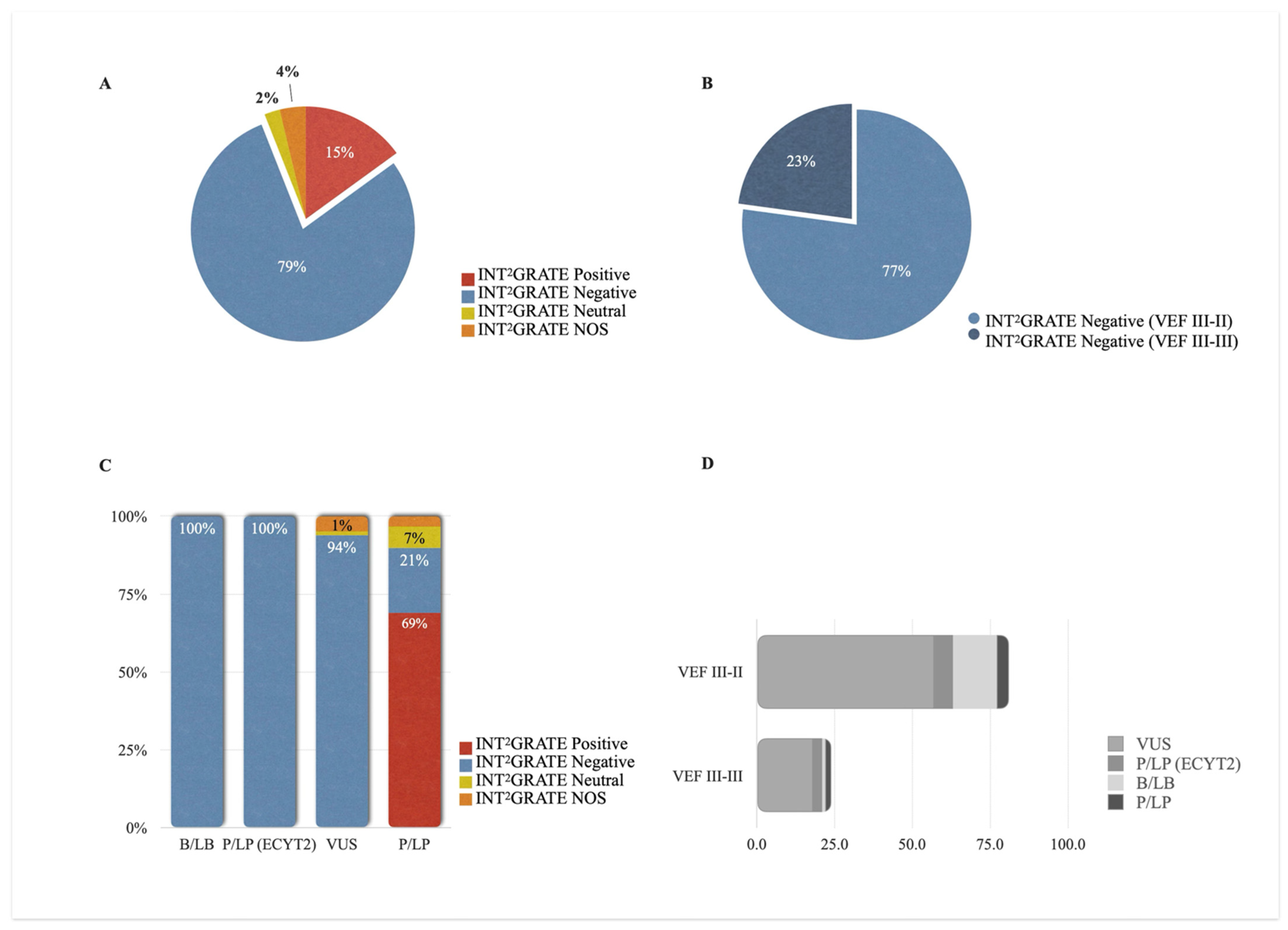
3.5. INT2GRATE Variants and Clinical Actionability

After establishing the patient-specific applications, we investigated whether the INT2GRATE|VEF could also support variant actionability more globally. To achieve this, we evaluated the INT2GRATE variants and their related ACMG classifications and case reports in the literature. For the ACMG assessment, we evaluated the variant classifications provided by the reporting laboratory on patient reports in this study, in addition to those publicly available from all submitters in ClinVar.
The INT2GRATE Negative categories accurately identified the benign VHL variants and those associated only with ECYT2. All variants classified as benign (n = 15) and those with a pathogenic classification related to congenital erythrocytosis (n = 9) were INT2GRATE Negative (Figure 4C,D, Supplementary Materials Tables S5, S6 and S8). These variants are not clinically actionable in the context of VHL syndrome, and their INT2GRATE Negative status reflects this absence of clinical actionability.
Notably, 94% (75/80) of the variants reported as VUS were assessed to be INT2GRATE Negative (Figure 4C, Supplementary Materials Tables S5 and S6). In the ClinVar database, these variants were reported either as VUS due to insufficient clinical evidence or as likely benign by other submitters. This observation suggests that the INT2GRATE assessment is consistent with the B/LB submissions. Furthermore, the comprehensive VHL-specific clinical evidence from INT2GRATE can provide crucial clinical data for the variants labeled as VUS due to the absence of clinical information.
Among the P/LP germline VHL variants, 69% (20/29) were INT2GRATE Positive, consistent with the patients’ classic VHL syndrome and the actionability of these germline variants (Figure 4C). A remaining 21% (6/29) of the P/LP variants were INT2GRATE Negative. Four variants collectively in six individuals were VHL:c.292T>C (p.Tyr98His) in two unrelated individuals, VHL:c.562C>G (p.Leu188Val) in two unrelated individuals, VHL:c.388G>A (p.Val130Ile), and VHL: c.429C>T (p.Asp143=). We investigated these variants to determine whether the INT2GRATE Negative status was specific to the patient in our study or if the evidence supported their lack of actionability more broadly. Regarding VHL:c.292T>C (p.Tyr98His), in addition to two individuals with no VHL-related evidence in this study, we have previously reported this variant in an 82-year-old patient with urothelial cancer negative for all VHL-related evidence (personal and family features, tumors, and somatic variants) [8]. In the literature, this variant has been reported as a founder variant in the Black Forest region of Germany in individuals with pheochromocytoma [18,19,20]. Regarding VHL:c.562C>G (p.Leu188Val), in addition to two individuals with no VHL-related evidence in this study, we have previously reported this variant in two family members negative for all VHL-related evidence (personal and family features, tumors, and somatic variants) [8]. In the literature, this variant has been reported in the compound heterozygous state in congenital erythrocytosis and polycythemia [21,22,23,24]. The variants VHL: c.429C>T (p.Asp143=) and VHL:c.388G>A (p.Val130Ile) have also been reported in individuals with erythrocytosis [25,26] and not VHL syndrome.

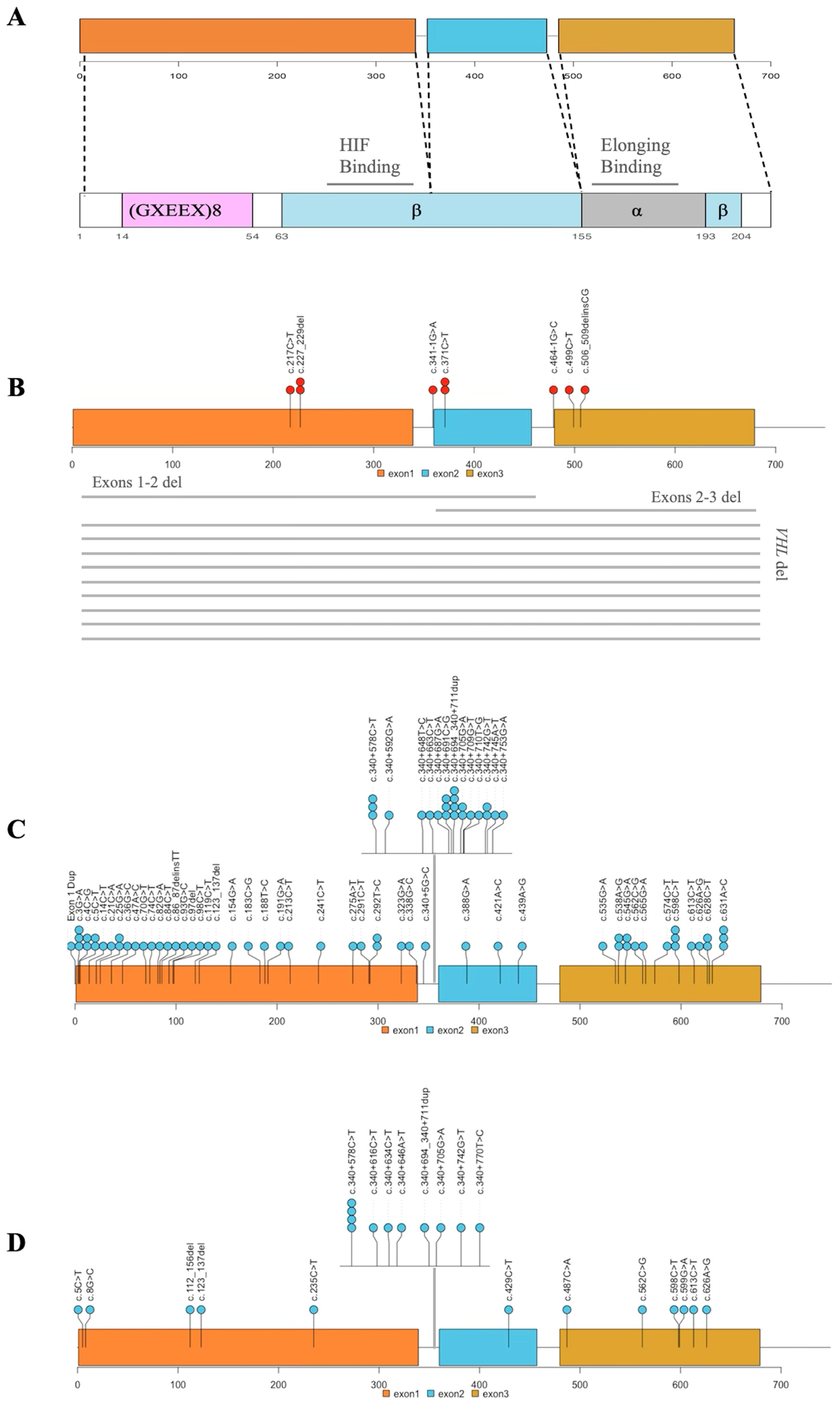
3.6. INT2GRATE Variants and Distribution Along VHL Exons

We assessed the distribution of the VHL variants across the three exons of the VHL gene to investigate their localization and to determine whether the variants from different INT2GRATE categories were confined to specific regions within the VHL (Figure 5). The majority of the INT2GRATE Positive variants (n = 11/20) were large deletions, either spanning multiple exons or the entire VHL gene, and null variants (c.341-1G>A, c.464-1G>C, c.506_509delinsCG, c.217C>T) (Figure 5A,B), Supplementary Materials Table S4). The number of missense variants was too small to provide insight into the potential enrichment of the loss-of-function missense variants in VHL in this study.
The INT2GRATE Negative variants were widely distributed. Several variants were categorized as III–II, and the same variants in other unrelated patients in this study fell in the III–II category (Figure 5C,D). Both INT2GRATE categories indicate the absence of VHL-specific evidence: patients with III–II variants displayed non-VHL tumors, and those with III–III variants had no tumors. While the variants in these overlapping INT2GRATE categories further signify that these VHL variants are not actionable, their distribution in both lollipop diagrams supports the lack of enrichment or the localization of INT2GRATE Negative variants in VHL regions.

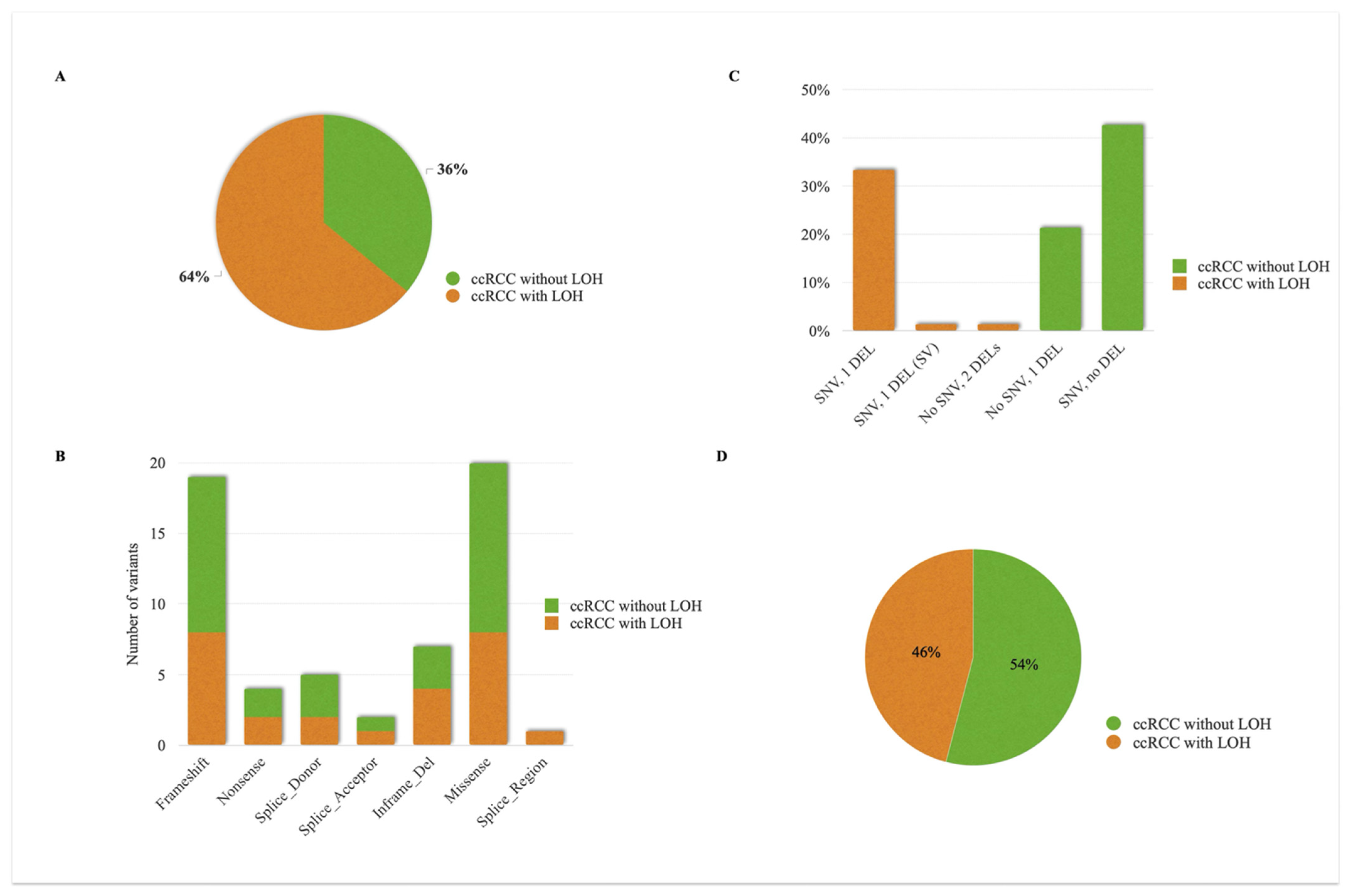
3.7. Analysis of Somatic VHL Allelic Tumors

Hemangioblastomas and ccRCC are relatively common in VHL syndrome, but they are also frequently observed as isolated sporadic tumors in the absence of germline VHL. In such cases, although these tumors are not part of any constitutional syndrome, their mere presence in a patient with a germline VHL variant could lead to the incorrect attribution of the tumor to the germline VHL. The comprehensive INT2GRATE|VHL is designed to distinguish somatic differential tumors.
To better understand the molecular landscape of these sporadic tumors, we investigated somatic VHL alterations in 2539 patients with no germline alterations related to VHL syndrome. Of the VHL alterations, 154 were SNVs and 2394 were CNs, of which 1817 were deletions (either one copy or two copy deletions). Somatic profiles and tumor pathology evaluations indicated that, in most cases, these somatic VHL alterations were passenger alterations due to genomic instability, and not the driver events in the development of tumors associated with the VHL gene. Only 75 patients were diagnosed with ccRCC, and 6 had pancreatic neuroendocrine tumors, while 2 had paragangliomas. The paraganglioma tumors were SDH-deficient and, therefore, excluded from the list of VHL-related tumors (Supplementary Materials Table S9). Among the tumors with somatic VHL alterations, ccRCC was the most frequent sporadic VHL-associated tumor.
Of the 75 patients with ccRCC, 36% (27/75) exhibited LOH due to biallelic somatic inactivating alterations in the VHL (Figure 6A). The most common LOH mechanism involved one somatic SNV and one somatic copy deletion in the VHL (Figure 6B). No significant differences were observed in the type of inactivating SNV (missense vs null) between the ccRCC cases with or without LOH (Figure 6C) or their distribution across the VHL exons (Supplementary Materials Figure S1). Null VHL variants (n = 37) were slightly more frequent in the ccRCC cases without LOH (54% or 17/37) compared to those with LOH (46% or 20/37) (Figure 6D). These results show that, in a proportion of the ccRCC cases, LOH and bilateral somatic VHL alterations may not be present. This could be attributed to assay limitations and the inability to detect possible complex or cryptic rearrangements in the VHL. Alternatively, alterations may not be present at the genomic level (e.g., epigenomic hypermethylation of the VHL promoter). This limitation in detecting LOH and somatic inactivating alterations in the VHL is addressed in the INT2GRATE|VEF to improve accuracy. The scenarios that require the somatic genetic entry in combination with other related evidence include I–II, I–III, II–I, II–II, II–III, and III–I (Supplementary Materials Table S1).

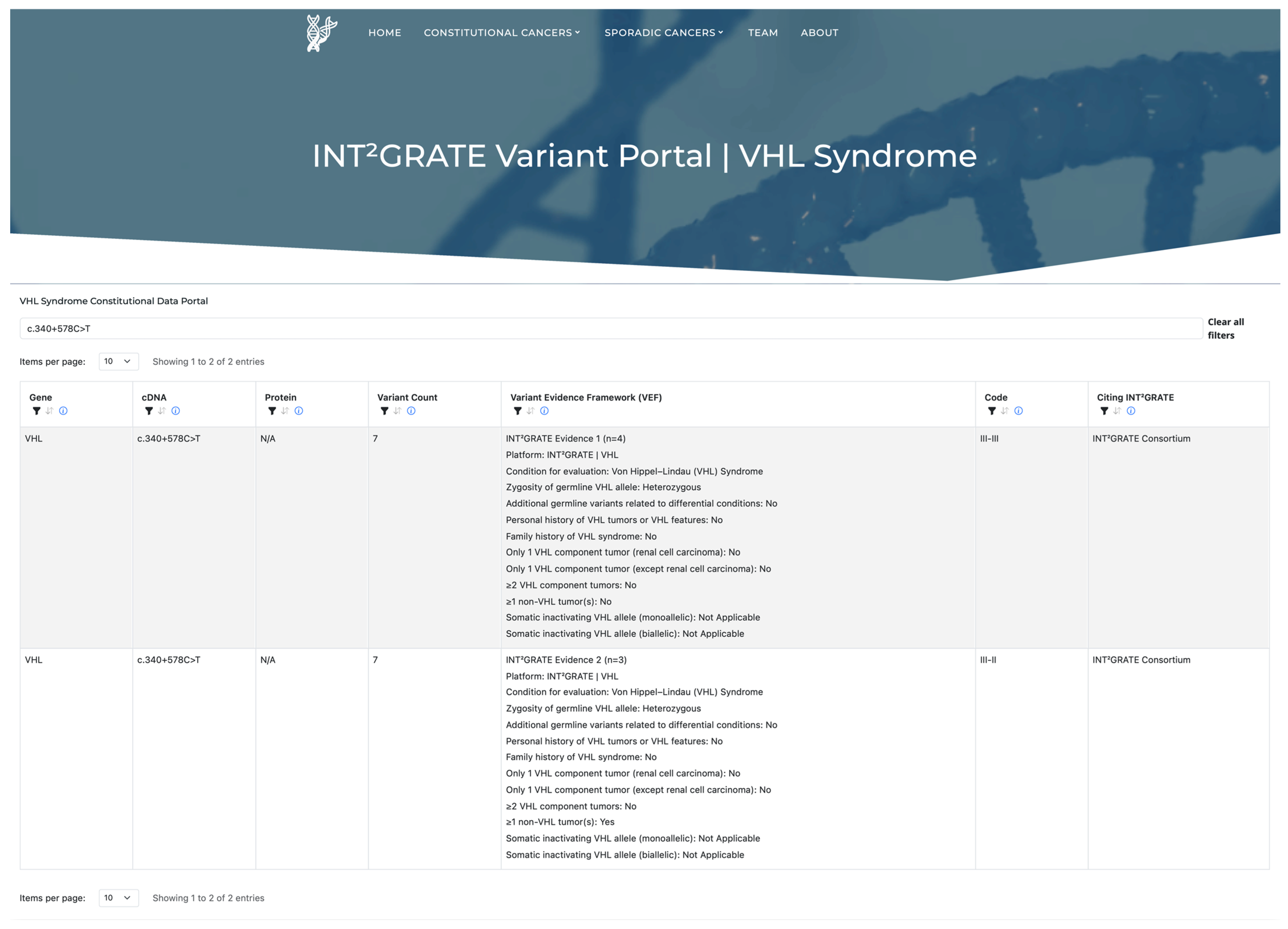
3.8. INT2GRATE|Variant Portal and Data Sharing

A primary goal of the INT2GRATE|Oncology Consortium is to advance the understanding of variant actionability in cancer through data sharing. To this end, we designed and launched the INT2GRATE Data Portal to publicly share the INT2GRATE variants and their associated evidence (INT2GRATEdata.bwh.harvard.edu). We programmatically assessed the frequency of each variant in the cohort, as well as the frequency of unique observations. A unique observation is defined when the same variant, even within the same INT2GRATE category, has a unique entry for a given parameter within the VEF framework (Figure 7). The portal facilitates the sharing of variants with detailed clinical information and frequency data, providing the scientific and clinical communities access to VHL-specific insights.

4. Discussion

An accurate assessment of the actionability of the germline variants is essential in cancer care. In VHL, clinical guidelines are largely based on clinical criteria, but establishing a molecular diagnosis requires the identification of a pathogenic or likely pathogenic germline VHL variant [9,15,16]. Similarly, clinical trials for targeted therapies in VHL disease, such as those involving hypoxia-inducible factor-2 alpha (HIF-2α) inhibitors or receptor tyrosine kinase (RTK) inhibitors, commonly have strict eligibility criteria based on genetic testing (ClinicalTrials.gov). Despite advances in molecular diagnostics, identifying the actionable germline variants remains challenging, as evidenced by the high prevalence of VUS among the reported variants. In VHL, these challenges are further compounded because tumors, such as RCC, that appear in hereditary VHL syndrome can also occur frequently as sporadic tumors without germline involvement. In fact, except for retinal hemangioblastomas, the majority of VHL component tumors are sporadic. As a result, clinicians may be inclined to overinterpret a VHL VUS in an individual with RCC or other component tumors, which could lead to over-screening of the patient and relatives. Furthermore, VHL tumors exhibit overlapping features with several differential genetic conditions (Supplementary Materials Table S2). Finally, the germline VHL variants could be genetically associated with the ECYT2 allelic disorder. The INT2GRATE|VHL addresses all these challenges through a comprehensive, evidence-based framework that incorporates constitutional, sporadic, differential, and allelic genetic conditions.
In developing the INT2GRATE|VEF, we incorporated a comprehensive set of evidence and parameters routinely used in clinical practice. We then crafted realistic scenarios based on actual clinical experiences and applied expert consensus to ensure conservative decision-making in each case. The VEF underwent review and finalization by the INT2GRATE’s multidisciplinary core committee, which includes board-certified medical geneticists specializing in cancer genetics and tumor profiling, clinicians and genetic counselors with expertise in hereditary cancer diagnostics, including VHL, and board-certified pathologists with expertise in molecular pathology and tumor profiling. The VEF parameters were shared with the INT2GRATE|Consortium members for data collection, and a digital version of the VEF was developed at the INT2GRATE Coordinating Center. All patient data were analyzed programmatically through the digital VEF to create the INT2GRATE Variants for analysis and sharing via the INT2GRATE Data Portal.
The underlying mechanism of VHL disease is loss of function, although the exact association between the genetic variants and the diverse VHL phenotypes is not yet fully understood. Recent research has demonstrated that splicing dysregulation in the E1’ and E2 regions of the VHL gene, leading to the formation of hypomorphic VHL variants, could be the underlying cause of erythrocytosis [25]. The E1’ region is a cryptic exon located deep in intron 1 of the VHL gene. Variants in this region create spliced isoforms containing the cryptic exon E1’, resulting in downregulation of VHL protein expression in patients with erythrocytosis. The INT2GRATE results were consistent with these findings, as all VHL variants across 33 individuals in the E1’ region were INT2GRATE Negative, showing the absence of all evidence related to VHL syndrome (Supplementary Materials Table S8). Furthermore, the synonymous VHL:c.429C>T (p.Asp143=) variant has been reported in homozygous or compound heterozygous states in erythrocytosis patients, and has been characterized to induce exon 2 skipping, resulting in decreased VHL expression [25]. The INT2GRATE Negative findings for the variants in E1’ and E2 aligned with their lack of association with VHL syndrome.
Consistent with the conservative INT2GRATE VEF, all 20 variants in the patients with classic VHL syndrome were INT2GRATE Positive, and all 24 benign variants or known variants associated with ECYT2 were INT2GRATE Negative. Interestingly, 94% (75/80) of the VUS variants were assessed to be INT2GRATE Negative. They were largely in the III–II category (n = 57), followed by the III–III category (n = 18), consistent with the absence of all comprehensive VHL-specific clinical evidence in these patients. Further investigation revealed that the variants in 20 individuals were located within the cryptic exon E1’ (deep intronic) in the VHL gene (as noted above; 33 variants were in E1’, 20 of which were VUS). The variants in this region are expressed at low levels in several human tissues, and have been associated with erythrocytosis [25]. The VUS classification in the remaining 60/80 cases is largely due to the absence of clinical information in the literature. Sharing these variants, along with their pertinent VHL-specific clinical evidence, on the INT2GRATE|Data Portal can provide critical support to clinical laboratories and expert groups in reclassifying these variants over time.
In our analysis, we identified six variants in four individuals as INT2GRATE negative [(n = 4, III–II; n = 2, III–III)], despite being reported as P/LP by the laboratory. We further examined these variants to determine whether the inconsistency was related to variations in VHL phenotypic expression among the individuals or a potential interpretation issue in assigning the P/LP classifications. Variants VHL:c.562C>G (p.Leu188Val), VHL: c.429C>T (p.Asp143=), and VHL:c.388G>A (p.Val130Ile) have been reported in association with erythrocytosis [21,22,23,24,25,26] and not VHL syndrome [8]. This observation demonstrates that the INT2GRATE program can efficiently differentiate between the involvement of variants in allelic disorders. The VHL:c.292T>C (p.Tyr98His) variant was present in two unrelated individuals (ages 81 and 83) with bladder and cervical cancer. We have previously reported this variant in an 82-year-old patient with urothelial cancer negative for all VHL-related evidence (personal and family features, tumors, and somatic variants) [8]. However, in the literature, this variant has been reported in association with pheochromocytoma and VHL Type 2C. The absence of all VHL-specific evidence in three of our patients, in contrast to the presence of pheochromocytoma in case reports, may suggest low penetrance of the variant or potential somatic allelic events causing sporadic pheochromocytoma. VHL tumors, other than retinal hemangioblastomas, are generally more common as sporadic tumors. VHL syndrome is estimated to account for 25% of CNS hemangioblastomas, less than 20% of pheochromocytomas, less than 10% of pancreatic neuroendocrine tumors, and 1% of RCCs [27,28,29]. A larger variant dataset could shed light on the full spectrum of VHL phenotypes in the individuals with these variants.
While VHL syndrome is typically considered an early-onset, high-penetrance condition, some variants may be associated with reduced penetrance. Clinical observations support age-related penetrance for component tumors in the individuals with VHL syndrome, with a mean age of diagnosis varying by tumor type: 22 years for endolymphatic sac tumors, 25 years for retinal hemangioblastoma, 30 years for pheochromocytoma, 30–32 years for CNS hemangioblastoma, 36 years for pancreatic lesions, and 39 years for renal cell carcinoma [30,31,32,33,34]. The risk for multiple tumors increases with age in people with VHL syndrome, and younger patients are less likely to be affected with one or more component tumors. We have considered age-related penetrance in the design of the VEF (INT2GRATE scenarios with codes II–V, II–VI, and II–VII), and variants from three patients fell in these categories (Supplementary Materials Table S7). One patient harbored VHL:c.208G>A, which was assigned code II–VI. This designation indicates that the VHL component tumors may be due to age-related penetrance and that the evaluation of the proband’s age should be considered in this variant assessment. The review of patient data confirmed this assessment, as the patient was 16 years old and had no VHL personal features yet, but her mother was diagnosed with VHL syndrome.
The INT2GRATE|VHL has several limitations. The platform analyzes the variants identified through DNA sequencing, but epigenetic alterations are not detectable by sequencing and, as such, are not evaluated. Studies have shown that methylation of the VHL promoter or consensus hypoxia-responsive element (HRE) can result in VHL loss, contributing to ccRCC progression [35,36,37]. Other limitations relate to somatic data. LOH at 3p is an early event in ccRCC, with 40–57% of alterations being somatic inactivating genetic alterations, and 10–15% resulting from epigenetic hypermethylation [38]. Our somatic VHL analysis showed that 36% of RCC tumors did not have LOH of the VHL locus. The LOH status in our study was based on genetic analysis and did not include copy-neutral LOH and methylation. Furthermore, cryptic alterations or complex rearrangements in the genome could not be assessed with standard testing and, therefore, were not investigated in this study. Another limitation is the lack of both somatic and germline sequencing data for every patient in this study. A subset of patients in Cohort 2 did not undergo constitutional cancer testing, and it is not clear whether the VHL alterations detected through tumor profiling—most of which were deletions—were truly somatic. Given that pathogenic germline VHL alterations, particularly deletions, are expected to be highly penetrant, and these patients did not have a syndromic tumor profile (i.e., had only one isolated component tumor), it is likely that these VHL alterations were somatic events. Nonetheless, the most accurate assessment requires a comprehensive analysis of both the germline and the somatic data for each individual. There are also limitations related to the sensitivity of the germline DNA testing. Studies have indicated that about 20% of the individuals with VHL syndrome have post-zygotic de novo alterations, and an estimated 5% of the individuals with VHL have somatic mosaicism [39]. Low-level mosaicism may not be detected or reported by the testing laboratories. All patients in this study have heterozygous VHL alterations, and therefore, the variants with low allelic fraction and the patients with no detectable germline VHL alterations were not included in this study.
It is noteworthy that INT2GRATE is primarily designed to evaluate the potential actionability of a given VHL variant in each patient rather than serving as a tool for the broad reclassification of variants. However, observing a consistent INT2GRATE pattern for a variant across multiple individuals over time could broaden its application in variant actionability. The INT2GRATE|Data Portal facilitates access to the variants and their VHL-related clinical evidence, as well as the frequency of variant recurrence. In this study, we identified several recurrent SNVs and indels with informative INT2GRATE categories. They include the INT2GRATE Negative variants (c.123_137del, c.25G>A, c.292T>C, c.340+705G>A, c.613C>T) in two unrelated patients; variants (c.340+691C>G, c.340+742G>T, c.3G>A, c.631A>C) in three unrelated patients; variants (c.340+694_340+711dup, c.545G>A, c.598C>T, c.5C>T, c.626A>G) in four unrelated patients; and c.340+578C>T in seven unrelated patients. Among the INT2GRATE Positive variants, c.227_229del and c.371C>T have been observed in two unrelated patients. As the variant database increases in size, the INT2GRATE|Data Portal will broaden its impact on variant actionability and may support the reclassification of variants. The portal facilitates the sharing of variants with detailed clinical information and frequency data, providing the scientific and clinical community access to VHL-specific insights.

5. Conclusions

In conclusion, the INT2GRATE|VHL presents a novel and comprehensive evidence-based framework that integrates germline, somatic, tumor, and associated clinical data, making them publicly accessible via the INT2GRATE variant portal. The INT2GRATE analysis of the VHL variants is an effective approach for determining the actionability of the constitutional VHL variants while excluding sporadic, differential, and allelic genetic conditions. The INT2GRATE Data Portal provides scientists, clinicians, and laboratories with unparalleled access to comprehensive variants and their associated clinical data. Through data sharing, the INT2GRATE Oncology Consortium is committed to advancing the clinical actionability assessment of genomic variants, fostering informed decision-making, and advancing precision oncology.

Supplementary Materials

The following supporting information can be downloaded at: https://www.mdpi.com/article/10.3390/cancers17132173/s1, Figure S1: Distribution of somatic SNVs across VHL exons in clear cell RCC (ccRCC tumors, with and without loss of heterozygosity (LOH); Table S1: INT2GRATE Variant Evidence Framework (VEF) for VHL Syndrome; Table S2: VHL-differential conditions and associated genes in the INT2GRATE | Variant Evidence Framework; Table S3: Distribution and prevalence of non-VHL tumors in patients with germline VHL variant in the INT2GRATE | VHL Program; Table S4: INT²GRATE Positive variants in the VHL gene; Table S5: INT2GRATE Negative III-II variants in the VHL gene; Table S6: INT2GRATE Negative III-III variants in the VHL gene; Table S7: Germline VHL variants in the INT2GRATE Neutral category; Table S8: Germline VHL variants in the cryptic exon E1’ and associated Iin the INT2GRATE category; Table S9: Somatic VHL allelic tumors in the INT2GRATE study.

Author Contributions

Conceptualization, A.A.G.; methodology, D.R.K., M.W., A.C., A.S.L. and A.A.G.; software, M.W. and P.C.O.; formal analysis, D.R.K., B.U., A.C., A.S.L., C.P.H., I.G., W.A.S.J., J.L.-E., S.C., R.M., N.W., N.B.A., D.K.M., H.Q.R. and A.A.G.; investigation, B.U., A.C., M.D.M., R.M.B., I.G., W.A.S.J., J.L.-E., S.C., R.M., N.W., N.B.A., D.K.M., J.A.B., J.L.H., N.I.L., L.M.S., H.Q.R., J.E.G. and A.A.G.; resources, I.G., W.A.S.J., J.L.-E., N.B.A., L.M.S., J.E.G. and A.A.G.; data curation, B.U., C.P.H. and A.A.G.; writing—original draft, D.R.K., M.W., B.U. and A.A.G.; writing—review and editing, D.R.K., M.W., B.U., A.C., A.S.L., C.P.H., P.C.O., M.D.M., R.M.B., I.G., W.A.S.J., J.L.-E., S.C., R.M., N.W., N.B.A., D.K.M., J.A.B., J.L.H., N.I.L., L.M.S., H.Q.R., J.E.G. and A.A.G.; visualization, M.W., P.C.O. and A.A.G.; supervision, A.A.G.; Project administration, A.A.G. All authors have read and agreed to the published version of the manuscript.

Funding

This research received no external funding.

Institutional Review Board Statement

The INT2GRATE Coordinating Center IRB BWH Protocol #: 2023P002760 on 12/05/2023; DFCI OHRS # 22-294 on 09/13/2022.

Informed Consent Statement

This research project utilizes secondary use of de-identified data. Therefore, informed consent from participants is not applicable.

Data Availability Statement

The original contributions presented in this study are included in the article, the public INT2GRATE Portal, and the Supplementary Materials. Further inquiries can be directed to the corresponding author.

Conflicts of Interest

The authors declare no conflicts of interest.

References

  1. Maher, E.R.; Iselius, L.; Yates, J.R.; Littler, M.; Benjamin, C.; Harris, R.; Sampson, J.; Williams, A.; Ferguson-Smith, M.A.; Morton, N. Von Hippel-Lindau disease: A genetic study. J. Med. Genet. 1991, 28, 443–447. [Google Scholar] [CrossRef] [PubMed]
  2. Varshney, N.; Kebede, A.A.; Owusu-Dapaah, H.; Lather, J.; Kaushik, M.; Bhullar, J.S. A Review of Von Hippel-Lindau Syndrome. J. Kidney Cancer VHL 2017, 4, 20–29. [Google Scholar] [CrossRef] [PubMed]
  3. Rednam, S.P.; Erez, A.; Druker, H.; Janeway, K.A.; Kamihara, J.; Kohlmann, W.K.; Nathanson, K.L.; States, L.J.; Tomlinson, G.E.; Villani, A.; et al. Von Hippel-Lindau and Hereditary Pheochromocytoma/Paraganglioma Syndromes: Clinical Features, Genetics, and Surveillance Recommendations in Childhood. Clin. Cancer Res. 2017, 23, e68–e75. [Google Scholar] [CrossRef]
  4. Semenza, G.L.; Nejfelt, M.K.; Chi, S.M.; Antonarakis, S.E. Hypoxia-inducible nuclear factors bind to an enhancer element located 3′ to the human erythropoietin gene. Proc. Natl. Acad. Sci. USA 1991, 88, 5680–5684. [Google Scholar] [CrossRef]
  5. Ivan, M.; Kondo, K.; Yang, H.; Kim, W.; Valiando, J.; Ohh, M.; Salic, A.; Asara, J.M.; Lane, W.S.; Kaelin, W.G. HIFalpha targeted for VHL-mediated destruction by proline hydroxylation: Implications for O2 sensing. Science 2001, 292, 464–468. [Google Scholar] [CrossRef]
  6. Maxwell, P.H.; Wiesener, M.S.; Chang, G.W.; Clifford, S.C.; Vaux, E.C.; Cockman, M.E.; Wykoff, C.C.; Pugh, C.W.; Maher, E.R.; Ratcliffe, P.J. The tumour suppressor protein VHL targets hypoxia-inducible factors for oxygen-dependent proteolysis. Nature 1999, 399, 271–275. [Google Scholar] [CrossRef]
  7. Zhang, K.; Qiu, J.; Yang, W.; Ma, K.; Li, L.; Xie, H.; Xu, Y.; Gong, Y.; Zhou, J.; Cai, L.; et al. Clinical characteristics and risk factors for survival in affected offspring of von Hippel-Lindau disease patients. J. Med. Genet. 2022, 59, 951–956. [Google Scholar] [CrossRef]
  8. Rana, H.Q.; Koeller, D.R.; Schwartz, A.; Manning, D.K.; Schneider, K.A.; Krajewski, K.M.; Choueiri, T.K.; Lindeman, N.I.; Garber, J.E.; Ghazani, A.A. Pathogenicity of VHL variants in families with non-syndromic von Hippel-Lindau phenotypes: An integrated evaluation of germline and somatic genomic results. Eur. J. Med. Genet. 2021, 64, 104359. [Google Scholar] [CrossRef]
  9. Adam, M.P.; Feldman, J.; Mirzaa, G.M.; Pagon, R.A.; Wallace, S.E.; Amemiya, A. GeneReviews; University of Washington: Seattle, WA, USA, 1993. [Google Scholar]
  10. Rana, H.Q.; Koeller, D.R.; Walker, M.; Unal, B.; Levine, A.S.; Chittenden, A.; Isidro, R.A.; Hayes, C.P.; Manam, M.D.; Buehler, R.M.; et al. Advancing Precision Oncology in Hereditary Paraganglioma-Pheochromocytoma Syndromes: Integrated Interpretation and Data Sharing of the Germline and Tumor Genomes. Cancers 2024, 16, 947. [Google Scholar] [CrossRef]
  11. Isidro, R.A.; Chittenden, A.; Walker, M.; Schwartz, A.; Koeller, D.R.; Hayes, C.P.; Unal, B.; Manam, M.D.; Buehler, R.M.; Manning, D.K.; et al. Development and evaluation of INT. Front. Oncol. 2023, 13, 1284690. [Google Scholar] [CrossRef]
  12. Manning, D.K.; Shivdasani, P.; Koeller, D.R.; Schwartz, A.; Rana, H.Q.; Garber, J.E.; Lindeman, N.I.; Ghazani, A.A. Assessment of genomic alterations in non-syndromic von Hippel-Lindau: Insight from integrating somatic and germline next generation sequencing genomic data. Data Brief 2021, 39, 107653. [Google Scholar] [CrossRef] [PubMed]
  13. Koeller, D.R.; Manning, D.K.; Schwartz, A.; Chittenden, A.; Hayes, C.P.; Abraamyan, F.; Rana, H.Q.; Lindeman, N.I.; Garber, J.E.; Ghazani, A.A. An optimized protocol for evaluating pathogenicity of. MethodsX 2022, 9, 101761. [Google Scholar] [CrossRef] [PubMed]
  14. Abo, R.P.; Ducar, M.; Garcia, E.P.; Thorner, A.R.; Rojas-Rudilla, V.; Lin, L.; Sholl, L.M.; Hahn, W.C.; Meyerson, M.; Lindeman, N.I.; et al. BreaKmer: Detection of structural variation in targeted massively parallel sequencing data using kmers. Nucleic Acids Res. 2015, 43, e19. [Google Scholar] [CrossRef]
  15. Louise, M.B.M.; Smerdel, M.; Borgwadt, L.; Beck Nielsen, S.S.; Madsen, M.G.; Moller, H.U.; Kiilgaard, J.F.; Friis-Hansen, L.; Harbud, V.; Cortnum, S.; et al. Von Hippel-Lindau disease: Updated guideline for diagnosis and surveillance. Eur. J. Med. Genet. 2022, 65, 104538. [Google Scholar] [CrossRef]
  16. Wolters, W.P.G.; Dreijerink, K.M.A.; Giles, R.H.; van der Horst-Schrivers, A.N.A.; van Nesselrooij, B.; Zandee, W.T.; Timmers, H.; Seute, T.; de Herder, W.W.; Verrijn Stuart, A.A.; et al. Multidisciplinary integrated care pathway for von Hippel-Lindau disease. Cancer 2022, 128, 2871–2879. [Google Scholar] [CrossRef]
  17. Shuch, B.; Vourganti, S.; Ricketts, C.J.; Middleton, L.; Peterson, J.; Merino, M.J.; Metwalli, A.R.; Srinivasan, R.; Linehan, W.M. Defining early-onset kidney cancer: Implications for germline and somatic mutation testing and clinical management. J. Clin. Oncol. 2014, 32, 431–437. [Google Scholar] [CrossRef]
  18. Boedeker, C.C.; Erlic, Z.; Richard, S.; Kontny, U.; Gimenez-Roqueplo, A.P.; Cascon, A.; Robledo, M.; de Campos, J.M.; van Nederveen, F.H.; de Krijger, R.R.; et al. Head and neck paragangliomas in von Hippel-Lindau disease and multiple endocrine neoplasia type 2. J. Clin. Endocrinol. Metab. 2009, 94, 1938–1944. [Google Scholar] [CrossRef]
  19. Bender, B.U.; Eng, C.; Olschewski, M.; Berger, D.P.; Laubenberger, J.; Altehöfer, C.; Kirste, G.; Orszagh, M.; van Velthoven, V.; Miosczka, H.; et al. VHL c.505 T>C mutation confers a high age related penetrance but no increased overall mortality. J. Med. Genet. 2001, 38, 508–514. [Google Scholar] [CrossRef]
  20. Favier, J.; Brière, J.J.; Burnichon, N.; Rivière, J.; Vescovo, L.; Benit, P.; Giscos-Douriez, I.; De Reyniès, A.; Bertherat, J.; Badoual, C.; et al. The Warburg effect is genetically determined in inherited pheochromocytomas. PLoS ONE 2009, 4, e7094. [Google Scholar] [CrossRef]
  21. Pastore, Y.; Jedlickova, K.; Guan, Y.; Liu, E.; Fahner, J.; Hasle, H.; Prchal, J.F.; Prchal, J.T. Mutations of von Hippel-Lindau tumor-suppressor gene and congenital polycythemia. Am. J. Hum. Genet. 2003, 73, 412–419. [Google Scholar] [CrossRef]
  22. Bento, M.C.; Chang, K.T.; Guan, Y.; Liu, E.; Caldas, G.; Gatti, R.A.; Prchal, J.T. Congenital polycythemia with homozygous and heterozygous mutations of von Hippel-Lindau gene: Five new Caucasian patients. Haematologica 2005, 90, 128–129. [Google Scholar] [PubMed]
  23. Lorenzo, F.R.; Yang, C.; Lanikova, L.; Butros, L.; Zhuang, Z.; Prchal, J.T. Novel compound VHL heterozygosity (VHL T124A/L188V) associated with congenital polycythaemia. Br. J. Haematol. 2013, 162, 851–853. [Google Scholar] [CrossRef] [PubMed]
  24. Erlic, Z.; Hoffmann, M.M.; Sullivan, M.; Franke, G.; Peczkowska, M.; Harsch, I.; Schott, M.; Gabbert, H.E.; Valimäki, M.; Preuss, S.F.; et al. Pathogenicity of DNA variants and double mutations in multiple endocrine neoplasia type 2 and von Hippel-Lindau syndrome. J. Clin. Endocrinol. Metab. 2010, 95, 308–313. [Google Scholar] [CrossRef] [PubMed]
  25. Lenglet, M.; Robriquet, F.; Schwarz, K.; Camps, C.; Couturier, A.; Hoogewijs, D.; Buffet, A.; Knight, S.J.L.; Gad, S.; Couvé, S.; et al. Identification of a new. Blood 2018, 132, 469–483. [Google Scholar] [CrossRef]
  26. Capodimonti, S.; Teofili, L.; Martini, M.; Cenci, T.; Iachininoto, M.G.; Nuzzolo, E.R.; Bianchi, M.; Murdolo, M.; Leone, G.; Larocca, L.M. Von hippel-lindau disease and erythrocytosis. J. Clin. Oncol. 2012, 30, e137–e139. [Google Scholar] [CrossRef]
  27. Shankar, G.M.; Taylor-Weiner, A.; Lelic, N.; Jones, R.T.; Kim, J.C.; Francis, J.M.; Abedalthagafi, M.; Borges, L.F.; Coumans, J.V.; Curry, W.T.; et al. Sporadic hemangioblastomas are characterized by cryptic VHL inactivation. Acta Neuropathol. Commun. 2014, 2, 167. [Google Scholar] [CrossRef]
  28. Maher, E.R.; Neumann, H.P.; Richard, S. Von Hippel-Lindau disease: A clinical and scientific review. Eur. J. Hum. Genet. 2011, 19, 617–623. [Google Scholar] [CrossRef]
  29. Arnon, L.; Halperin, R.; Tirosh, A. Impact of Pancreatic Neuroendocrine Tumor on Mortality in Patients with von Hippel-Lindau Disease. Endocr. Pract. 2021, 27, 1040–1045. [Google Scholar] [CrossRef]
  30. Webster, A.R.; Maher, E.R.; Moore, A.T. Clinical characteristics of ocular angiomatosis in von Hippel-Lindau disease and correlation with germline mutation. Arch. Ophthalmol. 1999, 117, 371–378. [Google Scholar] [CrossRef]
  31. Kreusel, K.M.; Bechrakis, N.E.; Krause, L.; Neumann, H.P.; Foerster, M.H. Retinal angiomatosis in von Hippel-Lindau disease: A longitudinal ophthalmologic study. Ophthalmology 2006, 113, 1418–1424. [Google Scholar] [CrossRef]
  32. Dollfus, H.; Massin, P.; Taupin, P.; Nemeth, C.; Amara, S.; Giraud, S.; Béroud, C.; Dureau, P.; Gaudric, A.; Landais, P.; et al. Retinal hemangioblastoma in von Hippel-Lindau disease: A clinical and molecular study. Investig. Ophthalmol. Vis. Sci. 2002, 43, 3067–3074. [Google Scholar]
  33. Huntoon, K.; Shepard, M.J.; Lukas, R.V.; McCutcheon, I.E.; Daniels, A.B.; Asthagiri, A.R. Hemangioblastoma diagnosis and surveillance in von Hippel-Lindau disease: A consensus statement. J. Neurosurg. 2022, 136, 1511–1516. [Google Scholar] [CrossRef] [PubMed]
  34. Lonser, R.R.; Glenn, G.M.; Walther, M.; Chew, E.Y.; Libutti, S.K.; Linehan, W.M.; Oldfield, E.H. Von Hippel-Lindau disease. Lancet 2003, 361, 2059–2067. [Google Scholar] [CrossRef] [PubMed]
  35. Robinson, C.M.; Lefebvre, F.; Poon, B.P.; Bousard, A.; Fan, X.; Lathrop, M.; Tost, J.; Kim, W.Y.; Riazalhosseini, Y.; Ohh, M. Consequences of VHL Loss on Global DNA Methylome. Sci. Rep. 2018, 8, 3313. [Google Scholar] [CrossRef]
  36. Herman, J.G.; Latif, F.; Weng, Y.; Lerman, M.I.; Zbar, B.; Liu, S.; Samid, D.; Duan, D.S.; Gnarra, J.R.; Linehan, W.M. Silencing of the VHL tumor-suppressor gene by DNA methylation in renal carcinoma. Proc. Natl. Acad. Sci. USA 1994, 91, 9700–9704. [Google Scholar] [CrossRef]
  37. Li, L.; Bao, H.; Xu, Y.; Yang, W.; Zhang, Z.; Ma, K.; Zhang, K.; Zhou, J.; Gong, Y.; Ci, W.; et al. Preliminary Study of Whole-Genome Bisulfite Sequencing and Transcriptome Sequencing in VHL Disease-Associated ccRCC. Mol. Diagn. Ther. 2023, 27, 741–752. [Google Scholar] [CrossRef]
  38. Singh, R.B.; Amare Kadam, P.S. Investigation of tumor suppressor genes apart from VHL on 3p by deletion mapping in sporadic clear cell renal cell carcinoma (cRCC). Urol. Oncol. 2013, 31, 1333–1342. [Google Scholar] [CrossRef]
  39. Chen, M.Y.; Chew, E.Y.; Reynolds, J.C.; Chao, D.L.; Oldfield, E.H. Metastatic brainstem pheochromocytoma in a patient with von Hippel-Lindau disease. Case illustration. J. Neurosurg. 2001, 94, 138. [Google Scholar] [CrossRef]
Figure 1. Possible genetic etiologies of an isolated VHL tumor. The diagram depicts the molecular mechanisms underlying the development of a VHL component tumor. An isolated VHL tumor, such as renal cell carcinoma, may be sporadic and not related to VHL syndrome. It may be related to differential genetic conditions with clinical features resembling those in VHL syndrome. Alternatively, a VHL component tumor can be related to a germline VHL variant with incomplete penetrance, age-related penetrance, or high penetrance but in a young patient.
Cancers 17 02173 g001
Figure 2. Schematic representation of the INT2GRATE|VHL. The INT2GRATE variant evidence framework (VEF) has the following four main components: (1) germline variants, including the VHL variants and the zygosity status, and variants in genes related to VHL differential conditions; (2) pertinent patient clinical genetics data related to personal and family history of VHL syndrome; (3) tumor-derived data, including VHL-related tumor counts and types; and (4) somatic genetic variants in the VHL gene. A comprehensive evaluation of the INT2GRATE variants can help differentiate their role in non-syndromic sporadic tumors or syndromic VHL, VHL allelic disorders, and differential conditions.
Cancers 17 02173 g002
Figure 3. The clinical history and VHL tumors across patient cohorts before the INT2GRATE analysis. The (A,B) panels show the proportion of patients with VHL personal features and/or tumors (A) and family history of VHL syndrome (B). The variant classifications (ACMG) provided by the reporting laboratory are included on the X-axis. Panel (C) shows the distribution of VHL tumors across the variants according to their ACMG classifications. P/LP: pathogenic/likely pathogenic variants; P/LP (ECYT2): pathogenic/likely pathogenic variants related to ECYT2; VUS: variants of uncertain significance; B/LB: benign and likely benign variants.
Cancers 17 02173 g003
Figure 4. Distribution of Informative INT2GRATE Variants in the VHL. An INT2GRATE analysis resulted in 94% of germline VHL variants having informative INT2GRATE categories. This includes 15% INT2GRATE Positive and 79% INT2GRATE Negative variants (A). The INT2GRATE Negative category does not support the involvement of the germline variants in VHL syndrome; 77% of these variants were in adult patients with tumors other than VHL tumors (Code III–II), and 23% were in patients with no tumors (III–III) (B). The distribution of variants with different INT2GRATE categories and their reported ACMG classifications are shown in (C), and the breakdown of INT2GRATE Negative III–II and III–III is shown in (D). INT2GRATE Neutral represents the variants with strong but insufficient evidence for a complete evaluation, and INT2GRATE NOS denotes cases where key evidence was absent, excluding them for evaluation.
Cancers 17 02173 g004
Figure 5. Distribution of the Informative INT2GRATE Variants Across the VHL Gene. (A) A schematic of the VHL gene is shown, including exons, the α- and β-structural domains, the HIF interaction domain, and the elongin C binding domain. Lollipop plots illustrate the distribution of variants across the gene: (B) the INT2GRATE Positive variants, (C) the INT2GRATE Negative variants in category III–II, and (D) the INT2GRATE Negative variants in category III–III. Variants located within the cryptic exon E1′, situated deep within intron 1, are shown in an inset within the intronic region in (C,D).
Cancers 17 02173 g005
Figure 6. Molecular landscape of somatic VHL alterations in clear cell renal cell carcinoma (ccRCC) tumors. (A) Among patients with sporadic ccRCC, 64% exhibited LOH due to biallelic somatic inactivating alterations in VHL. (B) Different types of genetic alterations in somatic alleles are shown in the ccRCC cases with and without LOH, with the most common LOH mechanism involving one somatic SNV and one somatic copy deletion. (C) The types and frequency of somatic inactivating SNVs in the VHL showed no significant difference between the ccRCC cases with LOH and those without LOH. (D) Null VHL variants were slightly more frequent in the ccRCC cases without LOH compared to those with LOH. SNV: single nucleotide variant; DEL: copy number deletion of VHL allele; LOH: loss of heterozygosity.
Cancers 17 02173 g006
Figure 7. The INT2GRATE Variant Portal, publicly accessible at INT2GRATE.bwh.harvard.edu. A screenshot showing the search for VHL:c.340+578C>T. The variant is shown along with comprehensive details of associated clinical evidence. This variant was observed in seven unrelated patients (variant recurrence = 7). In four patients, the pattern of evidence was consistent with INT2GRATE Negative III–III (i.e., no tumor), and in three patients, it was consistent with INT2GRATE III–II (i.e., VHL tumor).
Cancers 17 02173 g007
Table 1. Patient Cohorts in the INT2GRATE|VHL Program.
Patient Cohorts Based on Availability of Data TypeNumbers
Total patients in the study2672
Breakdown of Cohort 1
  Patients had somatic VHL variants and detailed tumor data, evaluated in genetics, and had germline VHL variants.11
  Patients had no somatic sequencing data, had detailed tumor data, were evaluated in genetics, and had germline VHL variants.122
Breakdown of Cohort 2
  Patients had somatic VHL variants and detailed tumor data, were evaluated in genetics, and had no germline VHL variants.638
  Patients had somatic VHL variants and detailed tumor data, were not evaluated in genetics for any hereditary cancer, and had no germline sequencing data *.1901
* Germline VHL status extrapolated from somatic sequence data and clinical data.
Table 2. The demographic of patients in the INT2GRATE|VHL program who were positive for the germline VHL variant.
Sex and Age Distribution of Patients with the Germline VHL VariantNumbers (%)
Female98 (74%)
Male35 (26%)
Median Age 54
Patient with VHL Component Tumor27 (20%)
Patient with no VHL Component Tumor106 (80%)
Patient with No Tumor or Cancer Diagnosis24 (18%)
Patient with Positive family History of VHL Diagnosis 17 (13%)
Patient with Negative Family History of VHL Diagnosis 114 (86%)
Patient with Unavailable Family History of VHL Diagnosis 2 (2%)
Table 3. Distribution and prevalence of VHL tumors in patients with germline VHL variant in the INT2GRATE|VHL program.
VHL Tumor TypeVHL
Tumors (%)
Females (%)Males (%)Mean AgePatients with Family History of VHL Diagnosis
Hemangioblastomas
 CNS hemangioblastoma16 (12%)15 (12%)1 (1%)36.911 (9%)
 Retinal hemangioblastoma11 (9%)9 (7%)2 (2%)32.38 (6%)
Renal lesions
 Multiple renal cysts13 (10%)11 (9%)2 (2%)32.910 (8%)
 Renal cell carcinoma9 (7%)4 (3%)5 (4%)44.35 (4%)
Pheochromocytoma4 (3%)2 (2%)2 (2%)30.53 (2%)
Paraganglioma3 (2%)2 (2%)1 (1%)311 (1%)
Pancreatic lesions
 Pancreatic cysts14 (11%)11 (9%)3 (2%)30.610
 Neuroendocrine tumors of the pancreas1 (1%)1 (1%)0540
Endolymphatic sac tumors1 (1%)1 (1%)0300
Epididymal and broad ligament cystadenomas00000
Disclaimer/Publisher’s Note: The statements, opinions and data contained in all publications are solely those of the individual author(s) and contributor(s) and not of MDPI and/or the editor(s). MDPI and/or the editor(s) disclaim responsibility for any injury to people or property resulting from any ideas, methods, instructions or products referred to in the content.

