Article

Applications for Circulating Cell-Free DNA in Oral Squamous Cell Carcinoma: A Non-Invasive Approach for Detecting Structural Variants, Fusions, and Oncoviruses

1
Azrieli Faculty of Medicine, Bar Ilan University, Safed 1311502, Israel
2
Otolaryngology, Head and Neck Surgery Department, Rabin Medical Center, Petah Tikva 4941492, Israel
3
Scojen Institute of Synthetic Biology, The Dina Recanati School of Medicine, Reichman University, Herzliya 4610101, Israel
4
Division of Head and Neck Surgery, Princess Margaret Cancer Center, University of Toronto, Toronto, ON M5G 2M9, Canada
5
Princess Margaret Cancer Centre, University Health Network, Toronto, ON M5G 2M9, Canada
6
Department of Pathology, Rabin Medical Centre, Petah Tikva 4941492, Israel
7
Biobank, Department of Pathology, Rabin Medical Centre, Petah Tikva 4941492, Israel
8
Department of Digital Medical Technologies, Holon Institute of Technology, Holon 5883754, Israel
*
Authors to whom correspondence should be addressed.
These authors contributed equally to this work.
Cancers 2025, 17(12), 1901; https://doi.org/10.3390/cancers17121901
Submission received: 5 April 2025 / Revised: 13 May 2025 / Accepted: 26 May 2025 / Published: 6 June 2025
(This article belongs to the Special Issue Circulating Tumour DNA and Liquid Biopsy in Oncology)

Simple Summary

A comprehensive study analyzed circulating cell-free DNA (cfDNA) from 30 oral squamous cell carcinoma (OSCC) patients, 199 public OSCC, and 192 normal samples to explore its potential in early-stage diagnostics and prognosis. The findings suggest that the cfDNA concentration correlates with the tumor stage, malignancy, and survival prognosis in OSCC patients. Deep genome sequencing of cfDNA revealed several genomic alterations: 1. Copy number variation analysis identified amplifications and deletions at loci 1q, 2q, 3p, 3q, and 8q22. Notably, amplifications of TP53 and PIK3CA, along with other genes related to keratinization, were identified in relapsed OSCC cases. Single nucleotide variations were observed in genes highly mutated in OSCC. 2. A novel fusion gene, TRMO-TRNT1, was detected in seven high-grade tumor samples. The parental genes, TRMO and TRNT1, are involved in tRNA modification and DNA repair, respectively. 3. Integrations of human papillomavirus, simian virus, and enterovirus were found in the OSCC samples, potentially indicating viral involvement in OSCC pathogenesis. These results underscore the utility of cfDNA as a non-invasive biomarker for OSCC, offering insights into tumor genomics and aiding in early detection and prognosis assessment.

Abstract

Background: Circulating cell-free DNA (cfDNA) has been widely used as a prognostic marker for different cancers. Objective: In this study, we used 30 cfDNA samples from oral squamous cell carcinoma (OSCC), 199 public OSCC samples, and 192 normal samples to study various correlation factors that could improve the early-stage diagnostics and/or prognosis of OSCC. Methods: The statistical correlation between healthy and OSCC patients was done and deep sequencing analyses was performed to study various genomic alterations likes copy number variation (CNV), and single nucleotide variants (SNVs), gene fusion and genomic integration of viruses. Results: We found that the OSCC patient cfDNA concentration can serve as an indicator of tumor stage, malignancy, and survival prognosis. Deep genome sequencing of cfDNA revealed genomic alterations, such as CNVs, fusion genes, and viral integrations. The CNV analysis suggested a correlation with amplification and deletion in chromosomes at loci 1q, 2q, 3p, 3q, and chromosome 8 at loci q22. Moreover, at these loci, amplification of TP53, PIK3CA, and other genes related to keratinization in OSCC patients was observed. In addition, we identified a novel abundant fusion gene, TRMO-TRNT1 ‘chimera’, in seven high-grade tumor samples. The parental genes of this chimera, TRMO and TRNT1, are known to play roles in tRNA modification and DNA repair, respectively. We have identified SNVs in our OSCC cohort. Some of these SNVs, like KMT2C, MUC3A, and MUC6, have been identified as common cases in different cancer populations. Finally, we detected contigs integrations of human papillomavirus, simian virus, and enterovirus in the OSCC samples, which may point to the potential causes of OSCC. Conclusions: Our results indicate that the liquid biopsy technique may thus serve as a sensitive tool to study OSCC patient genomic alterations by exploring cfDNA circulating in the plasma, providing an easy-to-use blood test in the future.

1. Introduction

Oral squamous cell carcinoma (OSCC) considered a part of head and neck squamous cell carcinoma (HNSCC) [1], affects the oral and buccal regions, including the gums, the floor of the mouth, and the tongue, palate, and lips [2]. OSCC is the 16th most common form of cancer and the 15th highest contributor to morbidity rates due to cancer [3]. Oral cancer is mostly associated with lifestyle, being linked to smoking and drinking, although some authors suggest that a predisposition to human papillomavirus (HPV) is also linked to OSCC [3,4,5,6]. While early diagnosis can help prevent metastasis and progression, OSCC often goes undiagnosed until a late stage when symptoms are observed, making radiotherapy and chemotherapy less effective [7].
The discovery of non-invasive methods, like a liquid biopsy, has led to progress in cancer diagnostics [8,9,10,11], specifically improving the diagnosis of lung, blood, colorectal, and cervical cancers, among others. At the same time, a liquid biopsy analysis of cell-free DNA (cfDNA) in plasma, saliva, and urine has helped determine the survival prognosis of patients [8,9,12,13,14,15]. CfDNA fragments appear in the plasma because of cellular apoptosis or necrosis. The amount of released cfDNA correlates with the tumor burden and metastasis [12,16,17,18,19,20,21]. These approaches offer rapid, repeatable sampling with minimal risk to the patient. As technology advances, liquid biopsy is increasingly being explored for early detection, treatment monitoring, and recurrence prediction across a broader spectrum of malignancies [8,9,12,13,22]. The cfDNA analyses have already been conducted on HNSCC patients, specifically on OSCC patients [23,24]. Human papillomavirus (HPV) is one of the major causes of HNSCC, with a significantly mutated HPV gene being the major cause of HNSCC [25,26,27]. The presence of HPV and other pathogens, as revealed by cfDNA analyses of OSCC patients, has made diagnosis and prognosis at the early stages possible [1,27,28,29].
Previous studies reported the presence of a second virus, herpes simplex virus (HSV), in HNSCC and other squamous cell carcinomas, like cervical cancer, and correlated the presence of HPV and HSV with the oncogenesis of these cancers [30,31,32]. Since OSCC is one of the most common head and neck cancers, we were interested in analyzing the cfDNA from patients belonging to an Israeli cohort to identify the genomic alterations associated with the disease, such as the copy number variation (CNV), single-nucleotide variation (SNV), integrated viral sequences, and fusion genes, along with a correlation with the tumor stage for better disease prognosis [27,33,34].
In this study, we analyzed cfDNA from 30 patients diagnosed with oral squamous cell carcinoma (OSCC) and identified a statistically significant positive Pearson’s correlation (at 5% false discovery rate (FDR) between the cfDNA concentration and tumor stage, the emergence of secondary malignancies, and the lesion size. Additionally, we observed that elevated cfDNA levels were associated with increased recurrence rates and reduced survival probability in OSCC patients. Our genomic analyses revealed frequent CNVs in cfDNA at loci containing key cancer-related genes, including TP53, TP63, FGFR1, and MLH1. We have identified SNVs like insertions, deletions, and frameshift mutations in our cohort. Common SNVs identified in our samples were KMT2C, MUC3A, and MUC6, which have been previously reported in other cohort. Furthermore, the presence of oncogenic viral sequences, such as HPV, HSV, simian virus (SV), and enterovirus (EV), were detected in several patient samples, suggesting a potential role for viral integration in the oncogenic progression of OSCC. Notably, we identified a novel fusion gene, TRMO-TRNT1, representing a chimera of two genes involved in tRNA processing. TRMO (located on chromosome 9q22) encodes a tRNA methyltransferase, while TRNT1 (on chromosome 3p26) is responsible for the addition of CCA nucleotides to tRNA molecules. According to the Genotype-Expression project (GTEx) data, both genes are ubiquitously expressed across human tissues [35]. Although TRMO has previously been linked to thyroid cancer, its role in OSCC has yet to be elucidated [36]. TRNT1, while not currently associated with cancer, is known to harbor mutations that cause sideroblastic anemia [37]. To validate our findings, we analyzed 199 publicly available RNA-seq datasets from OSCC patients and 192 from healthy controls. We observed TRNT1-TRMO in the OSCC data while this chimera was not observed in corresponding normal tissues. We further observed approximately 25% of the chimeric transcripts identified in our OSCC liquid biopsies were also detected in public OSCC tissue biopsy datasets and absent in healthy controls. Notably, ten chimeras exhibited expression levels as high as 50 reads per million underscoring their potential relevance. Collectively, our findings contribute to a more comprehensive understanding of the molecular landscape of OSCC. The identification of recurrent cfDNA alterations, viral integrations, and novel chimeric transcripts may support the development of minimally invasive biomarkers for early diagnosis, prognostication, and personalized therapeutic strategies in OSCC.

2. Materials and Methods

2.1. Methodology for Sample Size Collection

The sample size in our study was determined based on the availability of clinically well-characterized OSCC patients and matched healthy controls within the approved timeframe and ethical guidelines. A total of 30 OSCC patients were enrolled, representing diverse tumor stages and anatomical sub-sites, along with 25 healthy individuals serving as the controls. All participants provided informed consent, and the study was conducted under the approval of the relevant institutional ethics committee (Helsinki Committee approval-OSCC #0813-16-RMC). The inclusion criteria ensured representation across primary and secondary malignancies, and sample collection was standardized to minimize technical variabilities. This sample size allowed for preliminary statistical analyses and correlation assessments between cfDNA levels and clinical parameters, and expansion in future cohorts for higher statistical power.

2.2. Sample Collection

Blood samples of 30 OSCC patients were drawn before surgery and collected in cfDNA preservation tubes at the Rabin Medical Center. The blood samples were centrifuged at 300× g for 20 min at room temperature (RT) to separate the plasma and PBMCs. The plasma samples were then collected, aliquoted into Eppendorf tubes, and stored at −80 °C until use.

2.3. cfDNA Extraction

Aliquots (1 mL) of plasma from 30 patients were processed for cfDNA extraction using the Qiagen QIAamp ccfDNA/RNA Kit (Cat. No. 55184), Redwood, CA, USA in accordance with the manufacturer’s protocol and following informed patient consent. All procedures were conducted under the approval of the institutional ethics committee.
Inclusion Criteria:
  • Patients diagnosed with histopathologically confirmed OSCC;
  • Age ≥ 18 years;
  • Availability of complete clinical and demographic data;
  • Ability to provide informed consent;
  • No prior systemic cancer therapy (chemotherapy or radiotherapy) at the time of sample collection.
Exclusion Criteria:
  • Patients with other concurrent malignancies;
  • A history of autoimmune or chronic inflammatory diseases;
  • Recent major surgical procedures or trauma within the past month;
  • An inadequate sample quality or volume for cfDNA analysis.
Appropriate buffers were added at the specified volume per 1 mL of serum and vortexed and incubated as instructed. Centrifugation was performed at 12,000× g for 3 min to pellet the precipitate. The supernatant was then transferred to a new tube, and ice-cold isopropanol was added in a 1:1 ratio, briefly vortexed, and centrifuged at 5000× g for 1 min. The washing step was conducted at 5000× g for 5 min. cfDNA was eluted in 50 µL of elution buffer.
Extracted cfDNA concentrations were measured using the Qubit dsDNA High Sensitivity Assay (Thermo Fisher Scientific, Waltham, MA, USA) with a Qubit 2.0 fluorometer. Additionally, a Bioanalyzer 2100 DNA High Sensitivity Assay (Agilent Technologies, Santa Clara, CA, USA) was performed to determine the cfDNA fragment size distribution.

2.4. Copy Number Analyses

Extracted cfDNA samples from all patients were sequenced using Illumina high-throughput whole genome sequencing (at least 50 million reads per sample, NextSeq 550). Paired-end sequencing was performed. For copy number analysis, the iChorCNA [38] and Gistic2.0 [39] pipelines were used with default parameters.

2.5. Chimera Identification and Validation

Using the in-house chimera detection pipeline ChiTaH [40], novel fusion genes were identified in sequenced cfDNA samples. To validate the ChiTaH results, the primers were designed upstream and downstream of the gene–gene junction sequence of a fusion gene (See Supplementary Table S2). PCR was performed using the Hylab Hy-Taq Ready Mix (2x) (Cat no. EZ3006/7/636) (For the PCR conditions, refer to Supplementary Table S3). PCR products were purified using a Macherey-Nagel NucleoSpin Gel and PCR Clean-up Kit (Cat no: 12303368), followed by Sanger sequencing done at Macrogen, Israel.

2.6. Gene Interaction Network Analysis

GeneMania [41] was used to find the gene regulators of the TRNT1 and TRMO genes. Only co-expressed and physical interaction partners were used for the study.

2.7. Identification of Coding Potential

The coding potential of a chimera was determined using the CPAT [42] and CNIT [43] web server tools, employing the default parameters.

2.8. Virus Detection in cfDNA

The VirusFinder2.0 [44] pipeline was employed to detect the presence of oncogenic viruses in circulating cfDNA samples using its default parameters This tool integrates sequence alignment and viral–host fusion detection to accurately identify the viral integration events, enabling the characterization of viral contributions to tumorigenesis. Its application in this study supports a non-invasive approach to uncovering virus-associated cancers and complements genomic analyses aimed at identifying the molecular drivers of the disease.

2.9. SNV Analysis

SNV analysis was performed using three tools. VarScanv2.3.9 [45] uses default parameters for analysis. Only exonic SNVs were considered for the study. The SNVs from VarScan were annotated using ANNOVAR (https://annovar.openbioinformatics.org/en/latest/user-guide/download/, accessed on 1 March 2025) [46]. DeepVariantv1.0 [47], and Mutect2 v4.1.4.1 [48]. The SNVs that were identified by all three tools were selected for the study.

2.10. Gene Ontology Analysis

Gene ontology analyses were performed using PantherDBv16 [49] software.

2.11. Statistical Analysis
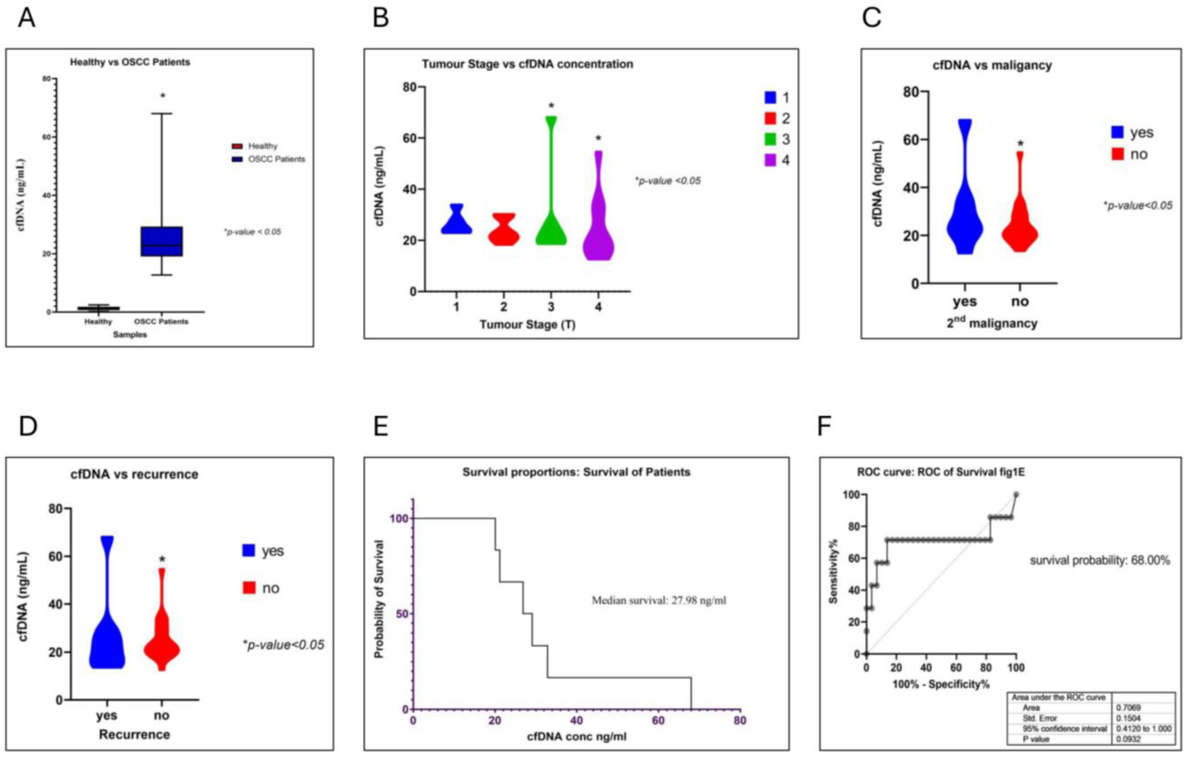
Statistical analysis was performed using GraphPad Prism 10.4.2. A Shapiro–Wilk test was used for various correlation analyses. The t-test was performed to analyze the significant difference between the cfDNA concentration of healthy and OSCC patients. Survival analysis was conducted using a Kaplan–Meyer curve, which was included in the Prism GraphPadv10.4.2 software. A p-value was considered at ≤0.05 for statistical significance at at 5% FDR.

3. Results

It is known that high concentrations of cfDNA are observed in tissue injuries, cancer, and various autoimmune diseases. The demography of all 30 OSCC patients and 25 healthy individuals was considered in our studies (see Supplementary Table S1A). The demographic parameters of all 30 OSCC patients with a clinical history were mentioned. (see Supplementary Table S1B). The cfDNA samples of OSCC and healthy individuals were extracted and subjected to BioanalyzerTM (Agilent Technologies, Santa Clara, CA, USA), evaluations (Figure S1, Supplementary Materials). Firstly, we studied the difference in the cfDNA concentrations of OSCC patients vs. healthy individuals and identified that the cfDNA in OSCC patients was significantly higher than the cfDNA concentrations of healthy individuals (Figure 1A) (p-value < 0.05, t-test), thereby suggesting that OSCC patients have a significantly higher cfDNA concentration than healthy individuals. Next, we sought to understand the correlation of increased cfDNA levels with various tumorigenic parameters, like the tumor stage, a second malignancy, patient survival, and more. In this study, cancers of the oral cavity sub-site samples were considered. These included twenty tongues, eight alveolar ridges, one floor of the mouth, and one retromolar trigone. Eleven patients showed mandibular involvement at presentation, meaning the bone of the mandible was invaded by the tumor at diagnosis; nine had undergone segmental mandibulectomy, and two had experienced marginal mandibulectomy procedures. To check if increased cfDNA levels had a direct correlation with the tumor stage, we performed Pearson’s correlation analyses. We observed that even though the average cfDNA concentrations in patients with a tumor stage 3 or 4 were high, as compared to patients with tumor stages 1 and 2, the overall distributions were similar, other than a few cases with stage 3 or 4 tumors that presented very high concentrations of cfDNA in their blood samples (Figure 1B). These results indicate that cfDNA concentrations specify the presence of stage 3 or 4 OSCC tumors at p-value < 0.05.
We also observed similar results with secondary vs. primary malignancies and observed that patients with secondary malignancies had a high cfDNA concentration at a p-value < 0.05 (Figure 1C). Furthermore, the increased cfDNA concentration was significantly associated with a higher risk of disease recurrence in OSCC patients (Figure 1D). Then, using the Kaplan–Meyer survival analysis method on data describing the OSCC recurrence and death vs. cfDNA concentration, we found that patients with cfDNA concentrations of 27 ng/mL or less had a higher survival probability (68%; Figure 1E,F). These results indicate that cfDNA concentrations above a threshold of 27 ng/mL can be used to recognize tumor stages 3 and 4, which, furthermore, can be associated with decreased survival. This study thus helped in identifying the probability of survival, underlining the tumor stages using the cfDNA concentration as a biomarker. In the future, this method could be used as an even more sensitive biomarker for the early detection of cancer.

3.1. Oncogenic Viruses Were Identified When Analyzing cfDNA

In the present study, we detected partial contigs of HPV and other oncogenic viruses. Specifically, nine of thirty OSCC patients showed the presence of viral DNA integration into cfDNA (Figure 2A). Moreover, we observed sequences from HSV strains 1, 3, and 5, SV, HPV, and EV (Figure 2A). HSV integrations were identified in six OSCC patients in the present study; three patients had HSV-1, and three patients had HSV-5 integrations. The relation of EV with cancer has not been completely established; however, some evidence shows a link to malignancy in gliomas and to the progression of cervical cancers. SV has been identified in the serum of lung cancer patients. However, SV and EV have not yet been recognized as causing OSCC. These results indicate that our study revealed the presence of more than two viruses in OSCC patient sera (Figure 2B). However, the status of these viruses cannot yet be statistically correlated with the tumor stage in our cohort. Nonetheless, the presence of viruses may serve some oncogenic role in OSCC, and more samples should be used to identify their characteristics in OSCC patients.

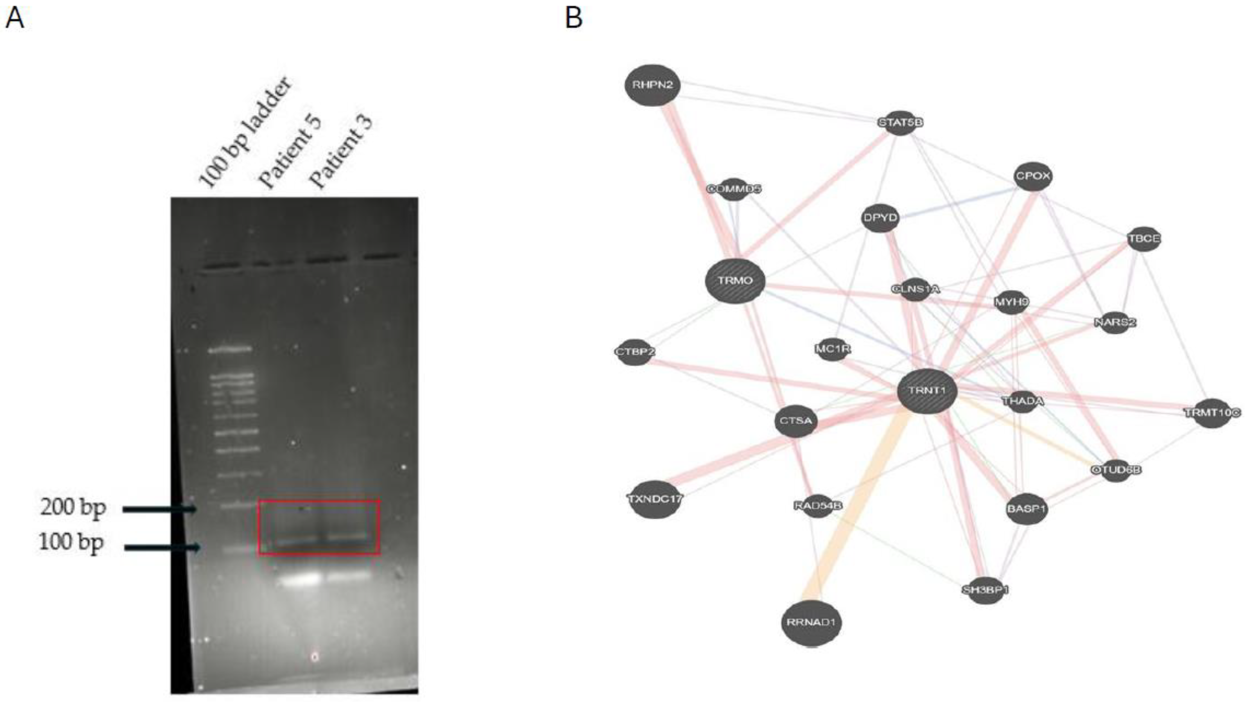
3.2. A Novel Chimera Identified in cfDNA

Fusion genes that are produced by chromosomal translocations are common in lymphomas and leukemias yet are extremely rare in HNSCC [27,34]. Using our in-house chimera analysis tool, ChiTaH, we identified the novel fusion TRMO-TRNT1 in the cfDNA samples of two patients (sequence, Figure S2, Supplementary Materials and Supplementary Data S1-Fusion_OSCC_30). TRMO (tRNA methyl-O-transferase) and TRNT1 (tRNA nucleotide transferase I) were highly expressed in all tissue types. To validate this chimera at both RNA and DNA levels, we designed primers and performed PCR on the total RNA and DNA from OSCC biopsies and used patient 3 and patient 5 as positive controls (Figure 3). Subsequently, we conducted PCR on all 30 cfDNA samples and observed a 108 bp fused sequence in seven of them (Figure S3A, Supplementary Materials). The band containing this fusion was excised from the agarose gel, purified, and then assessed by Sanger sequencing, which confirmed the fusion junction of the two parental genes, TRMO and TRN1 (Figure S3B, Supplementary Materials). This fusion was identified in seven patients using PCR against the junction sequence and the Sanger sequencing result, confirming the same, showing that the tumor has advanced malignancy and proliferation status. The altered expression of TRMO has been observed in patients with thyroid carcinoma [36], while altered TRNT1 expression was seen in colorectal cancer patients [37]. These results suggest that fusion genes could be used as biomarkers and explored in the search for additional OSCC patients, which may be identified by means of the deep sequencing of cfDNA samples. Finally, we checked the coding potential of the observed chimera to produce some functional protein. For this, we utilized three tools commonly used for analyzing the coding/non-coding potential of a given DNA sequence. The scores obtained by the CPAT and CNIT programs suggested that the fusion has a <0.3% coding probability. Therefore, the fusion does not have coding potential (Figure S4A,B) but it still may serve as a biomarker of the OSCC progression through a highly abundant non-coding cfDNA fragment. Next, we checked for the common interactors of TRNT1-TRMO genes (Figure 3B). Three common interactors are identified as THADA, COMMD2, and CTBP2. In addition, we downloaded 199 OSCC and 192 normal RNA-seq datasets from public databases (Supplementary Data S2 Fusions in 192 OSCC patients from public dataset) and considered only those chimeras expressed in tumors while absent in healthy controls for our study. We found that 4 out of 16 chimeras identified in the OSCC plasma samples were also observed in the public biopsies of OSCC patients at 50 RPKM. We observed the presence of TRNT1-TRMO fusion in few OSCC patients from public dataset at a frequency of 5%. Our findings may help to further characterize novel abundant chimeras as biomarkers in OSCC for the early diagnostics and survival prognostics.

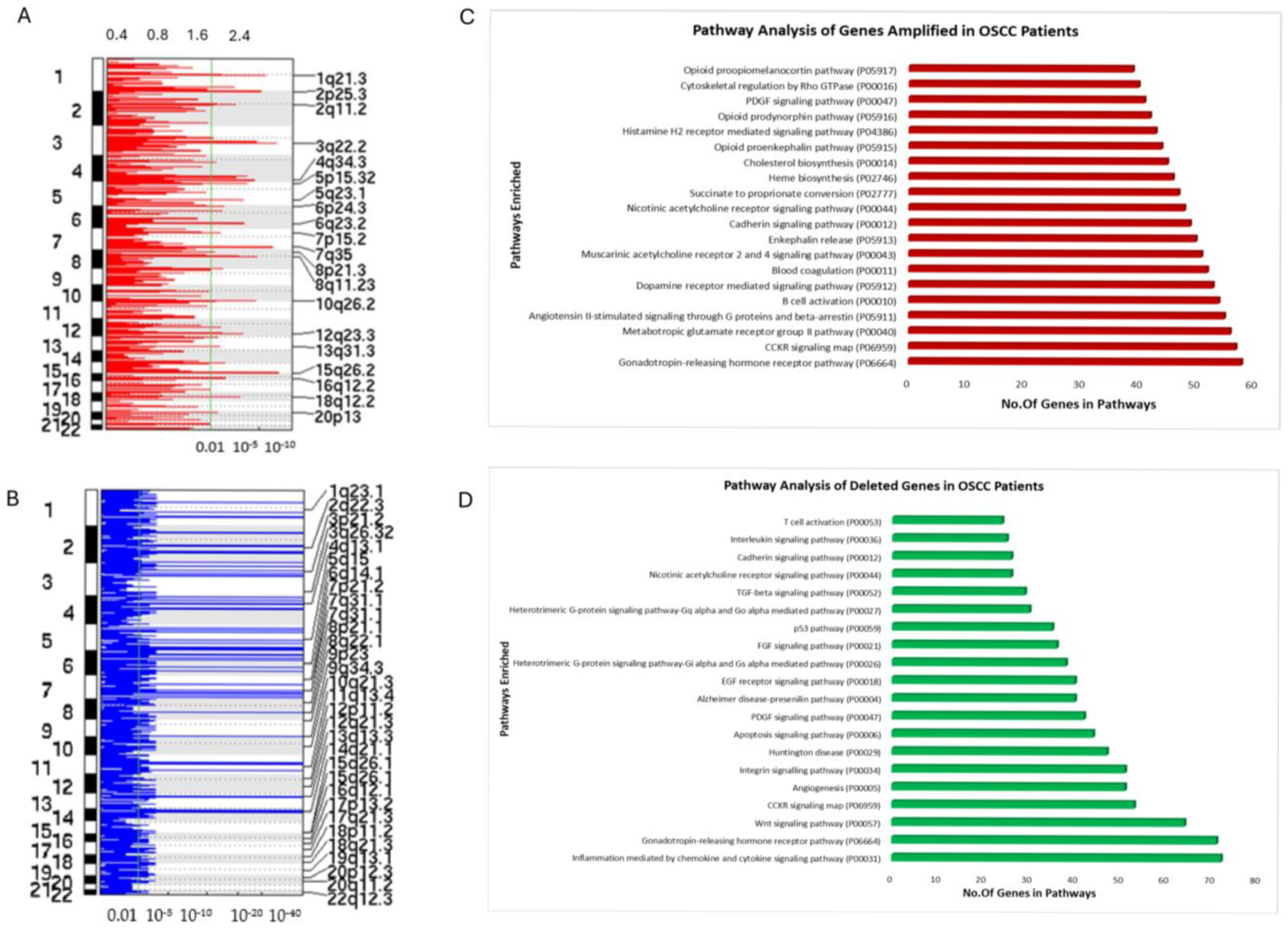
3.3. Copy Number Variation (CNV) Analysis of OSCC Patient cfDNA

CNV is commonly observed in many cancers. Various studies have identified CNV and somatic mutations using cfDNA [27,33,50,51,52]. Our aim here was to identify significant changes in the CNVs for OSCC patients using cfDNA genomic data. We performed CNV analysis on the sequenced cfDNA of OSCC patients using Gistic2.0, which assesses low pass copy number variations. The Q-value was set at 0.01 for the significance level. We observed significant amplifications in chromosomes 3q22.2, 7q35, and 15q26. Additional amplifications were also seen in chromosomes 8p21.3, 10q26,2, 2p25, and 1q21.3 (Figure 4A) (Supplementary Data S3 OSCC_Copy number variations). Some of our findings correlate with the TCGA HNSCC data for CNV and significantly mutated genes. Next, we performed functional enrichment analysis using PantherDB and observed that commonly amplified genes in the present study were predicted to play a significant role in the GPCR pathways, opioid pathways, immune system activation pathways, apoptosis, and angiogenesis activation pathways (Figure 4C). Particularly, we identified deleterious regions in patient cfDNA samples. In our cohort, we observed that chromosomes 1q23, 2q22, 3p21, 3q26, 7q31, 8p21, 9q34, 13q13, and 14q21 had significant deletions (Figure 4B). Significantly mutated genes, like TP53, TP63, FGFR1, or MLH1, have been shown to present deletions in those regions and were also identified in the present study (Supplementary Data S2) [27,53,54,55]. We also found deleted regions of THADA in our cohort. Amplification of regions of the CTBP2 genes has been observed in the present study. Both THADA and CTBP2 are common interactors of fusion genes TRNT1-TRMO (Figure 3B). Our gene ontology (GO) analysis of amplification regions found that the deleterious genes in our samples assigned to the affected genes were mostly involved in immune activation pathways, apoptosis signaling pathways (particularly the CCKR gene), cell cycle pathways, and various metabolic pathways (Figure 4D). These results indicate that cfDNA analyses may reveal significant CNV in the OSCC samples and may be used for OSCC’s early diagnosis and prognosis.

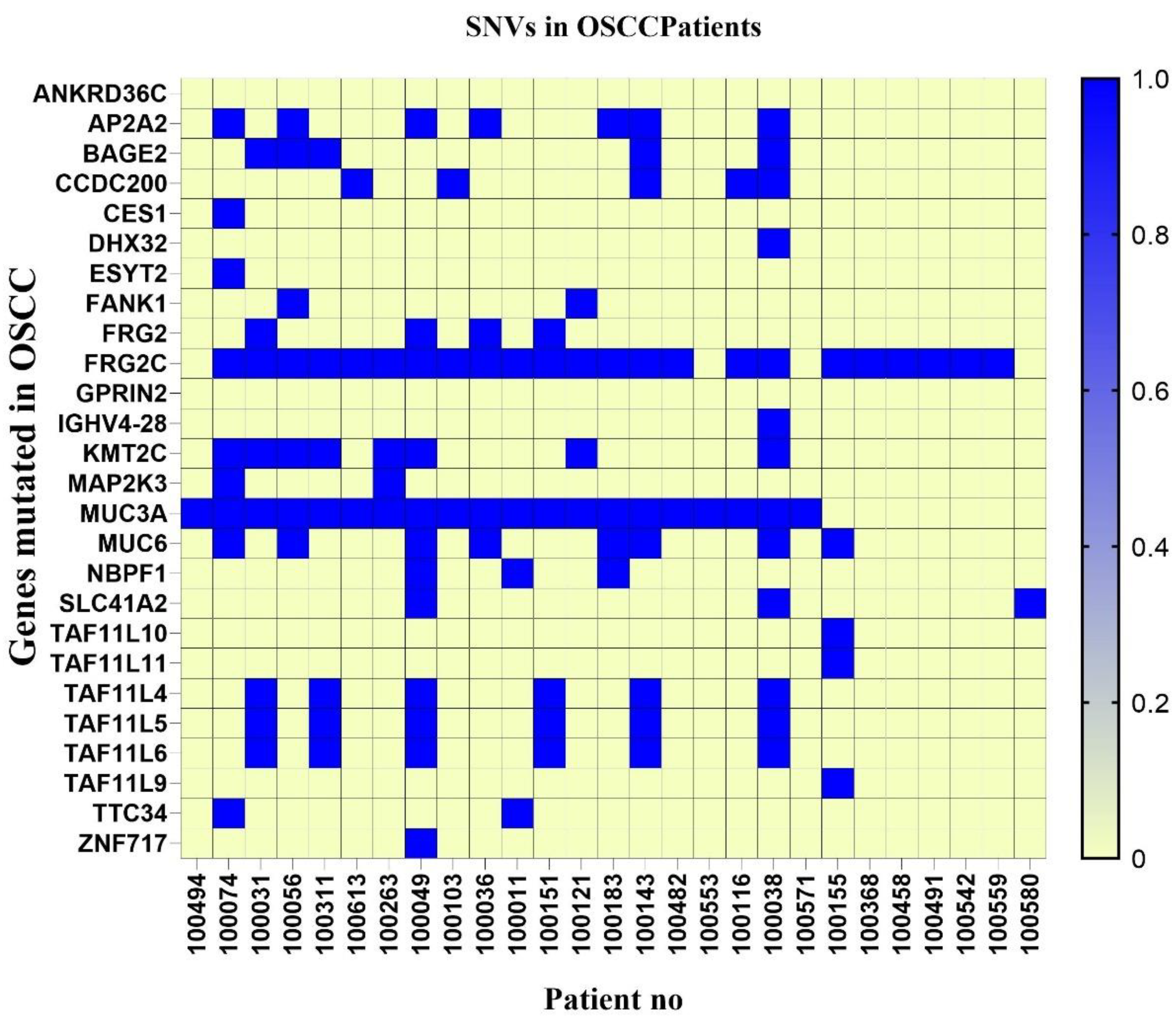
3.4. SNV Analysis of OSCC Patients’ cfDNA

SNV analysis suggests structural variants and is key to identifying the mutations that cause cancer [53]. We used three tools to perform the SNV analysis of the cfDNA of OSCC patients (Supplementary Data S4 Somatic nucleotide variations, annotation and filtering). We identified common SNVs present in our samples, such as MUC6 (27%), MUC3A (70%), FRG2C (80%), KMT2C (26%), AP2A2 (23%), BAGE (17%), and CCDC200 (20%) as well as TAF11L4, TAF11L5, and TAF11L6 (23% each) in our cohort, (Figure 5). In the present study, we considered non-synonymous mutations, frameshifts, and in-Del mutations in exonic regions. Most mutations observed were C > T, C > A, G > T, G > A. Some deletions in MUC3A and FRGC2 have been observed. These observations are in concordance with other studies reported for HNSCC. These mutations can be used to diagnose OSCC.
Approximately 4% of the SNVs found had deletions in KMT2C and MUC3A, 1% were frameshift mutations observed in MUC6, and 20% were non-synonymous substitution mutations observed mainly in KMT2C, MUC6, MUC3A, ZNF717, and SLC41A2 (Table 1). These mutations have been identified as significant mutations across various studies in HNSCC and OSCC. The mutations in these genes have been identified to play oncogenic roles in HNSCC.

4. Discussion

cfDNA has been explored as a prognostic and diagnostic biomarker in many cancers [11,12,14,16,18,24,56,57,58]. A correlation of cfDNA levels with various tumors has been established in glioblastoma and neuroblastoma. High cfDNA concentrations were observed in samples with cancer, as compared to the cfDNA from healthy samples [17,34]. Many studies have found that the concentration of cfDNA was correlated with tumor burden [19,59]. In the present study, we observed that the cfDNA concentration of OSCC patients was significantly higher than the cfDNA concentration of healthy patients. This result suggests that the cfDNA concentration of OSCC patients could be used for early and late diagnostics. Then, we explored the correlation between the cfDNA concentration from 30 OSCC patients and tumor stages. High cfDNA levels were observed in patients with stage 3 or 4 tumors, while comparatively lower cfDNA concentrations were observed in patients with stage 1 or 2 tumors. We also observed a direct correlation between a higher cfDNA concentration at presentation and the possibility of future recurrence and a secondary malignancy at a 5% FDR. We also observed that the threshold of a maximum cfDNA concentration for prognostication was <27 ng/mL. The Kaplan–Meyer survival analysis showed that the probability of survival was 68% for patients with OSCC at cfDNA concentrations above 27 ng/mL. Therefore, our study suggests that cfDNA could be used as a non-invasive method to detect OSCC at an early stage. Still, the analysis of more samples could have resulted in a better level of significance for the correlation between the cfDNA concentration and the tumor parameters.
Various genomic alterations in the cfDNA of OSCC patients have also been addressed. One such alteration was observed as the presence of viral DNA integrations. Deep sequencing of the cfDNA of OSCC patients revealed the presence of HPV, SV, HSV, and EV integrations. While a high proportion of patients (48%) did not show the presence of any virus, a few patients presented more than one virus. Interestingly, HPV has been studied as a cause of HNSCC, cervical cancer, and other gynecological and urological cancers [60,61,62,63,64,65,66,67,68,69]. We identified HPV in six out of thirty samples. HSV strains 1 and 5 were identified in six of thirty samples. HSV was previously reported in samples from HNSCC and OSCC patients, and its association with the progression of cancer was claimed [30,31,32]. Ev has been associated with malignant glioma [70]. SV was reported in samples of osteosarcoma and prostate cancer [71,72,73]. However, these viruses have not yet been associated with OSCC. Future studies could establish the association of these viruses with OSCC and determine if they are involved in oncogenic progression.
Using the in-house ChiTaH pipeline we identified the novel fusion TRMO-TRNT1. PCR and Sanger sequencing validated the presence of this fusion in seven samples from patients with late-stage tumors. A mutation in TRMO has been associated with thyroid carcinoma [36]. TRNT1 has not been associated with any cancer thus far, although its mutation is responsible for causing congenital disease and sideroblastic anemia [37]. The fusion product does not have any coding potential, according to CPAT [42] and CNIT [43]. Gene regulatory studies revealed three potential regulators of both TRNT1 and TRMO: THADA, CTBP2, and COMMD2. This fusion was observed in RNA-seq of OSCC tissue biopsy and is absent in healthy controls. Apart from TRNT1-TRMO, three other fusions were identified in cfDNA of our cohort that were observed in RNA seq of public OSCC tissue biopsy samples. Future research could explore the mechanism of action of this fusion gene in OSCC development.
CNV in tissues, as well as in cfDNA, has been studied and has been shown to be associated with many cancers, including OSCC [33,50]. In the present study, we found regions of chromosomes 3q22.2, 7q35, and 15q26 to be amplified. Amplifications were also seen in chromosomes 8p21.3, 10q26,2, 2p25, and 1q21.3. Gene ontology pathway analysis of the genes at these loci revealed them to be associated with angiogenesis, immune activation pathways, and GPCR, as well as interleukin, VEGF, FGF, and vasopressin activation pathways. The deleterious regions at chromosomes 1q23, 2q22, 3p21, 3q26, 7q31, 8p21, 9q34, 13q13, and 14q21 affect the genes associated with apoptosis, CCKR, the cell cycle, and immune activation and cell signaling pathways. Mutations in these genes and the structural variations associated with these loci have been studied in TCGA and by other groups. Our study agrees with TCGA and previous studies [27,54,55,74,75]. We also identified deleted regions of THADA in our cohort. Amplification of regions of the CTBP2 gene was observed in the present study. Both THADA and CTBP2 are common regulators of the fusion gene TRNT1-TRMO. In the future, it will be interesting to investigate whether structural variations in THADA, COMMD2, and CTBP2 play a significant role in OSCC progression and whether the fusion gene TRNT1-TRMO is involved in CNV. Additionally, targeted sequencing of these genes or CNV analysis of these chromosomal loci could be used as potential biomarkers for OSCC. Additionally, from the 199 public OSCC RNA-seq datasets, we found that 50% of the chimeras identified in the OSCC liquid biopsies were also observed in public biopsies. Furthermore, 10 chimeras exhibited abundances as high as 88 RPKM and may serve as potential biomarkers for OSCC. These findings could help explore their association with cancer and enhance applications in early diagnostics and survival prognostics.
SNV analysis helps in understanding the key mutations leading to oncogenesis [27,74,76,77,78]. They help in identifying potential risk mutations that can lead to carcinogenesis. SNVs can be a result of various factors, including environmental factors, smoking status, chewing betel quid, and viral integration status [67,79,80,81]. SNV analysis was performed in the present study to identify significant mutations in genes. Our study suggested common mutations in genes such as MUC6 (27%), MUC3A (70%), FRG2C (80%), KMT2C (26%), AP2A2 (23%), BAGE (17%), and CCDC200 (20%) in our cohort. Mutational profiles of TAF11L4, TAF11L5, and TAF11L6 (23% each) have not been reported in HNSCC. Previous studies reported that the Mucin genes were altered in OSCC. Most mutated Mucin genes are MUC1, MUC4, MUC3, and MUC6 [82,83,84]. MUC1 and MUC4 have been identified as prognostic biomarkers for OSCC, which leads to oral mucosa carcinogenesis [82,83,84,85]. MUC6 has been recently identified as a potential biomarker in Asian OSCC patients. The mutations in MUC6 have been associated with malignancy and tumorigenesis of oral carcinoma [84,85,86]. Recently, MUC3A has also been associated with the progression of oral cancer. Mutations in MUC3A have been linked to oncogenesis [84,85,86,87]. Another study showed that ZNF717 and MUC3A are mutated in tongue squamous cell carcinoma [87]. In the present study, we identified the SNV of ZNF717 in one sample. FRG2C is a signature gene mutated in ESCC (esophageal squamous cell carcinoma) and proliferative verrucous leukoplakia (PVL) [88,89]. Another gene highly mutated in the present study cohort, KMT2C, was observed to be rarely mutated in South Asian patients with HNSCC [89,90]. BAGE was identified as a clinically significant immunogenic antigen in HNSCC patients [91]. AP2A2, an adapting protein family molecule, has not been reported to be oncogenic. However, it has been linked to inhibiting the activity of Erlotinib, an anti-cancer drug for lung cancer [92].
All these mutational signatures of genes can be drug targets for precision medicines. Also, these genes can be used further to study the relevance of mutation in OSCC progression and thereby can be used as a biomarker for non-invasive cancer diagnosis and prognosis.

5. Conclusions

Our study highlights the potential of circulating cfDNA as a non-invasive biomarker for the diagnosis, prognosis, and therapeutic monitoring of OSCC. Elevated cfDNA levels correlated significantly with the tumor stage and patient outcomes, suggesting its value in early detection and recurrence risk assessment. We also uncovered a spectrum of genomic alterations in cfDNA, including viral integrations, chimeric transcripts, copy number variations (CNVs), and single-nucleotide variants (SNVs), many of which have known or emerging relevance to OSCC progression. Notably, we identified and validated a novel fusion gene, TRMO-TRNT1, which is associated with late-stage tumors, and observed recurring mutations in genes such as MUC3A, MUC6, FRG2C, and KMT2C, with implications for oncogenesis and potential as therapeutic targets. CNV analysis revealed altered regions linked to the immune response and oncogenic pathways, supporting the use of these alterations as molecular biomarkers. The consistency of several chimeric transcripts with public datasets further underscores their potential clinical relevance. The limitation of the present study is the low number of samples studied. Altogether, our findings establish a robust foundation for integrating cfDNA-based assays into OSCC diagnostics and precision medicine frameworks while pointing toward novel molecular targets for future therapeutic exploration. Continued research with larger cohorts and functional validation will be essential to fully realize the translational potential of these biomarkers.

Supplementary Materials

The following supporting information can be downloaded at: https://www.mdpi.com/article/10.3390/cancers17121901/s1, Figure S1: Bioanalyzer graph of cfDNA size of OSCC patients; Figure S2: Fusion sequence; Figure S3: Fusion validation in other OSCC samples; Figure S4: Coding potential of the fusion sequence; Table S1A: Demographic information of healthy vs. OSCC patients; Table S1B: Details of OSCC patients, Table S2: Fusion primers, Table S3: PCR conditions for fusions File S1: Fusions in 30 OSCC samples. File S2: Fusions in 192 OSCC patients from a public dataset File S3: OSCC_copy number variations. File S4: somatic variant calling, filtering, and annotation.

Author Contributions

Conceptualization: M.B., D.Y., G.B. (Gideon Baum), G.B. (Gideon Bachar), and M.F.M.; methodology: M.B., G.B. (Gideon Baum), and M.F.M.; software: D.P.D., and R.D.; validation: M.B., G.B. (Gideon Baum), D.P.D., and R.D.; formal analysis: M.B., D.Y., G.B. (Gideon Bachar), and M.F.M.; resources: M.F.M., G.B. (Gideon Bachar), S.T., T.W., E.Y., and A.L.-B.; investigation: M.B., D.Y., and M.F.M.; data curation: D.P.D. and R.D.; writing—original draft: M.F.M., M.B. and D.Y.; writing—review and editing: M.B., D.Y., A.M., D.P.D., G.B. (Gideon Baum), G.B. (Gideon Bachar), and M.F.M.; supervision: M.F.M. and G.B. (Gideon Bachar); project administration: M.F.M.; funding acquisition: M.F.M. and G.B. (Gideon Bachar). All authors have read and agreed to the published version of the manuscript.

Funding

This work was funded by Bar-Ilan University core funding, Scojen Institute of Synthetic Biology (Reichman University) fund and by the Rabin Medical Centre.

Institutional Review Board Statement

This study was conducted in accordance with the Declaration of Helsinki and approved by the Institutional Review Board of Rabin Medical Centre (protocol code OSCC #0813-16-RMCand date of approval: 17 October 2019).

Informed Consent Statement

Informed consent was obtained from all subjects involved in this study.

Data Availability Statement

All data are available upon publication.

Conflicts of Interest

The authors state no conflicts of interest.

References

  1. Sinevici, N.; O’sullivan, J. Oral Cancer: Deregulated Molecular Events and Their Use as Biomarkers. Oral. Oncol. 2016, 61, 12–18. [Google Scholar] [CrossRef] [PubMed]
  2. Montero, P.H.; Patel, S.G. Cancer of the Oral Cavity. Surg. Oncol. Clin. N. Am. 2015, 24, 491–508. [Google Scholar] [CrossRef] [PubMed]
  3. Inchingolo, F.; Santacroce, L.; Ballini, A.; Topi, S.; Dipalma, G.; Haxhirexha, K.; Bottalico, L.; Charitos, I.A. Oral Cancer: A Historical Review. Int. J. Environ. Res. Public Health 2020, 17, 3168. [Google Scholar] [CrossRef] [PubMed]
  4. Katirachi, S.K.; Grønlund, M.P.; Jakobsen, K.K.; Grønhøj, C.; von Buchwald, C. The Prevalence of HPV in Oral Cavity Squamous Cell Carcinoma. Viruses 2023, 15, 451. [Google Scholar] [CrossRef]
  5. Hernandez, B.Y.; Lynch, C.F.; Chan, O.T.M.; Goodman, M.T.; Unger, E.R.; Steinau, M.; Thompson, T.D.; Gillison, M.; Lyu, C.; Saraiya, M. Human Papillomavirus DNA Detection, P16 INK4a, and Oral Cavity Cancer in a U.S. Population. Oral. Oncol. 2019, 91, 92–96. [Google Scholar] [CrossRef]
  6. Orrù, G.; Mameli, A.; Demontis, C.; Rossi, P.; Ratto, D.; Occhinegro, A.; Piras, V.; Kuqi, L.; Berretta, M.; Taibi, R.; et al. Oral Human Papilloma Virus Infection: An Overview of Clinical-Laboratory Diagnosis and Treatment. Eur. Rev. Med. Pharmacol. Sci. 2019, 23, 8148–8157. [Google Scholar]
  7. Nakagaki, T.; Tamura, M.; Kobashi, K.; Omori, A.; Koyama, R.; Idogawa, M.; Ogi, K.; Hiratsuka, H.; Tokino, T.; Sasaki, Y. Targeted Next-Generation Sequencing of 50 Cancer-Related Genes in Japanese Patients with Oral Squamous Cell Carcinoma. Tumor Biol. 2018, 40, 1010428318800180. [Google Scholar] [CrossRef]
  8. Lousada-Fernandez, F.; Rapado-Gonzalez, O.; Lopez-Cedrun, J.-L.; Lopez-Lopez, R.; Muinelo-Romay, L.; Suarez-Cunqueiro, M. Liquid Biopsy in Oral Cancer. Int. J. Mol. Sci. 2018, 19, 1704. [Google Scholar] [CrossRef]
  9. Siravegna, G.; Marsoni, S.; Siena, S.; Bardelli, A. Integrating Liquid Biopsies into the Management of Cancer. Nat. Rev. Clin. Oncol. 2017, 14, 531–548. [Google Scholar] [CrossRef]
  10. Naito, Y.; Honda, K. Liquid Biopsy for Oral Cancer Diagnosis: Recent Advances and Challenges. J. Pers. Med. 2023, 13, 303. [Google Scholar] [CrossRef]
  11. Salvi, S.; Gurioli, G.; De Giorgi, U.; Conteduca, V.; Tedaldi, G.; Calistri, D.; Casadio, V. Cell-Free DNA as a Diagnostic Marker for Cancer: Current Insights. Onco Targets Ther. 2016, 9, 6549–6559. [Google Scholar] [CrossRef] [PubMed]
  12. Di Meo, A.; Bartlett, J.; Cheng, Y.; Pasic, M.D.; Yousef, G.M. Liquid Biopsy: A Step Forward towards Precision Medicine in Urologic Malignancies. Mol. Cancer 2017, 16, 1–14. [Google Scholar] [CrossRef] [PubMed]
  13. Sumanasuriya, S.; Lambros, M.B.; de Bono, J.S. Application of Liquid Biopsies in Cancer Targeted Therapy. Clin. Pharmacol. Ther. 2017, 102, 745–747. [Google Scholar] [CrossRef] [PubMed]
  14. Kang, Z.; Stevanovic, S.; Hinrichs, C.S.; Cao, L. Circulating Cell-Free DNA for Metastatic Cervical Cancer Detection, Genotyping, and Monitoring. Clin. Cancer Res. 2017, 23, 6856–6862. [Google Scholar] [CrossRef]
  15. Arantes, L.M.R.B.; De Carvalho, A.C.; Melendez, M.E.; Carvalho, A.L. Serum, Plasma and Saliva Biomarkers for Head and Neck Cancer. Expert Review of Molecular Diagnostics. Expert Rev. Mol. Diagn. 2017, 18, 85–112. [Google Scholar] [CrossRef]
  16. Cordeiro Mitchell, C.N.; Murdock, T.; Fader, A.N.; Stone, R.L. Advanced Ovarian Cancer Treated in Pregnancy and Detected by Cell-Free DNA Aneuploidy Screening. Gynecol. Oncol. Rep. 2018, 24, 48–50. [Google Scholar] [CrossRef]
  17. Wang, X.; Wang, L.; Su, Y.; Yue, Z.; Xing, T.; Zhao, W.; Zhao, Q.; Duan, C.; Huang, C.; Zhang, D.; et al. Plasma Cell-Free DNA Quantification Is Highly Correlated to Tumor Burden in Children with Neuroblastoma. Cancer Med. 2018, 7, 3022–3030. [Google Scholar] [CrossRef]
  18. Fawzy, A.; Sweify, K.M.; El-Fayoumy, H.M.; Nofal, N. Quantitative Analysis of Plasma Cell-Free DNA and Its DNA Integrity in Patients with Metastatic Prostate Cancer Using ALU Sequence. J. Egypt. Natl. Cancer Inst. 2016, 28, 235–242. [Google Scholar] [CrossRef]
  19. Grisanti, S.; Almici, C.; Consoli, F.; Buglione, M.; Verardi, R.; Bolzoni-Villaret, A.; Bianchetti, A.; Ciccarese, C.; Mangoni, M.; Ferrari, L.; et al. Circulating Tumor Cells in Patients with Recurrent or Metastatic Head and Neck Carcinoma: Prognostic and Predictive Significance. PLoS ONE 2014, 9, e103918. [Google Scholar] [CrossRef]
  20. Krebs, M.G.; Metcalf, R.L.; Carter, L.; Brady, G.; Blackhall, F.H.; Dive, C. Molecular Analysis of Circulating Tumour Cells—Biology and Biomarkers. Nat. Rev. Clin. Oncol. 2014, 11, 129–144. [Google Scholar] [CrossRef]
  21. Yu, M.; Stott, S.; Toner, M.; Maheswaran, S.; Haber, D.A. Circulating Tumor Cells: Approaches to Isolation and Characterization. J. Cell Biol. 2011, 192, 373–382. [Google Scholar] [CrossRef] [PubMed]
  22. Wang, J.; Chang, S.; Li, G.; Sun, Y. Application of Liquid Biopsy in Precision Medicine: Opportunities and Challenges. Front. Med. 2017, 11, 522–527. [Google Scholar] [CrossRef] [PubMed]
  23. Alpaugh, R.K.; Zhou, L.L.; Dicker, D.T.; Matthew, E.; El-Deiry, W.S. Circulating Tumor Cells: Silent Predictors of Metastasis. F1000Research 2017, 6, 1445. [Google Scholar] [CrossRef]
  24. Lin, L.H.; Chang, K.W.; Kao, S.Y.; Cheng, H.W.; Liu, C.J. Increased Plasma Circulating Cell-Free DNA Could Be a Potential Marker for Oral Cancer. Int. J. Mol. Sci. 2018, 19, 3303. [Google Scholar] [CrossRef]
  25. Mazurek, A.M.; Rutkowski, T.; Fiszer-Kierzkowska, A.; Małusecka, E.; Składowski, K. Assessment of the Total CfDNA and HPV16/18 Detection in Plasma Samples of Head and Neck Squamous Cell Carcinoma Patients. Oral. Oncol. 2016, 54, 36–41. [Google Scholar] [CrossRef]
  26. Morgan, I.M.; Dinardo, L.J.; Windle, B. Integration of Human Papillomavirus Genomes in Head and Neck Cancer: Is It Time to Consider a Paradigm Shift? Viruses 2017, 9, 208. [Google Scholar] [CrossRef]
  27. Lawrence, M.S.; Sougnez, C.; Lichtenstein, L.; Cibulskis, K.; Lander, E.; Gabriel, S.B.; Getz, G.; Ally, A.; Balasundaram, M.; Birol, I.; et al. Comprehensive Genomic Characterization of Head and Neck Squamous Cell Carcinomas. Nature 2015, 517, 576–582. [Google Scholar] [CrossRef]
  28. Wang, Y.; Springer, S.; Mulvey, C.L.; Silliman, N.; Schaefer, J.; Sausen, M.; James, N.; Rettig, E.M.; Guo, T.; Pickering, C.R.; et al. Detection of Somatic Mutations and HPV in the Saliva and Plasma of Patients with Head and Neck Squamous Cell Carcinomas. Sci. Transl. Med. 2015, 7, 293ra104. [Google Scholar] [CrossRef]
  29. Chattopadhyay, I.; Verma, M.; Panda, M. Role of Oral Microbiome Signatures in Diagnosis and Prognosis of Oral Cancer. Technol. Cancer Res. Treat. 2019, 18, 1533033819867354. [Google Scholar] [CrossRef]
  30. Wołaçewicz, M.; Becht, R.; Grywalska, E.; Niedźwiedzka-Rystwej, P. Herpesviruses in Head and Neck Cancers. Viruses 2020, 12, 172. [Google Scholar] [CrossRef]
  31. Brown, S.H.; States, V.A.R.; Afghan, A.K.; Satyanarayana, G. Herpes Simplex Virus-Infected Squamous Cell Carcinoma: A Case Report. BMC Infect. Dis. 2022, 22, 1–4. [Google Scholar] [CrossRef] [PubMed]
  32. Mosmann, J.P.; Talavera, A.D.; Criscuolo, M.I.; Venezuela, R.F.; Kiguen, A.X.; Panico, R.; Ferreyra De Prato, R.; López De Blanc, S.A.; Ré, V.; Cuffini, C.G. Sexually Transmitted Infections in Oral Cavity Lesions: Human Papillomavirus, Chlamydia Trachomatis, and Herpes Simplex Virus. J. Oral. Microbiol. 2019, 11, 1632129. [Google Scholar] [CrossRef] [PubMed]
  33. Shlien, A.; Malkin, D. Copy Number Variations and Cancer. Genome Med. 2009, 1, 1–9. [Google Scholar] [CrossRef]
  34. Palande, V.; Siegal, T.; Detroja, R.; Gorohovski, A.; Glass, R.; Flueh, C.; Kanner, A.A.; Laviv, Y.; Har-Nof, S.; Levy-Barda, A.; et al. Detection of Gene Mutations and Gene-Gene Fusions in Circulating Cell-Free DNA of Glioblastoma Patients: An Avenue for Clinically Relevant Diagnostic Analysis. Mol. Oncol. 2022, 16, 2098. [Google Scholar] [CrossRef] [PubMed]
  35. Lonsdale, J.; Thomas, J.; Salvatore, M.; Phillips, R.; Lo, E.; Shad, S.; Hasz, R.; Walters, G.; Garcia, F.; Young, N.; et al. The Genotype-Tissue Expression (GTEx) Project. Nat. Genet. 2013, 45, 580. [Google Scholar] [CrossRef]
  36. Kulkarni, O.; Sugier, P.E.; Guibon, J.; Boland-Augé, A.; Lonjou, C.; Bacq-Daian, D.; Olaso, R.; Rubino, C.; Souchard, V.; Rachedi, F.; et al. Gene Network and Biological Pathways Associated with Susceptibility to Differentiated Thyroid Carcinoma. Sci. Rep. 2021, 11, 8932. [Google Scholar] [CrossRef]
  37. Wedatilake, Y.; Niazi, R.; Fassone, E.; Powell, C.A.; Pearce, S.; Plagnol, V.; Saldanha, J.W.; Kleta, R.; Chong, W.K.; Footitt, E.; et al. TRNT1 Deficiency: Clinical, Biochemical and Molecular Genetic Features. Orphanet J. Rare Dis. 2016, 11, 1–14. [Google Scholar] [CrossRef]
  38. Adalsteinsson, V.A.; Ha, G.; Freeman, S.S.; Choudhury, A.D.; Stover, D.G.; Parsons, H.A.; Gydush, G.; Reed, S.C.; Rotem, D.; Rhoades, J.; et al. Scalable Whole-Exome Sequencing of Cell-Free DNA Reveals High Concordance with Metastatic Tumors. Nat. Commun. 2017, 8, 1324. [Google Scholar] [CrossRef]
  39. Mermel, C.H.; Schumacher, S.E.; Hill, B.; Meyerson, M.L.; Beroukhim, R.; Getz, G. GISTIC2.0 Facilitates Sensitive and Confident Localization of the Targets of Focal Somatic Copy-Number Alteration in Human Cancers. Genome Biol. 2011, 12, R41. [Google Scholar] [CrossRef]
  40. Detroja, R.; Gorohovski, A.; Giwa, O.; Baum, G.; Frenkel-Morgenstern, M. ChiTaH: A Fast and Accurate Tool for Identifying Known Human Chimeric Sequences from High-Throughput Sequencing Data. NAR Genom. Bioinform. 2021, 3, lqab112. [Google Scholar] [CrossRef]
  41. Warde-Farley, D.; Donaldson, S.L.; Comes, O.; Zuberi, K.; Badrawi, R.; Chao, P.; Franz, M.; Grouios, C.; Kazi, F.; Lopes, C.T.; et al. The GeneMANIA Prediction Server: Biological Network Integration for Gene Prioritization and Predicting Gene Function. Nucleic Acids Res. 2010, 38, W214–W220. [Google Scholar] [CrossRef] [PubMed]
  42. Wang, L.; Park, H.J.; Dasari, S.; Wang, S.; Kocher, J.P.; Li, W. CPAT: Coding-Potential Assessment Tool Using an Alignment-Free Logistic Regression Model. Nucleic Acids Res. 2013, 41, e74. [Google Scholar] [CrossRef]
  43. Guo, J.C.; Fang, S.S.; Wu, Y.; Zhang, J.H.; Chen, Y.; Liu, J.; Wu, B.; Wu, J.R.; Li, E.M.; Xu, L.Y.; et al. CNIT: A Fast and Accurate Web Tool for Identifying Protein-Coding and Long Non-Coding Transcripts Based on Intrinsic Sequence Composition. Nucleic Acids Res. 2019, 47, W516–W522. [Google Scholar] [CrossRef]
  44. Wang, Q.; Jia, P.; Zhao, Z. VirusFinder: Software for Efficient and Accurate Detection of Viruses and Their Integration Sites in Host Genomes through next Generation Sequencing Data. PLoS ONE 2013, 8, e64465. [Google Scholar] [CrossRef]
  45. Koboldt, D.C.; Chen, K.; Wylie, T.; Larson, D.E.; Mclellan, M.D.; Mardis, E.R.; Weinstock, G.M.; Wilson, R.K.; Ding, L. VarScan: Variant Detection in Massively Parallel Sequencing of Individual and Pooled Samples. Bioinform. Appl. Note 2009, 25, 2283–2285. [Google Scholar] [CrossRef] [PubMed]
  46. Wang, K.; Li, M.; Hakonarson, H. ANNOVAR: Functional Annotation of Genetic Variants from High-Throughput Sequencing Data. Nucleic Acids Res. 2010, 38, e164. [Google Scholar] [CrossRef]
  47. Poplin, R.; Chang, P.C.; Alexander, D.; Schwartz, S.; Colthurst, T.; Ku, A.; Newburger, D.; Dijamco, J.; Nguyen, N.; Afshar, P.T.; et al. A Universal SNP and Small-Indel Variant Caller Using Deep Neural Networks. Nat. Biotechnol. 2018, 36, 983–987. [Google Scholar] [CrossRef] [PubMed]
  48. Benjamin, D.; Sato, T.; Cibulskis, K.; Getz, G.; Stewart, C.; Lichtenstein, L. Calling Somatic SNVs and Indels with Mutect2. Nat. Biotechnol. 2018, 36, 983–987. [Google Scholar] [CrossRef]
  49. Mi, H.; Ebert, D.; Muruganujan, A.; Mills, C.; Albou, L.P.; Mushayamaha, T.; Thomas, P.D. PANTHER Version 16: A Revised Family Classification, Tree-Based Classification Tool, Enhancer Regions and Extensive API. Nucleic Acids Res. 2021, 49, D394–D403. [Google Scholar] [CrossRef]
  50. Zhang, N.; Wang, M.; Zhang, P.; Huang, T. Classification of Cancers Based on Copy Number Variation Landscapes. Biochim. Biophys. Acta Gen. Subj. 2016, 1860, 2750–2755. [Google Scholar] [CrossRef]
  51. Tan, E.S.; Knepper, T.C.; Wang, X.; Permuth, J.B.; Wang, L.; Fleming, J.B.; Xie, H. Copy Number Alterations as Novel Biomarkers and Therapeutic Targets in Colorectal Cancer. Cancers 2022, 14, 2223. [Google Scholar] [CrossRef] [PubMed]
  52. Kaveh, F.; Baumbusch, L.O.; Nebdal, D.; Børresen-Dale, A.L.; Lingjærde, O.C.; Edvardsen, H.; Kristensen, V.N.; Solvang, H.K. A Systematic Comparison of Copy Number Alterations in Four Types of Female Cancer. BMC Cancer 2016, 16, 1–15. [Google Scholar] [CrossRef] [PubMed]
  53. Bailey, M.H.; Tokheim, C.; Porta-Pardo, E.; Sengupta, S.; Bertrand, D.; Weerasinghe, A.; Colaprico, A.; Wendl, M.C.; Kim, J.; Reardon, B.; et al. Comprehensive Characterization of Cancer Driver Genes and Mutations. Cell 2018, 173, 371–385.e18. [Google Scholar] [CrossRef] [PubMed]
  54. Wang, C.-I.; Kao, H.-K.; Chen, T.-W.; Huang, Y.; Cheng, H.-W.; Yi, J.-S.; Hung, S.-Y.; Wu, C.-S.; Lee, Y.-S.; Chang, K.-P. Characterization of Copy Number Variations in Oral Cavity Squamous Cell Carcinoma Reveals a Novel Role for MLLT3 in Cell Invasiveness. Oncologist 2019, 24, e1388–e1400. [Google Scholar] [CrossRef]
  55. Rhie, A.; Park, W.S.; Choi, M.K.; Kim, J.H.; Ryu, J.; Ryu, C.H.; Kim, J.I.; Jung, Y.S. Genomic Copy Number Variations Characterize the Prognosis of Both P16-Positive and P16-Negative Oropharyngeal Squamous Cell Carcinoma after Curative Resection. Medicine 2015, 94, e2187. [Google Scholar] [CrossRef]
  56. Rapado-González, Ó.; López-Cedrún, J.L.; Lago-Lestón, R.M.; Abalo, A.; Rubin-Roger, G.; Salgado-Barreira, Á.; López-López, R.; Muinelo-Romay, L.; Suárez-Cunqueiro, M.M. Integrity and Quantity of Salivary Cell-Free DNA as a Potential Molecular Biomarker in Oral Cancer: A Preliminary Study. J. Oral. Pathol. Med. 2022, 51, 429–435. [Google Scholar] [CrossRef]
  57. van Ginkel, J.H.; Slieker, F.J.B.; de Bree, R.; van Es, R.J.J.; Van Cann, E.M. Cell-Free Nucleic Acids in Body Fluids as Biomarkers for the Prediction and Early Detection of Recurrent Head and Neck Cancer: A Systematic Review of the Literature. Oral. Oncol. 2017, 75, 8–15. [Google Scholar] [CrossRef]
  58. Johann, D.J.; Steliga, M.; Shin, I.J.; Yoon, D.; Arnaoutakis, K.; Hutchins, L.; Liu, M.; Liem, J.; Walker, K.; Pereira, A.; et al. Liquid Biopsy and Its Role in an Advanced Clinical Trial for Lung Cancer. Exp. Biol. Med. 2018, 243, 262–271. [Google Scholar] [CrossRef] [PubMed]
  59. Hamana, K.; Uzawa, K.; Ogawara, K.; Shiiba, M.; Bukawa, H.; Yokoe, H.; Tanzawa, H. Monitoring of Circulating Tumour-Associated DNA as a Prognostic Tool for Oral Squamous Cell Carcinoma. Br. J. Cancer 2005, 92, 2181–2184. [Google Scholar] [CrossRef]
  60. Serrano, B.; Brotons, M.; Bosch, F.X.; Bruni, L. Epidemiology and Burden of HPV-Related Disease. Best. Pr. Pract. Res. Clin. Obs. Obstet. Gynaecol. 2018, 47, 14–26. [Google Scholar] [CrossRef]
  61. Bodelon, C.; Untereiner, M.E.; Machiela, M.J.; Vinokurova, S.; Wentzensen, N. Genomic Characterization of Viral Integration Sites in HPV-Related Cancers. Int. J. Cancer 2016, 139, 2001–2011. [Google Scholar] [CrossRef] [PubMed]
  62. McBride, A.A.; Warburton, A. The Role of Integration in Oncogenic Progression of HPV-Associated Cancers. PLoS Pathog. 2017, 13, e1006211. [Google Scholar] [CrossRef]
  63. Nees, M.; Van Wijngaarden, E.; Bakos, E.; Schneider, A.; Dürst, M. Identification of Novel Molecular Markers Which Correlate with HPV-Induced Tumor Progression. Oncogene 1998, 16, 2447–2458. [Google Scholar] [CrossRef]
  64. Perri, F.; Longo, F.; Della Vittoria Scarpati, G.; Pisconti, S.; Longo, V.; Addeo, R.; Carducci, F.; Buonerba, C.; Fulciniti, F.; Solla, R. Metastatic HPV-Related Oropharyngeal Carcinoma Cured with Chemoradiotherapy: Importance of Pretherapy Biomolecular Assessment. Clin. Case Rep. 2018, 6, 56–62. [Google Scholar] [CrossRef]
  65. Ngan, H.L.; Wang, L.; Lo, K.W.; Lui, V.W.Y. Genomic Landscapes of EBV-Associated Nasopharyngeal Carcinoma vs. HPV-Associated Head and Neck Cancer. Cancers 2018, 10, 210. [Google Scholar] [CrossRef] [PubMed]
  66. Sastre-Garau, X.; Diop, M.; Martin, F.; Dolivet, G.; Marchal, F.; Charra-Brunaud, C.; Peiffert, D.; Leufflen, L.; Dembele, B.; Demange, J.; et al. A NGS-Based Blood Test for the Diagnosis of Invasive HPV-Associated Carcinomas with Extensive Viral Genomic Characterization. Clin. Cancer Res. 2021, 27, 5307–5316. [Google Scholar] [CrossRef] [PubMed]
  67. Panda, B.; Krishnan, N.; Gupta, S.; Palve, V.; Varghese, L.; Pattnaik, S.; Jain, P.; Khyriem, C.; Hariharan, A.; Dhas, K.; et al. Integrated Analysis of Oral Tongue Squamous Cell Carcinoma Identifies Key Variants and Pathways Linked to Risk Habits, HPV, Clinical Parameters and Tumor Recurrence. F1000Research 2015, 4, 1215. [Google Scholar] [CrossRef]
  68. Emmett, S.; Whiteman, D.C.; Panizza, B.J.; Antonsson, A. An Update on Cellular MicroRNA Expression in Human Papillomavirus-Associated Head and Neck Squamous Cell Carcinoma. Oncology 2018, 95, 193–201. [Google Scholar] [CrossRef]
  69. Ryoo, N.-K.; Kim, J.-E.; Choung, H.-K.; Kim, N.; Lee, M.-J.; Khwarg, S.-I. Human Papilloma Virus in Retinoblastoma Tissues from Korean Patients. Korean J. Ophthalmol. 2013, 27, 368. [Google Scholar] [CrossRef]
  70. Zhang, X.; Wang, H.; Sun, Y.; Qi, M.; Li, W.; Zhang, Z.; Zhang, X.E.; Cui, Z. Enterovirus A71 Oncolysis of Malignant Gliomas. Mol. Ther. 2020, 28, 1533–1546. [Google Scholar] [CrossRef]
  71. Shah, K.V.; Galloway, D.A.; Knowles, W.A.; Viscidi, R.P. Simian Virus 40 (SV40) and Human Cancer: A Review of the Serological Data. Rev. Med. Virol. 2004, 14, 231–239. [Google Scholar] [CrossRef] [PubMed]
  72. Mazzoni, E.; Benassi, M.S.; Corallini, A.; Barbanti-Brodano, G.; Taronna, A.; Picci, P.; Guerra, G.; D’Agostino, A.; Trevisiol, L.; Nocini, P.F.; et al. Significant Association between Human Osteosarcoma and Simian Virus 40. Cancer 2015, 121, 708–715. [Google Scholar] [CrossRef] [PubMed]
  73. De Sanjose, S.; Shah, K.V.; Domingo-Domenech, E.; Engels, E.A.; De Sevilla, A.F.; Alvaro, T.; Garcia-Villanueva, M.; Romagosa, V.; Gonzalez-Barca, E.; Viscidi, R.P. Lack of Serological Evidence for an Association between Simian Virus 40 and Lymphoma. Int. J. Cancer 2003, 104, 522–524. [Google Scholar] [CrossRef]
  74. Solomon, B.; Young, R.J.; Rischin, D. Head and Neck Squamous Cell Carcinoma: Genomics and Emerging Biomarkers for Immunomodulatory Cancer Treatments. Semin. Cancer Biol. 2018, 52, 228–240. [Google Scholar] [CrossRef] [PubMed]
  75. Wu, F.; Du, Y.; Hou, X.; Cheng, W. A Prognostic Model for Oral Squamous Cell Carcinoma Using 7 Genes Related to Tumor Mutational Burden. BMC Oral. Health 2022, 22, 1–15. [Google Scholar] [CrossRef]
  76. Chen, R.; Davydov, E.V.; Sirsota, M.; Butte, A.J. Non-Synonymous and Synonymous Coding SNPS Show Similar Likelihood and Effect Size of Human Disease Association. PLoS ONE 2010, 5, e13574. [Google Scholar] [CrossRef]
  77. Stransky, N.; Egloff, A.M.; Tward, A.D.; Kostic, A.D.; Cibulskis, K.; Sivachenko, A.; Kryukov, G.V.; Lawrence, M.S.; Sougnez, C.; McKenna, A.; et al. The Mutational Landscape of Head and Neck Squamous Cell Carcinoma. Science 2011, 333, 1157–1160. [Google Scholar] [CrossRef]
  78. Perdomo, S.; Avogbe, P.H.; Foll, M.; Abedi-Ardekani, B.; Lescher Facciolla, V.; Anantharaman, D.; Chopard, P.; Le Calvez-Kelm, F.; Vilensky, M.; Polesel, J.; et al. Circulating Tumor DNA Detection in Head and Neck Cancer: Evaluation of Two Different Detection Approaches. Oncotarget 2017, 8, 72621–72632. [Google Scholar] [CrossRef]
  79. Ghasemi, F.; Prokopec, S.D.; MacNeil, D.; Mundi, N.; Gameiro, S.F.; Howlett, C.; Stecho, W.; Plantinga, P.; Pinto, N.; Ruicci, K.M.; et al. Mutational Analysis of Head and Neck Squamous Cell Carcinoma Stratified by Smoking Status. JCI Insight 2019, 4, e123443. [Google Scholar] [CrossRef]
  80. Maitra, A.; Biswas, N.K.; Amin, K.; Kowtal, P.; Kumar, S.; Das, S.; Sarin, R.; Majumder, P.P.; Bagchi, I.; Bairagya, B.B.; et al. Mutational Landscape of Gingivo-Buccal Oral Squamous Cell Carcinoma Reveals New Recurrently-Mutated Genes and Molecular Subgroups. Nat. Commun. 2013, 4, 2873. [Google Scholar] [CrossRef]
  81. Su, S.C.; Chang, L.C.; Lin, C.W.; Chen, M.K.; Yu, C.P.; Chung, W.H.; Yang, S.F. Mutational Signatures and Mutagenic Impacts Associated with Betel Quid Chewing in Oral Squamous Cell Carcinoma. Hum. Genet. 2019, 138, 1379–1389. [Google Scholar] [CrossRef] [PubMed]
  82. John, S.; Sharma, K.; Singh, P.; Chandra, S.; Singh, G.; Gupta, S. Quantitative Proteogenomic Characterization in MUC1 and MUC4 in Oral Squamous Cell Carcinoma, Oral Potentially Malignant Disorders, and Normal Oral Mucosa in Carcinogenesis. Eur. Arch. Oto-Rhino-Laryngol. 2024, 282, 1–10. [Google Scholar] [CrossRef]
  83. Xia, P.; Choi, A.H.; Deng, Z.; Yang, Y.; Zhao, J.; Wang, Y.; Hardwidge, P.R.; Zhu, G. Cell Membrane-Anchored MUC4 Promotes Tumorigenicity in Epithelial Carcinomas. Oncotarget 2017, 8, 14147–14157. [Google Scholar] [CrossRef]
  84. Lu, H.; Liang, D.; Zhu, Y.; Xu, W.; Zhou, K.; Liu, L.; Liu, S.; Yang, W. Prognostic and Clinicopathological Significance of MUC Expression in Head and Neck Cancer: A Systematic Review and Meta-Analysis. Oncotarget 2017, 8, 96359. [Google Scholar] [CrossRef] [PubMed]
  85. Mahomed, F. Recent Advances in Mucin Immunohistochemistry in Salivary Gland Tumors and Head and Neck Squamous Cell Carcinoma. Oral. Oncol. 2011, 47, 797–803. [Google Scholar] [CrossRef] [PubMed]
  86. Hua, C.H.; Chuang, C.Y.; Chien, Y.C.; Su, C.W.; Chen, S.C.; Liu, L.C.; Yang, S.F.; Yu, Y.L. Analysis of MUC6 Polymorphisms on the Clinicopathologic Characteristics of Asian Patients with Oral Squamous Cell Carcinoma. J. Cell Mol. Med. 2023, 27, 2594–2602. [Google Scholar] [CrossRef]
  87. Zheng, X.; Sun, Y.; Li, Y.; Ma, J.; Lv, Y.; Hu, Y.; Zhou, Y.; Zhang, J. A Novel Tongue Squamous Cell Carcinoma Cell Line Escapes from Immune Recognition Due to Genetic Alterations in HLA Class I Complex. Cells 2023, 12, 35. [Google Scholar] [CrossRef]
  88. Zhang, R.; Li, C.; Wan, Z.; Qin, J.; Li, Y.; Wang, Z.; Zheng, Q.; Kang, X.; Chen, X.; Li, Y.; et al. Comparative Genomic Analysis of Esophageal Squamous Cell Carcinoma among Different Geographic Regions. Front. Oncol. 2023, 12, 999424. [Google Scholar] [CrossRef]
  89. Farah, C.S.; Shearston, K.; Melton, P.E.; Fox, S.A. Genome-Wide Characterization of the Mutational Landscape of Proliferative Verrucous Leukoplakia. Oral. Surg. Oral. Med. Oral. Pathol. Oral. Radiol. 2024, 138, 99–111. [Google Scholar] [CrossRef]
  90. Ghias, K.; Rehmani, S.S.; Razzak, S.A.; Madhani, S.; Azim, M.K.; Ahmed, R.; Khan, M.J. Mutational Landscape of Head and Neck Squamous Cell Carcinomas in a South Asian Population. Genet. Mol. Biol. 2019, 42, 526–542. [Google Scholar] [CrossRef]
  91. Expression of cancer-testis antigens as possible targets for antigen-specific immunotherapy in head and neck squamous cell carcinoma. Cancer Biol. Ther. 2006, 5, 1218–1225. [CrossRef] [PubMed]
  92. Shin, J.; Nile, A.; Oh, J.W. Role of Adaptin Protein Complexes in Intracellular Trafficking and Their Impact on Diseases. Bioengineered 2021, 12, 8259–8278. [Google Scholar] [CrossRef] [PubMed]
Figure 1. Correlation of cfDNA concentration with different tumor profiles. (A) cfDNA concentration of healthy vs. OSCC patients (p-value ≤ 0.05) using t-test. (B) cfDNA concentration compared with tumor stage using Shapiro–Wilk normality. (C) cfDNA vs. secondary malignancy. (D) cfDNA vs. recurrence. (E) The probability of survival was determined using a survival curve, with the threshold of survival being 27 ng/mL. (F) Survival analysis using the Kaplan–Meyer method showed a 68% probability of survival within the cohort. * Indicates statistical significance at p-value < 0.05.
Cancers 17 01901 g001
Figure 2. Identification of viral DNA in cfDNA samples. (A) Pie chart showing the percentage of samples containing viral DNA. (B) Presence of different viral DNA contigs in the samples. Some patients had DNA from more than one virus in their sample.
Cancers 17 01901 g002
Figure 3. Identification of a novel fusion. (A) Agarose gel of PCR products from two confirmed patient samples with the fusion highlighted by a red box. (B) Gene–gene interaction network analysis. The orange line represents the co-expressed genes, and the purple line represents physical interactions. CTBP2, COMMD2, and THADA are the common interactors of the parental genes.
Cancers 17 01901 g003
Figure 4. Copy number variation analyses of cfDNA sequences: CNV analysis was conducted on 30 patient cfDNA samples using Gistic2.0. An amplification plot suggests (A) amplification in the regions 8p21.3, 10q26,2, 2p25, and 1q21.3 and (B) deletions in chromosomes 1q23, 2q22, 3p21, 3q26, 7q31, 8p21, 9q34, 13q13, and 14q21. (C,D) Gene ontology analysis of amplified (C) and deleted regions (D) using PantherDB. Genes related to various signaling pathways and the apoptosis pathway were enriched.
Cancers 17 01901 g004
Figure 5. SNV analysis of OSCC patients. The heatmap shows SNVs identified in our OSCC cohort. Blue indicates SNVs in the sample, and yellow indicates no SNVs identified. On the Y-axis are genes that are mutated in our cohort. MUC3A, MUC6, FRGC2, TAF11, and KMT2C have high mutation rates in patients’ samples.
Cancers 17 01901 g005
Table 1. SNVs in OSCC samples. The frequencies of non-synonymous mutations in OSCC patients’ cfDNA.
MutationsObserved Frequencies
Deletions4%
Non-synonymous20%
Frameshift1%
Insertion0.1%
Transversion1.2%
Disclaimer/Publisher’s Note: The statements, opinions and data contained in all publications are solely those of the individual author(s) and contributor(s) and not of MDPI and/or the editor(s). MDPI and/or the editor(s) disclaim responsibility for any injury to people or property resulting from any ideas, methods, instructions or products referred to in the content.

