Article

A Comprehensive Evaluation of Clinicopathologic Characteristics, Molecular Features and Prognosis in Lung Adenocarcinoma with an Acinar Component

by
Hanie Abolfathi
1,
Manal Kordahi
1,
Victoria Saavedra Armero
1,
Nathalie Gaudreault
1,
Dominique K. Boudreau
1,
Andréanne Gagné
1,
Michèle Orain
1,
Pierre Oliver Fiset
2,
Patrice Desmeules
1,
Fabien Claude Lamaze
1,
Yohan Bossé
1 and
Philippe Joubert
1,*
1
Institut Universitaire de Cardiologie et de Pneumologie de Québec, Laval University, Quebec, QC G1V 4G5, Canada
2
Department of Pathology, McGill University, Montreal, QC H3A 0G4, Canada
*
Author to whom correspondence should be addressed.
Cancers 2025, 17(11), 1825; https://doi.org/10.3390/cancers17111825
Submission received: 8 April 2025 / Revised: 26 May 2025 / Accepted: 27 May 2025 / Published: 30 May 2025
(This article belongs to the Section Molecular Cancer Biology)

Simple Summary

Lung adenocarcinoma (LUAD) is the most common type of lung cancer, and its prognosis often depends on the tumor’s microscopic structure. Acinar-predominant, the most frequent histological pattern, is associated with an intermediate prognosis. However, it remains unclear how minor acinar components influence patient outcomes. In this study, we examined over 1200 LUAD cases to compare patients with acinar-predominant tumors to those with tumors containing a minor acinar component. We analyzed the clinical characteristics, common driver mutations, and recurrence-free survival. We also evaluated the effect of EGFR tyrosine kinase inhibitors (TKIs) on post-recurrence survival in EGFR-mutated LUAD patients harboring an acinar component. Our results show that even small acinar components can worsen outcomes when combined with more aggressive patterns. This research suggests that looking beyond the predominant histological pattern and integrating molecular information may improve prognostic assessments and help guide personalized treatment decisions for patients with LUAD.

Abstract

Introduction: Lung adenocarcinoma (LUAD) is the leading cause of cancer-related mortality worldwide. Acinar is the most prevalent architectural pattern and is associated with an intermediate prognosis. Several studies have investigated the prognosis of acinar-predominant LUAD patients. Here, we aimed to move beyond the acinar-predominant classification and gain a more comprehensive understanding of how acinar minor components influence prognosis specifically when accompanying other histological patterns in LUAD. Methods: Patients were grouped by the proportion of acinar patterns in their tumors: acinar-predominant (AP), and acinar component (AC; non-acinar predominant LUAD with an acinar component of ≥5%). The clinicopathologic characteristics, recurrence-free survival (RFS), and a panel of well-characterized driver mutations, including KRAS, EGFR, BRAF, MET, and PIK3CA, were investigated in the two groups of patients. Results: Among 1263 LUAD patients, 716 (56.7%) were AP, and 547 (43.3%) were AC. In AP, the frequency of EGFR exon 19 deletions (EGFR-Del 19) was significantly higher than in AC (p = 0.014). AC demonstrated a worse RFS than AP in the unadjusted analysis (log-rank p: 0.006). In stage I, the difference in the RFS of AC in comparison to AP remained significant (p = 0.048). In the multivariable analysis, AC was significantly associated with a worse RFS in comparison to AP (hazard ratio [HR] AC vs. AP: 1.240, 95% CI: 1.103–1.312, p: 0.04), even after adjusting for other histological patterns, the mutational status, and relevant clinicopathological features. The post-recurrence survival was significantly better in patients with an acinar component of ≥5% who received EGFR tyrosine kinase inhibitors (TKIs) compared to those who did not receive TKIs (p = 0.033). Conclusions: While the predominant pattern primarily dictates prognosis in LAUD, the presence of an acinar minor component alongside other high-grade patterns may further worsen outcomes. This underscores the necessity of considering the broader histological landscape rather than focusing solely on predominant patterns, as our findings show that minor acinar components can impact RFS alongside other histological patterns.

1. Introduction

Lung cancer is the most common cause of cancer-related death worldwide, among both men and women [1]. The most common histological type of lung cancer is LUAD, accounting for 40% of all lung cancer cases [2]. In 2011, a new classification based on six major architectural patterns was introduced for invasive and non-mucinous-LUAD (NM-LUAD), including lepidic, acinar, papillary, micropapillary, solid, and complex glandular patterns (CGPs; cribriform and fused gland) [3,4]. Each predominant pattern is associated with a distinct grade and prognosis (grade 1: lepidic-predominant; grade 2: acinar- or papillary-predominant; grade 3: solid and micropapillary-predominant). The International Association for the Study of Lung Cancer (IASLC) pathology committee updated this classification in 2021 to incorporate CGPs as a distinct category and redefined grade 3 tumors. According to the revised classification, grade 3 tumors must comprise at least 20% of high-grade patterns, such as solid, micropapillary, or CGPs [2,5]. The prognostic impact of the acinar pattern ranges widely, and identifying these CGPs may decrease the heterogeneity in the prognosis of acinar-predominant LUAD [6,7,8,9,10].
The acinar pattern is the most prevalent architectural pattern and consists of round-to-oval-shaped malignant glands invading the fibrous stroma [3,8]. Some specific tumor features, such as spread through air spaces (STAS) and lymphovascular invasion (LVI), are associated with acinar-predominant LUAD [11,12,13,14]. Molecularly, EGFR mutations are more frequently observed in acinar-predominant, whereas KRAS mutations are less common compared to their prevalence in solid and micropapillary patterns [15,16,17].
Studies suggest that the vast majority of LUAD cases (~80–90%) contain at least 5% of the acinar pattern, and acinar-predominant LUAD accounts for approximately 40–50% of all LUAD cases [18]. Patients with acinar-predominant tumors have an intermediate prognosis, better than micropapillary- or solid-predominant tumors, but worse than lepidic-predominant [14,19,20,21]. Earlier research has examined the prognostic value of LUAD with an acinar pattern in comparison to those without this pattern or acinar pr-dominant LUAD compared to other histologic subtypes.
While prior research has focused on acinar-predominant tumors, the prognostic significance of acinar components in non-acinar-predominant LUAD, especially alongside other histological patterns, remains unclear. Since minor histologic components can contribute to tumor behavior, a more comprehensive approach that considers both predominant and accompanying acinar patterns is essential for refining prognostic assessments.
This study leverages the updated IASLC grading system to refine the classification of acinar components in LUAD. To gain deeper insight into their prognostic importance, we investigated the clinicopathologic characteristics, molecular profiles, and outcomes of LUAD cases harboring an acinar component of at least 5%. Patients were categorized into two groups: acinar-predominant (AP), and acinar component (AC; non-acinar predominant LUAD with an acinar component of ≥5%).
Specifically, we first compared the clinicopathologic features and the frequency of common driver mutations between the two groups. We then conducted survival analyses focusing on recurrence-free survival (RFS) including stratified and multivariable models. Finally, we evaluated post-recurrence survival in EGFR-mutated LUAD patients with an acinar component of ≥5%, according to the administration of EGFR tyrosine kinase inhibitors (TKIs). This comprehensive approach allowed us to assess whether the presence of a minor acinar component influences prognosis when accompanying other histological patterns.

2. Materials and Methods

2.1. Study Population

A cohort of 1263 consecutive LUAD patients who underwent lung surgical resection between March 2006 and February 2021 was collected at the Institut universitaire de cardiologie et de pneumologie de Québec-Université Laval (IUCPQ-UL). The inclusion criteria were as follows: (1) diagnosis of LUAD; (2) complete surgical resection with negative margins (R0), and (3) the availability of hematoxylin and eosin (H&E) slides and tissue for histology and molecular characterization. The exclusion criteria were as follows: (1) patients who received neoadjuvant treatment, (2) those with multifocal or synchronous tumors, (3) cases of combined carcinomas, and (4) the cases with <5% acinar pattern. This project was approved by the IUCPQ-UL ethics committee (Number: MP-10-2022-3752, 22156).

2.2. Clinicopathological Data

Data on the clinical and pathological characteristics of the patients were collected. The following data were retrieved from the clinical chart: patient age, sex, smoking status, tumor location, type of surgical resection, and adjuvant therapy. RFS was defined as the time from surgery to the first recurrence or the last follow-up.

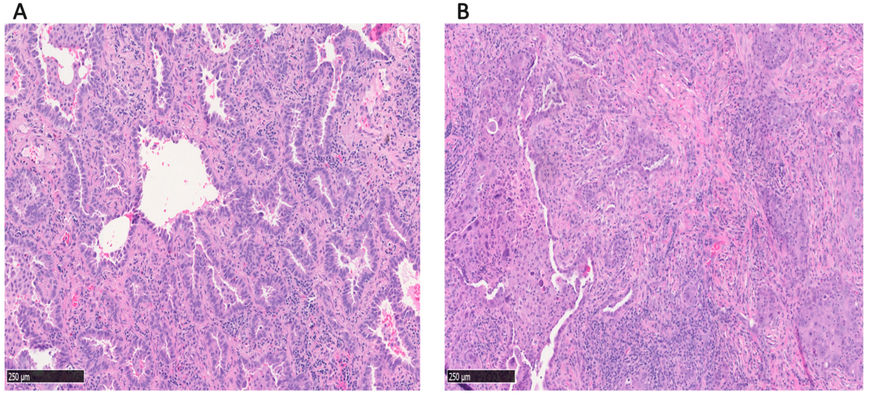
2.3. Histological Evaluation

The H&E slides from each tumor were retrieved and reviewed by thoracic pathologists (PJ, AG, PD, POF, MK). The grading followed the 2021 IASLC system for LUAD [2]. Patients were grouped by the proportion of acinar patterns in their tumors: acinar-predominant (AP), and acinar component (AC; non-acinar predominant LUAD with an acinar component of ≥5%). The following features were recorded: the architectural patterns, lymphovascular invasion (LVI), visceral pleural invasion (VPI), spread through air spaces (STAS), tumor size, and TNM stage (based on the 9th edition AJCC cancer staging) [22]. LUAD classification was based on the sum of six predominant patterns (acinar, papillary, solid, lepidic, micropapillary, and CGPs), totaling 100%.

2.4. Mutational Analysis

For the mutational analysis, DNA/RNA was extracted from either snap-frozen samples (n = 1109 for DNA and 1113 for RNA) or formalin-fixed paraffin-embedded (FFPE) samples (n = 154 for DNA and 150 for RNA). Nucleic acids were extracted and analyzed using the Oncomine™ Precision Assay GX Gene Panel (Thermo Fisher Scientific, Waltham, MA, USA), which includes 50 prevalent driver mutations and fusions in LUAD [23,24,25,26]. Next-generation sequencing (NGS) and alignment were conducted on the Ion Torrent Genexus platform according to the manufacturer’s recommendations. Each sample’s variant call report was reviewed by a pathologist (PJ or PD) for validation. To ensure the presence of tumors and assess tumor cellularity, a fraction of the samples was evaluated to determine the tumor content, with assessments primarily conducted using FFPE slides. All evaluated samples showed a tumor content of more than 10%.

2.5. Statistical Analysis and Visualization

To investigate the correlation of clinicopathological and molecular features with the acinar pattern components, the features were compared in AC and AP patients. The Mann–Whitney U test (Wilcoxon Rank Sum Test) was used to evaluate differences in continuous variables such as age and tumor size, while the Chi-square test or Fisher’s exact test was used for categorical variables to examine associations between the acinar pattern components and other clinicopathological features, as well as genetic variations. The RFS was analyzed using the Kaplan–Meier method, and group differences were evaluated with the log-rank test. To evaluate the prognostic value of the acinar pattern, the Cox proportional hazards regression model was used incorporating clinicopathological features, histological patterns, and molecular characteristics. All p-values were two-tailed, and a threshold of ≤0.05 was considered statistically significant. Statistical analyses were performed using the R statistical language (version 4.2.3, RStudio, Boston, MA, USA). Cox proportional hazards regression models, Kaplan–Meier analysis, and corresponding plots were generated using the R packages survival and survminer [27].

3. Results

3.1. Clinicopathologic Factors

The clinicopathologic characteristics of LUAD patients, both overall and based on acinar components, are presented in Table 1. A total of 1263 patients with LUAD were included in this study, comprising 716 AP and 547 AC cases. Representative images of AP and AC are shown in Figure 1. Of 547 AC patients, the predominant histologic subtype was lepidic in 136 patients, papillary in 50 patients, and ≥20% solid, micropapillary, or CGPs in 361 patients.
Among all, 785 (62.1%) were female and 478 (37.9%) were male, ranging in age from 26 to 88 years (median: 66 years). The distribution of tumor stages was as follows: 923 (73.1%) cases were stage I, 219 (17.3%) were stage II, and 121 (9.6%) were stage III.
Compared to AP, AC cases had a significantly lower proportion of stage I disease (69.5% vs. 75.9%) and a higher proportion of stage III disease (12.2% vs. 7.5%) (p = 0.009). The median tumor size was slightly larger in AC (2.5 cm) than in AP (2.3 cm, p = 0.0009).
Histopathologically, there were significant differences in the tumor grade distribution between AP and AC groups (p < 0.00001). Grade 3 tumors were significantly more common in AC than in AP (66.0% vs. 45.9%).
Tumor localization differed significantly between the groups (p = 0.045). AP tumors were more frequently located in the right upper lobe (41.2% vs. 34.5%), while AC tumors were more common in the right lower lobe (18.8% vs. 14.1%). These findings suggest potential differences in the tumor origin or spread patterns between histologic subtypes.
To further investigate whether the observed differences were specific to the acinar type or could be attributed to the presence of other histologic subtypes, we performed an additional comparative analysis including cases with acinar-predominant, papillary-predominant, or lepidic-predominant and tumors with ≥20% high-grade patterns (solid, micropapillary, or CGPs), together (Supplementary Table S1).
We found that the EGFR-Del19 mutation, which was significantly different between AP and AC, was not different across all histologic patterns, suggesting that these differences may be intrinsic to the acinar type. Conversely, features like LVI, VPI, and STAS were not significantly different between AP and AC but showed differences among the subgroups of all histological patterns, suggesting that these may be driven by the presence of high-grade and not intrinsic to the acinar type.

3.2. Status of Common Driver Mutations

We evaluated the frequencies of the most common driver mutations, including KRAS, EGFR, BRAF, MET, and PIK3CA, by sub-mutations. Less frequent mutations, such as those in ARAF, CTNNB1, ERBB2, MAP2K1, NRAS, GNAS, and FGFR2/3, were grouped together as “Other”. Among the 1263 LUAD cases, 907 cases had a detected driver mutation, while 356 cases were classified as wild-type (WT) with no detected mutations. The most frequently observed driver mutations were KRAS-G12C (20.9%), KRAS-G12V (9.4%), KRAS-G12D (4.9%), EGFR-L858R (4.9%), and EGFR-Del-19 (4.7%).
To investigate the correlation between molecular features and acinar pattern components, we compared the frequency of the most prevalent driver mutations between the two groups of AP and AC. Among the analyzed mutations, only the frequency of the EGFR-Del-19 mutation demonstrated a statistically significant difference between the two groups (p = 0.014) (Figure 2, Table 1).
As EGFR-Del-19 was the only mutation significantly correlated with the acinar histology among all evaluated mutations, we further assessed its relationship with the acinar pattern components through univariable and multivariable logistic regression analysis (Table 2). Since the AC group is heterogeneous and includes other predominant histological patterns, we used a multivariable model to adjust for these histological patterns, along with relevant clinical and pathological variables.
The results indicated that the presence of the EGFR-Del-19 mutation remained significantly associated with the acinar pattern even after an adjustment (Table 2). Specifically, AP was associated with a significantly higher likelihood of the presence of EGFR-Del-19 compared to AC in the multivariable model (OR = 1.951, 95% CI: 1.701–2.205, p = 0.03).
These findings highlight the molecular heterogeneity within LUAD and underscore the importance of considering minor components of the acinar type beyond the predominant classification. Since non-predominant acinar components may influence the mutational landscape, their presence should be assessed alongside other histological patterns to refine prognostic evaluations and potential therapeutic strategies. Incorporating AC provides a more comprehensive understanding, as AC was associated with a significantly lower likelihood of the presence of EGFR-Del-19, even after adjusting for other histological patterns and clinicopathological features.

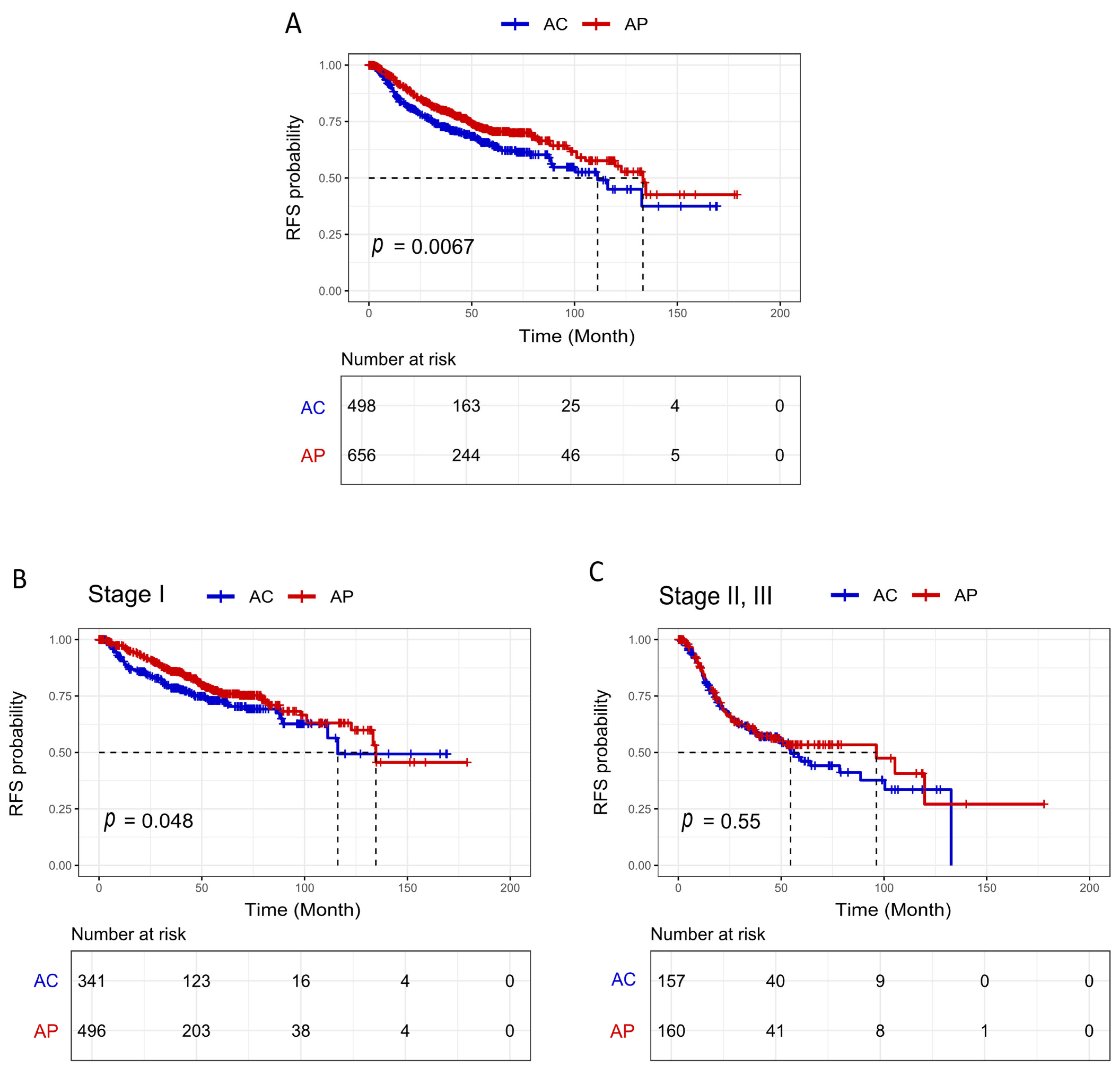
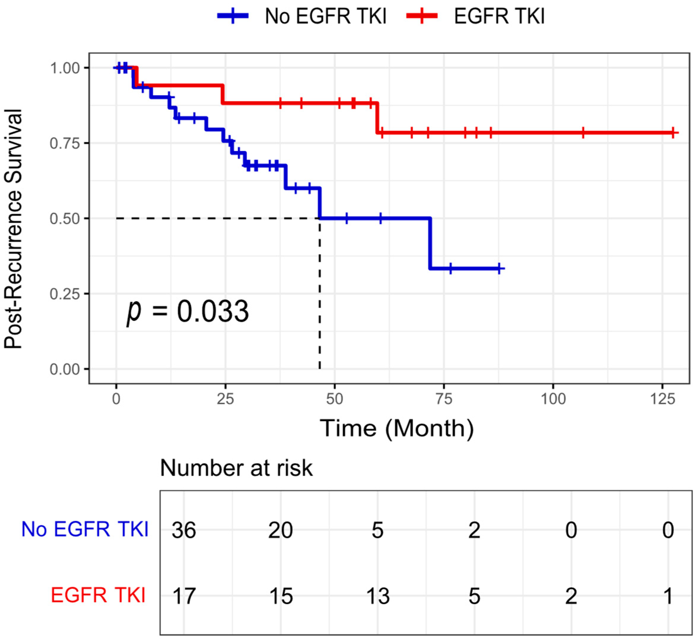
3.3. Survival Analysis

For the survival analysis, our primary endpoint was RFS. In our cohort, 73% of patients were diagnosed at stage I. In early-stage LUAD, RFS is a more relevant measure of tumor aggressiveness and treatment efficacy. RFS provides valuable insights into disease recurrence patterns, which have direct implications for clinical decision-making, including adjuvant therapy strategies. By focusing on RFS, we aim to better assess the prognostic value of ACs in LUAD while minimizing external influences on survival outcomes.
Among all, 1154 (91.3%) had at least one post-surgery visit to calculate the RFS. Survival outcomes were compared between 716 patients with AP and 547 patients with AC. The median follow-up time (95% CI) of these patients was 122.7 months (105-NA), and the recurrence rate was 27% (312 cases). The median RFS time (95% CI) was 133.3 (119.7-NA) and 111.2 (88.6-NA) for AP and AC patients, respectively. Among all, 163 (22.7%) AP and 149 (27.2%) AC patients had recurrent disease. In the unadjusted analysis (log-rank test), AC LUAD patients had a significantly worse RFS (p = 0.006) than AP patients (Figure 3A).
The survival analysis was followed by stratifying cases based on the TNM stage to stage I (Figure 3B) and stages II–III (Figure 3C). Compared to AP patients, AC patients had a significantly worse RFS in the stages I group (p = 0.048, HR: 0.744, 95% CI: 0.674–0.822) (Figure 3C).
We then focused on the univariable and multivariable analysis of RFS using the Cox regression hazards model (Table 3). In the univariate analysis, AC had a worse RFS than AP [HR AC vs. AP: 1.358, 95% CI: 1.188–1.541, p: 0.006]. An older age at diagnosis (p = 0.002), TNM stage (II–III vs. I, p = 2.76 × 10⁻¹⁵), higher tumor grade (3 vs. 1, 2, p = 3.38 × 10⁻⁹), the presence of LVI (p = 2 × 10⁻⁸), VPI (p = 0.0005), STAS (p = 0.003), and the presence of solid-predominant (p = 0.0001), micropapillary-predominant (p = 0.005), and CGP-predominant (0.005) were significantly associated with a worse RFS. In contrast, the presence of lepidic-predominant (p = 4.1 × 10⁻⁶) was associated with an improved RFS.
Of the 547 AC patients, the predominant histologic subtype was lepidic in 120 patients, papillary in 49 patients, and ≥20% solid, micropapillary, or CGPs in 329 patients. We performed a multivariable analysis adjusting for variables that showed a significant association with RFS in the univariable analysis. These included age, TNM stage, tumor grade, LVI, VPI, STAS, and the histological patterns of lepidic, micropapillary, solid, and CGPs. We selected AC as the reference group and compared it with AP.
In the multivariate analysis, acinar (HR AC vs. AP = 1.240, 95% CI: 1.103–1.312, p = 0.04), TNM stage (HR II, III vs. I = 1.830, 95% CI: 1.533–2.118, p = 1.25 × 10⁻⁶), and older age (HR = 1.293, 95% CI: 1.199–1.321, p = 0.014) were the independent predictors of a worse RFS. In addition, the presence of micropapillary-predominant (HR = 1.296, 95% CI: 1.107–1.367, p = 0.043) was significantly associated with a worse RFS. However, solid-predominant (p = 0.951) and CGP-predominant (p = 0.303) did not show a significant impact on the RFS. This finding suggests that micropapillary, a high-grade pattern associated with a worse prognosis, can lead to a poorer RFS when accompanied by a minor acinar component.
We further evaluated the effect of EGFR-TKIs on post-recurrence survival in EGFR-mutated LUAD patients harboring an acinar component of ≥5% (Figure 4). Among 53 patients who experienced recurrence during follow-up, 17 received EGFR-TKI therapy (gefitinib or erlotinib). An objective response was observed in 12 of the 17 patients (1 complete response and 11 partial responses), while the remaining 5 had stable disease at the first evaluation. The median post-recurrence survival was not reached (NA) in the EGFR-TKI group compared to 46.9 months in the non-TKI group. The post-recurrence survival was significantly better in patients who received EGFR-TKIs compared to those who did not (log-rank p = 0.033). These findings support the potential clinical benefit of EGFR-TKI therapy for recurrent EGFR-mutated LUAD with an acinar component and highlight the importance of molecular testing in guiding post-recurrence treatment decisions.
Our findings highlight the heterogeneity of the AC group, showing that prognosis is primarily dictated by the predominant pattern, but the minor components of acinar alongside high-grade patterns can worsen outcomes. While AC alone is not an independent prognostic factor, its presence alongside other predominant patterns contributes to a worse prognosis, reinforcing the importance of comprehensive histopathological evaluations in LUAD.

4. Discussion

Based on the IASLC/ATS/ERS classification system, LUAD resection specimens are classified based on the predominant histologic pattern, following a comprehensive histologic evaluation that involves subtyping in 5% increments of each architecture pattern [3,28]. These patterns include lepidic, papillary, acinar, solid, micropapillary, and CGPs. In 2021, the previous grading system of LUAD was updated by introducing CGPs and a new definition of grade 3 tumors to better prognosticate patient evolution [2]. The acinar pattern is the most common architectural pattern in LUAD and is associated with an intermediate prognosis. Previous studies have examined the prognostic value of acinar-predominant LUAD compared to other histologic subtypes [14,20].
Given the heterogeneity of LUAD, the prognosis is primarily dictated by the predominant histologic pattern. However, the minor acinar components coexist with other patterns in 40–50% of all LUAD cases, raising the question of whether their presence influences outcomes beyond the predominant classification. While prior research has focused on acinar-predominant tumors, little is known about the prognostic significance of AC in non-acinar-predominant LUAD. Since minor histologic components can contribute to tumor behavior, a more comprehensive approach that considers both predominant and accompanying patterns is essential for refining prognostic assessments.
As far as we know, this is the first study to compare AP with non-acinar-predominant LUAD containing an acinar component of at least 5%, focusing on the clinicopathologic characteristics, mutational spectrum, and RFS. This study can offer valuable insights for assessing the relapse risk after surgical resection.
The distribution of gene mutations and rearrangements in LUAD-predominant histologic subtypes is well-documented. Notably, EGFR mutations are significantly more prevalent in AP-LUAD [15,16,17,29,30,31]. In this study, we evaluated the frequency of the most common driver mutations, including KRAS, EGFR, BRAF, MET, and PIK3CA, by sub-mutations. we demonstrated that the frequency of the EGFR-Del 19 mutation is higher in AP-LUAD than in AC. Specifically, AP was associated with a significantly higher likelihood of the presence of EGFR-Del-19 compared to AC in the multivariable model with an adjustment for the other histological patterns and clinicopathological features.
Since non-predominant acinar components may influence the mutational landscape, their presence should be assessed alongside other histological patterns to refine prognostic evaluations and potential targeted therapies. Incorporating the AC provides a more comprehensive understanding, as AC was associated with a significantly lower likelihood of the presence of EGFR-Del-19, even after adjusting for other histological patterns and clinicopathological features.
AP-LUAD has been reported to exhibit specific clinicopathological characteristics [11,12,13,32]. Caso et al. found that among AP-LUAD tumors, VPI was independently associated with an increased risk of recurrence [11]. Kim et al. identified STAS as an independent prognostic biomarker for RFS in an AP-LUAD cohort [14]. In our study, the frequencies of STAS, LVI, and VPI did not differ significantly between AP and AC.
Previous studies have shown that patients with AP had better overall survival (OS) than those with solid, micropapillary, or CGP-predominant patterns [14,19,20] but worse OS than those with lepidic-predominant patterns [33]. Our specific focus was on the comparison between AP tumors and non-acinar-predominant tumors with an acinar component of ≥5%, adjusting for other histological patterns and clinicopathological features.
For the survival analysis, our primary endpoint was RFS rather than OS for several reasons. First, in early-stage LUAD, RFS is a more relevant measure of tumor aggressiveness and treatment efficacy, as OS can be influenced by non-cancer-related factors, such as comorbidities and unrelated causes of death. Second, RFS provides valuable insights into disease recurrence patterns, which have direct implications for clinical decision-making, including adjuvant therapy strategies. Third, RFS events occur earlier than OS events, reducing the impact of confounding factors, such as variations in post-recurrence treatment and prolonged follow-up durations. Notably, in our cohort, 73% of patients were diagnosed at stage I, further supporting the use of RFS as a more appropriate endpoint to evaluate early disease progression and recurrence risk. By focusing on RFS, we aim to better assess the prognostic value of acinar patterns in LUAD while minimizing external influences on survival outcomes.
We found that AP patients had significantly better RFS than AC patients. In our cohort, the AC group had a higher proportion of high-grade tumor patterns, including solid and micropapillary, which are associated with poorer prognosis. To address this imbalance, we performed a multivariable analysis adjusting for the mutational status, clinicopathological variables, and other histopathological patterns, including the high-grade patterns. We selected KRAS, EGFR, BRAF, and MET mutations for inclusion in the multivariable model because they are among the most commonly altered driver mutations in LUAD and are known to significantly influence tumor behavior, prognosis, and response to targeted therapies. Even after the adjustment, AC continued to show a worse RFS than AP. When survival analysis was stratified by stage, the difference in RFS between AC and AP remained significant in stage I and not in stage II, III. Additionally, the presence of micropapillary-predominant was associated with a worse RFS, suggesting that even minor acinar components can amplify the adverse prognostic impact of the micropapillary-predominant type, further worsening RFS.
Our findings highlight the heterogeneity of the AC group, demonstrating that while AC alone is not an independent prognostic factor, prognosis is primarily dictated by the predominant pattern. However, the presence of a minor acinar component alongside high-grade patterns can exacerbate poor outcomes.
Several clinicopathological features, including the TNM stage, lymph node metastasis, tumor size, and histological patterns, were shown to be associated with the response to targeted therapy in LUAD [31,34]. The AP was shown to have an intermediate response to adjuvant therapy, better than solid and micropapillary, but worse than lepidic [2,35]. In our study, we examined the effect of EGFR-TKIs on the post-recurrence survival of patients with EGFR-mutated LUAD harboring an acinar component of ≥5%. The post-recurrence survival was significantly better in patients who received EGFR-TKIs compared to those who did not receive TKIs.
Our results do not suggest that the acinar component alone serves as an independent prognostic factor, given its heterogeneity. Instead, when considered alongside other histological patterns, the presence of a minor acinar component can contribute to a worse prognosis. This reinforces the importance of evaluating minor acinar components within the comprehensive histopathological context to refine prognostic assessments and treatment strategies in LUAD.
Unlike previous studies that primarily focused on acinar-predominant LUAD or compared histologic subtypes without considering coexisting patterns, our methodology offers a more granular and comprehensive approach by isolating the prognostic role of acinar components in non-acinar-predominant tumors. A key strength of our study is the integration of a detailed histological assessment with advanced molecular and clinical analyses. We employed the Oncomine™ Precision Assay GX, a broad and clinically validated next-generation sequencing panel, to evaluate a wide spectrum of prevalent driver mutations in LUAD, including EGFR, KRAS, BRAF, MET, and PIK3CA. Importantly, we analyzed associations at the level of specific sub-mutations, such as EGFR-Del19 and KRAS-G12C, allowing for deeper genotype–phenotype correlations than previous studies. Furthermore, we applied the 9th edition of the TNM staging system, recently proposed in 2024, which enhances the accuracy of the prognostic classification. By adjusting for coexisting high-grade histological patterns and incorporating post-recurrence treatment outcomes, our study provides a refined and clinically actionable stratification framework for LUAD patients that surpasses conventional predominant-pattern-based approaches.
Our study has limitations. The prevalence of EGFR-TKI therapy was low, with only 53 cases qualifying for an evaluation of these modalities. A larger prospective clinical trial would be valuable to validate our findings and further explore the impact of these therapies on the prognosis of acinar-predominant LUAD patients.
Pathology reviews are typically conducted by a single pathologist to minimize variability in interpretation. In this study, five pathologists were involved, which may have increased inter-observer variability, as differences between reviewers are well-documented in the literature. However, in cases of disagreement, the pathologists discussed their findings to reach a consensus, likely reducing the impact of this variability. Lastly, the genotypes and phenotypes of patients may be influenced by ethnicity. In this study, all cases were French-Canadian. Therefore, conducting similar studies with more diverse populations would be valuable to validate our results across different ethnic backgrounds.

5. Conclusions

In conclusion, our study highlights the importance of considering the minor components of the acinar pattern in the prognosis and management of LUAD patients. Although the AP LUAD is used in clinical analysis, its minor components may have an impact on the prognosis alongside other histological patterns. For stage I, AC was even more likely to recur than AP and should be distinguished from AP. Additionally, our results underscore the potential benefit of EGFR-TKI therapy for EGFR-mutated LUAD with acinar components, suggesting the importance of integrating molecular data with histopathological classification for therapeutic decision-making. Future studies should focus on validating these findings using multi-ethnic and prospective cohorts and exploring the biological mechanisms by which minor acinar components interact with high-grade patterns to influence tumor behavior. Further investigation into the predictive role of specific mutations in relation to histologic subtypes could also inform personalized therapeutic strategies.

Supplementary Materials

The following supporting information can be downloaded at: https://www.mdpi.com/article/10.3390/cancers17111825/s1, Table S1: Clinicopathologic and molecular characteristics across LUAD histological patterns.

Author Contributions

Conceptualization, A.G. and P.J.; Methodology, H.A., P.D., F.C.L. and Y.B.; Software, H.A., V.S.A., N.G. and D.K.B.; Validation, H.A. and P.D.; Formal analysis, H.A., M.O. and F.C.L.; Investigation, H.A. and P.O.F.; Data curation, H.A., M.K., V.S.A., N.G., D.K.B., P.D. and Y.B.; Writing—original draft, H.A.; Writing—review & editing, M.K., V.S.A., N.G., D.K.B., A.G., P.D., P.O.F., M.O., F.C.L., Y.B. and P.J.; Visualization, H.A.; Supervision, P.J.; Project administration, Y.B. and P.J.; Funding acquisition, P.J. All authors have read and agreed to the published version of the manuscript.

Funding

This work was supported by the IUCPQ Foundation, owing to a generous donation from Mr. Normand Lord. This work was also supported by the IUCPQ Biobank of the Quebec Respiratory Health research network.

Institutional Review Board Statement

The study was approved by the ethics committee of the IUCPQ-UL, part of the Quebec Respiratory Health Research Network, approval number: MP-10-2022-3752, 22156, approval date: 20 December 2021. The entire research was performed in accordance with the Declaration of Helsinki.

Informed Consent Statement

Informed consent was obtained from all subjects involved in this study. Written informed consent has been obtained from the patient(s) to publish this paper.

Data Availability Statement

The data presented here are available from the corresponding author Philippe Joubert.

Acknowledgments

The authors would like to thank all patients included in this study and the IUCQP-UL Biobank, part of the Quebec Respiratory Health Research Network, for providing the clinical data.

Conflicts of Interest

The authors declare that the research was conducted in the absence of any commercial or financial relationships that could be construed as potential conflicts of interest.

References

  1. Adjei, A.A. Lung Cancer Worldwide. J. Thorac. Oncol. 2019, 14, 956. [Google Scholar] [CrossRef] [PubMed]
  2. Moreira, A.L.; Ocampo, P.S.; Xia, Y.; Zhong, H.; Russell, P.A.; Minami, Y.; Cooper, W.A.; Yoshida, A.; Bubendorf, L.; Papotti, M.; et al. A Grading System for Invasive Pulmonary Adenocarcinoma: A Proposal From the International Association for the Study of Lung Cancer Pathology Committee. J. Thorac. Oncol. 2020, 15, 1599–1610. [Google Scholar] [CrossRef] [PubMed]
  3. Travis, W.D.; Brambilla, E.; Noguchi, M.; Nicholson, A.G.; Geisinger, K.R.; Yatabe, Y.; Beer, D.G.; Powell, C.A.; Riely, G.J.; Van Schil, P.E.; et al. International Association for the Study of Lung Cancer/American Thoracic Society/European Respiratory Society International Multidisciplinary Classification of Lung Adenocarcinoma. J. Thorac. Oncol. 2011, 6, 244–285. [Google Scholar] [CrossRef] [PubMed]
  4. Zhang, Y.; Wang, R.; Cai, D.; Li, Y.; Pan, Y.; Hu, H.; Wang, L.; Li, H.; Ye, T.; Luo, X.; et al. A comprehensive investigation of molecular features and prognosis of lung adenocarcinoma with micropapillary component. J. Thorac. Oncol. 2014, 9, 1772–1778. [Google Scholar] [CrossRef]
  5. Deng, C.; Zheng, Q.; Zhang, Y.; Jin, Y.; Shen, X.; Nie, X.; Fu, F.; Ma, X.; Ma, Z.; Wen, Z.; et al. Validation of the Novel International Association for the Study of Lung Cancer Grading System for Invasive Pulmonary Adenocarcinoma and Association with Common Driver Mutations. J. Thorac. Oncol. 2021, 16, 1684–1693. [Google Scholar] [CrossRef]
  6. Bossé, Y.; Gagné, A.; Althakfi, W.A.; Orain, M.; Couture, C.M.; Trahan, S.; Pagé, S.; Joubert, D.; Fiset, P.O.; Desmeules, P.M.; et al. A Simplified Version of the IASLC Grading System for Invasive Pulmonary Adenocarcinomas with Improved Prognosis Discrimination. Am. J. Surg. Pathol. 2023, 47, 686–693. [Google Scholar] [CrossRef]
  7. She, Y.; Zhong, Y.; Hou, L.; Zhao, S.; Zhang, L.; Xie, D.; Zhu, Y.; Wu, C.; Chen, C. Application of the Novel Grading System of Invasive Pulmonary Adenocarcinoma in a Real Diagnostic Scenario: A Brief Report of 9353 Cases. JTO Clin. Res. Rep. 2023, 4, 100465. [Google Scholar] [CrossRef]
  8. Borczuk, A.C. Updates in grading and invasion assessment in lung adenocarcinoma. Mod. Pathol. 2022, 35, 28–35. [Google Scholar] [CrossRef]
  9. Li, P.; Liu, L.; Wang, D.; Yang, R.; Xuan, Y.; Han, Y.; Wang, J.; Guo, L.; Zhang, L.; Zhang, S.; et al. Genomic and clinicopathological features of lung adenocarcinomas with micropapillary component. Front. Oncol. 2022, 12, 989349. [Google Scholar] [CrossRef]
  10. Bai, J.; Deng, C.; Zheng, Q.; Li, D.; Fu, F.; Li, Y.; Zhang, Y.; Chen, H. Comprehensive analysis of mutational profile and prognostic significance of complex glandular pattern in lung adenocarcinoma. Transl. Lung Cancer Res. 2022, 11, 1337–1347. [Google Scholar] [CrossRef]
  11. Lin, G.; Li, H.; Kuang, J.; Tang, K.; Guo, Y.; Han, A.; Xie, C. Acinar-Predominant Pattern Correlates with Poorer Prognosis in Invasive Mucinous Adenocarcinoma of the Lung. Am. J. Clin. Pathol. 2018, 149, 373–378. [Google Scholar] [CrossRef] [PubMed]
  12. Tsubokawa, N.; Mimae, T.; Miyata, Y.; Sasada, S.; Yoshiya, T.; Kushitani, K.; Takeshima, Y.; Murakami, S.; Yokose, T.; Ito, H.; et al. Prognostic significance of vascular invasion in intermediate-grade subtype of lung adenocarcinoma. Ultrasound Med. Biol. 2016, 46, 1015–1021. [Google Scholar] [CrossRef] [PubMed]
  13. Zuo, Z.-C.; Wang, L.-D.; Peng, K.; Yang, J.; Li, X.; Zhong, Z.; Zhang, H.-M.; Ouyang, X.; Xue, Q. Development and Validation of a Nomogram for Predicting the 1-, 3-, and 5-year Survival in Patients with Acinar-predominant Lung Adenocarcinoma. Curr. Med. Sci. 2022, 42, 1178–1185. [Google Scholar] [CrossRef] [PubMed]
  14. Kim, M.; Chung, Y.S.; A Kim, K.; Shim, H.S. Prognostic factors of acinar- or papillary-predominant adenocarcinoma of the lung. Lung Cancer 2019, 137, 129–135. [Google Scholar] [CrossRef]
  15. Dong, Y.-J.; Cai, Y.-R.; Zhou, L.-J.; Su, D.; Mu, J.; Chen, X.-J.; Zhang, L. Association between the histological subtype of lung adenocarcinoma, EGFR/KRAS mutation status and the ALK rearrangement according to the novel IASLC/ATS/ERS classification. Oncol. Lett. 2016, 11, 2552–2558. [Google Scholar] [CrossRef]
  16. Kadota, K.; Sima, C.S.; Arcila, M.E.; Hedvat, C.; Kris, M.G.; Jones, D.R.; Adusumilli, P.S.M.; Travis, W.D. KRAS Mutation Is a Significant Prognostic Factor in Early-stage Lung Adenocarcinoma. Am. J. Surg. Pathol. 2016, 40, 1579–1590. [Google Scholar] [CrossRef]
  17. Russell, P.A.; Barnett, S.A.; Walkiewicz, M.; Wainer, Z.; Conron, M.; Wright, G.M.; Gooi, J.; Knight, S.; Wynne, R.; Liew, D.; et al. Correlation of Mutation Status and Survival with Predominant Histologic Subtype According to the New IASLC/ATS/ERS Lung Adenocarcinoma Classification in Stage III (N2) Patients. J. Thorac. Oncol. 2013, 8, 461–468. [Google Scholar] [CrossRef]
  18. Liu, W.; Zhang, Q.; Zhang, T.; Li, L.; Xu, C. Minor histological components predict the recurrence of patients with resected stage I acinar- or papillary-predominant lung adenocarcinoma. Front. Oncol. 2022, 12, 1090544. [Google Scholar] [CrossRef]
  19. Tang, E.R.; Schreiner, A.M.; Pua, B.B. Advances in lung adenocarcinoma classification: A summary of the new international multidisciplinary classification system (IASLC/ATS/ERS). J. Thorac. Dis. 2014, 6, S489–S501. [Google Scholar] [CrossRef]
  20. Lu, D.; Yang, J.; Liu, X.; Feng, S.; Dong, X.; Shi, X.; Zhai, J.; Mai, S.; Jiang, J.; Wang, Z.; et al. Clinicopathological features, survival outcomes, and appropriate surgical approaches for stage I acinar and papillary predominant lung adenocarcinoma. Cancer Med. 2020, 9, 3455–3462. [Google Scholar] [CrossRef]
  21. Rokutan-Kurata, M.; Yoshizawa, A.; Ueno, K.; Nakajima, N.; Terada, K.; Hamaji, M.; Sonobe, M.; Menju, T.; Date, H.; Morita, S.; et al. Validation Study of the International Association for the Study of Lung Cancer Histologic Grading System of Invasive Lung Adenocarcinoma. J. Thorac. Oncol. 2021, 16, 1753–1758. [Google Scholar] [CrossRef] [PubMed]
  22. Detterbeck, F.C.; Woodard, G.A.; Bader, A.S.; Dacic, S.; Grant, M.J.; Park, H.S.; Tanoue, L.T. The Proposed Ninth Edition TNM Classification of Lung Cancer. Chest 2024, 166, 882–895. [Google Scholar] [CrossRef] [PubMed]
  23. Melosky, B.; Kambartel, K.; Häntschel, M.; Bennetts, M.; Nickens, D.J.; Brinkmann, J.; Kayser, A.; Moran, M.; Cappuzzo, F. Worldwide Prevalence of Epidermal Growth Factor Receptor Mutations in Non-Small Cell Lung Cancer: A Meta-Analysis. Mol. Diagn. Ther. 2021, 26, 7–18. [Google Scholar] [CrossRef] [PubMed]
  24. Zhang, L.; Zheng, L.; Yang, Q.; Sun, J. The Evolution of BRAF Activation in Non-Small-Cell Lung Cancer. Front. Oncol. 2022, 12, 882940. [Google Scholar] [CrossRef]
  25. Fujino, T.; Suda, K.; Mitsudomi, T. Lung Cancer with MET exon 14 Skipping Mutation: Genetic Feature, Current Treatments, and Future Challenges. Lung Cancer Targets Ther. 2021, 12, 35–50. [Google Scholar] [CrossRef]
  26. Ghimessy, A.; Radeczky, P.; Laszlo, V.; Hegedus, B.; Renyi-Vamos, F.; Fillinger, J.; Klepetko, W.; Lang, C.; Dome, B.; Megyesfalvi, Z. Current therapy of KRAS-mutant lung cancer. Cancer Metastasis Rev. 2020, 39, 1159–1177. [Google Scholar] [CrossRef]
  27. Tibshirani, R. The lasso method for variable selection in the Cox model. Stat. Med. 1997, 16, 385–395. [Google Scholar] [CrossRef]
  28. Zugazagoitia, J.; Enguita, A.B.; Nuñez, J.A.; Iglesias, L.; Ponce, S. The new IASLC/ATS/ERS lung adenocarcinoma classification from a clinical perspective: Current concepts and future prospects. J. Thorac. Dis. 2014, 6, S526–S536. [Google Scholar] [CrossRef]
  29. Tsuta, K.; Kawago, M.; Inoue, E.; Yoshida, A.; Takahashi, F.; Sakurai, H.; Watanabe, S.-I.; Takeuchi, M.; Furuta, K.; Asamura, H.; et al. The utility of the proposed IASLC/ATS/ERS lung adenocarcinoma subtypes for disease prognosis and correlation of driver gene alterations. Lung Cancer 2013, 81, 371–376. [Google Scholar] [CrossRef]
  30. de Melo, A.C.; de Sá, V.K.; Sternberg, C.; Olivieri, E.R.; Werneck da Cunha, I.; Fabro, A.T.; Carraro, D.M.; de Barros e Silva, M.J.; Inada, H.K.P.; de Mello, E.S.; et al. Mutational Profile and New IASLC/ATS/ERS Classification Provide Additional Prognostic Information about Lung Adenocarcinoma: A Study of 125 Patients from Brazil. Oncology 2015, 89, 175–186. [Google Scholar] [CrossRef]
  31. Arrieta, O.; Cardona, A.F.; Corrales, L.; Campos-Parra, A.D.; Sánchez-Reyes, R.; Amieva-Rivera, E.; Rodríguez, J.; Vargas, C.; Carranza, H.; Otero, J.; et al. The impact of common and rare EGFR mutations in response to EGFR tyrosine kinase inhibitors and platinum-based chemotherapy in patients with non-small cell lung cancer. Lung Cancer 2014, 87, 169–175. [Google Scholar] [CrossRef] [PubMed]
  32. Caso, R.; Sanchez-Vega, F.; Tan, K.S.; Mastrogiacomo, B.; Zhou, J.; Jones, G.D.; Nguyen, B.; Schultz, N.; Connolly, J.G.; Brandt, W.S.; et al. The Underlying Tumor Genomics of Predominant Histologic Subtypes in Lung Adenocarcinoma. J. Thorac. Oncol. 2020, 15, 1844–1856. [Google Scholar] [CrossRef] [PubMed]
  33. Renaud, S.; Seitlinger, J.; Falcoz, P.-E.; Schaeffer, M.; Voegeli, A.-C.; Legrain, M.; Beau-Faller, M.; Massard, G. Specific KRAS amino acid substitutions and EGFR mutations predict site-specific recurrence and metastasis following non-small-cell lung cancer surgery. Br. J. Cancer 2016, 115, 346–353. [Google Scholar] [CrossRef] [PubMed]
  34. Lee, C.-Y.; Lee, S.-W.; Hsu, Y.-C. Drug Resistance in Late-Stage Epidermal Growth Factor Receptor (EGFR)-Mutant Non-Small Cell Lung Cancer Patients After First-Line Treatment with Tyrosine Kinase Inhibitors. Int. J. Mol. Sci. 2025, 26, 2042. [Google Scholar] [CrossRef]
  35. Nicholson, A.G.; Moreira, A.L.; Mino-Kenudson, M.; Popat, S. Grading in Lung Adenocarcinoma: Another New Normal. J. Thorac. Oncol. 2021, 16, 1601–1604. [Google Scholar] [CrossRef]
Figure 1. Representative images of (A) acinar-predominant and (B) acinar component; non-acinar-predominant LUAD with an acinar component of ≥5%.
Cancers 17 01825 g001
Figure 2. Summary of the most common driver mutations in LUAD patients in two groups of AP and AC. AP: acinar-predominant, AC: acinar component (non-acinar predominant LUAD with an acinar component of ≥5%), WT: wild-type, other refers to the less frequent mutations, such as those in ARAF, CTNNB1, ERBB2, MAP2K1, NRAS, GNAS, and FGFR2/3. Note: The dotted squares represent the statistically significant difference in the frequency of EGFR-Del 19 mutations between the two groups of AP and AC (p = 0.014).
Cancers 17 01825 g002
Figure 3. (A) RFS probability of all patients, and (B) RFS probability of stage I, and (C) Stage II, III. AP: acinar-predominant, AC: acinar component (non-acinar predominant LUAD with an acinar component of ≥5%), RFS: recurrence-free survival. Note: “p” represents the log-rank p-value, which was calculated through an unadjusted analysis (log-rank test). The dotted lines indicate the time (in months) at which the RFS probability reaches 0.5 for each group, representing the median RFS.
Cancers 17 01825 g003
Figure 4. Post-recurrence survival of patients with EGFR-mutated lung adenocarcinoma harboring an acinar component of ≥5% according to the administration of EGFR tyrosine kinase inhibitors (TKI). Note: “p” represents the log-rank p-value, which was calculated via an unadjusted analysis (log-rank test). EGFR-TKI refers to cases that received EGFR-TKIs (gefitinib or erlotinib), while No EGFR-TKI refers to cases that did not receive these treatments. The dotted lines indicate the time (in months) at which the post-recurrence survival probability reaches 0.5 for each group, representing the median post-recurrence survival.
Cancers 17 01825 g004
Table 1. Characteristics of LUAD patients, overall and based on acinar components.
CharacteristicsOverall (N = 1263)AP (N = 716)AC (N = 547)p-Value (AP vs. AC)
Age
Median6666660.34
Min–Max26–8837–8826–84
Sex
Male478 (37.9%)255 (35.6%)225 (41.1%)0.045
Female785 (62.1%)461 (64.4%)322 (58.9%)
Smoking status
Ever1194 (94.5%)675 (94.3%)519 (94.9%)0.637
Never69 (5.5%)41 (5.7%)28 (5.1%)
N status
N0497 (39.3%)270 (37.7%)227 (41.5%)0.171
N1/2766 (60.7%)446 (62.3%)320 (58.5%)
Stage 0.009
I923 (73.1%)543 (75.9%)380 (69.5%)
II219 (17.3%)119 (16.6%)100 (18.3%)
III121 (9.6%)54 (7.5%)67 (12.2%)
Tumor size (cm)
Median2.32.32.50.0009
Range1–151–151–12.5
Grade <0.00001
1136 (10.8%)0 (0.0%)136 (24.9%)
2437 (34.6%)387 (54.1%)50 (9.1%)
3690 (54.6%)329 (45.9%)361 (66.0%)
Type of surgery 0.961
Lobectomy954 (75.6%)539 (75.3%)415 (75.9%)
Segmentectomy132 (10.5%)75 (10.5%)57 (10.4%)
Other177 (14.0%)102 (14.3%)75 (13.7%)
Tumor localization 0.045
Left Upper Lobe323 (25.6%)186 (26.0%)137 (25.1%)
Left Lower Lobe153 (12.1%)84 (11.7%)69 (12.6%)
Right Upper Lobe486 (38.5%)295 (41.2%)189 (34.5%)
Right Lower Lobe202 (16.0%)101 (14.1%)103 (18.8%)
Other99 (7.8%)50 (7.0%)49 (9.0%)
STAS
Yes537 (42.5%)312 (43.6%)225 (41.1%)0.384
No726 (57.5%)404 (56.4%)322 (58.9%)
LVI
Yes592 (46.9%)329 (46.0%)263 (48.1%)0.452
No571 (45.2%)387 (54.0%)284 (51.9%)
VPI
Yes343 (27.2%)194 (27.1%)149 (27.2%)0.954
No920 (72.8%)522 (72.9%)398 (72.8%)
Mutational status
KRAS-G12C264 (20.9%)149 (20.8%)115 (21.0%)0.981
KRAS-G12V119 (9.4%)67 (9.4%)52 (9.5%)1.000
KRAS-G12D62 (4.9%)33 (4.6%)29 (5.3%)0.664
KRAS-G12A39 (3.1%)22 (3.1%)17 (3.1%)1.000
KRAS-G12X24 (1.9%)12 (1.7%)12 (2.2%)0.645
KRAS-G13X34 (2.7%)18 (2.5%)16 (2.9%)0.785
KRAS-Q61H33 (2.6%)19 (2.7%)14 (2.6%)1.000
KRAS-Q61L11 (0.9%)5 (0.7%)6 (1.1%)0.652
EGFR-Del-1959 (4.7%)43 (6.0%)16 (2.9%)0.014
EGFR-L858R62 (4.9%)39 (5.4%)23 (4.2%)0.378
EGFR-Ins 2013 (1.0%)8 (1.1%)5 (0.9%)0.941
EGFR-Other35 (2.8%)19 (2.7%)16 (2.9%)0.905
MET-Exon 1443 (3.4%)23 (3.2%)20 (3.7%)0.783
BRAF-V600E13 (1.0%)8 (1.1%)5 (0.9%)0.941
BRAF-Other41 (3.2%)25 (3.5%)16 (2.9%)0.687
PIK3CA17 (1.3%)7 (1.0%)10 (1.8%)0.292
Other 38 (3.0%)24 (3.4%)14 (2.6%)0.515
WT356 (28.2%)195 (27.2%)161 (29.4%)0.425
AP: acinar-predominant, AC: acinar component (non-acinar predominant LUAD with an acinar component of ≥5%), N status: nodal status, STAS: tumor spread through air spaces, VPI: visceral pleural invasion, LVI: lymphovascular invasion, WT: wild-type. Note: Bold emphasis is used to indicate statistically significant comparisons. Italic emphasis is used to describe “age” and “tumor size” as a continuous characteristic. Bold emphasis is used to indicate statistically significant p-values. The chi-squared test or Fisher’s exact test was used to evaluate the categorical variables, and The Mann–Whitney U test (Wilcoxon Rank Sum Test) was performed to evaluate the continuous characteristics.
Table 2. Logistic regression analyses of the EGFR-Del-19 mutation.
VariableUnivariableMultivariable
p-ValueOR (95% CI)p-ValueOR (95% CI)
Acinar: AP (vs. AC)0.0111.187 (1.101–1.219)0.0301.951 (1.701–2.205)
TNM stage: II, III (vs. I)0.7910.922 (0.490–1.641)
Age0.8211.003 (0.971–1.038)
Sex: female (vs. Male)0.1461.538 (0.877–2.795)
Smoking: never (vs. ever)1.85 × 10−97.260 (6.107–8.004)1.39 × 10−76.22 (5.066–7.060)
Grade: 3 (vs. 1, 2)0.0070.477 (0.324–0.551)0.7730.905 (0.457–1.762)
LVI: presence (vs. absence)0.3300.767 (0.446–1.229)
VPI: presence (vs. absence)0.2310.673 (0.337–1.243)
STAS: presence (vs. absence)0.0580.579 (0.331–1.004)
Lepidic-predominant: Yes (vs. No)0.0022.198 (1.877–3.022)0.0552.158 (1.019–5.021)
Papillary-predominant: Yes (vs. No)0.6650.875 (0.695–0.978)
Micropapillary-predominant: Yes (vs. No)0.9390.979 (0.576–1.688)
Solid-predominant: Yes (vs. No)0.0190.520 (0.396–0.692)0.5300.814 (0.424–1.537)
CGP-predominant: Yes (vs. No)0.7850.910 (0.555–1.492)
AP: acinar-predominant, AC: acinar component (non-acinar predominant LUAD with an acinar component of ≥5%), CGPs: complex glandular patterns (cribriform and fused gland), STAS: tumor spread through air spaces, VPI: visceral pleural invasion, LVI: lymphovascular invasion, OR: odds ratio, CI: confidence interval. Note: Bold emphasis is used to indicate statistically significant p-values.
Table 3. Univariable and multivariable analysis of RFS using Cox regression hazards model.
VariableUnivariableMultivariable
HR (95% CI)p-ValueHR (95% CI)p-Value
Acinar: AC (vs. AP)1.358 (1.188–1.541)0.0061.240 (1.103–1.312)0.04
TNM stage: II, III (vs. I)2.481 (1.980–3.109)2.76 × 10−151.830 (1.533–2.118)1.25 × 10⁻⁶
Age1.379 (1.265–0.422)0.0021.293 (1.199–1.321)0.014
Sex: Female (vs. Male)0.926 (0.738–1.162)0.506
Smoking status: Never (vs. Ever)0.745 (0.427–1.299)0.300
Grade: 3 (vs. 1/2)2.049 (1.816–2.401)3.38 × 10−91.281 (0.981–1.445)0.114
LVI: Present (vs. Absent)1.935 (1.737–2.007)2 × 10−81.270 (0.974–1.356)0.076
VPI: Present (vs. Absent)1.500 (1.391–1.689)0.00051.130 (0.985–1.343)0.324
STAS: Present (vs. Absent)1.399 (1.279–1.549)0.0031.119 (0.082–1.220)0.352
Lepidic-predominant: Yes (vs. No)0.592 (0.473–0.660)4.1 × 10−60.859 (0.769–1.003)0.234
Papillary-predominant: Yes (vs. No)0.927 (0.721–1.191)0.555
Micropapillary-predominant: Yes (vs. No)1.383 (1.230–1.540)0.0051.296 (1.107–1.367)0.043
Solid-predominant: Yes (vs. No)1.551 (1.335–1.646)0.00011.008 (0.876–1.127)0.951
CGP-predominant: Yes (vs. No)1.417 (1.297–1.513)0.0051.146 (0.983–1.289)0.303
KRAS: Mutant (vs. WT)1.151 (0.921–1.438)0.214
EGFR: Mutant (vs. WT)0.997 (0.715–1.391)0.989
BRAF: Mutant (vs. WT)1.101 (0.655–1.852)0.716
MET: Mutant (vs. WT)0.615 (0.305–1.243)0.176
AP: acinar-predominant, AC: acinar component (non-acinar predominant LUAD with an acinar component of ≥5%), CI: confidence interval; HR: hazard ratio, WT: wild-type, LVI: lymphovascular invasion, VPI: visceral pleural invasion, STAS: spread through air space, CGPs: complex glandular patterns (cribriform and fused gland). Note: Bold emphasis is used to indicate statistically significant p-values.
Disclaimer/Publisher’s Note: The statements, opinions and data contained in all publications are solely those of the individual author(s) and contributor(s) and not of MDPI and/or the editor(s). MDPI and/or the editor(s) disclaim responsibility for any injury to people or property resulting from any ideas, methods, instructions or products referred to in the content.

