Simple Summary
Melanoma is the most lethal form of skin cancer and has seen a rising incidence globally over the last 10 years. A majority of melanomas arise from the skin as a result of exposure to ultraviolet radiation. However, even within this group of “cutaneous melanomas”, there is considerable clinical and molecular heterogeneity. In this study, we sought to understand how different genetic “drivers” in a group of 254 patients with cutaneous melanomas influenced overall survival and response to immunotherapy. We found that NRAS mutation correlated with decreased survival and that tumor mutational burden (an estimate of the amount of genetic change within each tumor) correlated with improved responses to immune checkpoint inhibition. We also found that almost all cutaneous melanomas have a driver mutation in the MAPK pathway, underscoring the key role of MAPK-targeted therapies in melanoma drug development.
Abstract
Background: Cutaneous melanoma (CM) can be molecularly classified into four groups: BRAF mutant, NRAS mutant, NF1 mutant and triple wild-type (TWT) tumors lacking any of these three alterations. In the era of immune checkpoint inhibition (ICI) and targeted molecular therapy, the clinical significance of these groups remains unclear. Here, we integrate targeted DNA sequencing with comprehensive clinical follow-up in CM patients. Methods: This was a retrospective cohort study that assessed clinical and molecular features from patients with localized or metastatic CM who underwent targeted next-generation sequencing as part of routine clinical care. A total of 254 patients with CM who had a CLIA-certified targeted sequencing assay performed on their tumor tissue were included. Results: Of the 254 patients with cutaneous melanoma, 77 were BRAF mutant (30.3%), 77 were NRAS mutant (30.3%), 47 were NF1 mutant (18.5%), 33 were TWT (13.0%) and the remaining 20 (7.9%) carried mutations in multiple driver genes (BRAF/NRAS/NF1 co-mutated). The majority of this co-mutation group carried mutations in NF1 (n = 19 or 90%) with co-occurring mutations in BRAF or NRAS, often with a weaker oncogenic variant. Consistently, NF1 mutant tumors harbored numerous significantly co-altered genes compared to BRAF or NRAS mutant tumors. The majority of TWT tumors (n = 29, 87.9%) harbor a pathogenic mutation within a known Ras/MAPK signaling pathway component. Of the 154 cases with available TMB data, the median TMB was 20 (range 0.7–266 mutations/Mb). A total of 14 cases (9.1%) were classified as having a low TMB (≤5 mutations/Mb), 64 of 154 (41.6%) had an intermediate TMB (>5 and ≤20 mutations/Mb), 40 of 154 (26.0%) had a high TMB (>20 and ≤50 mutations/Mb) and 36 of 154 (23.4%) were classified as having a very high TMB (>50 mutations/Mb). NRAS mutant melanoma demonstrated significantly decreased overall survival on multivariable analysis (HR for death 2.95, 95% CI 1.13–7.69, p = 0.027, log-rank test) compared with other TCGA molecular subgroups. Of the 116 patients in our cohort with available treatment data, 36 received a combination of dual ICI with anti-CTLA4 and anti-PD1 inhibition as first-line therapy. Elevated TMB was associated with significantly longer progression-free survival following dual-agent ICI (HR 0.26, 95% CI 0.07–0.90, p = 0.033, log-rank test). Conclusions: NRAS mutation in CMs correlated with significantly worse overall survival. Elevated TMB was associated with increased progression-free survival for patients treated with a combination of dual ICI, supporting the potential utility of TMB as a predictive biomarker for ICI response in melanoma.
Keywords:
melanoma; MAPK pathway; BRAF; NRAS; NF1; triple wild type; immune checkpoint inhibitors; biomarkers 1. Introduction
Cutaneous melanoma (CM) is associated with Ras mutations that activate downstream mitogen-activated protein kinase (MAPK) signaling [1,2,3,4]. CM can accordingly be molecularly classified based on the MAPK pathway molecular driver, which includes BRAF mutations, NRAS mutations and NF1 deficiency, accounting for ~80% of tumors [2,4]. BRAF, NRAS and NF1 are generally thought to be mutually exclusive oncogenic drivers, although co-occurring driver mutations can be seen, particularly with NF1 loss [4]. BRAF and NRAS mutant melanomas histopathologically correlate with low cumulative sun damage (low-CSD) melanomas, while NF1 mutant melanomas are classified as high-CSD [5]. The remaining 20% of melanomas are deemed “triple wild-type” (TWT) and are clinically comprised less common melanoma subtypes such as acral, mucosal and uveal melanomas occurring in sun-shielded locations (classified as non-CSD melanomas) and often driven by alternate pathways of melanomagenesis.
While driver group is associated with distinct demographic and pathologic features [4,6,7,8], the relationship between molecular driver, clinical outcomes and ICI response remains unclear. Prior large-scale genomic studies, including the TCGA landscape study of 333 primary and metastatic melanomas, showed no significant correlations between genomic classification and clinical outcome [4]. However, this was published prior to widespread ICI use, only one year after the approval of anti-PD1 agents in metastatic melanoma [9,10,11,12].
Therapeutic advances in melanoma have resulted in the FDA approval of immune checkpoint inhibitors and several generations of BRAF-targeted therapies for patients with BRAF V600E mutant tumors. Accordingly, BRAF mutant melanomas exhibit improved overall survival compared with other cutaneous melanomas [13]. BRAF mutation was independently associated with improved recurrent-free survival (RFS) in patients with stage three melanoma who were treated with adjuvant pembrolizumab [14]. In addition, patients with BRAF mutations were shown to have superior outcomes when treated with first-line ICI compared with BRAF and MEK inhibition, and this has now become standard practice for a majority of patients [15]. Improved survival outcomes in BRAF mutant melanomas may be related to disease biology in addition to the availability of multiple therapeutic avenues.
Pre-clinical work and retrospective genomic studies have implicated NRAS mutation as an overall poor prognostic factor, with NRAS mutations associated with increased Breslow thickness [16,17]. However, data correlating clinical outcomes and responses to ICI for NRAS mutant melanoma are mixed [16,18,19,20]. The phase III IMspire170 trial compared cobimetinib + atezolizumab to pembrolizumab in patients with BRAF wild-type melanoma and found no differences in PFS or ORR based on NRAS, NF1 or TWT molecular drivers in either of the two study arms [21]. In a phase II trial evaluating ipilimumab vs. ipilimumab/nivolumab, NRAS mutations were noted to be enriched in patients who experienced clinical benefit [18], suggesting that molecular driver disease biology may affect ICI response in this patient population. With regard to other biomarkers of ICI response, the only robustly associated tumor-specific molecular feature is tumor mutational burden (TMB) [22]. A high TMB correlates with improved ICI response and overall survival across a range of cancer types, including melanoma [23]. Given that almost all cutaneous melanomas have an intermediate or high TMB, it remains unclear whether there exists a TMB threshold above which the predictive significance of this biomarker becomes less robust.
Taken together, there remains a critical need to determine the precise relationship between molecular–genetic tumor alterations and clinical outcomes with contemporaneous systemic therapy regimens [24]. Here, we retrospectively identified a real-world cohort of 254 patients with CM undergoing targeted DNA sequencing of recurrently mutated cancer to evaluate whether any tumor-specific genomic features, particularly TCGA driver and tumor mutational burden, correlated with clinical outcomes in the real-world setting. We additionally assessed alternate mutations in TWT sun-exposed cutaneous melanomas to better understand this under-studied and poorly understood cutaneous melanoma phenotype. We found that NRAS mutation was associated with poorer overall survival (OS), and TMB was predictive of dual ICI response, suggesting that tumor genetics may help serve as an informative biomarker predictive of ICI response and clinical outcomes for cutaneous melanoma.
2. Methods
2.1. Cohort Selection/Identification and Targeted DNA Sequencing Analysis
A total of 330 patients with melanoma who underwent in-house-targeted next-generation DNA sequencing were retrospectively identified using the UCSF tumor registry, from which medical records, baseline demographics and pathologic and clinical outcome data were extracted. Uveal, acral, mucosal, primary CNS/meningeal and pediatric melanomas were excluded. Melanomas of unknown primary (n = 68) sites were assumed to be predominantly cutaneous and were included, leading to a total of 254 cases included for subsequent analysis. All cases were re-reviewed by a board-certified pathologist for inclusion in the present cohort. DNA sequencing was performed at the UCSF Clinical Cancer Genomics Laboratory (CCGL) using the UCSF500 (https://genomics.ucsf.edu/UCSF500, accessed on 1 May 2022) CLIA-certified and targeted DNA next-generation sequencing assay obtained as part of routine clinical care. Briefly, this assay uses a custom bait library (Roche Nimblegen) to cover the genomic sequence of 529 cancer-related genes and select introns of 47 genes. The high throughput sequencing of captured libraries was performed using Illumina NovaSeq6s000, aligned to the human reference genome, and variants were called using an internal pipeline and then filtered before undergoing manual review to assign functions based on known pathogenic alterations and predicted effects on proteins by a team of board-certified pathologists and the UCSF CCGL team. Microsatellite instability (MSI) was quantified from targeted sequencing data using MSIsensor [25]. A cutoff of 30% or greater was used to classify tumors as “MSI-high” per our UCSF500 clinical testing protocol. Given that the highest percentage of microsatellite instability in the cohort was 4.7%, none of the cases were deemed MSI-high.
2.2. Outcomes
The primary outcome assessed in this retrospective cohort was overall survival from the time of initial melanoma diagnosis. Secondary outcomes included progression-free survival evaluated in the context of systemic therapy for metastatic disease and recurrence-free survival for patients with localized disease treated with surgical intervention +/− adjuvant therapy. For the purposes of this study, progression-free survival was defined as the time from initiation of first-line systemic therapy to disease progression or death from any cause. Recurrence-free survival was defined as the time from the date of primary resection for localized disease to the time of recurrent melanoma or death from any cause. Patients who developed recurrent disease and then subsequently received systemic therapy were included in both the recurrence-free survival and progression-free survival analysis. Detailed information regarding the systemic therapy regimen and timing for metastatic disease is available for 116 patients (Table 1). The majority of these patients received immunotherapy as first-line systemic therapy (n = 108, 93.1%), with 8 patients receiving first-line BRAF/MEK inhibition. Outcomes were assessed separately for patients who received first-line anti-PD1 (n = 65) and for patients who received a first-line combination of dual ICI (anti-CTLA4 + anti-PD1, n = 36), as well as for all patients who received any form of immunotherapy in the first-line metastatic setting (including rare cases of anti-CLTA4 monotherapy, n = 7).

Table 1.
Baseline clinical characteristics of patients with metastatic melanoma undergoing target DNA sequencing (n = 254 total patients).
2.3. Statistical Analysis
An evaluation of categorical variables such as sex, ECOG status, stage, TMB group and TCGA driver gene was performed using either the chi-squared or Fisher’s exact test. Fisher’s exact test was used to assess proportions within two categorical variables. A chi-squared test was used to assess proportions within three or more categorical variables. An unpaired t-test was used forcomparison of continuous variables such as age, TMB and MSI (%). All hypothesis tests were 2-sided and considered significant at a value < 0.05. Univariable Cox Proportional Hazards (CPHs) were performed using STATA to evaluate overall survival, progression-free survival, recurrence-free survival and the following variables: age, sex, TMB, MSI (%), ECOG, presence of CNS disease, stage, TCGA mutation driver group (assessed independently for BRAF, NRAS, NF1, TWT and co-mutation group), any NF1 mutation and TERT mutation status. Any variables found to have p < 0.1 in univariable CPH analysis were included in a multivariable CPH model for overall survival (OS), progression-free survival (PFS) and recurrence-free survival (RFS). Kaplan–Meier curves were then generated for OS, PFS and RFS using PRISM.
2.4. Co-Mutation Analysis
In an effort to uncover other genes that were commonly altered together in this patient cohort, a co-mutation analysis was also performed. Each gene was first examined for the frequency at which it was altered across all patients, and the resulting gene list was filtered to only include the top 5% of genes that were most commonly altered. The ensuing threshold of ≥40 alterations resulted in 30 candidate genes for co-mutation analysis. Patients were then examined for the presence of alterations in these 30 genes, and the associations between genes were subsequently examined via Pearson’s correlation analysis. p-values were adjusted for multiple comparisons using the Bonferroni correction method, and significance was defined as an adjusted p-value < 0.05. All co-mutation analyses were conducted in R (R Foundation for Statistical Computing), version 4.2.2.
3. Results
3.1. Molecular Melanoma Groups Display Characteristic Phenotypic and Demographic Features
The baseline demographic features for the 254 patients with cutaneous melanoma who underwent targeted DNA sequencing are summarized in Table 1. Targeted sequencing was performed at the time of initial melanoma diagnosis in 103/225 (45.8%) of cases and performed at a later time point in clinical care for melanoma in 122/225 cases (54.2%). The majority of cases (136/232, 58.6%) were metastatic at the time of tissue sampling for next-generation sequencing. The median age for the overall cohort was 61 (range 20–100). Patients were predominantly male (n = 170/253, 67.2%), consistent with previously reported demographics for cutaneous melanoma in the United States [26]. The median follow-up time for all patients was 39 months (IQR 13–79 months). A total of 186 cases were confirmed cutaneous melanoma (73.2%), while a total of 68 cases (26.8%) were melanoma of unknown primary (MUP) sites. MUPs were included in our analysis based on histopathologic review by board-certified dermatopathologists and prior data that suggest a majority of MUPs exhibit mutational profiles consistent with a sun-exposed cutaneous origin (Table 1) [27,28].
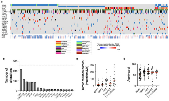
Cases were classified based on previously reported TCGA driver genes, including BRAF mutation (n = 77, 30.3%), NRAS mutation (n = 77, 30.3%), NF1 mutation (n = 47, 18.5%) and TWT (n = 33, 13.0%) (Figure 1a) [4]. A small cohort of cases demonstrated mutations in multiple TCGA driver genes (n = 20, 7.9%), and within this subset, a majority harbored mutations in NF1 as well as either BRAF or NRAS (n = 18 of 20, 90%) (Figure 1a). In addition to TCGA driver genes (BRAF, NRAS, NF1), we evaluated recurrent molecular alterations observed in at least 5% of the entire cohort, the majority of which were established oncogenes or tumor suppressors (Figure 1b) [29,30,31,32,33,34,35,36,37,38,39,40,41,42]. Recurrently altered genes included TERT (n = 216), CDKN2A/B (n = 116) TP53 (n = 93), ARID2 (n = 32), PTEN (n = 25), RASA2 (n = 22), PTEN (n = 25), NFKBIE (n = 20), KMT2B (n = 16), PTPRD (n = 15), PTPRT (n = 15), MAP2K1 (n = 14), RAC1 (n = 14), GRIN2A (n = 13), PTPRB (n = 13 or 5%), PREX2 (n = 13 or 5%), and SETD2 (n = 13 or 5%). The three most commonly co-altered genes included activating TERT promoter mutations and loss of the tumor suppressors CDKN2A/B and TP53. A total of 121 cases demonstrated mutations in 2 of these 3 genes, and an additional 23 cases were “triple hit” in terms of alterations in TERT, CDKN2A/B and TP53. In addition, there were 25 cases harboring the PTEN alteration, of which 12 co-occurred with CDKN2A/B loss and 10 co-occurred with TP53 mutation.

Figure 1.
Mutational analysis of metastatic melanoma from a real-world cohort recapitulates TCGA molecular drivers. (a) Targeted DNA sequencing analysis using a CLIA-certified clinical genomics assay of 254 patients undergoing clinical care at a tertiary cancer center reveals the following five molecular groups: BRAF mutant (n = 77), NRAS mutant (n = 77), NF1 mutant (n = 47), triple wild-type (n = 33), and co-mutated tumors with multiple alterations in BRAF, NRAS, and/or NF1 (n = 20) consistent with TCGA molecular groups. (b) A total of 19 genes were recurrently altered in at least 5% of the cutaneous melanoma cohort including TERT (n = 216), CDKN2A/B (n = 116), TP53 (n = 93), NRAS (n = 91), BRAF (n = 87), NF1 (n = 85), ARID2 (n = 32), PTEN (n = 25), RASA2 (n = 22), NFKBIE (n = 20), KMT2B (n =16), PTPRD (n = 15), PTPRT (n = 15), RAC1 (n = 14), MAP2K1 (n = 14), SETD2 (n = 13), PTPRB (n = 13), GRIN2A (n = 13) and PREX2 (n = 13). (c) Median tumor mutation burden (shown by the red lines) is significantly increased in NF1 mutant and co-mutated tumors (as denoted by the *) compared to BRAF, NRAS, or TWT tumors (ANOVA, p < 0.001). (d) Patients with BRAF mutant tumors are significantly younger, while NF1 mutant patients are significantly older than other molecular groups (ANOVA, p = 0.001).
BRAF mutant melanoma tended to occur in younger patients with a median age of 54 (p = 0.0001, one-way ANOVA), which may account, in part, for previous reports of improved outcomes in BRAF mutant melanoma, while NF1 mutant tumors were seen in older patients with a median age of 67 (p = 0.005, one-way ANOVA) compared with other driver genes (Figure 1d). Tumor mutational burden was significantly higher in NF1 mutant tumors (median 70.2 mutations/Mb) and co-mutated tumors (median 66.5 mutations/Mb) (p < 0.001, one-way ANOVA) compared with other TCGA driver genes (Figure 1c). BRAF mutant tumors had the lowest median TMB (12.4 mutations/Mb), followed by NRAS mutant tumors (median 17.5 mutations/Mb) and TWT melanomas (28.2 mutations/Mb) (Figure 1c). Patients with BRAF mutant tumors were associated with increased rates of non-metastatic disease at diagnosis (p < 0.0001, chi-square test), while patients with TWT tumors were associated with poorer performance status (ECOG 1+) (n = 7/20, 35%) compared with other TCGA driver genes (n = 21/144, 14.6%; p = 0.028, Fisher’s exact test) (Figure S1c,d). With regard to anatomic location, NF1 mutant tumors (n = 22/47, 46.8%), TWT melanomas (n = 14/33, 42.4%) and co-mutant tumors (n = 7/20, 35%) occurred more frequently on the head/neck compared with BRAF or NRAS mutant tumors (n = 24/154, 15.6%; p < 0.001, chi-squared test) (Figure S1a). There were otherwise no statistically significant differences in baseline features between TCGA driver genes (Table 1; Figure S1b,e,f).
3.2. BRAF, NRAS and NF1 Mutant Melanomas Exhibit Distinct Alteration and Co-Mutation Patterns
BRAF mutations can be classified into the following three groups: Class I, II or III [43]. Class I and Class II mutations lead to Ras-independent Raf activation, with Class I mutations involving V600 to create monomers with high kinase activity and Class II mutations creating activated Raf dimers with intermediate kinase activity. In contrast, class III mutations comprise weaker oncogenic variants leading to enhanced Ras binding with impaired kinase activity that drives CRAF activation for enhanced downstream MAPK pathway signaling [44]. In our cohort, BRAF mutations were predominantly class I (Figure 2a), particularly for the BRAF driver group, in which 68 of 77 cases (88.3%) had class I BRAF mutations. Only one class III BRAF mutation was noted in this group (BRAF G466E), with several class II mutations [43]. Multiple fusions and gene rearrangements were also detected involving AGK and BRAF (n = 3) and ATF7 and BRAF (n = 1). In contrast, only one of the 11 BRAF mutations identified in the BRAF/NRAS/NF1 co-mutation group was a class I BRAF mutation. When occurring in the co-mutation group, the majority (8 of 11 or 73%) of BRAF mutations were class III, consistent with prior work suggesting these weaker oncogenic BRAF variants require additional hits to drive melanomagenesis [43].

Figure 2.
Driver mutation-specific analysis of BRAF, NRAS and NF1 mutant tumors reveals specific variant and co-mutation patterns. (a) BRAF mutant tumors were primarily composed of class I BRAF mutations (n = 68 or 86%). The majority were V600E (n = 58, shown in red) with 8 V600K mutations and 2 V600R mutations. A total of 3 BRAF fusions were detected, as well as 2 gene re-arrangements, most of which involved AGK and BRAF. Other non-class I loci in this group included K601, G469, G466, F247 and E586. There were no associated differences in outcomes with class I mutations compared to other BRAF classes or to non-BRAF mutant tumors. Only 1 of 10 BRAF mutant cases with co-occurring NRAS or NF1 mutations (10%) had a class I BRAF mutation. S467L mutations were particularly common in this cohort (n = 4 or 40%). Other loci included N581, G469, L597 and N580. (b) The majority of NRAS mutant tumors had mutations at Q61 (N = 66 or 87%) including Q61R (n = 30), Q61K (n = 24), Q61L (n = 8) and Q61H (n = 4). These correspond to color coded bars in the figure. Other mutations identified included G13R (n = 5), G12A (n = 3), G13C (n = 1) and T50I (n = 1). Two amplifications were noted, as well as one case with copy-number-neutral LOH at Q61K. Of the 15 NRAS mutations identified in the co-mutation group, 10 were Q61K/L/R (67%), 2 were G12A/D, 2 were G13D, and 1 was T50I. (c) A wide spectrum of variants, enriched on the 5′ ends of the gene, was identified in NF1 mutant tumors, with 12 of 47 cases in the NF1 driver group (26%) and 8 of 18 NF1 mutant cases (44%) in the BRAF/NRAS/NF1 group having more than one mutation in NF1. The presence of multiple NF1 mutations did not have a significant impact on overall survival when compared with tumors with only one NF1 alteration. The majority of the BRAF/NRAS/NF1 co-mutant tumor group had mutations in NF1 (n = 18 or 90%). There were no significant survival outcome differences between this group and the NF1 driver mutation group. (d) Co-mutation correlation analysis (threshold Pearson > 0.3 at p < 0.05) found that BRAF mutations did not significantly correlate with any other alterations, NRAS mutations correlated with TERT mutations and NF1 mutations were significantly correlated with several genes, including RASA2, PTPRT, TP53, PTPRB and TERT. Notably, BRAF and NRAS mutations were anti-correlated, consistent with their mutual exclusivity as drivers. (e) The co-mutation breakdown of NF1 mutant tumors and significantly co-occurring alterations.
NRAS mutations were predominantly Q61 variants regardless of whether the tumors were in the NRAS driver (n = 67/77 or 86%) or co-mutated group (n = 9/14 or 64.3%) (Figure 2b). A total of 67 putative loss of function variants in NF1 were identified, with the majority clustered at the 5′ end of the gene, suggesting that the upstream loss of function mutations is enriched in NF1 (Figure 2c). There were no differences in specific NF1 variants when co-mutated with other driver genes compared to tumors with the NF1 mutation alone.
Correlation analysis of all identified genes found NF1 mutations to correlate with several genomic alterations, including, most frequently, RASA2 loss (correlation coefficient 0.37, adjusted p < 0.001) but also frequently with PTPRT mutations (correlation coefficient 0.33, adjusted p < 0.001), TP53 mutations (correlation coefficient 0.31, adjusted p < 0.001), PTPRB mutations (correlation coefficient 0.30, adjusted p < 0.001) and 39 other less commonly co-mutated genes (Figure 2d,e and Figure S2). NRAS correlated most with TERT (correlation coefficient 0.33, p < 0.001), BRAF was not significantly co-altered with any genes examined, and BRAF and NRAS demonstrated significant mutual exclusivity (Figure 2d and Figure S2).
3.3. Triple Wild-Type Tumors Contain Frequent Alterations in Other Components of MAPK Pathway Signaling
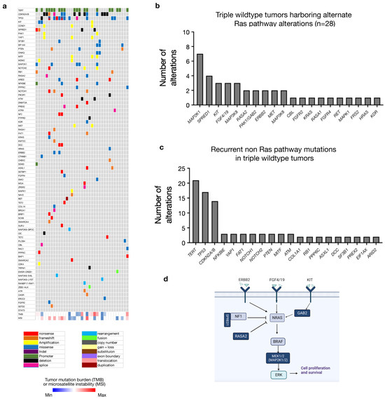
The majority of TWT tumors demonstrated a pathogenic variant in the Ras pathway (n = 28/33 or 84.8%) (Figure 3a,b). These included MAP2K1 (n = 7), SPRED1 (n = 4), KIT (n = 3), FGF4/19 (n = 3), MAP3K8 (n = 2), RASA2 (n = 2), ERBB2 (n = 2), MET (n = 2), NF2 (n = 2) and GAB2 (n = 2). (Figure 3b,d). In tumors harboring a BRAF, NRAS, or NF1 mutation (non-TWT tumors), MAP2K1 mutations were identified in 7 of 221 (or 3.2%) tumors, SPRED1 mutations were found in 5 of 221 (or 2.3%) tumors, KIT in 7 of 221 (or 3.2%) tumors and FGF4/19 in 10 of 221 (or 4.5%) tumors. In addition, triple wild-type tumors often harbored non-Ras pathway alterations found in other TCGA molecular groups including TERT (n = 21), TP53 (n = 17), CDKN2A/B (n = 12), NFKBIE (n = 3), YAP1 (n = 3), FAT1 (n = 3), NOTCH1 (n = 3), NOTCH2 (n = 3), PTEN (n = 3), MITF (n = 3) and ATM (n = 3) (Figure 3c,d). A total of 5 of 33 TWT cases demonstrated no known or suspected Ras pathway alterations and demonstrated presumed driver mutations in EIF1AX, GNAQ, ATRX and ASXL1 (Figure 3a). The GNAQ mutation was found in a melanoma of unknown primary site, while the EIF1AX mutation was found in a cutaneous melanoma located in the neck.

Figure 3.
Triple wild-type tumors harbor alternate Ras pathway alterations in the majority of tumors lacking a classical TCGA driver mutation. (a) In triple wild-type tumors, recurrently altered genes include TERT (n = 21), TP53 (n = 18), CDKN2A/B (n = 14), MAP2K1 (n = 7) and SPRED1 (n = 4). (b) In 28 of 33 cases (84.8%), pathogenic variants in known Ras pathway components are observed as putative driver mutations. (c) Recurrent alterations in non-Ras pathway genes in the triple wild-type group are predominantly the TERT and TP53 mutation, similar to other molecular groups, with additional mutations comprising potential drivers in tumors without a classic Ras pathway mutation. (d) Schematic depicting the mutations occurring in triple wild-type tumors (dark blue) within the Ras signaling pathway.
3.4. NRAS Mutation Is Prognostic, and Tumor Mutational Burden (TMB) Is Predictive of Response to Dual Checkpoint Blockade
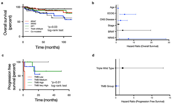
We next assessed clinical outcomes for the entire 254-patient cohort. With regard to overall survival (OS), univariable CPH analysis revealed that age, ECOG status, CNS disease, stage, BRAF mutation and NRAS mutation were significantly correlated with OS (p < 0.1; Table S2). For multivariable analysis, NRAS mutant tumors remained correlated with worse OS (HR 2.95, 95% CI 1.13–7.69, p = 0.027; log-rank test) (Figure 4a, Table S2). Older age (HR 1.06, 95% CI 1.02–1.11, p = 0.003; log-rank test) and higher ECOG scores (HR 4.84, 95% CI 2.12–11.05, p < 0.001; log-rank test) also correlated with decreased overall survival (Figure 4b, Table S2). Tumor mutational burden did not correlate with overall survival in our cohort (Figure S3a), and the TCGA molecular group was not associated with differences in progression-free survival or recurrence-free survival (Figure S3b,c). Multivariable analysis for recurrence-free survival found age to be the only variable correlated with RFS (HR 1.03, 95% CI 1.02–1.05, p < 0.001, log-rank test) (Table S4).

Figure 4.
Molecular subgroup is prognostic for overall survival, and tumor mutation burden (TMB) is predictive of dual checkpoint blockade response in cutaneous melanoma. (a) Overall survival stratified by 5 pre-specified molecular subgroups (BRAF mutant, NRAS mutant, NF1 mutant, triple wild-type and co-mutated BRAF/NRAS/NF1) shows statistically significant differences in outcome based on molecular driver (p = 0.03) with NRAS mutation associated with particularly worse outcomes. (b) Forest plot of multivariable hazard ratios for OS including covariates with p ≤ 0.1 in univariable survival analysis (ECOG status, presence of CNS disease, stage, BRAF, NRAF). In addition to NRAS mutation, age, ECOG status, and the presence of CNS disease were independent factors significantly associated with overall survival. (c) Kaplan–Meier curve depicting progression-free survival stratified by tumor mutation burden (TMB) for 36 out of 116 patients (31.0%) who received the combination of anti-PD1/PDL1 + anti-CTLA4 as the first-line systemic therapy for metastatic disease. This showed a statistically significant difference in PFS based on TMB group (p = 0.01). (d) TMB was the only covariate found to significantly impact PFS on combination ICI in multivariable CPH analysis.
Of the 116 patients treated with systemic therapy for metastatic or locally advanced/unresectable disease, the majority were treated with first-line ICI: 65 out of 116 (56.0%) patients received anti-PD1 monotherapy, 36 of 116 (31.0%) patients received dual checkpoint inhibition with a combination of anti-PD1 and anti-CTLA4, and 7 of 116 patients (6.0%) received anti-CTLA4 monotherapy (Table 1). A small proportion were treated with BRAF/MEK inhibitors in the first-line setting (n = 8/116 or 6.9%). Multivariable analysis found no significant associations with progression-free survival on any first-line immune checkpoint inhibition (Table S3, Figure S3d,e). There was a trend towards improved progression-free survival for patients with mutations in at least 2 of the 3 most commonly mutated tumor suppressor genes (TERT, CKDN2A/B and TP53) with HR for progressions of 0.613 (95% CI 0.373–1.009, p = 0.054), although this did not reach statistical significance. When further stratified based on specific treatment type, TMB was the only variable significantly associated with response to dual ICI; no variables were associated with response to single-agent anti-PD1 (Figure 4c, Tables S5 and S6). A low TMB (≤5 mutations/Mb) correlated with poorer PFS in patients receiving dual-agent ICI (HR 20.9, 95% CI 1.77–245.9; p = 0.016; log-rank test) (Figure 4d, Table S6). Given the overall small sample size, particularly in the low TMB cohort, which included only 14 patients, this relationship between TMB and clinical outcome requires further corroboration with larger, ideally multi-institutional datasets.
4. Discussion
We present clinical and molecular data from 254 patients with melanoma who underwent targeted DNA sequencing and were treated in the ICI era. Our analysis confirmed CM classification based on TCGA genetic drivers [4] and previously reported associated demographic and phenotypic features, including age of onset (BRAF mutations in younger patients and NF1 mutations found in older patients) and significantly higher median TMB in NF1 mutant tumors [4,8] (Figure 1c,d). BRAF mutations in the entire 254-patient cohort were predominantly class I (n = 68 of 88 or 77.3%) (Figure 2a). While cases with BRAF TCGA driver mutations alone were consistent with previously reported data suggesting a predominance of class I mutations in BRAF mutant melanoma, only one of eleven BRAF co-mutated cases (co-occurring with either NRAS or NF1) exhibited a class I BRAF mutation. The majority of co-mutated cases had weaker class III BRAF mutations, which have been associated with improved prognosis in melanoma compared with class I BRAF mutations [45]. We found NF1 to be the only TCGA driver in our cohort that significantly correlated with multiple other genes, most frequently RASA2, as previously reported [46]. NF1 co-mutations with either BRAF or NRAS, as well as frequently with other melanoma-associated oncogenes, suggest that this is overall a weaker oncogenic driver that may cooperate with additional MAPK pathway alterations to drive melanomagenesis. NRAS mutant tumors correlated with poorer overall survival compared with other TCGA driver gene groups. The presence of an NRAS mutation has historically been correlated with poorer overall survival in patients with advanced melanoma, but its prognostic impact in the modern treatment era is less clear [47,48]. Our analysis of patients treated in the immunotherapy era (from 2013–2023) found NRAS mutation to be an independent factor significantly associated with poorer overall survival, underscoring the need for further drug development in this particular patient population.
The majority of TWT tumors carried at least one putative driver mutation involving the MAPK pathway, including MAP2K1, SPRED1, KIT, FGF4/19, RASA2 and ERBB2 (Figure 3). Triple wild-type tumors also demonstrated a median TMB of 28.2, suggesting that the correlation between low TMB and triple wild-type status may be most relevant to sun-shielded or non-cutaneous melanomas (Figure 1c). Only 5 of 33 TWT melanomas in our cohort lacked a putative driver mutation involving the MAPK pathway. Our analysis suggests that the majority of TWT cutaneous melanomas harbor a Ras/MAPK pathway mutation, with some confirmed cutaneous melanomas demonstrating mutations more commonly identified in uveal melanoma, including EIF1AX and GNAQ.
Beyond TCGA drivers, several studies have focused on identifying additional prognostic or predictive tumor-specific genomic aberrations in cutaneous melanoma, particularly in terms of ICI response. Mutations in KMT2, PTPRT, MAP2K1 and SETD2 have been associated with improved responses to immune checkpoint inhibitors, while PTEN loss of function is thought in some studies to be associated with innate resistance to immune checkpoint blockade [49,50,51,52,53]. We did not find any specific genomic alterations other than the presence of an NRAS mutation to correlate with clinical outcomes in our 254-patient cohort. TMB was the only predictive biomarker of response to immune checkpoint inhibition identified in our analysis. Notably, TMB was not associated with PFS on single-agent anti-PD1 but was predictive for patients treated with dual-agent immune checkpoint inhibition in our patient population. Interestingly, despite a significantly higher TMB than other driver groups, NF1 mutant and co-mutant tumors did not demonstrate improved PFS when treated with first-line immunotherapy. This may be related to sample size and the wide range of TMB findings within the NF1 mutant group. Given that a higher TMB has been correlated with response to ICI, both in our study and in several others, and TMB in NF1 mutant melanomas is generally very high, further investigation is warranted as to why NF1 mutation alone did not correlate with improved ICI response.
5. Conclusions
Our real-world retrospective analysis of 254 patients with melanoma who underwent in-house DNA sequencing at our institution generated several novel and notable findings. NRAS mutant tumors were associated with worse overall survival outcomes. We also demonstrate that triple wild-type cutaneous melanomas that lack mutations in BRAF, NRAS or NF1 have a similar TMB to the overall median and, in the majority of cases, demonstrate putative drivers in the MAPK pathway, suggesting that disease biology in these cases is likely similar to those melanomas with known Ras drivers. Finally, TCGA driver group was not associated with progression-free survival on any first-line immunotherapy despite prognostic significance in terms of overall survival. Tumor mutational burden was associated with response to a combination of immune checkpoint inhibition with anti-CTLA4 and anti-PD1 but did not have a statistically significant association with progression-free survival on anti-PD1 monotherapy. Taken together, these data support prognostic differences based on driver mutation and the predictive utility for TMB in cutaneous melanoma. Additional investigation into “triple wild-type” cutaneous melanomas and their reliance on Ras/MAPK signaling is an area for further study, and poor outcomes in NRAS mutant tumors highlight an urgent, unmet clinical need for therapeutic development.
Our study has several limitations, most notably that it is a retrospective single-institution investigation with all the caveats such an approach entails. In addition, detailed follow-up and treatment data were available only for a subset of the 254 patients that underwent molecular profiling, and both metastatic and non-metastatic sites were included with different patterns of follow-up, making results difficult to meaningfully interpret and apply to distinct clinical populations. Further prospective studies that assess the prognostic significance of driver mutations and the predictive and prognostic significance of tumor mutational burden are certainly warranted.
Supplementary Materials
The following supporting information can be downloaded at: https://www.mdpi.com/article/10.3390/cancers16071347/s1, Figure S1: Baseline clinical correlations based on molecular group; Figure S2: Analysis of co-mutation patterns; Figure S3: Clinical outcomes based on molecular signatures and treatment; Table S1: Compiled variant list from the UCSF500 assay, a CLIA certified, capture based targeted DNA sequencing assay of 529 cancer-associated genes; Table S2: Univariable and multivariable analysis for OS; Table S3: Univariable and multivariable analysis for PFS on any first line immunotherapy; Table S4: Univariable and multivariable analysis for RFS; Table S5: Univariable and multivariable analysis for PFS on single agent immune checkpoint inhibition; Table S6: Univariable and multivariable analysis for PFS on dual agent immune checkpoint inhibition.
Author Contributions
Conceptualization, A.M.H., K.K.T., A.D. and H.N.V.; methodology, A.M.H., R.C.O., R.A.F., M.E.T., M.T., A.D. and H.N.V.; formal analysis, A.M.H., R.C.O., R.A.F., M.T. and H.N.V.; investigation, A.M.H., R.C.O., R.A.F., M.T. and H.N.V.; resources, R.A.F., K.K.T. and M.T.; data curation, A.M.H., R.C.O., R.A.F., M.E.T., M.T., A.D. and H.N.V.; writing—original draft preparation, A.M.H. and H.N.V.; writing—review and editing, all authors; visualization, A.M.H., R.C.O. and H.N.V.; supervision, A.D. and H.N.V.; project administration, A.D. and H.N.V.; funding acquisition, A.M.H. All authors have read and agreed to the published version of the manuscript.
Institutional Review Board Statement
The study was conducted in accordance with the Declaration of Helsinki, and approved by the Institutional Review Board of the University of California San Francisco (protocol code 22-37134 approved on 8 February 2022).
Informed Consent Statement
Not applicable.
Data Availability Statement
All data is available in Supplementary tables, and more detailed information, specifically clinical annotations, can be obtained by contacting the corresponding author.
Conflicts of Interest
The authors declare no conflict of interest.
References
- Newell, F.; Johansson, P.A.; Wilmott, J.S.; Nones, K.; Lakis, V.; Pritchard, A.L.; Lo, S.N.; Rawson, R.V.; Kazakoff, S.H.; Colebatch, A.J.; et al. Comparative Genomics Provides Etiologic and Biological Insight into Melanoma Subtypes. Cancer Discov. 2022, 12, 2856–2879. [Google Scholar] [CrossRef]
- Shain, A.H.; Yeh, I.; Kovalyshyn, I.; Sriharan, A.; Talevich, E.; Gagnon, A.; Dummer, R.; North, J.; Pincus, L.; Ruben, B.; et al. The Genetic Evolution of Melanoma from Precursor Lesions. N. Engl. J. Med. 2015, 373, 1926–1936. [Google Scholar] [CrossRef]
- Shain, A.H.; Joseph, N.M.; Yu, R.; Benhamida, J.; Liu, S.; Prow, T.; Ruben, B.; North, J.; Pincus, L.; Yeh, I.; et al. Genomic and Transcriptomic Analysis Reveals Incremental Disruption of Key Signaling Pathways during Melanoma Evolution. Cancer Cell 2018, 34, 45–55.e44. [Google Scholar] [CrossRef]
- Akbani, R.; Akdemir, K.C.; Aksoy, B.A.; Albert, M.; Ally, A.; Amin, S.B.; Arachchi, H.; Arora, A.; Auman, J.T.; Ayala, B.; et al. Genomic Classification of Cutaneous Melanoma. Cell 2015, 161, 1681–1696. [Google Scholar] [CrossRef]
- Elder, D.E.; Bastian, B.C.; Cree, I.A.; Massi, D.; Scolyer, R.A. The 2018 World Health Organization Classification of Cutaneous, Mucosal, and Uveal Melanoma: Detailed Analysis of 9 Distinct Subtypes Defined by Their Evolutionary Pathway. Arch. Pathol. Lab. Med. 2020, 144, 500–522. [Google Scholar] [CrossRef]
- Ellerhorst, J.A.; Greene, V.R.; Ekmekcioglu, S.; Warneke, C.L.; Johnson, M.M.; Cooke, C.P.; Wang, L.E.; Prieto, V.G.; Gershenwald, J.E.; Wei, Q.; et al. Clinical correlates of NRAS and BRAF mutations in primary human melanoma. Clin. Cancer Res. 2011, 17, 229–235. [Google Scholar] [CrossRef]
- Guhan, S.; Klebanov, N.; Tsao, H. Melanoma genomics: A state-of-the-art review of practical clinical applications. Br. J. Dermatol. 2021, 185, 272–281. [Google Scholar] [CrossRef]
- Cirenajwis, H.; Lauss, M.; Ekedahl, H.; Törngren, T.; Kvist, A.; Saal, L.H.; Olsson, H.; Staaf, J.; Carneiro, A.; Ingvar, C.; et al. NF1-mutated melanoma tumors harbor distinct clinical and biological characteristics. Mol. Oncol. 2017, 11, 438–451. [Google Scholar] [CrossRef]
- Wolchok, J.D.; Chiarion-Sileni, V.; Gonzalez, R.; Rutkowski, P.; Grob, J.J.; Cowey, C.L.; Lao, C.D.; Wagstaff, J.; Schadendorf, D.; Ferrucci, P.F.; et al. Overall Survival with Combined Nivolumab and Ipilimumab in Advanced Melanoma. N. Engl. J. Med. 2017, 377, 1345–1356. [Google Scholar] [CrossRef]
- Larkin, J.; Chiarion-Sileni, V.; Gonzalez, R.; Grob, J.J.; Cowey, C.L.; Lao, C.D.; Schadendorf, D.; Dummer, R.; Smylie, M.; Rutkowski, P.; et al. Combined Nivolumab and Ipilimumab or Monotherapy in Untreated Melanoma. N. Engl. J. Med. 2015, 373, 23–34. [Google Scholar] [CrossRef]
- Robert, C.; Schachter, J.; Long, G.V.; Arance, A.; Grob, J.J.; Mortier, L.; Daud, A.; Carlino, M.S.; McNeil, C.; Lotem, M.; et al. Pembrolizumab versus Ipilimumab in Advanced Melanoma. N. Engl. J. Med. 2015, 372, 2521–2532. [Google Scholar] [CrossRef]
- Hodi, F.S.; O’Day, S.J.; McDermott, D.F.; Weber, R.W.; Sosman, J.A.; Haanen, J.B.; Gonzalez, R.; Robert, C.; Schadendorf, D.; Hassel, J.C.; et al. Improved survival with ipilimumab in patients with metastatic melanoma. N. Engl. J. Med. 2010, 363, 711–723. [Google Scholar] [CrossRef]
- Wolchok, J.D.; Chiarion-Sileni, V.; Gonzalez, R.; Grob, J.J.; Rutkowski, P.; Lao, C.D.; Cowey, C.L.; Schadendorf, D.; Wagstaff, J.; Dummer, R.; et al. Long-Term Outcomes With Nivolumab Plus Ipilimumab or Nivolumab Alone Versus Ipilimumab in Patients with Advanced Melanoma. J. Clin. Oncol. 2022, 40, 127–137. [Google Scholar] [CrossRef]
- Eckardt, J.; Schroeder, C.; Martus, P.; Armeanu-Ebinger, S.; Kelemen, O.; Gschwind, A.; Bonzheim, I.; Eigentler, T.; Amaral, T.; Ossowski, S.; et al. TMB and BRAF mutation status are independent predictive factors in high-risk melanoma patients with adjuvant anti-PD-1 therapy. J. Cancer Res. Clin. Oncol. 2023, 149, 833–840. [Google Scholar] [CrossRef]
- Atkins, M.B.; Lee, S.J.; Chmielowski, B.; Tarhini, A.A.; Cohen, G.I.; Truong, T.G.; Moon, H.H.; Davar, D.; O’Rourke, M.; Stephenson, J.J.; et al. Combination Dabrafenib and Trametinib Versus Combination Nivolumab and Ipilimumab for Patients With Advanced BRAF-Mutant Melanoma: The DREAMseq Trial-ECOG-ACRIN EA6134. J. Clin. Oncol. 2023, 41, 186–197. [Google Scholar] [CrossRef]
- Devitt, B.; Liu, W.; Salemi, R.; Wolfe, R.; Kelly, J.; Tzen, C.Y.; Dobrovic, A.; McArthur, G. Clinical outcome and pathological features associated with NRAS mutation in cutaneous melanoma. Pigment Cell Melanoma Res. 2011, 24, 666–672. [Google Scholar] [CrossRef]
- Gutiérrez-Castañeda, L.D.; Nova, J.A.; Tovar-Parra, J.D. Frequency of mutations in BRAF, NRAS, and KIT in different populations and histological subtypes of melanoma: A systemic review. Melanoma Res. 2020, 30, 62–70. [Google Scholar] [CrossRef]
- Friedman, C.F.; Spencer, C.; Cabanski, C.R.; Panageas, K.S.; Wells, D.K.; Ribas, A.; Tawbi, H.; Tsai, K.; Postow, M.; Shoushtari, A.; et al. Ipilimumab alone or in combination with nivolumab in patients with advanced melanoma who have progressed or relapsed on PD-1 blockade: Clinical outcomes and translational biomarker analyses. J. Immunother. Cancer 2022, 10, e003853. [Google Scholar] [CrossRef]
- Johnson, D.B.; Lovly, C.M.; Flavin, M.; Panageas, K.S.; Ayers, G.D.; Zhao, Z.; Iams, W.T.; Colgan, M.; DeNoble, S.; Terry, C.R.; et al. Impact of NRAS mutations for patients with advanced melanoma treated with immune therapies. Cancer Immunol. Res. 2015, 3, 288–295. [Google Scholar] [CrossRef]
- Heppt, M.V.; Siepmann, T.; Engel, J.; Schubert-Fritschle, G.; Eckel, R.; Mirlach, L.; Kirchner, T.; Jung, A.; Gesierich, A.; Ruzicka, T.; et al. Prognostic significance of BRAF and NRAS mutations in melanoma: A German study from routine care. BMC Cancer 2017, 17, 536. [Google Scholar] [CrossRef]
- Gogas, H.; Dréno, B.; Larkin, J.; Demidov, L.; Stroyakovskiy, D.; Eroglu, Z.; Francesco Ferrucci, P.; Pigozzo, J.; Rutkowski, P.; Mackiewicz, J.; et al. Cobimetinib plus atezolizumab in BRAF(V600) wild-type melanoma: Primary results from the randomized phase III IMspire170 study. Ann. Oncol. 2021, 32, 384–394. [Google Scholar] [CrossRef]
- McGrail, D.J.; Pilié, P.G.; Rashid, N.U.; Voorwerk, L.; Slagter, M.; Kok, M.; Jonasch, E.; Khasraw, M.; Heimberger, A.B.; Lim, B.; et al. High tumor mutation burden fails to predict immune checkpoint blockade response across all cancer types. Ann. Oncol. 2021, 32, 661–672. [Google Scholar] [CrossRef]
- Gandara, D.R.; Agarwal, N.; Gupta, S.; Klempner, S.J.; Andrews, M.C.; Mahipal, A.; Subbiah, V.; Eskander, R.N.; Carbone, D.P.; Snider, J.; et al. Tumor mutational burden (TMB) measurement from an FDA-approved assay and real-world overall survival (rwOS) on single-agent immune checkpoint inhibitors (ICI) in over 8000 patients across 24 cancer types. J. Clin. Oncol. 2023, 41 (Suppl. S16), 2503. [Google Scholar] [CrossRef]
- Panning, A.; Samlowski, W.; Allred, G. Lack of Influence of Non-Overlapping Mutations in BRAF, NRAS, or NF1 on 12-Month Best Objective Response and Long-Term Survival after Checkpoint Inhibitor-Based Treatment for Metastatic Melanoma. Cancers 2023, 15, 3527. [Google Scholar] [CrossRef]
- Niu, B.; Ye, K.; Zhang, Q.; Lu, C.; Xie, M.; McLellan, M.D.; Wendl, M.C.; Ding, L. MSIsensor: Microsatellite instability detection using paired tumor-normal sequence data. Bioinformatics 2014, 30, 1015–1016. [Google Scholar] [CrossRef]
- Bellenghi, M.; Puglisi, R.; Pontecorvi, G.; De Feo, A.; Carè, A.; Mattia, G. Sex and Gender Disparities in Melanoma. Cancers 2020, 12, 1819. [Google Scholar] [CrossRef]
- Dutton-Regester, K.; Kakavand, H.; Aoude, L.G.; Stark, M.S.; Gartside, M.G.; Johansson, P.; O’Connor, L.; Lanagan, C.; Tembe, V.; Pupo, G.M.; et al. Melanomas of unknown primary have a mutation profile consistent with cutaneous sun-exposed melanoma. Pigment Cell Melanoma Res. 2013, 26, 852–860. [Google Scholar] [CrossRef]
- Egberts, F.; Bergner, I.; Krüger, S.; Haag, J.; Behrens, H.M.; Hauschild, A.; Röcken, C. Metastatic melanoma of unknown primary resembles the genotype of cutaneous melanomas. Ann. Oncol. 2014, 25, 246–250. [Google Scholar] [CrossRef]
- Shain, A.H.; Garrido, M.; Botton, T.; Talevich, E.; Yeh, I.; Sanborn, J.Z.; Chung, J.; Wang, N.J.; Kakavand, H.; Mann, G.J.; et al. Exome sequencing of desmoplastic melanoma identifies recurrent NFKBIE promoter mutations and diverse activating mutations in the MAPK pathway. Nat. Genet. 2015, 47, 1194–1199. [Google Scholar] [CrossRef]
- Cannon, A.C.; Uribe-Alvarez, C.; Chernoff, J. RAC1 as a Therapeutic Target in Malignant Melanoma. Trends Cancer 2020, 6, 478–488. [Google Scholar] [CrossRef]
- Williams, E.A.; Montesion, M.; Shah, N.; Sharaf, R.; Pavlick, D.C.; Sokol, E.S.; Alexander, B.; Venstrom, J.; Elvin, J.A.; Ross, J.S.; et al. Melanoma with in-frame deletion of MAP2K1: A distinct molecular subtype of cutaneous melanoma mutually exclusive from BRAF, NRAS, and NF1 mutations. Mod. Pathol. 2020, 33, 2397–2406. [Google Scholar] [CrossRef]
- Krebs, F.S.; Moura, B.; Missiaglia, E.; Aedo-Lopez, V.; Michielin, O.; Tsantoulis, P.; Bisig, B.; Trimech, M.; Zoete, V.; Homicsko, K. Response and Resistance to Trametinib in MAP2K1-Mutant Triple-Negative Melanoma. Int. J. Mol. Sci. 2023, 24, 4520. [Google Scholar] [CrossRef]
- Weng, X.; Chen, W.; Hu, W.; Xu, K.; Qi, L.; Chen, J.; Lu, D.; Shao, Y.; Zheng, X.; Ye, C.; et al. PTPRB promotes metastasis of colorectal carcinoma via inducing epithelial-mesenchymal transition. Cell Death Dis. 2019, 10, 352. [Google Scholar] [CrossRef]
- Ding, L.; Kim, M.; Kanchi, K.L.; Dees, N.D.; Lu, C.; Griffith, M.; Fenstermacher, D.; Sung, H.; Miller, C.A.; Goetz, B.; et al. Clonal architectures and driver mutations in metastatic melanomas. PLoS ONE 2014, 9, e111153. [Google Scholar] [CrossRef]
- Berger, M.F.; Hodis, E.; Heffernan, T.P.; Deribe, Y.L.; Lawrence, M.S.; Protopopov, A.; Ivanova, E.; Watson, I.R.; Nickerson, E.; Ghosh, P.; et al. Melanoma genome sequencing reveals frequent PREX2 mutations. Nature 2012, 485, 502–506. [Google Scholar] [CrossRef]
- Moreno, T.; Monterde, B.; González-Silva, L.; Betancor-Fernández, I.; Revilla, C.; Agraz-Doblas, A.; Freire, J.; Isidro, P.; Quevedo, L.; Blanco, R.; et al. ARID2 deficiency promotes tumor progression and is associated with higher sensitivity to chemotherapy in lung Cancer. Oncogene 2021, 40, 2923–2935. [Google Scholar] [CrossRef]
- Arafeh, R.; Qutob, N.; Emmanuel, R.; Keren-Paz, A.; Madore, J.; Elkahloun, A.; Wilmott, J.S.; Gartner, J.J.; Di Pizio, A.; Winograd-Katz, S.; et al. Recurrent inactivating RASA2 mutations in melanoma. Nat. Genet. 2015, 47, 1408–1410. [Google Scholar] [CrossRef]
- Maitituoheti, M.; Keung, E.Z.; Tang, M.; Yan, L.; Alam, H.; Han, G.; Singh, A.K.; Raman, A.T.; Terranova, C.; Sarkar, S.; et al. Enhancer Reprogramming Confers Dependence on Glycolysis and IGF Signaling in KMT2D Mutant Melanoma. Cell Rep. 2020, 33, 108293. [Google Scholar] [CrossRef]
- Walia, V.; Prickett, T.D.; Kim, J.S.; Gartner, J.J.; Lin, J.C.; Zhou, M.; Rosenberg, S.A.; Elble, R.C.; Solomon, D.A.; Waldman, T.; et al. Mutational and functional analysis of the tumor-suppressor PTPRD in human melanoma. Hum. Mutat. 2014, 35, 1301–1310. [Google Scholar] [CrossRef]
- Chen, C.; Liu, H.; Xu, Q.; Zhang, X.; Mu, F.; Liu, J. Association of PTPRT Mutations with Cancer Metastasis in Multiple Cancer Types. Biomed. Res. Int. 2022, 2022, 9386477. [Google Scholar] [CrossRef]
- Chen, R.; Zhao, W.Q.; Fang, C.; Yang, X.; Ji, M. Histone methyltransferase SETD2: A potential tumor suppressor in solid cancers. J. Cancer 2020, 11, 3349–3356. [Google Scholar] [CrossRef] [PubMed]
- Prickett, T.D.; Zerlanko, B.J.; Hill, V.K.; Gartner, J.J.; Qutob, N.; Jiang, J.; Simaan, M.; Wunderlich, J.; Gutkind, J.S.; Rosenberg, S.A.; et al. Somatic mutation of GRIN2A in malignant melanoma results in loss of tumor suppressor activity via aberrant NMDAR complex formation. J. Investig. Dermatol. 2014, 134, 2390–2398. [Google Scholar] [CrossRef] [PubMed]
- Yao, Z.; Yaeger, R.; Rodrik-Outmezguine, V.S.; Tao, A.; Torres, N.M.; Chang, M.T.; Drosten, M.; Zhao, H.; Cecchi, F.; Hembrough, T.; et al. Tumours with class 3 BRAF mutants are sensitive to the inhibition of activated RAS. Nature 2017, 548, 234–238. [Google Scholar] [CrossRef] [PubMed]
- Dankner, M.; Rose, A.A.N.; Rajkumar, S.; Siegel, P.M.; Watson, I.R. Classifying BRAF alterations in cancer: New rational therapeutic strategies for actionable mutations. Oncogene 2018, 37, 3183–3199. [Google Scholar] [CrossRef] [PubMed]
- Wu, X.; Yan, J.; Dai, J.; Ma, M.; Tang, H.; Yu, J.; Xu, T.; Yu, H.; Si, L.; Chi, Z.; et al. Mutations in BRAF codons 594 and 596 predict good prognosis in melanoma. Oncol. Lett. 2017, 14, 3601–3605. [Google Scholar] [CrossRef] [PubMed]
- Arafeh, R.; Di Pizio, A.; Elkahloun, A.G.; Dym, O.; Niv, M.Y.; Samuels, Y. RASA2 and NF1, two-negative regulators of Ras with complementary functions in melanoma. Oncogene 2019, 38, 2432–2434. [Google Scholar] [CrossRef]
- Jakob, J.A.; Bassett, R.L., Jr.; Ng, C.S.; Curry, J.L.; Joseph, R.W.; Alvarado, G.C.; Rohlfs, M.L.; Richard, J.; Gershenwald, J.E.; Kim, K.B.; et al. NRAS mutation status is an independent prognostic factor in metastatic melanoma. Cancer 2012, 118, 4014–4023. [Google Scholar] [CrossRef] [PubMed]
- Thumar, J.; Shahbazian, D.; Aziz, S.A.; Jilaveanu, L.B.; Kluger, H.M. MEK targeting in N-RAS mutated metastatic melanoma. Mol. Cancer 2014, 13, 45. [Google Scholar] [CrossRef] [PubMed]
- Zhang, P.; Huang, Y. Genomic alterations in KMT2 family predict outcome of immune checkpoint therapy in multiple cancers. J. Hematol. Oncol. 2021, 14, 39. [Google Scholar] [CrossRef]
- Zhang, W.; Shi, F.; Kong, Y.; Li, Y.; Sheng, C.; Wang, S.; Wang, Q. Association of PTPRT mutations with immune checkpoint inhibitors response and outcome in melanoma and non-small cell lung Cancer. Cancer Med. 2022, 11, 676–691. [Google Scholar] [CrossRef]
- Ye, T.; Zhang, J.Y.; Liu, X.Y.; Zhou, Y.H.; Yuan, S.Y.; Yang, M.M.; Xie, W.Z.; Gao, C.; Chen, Y.X.; Huang, M.L.; et al. The Predictive Value of MAP2K1/2 Mutations on Efficiency of Immunotherapy in Melanoma. Front. Immunol. 2021, 12, 785526. [Google Scholar] [CrossRef] [PubMed]
- Lu, M.; Zhao, B.; Liu, M.; Wu, L.; Li, Y.; Zhai, Y.; Shen, X. Pan-cancer analysis of SETD2 mutation and its association with the efficacy of immunotherapy. NPJ Precis. Oncol. 2021, 5, 51. [Google Scholar] [CrossRef] [PubMed]
- Lim, S.Y.; Shklovskaya, E.; Lee, J.H.; Pedersen, B.; Stewart, A.; Ming, Z.; Irvine, M.; Shivalingam, B.; Saw, R.P.M.; Menzies, A.M.; et al. The molecular and functional landscape of resistance to immune checkpoint blockade in melanoma. Nat. Commun. 2023, 14, 1516. [Google Scholar] [CrossRef] [PubMed]
Disclaimer/Publisher’s Note: The statements, opinions and data contained in all publications are solely those of the individual author(s) and contributor(s) and not of MDPI and/or the editor(s). MDPI and/or the editor(s) disclaim responsibility for any injury to people or property resulting from any ideas, methods, instructions or products referred to in the content. |
© 2024 by the authors. Licensee MDPI, Basel, Switzerland. This article is an open access article distributed under the terms and conditions of the Creative Commons Attribution (CC BY) license (https://creativecommons.org/licenses/by/4.0/).