Article

Long-Term Outcomes of Ablative Carbon-Ion Radiotherapy for Central Non-Small Cell Lung Cancer: A Single-Center, Retrospective Study

1
QST Hospital, National Institutes for Quantum Science and Technology, 4-9-1 Anagawa, Inage-ku, Chiba 263-8555, Japan
2
Department of Radiology, University of Tokyo Hospital, 3-7-1 Hongo, Tokyo 113-8655, Japan
3
Department of Radiation Oncology, Juntendo University Graduate School of Medicine, 2-1-1 Hongo, Tokyo 113-8421, Japan
4
Department of Pulmonary Medicine, International University of Health and Welfare, Narita Hospital, Hatakeda 852, Chiba 286-8520, Japan
5
Department of Respiratory Surgery, Social Welfare Organization Saiseikai Imperial Gift Foundation, Chibaken Saiseikai Narashino Hospital, 1-1-1 Izumi-cho, Chiba 275-8580, Japan
6
Department of Thoracic Surgery, International University of Health and Welfare, Narita Hospital, Hatakeda 852, Chiba 286-8520, Japan
*
Author to whom correspondence should be addressed.
Cancers 2024, 16(5), 933; https://doi.org/10.3390/cancers16050933
Submission received: 11 January 2024 / Revised: 14 February 2024 / Accepted: 23 February 2024 / Published: 25 February 2024
(This article belongs to the Special Issue Oncology: State-of-the-Art Research and Initiatives in Japan)

Abstract

:

Simple Summary

Ablative radiotherapy for central early stage non-small cell lung cancer (NSCLC) is controversial in its feasibility and optimal dose prescription due to the risk of severe pneumonia and adverse events associated with mediastinal organs. Carbon-ion radiotherapy (CIRT) is a promising modality of radiotherapy with steep and linear dose distribution and high biological efficacy. We retrospectively analyzed the long-term results of ablative CIRT using 68.4 Gy in 12 fractions for central early stage NSCLC. Irradiation doses to lungs and mediastinal organs such as bronchus and esophagus were controlled and no serious mediastinal organ-related adverse events occurred. In addition, the efficacy was not inferior to that reported in previous studies, including photon SBRT or proton radiotherapy. This means that our CIRT regimen is expected to have a relatively high safety profile for central NSCLC with tumor and organ at risk (OAR) proximity.

Abstract

The aim of this study is to assess the efficacy and safety of ablative carbon ion radiotherapy (CIRT) for early stage central non-small cell lung cancer (NSCLC). We retrospectively reviewed 30 patients who had received CIRT at 68.4 Gy in 12 fractions for central NSCLC in 2006–2019. The median age was 75 years, and the median Karnofsky Performance Scale score was 90%. All patients had concomitant chronic obstructive pulmonary disease, and 20 patients (67%) were considered inoperable. In DVH analysis, the median lung V5 and V20 were 15.5% and 10.4%, and the median Dmax, D0.5cc, D2cc of proximal bronchial tree was 65.6 Gy, 52.8 Gy, and 10.0 Gy, respectively. At a median follow-up of 43 months, the 3-year overall survival, disease-specific survival, and local control rates were 72.4, 75.8, and 88.7%, respectively. Two patients experienced grade 3 pneumonitis, but no grade ≥3 adverse events involving the mediastinal organs occurred. Ablative CIRT is feasible and effective for central NSCLC and could be considered as a treatment option, especially for patients who are intolerant of other curative treatments.

1. Introduction

Surgery is the standard treatment for early stage non-small cell lung cancer (NSCLC) [1,2,3]. However, the increasing elderly population with multiple comorbidities also requires minimally invasive alternatives. Stereotactic body radiotherapy (SBRT) has been established as an alternative treatment for patients with inoperable peripheral early stage NSCLC, achieving favorable local control and safety [4,5,6,7]. On the other hand, for central early stage NSCLC, both surgery and SBRT need careful consideration due to the high risk of injury to mediastinal organs and loss of respiratory function [8,9,10,11,12,13,14,15,16,17]. Indeed, SBRT for centrally located NSCLC causes 11 times as many severe AEs as it does for peripheral NSCLC [10]. Therefore, the appropriate dose prescription for central SBRT remains controversial.
Meanwhile, carbon-ion radiotherapy (CIRT) is a type of particle RT using carbon ions that was first clinically applied in the world at QST hospital in Japan. The advantages of particle RT, particularly CIRT, can be summarized mainly in two respects: physical and biological. Physically, CIRT has a peak in the dose spread called the Bragg peak, which can be expanded according to the shape and location of the tumor (spread-out Bragg peak; SOBP). SOBP allows CIRT to deliver sufficient doses focused on the tumor with a few numbers of beams [18,19]. Therefore, a steep dose gradient can be created between the tumor and the mediastinal OAR, which is expected to reduce severe AEs, even in central lung tumors. Biologically, carbon ions also have higher linear energy transfer (LET) values compared to photons and protons, resulting in high relative biological effects (RBE) and cytotoxicity against X-ray resistant tumor cells [20]. In addition, the LET of carbon-ion beams advances to the peak region in the body, resulting in an increased biological effect on deep-located tumors. This is also advantageous from a therapeutic point of view [21,22].
We have been exploring CIRT for lung tumors since 1994 and have developed high-dose CIRT for localized NSCLC, achieving better results relative to photon SBRT [23,24]. Currently, the protocol of 50 Gy (described as a relative biological effect [RBE]-weighted dose based on the modified microdosimetric kinetic model) in a single fraction (fr) is employed for peripheral, early stage NSCLC and provides tumor control comparable to SBRT but with an extremely high safety profile [25]. In contrast, for central NSCLC, specific safety-oriented protocols were developed in 2006, i.e., 68.4 Gy/12 fr, has been used to date but not adequately evaluated. In this study, we evaluated the efficacy and safety of CIRT for the treatment of central NSCLC.

2. Materials and Methods

2.1. Patients

Patients with central lung tumors who had received CIRT with 68.4 Gy in 12 Fr at QST Hospital in Chiba, Japan, between July 2006 and September 2019 were retrospectively analyzed. All patients were treated according to the institutional protocols for hilar and hilar-adjacent NSCLC with extrabronchial masses. This study was approved by the hospital Ethics Committee (approval number: N22-023), and written informed consent was obtained from all patients.
Inclusion criteria were as follows: (1) Japanese patients with pathologically confirmed NSCLC, (2) stage T1-T2N0M0 (International Union for Cancer Control; UICC, 7th edition) tumors ≤7 cm without lymph node or distant metastasis, and (3) World Health Organization performance status of 0 to 2. Central lesions were defined as tumors within 2 cm of the proximal bronchial tree (PBT) or immediately adjacent to the mediastinal or pericardial pleura, as described in the Radiation Therapy Oncology Group Trial (RTOG) 0813 [15]. Ultra-central lung tumors (i.e., tumors invading the hilar structures and bronchial tree proximal to the main bronchus) were excluded from the present study as they were not considered safe targets for this regimen [26,27,28,29], and were treated with a reduced dose of 48 Gy in 12 Fr. Patients receiving concurrent chemotherapy and those with prior irradiation in the same area were also excluded. The pretreatment NSCLC stage was determined based on the UICC for International Cancer Control 7th edition using thin-slice computed tomography (CT) and positron emission tomography (PET)-CT. In the retrospective analysis of the clinical data, the stage was re-evaluated based on the 8th edition. No patients were excluded due to respiratory function.

2.2. Treatment Planning

CIRT treatment planning was performed using four-dimensional (4D) CT at 2.5 mm or 5.0 mm intervals. Patients were immobilized in the supine or abdominal position on a body frame under free-breathing conditions using an external respiratory monitoring system (PSM1520; Toyonaka Lab, Osaka, Japan for 2015–2016; AZ-733 V®; Anzai Medical, Tokyo, Japan for 2016). The HIPLAN (National Institute for Radiological Science, Chiba, Japan) and Xio-N (ELECTA; Stockholm, Sweden; Mitsubishi Electric, Tokyo, Japan) systems were used for CT planning until July 2013. A respiratory gating system was developed to minimize the effects of breath-induced tumor motion.
The gross tumor volume (GTV) was delineated, including the lung tumor at the lung window (1600 Hounsfield units (HU); level, −300 HU). The clinical target volume (CTV) was defined as the GTV plus a 0.5 to 1.0 cm margin. In addition, we defined the beam field-specific target volume (FTV) by extending the 3D treatment planning technique to 4D [30] and a 2 to 3 mm setup margin to create the planning target volume (PTV). Major OARs (heart, lungs, esophagus, spinal cord, and proximal tracheobronchial tree) were contoured according to the RTOG 0236 guidelines [28,31].
The available treatment angles were increased by tilting the treatment bed up to ±20° for beams delivered horizontally or vertically, and several different angular ports were selected and combined for OAR protection. CIRT planning and irradiation details at our institution have been previously reported [23,24,32,33]. The carbon-ion dose is expressed in Gy and calculated by multiplying the physical dose by the RBE [34]. A fixed dose of 68.4 Gy was delivered in 12 Fr (i.e., four consecutive days per week over three consecutive weeks). The total dose was prescribed to the isocenters and adjusted to cover the PTV with a 95% isodose line of the prescribed dose. Dose constraints for organs at risk (OARs) were strictly adhered to, with spinal (maximum point dose; Dmax) < 28 Gy, esophageal (dose to the hottest 0.2cc; D0.2cc) < 50 Gy, and mainstem bronchus (D2cc) < 50 Gy, taking priority over target coverage. No dose constraints on the lung doses were defined. Figure 1 shows examples of the CIRT treatment plans.

2.3. Follow-Up and Statistical Analysis

Follow-up after CIRT consisted of a physical examination and a contrast-enhanced CT performed every 3 months for the first 2 years and at least every 6 months thereafter. Local recurrence was defined as progressive and increased CT abnormality. If local recurrence was suspected, 18F-fluorodeoxyglucose (FDG) tomography/computed tomography and a biopsy were performed. Acute and late AEs were assessed according to the Common Terminology Criteria for Adverse Events Version 5.0 [35]. AEs occurring within 3 months were defined as acute AEs, and those occurring thereafter were defined as late effects. The dosimetric evaluation was performed using dose-volume histogram (DVH) analysis to verify treatment quality. Doses to the OARs were calculated for the following structures: for PBT/esophagus/spinal cord, maximum dose to a point (Dmax) or small volumes (D0.2/0.5/1/2cc), for lungs, mean lung total dose (MLD total) and percentage of lung volume irradiated above 5/20 Gy (lung V5/V20).
For categorical variables, between-group comparisons were performed using Pearson’s χ2 test. Overall survival (OS), disease-specific survival (DSS), and local control (LC) rates were calculated from the first day of CIRT until the event or last patient contact using Kaplan–Meier curves. Statistical analyses were performed using R software (https://www.r-project.org/, accessed on 15 January 2024), and statistical significance was set at p < 0.05.

3. Results

3.1. Patients

A total of 30 patients with a median age of 75 (range 55–85) years who had been treated with 68.4 Gy in 12 Fr were evaluated (Table 1). The median Karnofsky Performance Scale (KPS) score was 90% (range 70–100%). All patients had concomitant chronic obstructive pulmonary disease (COPD). According to the Chronic Obstructive Lung Disease (COLD) spirometry grade [36], the numbers of patients with grades 1, 2, 3, and 4 were 11, 14, 5, and 1, respectively, with six cases (20%) ≥ grade 3. Twenty (67%) patients were deemed inoperable. The median follow-up time was 42 (range 8–163) months for all patients and 63 months for the surviving patients.

3.2. Treatment Planning

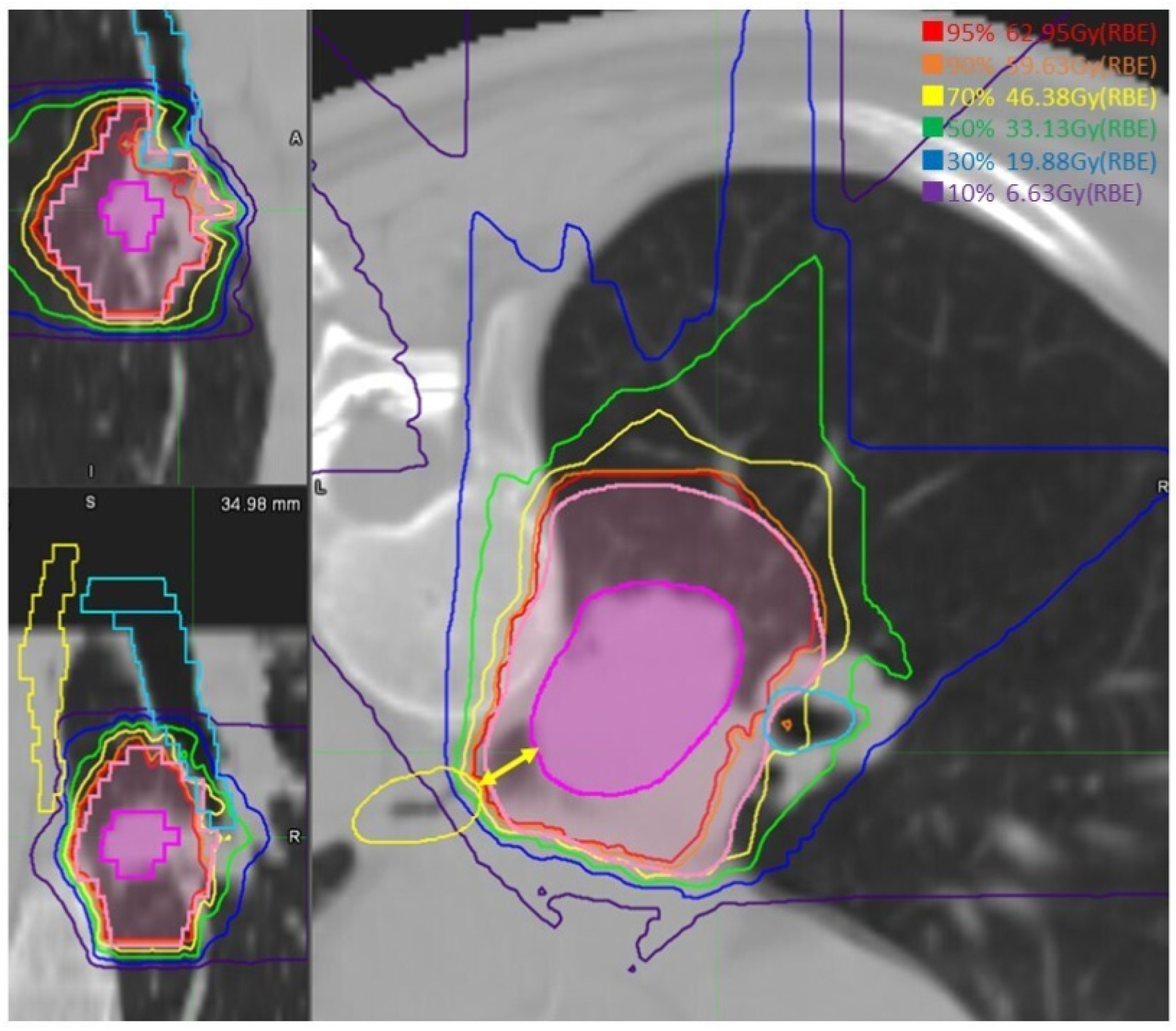
DVH was available for analysis in 26 patients, as the dose data were partially missing for the remaining four patients. Median GTV volume was 22.4 (0.9–98.6) cm3. The GTV dose was within the prescribed range of ±5% in all cases. For the lungs, median lung V5 and V20 were 15.5 (10.2–24.1) % and 10.4 (3.7–18.2) %, respectively. The MLD total was 5.6 (2.6–10.9) Gy. PBT-tumor distance was 2 (0–18) mm, median Dmax of PBT was 65.6 (3.8–71.2) Gy, median D0.2cc, D0.5cc, D1cc, and D2cc were 58.8 (2.8–70.7) Gy, 52.8 (2.7–69.2) Gy, 39.5 (2.4–68.6) Gy, and 10.0 (0.2–51.6) Gy, respectively. The median of the maximum dose to the PBT in the 8 patients with tumors touching the PBT was 67.8 (61.8–70.6) Gy, and in the 16 patients with tumors at 1 to 15 mm from the PBT, it was 61.4 (50.5–71.2) Gy. In contrast, in the remaining 2 patients with tumor distances of 18–20 mm from the PBT, the maximum PBT doses were extremely low at 3.8 and 1.9 Gy, respectively. The median maximum dose to the esophagus was 9.9 (1.8–60.7) Gy. In the highest case, where the distance to the GTV was 8.6 mm and that to the CTV was 2.0 mm, the maximum dose point was 60.7 Gy, which was 5.4 Gy in the D1cc setting (Figure 2). In contrast, the maximum dose to the esophagus was <20 Gy in 21 of the 23 cases with a distance to the tumor of ≥20 mm, and <10 Gy in all 12 cases with a distance to the tumor of ≥30 mm. The median D0.2cc, D0.5cc, and D1cc into the esophagus were 4.2 (1.4–37.5), 3.6 (0.8–30.9), and 3.0 (0.7–23.5) Gy, respectively. The doses delivered to the main OARs are listed in Table 2.

3.3. Treatment Effects

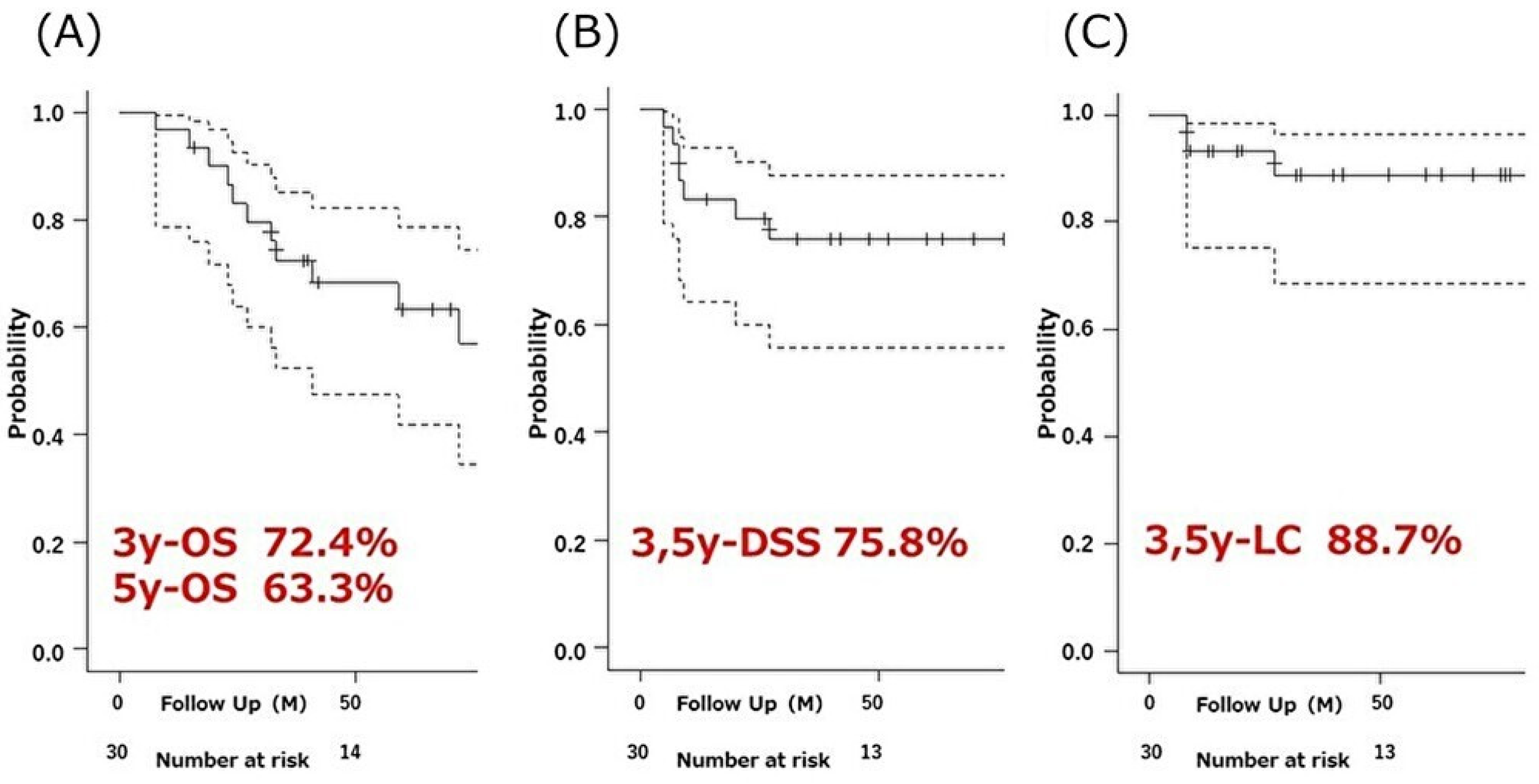
At the last follow-up, 14 patients (47%) had died, of which two were cause-specific deaths. Among the other 12, nine deaths were lung-related but treatment-unrelated (e.g., pneumonitis), 2 were cancer-related but without the recurrence of lung cancer, and 1 was from heart failure. The median OS was 77 months (range 41–NA). The 1-, 3-, and 5-year OS rates were 96.7% (95% CI: 78.6–99.5%), 72.4% (95% CI: 52.3–85.2%), and 63.3% (95% CI: 42.0–78.6%), respectively (Figure 3A). Seven patients (23%) had recurrences; three had local recurrence, five had non-local recurrences, and one had both. The five non-local recurrences included two patients with intrathoracic metastases (intrapulmonary or mediastinal lymph nodes), one with clavicular fossa lymph node metastasis, and two with distant organ metastasis (brain, bone, and liver). The 1-, 3-, and 5-year DSS rates were 83.2% (95% CI: 64.2–92.6%), 75.8% (95% CI: 64.2–92.6%), and 75.8% (95% CI: 55.7–87.7%), respectively (Figure 3B). The 1-, 3-, and 5-year LC rates were 93.1% (95% CI: 75.1–98.2%), 88.7% (95% CI: 68.5–96.2%), and 88.7% (95% CI: 68.5–96.2%), respectively (Figure 3C).
The results of the univariate analyses of the factors predicting OS are summarized in Table 3. Age (≤75 vs. >75 years, p = 0.047) and histology (squamous vs. other, p = 0.015) were significant prognosticators. In contrast, the KPS score, cancer stage, and COPD severity, which are known to affect life expectancy, were not significant factors.

3.4. Adverse Events

In the acute phase, grade 2 pneumonitis was observed in three patients (10%), and in the late phase, grade 2 and 3 pneumonitis developed in two cases (6.7%), respectively. The two grade 3 cases had stage 3 COPD prior to treatment and were assessed and treated for their symptoms rather than radiological findings. Grade 2 bronchial stenosis with wheezing and coughing occurred in one other case. There were no cases of grade ≥2 esophagitis, but grade 1 occurred in two patients (6.7%); of these, one had received the highest esophageal dose, 60.7 Gy. No grade ≥4 AEs occurred, and we did not observe serious AEs specific to central irradiation (e.g., hemoptysis, tracheoesophageal perforations) during the long-term observation period. Acute and late AEs are summarized in Table 4.

4. Discussion

4.1. Radiation Therapy for Central NSCLC

The usefulness of SBRT for peripheral early stage NSCLC has been confirmed [4,5,6,7,37,38]; however, its use in treating central lesions remains controversial due to the increased risk of serious AEs [10,11]. Central tumors are independent risk factors for severe AEs even when ultra-central tumors are excluded, and researchers have documented the high toxicity of central SBRT [10,12,14,15,39,40,41,42,43,44,45]. Table 5 summarizes reports on hypo fractionated RT for central lung tumors, mainly SBRT, for the treatment of NSCLC. Severe pneumonitis is the most common AE after central chest irradiation [10,14,40,42,43], grade 5 hemoptysis, tracheoesophageal perforation, and cardiovascular events have often been reported [10,14,39,40,41,42,43,44,45,46]. These AEs are specific to central SBRT with a high dose to the mediastinal organs; although rare, their onset can be sudden and difficult to predict and treat.

4.2. Tumor Control

Although there is no established dose prescription for photon SBRT to control stage I NSCLC, BED10 ≥ 100 Gy is considered a significant predictor of LC [42,47,48]. Results from a multicenter study in Japan [48] showed that SBRT for stage I NSCLC with BED10 ≥ 100 Gy significantly improved 3-year OS and LC. Another study reported a 2-year LC rate of 94% for BED10 ≥ 100 Gy and 80% for BED10 < 100 Gy (p = 0.02) [42]. While lower dose prescriptions are often used in central NSCLC due to the risk of AEs, SBRT using BED10 ≥ 100 Gy has been reported to provide acceptable tumor control [10,14,15,41].
To the best of our knowledge, there are no reports of ablative CIRT for central early stage lung cancer. However, there have been several reports on similar irradiation using proton beams. Kanemoto et al. [49] reported the results of PBT for stage I NSCLC using 72.6 Gy in 22 Fr (BED10 96.6 Gy) for central tumors and 66 Gy in 10–12 Fr (BED10 102.3–109.56 Gy) for peripheral tumors. The 3-year LC rates were 63.9% and 88.4%, respectively, suggesting insufficient dosing for central NSCLCs. Nakamura et al. [50] retrospectively studied cases of hypo fractionated proton beam therapy for central early stage lung cancer and reported that patients who received less than 110 Gy of BED10 had a lower progression-free survival. As with SBRT, it may be that a dose of BED10 ≥ 100 Gy is required for tumor control in proton beam therapy. Assuming the same estimation as for SBRT, the dose prescription in the present study was a uniform 107.4 Gy in BED10, a dose that is expected to provide adequate tumor control. Indeed, tumor control in this study was comparable to that reported for SBRT using a BED10 ≥ 100 Gy with regard to long-term results. Considering that the present study included relatively large tumors of T2 and T3 (UICC 8th), accounting for 60% and 13% of the cases, respectively, these results seem encouraging. Age (≤75 years) and pathology (squamous cell carcinoma) were identified as prognostic factors for OS in the univariate statistics, consistent with previous reports [51,52]. The absence of effects on tumor size and COPD severity may be attributable to the small sample size.
The risk of AEs will continue to make it difficult to prescribe a significant dose escalation for central lung tumors. However, as shown in a recent paper by Chang JY et al., SBRT combined with ICI has been reported to prolong event-free survival, even in Stage I NSCLC [53]. To improve prognosis, combined ICI could be a promising approach in the future, rather than increasing dose prescription or treatment margins of radiotherapy itself.

4.3. CIRT Safety

We identified two major clinical safety advantages of ablative CIRT for central early stage NSCLC. The first is the lower risk of radiation pneumonitis; the sharp dose distribution of CIRT has the advantage of decreased dosing to normal lung tissue [23,54]. Indeed, the doses to the lungs in the present study were sufficiently low across all indices, with notable differences from previous SBRT studies, particularly the mean lung dose (MLD) and lung V5 [14,55,56,57]. In recent years, in addition to lung V20 [51,52], low-dose lung areas, evaluated using lung V5, 10, etc., and MLD have received increased attention as indicators of dose-associated clinical pneumonitis [14,55,56,57]. The reduced low-dose lung area may have led to lower toxicity. However, previous reports of RT for central lung tumors suggest that even if the irradiation area is similarly limited, central radiation pneumonitis with stenosis in BPT tends to be severe. In the present study, two cases of grade 3 pneumonitis were observed, indicating a higher risk compared to that associated with CIRT for peripheral early stage lung cancers. However, most patients had interstitial pneumonitis or lung dysfunction that made other treatments difficult, which may also be related to the occurrence of severe pneumonitis. The second and greatest benefit of CIRT is the reduced risk of damage to mediastinal regions, such as the trachea, esophagus, and cardiovascular organs. In these organs, even small areas of high-dose irradiation can cause injuries, such as perforation, ulceration, and bleeding, which are rare but fatal AEs with sudden onset and poor predictability. In this study, no serious AEs occurred in the mediastinal organs during the long-term follow-up period. Although the sample size was small, these results indicate the safety of central CIRT.
Compared to photon SBRT, which uses multiportal and rotational techniques, CIRT allows for sufficient dose control with fewer beams, creating a linear dose gradient between the tumor and mediastinal OARs. As a result, it is possible to keep these OAR doses low even in central tumors with less than 2 cm between the tumor and the bronchial tree or other mediastinal organs. In previous reports, several dose parameters such as Dmax, D0.5cc, and D2cc have been used to describe the association between mediastinal OAR dose, AEs, and survival, with some reports using relatively large volume indicators such as D10cc [45,58,59]. Their views on volume effects on mediastinal organs suggest that a reduction in high dose irradiated volumes could lead to fewer serious AEs. The HILUS study concluded that D0,2cc, D0.5cc, and D1cc of the main bronchus plus trachea was most associated with grade 5 toxicity [29]. Farrugia M. et al. reported that D4cc (>18 Gy in 5 fr) for PBT correlated most with worse non-cancer-related survival [59]. In the present study, we evaluated OAR doses using Dmax, D0.2cc, D0.5cc, D1cc, and D2cc, and found that D > 0.5 cc, which is considered clinically important, was well controlled even in cases with high Dmax. This means that in CIRT, even in cases where the OAR is in proximity/contact with the target and partly exposed to high doses, it could be possible to reduce the extension of the high dose area, thereby avoiding serious AEs. Although there are no data directly comparing CIRT with other treatment modalities, a dosimetric report on the superiority of CIRT supports the present results [60,61]. Of course, the larger number of fractions than most reports on SBRT may have contributed to the safety profile of the present study, but at least in the treatment of central NSCLC, the CIRT regimen performed at our institution would be expected to have a relatively high safety profile. However, the advantages of CIRT may not be fully exploited for ultra-central tumors, particularly those touching or invading the esophagus or trachea, and the optimal dose prescription requires more careful exploration [29,30,46].

4.4. Limitations

The present study had several limitations. First, this was a retrospective study conducted at a single institution. Second, the small sample size may have limited the statistical power. Third, our ongoing treatment regimen development resulted in minor modifications to the irradiation protocol within the case series. Furthermore, methods for comparing the biological effects of CIRT and photon SBRT have not yet been established, and it remains difficult to compare data across institutions. Future basic and multicenter studies are required to explore the role of appropriate doses of CIRT in treating central NSCLC.

5. Conclusions

We reported the results of ablative CIRT (68.4 Gy in 12 Fr) for central NSCLC. CIRT is considered to be a safe and effective treatment for central NSCLC, as dose reduction to intrathoracic OARs reduces the risk of fatal AEs. Further research to clarify the most effective indications and dose constraints for CIRT could make a significant contribution to the treatment of central NSCLC.

Author Contributions

Conceptualization, S.A., M.N. and H.I.; Methodology, H.I., N.Y. and S.M.; Validation, S.A., S.M. and N.O.; Formal Analysis, S.A. and H.I.; Investigation, S.A. and M.N.; Resources, M.N. and S.A.; Data Curation, S.A., S.M., N.O. and M.N.; Writing—Original Draft Preparation, S.A.; Writing—Review and Editing, H.I.; Supervision, H.I., S.Y., M.W., K.M., Y.T. and T.M.; Project Administration, H.I., I.Y. and S.Y. All authors have read and agreed to the published version of the manuscript.

Funding

This work was supported by JSPS KAKENHI Grant Number JP50773496.

Institutional Review Board Statement

This study was approved by our institution’s ethics committee (approval number: N22-023) and registered with the University Hospital Medical Information Network (UMIN) (identification number: 23K14666). This study was conducted in accordance with the principles of the Declaration of Helsinki.

Informed Consent Statement

Informed consent was obtained from all subjects involved in this study.

Data Availability Statement

The data presented in this study are available on request from the corresponding author.

Conflicts of Interest

The authors declare no conflicts of interest.

References

  1. de Perrot, M.; Licker, M.; Reymond, M.A.; Robert, J.; Spiliopoulos, A. Influence of age on operative mortality and long-term survival after lung resection for bronchogenic carcinoma. Eur. Respir. J. 1999, 14, 419–422. [Google Scholar] [CrossRef] [PubMed]
  2. Postmus, P.E.; Kerr, K.M.; Oudkerk, M.; Senan, S.; Waller, D.A.; Vansteenkiste, J.; Escriu, C.; Peters, S. ESMO Guidelines Committee. Early and locally advanced non-small-cell lung cancer (NSCLC): ESMO Clinical Practice Guidelines for diagnosis, treatment and follow-up. Ann. Oncol. 2017, 28 (Suppl. S4), iv1–iv21. [Google Scholar] [CrossRef]
  3. Howington, J.A.; Blum, M.G.; Chang, A.C.; Balekian, A.A.; Murthy, S.C. Treatment of stage I and II non-small cell lung cancer: Diagnosis and management of lung cancer, 3rd ed: American College of Chest Physicians evidence-based clinical practice guidelines. Chest 2013, 143 (Suppl. S5), e278S–e313S. [Google Scholar] [CrossRef] [PubMed]
  4. Nagata, Y.; Hiraoka, M.; Shibata, T.; Onishi, H.; Kokubo, M.; Karasawa, K.; Shioyama, Y.; Onimaru, R.; Kozuka, T.; Kunieda, E.; et al. Prospective Trial of Stereotactic Body Radiation Therapy for Both Operable and Inoperable T1N0M0 Non-Small Cell Lung Cancer: Japan Clinical Oncology Group Study JCOG0403. Int. J. Radiat. Oncol. Biol. Phys. 2015, 93, 989–996. [Google Scholar] [CrossRef]
  5. Tandberg, D.J.; Tong, B.C.; Ackerson, B.G.; Kelsey, C.R. Surgery versus stereotactic body radiation therapy for stage I non-small cell lung cancer: A comprehensive review. Cancer 2018, 124, 667–678. [Google Scholar] [CrossRef] [PubMed]
  6. Timmerman, R.D.; Hu, C.; Michalski, J.M. Bradley JC, Galvin J, Johnstone DW, Choy H. Long-term Results of Stereotactic Body Radiation Therapy in Medically Inoperable Stage I Non-Small Cell Lung Cancer. JAMA Oncol. 2018, 4, 1287–1288. [Google Scholar] [CrossRef] [PubMed]
  7. Timmerman, R.D.; Hu, C.; Michalski, J.M.; Bradley, J.C.; Galvin, J.; Johnstone, D.W.; Straube, W.L.; Nedzi, L.A.; McGarry, R.C.; Robinson, C.G.; et al. Stereotactic Body Radiation Therapy for Operable Early-Stage Lung Cancer: Findings From the NRG Oncology RTOG 0618 Trial. JAMA Oncol. 2018, 4, 1263–1266. [Google Scholar] [CrossRef]
  8. Qu, R.; Ping, W.; Hao, Z.; Cai, Y.; Zhang, N.; Fu, X. Surgical outcomes of segmental bronchial sleeve resection in central non-small cell lung cancer. Thorac. Cancer 2020, 11, 1319–1325. [Google Scholar] [CrossRef]
  9. Berthet, J.P.; Paradela, M.; Jimenez, M.J.; Molins, L.; Gómez-Caro, A. Extended sleeve lobectomy: One more step toward avoiding pneumonectomy in centrally located lung cancer. Ann. Thorac. Surg. 2013, 96, 1988–1997. [Google Scholar] [CrossRef]
  10. Timmerman, R.; McGarry, R.; Yiannoutsos, C.; Papiez, L.; Tudor, K.; DeLuca, J.; Ewing, M.; Abdulrahman, R.; DesRosiers, C.; Williams, M.; et al. Excessive toxicity when treating central tumors in a phase II study of stereotactic body radiation therapy for medically inoperable early-stage lung cancer. J. Clin. Oncol. 2006, 24, 4833–4839. [Google Scholar] [CrossRef]
  11. Chang, J.Y.; Li, Q.Q.; Xu, Q.Y.; Allen, P.K.; Rebueno, N.; Gomez, D.R.; Balter, P.; Komaki, R.; Mehran, R.; Swisher, S.G.; et al. Stereotactic ablative radiation therapy for centrally located early stage or isolated parenchymal recurrences of non-small cell lung cancer: How to fly in a “no fly zone”. Int. J. Radiat. Oncol. Biol. Phys. 2014, 88, 1120–1128. [Google Scholar] [CrossRef]
  12. Modh, A.; Rimner, A.; Williams, E.; Foster, A.; Shah, M.; Shi, W.; Zhang, Z.; Gelblum, D.Y.; Rosenzweig, K.E.; Yorke, E.D.; et al. Local control and toxicity in a large cohort of central lung tumors treated with stereotactic body radiation therapy. Int. J. Radiat. Oncol. Biol. Phys. 2014, 90, 1168–1176. [Google Scholar] [CrossRef]
  13. Roach, M.C.; Robinson, C.G.; DeWees, T.A.; Ganachaud, J.; Przybysz, D.; Drzymala, R.; Rehman, S.; Kashani, R.; Bradley, J.D. Stereotactic Body Radiation Therapy for Central Early-Stage NSCLC: Results of a Prospective Phase I/II Trial. J. Thorac. Oncol. 2018, 13, 1727–1732. [Google Scholar] [CrossRef]
  14. Aoki, S.; Yamashita, H.; Haga, A.; Ota, T.; Takahashi, W.; Ozaki, S.; Nawa, K.; Imae, T.; Abe, O.; Nakagawa, K. Stereotactic body radiotherapy for centrally-located lung tumors with 56 Gy in seven fractions: A retrospective study. Oncol. Lett. 2018, 16, 4498–4506. [Google Scholar] [CrossRef]
  15. Bezjak, A.; Paulus, R.; Gaspar, L.E.; Timmerman, R.D.; Straube, W.L.; Ryan, W.F.; Garces, Y.I.; Pu, A.T.; Singh, A.K.; Videtic, G.M.; et al. Safety and Efficacy of a Five-Fraction Stereotactic Body Radiotherapy Schedule for Centrally Located Non-Small-Cell Lung Cancer: NRG Oncology/RTOG 0813 Trial. J. Clin. Oncol. 2019, 37, 1316–1325. [Google Scholar] [CrossRef]
  16. Arnett, A.L.H.; Mou, B.; Owen, D.; Park, S.S.; Nelson, K.; Hallemeier, C.L.; Sio, T.; Garces, Y.I.; Olivier, K.R.; Merrell, K.W. Long-term Clinical Outcomes and Safety Profile of SBRT for Centrally Located NSCLC. Adv. Radiat. Oncol. 2019, 4, 422–428. [Google Scholar] [CrossRef] [PubMed]
  17. Owen, D.; Sio, T.T. Stereotactic body radiotherapy (SBRT) for central and ultra-central node-negative lung tumors. J. Thorac. Dis. 2020, 12, 7024–7031. [Google Scholar] [CrossRef] [PubMed]
  18. Kanai, T.; Furusawa, Y.; Fukutsu, K.; Itsukaichi, H.; Eguchi-Kasai, K.; Ohara, H. Irradiation of mixed beam and design of spread-out Bragg peak for heavy-ion radiotherapy. Radiat. Res. 1997, 147, 78–85. [Google Scholar] [CrossRef] [PubMed]
  19. Schulz-Ertner, D.; Tsujii, H. Particle radiation therapy using proton and heavier ion beams. J. Clin. Oncol. 2007, 25, 953–964. [Google Scholar] [CrossRef] [PubMed]
  20. Kanematsu, N.; Inaniwa, T. Biological dose representation for carbon-ion radiotherapy of unconventional fractionation. Phys. Med. Biol. 2017, 62, 1062–1075. [Google Scholar] [CrossRef] [PubMed]
  21. Hamada, N.; Imaoka, T.; Masunaga, S.; Ogata, T.; Okayasu, R.; Takahashi, A.; Kato, T.A.; Kobayashi, Y.; Ohnishi, T.; Ono, K.; et al. Recent advances in the biology of heavy-ion cancer therapy. J. Radiat. Res. 2010, 51, 365–383. [Google Scholar] [CrossRef]
  22. Blakely, E.A.; Kronenberg, A. Heavy-ion radiobiology: New approaches to delineate mechanisms underlying enhanced biological effectiveness. Radiat. Res. 1998, 150 (Suppl. S5), S126–S145. [Google Scholar] [CrossRef] [PubMed]
  23. Miyamoto, T.; Yamamoto, N.; Nishimura, H.; Koto, M.; Tsujii, H.; Mizoe, J.E.; Hirasawa, N.; Sugawara, T.; Yamamoto, N.; Koto, M.; et al. Carbon ion radiotherapy for stage I non-small cell lung cancer using a regimen of four fractions during 1 week. J. Thorac. Oncol. 2007, 2, 916–926. [Google Scholar] [CrossRef]
  24. Yamamoto, N.; Miyamoto, T.; Nakajima, M.; Karube, M.; Hayashi, K.; Tsuji, H.; Tsujii, H.; Kamada, T.; Fujisawa, T. A Dose Escalation Clinical Trial of Single-Fraction Carbon Ion Radiotherapy for Peripheral Stage I Non-Small Cell Lung Cancer. J. Thorac. Oncol. 2017, 12, 673–680. [Google Scholar] [CrossRef]
  25. Ono, T.; Yamamoto, N.; Nomoto, A.; Nakajima, M.; Isozaki, Y.; Kasuya, G.; Ishikawa, H.; Nemoto, K.; Tsuji, H. Long Term Results of Single-Fraction Carbon-Ion Radiotherapy for Non-small Cell Lung Cancer. Cancers 2020, 13, 112. [Google Scholar] [CrossRef]
  26. Lodeweges, J.E.; van Rossum, P.S.N.; Bartels, M.M.T.J.; van Lindert, A.S.R.; Pomp, J.; Peters, M.; Verhoeff, J.J.C. Ultra-central lung tumors: Safety and efficacy of protracted stereotactic body radiotherapy. Acta Oncol. 2021, 60, 1061–1068. [Google Scholar] [CrossRef]
  27. Guillaume, E.; Tanguy, R.; Ayadi, M.; Claude, L.; Sotton, S.; Moncharmont, C.; Magné, N.; Martel-Lafay, I. Toxicity and efficacy of stereotactic body radiotherapy for ultra-central lung tumours: A single institution real life experience. Br. J. Radiol. 2022, 95, 20210533. [Google Scholar] [CrossRef]
  28. Xiao, Y.; Papiez, L.; Paulus, R.; Timmerman, R.; Straube, W.L.; Bosch, W.R.; Michalski, J.; Galvin, J.M. Dosimetric evaluation of heterogeneity corrections for RTOG 0236: Stereotactic body radiotherapy of inoperable stage I-II non-small-cell lung cancer. Int. J. Radiat. Oncol. Biol. Phys. 2009, 73, 1235–1242. [Google Scholar] [CrossRef]
  29. Lindberg, K.; Grozman, V.; Karlsson, K.; Lindberg, S.; Lax, I.; Wersäll, P.; Persson, G.F.; Josipovic, M.; Khalil, A.A.; Moeller, D.S.; et al. The HILUS-Trial-a Prospective Nordic Multicenter Phase 2 Study of Ultracentral Lung Tumors Treated With Stereotactic Body Radiotherapy. J. Thorac. Oncol. 2021, 16, 1200–1210. [Google Scholar] [CrossRef] [PubMed]
  30. Mori, S.; Furukawa, T.; Inaniwa, T.; Zenklusen, S.; Nakao, M.; Shirai, T.; Noda, K. Systematic evaluation of four-dimensional hybrid depth scanning for carbon-ion lung therapy. Med. Phys. 2013, 40, 031720. [Google Scholar] [CrossRef]
  31. Kong, F.M.; Ritter, T.; Quint, D.J.; Senan, S.; Gaspar, L.E.; Komaki, R.U.; Hurkmans, C.W.; Timmerman, R.; Bezjak, A.; Bradley, J.D.; et al. Consideration of dose limits for organs at risk of thoracic radiotherapy: Atlas for lung, proximal bronchial tree, esophagus, spinal cord, ribs, and brachial plexus. Int. J. Radiat. Oncol. Biol. Phys. 2011, 81, 1442–1457. [Google Scholar] [CrossRef]
  32. Minohara, S.; Kanai, T.; Endo, M.; Noda, K.; Kanazawa, M. Respiratory gated irradiation system for heavy-ion radiotherapy. Int. J. Radiat. Oncol. Biol. Phys. 2000, 47, 1097–1103. [Google Scholar] [CrossRef] [PubMed]
  33. Mori, S.; Shirai, T.; Takei, Y.; Furukawa, T.; Inaniwa, T.; Matsuzaki, Y.; Kumagai, M.; Murakami, T.; Noda, K. Patient handling system for carbon ion beam scanning therapy. J. Appl. Clin. Med. Phys. 2012, 13, 3926. [Google Scholar] [CrossRef] [PubMed]
  34. Karger, C.P.; Peschke, P. RBE and related modeling in carbon-ion therapy. Phys. Med. Biol. 2017, 63, 01TR02. [Google Scholar] [CrossRef] [PubMed]
  35. Common Terminology Criteria for Adverse Events (CTCAE) Version 5.0. Available online: http://evs.nci.nih.gov/ftp1/CTCAE/CTCAE_4.03_2010-06-14_QuickReference_8.5x11.pdf (accessed on 15 January 2024).
  36. 2020 Global Initiative for Chronic Obstructive Lung Disease, Inc. 2011. Available online: https://goldcopd.org/wp-content/uploads/2020/11/GOLD-REPORT-2021-v1.1-25Nov20_WMV.pdf (accessed on 15 January 2024).
  37. Nishio, T.; Kunieda, E.; Shirato, H.; Ishikura, S.; Onishi, H.; Tateoka, K.; Hiraoka, M.; Narita, Y.; Ikeda, M.; Goka, T. Dosimetric verification in participating institutions in a stereotactic body radiotherapy trial for stage I non-small cell lung cancer: Japan clinical oncology group trial (JCOG0403). Phys. Med. Biol. 2006, 51, 5409–5417. [Google Scholar] [CrossRef] [PubMed]
  38. Onimaru, R.; Onishi, H.; Ogawa, G.; Hiraoka, M.; Ishikura, S.; Karasawa, K.; Matsuo, Y.; Kokubo, M.; Shioyama, Y.; Matsushita, H.; et al. Final report of survival and late toxicities in the Phase I study of stereotactic body radiation therapy for peripheral T2N0M0 non-small cell lung cancer (JCOG0702). Jpn. J. Clin. Oncol. 2018, 48, 1076–1082. [Google Scholar] [CrossRef] [PubMed]
  39. Milano, M.T.; Chen, Y.; Katz, A.W.; Philip, A.; Schell, M.C.; Okunieff, P. Central thoracic lesions treated with hypofractionated stereotactic body radiotherapy. Radiother. Oncol. 2009, 91, 301–306. [Google Scholar] [CrossRef]
  40. Song, S.Y.; Choi, W.; Shin, S.S.; Lee, S.W.; Ahn, S.D.; Kim, J.H.; Je, H.U.; Park, C.I.; Lee, J.S.; Choi, E.K. Fractionated stereotactic body radiation therapy for medically inoperable stage I lung cancer adjacent to central large bronchus. Lung Cancer 2009, 66, 89–93. [Google Scholar] [CrossRef]
  41. Haasbeek, C.J.; Lagerwaard, F.J.; Slotman, B.J.; Senan, S. Outcomes of stereotactic ablative radiotherapy for centrally located early-stage lung cancer. J. Thorac. Oncol. 2011, 6, 2036–2043. [Google Scholar] [CrossRef]
  42. Rowe, B.P.; Boffa, D.J.; Wilson, L.D.; Kim, A.W.; Detterbeck, F.C.; Decker, R.H. Stereotactic body radiotherapy for central lung tumors. J. Thorac. Oncol. 2012, 7, 1394–1399. [Google Scholar] [CrossRef]
  43. Tekatli, H.; Spoelstra, F.O.B.; Palacios, M.; van Sornsen de Koste, J.; Slotman, B.J.; Senan, S. Stereotactic ablative radiotherapy (SABR) for early-stage central lung tumors: New insights and approaches. Lung Cancer 2018, 123, 142–148. [Google Scholar] [CrossRef] [PubMed]
  44. Haseltine, J.M.; Rimner, A.; Gelblum, D.Y.; Modh, A.; Rosenzweig, K.E.; Jackson, A.; Yorke, E.D.; Wu, A.J. Fatal complications after stereotactic body radiation therapy for central lung tumors abutting the proximal bronchial tree. Pract. Radiat. Oncol. 2016, 6, e27–e33. [Google Scholar] [CrossRef] [PubMed]
  45. Li, Q.; Swanick, C.W.; Allen, P.K.; Gomez, D.R.; Welsh, J.W.; Liao, Z.; Balter, P.A.; Chang, J.Y. Stereotactic ablative radiotherapy (SABR) using 70 Gy in 10 fractions for non-small cell lung cancer: Exploration of clinical indications. Radiother. Oncol. 2014, 112, 256–261. [Google Scholar] [CrossRef]
  46. Wang, C.; Rimner, A.; Gelblum, D.Y.; Dick-Godfrey, R.; McKnight, D.; Torres, D.; Flynn, J.; Zhang, Z.; Sidiqi, B.; Jackson, A.; et al. Analysis of pneumonitis and esophageal injury after stereotactic body radiation therapy for ultra-central lung tumors. Lung Cancer 2020, 147, 45–48. [Google Scholar] [CrossRef]
  47. Jones, B.; Dale, R.G.; Deehan, C.; Hopkins, K.I.; Morgan, D.A. The role of biologically effective dose (BED) in clinical oncology. Clin. Oncol. 2001, 13, 71–81. [Google Scholar]
  48. Onishi, H.; Araki, T.; Shirato, H.; Nagata, Y.; Hiraoka, M.; Gomi, K.; Yamashita, T.; Niibe, Y.; Karasawa, K.; Hayakawa, K.; et al. Stereotactic hypofractionated high-dose irradiation for stage I nonsmall cell lung carcinoma: Clinical outcomes in 245 subjects in a Japanese multiinstitutional study. Cancer 2004, 101, 1623–1631. [Google Scholar] [CrossRef] [PubMed]
  49. Kanemoto, A.; Okumura, T.; Ishikawa, H.; Mizumoto, M.; Oshiro, Y.; Kurishima, K.; Homma, S.; Hashimoto, T.; Ohkawa, A.; Numajiri, H.; et al. Outcomes and prognostic factors for recurrence after high-dose proton beam therapy for centrally and peripherally located stage I non-small-cell lung cancer. Clin. Lung Cancer 2014, 15, e7–e12. [Google Scholar] [CrossRef]
  50. Nakamura, N.; Hotta, K.; Zenda, S.; Baba, H.; Kito, S.; Akita, T.; Motegi, A.; Hojo, H.; Nakamura, M.; Parshuram, R.V.; et al. Hypofractionated proton beam therapy for centrally located lung cancer. J. Med. Imaging Radiat. Oncol. 2019, 63, 552–556. [Google Scholar] [CrossRef]
  51. Katagiri, Y.; Jingu, K.; Yamamoto, T.; Matsushita, H.; Umezawa, R.; Ishikawa, Y.; Takahashi, N.; Takeda, K.; Tasaka, S.; Kadoya, N. Differences in patterns of recurrence of squamous cell carcinoma and adenocarcinoma after radiotherapy for stage III non-small cell lung cancer. Jpn. J. Radiol. 2021, 39, 611–617. [Google Scholar] [CrossRef]
  52. Wang, B.Y.; Huang, J.Y.; Chen, H.C.; Lin, C.H.; Lin, S.H.; Hung, W.H.; Cheng, Y.F. The comparison between adenocarcinoma and squamous cell carcinoma in lung cancer patients. J. Cancer Res. Clin. Oncol. 2020, 146, 43–52. [Google Scholar] [CrossRef]
  53. Chang, J.Y.; Lin, S.H.; Dong, W.; Liao, Z.; Gandhi, S.J.; Gay, C.M.; Zhang, J.; Chun, S.G.; Elamin, Y.Y.; Fossella, F.V.; et al. Stereotactic ablative radiotherapy with or without immunotherapy for early-stage or isolated lung parenchymal recurrent node-negative non-small-cell lung cancer: An open-label, randomised, phase 2 trial. Lancet 2023, 402, 871–881. [Google Scholar] [CrossRef]
  54. Nakajima, M.; Yamamoto, N.; Hayashi, K.; Karube, M.; Ebner, D.K.; Takahashi, W.; Anzai, M.; Tsushima, K.; Tada, Y.; Tatsumi, K.; et al. Carbon-ion radiotherapy for non-small cell lung cancer with interstitial lung disease: A retrospective analysis. Radiat. Oncol. 2017, 12, 144. [Google Scholar] [CrossRef]
  55. Rosca, F.; Kirk, M.; Soto, D.; Sall, W.; McIntyre, J. Reducing the low-dose lung radiation for central lung tumors by restricting the IMRT beams and arc arrangement. Med. Dosim. 2012, 37, 280–286. [Google Scholar] [CrossRef]
  56. Devpura, S.; Feldman, A.M.; Rusu, S.D.; Mayyas, E.; Movsas, A.; Brown, S.L.; Cook, A.; Simoff, M.J.; Sun, Z.; Lu, M.; et al. An Analysis of Clinical Toxic Effects and Quality of Life as a Function of Radiation Dose and Volume After Lung Stereotactic Body Radiation Therapy. Adv. Radiat. Oncol. 2021, 6, 100815. [Google Scholar] [CrossRef] [PubMed]
  57. Lin, J.B.; Hung, L.C.; Cheng, C.Y.; Chien, Y.A.; Lee, C.H.; Huang, C.C.; Chou, T.W.; Ko, M.H.; Lai, Y.C.; Liu, M.T.; et al. Prognostic significance of lung radiation dose in patients with esophageal cancer treated with neoadjuvant chemoradiotherapy. Radiat. Oncol. 2019, 14, 85. [Google Scholar] [CrossRef] [PubMed]
  58. Duijm, M.; Schillemans, W.; Aerts, J.G.; Heijmen, B.; Nuyttens, J.J. Dose and Volume of the Irradiated Main Bronchi and Related Side Effects in the Treatment of Central Lung Tumors with Stereotactic Radiotherapy. Semin. Radiat. Oncol. 2016, 26, 140–148. [Google Scholar] [CrossRef] [PubMed]
  59. Farrugia, M.; Ma, S.J.; Hennon, M.; Nwogu, C.; Dexter, E.; Picone, A.; Demmy, T.; Yendamuri, S.; Yu, H.; Fung-Kee-Fung, S.; et al. Exceeding Radiation Dose to Volume Parameters for the Proximal Airways with Stereotactic Body Radiation Therapy Is More Likely for Ultra-central Lung Tumors and Associated with Worse Outcome. Cancers 2021, 13, 3463. [Google Scholar] [CrossRef]
  60. Kubo, N.; Saitoh, J.I.; Shimada, H.; Shirai, K.; Kawamura, H.; Ohno, T.; Nakano, T. Dosimetric comparison of carbon ion and X-ray radiotherapy for Stage IIIA non-small cell lung cancer. J. Radiat. Res. 2016, 57, 548–554. [Google Scholar] [CrossRef]
  61. Ebara, T.; Shimada, H.; Kawamura, H.; Shirai, K.; Saito, J.; Kawashima, M.; Tashiro, M.; Ohno, T.; Kanai, T.; Nakano, T. Dosimetric analysis between carbon ion radiotherapy and stereotactic body radiotherapy in stage I lung cancer. Anticancer Res. 2014, 34, 5099–5104. [Google Scholar] [PubMed]
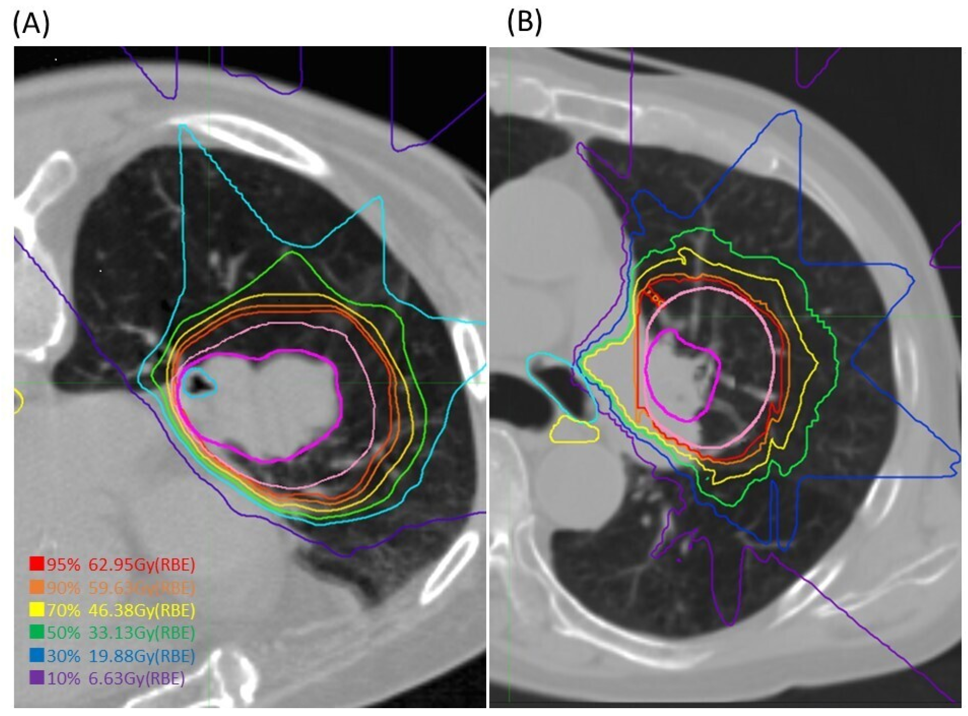
Figure 1. Dose distributions of CIRT for central NSCLC. The red, orange, yellow, green, blue, and purple lines show doses of 95%, 90%, 70%, 50%, 30%, and 10%, respectively; GTV was drawn in magenta, and CTV in pale pink. In cases where the OAR (in this case the lobe bronchus) is within the tumour, the OAR dose is sometimes as high as the tumour (A), but if there is a small distance from the tumour, the OAR can be successfully avoided by adjusting the beam direction (B).
Cancers 16 00933 g001
Figure 2. Dose distribution for one case with the highest maximum dose to the esophagus. The point dose was high at 60.7 Gy, while the D1cc dose was 5.4 Gy. The minimum distance between the GTV (magenta) and the esophagus, shown by a yellow arrow, was 8.6 mm.
Cancers 16 00933 g002
Figure 3. Kaplan–Meier curves of (A) overall survival (OS), (B) disease-specific survival (DSS), and (C) local control (LC) in the whole cohort.
Cancers 16 00933 g003
Table 1. Patient characteristics (n = 30).
Age (y)median (range)75 (55–85)
GenderMale/Female21/9
ECOG performance status0/124/6
OperabilityYes/No10/20
Severity of COPD: GOLD score1/2/3/4
HypertensionYes/No13/17
Diabetes mellitusYes/No5/25
Cardiovascular diseaseYes/No6/24
HistologyAdeno15
Squamous13
Others2
c-Stage (UICC 8th)1A8
1B12
2A6
2B4
Most constrained OARMain bronchus4
Lobar bronchus23
Others3
Baseline pulmonary functionFEV1 (liter, median)1.7 (0.4–2.8)
FEV1% (median)73.8 (28.3–90.0)
VC (liter, median)2.6 (1.2–4.5)
VC% (median)87.8 (65.6–138.1)
Age (y)median (range)75 (55–85)
Abbreviations. ECOG, Eastern Cooperative Oncology Group; COPD, chronic obstructive pulmonary disease; GOLD, Global Initiative for Chronic Obstructive Lung Disease; c-Stage, clinical stage; OAR, organ at risk; FEV1, forced expiratory volume in 1 s; VC, vital capacity.
Table 2. Irradiation doses for major OARs.
OARsDose ParametersTotal Dose
(Median [Range])
LungV5_%15.2 (10.2–24.1)
V20_%10.4 (4.1–18.2)
MLD_Gy5.6 (2.7–10.9)
PBTDmax_Gy65.6 (3.8–71.2)
D0.2cc_Gy58.8 (2.8–70.7)
D0.5cc_Gy52.8 (2.7–69.2)
D1cc_Gy39.5 (2.4–68.6)
D2cc_Gy10.0 (0.2–51.6)
EsophagusDmax_Gy9.9 (1.8–60.7)
D0.2cc_Gy4.2 (1.4–37.5)
D0.5cc_Gy3.6 (0.8–30.9)
D1cc_Gy3.0 (0.7–23.5)
Spinal cordDmax_Gy2.4 (0–18.2)
D0.2cc_Gy1.5 (0.0–17.4)
Abbreviations. PBT, proximal bronchial tree; V5 (20), percentage of the volume of an organ receiving 5 (20) Gy; MLD, mean lung dose; Dmax, maximum point dose; D0.2cc/0.5cc/1cc/2cc, dose to the hottest 0.2cc/0.5cc/1cc/2cc of OAR.
Table 3. Factors Associated with OS.
No. of PatientsOS
5y (%)p-Value
Age≤751485.10.047 *
>751646.4
GenderMale2150.30.18
Female988.9
PS02473.80.088
1633.3
Severity of COPDGOLD1,22467.70.16
GOLD3,4641.7
c-Stage12074.70.75
21040.5
HistologySquamous1336.90.015 *
the others1784.7
Abbreviations. ECOG, Eastern Cooperative Oncology Group; c-Stage, clinical stage; OS, overall survival; *, p < 0.05.
Table 4. Acute and late adverse events of all patients (n = 30).
Acute Adverse EventsGrade2Grade3Grade4–5
Pneumonitis3 (10.0%)00
Dermatitis000
Esophagitis000
Late Adverse EventsGrade2Grade3Grade4–5
Pneumonitis2 (6.7%)2 (6.7%)0
Dermatitis000
Esophagitis000
Chest wall pain000
Bronchial stenosis1 (3.3%)00
Table 5. Previous reports of ablative radiation therapy for central lung tumors.
Author (Year)DiseaseNo. of ptsPrescription
(Gy/Fr)
BED10
(Gy)
Follow
(m)
LC (%)OS (%)ToxicityDetails of G5 Toxicity
G ≥ 3 (%)G5 (%)
Timmerman (2006) [13]T1–2N07060–66/3180–211.21895 (2 y)54.7 (2 y)208.61 hemorrhage,
1 peripheral effusion
4 pneumonias
Milano (2009) [39]T1–3N0 or M15330–63
(2.5–5 Gy/Fr)
39–82.51073 (2 y)44 (2 y)11 (n = 45)8.93 pulmonary declines,
1 hemoptysis
Song (2009) [40]T1–2aN03240–60/4–680–1802785.3 (2 y)38.5 (2 y)33.3 (n = 9)11.11 hemorrhage
Haasbeek (2011) [41]T1–3N06360/81053592.6 (3 y)64.3 (3 y)20.614.39 cardiopulmonary causes
Rowe (2012) [42]T1–2N0 or M14750/476 (60–151.2)1194 (2 y)N/R10.62.21 hemorrhage
Modh (2014) [15]T1–4N0 or M112530–60/2–585.5 (43–180)1779 (2 y, BED ≥ 80)64 (2 y, BED ≥ 80)861 hemorrhage
Tekatli (2015) [43]stage 1A-48060/81054562 (2 y), 53 (3 y)N/R13.8 (n = 78)7.53 respiratory failures,
2 hemorrhages,
1 sudden death
Aoki (2018) [14]T1–3N0 or M13556/7100.813.196 (2 y)40.4 (2 y)265.71 hemorrhage, 1 pneumonia
Bezjak (2019) [15]T1–2N07157.5–60/5123.6, 1323389.4, 87.9 (2 y)67.9, 72.7 (2 y)215.63 hemorrhages,
1 esophageal ulcer
Lindberg. K
(2021) [29]
≤5 cm
(NSCLC/LM)
6556/895.22483 (2 y)58 (2 y)39.317.98 hemorrhage, 1 pneumonitis, 1 fistula
Current studyT1–3N03068.4/12107.44288.7 (3 y, 5 y)72.4 (3 y), 63.3 (5 y)6.70
Abbreviations: pt, patients; BED, biological equivalent dose; LC, local control; OS, overall survival; G, grade; Fr, fraction; y, years; n, number; N/R, data not reported; NSCLC, non-small cell lung cancer; LM, lung metastases.
Disclaimer/Publisher’s Note: The statements, opinions and data contained in all publications are solely those of the individual author(s) and contributor(s) and not of MDPI and/or the editor(s). MDPI and/or the editor(s) disclaim responsibility for any injury to people or property resulting from any ideas, methods, instructions or products referred to in the content.