Article

Chondrosarcoma of the Femur: Is Local Recurrence Influenced by the Presence of an Extraosseous Component?

1
Department of Orthopaedics and Traumatology, Helsinki University Hospital, University of Helsinki, 00100 Helsinki, Finland
2
Royal Orthopaedic Hospital, Birmingham and Aston University Medical School, Aston University, Birmingham B4 7ET, UK
3
Royal Orthopaedic Hospital, Birmingham B31 2AP, UK
4
Royal Orthopaedic Hospital, Faculty of Health Sciences, Aston University, Birmingham B4 7ET, UK
*
Author to whom correspondence should be addressed.
Cancers 2024, 16(2), 363; https://doi.org/10.3390/cancers16020363
Submission received: 7 December 2023 / Revised: 2 January 2024 / Accepted: 12 January 2024 / Published: 15 January 2024
(This article belongs to the Special Issue Advances in Bone Tumor and Sarcoma)

Abstract

:

Simple Summary

This study focused on conventional central chondrosarcoma (CS) in the femur, investigating the impact of an extraosseous tumor component on local recurrence-free survival (LRFS) and disease-specific survival (DSS). Analyzing data from 202 patients, we identified factors influencing LRFS and DSS, highlighting the significance of the extraosseous tumor component, histological grade, and achieved surgical margins. Notably, the location of the extraosseous component within the femur significantly affected local recurrence rates, emphasizing the challenge of achieving adequate margins, especially in the region around the greater trochanter. The study underscores the importance of precise imaging and the need for aggressive resection to prevent local recurrence, particularly in cases involving the proximal extremity of the femur. Understanding these factors aids in tailoring surgical approaches for improved outcomes in managing femoral CS.

Abstract

Background: Chondrosarcoma (CS) is the second most common surgically treated primary malignancy of the bone. The current study explored the effect of the margin and extraosseous tumor component in CS in the femur on local recurrence (LR), LR-free survival (LRFS), and disease-specific survival (DSS). Methods: Among 202 patients, 115 were in the proximal extremity of the femur, 4 in the corpus of the femur, and 83 in the distal extremity of femur; 105 patients had an extraosseous tumor component. Results: In the Kaplan–Meier analysis, factors significant for decreased LRFS were the extraosseous tumor component (p < 0.001), extraosseous tumor component arising from the superior aspect (p < 0.001), histological grade (p = 0.031), and narrow surgical margin < 3 mm (p < 0.001). Factors significantly affecting DSS were the histological grade (p < 0.001), extraosseous component (p < 0.001), LR (p < 0.001), metastases (p < 0.001), and surgical margin (p < 0.001). Conclusions: In CS of the femur, the presence of an extraosseous tumor component has a predictive role in LRFS, and extraosseous tumor component arising from the superior aspect was significant for decreased LRFS. Wide margins were more commonly achieved when the tumor had only an intraosseous component, and the rate of LR was significantly higher in cases with an extraosseous tumor component. When the extraosseous component arose from the superior aspect of the femur, LR occurred more frequently despite achieving adequate margins.

1. Introduction

Chondrosarcoma (CS) is the second most common surgically treated primary malignant bone tumor, accounting for approximately 20% of all bone sarcomas [1,2,3,4]. Conventional CSs constitute about 85% of all CSs and can be categorized according to their location within the bone, as central or (secondary) peripheral CS [5]. Central and peripheral CS are histologically similar and graded as low grade (grade 1) or high grade (grades 2 and 3). Grade 1 CS has more recently been classified as an atypical cartilaginous tumor (ACT) in the extremities, an intermediate tumor that is locally aggressive but does not metastasize [3]. When arising from the pelvis or axial skeleton, grade 1 CS behaves in a more aggressive manner and can metastasize [4,6,7,8,9].
Due to a relative insensitivity to chemotherapy and radiotherapy, CS is considered to be a surgical disease [4,10,11]. In managing CS, considering tumor grade and stage at diagnosis is vital for treatment planning and predicting the overall disease-specific survival (DSS) [12,13,14]. Additionally, achieving a sufficient surgical margin during resection significantly influences the likelihood of local recurrence (LR), playing a crucial role in long-term survival [15,16,17].
The anatomic location also predicts the behavior of the tumor; pelvic and axial CSs behave more aggressively than CS in an extremity, despite comparable grades. Acral CS and CS of the proximal humerus demonstrate improved overall survival compared with other locations in the extremities or pelvis [13]. The presence of a cortical breach and extraosseous tumor within the soft tissue has also been shown to have a major impact on survival [18,19].
Tumors arising from the femur, especially the proximal extremity of femur, raise specific challenges. The relation of the proximal extremity of the femur to the surrounding musculature, particularly the adductors of the hip and vasti muscle origin, means that tumors extending beyond the cortex can expand into the surrounding muscles without compartments to limit their growth [20]. The posterior aspect of the distal extremity of the femur also has no natural barriers, and extraosseous tumor components within the soft tissue make achieving a clear margin during resection even more challenging.
The aim of this study was to investigate the effect of an extraosseous tumor component of conventional central CS arising in the femur on LR and local recurrence-free survival (LRFS).

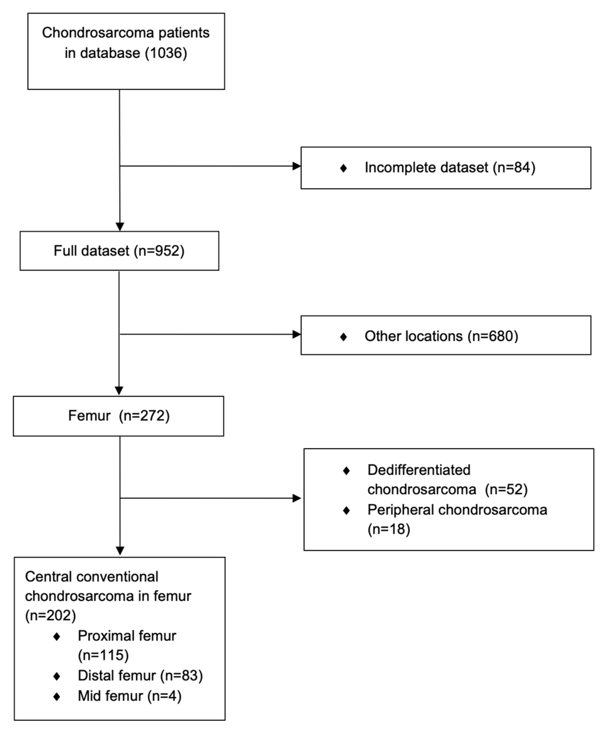
2. Materials and Methods

The study comprised a retrospective analysis of patients treated for central conventional CS in the femur at a singly tertiary sarcoma center—specifically, at the Royal Orthopaedic Hospital in Birmingham, UK—between January 1995 and October 2020. This review, approved by the institutional ethical review board, included patients diagnosed and treated at the hospital, excluding cases where patients were initially treated elsewhere or referred for managing recurrent tumors. Survivors were required to have at least 2 years follow-up, during which they were monitored for LR or metastases in line with European Society for Medical Oncology (ESMO) guidelines [21].
Details were collected on the clinical data and oncological outcomes, including LRFS. Primary surgery was defined as the method that concluded the first-line treatment. Resection specimens were examined by specialist bone sarcoma pathologists for grade, margin status, tumor location, and cortical breach in relation to the superior aspect of the greater trochanter or elsewhere within the specimen (Figure 1).
Grade was defined by internationally agreed upon standards and described according to the World Health Organization classification [1,9]. The diagnostic criteria governing histological grade included cellularity, nuclear size, the presence of an abundant hyaline cartilage matrix or mucomyxoid matrix, and the presence of mitosis. The highest grade seen in histology was then recorded, even when this higher grade comprised only a small number of cells. The margin was defined according to the Enneking classification as wide, marginal, or intralesional [22]. Because the Enneking definition of marginal and wide excision is inherently subjective and may vary depending on who is assessing the margin, we assessed the width of the surgical margin in millimeters. The histological status of the surgical margin was defined as follows: intralesional as microscopically positive (0 mm), marginal when margin was <3 mm, and wide when margin was ≥3 mm. The tumor was classified as proximal extremity of femur when the largest tumor mass was located in the subtrochanteric area, between the tip of the trochanter and 5 cm below the lesser trochanter. Tumors were classified as being located in the corpus of femur when the largest tumor mass was located in the diaphyseal area of thick cortical bone. Tumors were classified as being located in the distal extremity of femur when the biggest mass was located in the distal third of the femur.
The histological diagnosis and treatment plan were defined by a multidisciplinary team comprising specialist surgeons, radiologists, and pathologists. The primary outcome measure was LRFS. Secondary outcome measures were factors that influenced LR and DSS.

Statistical Analysis

Patient survival rates were assessed using the Kaplan–Meier method with 95% confidence intervals (CIs) as median observation times to estimate the LRFS and DSS. DSS was defined as the time from diagnosis to disease-related death and was censored at the date of the latest follow-up examination or death due to other causes. LRFS was defined as the time from the surgical procedure to LR and was censored at the date of the latest follow-up visit or death. LR was defined as tumor relapse evidenced by radiological confirmation and subsequent histological confirmation from biopsy or by an interval increase of 1 cm in the size of abnormal lesions on sequential imaging. Age was normally distributed and tested using the Shapiro–Wilk test.
Continuous variables are reported as medians and ranges, and between-group differences were analyzed using the one-way Mann–Whitney test. The Pearson chi-squared test was used to compare variables between groups and the Mann–Whitney u-test for medians between groups. Univariate analysis was performed by comparing groups using the log-rank test with subsequent univariate and multivariate Cox proportional hazards analysis of continuous variables to identify predictors of LRFS and DSS.
The subdistribution hazard ratio (SHR) of the effect of LR on survival was calculated with a competing risk analysis. Synchronous metastases (metastases developed before LR, at the time of LR, or within 90 days after LR) and death due to other reasons were considered as competing events in analyses of the effect of LR on DSS. Statistical analyses were performed using SPSS Statistics 27.0 (IBM, New York, NY, USA) and STATA 17 (Stata, College Station, TX, USA). A p-value < 0.05 was considered significant.

3. Results

The final study population comprised 202 patients with conventional central CS in the femur who were identified from a database of 1034 primary CS patients (Figure 2).
Patient demographics are summarized in Table 1.
Information regarding the presence of an extraosseous tumor component was available for 189 patients (94%); 105 (56%) had evidence of an extraosseous component. Of these extraosseous components, 28 (36%) involved the superior aspect of the proximal extremity of the femur.
When looking at the margin status following resection in relation to the location of the extraosseous component, if the tumor had an extraosseous component in the superior aspect of the proximal extremity of the femur, the margin was intralesional in 18% (5/28), marginal in 54% (15/28), and wide in 29% (8/28). When the extraosseous component of the tumor arose elsewhere within the proximal extremity of the femur, not involving the greater trochanter, the margin achieved at resection was intralesional in 8% (4/49), marginal in 25% (12/49), and wide in 67% (33/49). In the distal extremity of femur, if the tumor had an extraosseous component, the margin was intralesional in 18% (4/22), marginal in 63% (12/19), and wide in 36% (13/36). There was a significant difference in margin status achieved at resection depending on the location of the extraosseous component (p = 0.005).

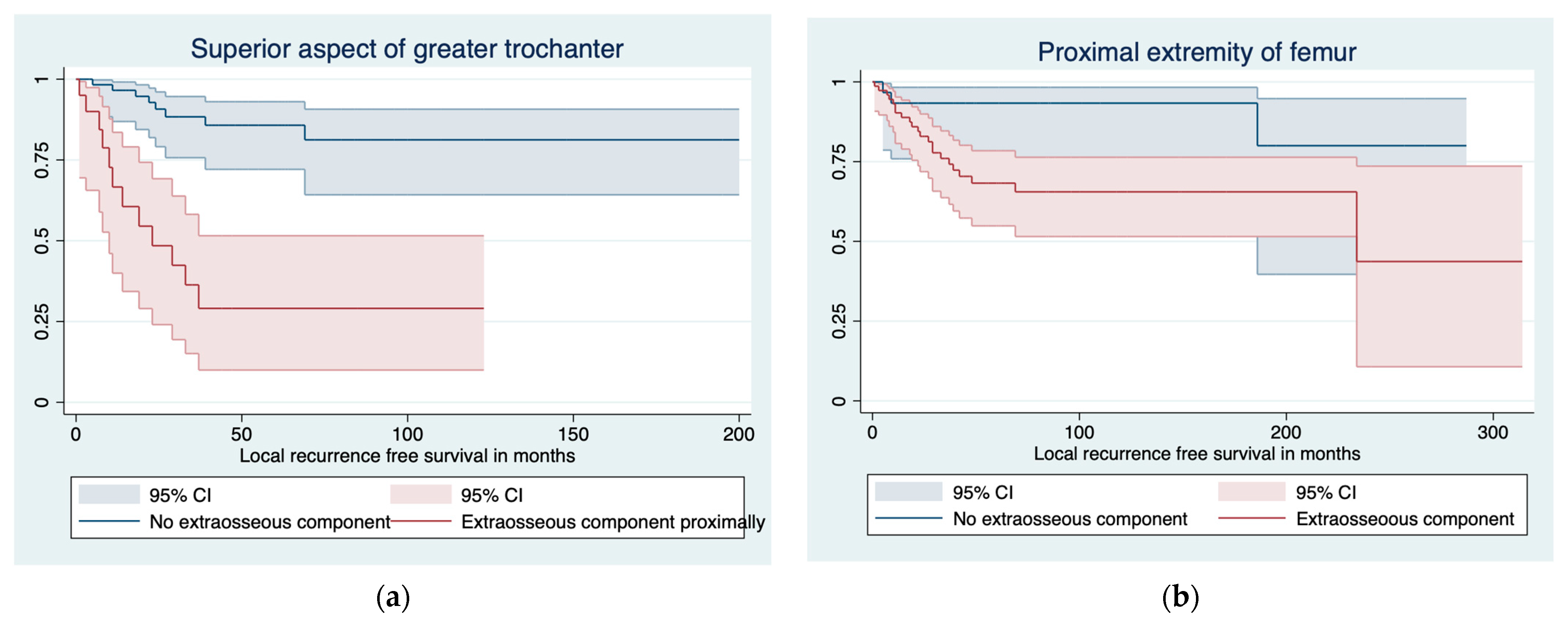
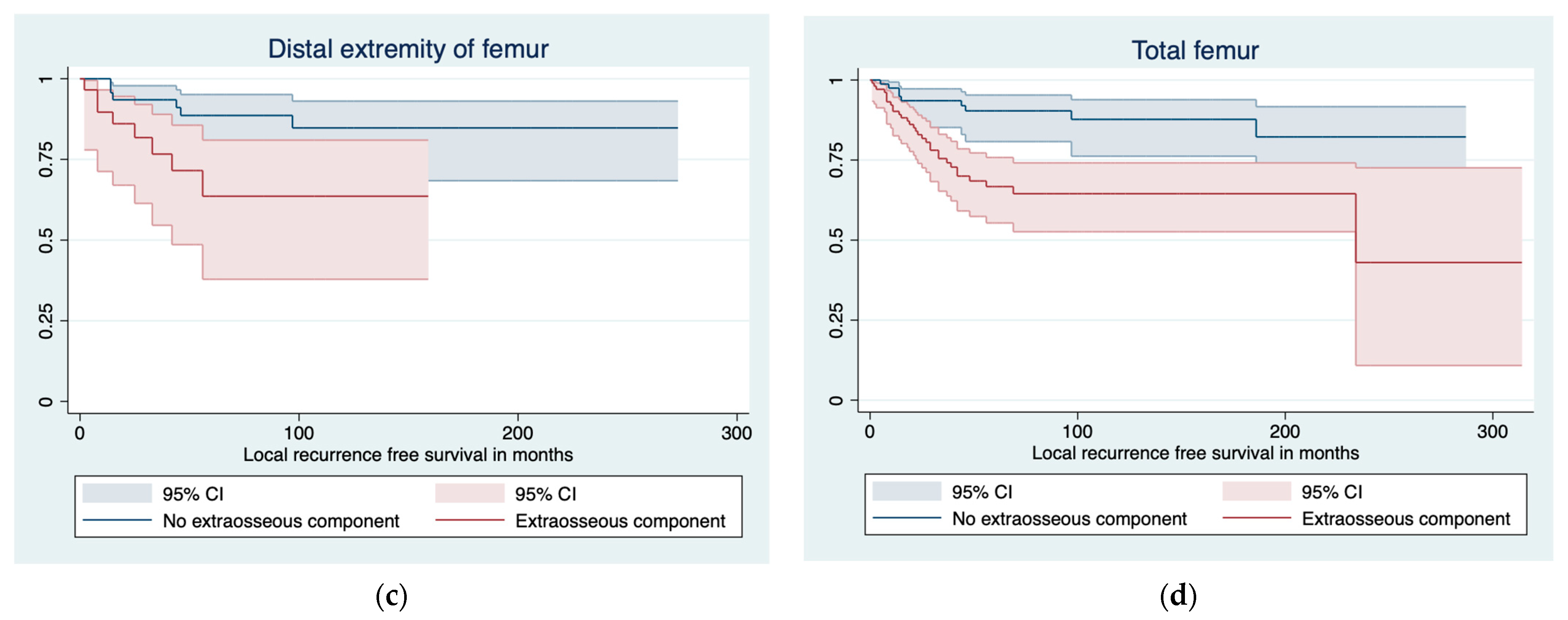
3.1. Predictors of LR and LRFS

The overall incidence of LR was 23% (47/202 patients). Margin as a factor affecting LR is summarized in Table 2. The extraosseous tumor component was a significant factor in LR (p = 0.005).
In grade 1 CS, the LRFS was 100% at 1 year, 93% (95% CI 85–100) at 3 years, and 88% (95% CI 79–97) at 5 and 10 years. In grade 2 CS, LRFS was 95% (95% CI 90–97) at 1 year, 81% (95% CI 73–89) at 3 years, and 70% (95% CI 60–81) at 5 and 10 years. In grade 3 CS, the LRFS was 82% (71–92) at 1 year and 69% (95% CI 56–82) at 3, 5, and 10 years.
In the Kaplan–Meier analysis, factors significant for LRFS were the extraosseous tumor component (p < 0.001), margin (p < 0.001), and histological grade (p = 0.031). The location of the extraosseous tumor component in relation to the superior aspect of the greater trochanter among the proximal extremity of the femur tumors was significant for LRFS (HR 7.4, 95% CI 3.0–18.2, p < 0.001; Figure 3). The phenomenon was similar in the distal extremity of the femur, with less significance (HR 3.1, 95% CI 1.0–8.9, p = 0.042).
In a multivariate Cox regression survival analysis, factors significant for LRFS were the extraosseous tumor component (HR 2.7; 95% CI 1.2–6.0, p = 0.021), histological grade (HR 2.9, 95% CI 1.7–5.2, p < 0.001), and margin achieved at resection (HR 0.2; 95% CI 0.11–0.33, p < 0.001).

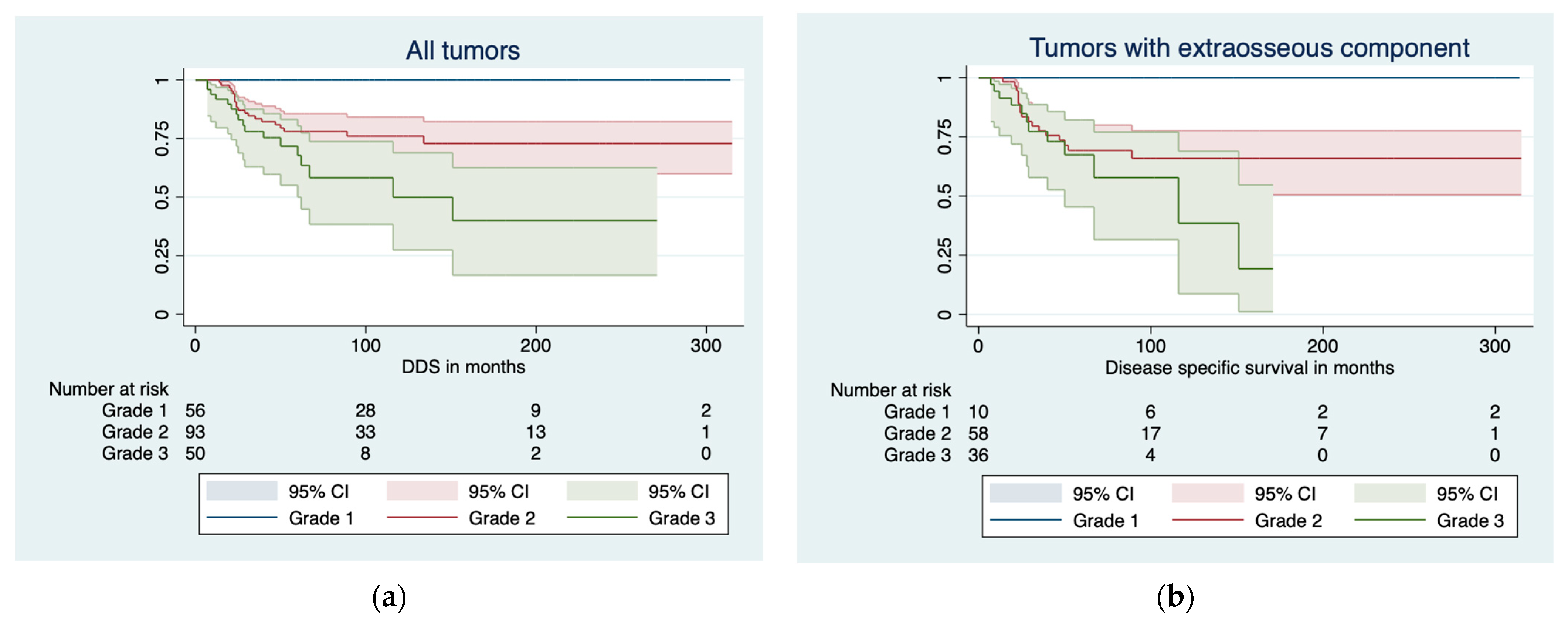
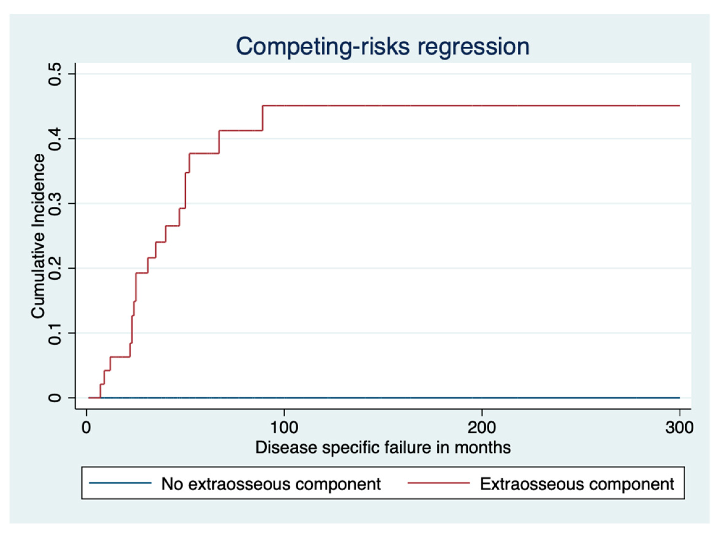
3.2. Disease-Specific Survival

The overall disease-specific death rate was 18% (37/202). DSS was 100% at 1, 3, 5, and 10 years for grade 1 CS. For grade 2 CS, the DSS was 100% at 1 year, 81% (95% CI 72–89) at 3 years, 78% (95% CI 69–87) at 5 years, and 76% (95% CI 66–86) at 10 years. For grade 3 CS, the DSS was 92% (95% CI 84–99) at 1 year, 78% (95% CI 66–90) at 3 years, 68% (95% CI 53–83) at 5 years, and 50% (95% CI 28–72) at 10 years (Figure 4).
In a Kaplan–Meier analysis, factors significantly affecting DSS were the histological grade (p < 0.001), extraosseous component (p < 0.001), LR (p < 0.001), metastases (p < 0.001), and surgical margin (p < 0.001). In a Cox regression analysis, factors affecting DSS were increasing size (HR 1.060, 95% CI 1.023–1.097, p < 0.001) and increasing age (HR 1.034, 95% CI 1.012–1.055, p = 0.002). Following a competing risk analysis, factors affecting disease-specific failure were the tumor grade (SHR 3.3, 95% CI 2.2–4.9, p < 0.001), extraosseous tumor component (SHR 1.1e07, p < 0.001; Figure 5), and LR (SHR 6.1, 95% CI 3.1–11.8, p < 0.001).

4. Discussion

The femur is a common location for primary malignant and benign bone tumors [23]. In this study, 21% of the CSs in the extremities and pelvis were in the femur, making the femoral location the second most common location after the pelvis. In opposition to other primary bone sarcomas, the proximal extremity of the femur was a more common location for CS than the distal extremity of the femur. Survival of the patients with proximal extremity of femur CS was less favorable than that of patients with a tumor in the distal extremity of the femur. This is most likely because tumors in the proximal extremity of the femur were more often of higher grade and large in size at the time of diagnosis. In femoral incidental cartilage tumor findings reported in the literature, the site of the origin in the femur affects the final diagnosis, as the ratio of benign to malignant is 11:1 in the distal extremity of the femur and 1:1.5 in the proximal extremity of the femur [24]. Therefore, any cartilage lesions in the proximal extremity of femur, especially if suspicious for malignant CS, should be reviewed prudently.
LR is closely related to the adequacy of the resection margin and plays a significant role in disease-free survival [15,25,26]. The extraosseous component has also been shown to be important in the local and systemic control of CS [13,19,26]. However, in the most recent work by Welling et al., the authors chose to include peripheral and dedifferentiated CSs, making their results difficult to interpret [19]. By definition, peripheral CSs are always associated with an extraosseous component; in dedifferentiated CS, the tumor presents as a low-grade, cartilaginous tumor juxtaposed to a generally high-grade, mesenchymal, non-cartilaginous component. In dedifferentiated CS, the high-grade non-cartilaginous component is thought to be more responsible for the diminished survival [27,28,29,30,31].
Could the effect of local control be the result of the difficulty in achieving an adequate margin when there is an extraosseous component? In this study, as well as in general, an adequate margin was more commonly achieved when the tumor only had an intraosseous component. The rate of LR was significantly higher in cases with an extraosseous tumor component. We specifically wanted to study the effect of an extraosseous component in the superior aspect of the proximal extremity of the femur, in the region of the greater trochanter, where the tendons for the gluteus minimus, gluteus medius, and piriformis are attached to the bone and sufficient soft tissue coverage can be difficult to achieve. Our results demonstrate a higher rate of LR in cases in which the extracortical breach of the tumor occurred in the superior aspect of the proximal extremity of the femur, which one could argue is the result of a higher incidence of an inadequate margin in this area. The importance of the abductors for hip stability may tempt the surgeon to preserve as much of the abductors as possible in the resection in an attempt to improve the function and stability of the hip [32,33,34]. However, this may compromise the margin and increase the rate of LR. In cases in which the tumor breaches the cortex in the superior aspect of the proximal extremity of the femur, the principle aim of the operation, as with all other locations, is to remove the tumor in its entirety with a sufficient soft tissue margin to minimize the risk of LR, even if this has a detrimental effect on function. However, our results have shown that, despite the margin achieved at resection, the rate of LR was affected by the location of the extraosseous component with the margin not having a significant effect on LR in cases with an extraosseous component in the superior aspect of the greater trochanter. In the distal extremity of femur, the phenomenon was similar but less significant.
This study has some limitations, including those inherent to its retrospective design, which must be acknowledged. Even though this is the largest study to date on conventional central CS of the femur, the small number of patients included in each group may have influenced the results because statistical significance was difficult to achieve. This will have influenced the duration of recruitment and follow-up period for the study. However, this remains the largest study yet concerning conventional central femur CS in which only grades 1, 2, and 3 were combined. As CS is commonly seen in elderly patients, death due to other causes frequently occurs. Therefore, our statistical method of using survival data calculations with competing risk analysis gives a more accurate assessment of overall survival, which differs from methods reported elsewhere in the literature.

5. Conclusions

In conclusion, in conventional central CS of the femur, the presence of an extraosseous tumor component has a predictive role in LRFS. Our results have shown the value of accurate, up-to-date imaging when managing CSs of the femur. Despite the outcomes seeming independent of margins achieved during resection, given the potential difficulty in accurately histologically assessing the soft tissue margins in this location, the presence of an extraosseous component should alert the treating surgeon to the need for a more aggressive resection in order to not break the first rule of oncology surgery and not compromise on margins. This is particularly important when dealing with tumors erupting from the greater trochanter, where the margin must not be compromised in an attempt to preserve function. CSs in the proximal extremity of the femur behave more aggressively because they are more often of higher grade and greater in size, which highlights the need for aggressive surgery.

Author Contributions

M.K.L. and L.M.J.: conceptualization, methodology, data analysis, and writing the paper; M.C.P.: writing and manuscript editing; J.D.S.: writing and manuscript editing; R.J.G.: writing and manuscript editing; G.V.M.: writing and manuscript editing; V.S.: pathology accuracy, writing, and manuscript editing. All authors have read and agreed to the published version of the manuscript.

Funding

This research received no external funding.

Institutional Review Board Statement

The study was conducted in accordance with the Declaration of Helsinki. This research was approved by the HUS ethic committee on 23 January 2023 (HUS/146/2023).

Informed Consent Statement

For the retrospective study, no informed consent statements were required.

Data Availability Statement

Deidentified patient data are available from the authors.

Conflicts of Interest

The authors declare no conflicts of interest.

References

  1. Kim, J.-H.; Lee, S.K. Classification of Chondrosarcoma: From Characteristic to Challenging Imaging Findings. Cancers 2023, 15, 1703. [Google Scholar] [CrossRef]
  2. Venneker, S.; Bovée, J.V.M.G. IDH Mutations in Chondrosarcoma: Case Closed or Not. Cancers 2023, 15, 3603. [Google Scholar] [CrossRef] [PubMed]
  3. Gilbert, A.; Tudor, M.; Montanari, J.; Commenchail, K.; Savu, D.I.; Lesueur, P.; Chevalier, F. Chondrosarcoma Resistance to Radiation Therapy: Origins and Potential Therapeutic Solutions. Cancers 2023, 15, 1962. [Google Scholar] [CrossRef] [PubMed]
  4. Gelderblom, H.; Hogendoorn, P.C.; Dijkstra, S.D.; van Rijswijk, C.S.; Krol, A.D.; Taminiau, A.H.; Bovée, J.V. The Clinical Approach Towards Chondrosarcoma. Oncologist 2008, 13, 320–329. [Google Scholar] [CrossRef] [PubMed]
  5. Laitinen, M.K.; Evans, S.; Stevenson, J.; Sumathi, V.; Kask, G.; Jeys, L.M.; Parry, M.C. Clinical differences between central and peripheral chondrosarcomas. Bone Jt. J. 2021, 103, 984–990. [Google Scholar] [CrossRef]
  6. Arshi, A.; Sharin, J.; Park, D.Y.; Park, H.Y.; Bernthal, N.M.; Yazdanshenas, H.; Shamie, A.N. Chondrosarcoma of the ossoeus spiine. J. Bone Jt. Surg. Br. 2005, 87, 1527–1530. [Google Scholar]
  7. Donati, D.; El Ghoneimy, A.; Bertoni, F.; Di Bella, C.; Mercuri, M. Surgical treatment and outcome of conventional pelvic chondrosarcoma. J. Bone Jt. Surg. 2013, 108, 19–27. [Google Scholar] [CrossRef]
  8. Mavrogenis, A.F.; Angelini, A.; Drago, G.; Merlino, B.; Ruggieri, P. Survival analysis of patients with chondrosarcomas of the pelvis. Spine 2017, 42, 644–652. [Google Scholar] [CrossRef]
  9. Fletcher, C.D.M.; Bridge, J.A.; Hogendoorn, P.C.W. Chondrogenic Tumors. In Soft Tissue and Bone Tumors, 4th ed.; IARC Pub-lisher: Lyon, France, 2013. [Google Scholar]
  10. Riedel, R.F.; Larrier, N.; Dodd, L.; Kirsch, D.; Martinez, S.; Brigman, B.E. The Clinical Management of Chondrosarcoma. Curr. Treat. Options Oncol. 2009, 10, 94–106. [Google Scholar] [CrossRef]
  11. Healey, J.H.; Lane, J.M. Chondrosarcoma. Clin. Orthop. Relat. Res. 1986, 204, 119–129. [Google Scholar] [CrossRef]
  12. Laitinen, M.K.; Stevenson, J.D.; Parry, M.C.; Sumathi, V.; Grimer, R.J.; Jeys, L.M. The role of grade in local recurrence and the disease-specific survival in chondrosarcomas. Bone Jt. J. 2018, 100, 662–666. [Google Scholar] [CrossRef] [PubMed]
  13. Fiorenza, F.; Abudu, A.; Grimer, R.J.; Carter, S.R.; Tillman, R.M.; Ayoub, K.; Mangham, D.C.; Davies, A.M. Risk factors for survival and local control in chondrosarcoma of bone. J. Bone Jt. Surg. 2002, 84, 93–99. [Google Scholar] [CrossRef]
  14. Angelini, A.; Guerra, G.; Mavrogenis, A.F.; Pala, E.; Picci, P.; Ruggieri, P. Clinical outcome of central conventional chondrosarcoma. J. Surg. Oncol. 2012, 106, 929–937. [Google Scholar] [CrossRef] [PubMed]
  15. Stevenson, J.D.; Laitinen, M.K.; Parry, M.C.; Sumathi, V.; Grimer, R.J.; Jeys, L.M. The role of surgical margins in chondrosarcoma. Eur. J. Surg. Oncol. 2018, 44, 1412–1418. [Google Scholar] [CrossRef] [PubMed]
  16. Andreou, D.; Fehlberg, S.; Pink, D.; Werner, M.; Tunn, P.-U. Survival amnd prognostic factors in chondrosarcoma. Results in 115 patients with long-term follow-up. Acta Orthop. 2011, 82, 749–755. [Google Scholar] [CrossRef]
  17. Kim, H.; Bindiganavile, S.S.; Han, I. Oncologic outcome after local recurrence of chondrosarcoma: Analysis of prognostic factors. J. Surg. Oncol. 2015, 111, 957–961. [Google Scholar] [CrossRef]
  18. Kask, G.; Laitinen, M.K.; Stevenson, J.; Evans, S.; Jeys, L.M.; Parry, M.C. Chondrosarcoma of the hands and feet. Bone Jt. J. 2021, 103, 562–568. [Google Scholar] [CrossRef]
  19. Wellings, E.P.; Mallett, K.E.; Parkes, C.W.; Labott, J.R.; Rose, P.S.; Houdek, M.T. Impact of tumour stage on the surgical outcomes of scapular chondrosarcoma. Int. Orthop. 2022, 46, 1175–1180. [Google Scholar] [CrossRef]
  20. Banskota, N.; Fang, X.; Yuan, D.; Lei, S.; Zhang, W.; Duan, H. Comparative Study of Proximal Femur Bone Tumor Patients Undergoing Hemiarthroplasty versus Total Hip Arthroplasty: A Meta-Analysis. J. Clin. Med. 2023, 12, 1209. [Google Scholar] [CrossRef]
  21. ESMO/European Sarcoma Network Working Group. Bone sarcomas: ESMO clinical practice guidelines for diagnosis, treatment and follow-up. Ann Oncol. 2014, 25, 113–123. [Google Scholar] [CrossRef]
  22. Enneking, W.F.; Spanier, S.S.; Goodman, M.A. A system for the surgical staging of musculoskeletal sarcoma. Clin. Orthop. Relat. Res. 1980, 415, 106–120. [Google Scholar] [CrossRef]
  23. Houdek, M.T.; Watts, C.D.; Wyles, C.C.; Rose, P.S.; Taunton, M.J.; Sim, F.H. Functional and oncologic outcome of cemented endoprosthesis for malignant proximal femoral tumors. J. Surg. Oncol. 2016, 114, 501–506. [Google Scholar] [CrossRef]
  24. Davies, A.; Patel, A.; Azzopardi, C.; James, S.; Botchu, R.; Jeys, L. The influence of site on the incidence and diagnosis of solitary central cartilage tumours of the femur. A 21st century perspective. J. Clin. Orthop. Trauma 2022, 32, 101953. [Google Scholar] [CrossRef]
  25. Kurisunkal, V.; Laitinen, M.K.; Kaneuchi, Y.; Kapanci, B.; Stevenson, J.; Parry, M.C.; Reito, A.; Fujiwara, T.; Jeys, L.M. Is 2 mm a wide margin in high-grade conventional chondrosarcomas of the pelvis? Bone Jt. J. 2021, 103, 1150–1154. [Google Scholar] [CrossRef]
  26. Thorkildsen, J.; Norum, O.J.; Myklebust, T.A.; Zaikova, O. Chondrosarcoma local recurrence in the Cancer Registry of Norway cohort (1990–2013): Patterns and impact. J. Surg. Oncol. 2021, 123, 510–520. [Google Scholar] [CrossRef]
  27. Fromm, J.; Klein, A.; Baur-Melnyk, A.; Knösel, T.; Lindner, L.; Birkenmaier, C.; Roeder, F.; Jansson, V.; Dürr, H.R. Survival and prognostic factors in conventional central chondrosarcoma. BMC Cancer 2018, 18, 849. [Google Scholar] [CrossRef]
  28. Frassica, F.J.; Unni, K.K.; Beabout, J.W.; Sim, F.H. Dedifferentiated chondrosarcoma. A report of the clinicopathological features and treatment of seventy-eight cases. J. Bone Jt. Surg. Am 1986, 68, 1197–1205. [Google Scholar] [CrossRef]
  29. Staals, E.L.; Bacchini, P.; Bertoni, F. Dedifferentiated central chondrosarcoma. Cancer 2006, 106, 2682–2691. [Google Scholar] [CrossRef]
  30. El Beaino, M.; Hoda, S.T.; Eldeib, A.J.; Masrouha, K. Dedifferentiated Chondrosarcoma: Diagnostic Controversies and Emerging Therapeutic Targets. Curr. Oncol. Rep. 2023, 25, 1117–1126. [Google Scholar] [CrossRef]
  31. Grimer, R.J.; Gosheger, G.; Taminiau, A.; Biau, D.; Matejovsky, Z.; Kollender, Y.; San-Julian, M.; Gherlinzoni, F.; Ferrari, C. Dedifferentiated chondrosarcoma: Prognostic factors and outcome from a European group. Eur. J. Cancer 2007, 43, 2060–2065. [Google Scholar]
  32. Bickels, J.; Meller, I.; Henshaw, R.M.; Malawer, M.M. Reconstruction of Hip Stability after Proximal and Total Femur Resections. Clin. Orthop. Relat. Res. 2000, 375, 218–230. [Google Scholar] [CrossRef]
  33. Elbuluk, A.M.; Coxe, F.R.; Schimizzi, G.V.; Ranawat, A.S.; Bostrom, M.P.; Sierra, R.J.; Sculco, P.K. Abductor Deficiency-Induced Recurrent Instability after Total Hip Arthroplasty. JBJS Rev. 2020, 8, e0164. [Google Scholar] [CrossRef]
  34. Henderson, E.R.; Keeney, B.J.; Pala, E.; Funovics, P.T.; Eward, W.C.; Groundland, J.S.; Ehrlichman, L.K.; Puchner, S.S.E.; Brigman, B.E.; Ready, J.E.; et al. The stability of the hip after the use of a proximal femoral endoprosthesis for oncological indications: Analysis of variables relating to the patient and the surgical technique. Bone Jt. J. 2017, 99, 531–537. [Google Scholar] [CrossRef]
Figure 1. Resection specimen showing an extraosseous tumor component in both the metaphyseal area and the superior aspect of the greater trochanter.
Cancers 16 00363 g001
Figure 2. Flowchart of identification of patients with conventional central chondrosarcomas of the femur.
Cancers 16 00363 g002
Figure 3. Local recurrence-free survival stratified by extraosseous tumor component in (a) the superior aspect of greater trochanter, (b) proximal extremity of femur, (c) distal extremity of femur, and (d) total femur.
Cancers 16 00363 g003aCancers 16 00363 g003b
Figure 4. Disease-specific survival stratified by grade (a) in all tumors and (b) in tumors with extraosseous tumor component.
Cancers 16 00363 g004
Figure 5. Disease-specific failure stratified by extraosseous tumor component in competing risk model.
Cancers 16 00363 g005
Table 1. Characteristics of 202 central chondrosarcoma in femur cases (values are presented in number of cases).
CharacteristicsTotalGrade 1Grade 2Grade 3
Eligible cases20257 (28%)94 (46%)51 (26%)
Site
      Proximal extremity115 (57%)20 (17%)60 (52%)35 (30%)
      Distal extremity83 (41%)36 (43%)32 (38%)15 (19%)
      Corpus4 (1.9%)1 (25%)2 (50%)1 (25%)
Male sex110 (55%)27 (25%)50 (46%)33 (30%)
      Proximal extremity65 (57%)10 (15%)33 (51%)22 (34%)
      Distal extremity43 (52%)17 (40%)16 (37%)10 (23%)
      Corpus2 (50%)-1 (100%)1 (100%)
Age at surgery, median (range)55 (8–95)46 (10–85)59 (8–95)59 (23–88)
      Proximal extremity59 (8–95)50 (10–85)60 (8–85)62 (25–87)
      Distal extremity51 (16–90)44 (16–85)57 (28–90)54 (23–88)
      Corpus53 (28–81)2864 (47–81)55
Mean tumor size, cm (range)12 (1.5–46)7.8 (1.5–46)12 (3.0–30)16 (4–40)
      Proximal extremity13 (2.0–40)7.3 (2.0–17)13 (3.5–30)17 (6.0–40)
      Distal extremity9.3 (1.5–60)8.0 (1.5–46)10 (3.0–25)11 (4.0–40)
      Corpus13 (6.0–19)6.017 (16–19)13
Extraosseous component105 (52%)10 (10%)58 (55%)37 (35%)
      Proximal extremity77 (67%)6 (8%%)42 (55%)28 (37%)
      Distal extremity28 (38%)4 (14%)16 (57%)8 (29%)
      Corpus1 (25%)--1 (100%)
Extraosseous component in cranial part of proximal extremity of femur28 (24%)1 (5%)17 (28%)10 (29%)
Median follow-up, months (range)90 (0–315)114 (0–314)92 (0–315)58 (0–271)
      Proximal extremity84 (0–315)117 (13–314)93 (0–315)50 (0–182)
      Distal extremity100 (0–273)114 (0–264)93 (8–273)78 (7–271)
      Corpus55 (10–104)7857 (10–104)29
Pathologic fracture20 (10%)1 (1.8%)12 (14%)7 (14%)
      Proximal extremity13 (11%)-10 (17%)3 (8.6%)
      Distal extremity6 (8.0%)1 (2.9%)2 (7.4%)3 (21%)
      Corpus1 (25%)--1 (100%)
Metastasis39 (19%)-21 (22%)18 (35%)
      Proximal extremity26 (23%)-14 (23%)12 (34%)
      Distal extremity12 (14%)-7 (22%)5 (33%)
      Corpus1--1
Median time to metastasis in months (range)22 (0–189)-26 (0–189)16 (4–41)
      Proximal extremity19 (0–73)-20 (0–73)18 (6–41)
      Distal extremity27 (0–189)-38 (0–189)11 (4–16)
      Corpus21--21
Local recurrence47 (23%)7 (12%)25 (27%)15 (29%)
      Proximal extremity31 (27%)2 (10%)20 (33%)9 (26%)
      Distal extremity15 (18%)5 (11%)5 (16%)5 (33%)
      Corpus1 (25%)--1 (100%)
Median time to LR in months (range)33 (0–234)54 (14–186)39 (0–234)12 (1–22)
      Proximal extremity36 (0–234)113 (39–186)40 (0–234)13 (1–22)
      Distal extremity28 (2–97)35 (14–97)38 (15–56)8 (2–18)
      Corpus21--21
Dead for disease37 (18%)-20 (21%)17 (33%)
      Proximal extremity25 (22%)-14 (23%)11 (31%)
      Distal extremity11 (13%)-6 (19%)5 (33%)
      Corpus1 (25%)--1 (100%)
Table 2. Role of surgery in local recurrence of 202 central chondrosarcoma in femur cases (values are presented in number of cases (percentage) with number and percentage of local recurrences in brackets).
TotalGrade 1Grade 2Grade 3
Curettage
      Proximal extremity9 (8%) [5, 45%]9 (45%) [2, 22%]--
      Distal extremity18 (22%) [2, 11%]16 (44%) [4, 25%]2 (6%) [1, 50%]-
      Corpus----
Resection
      Proximal extremity96 (85%) [29, 30%]11 (55%) [0]58 (97%) [20, 34%]27 (82%) [9, 33%]
      Distal extremity58 (70%) [8, 32%]20 (56%) [1, 5%]27 (84%) [4, 15%]11 (73%) [3, 27%]
      Corpus4 (100%) [1]1 (100%) [0]2 (100%) [0]1 (100%) [1, 100%]
Amputation
      Proximal extremity6 (5%) [0]-2 (3%) [0]4 (12%) [0]
      Distal extremity7 (8%) [2, 35%]-3 (9%) [0]4 (27%) [2, 50%]
      Corpus----
Hindquarter
      Proximal extremity2 (2%) [0]--2 (6%) [0]
      Distal extremity----
      Corpus----
Disclaimer/Publisher’s Note: The statements, opinions and data contained in all publications are solely those of the individual author(s) and contributor(s) and not of MDPI and/or the editor(s). MDPI and/or the editor(s) disclaim responsibility for any injury to people or property resulting from any ideas, methods, instructions or products referred to in the content.

