Article

The Association between Sampling and Survival in Patients with Pancreatic Ductal Adenocarcinoma Who Received Neoadjuvant Therapy and Pancreaticoduodenectomy

by
Mehran Taherian
1,
Matthew H. G. Katz
2,
Laura R. Prakash
2,
Dongguang Wei
1,
Yi Tat Tong
1,
Zongshan Lai
1,
Deyali Chatterjee
1,
Hua Wang
3,
Michael Kim
2,
Ching-Wei D. Tzeng
2,
Naruhiko Ikoma
2,
Robert A. Wolff
3,
Dan Zhao
3,
Eugene J. Koay
4,
Anirban Maitra
1 and
Huamin Wang
1,*
1
Department of Pathology, The University of Texas MD Anderson Cancer Center, Houston, TX 77030, USA
2
Department of Surgical Oncology, The University of Texas MD Anderson Cancer Center, Houston, TX 77030, USA
3
Department of Gastrointestinal Medical Oncology, The University of Texas MD Anderson Cancer Center, Houston, TX 77030, USA
4
Department of Radiation Oncology, The University of Texas MD Anderson Cancer Center, Houston, TX 77030, USA
*
Author to whom correspondence should be addressed.
Cancers 2024, 16(19), 3312; https://doi.org/10.3390/cancers16193312
Submission received: 31 July 2024 / Revised: 12 September 2024 / Accepted: 25 September 2024 / Published: 27 September 2024
(This article belongs to the Special Issue Histology and Pathology of Pancreatic Cancer)

Abstract

:

Simple Summary

We examined the association of the entire submission of the tumor (ESOT) and the entire submission of the pancreas (ESOP) with clinicopathologic features and survival in 627 pancreatic cancer patients who received neoadjuvant therapy (NAT). We demonstrated that both ESOT and ESOP were associated with lower ypT, less frequent perineural invasion, and better tumor response. Both ESOT and ESOP were associated with less frequent recurrence/metastasis, better disease-free survival (DFS), and overall survival (OS) in the overall study population. ESOP was associated with better DFS and OS in patients with ypT0/ypT1 or ypN0 tumors and better OS in patients with complete or near-complete response. ESOT was associated with better OS in patients with ypT0/ypT1 or ypN0 tumors. Both ESOT and ESOP were independent prognostic factors for OS in multivariate survival analyses. Therefore, ESOP and ESOT are associated with the prognosis of PDAC patients with complete or near-complete response and a ypT0/ypT1 tumor after NAT.

Abstract

Adequate sampling is essential to an accurate pathologic evaluation of pancreatectomy specimens resected for pancreatic ductal adenocarcinoma (PDAC) after neoadjuvant therapy (NAT). However, limited data are available for the association between the sampling and survival in these patients. We examined the association of the entire submission of the tumor (ESOT) and the entire submission of the pancreas (ESOP) with disease-free survival (DFS) and overall survival (OS), as well as their correlations with clinicopathologic features, for 627 patients with PDAC who received NAT and pancreaticoduodenectomy. We demonstrated that both ESOT and ESOP were associated with lower ypT, less frequent perineural invasion, and better tumor response (p < 0.05). ESOP was also associated with a smaller tumor size (p < 0.001), more lymph nodes (p < 0.001), a lower ypN stage (p < 0.001), better differentiation (p = 0.02), and less frequent lymphovascular invasion (p = 0.009). However, since ESOP and ESOT were primarily conducted for cases with no grossly identifiable tumor or minimal residual carcinoma in initial sections, potential bias cannot be excluded. Both ESOT and ESOP were associated with less frequent recurrence/metastasis and better DFS and OS (p < 0.05) in the overall study population. ESOP was associated with better DFS and better OS in patients with ypT0/ypT1 or ypN0 tumors and better OS in patients with complete or near-complete response (p < 0.05). ESOT was associated with better OS in patients with ypT0/ypT1 or ypN0 tumors (p < 0.05). Both ESOT and ESOP were independent prognostic factors for OS according to multivariate survival analyses. Therefore, accurate pathologic evaluation using ESOP and ESOT is associated with the prognosis in PDAC patients with complete or near-complete pathologic response and ypT0/ypT1 tumor after NAT.

1. Introduction

Pancreatic ductal adenocarcinoma (PDAC), the most common type of pancreatic cancer, is one of the most fatal human cancers with an average 5-year survival rate of 10.8% [1]. Neoadjuvant therapy (NAT) is increasingly used in the treatment of patients with potentially resectable PDAC [2], and it has been shown to improve disease-free survival (DFS) and overall survival (OS) in patients with borderline resectable PDAC [3,4,5,6]. The current National Comprehensive Cancer Network (NCCN) guidelines and the guidelines from the American Society of Clinical Oncology (ASCO) recommend NAT as the standard of care for patients with borderline resectable PDAC, high-risk patients with resectable disease, and selected patients with locally advanced disease [7,8].
Previous studies have shown that the systematic histopathologic assessment of post-therapy pancreatectomy specimens resected for PDAC is important not only for prognostication but also for the post-operative management of these patients [9,10,11,12]. Multiple pathologic parameters, including the post-treatment tumor stage (ypT), lymph node stage (ypN), tumor response grading to NAT, integrated pathologic score, vascular invasion, perineural invasion, and margin status, are important prognosticators in patients with PDAC who received NAT and pancreatectomy [9,10,11,12,13,14,15,16,17,18,19,20,21,22,23,24,25,26].
The pathologic examination of post-therapy pancreatectomy specimens is complex and challenging because NAT often induces a heterogeneous response in different areas of the tumor and extensive fibrosis in both the tumor and adjacent non-neoplastic pancreas [14]. Therapy-induced fibrosis and preexisting fibrosis related to obstructive changes in the adjacent pancreatic tissue obscure the tumor boundaries, which makes the identification of a tumor and tumor-size measurements extremely difficult [14,27,28]. In addition, microscopic tumor invasion beyond the grossly identified tumor areas into the adjacent pancreas, peripancreatic soft tissue, or organs is common for patients with PDAC who underwent surgical resection with or without receiving NAT. For patients whose tumors have a marked treatment response and no grossly identifiable tumor, extensive sampling of the pancreas and adjacent tissue often reveals a significant amount of residual carcinoma, which cannot be accurately identified through a gross examination, or microscopic foci of viable carcinoma [14]. Therefore, the adequate sampling of the tumor and adjacent pancreatic tissue is important for the accurate pathologic evaluation and staging of post-therapy pancreatectomy specimens. The recent recommendations from the Pancreatobiliary Pathology Society (PBPS) recommended systemic and standardized examination and reporting for pancreatectomy specimens resected after NAT [12]. The PBPS recommended that smaller tumors ≤2.0 cm should be entirely submitted, and the larger tumors may be sampled generously with at least 2 sections per centimeter of the tumor. For cases with a marked pathologic response, which have either no grossly visible tumor or only microscopic residual tumor in the initial representative sections of the tumor, the PBPS and other studies emphasized the submission of the entire pancreas or rigorous, extensive sampling of the pancreas and peripancreatic soft tissue, common bile duct, ampulla of Vater, and duodenum adjacent to the pancreas [12,29]. However, limited data are available on the association between the sampling of the tumor or pancreas of post-therapy pancreatectomy specimens and the clinical outcome and survival of patients with PDAC who received NAT. In this large, retrospective study, we examined the impact of the entire submission of the tumor and the pancreas in 627 patients with PDAC who received NAT. Our results demonstrated that the entire submission of the tumor and the pancreas through post-therapy pancreatectomy are important prognosticators for survival.

2. Materials and Methods

2.1. Study Population

Approval from the institutional review board was obtained, and a waiver of consent was granted for using patients’ clinical and pathologic information for research. Our study population consisted of 627 patients with PDAC who received NAT followed by pancreaticoduodenectomy (PD) at our institution between January 1999 and December 2019. There were 283 females (45.1%) and 344 males (54.9%) with a median age of 64.5 years (range: 30.3 to 85.4 years). Fifty-seven patients with PDAC who underwent distal pancreatectomy and 11 patients who underwent total pancreatectomy after being treated with NAT were excluded since the aim of this study was to focus on the impact of sampling PD specimens on survival. The pretreatment pathologic diagnosis of PDAC was confirmed in all cases by reviewing fine-needle aspiration cytology and/or biopsies.

2.2. Pathologic Evaluation of Pancreaticoduodenectomies

The PD specimens were grossly and microscopically evaluated and reported using a standardized protocol, which included the tumor location, size, tumor type, differentiation, tumor involvement of extra-pancreatic tissue/organ(s), histologic tumor response grade, presence or absence of lymphovascular or perineural invasion, number of positive and total lymph nodes, and margin status, etc. The pathology of all cases was re-reviewed. The uncinate margin was entirely submitted perpendicularly for a histologic examination in all cases. All cases had ≥12 lymph nodes to ensure accurate lymph-node (pN) staging. The grading of the extent of residual carcinoma in PD specimens was performed using the MD Anderson and the College of American Pathologists (CAP) grading schemes. The post-treatment pathologic staging was grouped according to the American Joint Committee on Cancer (AJCC) Staging Manual, 8th edition.
The information on sampling of the entire tumor and pancreas, and the number of blocks from the pancreas, were collected based on the review of the gross description and section codes from the pathology report of each case. Only cases for which the entire head of the pancreas, common bile duct, peripancreatic tissue, and ampulla of Vater were submitted for histologic examination were classified as an entire submission of the pancreas (ESOP).

2.3. Clinical and Follow-Up Data

Clinical and follow-up information, including age at diagnosis, gender, tumor resectability, neoadjuvant chemotherapy regimen, neoadjuvant radiation, type of surgery, the date and site of recurrence, and the date and cause of death, were retrieved from a prospectively maintained database for patients with pancreatic cancer. All clinical and follow-up information was verified by reviewing patients’ medical records and, if necessary, the United States Social Security Index. Disease recurrence or metastasis was determined largely based on radiographic and clinical suspicion, as definite confirmatory biopsies were not routinely performed on all patients.

2.4. Statistical Analyses

The categorized clinicopathologic features were compared between groups with and without the entire submission of the tumor or pancreas using Chi-square analyses. Independent-sample T tests were used to compare continuous variables. Disease-free survival (DFS) was calculated from the date of PD to the date of the first recurrence after PD in patients with recurrence or to the last follow-up date in patients who had no recurrence. Overall survival (OS) was calculated from the date of diagnosis to the date of death or the date of last follow-up if death did not occur. The follow-up time ranged from 6.7 to 257.5 months with a median of 33.0 months in the overall study group. The Kaplan–Meier method and log-rank test were used to compare the group with entire submissions of the tumor or pancreas to the group without an entire submission of the tumor or pancreas. Multivariate Cox regression analyses were performed to predict the DFS and OS based on the entire submission of the tumor or pancreas combined with other clinicopathologic covariates. A statistical analysis was performed using the Statistical Package for Social Sciences software for Windows (Version 26, SPSS Inc., Chicago, IL, USA). A 2-sided significance level of 0.05 was used for all statistical analyses.

3. Results

3.1. Correlations of Entire Submission of the Tumor or Pancreas with Clinicopathologic Parameters

Among the 627 patients, 17 (2.7%), 205 (32.7%), 325 (51.8%), and 80 (12.8%) cases were ypT0, ypT1, ypT2, and ypT3, respectively, and 284 (45.3%), 223 (35.6%) and 120 (19.1%) cases were ypN0, ypN1, and ypN2, respectively. There were 94 patients with a microscopically positive margin (R1, 15%), and 533 (85%) had a negative resection margin (R0). CAP grade 0, 1, 2, and 3 responses were observed in 17 (2.7%), 75 (12.0%), 330 (52.6%), and 205 (32.7%) cases, respectively. MD Anderson grade 0, 1, and 2 responses were observed in 17 (2.7%), 75 (12.0%), and 535 (85.3%) cases, respectively. The number of blocks submitted from the tumor and pancreas (including tumor blocks) ranged from 3 to 73 (median: 21). Only 16 (2.6%) had ≤10 blocks submitted from the pancreas in our study population. The entire tumor and entire pancreas were submitted in 333 (53.1%) and 101 (16%) cases, respectively.
The correlations between the entire submission of the tumor (ESOT) and the entire submission of the pancreas (ESOP) and clinicopathologic parameters are shown in Table 1. Both ESOT and ESOP were associated with lower ypT stages, better tumor responses using either the CAP or the MD Anderson grading scheme, and less frequent perineural invasion and recurrence/metastasis (p < 0.05). In addition, ESOP was also associated with better tumor differentiation, less frequent lymphovascular invasion, and lower ypN stages (p < 0.05). Compared to the group without ESOP, the ESOP group had smaller tumors (1.89 ± 1.82 cm vs. 2.78 ± 1.19 cm, p < 0.001) and more lymph nodes (29.7 ± 11.3 vs. 25.2 ± 9.8, p < 0.001), but there was no significant difference in the number of positive lymph nodes between these two groups (1.71 ± 4.0 vs. 2.00 ± 3.0, p = 0.40).

3.2. Correlation between Entire Submission of the Tumor or the Pancreas and Survival

In the overall study group, ESOT was associated with better OS compared to the group without ESOT (median OS: 45.5 ± 6.1 months vs. 35.5 ± 3.0 months; p = 0.003), but not DFS (p = 0.10, Figure 1A,B). The median DFS and OS for the group with ESOP were 25.8 ± 5.9 months and 83.7 ± 20.7 months, respectively, compared to 13.9 ± 1.2 months (p = 0.001) and 37.8 ± 2.3 months (p < 0.001), respectively, for the group without ESOP (Figure 1C,D).
Among the patients who had a complete or near-complete pathologic response (MD Anderson grade 0 or 1), ESOP was associated with better OS compared to those without ESOP (p = 0.03), but there was no difference in DFS between the group with ESOP and those without ESOP (p = 0.36, Figure 2A,B). There were no differences in either DFS (p = 0.77) or OS (p = 0.28) between the group with ESOT and those without ESOT (p > 0.05). Among the patients with MD Anderson grade 2, CAP grade 2, or CAP grade 3 responses, no significant associations between ESOT or ESOP and either DFS or OS were observed (p > 0.05).
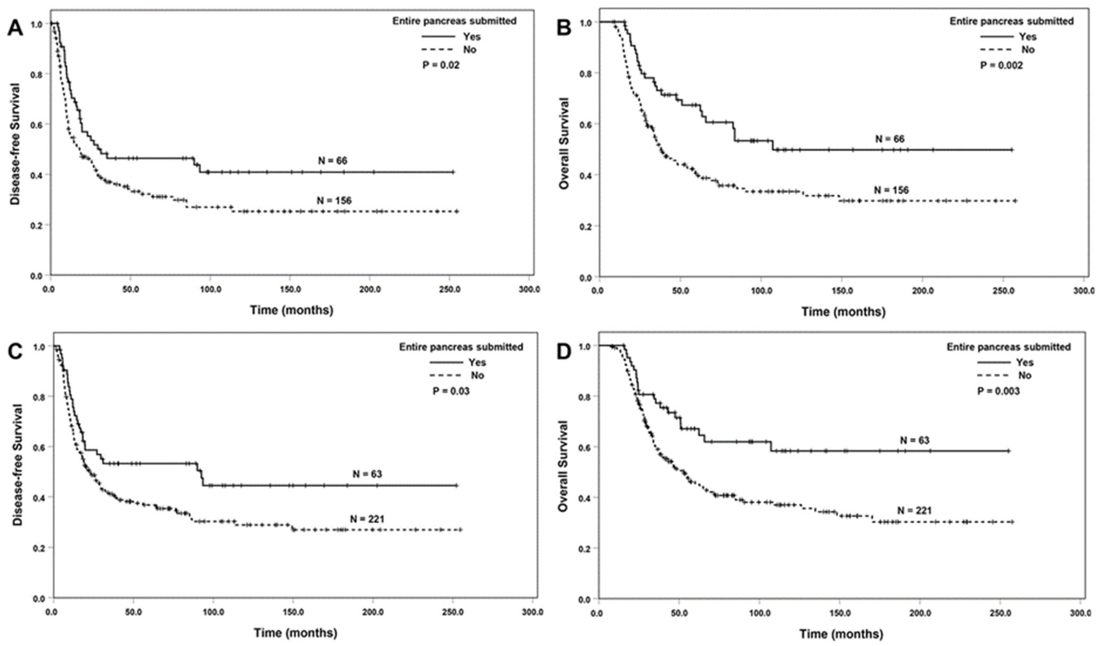
ESOP was associated with better DFS and OS among the patients with ypT0/T1 or ypN0 tumors (p < 0.05, Figure 3A–D), but not among the patients with ypT2/ypT3 or patients with positive lymph nodes (p > 0.05). ESOT was associated with better OS, but not DFS among the patients with ypT0/T1 or ypN0 tumors (p < 0.05, Figure 4A,B). There were no significant associations between ESOT and DFS or OS among the patients with a ypT2/ypT3 tumor or the patients with positive lymph nodes compared to the group without ESOT (p > 0.05).

3.3. Univariate and Multivariate Survival Analyses

To further examine the association between ESOT and ESOP and patient survival, we performed univariate and multivariate Cox regression analyses (Table 2, Table 3 and Table 4). We found that both ESOP [hazard ratio (HR): 0.72; 95% CI: 0.53–0.99; p = 0.04] and ESOT (HR: 0.80; 95% CI: 0.65–0.98; p = 0.03) were independent prognostic factors for OS, but not for DFS. In addition, ypN stage, tumor response grading, and tumor differentiation were also independent prognostic factors for both DFS and OS (p < 0.05, Table 2 and Table 3).

4. Discussion

The adequate sampling of the pancreatectomy specimens from patients with PDAC who received NAT is critical for the accurate histopathologic evaluation of tumor and lymph-node staging, the tumor response to NAT, and other pathologic parameters. However, limited data are available on the optimal sampling approach and the association of tumor and pancreas sampling with the clinical outcomes and survival. In this study, we retrospectively reviewed the association between the entire submission of the tumor and the entire submission of the pancreas and the clinicopathologic parameters and survival in a large cohort of 627 patients with PDAC who underwent PD after receiving NAT. We demonstrated for the first time that ESOT and ESOP are associated with lower ypT, less frequent perineural invasion, and better tumor response grade using either MD Anderson or the CAP grading system. ESOP was also associated with a lower ypN stage, better tumor differentiation and less frequent lymphovascular invasion, a smaller tumor size, and more lymph nodes. More importantly, we demonstrated that ESOT and ESOP were associated with less frequent recurrence/metastasis and better survival and were independent prognostic factors for OS. Our results provide much-needed evidence that adequate sampling of the tumor and the pancreas for pancreatectomies in patients with PDAC who received NAT has major implications on patient survival.
The recently published consensus paper from the PBPS on the pathologic examination of pancreatic specimens resected for PDAC after being treated with NAT recommended the entire submission of the tumor for cases with a tumor size of ≤2.0 cm and generous sampling (≥2 sections per cm of the tumor) for tumors that are >2.0 cm [12]. In this study, we demonstrated that ESOT was associated with better OS in patients with ypT0/T1 and ypN0 tumors. However, we did not observe significant associations between ESOT and either DFS or OS in patients with ypT2/ypT3 or positive lymph nodes. Our results support the recommendations from the PBPS that the entire tumor should be submitted for histologic examination if the tumor is 2.0 cm or less.
The consensus paper from the PBPS also recommended that the pancreas, peripancreatic soft tissue, common bile duct, ampulla of Vater, and duodenum adjacent to the pancreas should be entirely submitted or rigorously and extensively sampled for histologic examination to rule out microscopic foci of residual carcinoma if a pancreatectomy specimen for PDAC after NAT has no grossly identifiable tumor or has no viable residual carcinoma in the initially submitted sections [12]. This approach often identifies microscopic foci of residual carcinoma in the pancreas or adjacent tissue, which would be otherwise missed using representative sampling approach, and correctly classifies the ypT stage and tumor response grading. In this study, we demonstrated that ESOP was associated with better OS in patients with PDAC who had complete or near-complete pathologic response (MD Anderson grade 0 or 1). In addition, we demonstrated that ESOP was associated with better DFS and OS in patients with ypT0/T1 tumors. Interestingly, we also observed a significant association between ESOP with better DFS and OS in patients with ypN0 disease. However, no significant differences in either DFS or OS were observed between the group with ESOP and those without ESOP in patients with MD Anderson grade 2, CAP grade 2, or CAP grade 3 responses and in patients with ypT2/ypT3 or patients with positive lymph nodes. Our results support the approach of the entire submission or rigorous and extensive sampling of the pancreas, peripancreatic soft tissue, common bile duct, ampulla of Vater, and duodenum adjacent to the pancreas in patients with PDAC who have a complete pathologic response or a minimal amount of residual tumor (MD Anderson grade 0 or 1 response). Given the concerns of the cost and the time required for the pathologist to review the additional sections, the decision for the entire submission of the pancreas, peripancreatic tissue, common bile duct, ampulla of Vater, and duodenum adjacent to the pancreas is best made judiciously by the pathologist who is reviewing the specimen.
As expected, we found a significantly higher total number of lymph nodes in the PD specimens from the group with ESOP in our study. An adequate number of lymph nodes is critical for the accurate classification of the ypN stage, which has been shown to be one of the most important prognosticators of patients with PDAC [10,30,31,32].
The major limitations of this study are as follows: (1) this was a retrospective study at a single tertiary cancer center, which may have introduced potential selection bias into our study population. (2) At our institution, the entire submission of the pancreas was performed mainly due to the lack of a grossly identifiable tumor or initial representative sections with no or a minimal amount of residual carcinoma. In this retrospective study, however, it was challenging to determine the frequency with which ESOP and ESOT were performed intentionally during initial tissue sampling or as a result of missed carcinoma in the initial sampling in a two-stage approach. It is not surprising that the group with the entire submission of the pancreas had a smaller tumor size and a better pathology response, as we demonstrated in this study, which could have led to a potential bias in our survival analyses. However, the results of our survival analyses in different subgroups, including patients with complete or near-complete pathologic response (MD Anderson grade 0 or 1), ypT0/T1, and ypN0, consistently showed that ESOP was associated with better survival. More importantly, our results from multivariate Cox regression analyses demonstrated that ESOP remains an independent prognosticator of OS survival after adjusting ypT, ypN, tumor response grading, margin status, and tumor differentiation. Our study is the first large study to systemically examine the association of ESOP and ESOT with the clinicopathologic parameters and survival in patients with PDAC who underwent pancreatectomy after receiving NAT.
It should be noted that sampling (ESOP or ESOT) itself does not directly impact patient survival. However, inadequate sampling can lead to the underestimation of tumor size, residual viable tumor, lymphovascular and perineural invasion, and lymph-node metastasis, especially in patients with a near-complete pathologic response or a minimal amount of residual tumor. The inaccurate pathologic evaluation of these parameters can significantly affect survival and may explain the observed association between ESOP or ESOT and survival in our study.

5. Conclusions

Our study demonstrated for the first time that ESOP and ESOT are associated with ypT, perineural invasion, and tumor response grading. In addition, ESOP was also associated with the ypN stage, tumor differentiation, and lymphovascular invasion. However, since ESOP and ESOT were primarily conducted for cases with no grossly identifiable tumor or minimal residual carcinoma in initial sections, potential bias cannot be excluded. More importantly, we demonstrated that ESOP and ESOT were associated with less frequent recurrence/metastasis and better survival and were independent prognostic factors for OS according to multivariate survival analyses. Therefore, an accurate pathologic evaluation using ESOP and ESOT is important for the prognosis of PDAC patients with a complete or near-complete pathologic response and a ypT0/ypT1 tumor after NAT.

Author Contributions

Conceptualization, M.T. and H.W. (Huamin Wang); data curation, M.T., M.H.G.K., L.R.P., D.W., Y.T.T., Z.L., D.C., H.W. (Hua Wang), M.K., C.-W.D.T., N.I., R.A.W., D.Z., E.J.K. and H.W. (Huamin Wang); formal analysis, M.T. and H.W. (Huamin Wang); funding acquisition, H.W. (Huamin Wang); investigation, M.T., L.R.P., D.W., Y.T.T., Z.L., D.C., M.K., C.-W.D.T., N.I., R.A.W., E.J.K., A.M. and H.W. (Huamin Wang); methodology, M.T., M.H.G.K., L.R.P., Y.T.T., D.C., H.W. (Hua Wang), A.M. and H.W. (Huamin Wang); project administration, H.W. (Huamin Wang); resources, M.H.G.K., L.R.P., R.A.W., D.Z., A.M. and H.W. (Huamin Wang); supervision, H.W. (Huamin Wang); validation, D.C. and H.W. (Huamin Wang); visualization, H.W. (Huamin Wang); writing—original draft, M.T.; Writing—review and editing, M.T., M.H.G.K., D.W., Z.L., H.W. (Hua Wang), M.K., C.-W.D.T., N.I., R.A.W., D.Z., E.J.K., A.M. and H.W. (Huamin Wang). All authors have read and agreed to the published version of the manuscript.

Funding

Supported by the National Institutes of Health grants (1R01CA196941, 1R01CA195651, U01CA196403, P01CA117969, and P50CA221707).

Institutional Review Board Statement

This study was approved by the Institutional Review Board of The University of Texas MD Anderson Cancer Center (LAB05-0854).

Informed Consent Statement

Patient consent was waived based on our approved protocol.

Data Availability Statement

The data are unavailable due to privacy or ethical restrictions.

Conflicts of Interest

The authors declare no conflicts of interest pertinent to this study.

References

  1. Miller, K.D.; Siegel, R.L.; Lin, C.C.; Mariotto, A.B.; Kramer, J.L.; Rowland, J.H.; Stein, K.D.; Alteri, R.; Jemal, A. Cancer treatment and survivorship statistics, 2016. CA Cancer J. Clin. 2016, 66, 271–289. [Google Scholar] [CrossRef] [PubMed]
  2. Youngwirth, L.M.; Nussbaum, D.P.; Thomas, S.; Adam, M.A.; Blazer, D.G., 3rd; Roman, S.A.; Sosa, J.A. Nationwide trends and outcomes associated with neoadjuvant therapy in pancreatic cancer: An analysis of 18 243 patients. J. Surg. Oncol. 2017, 116, 127–132. [Google Scholar] [CrossRef] [PubMed]
  3. Jang, J.Y.; Han, Y.; Lee, H.; Kim, S.W.; Kwon, W.; Lee, K.H.; Oh, D.Y.; Chie, E.K.; Lee, J.M.; Heo, J.S.; et al. Oncological Benefits of Neoadjuvant Chemoradiation with Gemcitabine Versus Upfront Surgery in Patients with Borderline Resectable Pancreatic Cancer: A Prospective, Randomized, Open-label, Multicenter Phase 2/3 Trial. Ann. Surg. 2018, 268, 215–222. [Google Scholar] [CrossRef] [PubMed]
  4. Reni, M.; Balzano, G.; Zanon, S.; Zerbi, A.; Rimassa, L.; Castoldi, R.; Pinelli, D.; Mosconi, S.; Doglioni, C.; Chiaravalli, M.; et al. Safety and efficacy of preoperative or postoperative chemotherapy for resectable pancreatic adenocarcinoma (PACT-15): A randomised, open-label, phase 2–3 trial. Lancet Gastroenterol. Hepatol. 2018, 3, 413–423. [Google Scholar] [CrossRef] [PubMed]
  5. Versteijne, E.; Suker, M.; Groothuis, K.; Akkermans-Vogelaar, J.M.; Besselink, M.G.; Bonsing, B.A.; Buijsen, J.; Busch, O.R.; Creemers, G.M.; van Dam, R.M.; et al. Preoperative Chemoradiotherapy Versus Immediate Surgery for Resectable and Borderline Resectable Pancreatic Cancer: Results of the Dutch Randomized Phase III PREOPANC Trial. J. Clin. Oncol. 2020, 38, 1763–1773. [Google Scholar] [CrossRef]
  6. Versteijne, E.; Vogel, J.A.; Besselink, M.G.; Busch, O.R.C.; Wilmink, J.W.; Daams, J.G.; van Eijck, C.H.J.; Groot Koerkamp, B.; Rasch, C.R.N.; van Tienhoven, G.; et al. Meta-analysis comparing upfront surgery with neoadjuvant treatment in patients with resectable or borderline resectable pancreatic cancer. Br. J. Surg. 2018, 105, 946–958. [Google Scholar] [CrossRef]
  7. Tempero, M.A.; Malafa, M.P.; Al-Hawary, M.; Behrman, S.W.; Berson III, A.B.; Cardin, D.B.; Cha, C.; Chiorean, E.G.; Chung, V.; Czito, B.; et al. NCCN Clinical Practice Guidelines in Oncology, Pancreatic Adenocarcinoma (Version 1.2021). Available online: https://www.nccn.org/professionals/physician_gls/PDF/pancreatic.pdf (accessed on 25 January 2021).
  8. Khorana, A.A.; Mangu, P.B.; Berlin, J.; Engebretson, A.; Hong, T.S.; Maitra, A.; Mohile, S.G.; Mumber, M.; Schulick, R.; Shapiro, M.; et al. Potentially Curable Pancreatic Cancer: American Society of Clinical Oncology Clinical Practice Guideline. J. Clin. Oncol. 2016, 34, 2541–2556. [Google Scholar] [CrossRef]
  9. Lee, S.M.; Katz, M.H.; Liu, L.; Sundar, M.; Wang, H.; Varadhachary, G.R.; Wolff, R.A.; Lee, J.E.; Maitra, A.; Fleming, J.B.; et al. Validation of a Proposed Tumor Regression Grading Scheme for Pancreatic Ductal Adenocarcinoma after Neoadjuvant Therapy as a Prognostic Indicator for Survival. Am. J. Surg. Pathol. 2016, 40, 1653–1660. [Google Scholar] [CrossRef]
  10. Estrella, J.S.; Rashid, A.; Fleming, J.B.; Katz, M.H.; Lee, J.E.; Wolf, R.A.; Varadhachary, G.R.; Pisters, P.W.; Abdalla, E.K.; Vauthey, J.N.; et al. Post-therapy pathologic stage and survival in patients with pancreatic ductal adenocarcinoma treated with neoadjuvant chemoradiation. Cancer 2012, 118, 268–277. [Google Scholar] [CrossRef]
  11. Sohn, A.J.; Taherian, M.; Katz, M.H.G.; Prakash, L.R.; Chatterjee, D.; Wang, H.; Kim, M.; Tzeng, C.D.; Lee, J.E.; Ikoma, N.; et al. Integrated Pathologic Score Effectively Stratifies Patients with Pancreatic Ductal Adenocarcinoma Who Received Neoadjuvant Therapy and Pancreaticoduodenectomy. Am. J. Surg. Pathol. 2023, 47, 421–430. [Google Scholar] [CrossRef]
  12. Wang, H.; Chetty, R.; Hosseini, M.; Allende, D.S.; Esposito, I.; Matsuda, Y.; Deshpande, V.; Shi, J.; Dhall, D.; Jang, K.T.; et al. Pathologic Examination of Pancreatic Specimens Resected for Treated Pancreatic Ductal Adenocarcinoma: Recommendations from the Pancreatobiliary Pathology Society. Am. J. Surg. Pathol. 2022, 46, 754–764. [Google Scholar] [CrossRef]
  13. Haeberle, L.; Esposito, I. Pathology of pancreatic cancer. Transl. Gastroenterol. Hepatol. 2019, 4, 50. [Google Scholar] [CrossRef] [PubMed]
  14. Nagaria, T.S.; Wang, H.; Chatterjee, D.; Wang, H. Pathology of Treated Pancreatic Ductal Adenocarcinoma and Its Clinical Implications. Arch. Pathol. Lab. Med. 2020, 144, 838–845. [Google Scholar] [CrossRef] [PubMed]
  15. Carpenter, E.L.; Van Decar, S.G.; McCarthy, P.M.; Valdera, F.A.; Adams, A.M.; O’Shea, A.E.; Smolinsky, T.; Thomas, K.; Clifton, G.T.; Newhook, T.E.; et al. The benefit of adjuvant chemotherapy following pancreaticoduodenectomy for pancreatic adenocarcinoma depends on response to neoadjuvant therapy. J. Surg. Oncol. 2024, 130, 109–116. [Google Scholar] [CrossRef] [PubMed]
  16. Donisi, G.; Nappo, G.; Pacilli, M.; Capretti, G.L.; Spaggiari, P.; Sollai, M.; Bozzarelli, S.; Zerbi, A. Pathologic tumor response to neoadjuvant therapy in resected pancreatic cancer: Does it affect prognosis? Updates Surg. 2023, 75, 1497–1508. [Google Scholar] [CrossRef] [PubMed]
  17. Goess, R.; Jager, C.; Perinel, J.; Pergolini, I.; Demir, E.; Safak, O.; Scheufele, F.; Schorn, S.; Muckenhuber, A.; Adham, M.; et al. Lymph node examination and survival in resected pancreatic ductal adenocarcinoma: Retrospective study. BJS Open 2024, 8, zrad125. [Google Scholar] [CrossRef]
  18. Hayasaki, A.; Mizuno, S.; Nagata, M.; Kaluba, B.; Maeda, K.; Shinkai, T.; Ito, T.; Gyoten, K.; Fujii, T.; Iizawa, Y.; et al. Extrapancreatic extension is a better adverse prognostic factor than tumor size in patients with localized pancreatic ductal adenocarcinoma treated with chemoradiotherapy-comparison of T category between the American Joint Committee on Cancer and Japan Pancreas Society. HPB 2023, 25, 1268–1277. [Google Scholar] [CrossRef]
  19. Javed, A.A.; Mahmud, O.; Fatimi, A.S.; Habib, A.; Grewal, M.; He, J.; Wolfgang, C.L.; Besselink, M.G.; Consortium, P.-P. Predictors for Long-Term Survival After Resection of Pancreatic Ductal Adenocarcinoma: A Systematic Review and Meta-Analysis. Ann. Surg. Oncol. 2024, 31, 4673–4687. [Google Scholar] [CrossRef]
  20. Kinny-Koster, B.; Ahmad, Y.; Pfluger, M.J.; Habib, J.R.; Fujikura, K.; Hutchings, D.; Cameron, J.L.; Shubert, C.R.; Lafaro, K.J.; Burkhart, R.A.; et al. Clinical Relevance of Cancerization of Ducts in Resected Pancreatic Ductal Adenocarcinoma. Pancreas 2024, 53, e528–e536. [Google Scholar] [CrossRef]
  21. Murata, Y.; Mizuno, S.; Kishiwada, M.; Uchida, K.; Noguchi, D.; Gyoten, K.; Hayasaki, A.; Fujii, T.; Iizawa, Y.; Tanemura, A.; et al. Clinical significance and predictors of complete or near-complete histological response to preoperative chemoradiotherapy in patients with localized pancreatic ductal adenocarcinoma. Pancreatology 2021, 21, 1482–1490. [Google Scholar] [CrossRef]
  22. Neyaz, A.; Tabb, E.S.; Shih, A.; Zhao, Q.; Shroff, S.; Taylor, M.S.; Rickelt, S.; Wo, J.Y.; Fernandez-Del Castillo, C.; Qadan, M.; et al. Pancreatic ductal adenocarcinoma: Tumour regression grading following neoadjuvant FOLFIRINOX and radiation. Histopathology 2020, 77, 35–45. [Google Scholar] [CrossRef] [PubMed]
  23. Park, Y.; Han, Y.B.; Kim, J.; Kang, M.; Lee, B.; Ahn, E.S.; Han, S.; Kim, H.; Na, H.Y.; Han, H.S.; et al. Microscopic tumor mapping of post-neoadjuvant therapy pancreatic cancer specimens to predict post-surgical recurrence: A prospective cohort study. Pancreatology 2024, 24, 562–571. [Google Scholar] [CrossRef]
  24. Pu, N.; Wu, W.; Liu, S.; Xie, Y.; Yin, H.; Chen, Q.; He, T.; Xu, Z.; Wang, W.; Yu, J.; et al. Survival benefit and impact of adjuvant chemotherapy following systemic neoadjuvant chemotherapy in patients with resected pancreas ductal adenocarcinoma: A retrospective cohort study. Int. J. Surg. 2023, 109, 3137–3146. [Google Scholar] [CrossRef]
  25. Servin-Rojas, M.; Fong, Z.V.; Fernandez-Del Castillo, C.; Lionetto, G.; Bolm, L.; Fagenholz, P.J.; Ferrone, C.R.; Rocha-Castellanos, D.M.; Lillemoe, K.D.; Qadan, M. Lymph Node Yield is Associated with Improved Overall Survival and Increased Time to Recurrence in Node-Negative Pancreatic Ductal Adenocarcinoma Following Neoadjuvant Therapy. Ann. Surg. 2024, 10, 1097. [Google Scholar] [CrossRef] [PubMed]
  26. Usui, M.; Uchida, K.; Hayasaki, A.; Kishiwada, M.; Mizuno, S.; Watanabe, M. Prognostic impact of the distance from the anterior surface to tumor cells in pancreatoduodenectomy with neoadjuvant chemoradiotherapy for pancreatic ductal adenocarcinoma. PLoS ONE 2024, 19, e0307876. [Google Scholar] [CrossRef] [PubMed]
  27. Hartman, D.J.; Krasinskas, A.M. Assessing treatment effect in pancreatic cancer. Arch. Pathol. Lab. Med. 2012, 136, 100–109. [Google Scholar] [CrossRef]
  28. Verbeke, C.; Haberle, L.; Lenggenhager, D.; Esposito, I. Pathology assessment of pancreatic cancer following neoadjuvant treatment: Time to move on. Pancreatology 2018, 18, 467–476. [Google Scholar] [CrossRef] [PubMed]
  29. Yokohira, M.; Oshima, M.; Yamakawa, K.; Ye, J.; Nakano-Narusawa, Y.; Haba, R.; Fukumura, Y.; Hirabayashi, K.; Yamaguchi, H.; Kojima, M.; et al. Adequate tissue sampling for the assessment of pathological tumor regression in pancreatic cancer. Sci. Rep. 2021, 11, 6586. [Google Scholar] [CrossRef]
  30. Huebner, M.; Kendrick, M.; Reid-Lombardo, K.M.; Que, F.; Therneau, T.; Qin, R.; Donohue, J.; Nagorney, D.; Farnell, M.; Sarr, M. Number of lymph nodes evaluated: Prognostic value in pancreatic adenocarcinoma. J. Gastrointest. Surg. 2012, 16, 920–926. [Google Scholar] [CrossRef]
  31. Shi, S.; Hua, J.; Liang, C.; Meng, Q.; Liang, D.; Xu, J.; Ni, Q.; Yu, X. Proposed Modification of the 8th Edition of the AJCC Staging System for Pancreatic Ductal Adenocarcinoma. Ann. Surg. 2019, 269, 944–950. [Google Scholar] [CrossRef]
  32. Vuarnesson, H.; Lupinacci, R.M.; Semoun, O.; Svrcek, M.; Julie, C.; Balladur, P.; Penna, C.; Bachet, J.B.; Resche-Rigon, M.; Paye, F. Number of examined lymph nodes and nodal status assessment in pancreaticoduodenectomy for pancreatic adenocarcinoma. Eur. J. Surg. Oncol. 2013, 39, 1116–1121. [Google Scholar] [CrossRef] [PubMed]
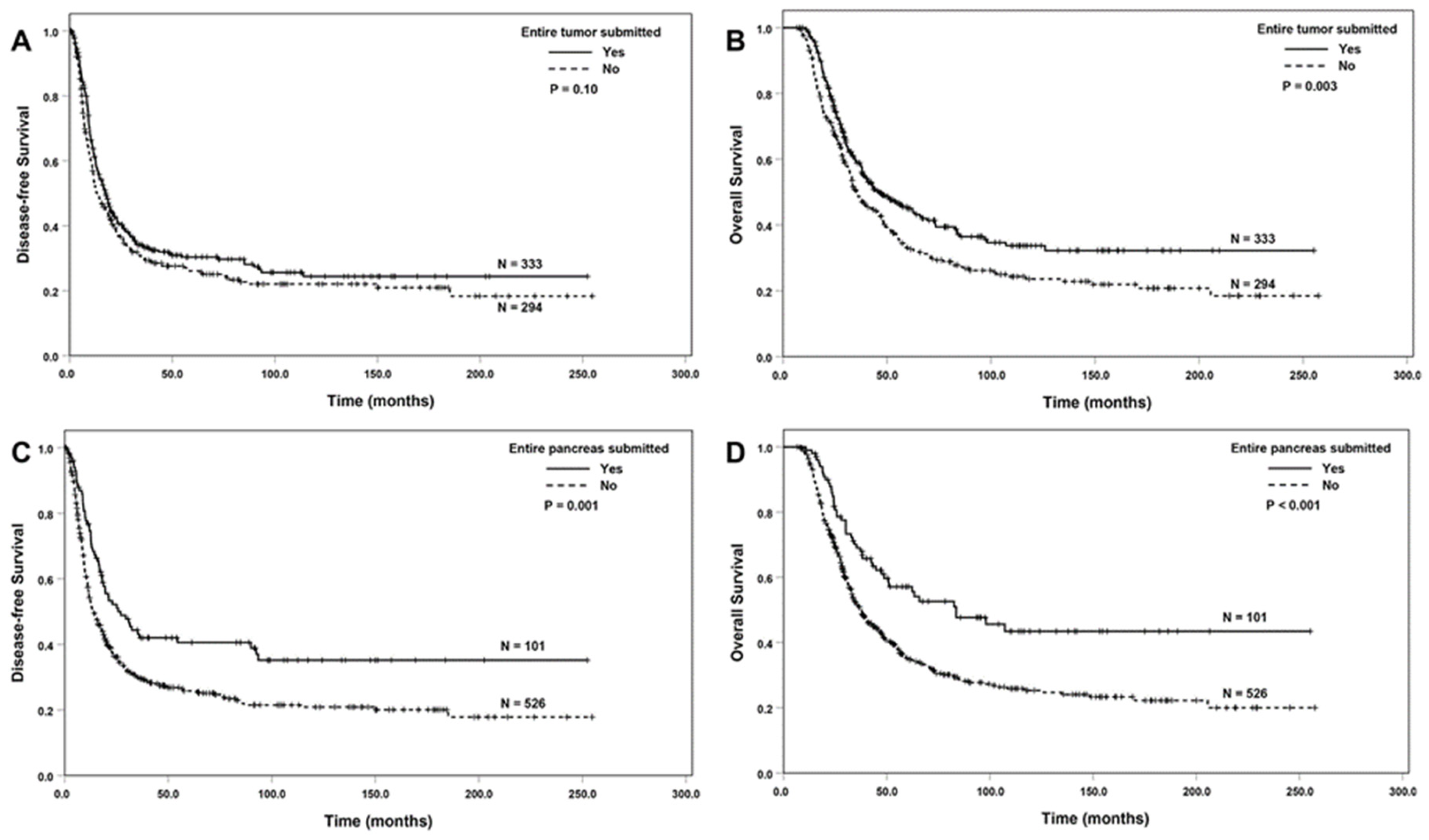
Figure 1. The Kaplan–Meier survival curves of disease-free survival and overall survival in the overall population (627 patients), comparing the group with ESOT (A,B) or ESOP (C,D) to the group without the submission of the tumor or the pancreas.
Cancers 16 03312 g001
Figure 2. The Kaplan–Meier survival curves of disease-free survival and overall survival in patients with a complete or near-complete pathologic response (MD Anderson grade 0 or 1). There was no difference in disease-free survival between the group with ESOP and those without ESOP ((A), p = 0.36). ESOP was associated with better overall survival ((B), p = 0.03).
Cancers 16 03312 g002
Figure 3. The Kaplan–Meier survival curves showing that ESOP was associated with better disease-free survival and overall survival compared to those without ESOP in patients with ypT0/ypT1 tumors (A,B) or ypN0 tumors (C,D).
Cancers 16 03312 g003
Figure 4. The Kaplan–Meier survival curves showing that ESOT was associated with better overall survival compared to those without ESOT in patients with ypT0/ypT1 tumors (A) and ypN0 tumors (B).
Cancers 16 03312 g004
Table 1. Correlations between the entire submission of the tumor and the pancreas with clinicopathologic features in pancreaticoduodenectomy specimens after neoadjuvant therapy.
CharacteristicsESOTpESOPp
Yes (n = 333)No (n = 294)Yes (n = 101)No (n = 526)
Age (Years, Mean ± SD)63.9 ± 9.9 63.5 ± 9.00.5764.2 ± 9.263.6 ± 9.70.56
Sex 0.96 0.27
Female150133 51232
Male183161 50294
Differentiation 0.56 0.02
Well/moderate213181 74320
Poor120113 27206
ypT * <0.001 <0.001
ypT0170 143
ypT112283 52153
ypT2146179 20305
ypT34832 1565
ypN stage * 0.46 0.001
ypN0156128 63221
ypN1111112 26197
ypN26654 12108
Lymphovascular invasion 0.34 0.009
No170138 62246
Yes163156 39280
Perineural invasion <0.001 <0.001
No9749 4799
Yes236245 54427
CAP TRG <0.001 <0.001
Score 0170 143
Score 15322 2748
Score 2169161 43287
Score 394111 17188
MD Anderson TRG <0.001 <0.001
Grade 0170 143
Grade 15322 2748
Grade 2263272 60475
Margin status 0.82 0.45
Negative282251 89444
Positive5143 1282
Recurrence/metastasis <0.001 0.03
No11581 42154
Local recurrence5455 1198
Distant metastasis164158 48274
* According to AJCC 8th edition; ESOT, entire submission of the tumor; ESOP, entire submission of the pancreas; TRG: tumor response grading; CAP: College of American Pathologists.
Table 2. Univariate Cox regression analysis of disease-free survival and overall survival.
Disease-Free SurvivalOverall Survival
Characteristics (N = 627)NHR (95% CI)pHR (95% CI)p
Gender
Female (ref)2831.00 1.00
Male3441.06 (0.88–1.28)0.561.09 (0.89–1.34)0.38
Age6271.00 (0.98–1.01)0.291.00 (0.99–1.01)0.61
Tumor differentiation
Well/Moderate (ref)3941.00 1.00
Poor2331.33 (1.09–1.61)0.0041.32 (1.07–1.62)0.009
Margin status
Negative (ref)5331.00 1.00
Positive941.35 (1.04–1.74)0.021.14 (0.85–1.53)0.39
ypT 0.003 0.01
ypT0/T1 (ref)2221.00 1.00
ypT23251.36 (1.10–1.68)0.0051.35 (1.08–1.69)0.009
ypT3801.60 (1.18–2.17)0.0031.49 (1.06–2.08)0.02
ypN stage <0.001 <0.001
ypN0 (ref)2841.00 1.00
ypN12231.54 (1.24–1.91)<0.0011.62 (1.28–2.04)<0.001
ypN21202.52 (1.97–3.23)<0.0012.48 (1.90–3.23)<0.001
MDA TRG
Grade 0/1 (ref)921.00 1.00
Grade 25351.94 (1.44–2.63)<0.0012.25 (1.61–3.15)<0.001
Entire submission of tumor
No (ref)2941.00 1.00
Yes3330.85 (0.71–1.03)0.100.74 (0.61–0.91)0.004
Entire submission of pancreas
No (ref)5261.00 1.00
Yes1010.63 (0.48–0.83)0.0010.56 (0.41–0.76)<0.001
MDA: MD Anderson Cancer Center; TRG: tumor response grading.
Table 3. Multivariate Cox regression analysis of disease-free survival and overall survival with the entire submission of the tumor.
Disease-Free SurvivalOverall Survival
Characteristics (N = 627)NHR (95% CI)pHR (95% CI)p
Tumor differentiation
Well/moderate (ref)3941.00 1.00
Poor2331.27 (1.05–1.54)0.021.28 (1.04–1.58)0.02
Margin status
Negative (ref)5331.00 1.00
Positive941.09 (0.83–1.42)0.530.88 (0.65–1.19)0.88
ypT 0.73 0.84
ypT0/T1 (ref)2221.00 1.00
ypT23251.03 (0.82–1.29)0.810.97 (0.77–1.24)0.83
ypT3801.14 (0.82–1.58)0.441.07 (0.75–1.53)0.70
ypN stage <0.001 <0.001
ypN0 (ref)2841.00 1.00
ypN12231.43 (1.15–1.78)0.0011.48 (1.17–1.87)0.001
ypN21202.32 (1.81–2.99)<0.0012.32 (1.77–3.03)<0.001
MDA TRG
Grade 0/1 (ref)921.00 1.00
Grade 25351.66 (1.22–2.25)0.0011.79 (1.26–2.52)0.001
Entire submission of tumor
No (ref)2941.00 1.00
Yes3330.89 (0.73–1.08)0.230.80 (0.65–0.98)0.03
MDA: MD Anderson Cancer Center; TRG: Tumor response grading.
Table 4. Multivariate Cox Regression Analysis of Disease-free Survival and Overall Survival with Entire Submission of Pancreas.
Disease-Free SurvivalOverall Survival
CharacteristicsNHR (95% CI)pHR (95% CI)p
Tumor differentiation
Well/moderate (ref)3941.00 1.00
Poor2331.26 (1.04–1.54)0.021.27 (1.03–1.56)0.02
Margin status
Negative (ref)5331.00 1.00
Positive941.09 (0.83–1.42)0.540.88 (0.65–1.17)0.41
ypT 0.74 0.79
ypT0/T1 (ref)2221.00 1.00
ypT23251.00 (0.80–1.25)0.990.95 (0.75–1.21)0.70
ypT3801.12 (0.81–1.55)0.491.06 (0.74–1.51)0.75
ypN stage <0.001 <0.001
ypN0 (ref)2841.00 1.00
ypN12231.42 (1.14–1.77)0.0021.47 (1.17–1.86)0.001
ypN21202.31 (1.79–2.97)<0.0012.24 (1.71–2.93)<0.001
MDA TRG
Grade 0/1 (ref)921.00 1.00
Grade 25351.53 (1.12–2.10)0.0081.73 (1.21–2.45)0.002
Entire pancreas submitted
No (ref)5261.00 1.00
Yes1010.77 (0.58–1.02)0.070.72 (0.53–0.99)0.04
MDA: MD Anderson Cancer Center; TRG: tumor response grading.
Disclaimer/Publisher’s Note: The statements, opinions and data contained in all publications are solely those of the individual author(s) and contributor(s) and not of MDPI and/or the editor(s). MDPI and/or the editor(s) disclaim responsibility for any injury to people or property resulting from any ideas, methods, instructions or products referred to in the content.