Review

Unraveling the Regulatory Role of HuR/microRNA Axis in Colorectal Cancer Tumorigenesis

by
Vikas Yadav
1,
Tejveer Singh
2,3,
Deepika Sharma
2,
Vivek Kumar Garg
4,
Payel Chakraborty
5,†,
Souvik Ghatak
5,† and
Shakti Ranjan Satapathy
1,*
1
Department of Translational Medicine, Clinical Research Centre, Lund University, 221 00 Malmö, Sweden
2
Translational Oncology Laboratory, Department of Zoology, Hansraj College, University of Delhi, New Delhi 110021, India
3
Division of Cyclotron and Radiopharmaceutical Sciences, Institute of Nuclear Medicine and Allied Sciences (INMAS-DRDO), New Delhi 110054, India
4
Department of Medical Lab Technology, Chandigarh University, Gharuan, Mohali 140413, Punjab, India
5
Amity Institute of Biotechnology, Amity University Kolkata, Kolkata 700135, West Bengal, India
*
Author to whom correspondence should be addressed.
These authors contributed equally to this work.
Cancers 2024, 16(18), 3183; https://doi.org/10.3390/cancers16183183
Submission received: 7 August 2024 / Revised: 4 September 2024 / Accepted: 13 September 2024 / Published: 18 September 2024
(This article belongs to the Special Issue miRNA in Colorectal Cancer)

Abstract

:

Simple Summary

MicroRNAs (miRNAs) play a significant role in the initiation and progression of colorectal cancer and are extensively well-studied in this context. However, the interplay between miRNAs and RNA-binding proteins, especially HuR, is yet to be fully understood. In this review, we summarize the regulatory role of the HuR-miRNA axis in colorectal cancer, highlighting its importance for an improved diagnostic or prognostic approach.

Abstract

Colorectal cancer (CRC) remains a significant global health burden with high incidence and mortality. MicroRNAs (miRNAs) are small non-protein coding transcripts, conserved throughout evolution, with an important role in CRC tumorigenesis, and are either upregulated or downregulated in various cancers. RNA-binding proteins (RBPs) are known as essential regulators of miRNA activity. Human antigen R (HuR) is a prominent RBP known to drive tumorigenesis with a pivotal role in CRC. In this review, we discuss the regulatory role of the HuR/miRNA axis in CRC. Interestingly, miRNAs can directly target HuR, altering its expression and activity. However, HuR can also stabilize or degrade miRNAs, forming complex feedback loops that either activate or block CRC-associated signaling pathways. Dysregulation of the HuR/miRNA axis contributes to CRC initiation and progression. Additionally, HuR-miRNA regulation by other small non-coding RNAs, circular RNA (circRNAs), or long-non-coding RNAs (lncRNAs) is also explored here. Understanding this HuR-miRNA interplay could reveal novel biomarkers with better diagnostic or prognostic accuracy.

1. Introduction

Currently, cancer is one of the leading causes of death globally, and its lethality is expected to increase in the upcoming years [1]. Colorectal cancer (CRC) is the third most frequently occurring cancer worldwide, accounting for a million occurrences globally with an annual death toll of half a million [2]. For gender, it is ranked second among women and third among men, and the incidences are higher in the USA, followed by Western Europe and Australia, with a lower prevalence noted in India, China, and Africa [3].
CRC is known to be driven by inflammation, a fact supported by the efficacy of anti-inflammatory drugs in its treatment [4]. Moreover, patients with chronic inflammatory bowel diseases like ulcerative colitis (UC) or Crohn’s disease are more susceptible to developing CRC in their lifetime compared to the general population, depending on the duration and severity of the disease [5,6,7]. Inflammatory mediators produced in the arachidonic acid pathway, leukotrienes, and prostaglandins have proven impact on CRC initiation and progression [4,8]. Cysteinyl leukotriene receptor 1, prostaglandin E2, and cyclooxygenase-2 (COX-2) are prominent names among other mediators responsible for poor patient outcomes in CRC [8,9].
CRC originates in mucosal epithelial cells, and 70% of CRCs are sporadic without any genetic predisposition [10]. Intestinal stem cells at the bottom of the crypt undergo mutations to form micro-lesions called ‘polyps’, which further progress to adenoma and adenocarcinoma before disseminating to metastasize in distant organs like the liver or lungs [11]. Only 10% of adenomas progress to become adenocarcinoma in 12–15 years [12]. However, this can accelerate with a history of familial adenomatous polyposis (FAP) or Lynch syndrome. The most common mutation in CRC is APC (adenomatous polyposis coli), which accounts for 85% of total cases, and an inherited APC germline mutation leads to FAP. Patients lacking APC mutation often possess β-catenin (CTNNB1) mutation as a part of hyperactivated canonical WNT signaling. Other important mutations in CRC involve the activation of proto-oncogenes KRAS and BRAF, inactivation of tumor suppressors TP53 and SMAD4, and alterations in PIK3CA (Phosphatidyl inositol 3-kinase) and TGFBR2 (Transforming growth factor receptor-2) [10]. While the KRAS mutation accounts for approximately 45%, BRAF accounts for 5–10% of all CRC cases. The TP53 mutation accounts for 50% of CRC cases and is associated with tumor aggressiveness and poor outcomes. Due to its slow progression from pre-malignant lesions to metastasis spreading, early detection is an integral part of an effective therapeutic regimen.
Major risk factors for CRC occurrence are divided into two categories: modifiable risk factors and non-modifiable risk factors [10]. The modifiable risk factors involve lifestyle choices, including a low-fiber diet, high consumption of red meat, alcohol, and smoking habits [13]. These risk factors are stressed by the fact that individuals who migrated from low-incidence countries to high-incidence ones may have an elevated risk of CRC. On the other hand, non-modifiable risk factors involve individual predisposition to develop CRC, where age is considered one of the major risk factors, with 30% of CRC patients diagnosed after 75–80 years of age. Although 50-year-old patients comprise only 4% of CRC patients, the incidence of CRC in younger patients from 25–35 years is on the rise [14]. Apart from age, genetic and family history could lead to FAP, or hereditary nonpolyposis colorectal cancer (HNPCC), which combined account for 20% of all CRC incidences [15,16]. Additionally, gender is associated with risk, progression, and localization in CRC. Men have a higher incidence of CRC than same-aged women, and right-sided CC is more prevalent in women, while rectal cancer is for men [17].
Screening methods like colonoscopy have improved the early detection of CRC. However, their invasive nature and high cost limit their widespread application. However, non-invasive screening tools like fecal occult blood test (FOBT) and stool DNA tests, although proven promising earlier, are unable to gather popularity due to low sensitivity yet expensive [18,19]. Hence, there is an alarming demand for new noninvasive biomarkers for both the diagnosis and prognosis of CRC.
Unlike other cancers, treatment response in CRC depends on mutation status and/or burden, which makes certain therapies effective for other solid tumors become completely ineffective for CRC patients, which poses a major bottleneck for better clinical outcomes. In CRC, conventional chemotherapy involves a combination of FOLFOX (5-fluorouracil, leucovorin, and oxaliplatin) or FOLFIRI (5-fluorouracil, leucovorin, and irinotecan), which targets the DNA damage repair pathway [20]. In addition, in metastatic CRC, anti-EGFR therapy (e.g., Cetuximab) [21] or anti-VEGFR therapy (e.g., Bevacizumab) [22] have shown promise, but immunotherapy (anti-PD1, Pembrolizumab) has only shown effectiveness in patients with high mutational burden owing to high microsatellite instability (MSI-H) [23]. In women, hormone replacement therapy (HRT) has shown a protective role against CRC. Therapeutic success in CRC is mostly context-dependent, which requires a better understanding of the mechanism of tumor progression. Additionally, biomarkers for early detection, survival prognosis, and therapy prediction are needed at this hour.
Non-coding RNAs such as microRNAs (miRNA), circular RNA (circRNA), and long non-coding RNA (lncRNA) involved in major physiological events are routinely dysregulated in cancers. Especially miRNAs are involved in CRC progression and metastasis by targeting common mutations [24,25]. Although the detailed mechanism of their action is yet to be fully understood, their role as biomarkers in various cancers has gathered attention [26]. Hence, the role of miRNAs could be explored further as a promising biomarker candidate for CRC.
RNA-binding proteins (RBPs) are known to modulate tumorigenesis by regulating the mRNA level or protein level of tumor promoters. In CRC, substantial information suggests the miRNA-mediated regulation of RBPs, an understanding of which could open new diagnostic or therapeutic avenues. In this review, we discuss the different modes of regulatory implications of miRNAs on the RBPs in CRC with a specific focus on Human antigen R (HuR).

2. The RBP HuR and Its Regulatory Role in CRC
RBPs play crucial roles in controlling the expression of specific genes. RBPs can create ribonucleoprotein (RNP) complexes through interactions with proteins and other types of RNAs, including mRNAs, ncRNAs, tRNAs, snRNAs, and snoRNAs [27]. RBPs influence the functions of target RNAs by recruiting different proteins and enzymes and forming distinct complexes in various combinations. In all eukaryotes, RBPs essentially affect post-transcriptional activities such as miRNA processing, mRNA splicing regulation, transport, translation, and decay [28]. RBPs are conserved in both humans and bacteria, showing their essential involvement in the cellular physiology of both species [28,29]. RBPs are affected by mutations, deletions, and/or autoimmune reactions that can change cellular functions, proper development, and a variety of illnesses. Changes in the expression of RBPs are frequently observed in a range of neurological conditions, including autoimmunity and cancer. RBP’s subcellular localization and associated RNA substrates influence their regulatory roles as well since post-transcriptional events are frequently performed in subcellular compartments that are divided by phases and membranes [30,31]. Improved targeted therapeutics may result from a greater understanding of the molecular mechanisms underlying aberrations of RBPs in several cancers, including CRC [30,31,32]. By establishing feed-forward and feedback loops, RBPs offer a way to combine the many gene expression processes and estimate the length of time a system will react to a disturbance. The scientific community believes that RBPs’ functions in producing and preserving RNA will grow along with the transcriptome’s diversity.
HuR, a prominent RBP, is one of the key regulators of cellular proliferation, which belongs to the Hu protein family and the Embryonic Lethal Abnormal Vision in Drosophila (ELAV) family [33]. The earliest evidence that ELAV/Hu proteins might play a role in growth regulation came from their identification as specific tumor antigens in the tumors of individuals with paraneoplastic neurological disorders [34]. It was later discovered that ELAV/Hu proteins, which include HuB, HuC, and HuD (mainly expressed in neuronal tissues), and the ubiquitous HuR, regulate the expression of labile mRNAs with AU- and U-rich sequences by improving their translation, stability, or both [35]. Many of these target mRNAs encode proteins crucial for cell division and growth.
HuR is a key protein in tumorigenesis [36], primarily localized in the nucleus, and can shuttle between the nucleus and cytoplasm in intestinal epithelial cells [37]. The stabilization of HuR-targets often results in increased mRNA stability and enhanced protein expression of HuR target genes involved in cell proliferation, apoptosis resistance, and metastasis [38,39].
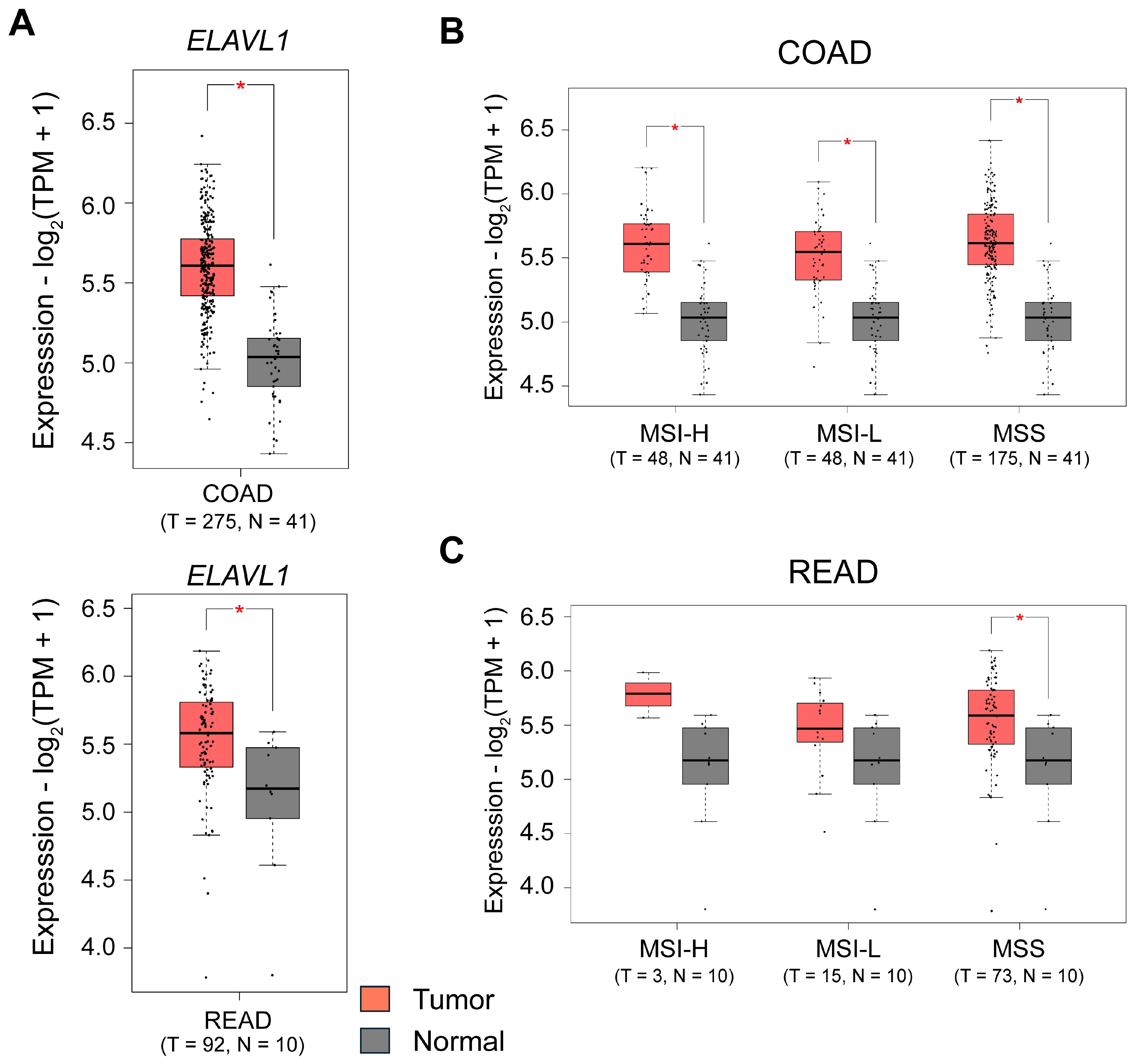
It is overexpressed in colorectal adenocarcinomas and binds to targets via AU-rich element (ARE) motifs, promoting increased mRNA stability (Figure 1) [40,41]. The functions of HuR in CRC pathogenesis have been well-studied. According to reports, HuR is increased in CRC [42,43,44,45]. It stabilizes several oncogenes, including COX-2 [43,45], VEGF [46], and IL-8 [46], which promotes the proliferation and tumorigenicity of CRC cells. Increased cytoplasmic HuR levels were reported to be strongly correlated with COX-2 expression and the stage of CC [43,45]. HuR markedly stimulates the formation of xenografted tumors in a mouse model of CRC [42].
In the last few years, evidence has shown that HuR’s influence on bound transcripts depends on its interaction with miRNAs, and competition between HuR and miRNAs enhances target gene expression, while their cooperation results in reduced expression [47]. MiRNAs are among the few known regulators of HuR expression levels and, hence, passively affect certain biological processes.
Figure 1. Expression of HuR in colon and rectal adenocarcinoma patients. Box plots showing expression of (A), ELAVL1 (gene encoding human antigen R; HuR) in colon adenocarcinoma (COAD) and rectal adenocarcinoma (READ) patients in The Cancer Genome Atlas (TCGA) COAD and READ cohorts generated using the Gene Expression Profiling Interactive Analysis 2 (GEPIA2) http://gepia2.cancer-pku.cn/) [48]. Box plots showing expression of ELAVL1 expression in (B), TCGA-COAD and (C), TCGA-READ cohorts in patients grouped based on the microsatellite stability status into microsatellite instability-high or low (MSI-h, MSI-l) or microsatellite stable (MSS). Comparisons are performed between cancer tissue and normal tissue. The number of patients (n) is indicated in the figure. The log2 fold change and p-value cut-offs were according to the default settings of GEPIA2. T = Tumor, N = Normal.
Cancers 16 03183 g001

3. Intriguing Role of miRNAs-HuR Axis in CRC

3.1. MiRNAs and Their Role in Cancer Pathogenesis

MiRNAs are so far the most studied type of small noncoding RNA molecules, typically 20–25 nucleotides long, that play a vital role in silencing target genes and regulating post-transcriptional processes [49,50]. They bind to specific mRNA molecules and inhibit protein production by promoting mRNA degradation or reducing translation efficiency. A single miRNA can target multiple mRNAs, and various miRNAs can act on the same mRNA as well [50,51,52,53]. Several computational tools have been developed to predict miRNA targets, which are then experimentally validated [54,55,56]. MiRNAs regulate nearly two-thirds of human protein-coding genes, influencing key cellular processes like migration, proliferation, differentiation, cell death, and metabolism [57,58]. However, because miRNA-mediated dynamics in biological networks are different, it is difficult to explain how miRNAs organize biological decision-making events [59].
Trieber et al. and several others have extensively discussed the biogenesis of miRNA before; therefore, we have not discussed it here in detail [60]. In miRNA biogenesis, a pri-miRNA (capped at the 5’ end and polyadenylated at the 3’ end) is transcribed by RNA Pol II. Endonucleases like DROSHA and DGCR8 process this precursor in the nucleus, producing an approximately 85-nucleotide stem-loop structure known as pre-miRNA [61,62]. Exportin-5 then transports the pre-miRNA to the cytoplasm, where the ribonuclease Dicer cleaves it into a double-stranded mature miRNA (22–30 nucleotides). The mature miRNA duplex associates with Argonaute proteins to form the RNA-induced silencing complex (RISC), which regulates translation by binding to complementary sequences in the 3’ UTR of target mRNAs via the seed region (2–8 nucleotides) [63,64]. Through these interactions, miRNAs form imperfect hybrids with target mRNAs, altering their stability and reducing the expression of around 30% of protein-coding genes [65].
Mutations or abnormal miRNA expression can contribute to cancer development and progression. Depending on the cellular context, miRNAs can function as either oncogenes or tumor suppressors [64,66]. Abnormal miRNA expression may result from gene amplification, deletion, or translocation of genomic regions containing miRNA genes. Additionally, miRNA genes can undergo epigenetic modifications, promoting oncogenesis in various cancers. These epigenetically altered miRNA genes are considered potential biomarkers for cancer diagnosis and monitoring disease progression [67,68].
More than 52% of miRNA genes associated with cancer suppression are located in fragile regions, which may lead to malfunction and abnormal miRNA expression in different types of cancer. These small molecules are crucial in regulating various cellular and biological processes, such as inflammation, stress response, migration, differentiation, apoptosis, metabolism, extracellular matrix regulation, MET transcription factors, and cancer initiation and progression. RBPs are also known to regulate miRNAs in several cancers [69]. They also impact tumor survival through pathophysiologic mechanisms like chemoresistance, chemosensitivity, and radiation resistance [70]. RBP HuR is reported to be associated with numerous miRNAs in different cancers (Figure 2) [47,71]. Oncogenic miRNAs, also known as oncomiRs, target and inhibit endogenous tumor suppressor genes, accelerating carcinogenesis [72].

3.1.1. Role of oncomiRs in HuR Regulation

Upregulation of oncomiRs is significantly known to impact the progression of CRC (Table 1). MiR-21 is a highly upregulated gene in CRC that targets and downregulates genes like PDCD4, TIAM1, SPRY2, PTEN, TGFBR2, and CDC25A [70,73]. It functions as an oncomiR, downregulating PTEN expression in CRC cells. MiR-21’s high expression is associated with resistance to chemotherapy of 5-FU, and its expression directly targets the 3’-UTR of tumor suppressor human DNA MutS homolog 2 (hMSH2), reducing 5-FU-induced damage arrest and apoptosis [74,75]. Overexpression of miR-21 enhances cell proliferation and invasion, inhibits apoptosis against 5-FU treatment, and vice versa [76]. These findings suggest miR-21 could be a non-invasive biomarker for CRC diagnosis and prognosis.
MiR-92a is a member of the oncomiR complex miR-17-92, which plays a key role in tumor cell proliferation, angiogenesis, and tumor progression in various cancers [77]. It is overexpressed in CRC, leading to suppression of E-cadherin and activation of β-catenin and vimentin, which promote EMT via the PTEN/PI3K/AKT pathway [78]. Elevated miR-92a, as shown by Nishida et al., is linked to lymphatic and venous invasion as well as liver metastasis through the TGF-β pathway [79]. CRC patients with high miR-92a expression have poor overall survival compared to those with low miR-92a expression [80]. Additionally, increased levels of miR-92a in CRC patients’ body fluids (e.g., plasma) have shown specificity and could be used for identifying individuals with CRC. This makes miR-92a a possible prognostic and diagnostic biomarker candidate for CRC [81].
MiR-96 is also upregulated in CRC patients and is linked to liver metastasis. It drives cell growth and proliferation by targeting TP53INP1, resulting in the downregulation of p53, FOXO1, and FOXO3a [82]. MiR-96 further enhances the effectiveness of 5-FU by inhibiting XIAP and UBE2N, leading to apoptosis [83]. Moreover, it also increases chemosensitivity to the platinum-based drug cisplatin and the PARP inhibitor AZD2281 by suppressing RAD51 recombinase and REV1 DNA polymerase, as evident in both in vitro and in vivo [83].
The miR-135 family has two isoforms: miR-135a and miR-135b. Both isoforms are conserved in mammals [84] and are considered oncogenic miRNAs in CRC, with miR-135b being overexpressed in cancer tissue than in normal tissue. Overexpression of miR-135 leads to the downregulation of APC, an essential component of the degradation complex in the Wnt signaling pathway. Furthermore, miR-135 serves as a biomarker for assessing the clinical stage of CRC [85,86].
Table 1. Role and function of miRNAs in CRC. MiRNAs, with their function as either oncomiR or tumor suppressor miR, are mentioned here.
No.microRNAFunctionReference
1.miR-21OncomiRs[74]
2.miR-92aOncomiRs[78]
3.miR-96OncomiRs[82]
4.miR-135OncomiRs[86]
5.miR-16Tumor suppressor miR[87]
6.miR-22Tumor suppressor miR[88]
7.miR-34Tumor suppressor miR[89,90]
8.miR-194Tumor suppressor miR[91,92,93,94]
9.miR-143Tumor suppressor miR[95,96]
10.miR-145Tumor suppressor miR[96]
11.miR-155-5pTumor suppressor miR[97]
12.miR-519cTumor suppressor miR[98,99]
13.miR-324-5pTumor suppressor miR[100,101]

3.1.2. Role of Tumor Suppressor miRs in HuR Regulation

MiRNAs also play a role as tumor suppressors by downregulating oncogenes involved in proliferation, apoptosis, invasion, and migration [72]. The miR-34 family is a prominent tumor suppressor miRNA due to its combinatorial effect with the tumor suppressor gene TP53.
It comprises three members, miR-34a, miR-34b, and miR-34c, located in the long arm 2 region of chromosome 11 [102]. MiR-34a, a tumor suppressor, regulates markers in cell proliferation, apoptosis, and senescence [103,104]. In cancer, miR-34a is downregulated, while HIF1 promotes angiogenesis by releasing VEGF from miRNA-mediated downregulation [103,104]. MiRNAs predominantly regulate target gene expression through a negative feedback loop or an incoherent feedforward loop [105]. MiR-34b-5p, another member of the miR-34 family, plays a crucial role in multiple diseases, including cancer. In CRC, long noncoding RNA (lncRNA) LINC02418 with a binding sequence for miR-34b-5p negatively affects patient survival, possibly via the miR-34b-5p/Bcl-2 axis.
The Lethal-7 (let-7) family, comprising 12 miRNAs, is essential in the epithelial-mesenchymal transition (EMT) process by targeting oncogenes like RAS, c-Myc, CDC25A, CDK6, and HMGA2. In CC tissues, let-7 is downregulated compared to adjacent noncancerous tissues, and high expression of let-7 is significantly correlated with better survival outcomes in CRC patients [106,107,108,109].
Additionally, miR-194 is mostly suppressed in CRC and is closely linked to overall survival and tumor size. Although it acts as a tumor suppressor in CRC, it is not associated with lymphatic invasion or distant metastasis. The expression profiles of the miR-194 target genes are in line with the oncogenic transcriptional regulator HMGA2, which is involved in restricting cell proliferation and migration, decreasing tumor growth in vivo, attenuating cell migration, suppressing EMT, and improving anticancer drug sensitivity in CRC [91,92,93,94].
MiR-143 and miR-145, located in a 1.6 kb cluster on the 5q32-33 chromosomal region, are frequently co-expressed from the same promoter. They are downregulated in several cancer types, including CRC, and function as anti-tumorigenic agents by altering various cancer-related cellular processes. The pathophysiology of synchronous CRC may be functionally associated with the deregulation of the miR-143-145 cluster [95,96].
Abdelmohsen et al. reported that miR-519 downregulated HuR expression in human CC cell lines RKO and HCT116 by suppressing HuR translation without affecting HuR mRNA stability [98]. HuR translation was more strongly suppressed by the coding region (CR) location of miR-519 interaction on HuR mRNA. When miR-519 levels increased, HuR levels decreased, suggesting an inverse correlation between the two. Normal tissues expressed low HuR and high miR-519 levels, but malignant tissues in cancer patients expressed high HuR and low miR-519 levels [98,99]. MiR-519’s potential as a tumor suppressor miRNA is also suggested by the fact that overexpression of the miR-519 reduced carcinogenesis by inhibiting DNA replication and cell division [99].
Using CRC patient tissue, Liu et al. have shown that tumor suppressor miR-22 was lowly expressed in CRC tissue compared to the normal tissue [88]. Expression of miR-22 was inversely correlated with HuR expression, and patients with high expression of miR-22 have a better prognosis. Furthermore, miR-22 directly interacts with the 3’-UTR of HuR, which inhibits HuR and hence limits the proliferation and migration of CRC cells in vitro.
Gu et al. have reported that MiR-324-5p is downregulated in CC cells (SW480 and SW620), while HuR (ELAVL1) expression is elevated in the same cell lines [100]. Cells treated with miR-324-5p mimics could downregulate the expression of ELAVL1 in both CC cells with a decrease in proliferation. Activation of miR-324-5p further limited the metastatic ability of CC cells by decreasing migration and invasion via targeting MMP-9 and the urokinase-type plasminogen activator system (uPA).
The inflammatory mediator, COX-2, produced in the arachidonic acid pathway, is well known as a tumor promoter in CRC [4]. HuR interacts with miR-16 to promote COX-2 mRNA stability and elevate its expression [87]. Due to this interaction, the steady-state levels of miR-16 decline rapidly. Cytoplasmic accumulation of HuR also leads to HuR binding to miR-16, causing a decrease in COX-2 abundance [87]. Although the molecular mechanism behind this reduction is unknown, these findings suggest that HuR could upregulate COX-2 expression by directly binding to COX-2 3’UTR and indirectly reducing miR-16 abundance. Additionally, antagomiR-155-5p reduces cytoplasmic HuR expression, facilitating HuR-mediated impact on the pro-inflammatory and pro-oncogenic mRNAs. Co-transfection with a blocker targeting TS1 ARE motifs reverses the reduction of HuR protein, suggesting that miR-155-5p affects HuR levels through mRNA expression rather than nucleus and cytoplasm reorganization [97].
Taken together, it is evident that a complex interplay between miRNAs and HuR possibly regulates tumor-promoting or tumor-suppressing functions. However, the HuR-miRNA axis is also known to be regulated by another type of non-coding RNA, circRNAs.

4. CircRNA Mediated Modulation of miRNAs-HuR Axis in CRC

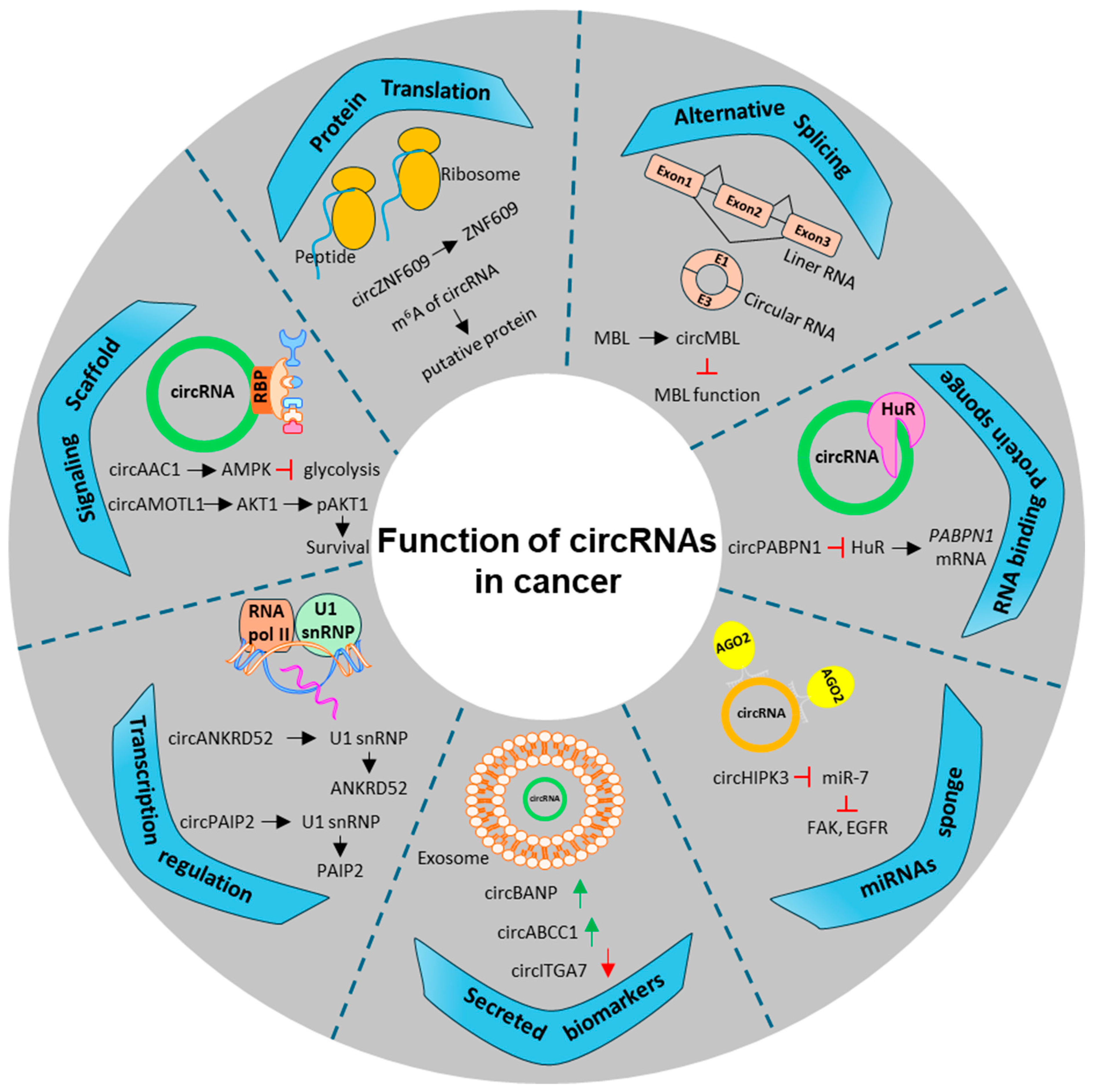
CircRNAs have recently gained significant attention for their diverse roles in gene regulation. Unlike linear RNA, circRNAs form continuous loops through covalent bonds generated by the back-splicing mechanism, making them more stable and resistant to degradation by RNA exonucleases [110]. Reports suggest that circRNAs can act as oncogenes or tumor suppressors by serving as miRNA sponges, binding to RBPs, regulating alternative splicing and transcription, encoding peptides, regulating host gene expression, or functioning as exosomal circRNAs (Figure 3) [111,112,113]. Additionally, circRNA concentrations, isoform expressions, and types are cell-type and tissue-specific [114,115,116,117], and they can be detected in human body fluids (e.g., saliva or plasma), which makes circRNAs and their host genes bona fide candidate biomarkers for early diagnosis, disease prognosis, and effectiveness of treatment for various tumors [118,119]. Although the biogenesis and function of circRNAs were previously poorly understood, with advances in sequencing techniques and bioinformatics analyses, researchers have better understood their roles and mechanisms.

4.1. CircRNA and Their Role in Gene Regulation

CircRNAs influence host gene transcription by interacting with RNA Pol II, recruiting various proteins, or forming R-loops to target regions regulating transcription [114,120,121,122,123,124,125]. Zhang et al. have shown that circANKRD52, circMCM5, and circSIRT7 accumulate at the transcription sites of their respective host genes, facilitating RNA Pol II-mediated transcription elongation and serving as positive regulators of transcription [114]. Moreover, reports show that circEIF3J and circPAIP2 bind to RNA Pol II, U1 snRNP, and promoters of their host genes, further amplifying transcription through a cis-acting positive feedback loop. Silencing these circRNAs results in reduced transcription levels of EIF3J and PAIP2 [120].
When RNA polymerase is silenced or RNA biogenesis is disrupted, R-loop structures composed of RNA-DNA hybrids and displaced single-stranded DNA can form [126,127], potentially interfering with replication and transcription processes. Recent studies indicate that circRNAs can create DNA hybrids or R-loops, increasing the efficiency of exon-deficient mRNA cleavage. Beyond altering linear transcript levels, circRNAs can trap mRNA, halt transcription, and enhance splicing factors, thereby regulating host gene expression [123,125]. Xu et al. identified that circSMARCA5 forms an R-loop to regulate its host gene expression [124]. Additionally, circRNAs are known to contain specific RNA-binding protein sites, which allow them to act as either protein decoys or scaffolds and guide proteins to certain promoter regions. This enables circRNAs to regulate transcription activation and host gene expression, potentially contributing to tumor progression. The following section will broadly discuss it, focusing on post-translational regulation by miRNA and protein sponges.

4.2. Regulatory Role of circRNA in the Translational and Post-Translational Modification of Their Host Genes

CircRNAs regulate the host gene translation by binding to the specific translation initiation-related proteins, influencing protein synthesis, and contributing to tumor development or progression. Yes-associated protein (YAP), a crucial factor in the Hippo pathway, is often dysregulated in CRC apart from other cancers [128]. CircYAP binds to YAP miRNA, translation initiation proteins eIF4G, and poly (A) binding protein (PABP), disrupting the interaction between eIF4G on the 5’-cap and PABP on the poly (A) tail of the YAP mRNA translation complex. This indicates that circYAP could act as a tumor suppressor gene, which could be validated with further investigations [129].
Furthermore, circRNAs can also serve as translation activators or inhibitors by influencing the binding of RBPs to host gene mRNA. For example, circPABPN1, derived from PABPN1, can compete with HuR by regulating PABPN1 [128,130] CircRNAs are also known to directly interact with host proteins to regulate post-translational modification, an essential process for maintaining protein turnover [131,132,133].

4.3. CircRNA as miRNA Sponges, Decoys, or Scaffolds

In addition to their above-mentioned role, circRNAs can also act as miRNA sponges or competing endogenous RNAs (ceRNAs) [134]. They contain miRNA response elements (MREs), which bind to and isolate miRNAs, preventing them from interacting with their target mRNAs. By sponging and binding to miRNAs, circRNAs block miRNAs from inhibiting their target genes. This leads to the upregulation of these target genes along with the protein-coding genes, which further regulate specific signaling cascades [135,136,137]. Moreover, some circRNAs are reported to act as ceRNAs in order to regulate the expression of other target genes and contribute to tumor development [137], like circEZH2/miR-133b/IGF2BP2/CREB1 [138]. CircCDR1as (ciRS-7) contains over 70 conserved miR-7 binding sites. CDR1as acts as a sponge by binding to miR-7 and regulating miR-7 target genes [139]. Circular RNAs may compete with non-coding RNAs or proteins for binding to regulatory molecules such as proteins RBPs during decoy activities [140]. Competition alters the availability and function of regulatory molecules, thus affecting gene expression. CircRNAs act as miRNA sponges, decoys, or scaffolds through distinct mechanisms by interacting and binding with regulatory molecules such as miRNAs and proteins.
In CRC, Tan et al. have reported that circRNA circ0104103 is downregulated in CRC tissue, and its activation leads to inhibition of cell proliferation, migration, and invasion, as evident using in vitro and in vivo models of CRC [141]. Circ0104103 acts as a ceRNA by negatively regulating endogenous miRNA-373-5P as a sponge, hence depleting the expression of HuR. Using both overexpression and silencing of circ0104103, authors have proved its tumor suppressor role in CRC cells.
Additionally, circAGO2 is over-expressed in CRC cells and directly interacts with HuR [142]. It induces the HuR translocation from nuclear to cytoplasm, further activating HuR protein and its associated miRNAs. The CircAGO2/HuR complex regulates cancer-promoting genes, like HNF4, NOTCH4, and SLC2A4, by regulating the miRNAs, miR-224-5p and miR-143-3p, located close to the ARE of these oncogenes. Anti-cancer therapies targeting the circAGO2/HuR complex could be effective in CC.
CircRNA can act as a protein sponge or decoy by binding to a specific region of the target promoter, regulating the activity, localization, or stability of the target proteins. RBPs, DNA demethylases, and DNA methyltransferases play crucial roles in this type of interaction. As a result, transcription, splicing, or other regulatory processes will be altered. CircFoxo3, a circRNA derived from the FOXO3 gene, binds to various proteins involved in cell cycle regulation, such as CDK2 and p21 [143,144], which in turn not only regulates their activity and cellular localization but also influences cell cycle progression. It has been demonstrated that some circRNAs transcriptionally activate host genes and their downstream target genes involved in either tumor promotion or inhibition [122,145].

4.4. Role of circRNA in the Regulation of miRNA-HuR Axis in CRC

CircRNAs regulate the miRNA-HuR axis, which contributes significantly to cancer progression and metastasis in CRC [146,147]. CircRNAs can serve as sponges for miRNAs that target HuR mRNA (Table 2). As circRNAs sequester these miRNAs, they prevent them from binding to and downregulating HuR mRNA. Through this interaction, HuR mRNA is stabilized and expressed more, which results in a greater level of HuR protein and subsequent effects on gene expression in CRC cells [142]. Among the well-studied circRNAs in CRC is circHIPK3, which acts as a sponge for miRNAs targeting HuR mRNAs, including miR-7, miR-124, and miR-29b [148,149]. As circHIPK3 binds to these miRNAs, it derepresses HuR expression, promoting CRC progression by inducing HuR-mediated proliferation and metastasis [150].
CircRNA microarray analysis identified that the two anti-metastatic circRNAs, circPPFIA1-L and circPPFIA1-s, are significantly downregulated in CRC cells [151]. CircPPFIA1-L and circPPFIA1-s acted as sponges to the oncomiR miR-155-5p. While circPPFIA1-s upregulated tumor suppressor CDX1 by decoying miR-155-5p; it downregulated oncogene RAB36 by disrupting the miR-155-5p/HuR complex and separating HuR. CircNOLC1 plays a vital role in CC liver metastasis [152]. CircNOLC1 upregulates the NOLC1 mRNA expression via HuR. It also sponges miR-212-5p to upregulate c-Met in liver metastasis.
Table 2. Role of circRNA/lncRNA in miRNA-HuR axis regulation in CRC.
No.microRNA (Expression)Regulation by Other Non-Coding RNAReference
1.miR-155-5pcircPPFIA1-L, circPPFIA1-S (circRNA)[151]
2.miR-373-5pcirc0104103 (circRNA)[141]
3.miR-143-3p
miR-224-5p
circAGO2 (circRNA)[142]
4.miR-212-5pcircNOLC1 (circRNA)[152]
5.miR-34b-5pOIP5-AS1 (lncRNA)[153]
6.miR-3121-3pTNFRSF10A-AS1 (lncRNA)[154]
CircPABPN1 and circRHOBTB3 have also been shown to promote CRC cell proliferation and migration by sponging miR-675, which targets HuR mRNA [155,156]. As a result of this circRNA-mediated regulation, HuR protein levels are upregulated, which facilitates tumor progression. CRC cell survival, proliferation, and metastasis are promoted by circRNA-mediated regulation of the miRNA-HuR axis. CRC progression may be associated with HuR’s ability to stabilize mRNAs encoding proteins involved in the cell cycle (e.g., cyclins, CDKs, and apoptosis resistance proteins, such as the Bcl-2 family) and epithelial-mesenchymal transition (EMT) genes. Circular RNAs that regulate the miRNA-HuR axis could potentially be targeted as therapeutics for CRC. The use of antisense oligonucleotides (ASOs) or small molecule inhibitors to inhibit or disrupt specific circRNA-miRNA interactions may offer novel approaches to treating CRCs. A circRNA-miRNA interaction affects the miRNA-HuR axis, modulating HuR expression and downstream gene targets in CRC. A better understanding of the miRNA-HuR axis provides insight into potential therapeutic strategies to intervene in the pathogenesis of CRCs by targeting the miRNA-HuR axis.

5. Intriguing Role of lncRNA in the Regulation of miRNA-HuR Axis in CRC

The last type of non-coding RNA we discuss here as a regulator of the HuR-miRNA axis is the lncRNAs. lncRNAs are produced from widespread genome transcription. These RNAs are longer than 200 nucleotides and cannot translate into functional proteins. This broad term comprises an enormous and tremendously varied group of transcripts with several genetic origins and biogenesis. The human genome has more than 16,000 lncRNA genes, which is shown by Human GENCODE Statistics, though a few other databases show that humans have more than 400,000 lncRNAs [157,158]. These RNAs control gene expression at various stages, including RNA splicing, chromatin structure, function, transcription of neighboring and distant genes, translation, and stability. Chromatin relaxation could happen when positively charged histone tails neutralize the negative charge of RNA molecules, which suggests that RNA-mediated chromatin relaxation could be an unusually fast event that leads to specific gene activation [159]. Numerous lncRNAs on chromatin have been revealed to interact with proteins, thus either upregulating or downregulating their binding and activity at specific sites of DNA. Additionally, direct lncRNA transcriptional effects on the gene of interest can be enabled by protein-assisted long-range chromatin interactions, like those facilitated by CTCF, a zinc finger protein [160,161,162]. About 20% of all lncRNAs in mammalian genomes are divergent lncRNAs transcribed in the opposite direction from neighboring protein-coding genes. Luo et al., through a genome-wide study, identified that there is a high correlation between the distribution of this class of lncRNAs and critical developmental regulating genes [163]. Additionally, lncRNAs are involved in the development and regulation of nuclear condensates and organelles [164]. Studies have shown that membrane-free RNA-protein compartments called nuclear condensates are essential for several biological activities [165]. There is a close relationship between the spatial regulation of gene expression during development and the abundance of lncRNAs associated with chromatin-modifying complexes, transcribed from enhancers, and nucleate phase separation of nuclear condensates and domains [166]. Using super-resolution microscopy, West and colleagues identified nuclear structures known as paraspeckles, constructed around the lncRNA Neat1. Neat1 controls several physiological functions, such as the development of the corpus luteum and the progression of cancer [167]. Recently, several authors have reviewed the detailed transcriptional regulatory activity of lncRNAs in the context of development and disease biology [164,166,168].
Dysregulated expression of lncRNAs and miRNAs has been identified as a measure of independent cancer patient prognosis and a characteristic of cancer progression [169,170]. Several studies have highlighted a strong relationship between miRNA and lncRNA in regulating the transcriptional regulation of RNA [169,171]. By sequestering miRNAs, lncRNAs can function as miRNA decoys, competing with endogenous RNAs and causing miRNA target genes to re-express. Furthermore, lncRNAs can enhance gene expression by competing with miRNAs for specific binding sites in the non-coding sections of mRNAs, thus blocking the transcriptional repression caused by miRNAs. The strong interaction between several kinds of non-coding RNA shows that some lncRNAs can be processed into miRNAs [169,172,173]. There is growing evidence that lncRNAs are important for the initiation and development of CC [174,175,176]. Forrest et al. used RNA-sequencing data from the TCGA dataset to find approximately 200 lncRNAs that were differentially expressed in colon tumors [174]. Kasagi and colleagues in their study found Colon-cancer Associated Transcript 2 (CCAT2), a substantially overexpressed lncRNA in microsatellite-stable CRC, to circumscribe a single nucleotide polymorphism (SNP) rs6983267, which is a high-risk allele for CRC [177]. In a separate study, Ling et al. demonstrated that CCAT2 transduced HCT116 cells exhibit enhanced migration capacity, and highly metastatic KM12SM silenced for CCAT2 exhibits diminished invasion rates [178]. The authors further found that CCAT2 physically interacts with TCFL2 and MYC-regulated miRNAs miR-17-5p and miR-20a. Notably, miR-20a was overexpressed during the change from colon mucosa to early adenoma and is expected to target several genes involved in the WNT pathway [178]. Alshahrani et al. and Rajtmajerova et al. have extensively reviewed more examples where the association between the lncRNA-miRNA axis and CRC development is described in detail [179,180].
To explore the regulatory mechanism of the lncRNA-miRNA axis in CRC development, Wang et al. found OIP5-AS1 lncRNA to positively interact between the HuR protein and miR-34b-5p [153]. Furthermore, they explored the carcinogenic potential of OIP5-AS1, revealing its overexpression in CRC patients along with HuR. The simultaneous inhibition of HuR reduces the levels of OIP5-AS1, leading to a concomitant decrease in CRC growth. Mechanistically, authors found that HuR binds to and stabilizes OIP5-AS1 expression in CRC cells, whereas miR-34b-5p competes with HuR in binding to OIP5-AS1 [153]. Similarly, Wang et al. revealed the carcinogenic potential of TNFRSF10A-AS1 in CRC progression, revealing its overexpression in CRC patients along with HuR [154]. Through in vitro luciferase assay and in silico analysis, the direct interaction between TNFRSF10A-AS1 and miR-3121-3p was verified. However, miR-3121-3p was found to be downregulated in CRC patients, suggesting a negative correlation with TNFRSF10A-AS1. Hence, the authors proposed that the oncogenic activity of TNFRSF10A-AS1 was via sponging miR-3121-3p. They showed that TNFRSF10A-AS1 binds to miR-3121-3p, thereby reducing the suppressing effect of miR-3121-3p on its downstream target, HuR. Conclusively, both studies highlighted contrasting but crucial insights on how the lncRNA/miRNA/HuR axis plays a significant role in the progression of CRC, thereby providing a platform for the diagnosis and treatment of CRC.

6. Conclusions and Future Directions

The HuR-miRNA interplay is complex, particularly in CRC. MiRNAs regulate HuR levels, while HuR, in turn, regulates the levels of miRNAs. The role of this ironic relationship between HuR-miRNA and its impact on mRNA translation in CRC remains poorly understood and requires further investigation [181]. A deeper understanding of the precise binding sites of HuR and miRNAs, their spatial arrangement, and how their interactions influence the secondary structure of the RNA is essential. Such studies would help understand how these interactions influence mRNA stability and translation in CRC. Hence, investigations focused on individual mRNAs are required [182,183]. While competitive regulation of target mRNA degradation by HuR and miRNAs has been observed, high-throughput analyses have not yet revealed more widespread instances of this process. To gain further insights, it would be beneficial to use experimental approaches such as identifying Ago2 binding sites under conditions of cellular stress, mitogenic signaling, or in the presence or absence of HuR [184]. Moreover, high-throughput techniques like polysome profiling, nascent translation assays, and pulsed SILAC should be used to investigate alterations in translation. This approach would help distinguish between direct miRNA-mediated regulations of HuR abundance and HuR-mediated regulation of miRNA levels, a distinction critical for developing novel biomarkers or therapeutic regimens for CRC [33,185]. Therapeutically, targeting the HuR-miRNA axis presents a promising avenue for CRC treatment. By manipulating this regulatory network, it may be possible to affect mRNA translation, which would either help suppress tumor growth or enhance the efficacy of existing treatments. Small molecules or antisense oligonucleotides designed to disrupt specific HuR-oncomiRs interactions could reduce HuR’s stabilizing effect on oncogenic mRNAs, thereby minimizing cancer progression. Additionally, therapies that modulate miRNA levels to alter HuR activity could benefit precision therapy against CRC. Although few preliminary studies discussed in the review article suggest intricate relationships of the HuR-miRNA axis in CRC progression, we believe that more studies are needed to strengthen such claims for diagnostic or therapeutic purposes in the future. Thus, advancing our understanding of the HuR-miRNA relationship will be crucial for developing biomarkers and targeted therapies and improving clinical outcomes in CRC.

Author Contributions

Conceptualization and design: V.Y. and S.R.S.; Writing, review, and/or revision of the manuscript: V.Y., T.S., D.S., V.K.G., P.C., S.G. and S.R.S.; Study supervision: S.R.S. All authors have read and agreed to the published version of the manuscript.

Funding

This research received no external funding.

Conflicts of Interest

The authors declare no conflicts of interest.

References

  1. Siegel, R.L.; Giaquinto, A.N.; Jemal, A. Cancer statistics, 2024. CA Cancer J. Clin. 2024, 74, 12–49. [Google Scholar] [CrossRef] [PubMed]
  2. Siegel, R.L.; Wagle, N.S.; Cercek, A.; Smith, R.A.; Jemal, A. Colorectal cancer statistics, 2023. CA Cancer J. Clin. 2023, 73, 233–254. [Google Scholar] [CrossRef] [PubMed]
  3. Bray, F.; Laversanne, M.; Sung, H.; Ferlay, J.; Siegel, R.L.; Soerjomataram, I.; Jemal, A. Global cancer statistics 2022: GLOBOCAN estimates of incidence and mortality worldwide for 36 cancers in 185 countries. CA Cancer J. Clin. 2024, 74, 229–263. [Google Scholar] [CrossRef] [PubMed]
  4. Wang, D.; Dubois, R.N. Eicosanoids and cancer. Nat. Rev. Cancer 2010, 10, 181–193. [Google Scholar] [CrossRef] [PubMed]
  5. Ekbom, A.; Helmick, C.; Zack, M.; Adami, H.O. Ulcerative colitis and colorectal cancer. A population-based study. N. Engl. J. Med. 1990, 323, 1228–1233. [Google Scholar] [CrossRef] [PubMed]
  6. Nadeem, M.S.; Kumar, V.; Al-Abbasi, F.A.; Kamal, M.A.; Anwar, F. Risk of colorectal cancer in inflammatory bowel diseases. Semin. Cancer Biol. 2020, 64, 51–60. [Google Scholar] [CrossRef] [PubMed]
  7. Shah, S.C.; Itzkowitz, S.H. Colorectal Cancer in Inflammatory Bowel Disease: Mechanisms and Management. Gastroenterology 2022, 162, 715–730.e713. [Google Scholar] [CrossRef] [PubMed]
  8. Savari, S.; Vinnakota, K.; Zhang, Y.; Sjolander, A. Cysteinyl leukotrienes and their receptors: Bridging inflammation and colorectal cancer. World J. Gastroenterol. 2014, 20, 968–977. [Google Scholar] [CrossRef] [PubMed]
  9. Satapathy, S.R.; Sjolander, A. Cysteinyl leukotriene receptor 1 promotes 5-fluorouracil resistance and resistance-derived stemness in colon cancer cells. Cancer Lett. 2020, 488, 50–62. [Google Scholar] [CrossRef] [PubMed]
  10. Kuipers, E.J.; Grady, W.M.; Lieberman, D.; Seufferlein, T.; Sung, J.J.; Boelens, P.G.; van de Velde, C.J.; Watanabe, T. Colorectal cancer. Nat. Rev. Dis. Primers 2015, 1, 15065. [Google Scholar] [CrossRef]
  11. Cunningham, D.; Atkin, W.; Lenz, H.J.; Lynch, H.T.; Minsky, B.; Nordlinger, B.; Starling, N. Colorectal cancer. Lancet 2010, 375, 1030–1047. [Google Scholar] [CrossRef] [PubMed]
  12. Testa, U.; Pelosi, E.; Castelli, G. Colorectal cancer: Genetic abnormalities, tumor progression, tumor heterogeneity, clonal evolution and tumor-initiating cells. Med. Sci. 2018, 6, 31. [Google Scholar] [CrossRef] [PubMed]
  13. Johnson, C.M.; Wei, C.; Ensor, J.E.; Smolenski, D.J.; Amos, C.I.; Levin, B.; Berry, D.A. Meta-analyses of colorectal cancer risk factors. Cancer Causes Control. 2013, 24, 1207–1222. [Google Scholar] [CrossRef] [PubMed]
  14. Vuik, F.E.; Nieuwenburg, S.A.; Bardou, M.; Lansdorp-Vogelaar, I.; Dinis-Ribeiro, M.; Bento, M.J.; Zadnik, V.; Pellisé, M.; Esteban, L.; Kaminski, M.F.; et al. Increasing incidence of colorectal cancer in young adults in Europe over the last 25 years. Gut 2019, 68, 1820–1826. [Google Scholar] [CrossRef]
  15. Burt, R.W.; DiSario, J.A.; Cannon-Albright, L. Genetics of colon cancer: Impact of inheritance on colon cancer risk. Annu. Rev. Med. 1995, 46, 371–379. [Google Scholar] [CrossRef]
  16. Tuohy, T.M.; Rowe, K.G.; Mineau, G.P.; Pimentel, R.; Burt, R.W.; Samadder, N.J. Risk of colorectal cancer and adenomas in the families of patients with adenomas: A population-based study in Utah. Cancer 2014, 120, 35–42. [Google Scholar] [CrossRef]
  17. Baraibar, I.; Ros, J.; Saoudi, N.; Salvà, F.; García, A.; Castells, M.R.; Tabernero, J.; Élez, E. Sex and gender perspectives in colorectal cancer. ESMO Open 2023, 8, 101204. [Google Scholar] [CrossRef]
  18. Jacobsson, M.; Wagner, V.; Kanneganti, S. Screening for Colorectal Cancer. Surg. Clin. N. Am. 2024, 104, 595–607. [Google Scholar] [CrossRef] [PubMed]
  19. Robertson, D.J.; Rex, D.K.; Ciani, O.; Drummond, M.F. Colonoscopy vs the Fecal Immunochemical Test: Which is Best? Gastroenterology 2024, 166, 758–771. [Google Scholar] [CrossRef] [PubMed]
  20. Loupakis, F.; Cremolini, C.; Masi, G.; Lonardi, S.; Zagonel, V.; Salvatore, L.; Cortesi, E.; Tomasello, G.; Ronzoni, M.; Spadi, R.; et al. Initial therapy with FOLFOXIRI and bevacizumab for metastatic colorectal cancer. N. Engl. J. Med. 2014, 371, 1609–1618. [Google Scholar] [CrossRef] [PubMed]
  21. Jonker, D.J.; O’Callaghan, C.J.; Karapetis, C.S.; Zalcberg, J.R.; Tu, D.; Au, H.J.; Berry, S.R.; Krahn, M.; Price, T.; Simes, R.J.; et al. Cetuximab for the treatment of colorectal cancer. N. Engl. J. Med. 2007, 357, 2040–2048. [Google Scholar] [CrossRef] [PubMed]
  22. Hurwitz, H.; Fehrenbacher, L.; Novotny, W.; Cartwright, T.; Hainsworth, J.; Heim, W.; Berlin, J.; Baron, A.; Griffing, S.; Holmgren, E.; et al. Bevacizumab plus irinotecan, fluorouracil, and leucovorin for metastatic colorectal cancer. N. Engl. J. Med. 2004, 350, 2335–2342. [Google Scholar] [CrossRef] [PubMed]
  23. Le, D.T.; Uram, J.N.; Wang, H.; Bartlett, B.R.; Kemberling, H.; Eyring, A.D.; Skora, A.D.; Luber, B.S.; Azad, N.S.; Laheru, D.; et al. PD-1 Blockade in Tumors with Mismatch-Repair Deficiency. N. Engl. J. Med. 2015, 372, 2509–2520. [Google Scholar] [CrossRef] [PubMed]
  24. Mohammadi, A.; Mansoori, B.; Baradaran, B. The role of microRNAs in colorectal cancer. Biomed. Pharmacother. 2016, 84, 705–713. [Google Scholar] [CrossRef] [PubMed]
  25. Balacescu, O.; Sur, D.; Cainap, C.; Visan, S.; Cruceriu, D.; Manzat-Saplacan, R.; Muresan, M.-S.; Balacescu, L.; Lisencu, C.; Irimie, A. The Impact of miRNA in Colorectal Cancer Progression and Its Liver Metastases. Int. J. Mol. Sci. 2018, 19, 3711. [Google Scholar] [CrossRef]
  26. Luo, X.; Burwinkel, B.; Tao, S.; Brenner, H. MicroRNA Signatures: Novel Biomarker for Colorectal Cancer? Cancer Epidemiol. Biomark. Prev. 2011, 20, 1272–1286. [Google Scholar] [CrossRef]
  27. Gerstberger, S.; Hafner, M.; Tuschl, T. A census of human RNA-binding proteins. Nat. Rev. Genet. 2014, 15, 829–845. [Google Scholar] [CrossRef]
  28. Tao, Y.; Zhang, Q.; Wang, H.; Yang, X.; Mu, H. Alternative splicing and related RNA binding proteins in human health and disease. Signal Transduct. Target. Ther. 2024, 9, 26. [Google Scholar] [CrossRef] [PubMed]
  29. Oliveira, C.; Faoro, H.; Alves, L.R.; Goldenberg, S. RNA-binding proteins and their role in the regulation of gene expression in Trypanosoma cruzi and Saccharomyces cerevisiae. Genet. Mol. Biol. 2017, 40, 22–30. [Google Scholar] [CrossRef]
  30. Pereira, B.; Billaud, M.; Almeida, R. RNA-Binding Proteins in Cancer: Old Players and New Actors. Trends Cancer 2017, 3, 506–528. [Google Scholar] [CrossRef]
  31. Kang, D.; Lee, Y.; Lee, J.S. RNA-Binding Proteins in Cancer: Functional and Therapeutic Perspectives. Cancers 2020, 12, 2699. [Google Scholar] [CrossRef] [PubMed]
  32. Qin, H.; Ni, H.; Liu, Y.; Yuan, Y.; Xi, T.; Li, X.; Zheng, L. RNA-binding proteins in tumor progression. J. Hematol. Oncol. 2020, 13, 90. [Google Scholar] [CrossRef] [PubMed]
  33. Meisner, N.-C.; Filipowicz, W. Properties of the regulatory RNA-binding protein HuR and its role in controlling miRNA repression. In Regulation of microRNAs; Springer: New York, NY, USA, 2010; pp. 106–123. [Google Scholar]
  34. Dalmau, J.; Furneaux, H.M.; Gralla, R.J.; Kris, M.G.; Posner, J.B. Detection of the anti-Hu antibody in the serum of patients with small cell lung cancer—A quantitative western blot analysis. Ann. Neurol. 1990, 27, 544–552. [Google Scholar] [CrossRef] [PubMed]
  35. Brennan, C.; Steitz*, J. HuR and mRNA stability. Cell. Mol. Life Sci. CMLS 2001, 58, 266–277. [Google Scholar] [CrossRef] [PubMed]
  36. Finan, J.M.; Sutton, T.L.; Dixon, D.A.; Brody, J.R. Targeting the RNA-Binding Protein HuR in Cancer. Cancer Res. 2023, 83, 3507–3516. [Google Scholar] [CrossRef] [PubMed]
  37. Liu, L.; Zhuang, R.; Xiao, L.; Chung, H.K.; Luo, J.; Turner, D.J.; Rao, J.N.; Gorospe, M.; Wang, J.Y. HuR Enhances Early Restitution of the Intestinal Epithelium by Increasing Cdc42 Translation. Mol. Cell Biol. 2017, 37, e00574-16. [Google Scholar] [CrossRef] [PubMed]
  38. Song, X.; Shi, X.; Li, W.; Zhang, F.; Cai, Z. The RNA-Binding Protein HuR in Digestive System Tumors. Biomed. Res. Int. 2020, 2020, 9656051. [Google Scholar] [CrossRef] [PubMed]
  39. Majumder, M.; Chakraborty, P.; Mohan, S.; Mehrotra, S.; Palanisamy, V. HuR as a molecular target for cancer therapeutics and immune-related disorders. Adv. Drug Deliv. Rev. 2022, 188, 114442. [Google Scholar] [CrossRef]
  40. Fan, X.C.; Steitz, J.A. Overexpression of HuR, a nuclear–cytoplasmic shuttling protein, increases the in vivo stability of ARE-containing mRNAs. EMBO J. 1998, 17, 3448–3460. [Google Scholar] [CrossRef]
  41. Dixon, D.A.; Kaplan, C.D.; McIntyre, T.M.; Zimmerman, G.A.; Prescott, S.M. Post-transcriptional control of cyclooxygenase-2 gene expression: The role of the 3′-untranslated region. J. Biol. Chem. 2000, 275, 11750–11757. [Google Scholar] [CrossRef]
  42. De Silanes, I.L.; Fan, J.; Yang, X.; Zonderman, A.B.; Potapova, O.; Pizer, E.S.; Gorospe, M. Role of the RNA-binding protein HuR in colon carcinogenesis. Oncogene 2003, 22, 7146–7154. [Google Scholar] [CrossRef]
  43. Young, L.E.; Sanduja, S.; Bemis–Standoli, K.; Pena, E.A.; Price, R.L.; Dixon, D.A. The mRNA binding proteins HuR and tristetraprolin regulate cyclooxygenase 2 expression during colon carcinogenesis. Gastroenterology 2009, 136, 1669–1679. [Google Scholar] [CrossRef] [PubMed]
  44. Embade, N.; Fernández-Ramos, D.; Varela-Rey, M.; Beraza, N.; Sini, M.; de Juan, V.G.; Woodhoo, A.; Martínez-López, N.; Rodríguez-Iruretagoyena, B.; Bustamante, F.J. Murine double minute 2 regulates Hu antigen R stability in human liver and colon cancer through NEDDylation. Hepatology 2012, 55, 1237–1248. [Google Scholar] [CrossRef]
  45. Dixon, D.A.; Tolley, N.D.; King, P.H.; Nabors, L.B.; McIntyre, T.M.; Zimmerman, G.A.; Prescott, S.M. Altered expression of the mRNA stability factor HuR promotes cyclooxygenase-2 expression in colon cancer cells. J. Clin. Investig. 2001, 108, 1657–1665. [Google Scholar] [CrossRef] [PubMed]
  46. Denkert, C.; Koch, I.; von Keyserlingk, N.; Noske, A.; Niesporek, S.; Dietel, M.; Weichert, W. Expression of the ELAV-like protein HuR in human colon cancer: Association with tumor stage and cyclooxygenase-2. Mod. Pathol. 2006, 19, 1261–1269. [Google Scholar] [CrossRef] [PubMed]
  47. Sobolewski, C.; Dubuquoy, L.; Legrand, N. MicroRNAs, Tristetraprolin Family Members and HuR: A Complex Interplay Controlling Cancer-Related Processes. Cancers 2022, 14, 3516. [Google Scholar] [CrossRef] [PubMed]
  48. Tang, Z.; Kang, B.; Li, C.; Chen, T.; Zhang, Z. GEPIA2: An enhanced web server for large-scale expression profiling and interactive analysis. Nucleic Acids Res. 2019, 47, W556–W560. [Google Scholar] [CrossRef]
  49. Bartel, D.P. MicroRNAs: Target recognition and regulatory functions. Cell 2009, 136, 215–233. [Google Scholar] [CrossRef]
  50. Iwakawa, H.-o.; Tomari, Y. The functions of microRNAs: mRNA decay and translational repression. Trends Cell Biol. 2015, 25, 651–665. [Google Scholar] [CrossRef]
  51. Eulalio, A.; Huntzinger, E.; Izaurralde, E. Getting to the root of miRNA-mediated gene silencing. Cell 2008, 132, 9–14. [Google Scholar] [CrossRef]
  52. O’Brien, J.; Hayder, H.; Zayed, Y.; Peng, C. Overview of microRNA biogenesis, mechanisms of actions, and circulation. Front. Endocrinol. 2018, 9, 388354. [Google Scholar] [CrossRef]
  53. Yadav, V.; Jena, M.K.; Parashar, G.; Parashar, N.C.; Joshi, H.; Ramniwas, S.; Tuli, H.S. Emerging role of microRNAs as regulators of protein kinase C substrate MARCKS and MARCKSL1 in cancer. Exp. Cell Res. 2024, 434, 113891. [Google Scholar] [CrossRef] [PubMed]
  54. Sun, W.; Julie Li, Y.-S.; Huang, H.-D.; Shyy, J.Y.; Chien, S. microRNA: A master regulator of cellular processes for bioengineering systems. Annu. Rev. Biomed. Eng. 2010, 12, 1–27. [Google Scholar] [PubMed]
  55. Hamilton, M.P.; Rajapakshe, K.; Hartig, S.M.; Reva, B.; McLellan, M.D.; Kandoth, C.; Ding, L.; Zack, T.I.; Gunaratne, P.H.; Wheeler, D.A. Identification of a pan-cancer oncogenic microRNA superfamily anchored by a central core seed motif. Nat. Commun. 2013, 4, 2730. [Google Scholar] [CrossRef]
  56. Agarwal, V.; Bell, G.W.; Nam, J.-W.; Bartel, D.P. Predicting effective microRNA target sites in mammalian mRNAs. eLife 2015, 4, e05005. [Google Scholar] [CrossRef] [PubMed]
  57. Lee, Y.; Ahn, C.; Han, J.; Choi, H.; Kim, J.; Yim, J.; Lee, J.; Provost, P.; Rådmark, O.; Kim, S. The nuclear RNase III Drosha initiates microRNA processing. Nature 2003, 425, 415–419. [Google Scholar] [CrossRef] [PubMed]
  58. Denli, A.M.; Tops, B.B.; Plasterk, R.H.; Ketting, R.F.; Hannon, G.J. Processing of primary microRNAs by the Microprocessor complex. Nature 2004, 432, 231–235. [Google Scholar] [CrossRef] [PubMed]
  59. Bartel, D.P. MicroRNAs: Genomics, biogenesis, mechanism, and function. Cell 2004, 116, 281–297. [Google Scholar] [CrossRef] [PubMed]
  60. Treiber, T.; Treiber, N.; Meister, G. Regulation of microRNA biogenesis and its crosstalk with other cellular pathways. Nat. Rev. Mol. Cell Biol. 2019, 20, 5–20. [Google Scholar] [CrossRef]
  61. Lee, Y.; Kim, M.; Han, J.; Yeom, K.H.; Lee, S.; Baek, S.H.; Kim, V.N. MicroRNA genes are transcribed by RNA polymerase II. EMBO J. 2004, 23, 4051–4060. [Google Scholar] [CrossRef]
  62. Borchert, G.M.; Lanier, W.; Davidson, B.L. RNA polymerase III transcribes human microRNAs. Nat. Struct. Mol. Biol. 2006, 13, 1097–1101. [Google Scholar] [CrossRef] [PubMed]
  63. MacFarlane, L.-A.; R Murphy, P. MicroRNA: Biogenesis, function and role in cancer. Curr. Genom. 2010, 11, 537–561. [Google Scholar] [CrossRef]
  64. Peng, Y.; Croce, C.M. The role of MicroRNAs in human cancer. Signal Transduct. Target. Ther. 2016, 1, 15004. [Google Scholar] [CrossRef] [PubMed]
  65. Majidinia, M.; Darband, S.G.; Kaviani, M.; Nabavi, S.M.; Jahanban-Esfahlan, R.; Yousefi, B. Cross-regulation between Notch signaling pathway and miRNA machinery in cancer. DNA Repair 2018, 66, 30–41. [Google Scholar] [CrossRef] [PubMed]
  66. Hayes, J.; Peruzzi, P.P.; Lawler, S. MicroRNAs in cancer: Biomarkers, functions and therapy. Trends Mol. Med. 2014, 20, 460–469. [Google Scholar] [CrossRef] [PubMed]
  67. Calin, G.; Croce, C. MicroRNAs and chromosomal abnormalities in cancer cells. Oncogene 2006, 25, 6202–6210. [Google Scholar] [CrossRef] [PubMed]
  68. Lujambio, A.; Calin, G.A.; Villanueva, A.; Ropero, S.; Sánchez-Céspedes, M.; Blanco, D.; Montuenga, L.M.; Rossi, S.; Nicoloso, M.S.; Faller, W.J. A microRNA DNA methylation signature for human cancer metastasis. Proc. Natl. Acad. Sci. USA 2008, 105, 13556–13561. [Google Scholar] [CrossRef] [PubMed]
  69. van Kouwenhove, M.; Kedde, M.; Agami, R. MicroRNA regulation by RNA-binding proteins and its implications for cancer. Nat. Rev. Cancer 2011, 11, 644–656. [Google Scholar] [CrossRef] [PubMed]
  70. Mehrgou, A.; Ebadollahi, S.; Seidi, K.; Ayoubi-Joshaghani, M.H.; Yazdi, A.A.; Zare, P.; Jaymand, M.; Jahanban-Esfahlan, R. Roles of miRNAs in colorectal cancer: Therapeutic implications and clinical opportunities. Adv. Pharm. Bull. 2021, 11, 233. [Google Scholar] [CrossRef]
  71. Zhou, H.; Rao, Y.; Sun, Q.; Liu, Y.; Zhou, X.; Chen, Y.; Chen, J. MiR-4458/human antigen R (HuR) modulates PBX3 mRNA stability in melanoma tumorigenesis. Arch. Dermatol. Res. 2020, 312, 665–673. [Google Scholar] [CrossRef]
  72. Hussen, B.M.; Hidayat, H.J.; Salihi, A.; Sabir, D.K.; Taheri, M.; Ghafouri-Fard, S. MicroRNA: A signature for cancer progression. Biomed. Pharmacother. 2021, 138, 111528. [Google Scholar] [CrossRef]
  73. Thomas, J.; Ohtsuka, M.; Pichler, M.; Ling, H. MicroRNAs: Clinical relevance in colorectal cancer. Int. J. Mol. Sci. 2015, 16, 28063–28076. [Google Scholar] [CrossRef] [PubMed]
  74. Asangani, I.A.; Rasheed, S.A.; Nikolova, D.; Leupold, J.; Colburn, N.; Post, S.; Allgayer, H. MicroRNA-21 (miR-21) post-transcriptionally downregulates tumor suppressor Pdcd4 and stimulates invasion, intravasation and metastasis in colorectal cancer. Oncogene 2008, 27, 2128–2136. [Google Scholar] [CrossRef] [PubMed]
  75. Xiong, B.; Cheng, Y.; Ma, L.; Zhang, C. MiR-21 regulates biological behavior through the PTEN/PI-3 K/Akt signaling pathway in human colorectal cancer cells. Int. J. Oncol. 2013, 42, 219–228. [Google Scholar] [CrossRef]
  76. Valeri, N.; Gasparini, P.; Braconi, C.; Paone, A.; Lovat, F.; Fabbri, M.; Sumani, K.M.; Alder, H.; Amadori, D.; Patel, T. MicroRNA-21 induces resistance to 5-fluorouracil by down-regulating human DNA MutS homolog 2 (hMSH2). Proc. Natl. Acad. Sci. USA 2010, 107, 21098–21103. [Google Scholar] [CrossRef] [PubMed]
  77. Mogilyansky, E.; Rigoutsos, I. The miR-17/92 cluster: A comprehensive update on its genomics, genetics, functions and increasingly important and numerous roles in health and disease. Cell Death Differ. 2013, 20, 1603–1614. [Google Scholar] [CrossRef] [PubMed]
  78. Zhang, G.; Zhou, H.; Xiao, H.; Liu, Z.; Tian, H.; Zhou, T. MicroRNA-92a functions as an oncogene in colorectal cancer by targeting PTEN. Dig. Dis. Sci. 2014, 59, 98–107. [Google Scholar] [CrossRef] [PubMed]
  79. Nishida, N.; Nagahara, M.; Sato, T.; Mimori, K.; Sudo, T.; Tanaka, F.; Shibata, K.; Ishii, H.; Sugihara, K.; Doki, Y. Microarray analysis of colorectal cancer stromal tissue reveals upregulation of two oncogenic miRNA clusters. Clin. Cancer Res. 2012, 18, 3054–3070. [Google Scholar] [CrossRef] [PubMed]
  80. Zhou, T.; Zhang, G.; Liu, Z.; Xia, S.; Tian, H. Overexpression of miR-92a correlates with tumor metastasis and poor prognosis in patients with colorectal cancer. Int. J. Color. Dis. 2013, 28, 19–24. [Google Scholar] [CrossRef] [PubMed]
  81. Chang, P.-Y.; Chen, C.-C.; Chang, Y.-S.; Tsai, W.-S.; You, J.-F.; Lin, G.-P.; Chen, T.-W.; Chen, J.-S.; Chan, E.-C. MicroRNA-223 and microRNA-92a in stool and plasma samples act as complementary biomarkers to increase colorectal cancer detection. Oncotarget 2016, 7, 10663. [Google Scholar] [CrossRef]
  82. Kim, S.-A.; Kim, I.; Yoon, S.K.; Lee, E.K.; Kuh, H.-J. Indirect modulation of sensitivity to 5-fluorouracil by microRNA-96 in human colorectal cancer cells. Arch. Pharmacal Res. 2015, 38, 239–248. [Google Scholar] [CrossRef]
  83. Wang, Y.; Huang, J.-W.; Calses, P.; Kemp, C.J.; Taniguchi, T. MiR-96 Downregulates REV1 and RAD51 to Promote Cellular Sensitivity to Cisplatin and PARP Inhibition. Cancer Res. 2012, 72, 4037–4046. [Google Scholar] [CrossRef] [PubMed]
  84. Yuki, D.; Lin, Y.-M.; Fujii, Y.; Nakamura, Y.; Furukawa, Y. Isolation of LEM domain-containing 1, a novel testis-specific gene expressed in colorectal cancers. Oncol. Rep. 2004, 12, 275–280. [Google Scholar] [CrossRef]
  85. Slaby, O.; Svoboda, M.; Michalek, J.; Vyzula, R. MicroRNAs in colorectal cancer: Translation of molecular biology into clinical application. Mol. Cancer 2009, 8, 102. [Google Scholar] [CrossRef]
  86. He, Y.-q.; Sheng, J.-q.; Ling, X.-l.; Fu, L.; Jin, P.; Yen, L.; Rao, J. Estradiol regulates miR-135b and mismatch repair gene expressions via estrogen receptor-β in colorectal cells. Exp. Mol. Med. 2012, 44, 723–732. [Google Scholar] [CrossRef]
  87. Young, L.E.; Moore, A.E.; Sokol, L.; Meisner-Kober, N.; Dixon, D.A. The mRNA stability factor HuR inhibits microRNA-16 targeting of COX-2. Mol. Cancer Res. 2012, 10, 167–180. [Google Scholar] [CrossRef] [PubMed]
  88. Liu, Y.; Chen, X.; Cheng, R.; Yang, F.; Yu, M.; Wang, C.; Cui, S.; Hong, Y.; Liang, H.; Liu, M.; et al. The Jun/miR-22/HuR regulatory axis contributes to tumourigenesis in colorectal cancer. Mol. Cancer 2018, 17, 11. [Google Scholar] [CrossRef] [PubMed]
  89. Zhang, Q.; Wang, J.; Li, N.; Liu, Z.; Chen, Z.; Li, Z.; Lai, Y.; Shen, L.; Gao, J. miR-34a increases the sensitivity of colorectal cancer cells to 5-fluorouracil in vitro and in vivo. Am. J. Cancer Res. 2018, 8, 280. [Google Scholar] [PubMed]
  90. Yamakuchi, M.; Ferlito, M.; Lowenstein, C.J. miR-34a repression of SIRT1 regulates apoptosis. Proc. Natl. Acad. Sci. USA 2008, 105, 13421–13426. [Google Scholar] [CrossRef] [PubMed]
  91. Chang, H.-Y.; Ye, S.-P.; Pan, S.-L.; Kuo, T.-T.; Liu, B.C.; Chen, Y.-L.; Huang, T.-C. Overexpression of miR-194 reverses HMGA2-driven signatures in colorectal cancer. Theranostics 2017, 7, 3889. [Google Scholar] [CrossRef] [PubMed]
  92. Wang, B.; Shen, Z.-l.; Gao, Z.-d.; Zhao, G.; Wang, C.-y.; Yang, Y.; Zhang, J.-z.; Yan, Y.-c.; Shen, C.; Jiang, K.-w. MiR-194, commonly repressed in colorectal cancer, suppresses tumor growth by regulating the MAP4K4/c-Jun/MDM2 signaling pathway. Cell Cycle 2015, 14, 1046–1058. [Google Scholar] [CrossRef] [PubMed]
  93. Wang, Z.-H.; Ren, L.-L.; Zheng, P.; Zheng, H.-M.; Yu, Y.-N.; Wang, J.-L.; Lin, Y.-W.; Chen, Y.-X.; Ge, Z.-Z.; Chen, X.-Y. miR-194 as a predictor for adenoma recurrence in patients with advanced colorectal adenoma after polypectomy. Cancer Prev. Res. 2014, 7, 607–616. [Google Scholar] [CrossRef] [PubMed]
  94. Zhao, H.-J.; Ren, L.-L.; Wang, Z.-H.; Sun, T.-T.; Yu, Y.-N.; Wang, Y.-C.; Yan, T.-T.; Zou, W.; He, J.; Zhang, Y. MiR-194 deregulation contributes to colorectal carcinogenesis via targeting AKT2 pathway. Theranostics 2014, 4, 1193. [Google Scholar] [CrossRef]
  95. Meng, W.-J.; Yang, L.; Ma, Q.; Zhang, H.; Adell, G.; Arbman, G.; Wang, Z.-Q.; Li, Y.; Zhou, Z.-G.; Sun, X.-F. MicroRNA expression profile reveals miR-17-92 and miR-143-145 cluster in synchronous colorectal cancer. Medicine 2015, 94, e1297. [Google Scholar] [CrossRef] [PubMed]
  96. Gomes, S.E.; Simões, A.E.; Pereira, D.M.; Castro, R.E.; Rodrigues, C.M.; Borralho, P.M. miR-143 or miR-145 overexpression increases cetuximab-mediated antibody-dependent cellular cytotoxicity in human colon cancer cells. Oncotarget 2016, 7, 9368. [Google Scholar] [CrossRef]
  97. Al-Haidari, A.; Algaber, A.; Madhi, R.; Syk, I.; Thorlacius, H. MiR-155-5p controls colon cancer cell migration via post-transcriptional regulation of Human Antigen R (HuR). Cancer Lett. 2018, 421, 145–151. [Google Scholar] [CrossRef]
  98. Abdelmohsen, K.; Srikantan, S.; Kuwano, Y.; Gorospe, M. miR-519 reduces cell proliferation by lowering RNA-binding protein HuR levels. Proc. Natl. Acad. Sci. USA 2008, 105, 20297–20302. [Google Scholar] [CrossRef] [PubMed]
  99. Abdelmohsen, K.; Kim, M.M.; Srikantan, S.; Mercken, E.M.; Brennan, S.E.; Wilson, G.M.; de Cabo, R.; Gorospe, M. miR-519 suppresses tumor growth by reducing HuR levels. Cell Cycle 2010, 9, 1354–1359. [Google Scholar] [CrossRef] [PubMed]
  100. Gu, C.; Zhang, M.; Sun, W.; Dong, C. Upregulation of miR-324-5p Inhibits Proliferation and Invasion of Colorectal Cancer Cells by Targeting ELAVL1. Oncol. Res. 2019, 27, 515–524. [Google Scholar] [CrossRef] [PubMed]
  101. Zhang, Y.; Tian, Z.; Ye, H.; Sun, X.; Zhang, H.; Sun, Y.; Mao, Y.; Yang, Z.; Li, M. Emerging functions of circular RNA in the regulation of adipocyte metabolism and obesity. Cell Death Discov. 2022, 8, 268. [Google Scholar] [CrossRef] [PubMed]
  102. Bai, X.; Zheng, L.; Xu, Y.; Liang, Y.; Li, D. Role of microRNA-34b-5p in cancer and injury: How does it work? Cancer Cell Int. 2022, 22, 381. [Google Scholar] [CrossRef] [PubMed]
  103. Zhao, C.; Popel, A.S. Computational model of microRNA control of HIF-VEGF pathway: Insights into the pathophysiology of ischemic vascular disease and cancer. PLoS Comput. Biol. 2015, 11, e1004612. [Google Scholar] [CrossRef] [PubMed]
  104. Slabáková, E.; Culig, Z.; Remšík, J.; Souček, K. Alternative mechanisms of miR-34a regulation in cancer. Cell Death Dis. 2017, 8, e3100. [Google Scholar] [CrossRef]
  105. Iyengar, B.R.; Pillai, B.; Venkatesh, K.; Gadgil, C.J. Systematic comparison of the response properties of protein and RNA mediated gene regulatory motifs. Mol. BioSystems 2017, 13, 1235–1245. [Google Scholar] [CrossRef]
  106. Takahashi, M.; Sung, B.; Shen, Y.; Hur, K.; Link, A.; Boland, C.R.; Aggarwal, B.B.; Goel, A. Boswellic acid exerts antitumor effects in colorectal cancer cells by modulating expression of the let-7 and miR-200 microRNA family. Carcinogenesis 2012, 33, 2441–2449. [Google Scholar] [CrossRef]
  107. Saridaki, Z.; Weidhaas, J.B.; Lenz, H.-J.; Laurent-Puig, P.; Jacobs, B.; De Schutter, J.; De Roock, W.; Salzman, D.W.; Zhang, W.; Yang, D. A let-7 microRNA-binding site polymorphism in KRAS predicts improved outcome in patients with metastatic colorectal cancer treated with salvage cetuximab/panitumumab monotherapy. Clin. Cancer Res. 2014, 20, 4499–4510. [Google Scholar] [CrossRef]
  108. Cappuzzo, F.; Sacconi, A.; Landi, L.; Ludovini, V.; Biagioni, F.; D’Incecco, A.; Capodanno, A.; Salvini, J.; Corgna, E.; Cupini, S. MicroRNA signature in metastatic colorectal cancer patients treated with anti-EGFR monoclonal antibodies. Clin. Color. Cancer 2014, 13, 37–45.e34. [Google Scholar] [CrossRef]
  109. Eslamizadeh, S.; Heidari, M.; Agah, S.; Faghihloo, E.; Ghazi, H.; Mirzaei, A.; Akbari, A. The role of microRNA signature as diagnostic biomarkers in different clinical stages of colorectal cancer. Cell J. 2018, 20, 220. [Google Scholar]
  110. Lasda, E.; Parker, R. Circular RNAs: Diversity of form and function. RNA 2014, 20, 1829–1842. [Google Scholar] [CrossRef] [PubMed]
  111. Chen, L.L. The expanding regulatory mechanisms and cellular functions of circular RNAs. Nat. Rev. Mol. Cell Biol. 2020, 21, 475–490. [Google Scholar] [CrossRef] [PubMed]
  112. Baumann, K. CircRNAs in lifespan. Nat. Rev. Mol. Cell Biol. 2020, 21, 420. [Google Scholar] [CrossRef] [PubMed]
  113. Conn, V.M.; Chinnaiyan, A.M.; Conn, S.J. Circular RNA in cancer. Nat. Rev. Cancer 2024, 24, 597–613. [Google Scholar] [CrossRef]
  114. Zhang, Y.; Zhang, X.O.; Chen, T.; Xiang, J.F.; Yin, Q.F.; Xing, Y.H.; Zhu, S.; Yang, L.; Chen, L.L. Circular intronic long noncoding RNAs. Mol. Cell 2013, 51, 792–806. [Google Scholar] [CrossRef] [PubMed]
  115. Rybak-Wolf, A.; Stottmeister, C.; Glazar, P.; Jens, M.; Pino, N.; Giusti, S.; Hanan, M.; Behm, M.; Bartok, O.; Ashwal-Fluss, R.; et al. Circular RNAs in the Mammalian Brain Are Highly Abundant, Conserved, and Dynamically Expressed. Mol. Cell 2015, 58, 870–885. [Google Scholar] [CrossRef]
  116. Patop, I.L.; Wust, S.; Kadener, S. Past, present, and future of circRNAs. EMBO J. 2019, 38, e100836. [Google Scholar] [CrossRef] [PubMed]
  117. Arizaca-Maquera, K.A.; Fernández-Álvarez, A.J. Circular RNAs: Molecules with past and future. Actual. Biol. 2022, 44, 1–12. [Google Scholar] [CrossRef]
  118. Wan, L.; Zhang, L.; Fan, K.; Cheng, Z.X.; Sun, Q.C.; Wang, J.J. Circular RNA-ITCH Suppresses Lung Cancer Proliferation via Inhibiting the Wnt/beta-Catenin Pathway. Biomed. Res. Int. 2016, 2016, 1579490. [Google Scholar] [CrossRef]
  119. Li, X.; Wang, J.; Zhang, C.; Lin, C.; Zhang, J.; Zhang, W.; Zhang, W.; Lu, Y.; Zheng, L.; Li, X. Circular RNA circITGA7 inhibits colorectal cancer growth and metastasis by modulating the Ras pathway and upregulating transcription of its host gene ITGA7. J. Pathol. 2018, 246, 166–179. [Google Scholar] [CrossRef]
  120. Li, Z.; Huang, C.; Bao, C.; Chen, L.; Lin, M.; Wang, X.; Zhong, G.; Yu, B.; Hu, W.; Dai, L.; et al. Exon-intron circular RNAs regulate transcription in the nucleus. Nat. Struct. Mol. Biol. 2015, 22, 256–264. [Google Scholar] [CrossRef]
  121. Chen, N.; Zhao, G.; Yan, X.; Lv, Z.; Yin, H.; Zhang, S.; Song, W.; Li, X.; Li, L.; Du, Z.; et al. A novel FLI1 exonic circular RNA promotes metastasis in breast cancer by coordinately regulating TET1 and DNMT1. Genome Biol. 2018, 19, 218. [Google Scholar] [CrossRef]
  122. Li, H.; Yang, F.; Hu, A.; Wang, X.; Fang, E.; Chen, Y.; Li, D.; Song, H.; Wang, J.; Guo, Y.; et al. Therapeutic targeting of circ-CUX1/EWSR1/MAZ axis inhibits glycolysis and neuroblastoma progression. EMBO Mol. Med. 2019, 11, e10835. [Google Scholar] [CrossRef] [PubMed]
  123. Conn, V.M.; Hugouvieux, V.; Nayak, A.; Conos, S.A.; Capovilla, G.; Cildir, G.; Jourdain, A.; Tergaonkar, V.; Schmid, M.; Zubieta, C.; et al. A circRNA from SEPALLATA3 regulates splicing of its cognate mRNA through R-loop formation. Nat. Plants 2017, 3, 17053. [Google Scholar] [CrossRef] [PubMed]
  124. Xu, X.; Zhang, J.; Tian, Y.; Gao, Y.; Dong, X.; Chen, W.; Yuan, X.; Yin, W.; Xu, J.; Chen, K.; et al. CircRNA inhibits DNA damage repair by interacting with host gene. Mol. Cancer 2020, 19, 128. [Google Scholar] [CrossRef] [PubMed]
  125. Liu, X.; Gao, Y.; Liao, J.; Miao, M.; Chen, K.; Xi, F.; Wei, W.; Wang, H.; Wang, Y.; Xu, X.; et al. Genome-wide profiling of circular RNAs, alternative splicing, and R-loops in stem-differentiating xylem of Populus trichocarpa. J. Integr. Plant Biol. 2021, 63, 1294–1308. [Google Scholar] [CrossRef] [PubMed]
  126. Li, M.; Klungland, A. Modifications and interactions at the R-loop. DNA Repair 2020, 96, 102958. [Google Scholar] [CrossRef]
  127. Garcia-Muse, T.; Aguilera, A. R Loops: From Physiological to Pathological Roles. Cell 2019, 179, 604–618. [Google Scholar] [CrossRef]
  128. Yu, F.X.; Guan, K.L. The Hippo pathway: Regulators and regulations. Genes. Dev. 2013, 27, 355–371. [Google Scholar] [CrossRef] [PubMed]
  129. Wu, N.; Yuan, Z.; Du, K.Y.; Fang, L.; Lyu, J.; Zhang, C.; He, A.; Eshaghi, E.; Zeng, K.; Ma, J.; et al. Translation of yes-associated protein (YAP) was antagonized by its circular RNA via suppressing the assembly of the translation initiation machinery. Cell Death Differ. 2019, 26, 2758–2773. [Google Scholar] [CrossRef] [PubMed]
  130. Abdelmohsen, K.; Panda, A.C.; Munk, R.; Grammatikakis, I.; Dudekula, D.B.; De, S.; Kim, J.; Noh, J.H.; Kim, K.M.; Martindale, J.L.; et al. Identification of HuR target circular RNAs uncovers suppression of PABPN1 translation by CircPABPN1. RNA Biol. 2017, 14, 361–369. [Google Scholar] [CrossRef]
  131. Haupt, Y.; Maya, R.; Kazaz, A.; Oren, M. Mdm2 promotes the rapid degradation of p53. Nature 1997, 387, 296–299. [Google Scholar] [CrossRef]
  132. Du, W.W.; Fang, L.; Yang, W.; Wu, N.; Awan, F.M.; Yang, Z.; Yang, B.B. Induction of tumor apoptosis through a circular RNA enhancing Foxo3 activity. Cell Death Differ. 2017, 24, 357–370. [Google Scholar] [CrossRef] [PubMed]
  133. Bradley, D. The evolution of post-translational modifications. Curr. Opin. Genet. Dev. 2022, 76, 101956. [Google Scholar] [CrossRef] [PubMed]
  134. Qi, X.; Zhang, D.H.; Wu, N.; Xiao, J.H.; Wang, X.; Ma, W. ceRNA in cancer: Possible functions and clinical implications. J. Med. Genet. 2015, 52, 710–718. [Google Scholar] [CrossRef] [PubMed]
  135. Hansen, T.B.; Jensen, T.I.; Clausen, B.H.; Bramsen, J.B.; Finsen, B.; Damgaard, C.K.; Kjems, J. Natural RNA circles function as efficient microRNA sponges. Nature 2013, 495, 384–388. [Google Scholar] [CrossRef]
  136. Li, F.; Zhang, L.; Li, W.; Deng, J.; Zheng, J.; An, M.; Lu, J.; Zhou, Y. Circular RNA ITCH has inhibitory effect on ESCC by suppressing the Wnt/beta-catenin pathway. Oncotarget 2015, 6, 6001–6013. [Google Scholar] [CrossRef] [PubMed]
  137. Zhong, Y.; Du, Y.; Yang, X.; Mo, Y.; Fan, C.; Xiong, F.; Ren, D.; Ye, X.; Li, C.; Wang, Y.; et al. Circular RNAs function as ceRNAs to regulate and control human cancer progression. Mol. Cancer 2018, 17, 79. [Google Scholar] [CrossRef]
  138. Yao, B.; Zhang, Q.; Yang, Z.; An, F.; Nie, H.; Wang, H.; Yang, C.; Sun, J.; Chen, K.; Zhou, J.; et al. CircEZH2/miR-133b/IGF2BP2 aggravates colorectal cancer progression via enhancing the stability of m(6)A-modified CREB1 mRNA. Mol. Cancer 2022, 21, 140. [Google Scholar] [CrossRef]
  139. Mehta, S.L.; Chokkalla, A.K.; Bathula, S.; Arruri, V.; Chelluboina, B.; Vemuganti, R. CDR1as regulates alpha-synuclein-mediated ischemic brain damage by controlling miR-7 availability. Mol. Ther. Nucleic Acids 2023, 31, 57–67. [Google Scholar] [CrossRef] [PubMed]
  140. Jiang, C.; Zeng, X.; Shan, R.; Wen, W.; Li, J.; Tan, J.; Li, L.; Wan, R. The Emerging Picture of the Roles of CircRNA-CDR1as in Cancer. Front. Cell Dev. Biol. 2020, 8, 590478. [Google Scholar] [CrossRef] [PubMed]
  141. Tan, P.; Sun, H.; Xu, M.; Liu, X.; Qin, J.; Nie, J.; Qin, X.; Wang, S.; Pan, Y. Circular RNA circ0104103 inhibits colorectal cancer progression through interactions with HuR and miR-373-5p. Cancer Sci. 2023, 114, 1396–1409. [Google Scholar] [CrossRef] [PubMed]
  142. Pellecchia, S.; Quintavalle, C.; Pallante, P. The control of tumor progression by circular RNAs: Novel prognostic and therapeutic insights resulting from the analysis of the circAGO2/human antigen R complex. Transl. Cancer Res. 2019, 8, S211–S215. [Google Scholar] [CrossRef] [PubMed]
  143. Du, W.W.; Yang, W.; Liu, E.; Yang, Z.; Dhaliwal, P.; Yang, B.B. Foxo3 circular RNA retards cell cycle progression via forming ternary complexes with p21 and CDK2. Nucleic Acids Res. 2016, 44, 2846–2858. [Google Scholar] [CrossRef] [PubMed]
  144. Yang, T.; Li, Y.; Zhao, F.; Zhou, L.; Jia, R. Circular RNA Foxo3: A Promising Cancer-Associated Biomarker. Front. Genet. 2021, 12, 652995. [Google Scholar] [CrossRef] [PubMed]
  145. Panda, A.C.; Grammatikakis, I.; Munk, R.; Gorospe, M.; Abdelmohsen, K. Emerging roles and context of circular RNAs. Wiley Interdiscip. Rev. RNA 2017, 8. [Google Scholar] [CrossRef]
  146. Singh, D.; Kesharwani, P.; Alhakamy, N.A.; Siddique, H.R. Accentuating CircRNA-miRNA-Transcription Factors Axis: A Conundrum in Cancer Research. Front. Pharmacol. 2021, 12, 784801. [Google Scholar] [CrossRef]
  147. Zhang, Y.; Luo, J.; Yang, W.; Ye, W.C. CircRNAs in colorectal cancer: Potential biomarkers and therapeutic targets. Cell Death Dis. 2023, 14, 353. [Google Scholar] [CrossRef] [PubMed]
  148. Shao, Q.; Huang, Y.; Zhang, C.; Gao, X.; Gao, S. Emerging landscape of circHIPK3 and its role in cancer and other diseases (Review). Mol. Med. Rep. 2021, 23. [Google Scholar] [CrossRef]
  149. Tang, X.; Ren, H.; Guo, M.; Qian, J.; Yang, Y.; Gu, C. Review on circular RNAs and new insights into their roles in cancer. Comput. Struct. Biotechnol. J. 2021, 19, 910–928. [Google Scholar] [CrossRef]
  150. Mao, Y.; Miao, J.; Xi, L.; Tong, H.; Shen, X.; Li, Q.; Yu, C. circSKA3 promotes colorectal cancer metastases through miR-1238 and methylation. Mol. Cell Biochem. 2024, 479, 941–950. [Google Scholar] [CrossRef]
  151. Ji, H.; Kim, T.W.; Lee, W.J.; Jeong, S.D.; Cho, Y.B.; Kim, H.H. Two circPPFIA1s negatively regulate liver metastasis of colon cancer via miR-155-5p/CDX1 and HuR/RAB36. Mol. Cancer 2022, 21, 197. [Google Scholar] [CrossRef]
  152. Yuan, M.; Zhang, X.; Yue, F.; Zhang, F.; Jiang, S.; Zhou, X.; Lv, J.; Zhang, Z.; Sun, Y.; Chen, Z.; et al. CircNOLC1 Promotes Colorectal Cancer Liver Metastasis by Interacting with AZGP1 and Sponging miR-212-5p to Regulate Reprogramming of the Oxidative Pentose Phosphate Pathway. Adv. Sci. 2023, 10, 2205229. [Google Scholar] [CrossRef] [PubMed]
  153. Wang, Y.; Lin, C.; Liu, Y. Molecular mechanism of miR-34b-5p and RNA binding protein HuR binding to lncRNA OIP5-AS1 in colon cancer cells. Cancer Gene Ther. 2022, 29, 612–624. [Google Scholar] [CrossRef] [PubMed]
  154. Wang, D.D.; Sun, D.L.; Yang, S.P.; Song, J.; Wu, M.Y.; Niu, W.W.; Song, M.; Zhang, X.L. Long noncoding RNA TNFRSF10A-AS1 promotes colorectal cancer through upregulation of HuR. World J. Gastroenterol. 2022, 28, 2184–2200. [Google Scholar] [CrossRef] [PubMed]
  155. Fontemaggi, G.; Turco, C.; Esposito, G.; Di Agostino, S. New Molecular Mechanisms and Clinical Impact of circRNAs in Human Cancer. Cancers 2021, 13, 3154. [Google Scholar] [CrossRef] [PubMed]
  156. Chen, J.; Wu, Y.; Luo, X.; Jin, D.; Zhou, W.; Ju, Z.; Wang, D.; Meng, Q.; Wang, H.; Fu, X.; et al. Circular RNA circRHOBTB3 represses metastasis by regulating the HuR-mediated mRNA stability of PTBP1 in colorectal cancer. Theranostics 2021, 11, 7507–7526. [Google Scholar] [CrossRef] [PubMed]
  157. Fang, S.; Zhang, L.; Guo, J.; Niu, Y.; Wu, Y.; Li, H.; Zhao, L.; Li, X.; Teng, X.; Sun, X.; et al. NONCODEV5: A comprehensive annotation database for long non-coding RNAs. Nucleic Acids Res. 2018, 46, D308–D314. [Google Scholar] [CrossRef] [PubMed]
  158. Yang, Y.; Wang, D.; Miao, Y.R.; Wu, X.; Luo, H.; Cao, W.; Yang, W.; Yang, J.; Guo, A.Y.; Gong, J. lncRNASNP v3: An updated database for functional variants in long non-coding RNAs. Nucleic Acids Res. 2023, 51, D192–D198. [Google Scholar] [CrossRef]
  159. Dueva, R.; Akopyan, K.; Pederiva, C.; Trevisan, D.; Dhanjal, S.; Lindqvist, A.; Farnebo, M. Neutralization of the Positive Charges on Histone Tails by RNA Promotes an Open Chromatin Structure. Cell Chem. Biol. 2019, 26, 1436–1449.e1435. [Google Scholar] [CrossRef]
  160. Xiang, J.F.; Yin, Q.F.; Chen, T.; Zhang, Y.; Zhang, X.O.; Wu, Z.; Zhang, S.; Wang, H.B.; Ge, J.; Lu, X.; et al. Human colorectal cancer-specific CCAT1-L lncRNA regulates long-range chromatin interactions at the MYC locus. Cell Res. 2014, 24, 513–531. [Google Scholar] [CrossRef]
  161. Yang, F.; Deng, X.; Ma, W.; Berletch, J.B.; Rabaia, N.; Wei, G.; Moore, J.M.; Filippova, G.N.; Xu, J.; Liu, Y.; et al. The lncRNA Firre anchors the inactive X chromosome to the nucleolus by binding CTCF and maintains H3K27me3 methylation. Genome Biol. 2015, 16, 52. [Google Scholar] [CrossRef]
  162. Saldaña-Meyer, R.; Rodriguez-Hernaez, J.; Escobar, T.; Nishana, M.; Jácome-López, K.; Nora, E.P.; Bruneau, B.G.; Tsirigos, A.; Furlan-Magaril, M.; Skok, J.; et al. RNA Interactions Are Essential for CTCF-Mediated Genome Organization. Mol. Cell 2019, 76, 412–422.e415. [Google Scholar] [CrossRef] [PubMed]
  163. Luo, S.; Lu, J.Y.; Liu, L.; Yin, Y.; Chen, C.; Han, X.; Wu, B.; Xu, R.; Liu, W.; Yan, P.; et al. Divergent lncRNAs Regulate Gene Expression and Lineage Differentiation in Pluripotent Cells. Cell Stem Cell 2016, 18, 637–652. [Google Scholar] [CrossRef] [PubMed]
  164. Statello, L.; Guo, C.J.; Chen, L.L.; Huarte, M. Gene regulation by long non-coding RNAs and its biological functions. Nat. Rev. Mol. Cell Biol. 2021, 22, 96–118. [Google Scholar] [CrossRef] [PubMed]
  165. Banani, S.F.; Lee, H.O.; Hyman, A.A.; Rosen, M.K. Biomolecular condensates: Organizers of cellular biochemistry. Nat. Rev. Mol. Cell Biol. 2017, 18, 285–298. [Google Scholar] [CrossRef]
  166. Mattick, J.S.; Amaral, P.P.; Carninci, P.; Carpenter, S.; Chang, H.Y.; Chen, L.L.; Chen, R.; Dean, C.; Dinger, M.E.; Fitzgerald, K.A.; et al. Long non-coding RNAs: Definitions, functions, challenges and recommendations. Nat. Rev. Mol. Cell Biol. 2023, 24, 430–447. [Google Scholar] [CrossRef] [PubMed]
  167. West, J.A.; Mito, M.; Kurosaka, S.; Takumi, T.; Tanegashima, C.; Chujo, T.; Yanaka, K.; Kingston, R.E.; Hirose, T.; Bond, C.; et al. Structural, super-resolution microscopy analysis of paraspeckle nuclear body organization. J. Cell Biol. 2016, 214, 817–830. [Google Scholar] [CrossRef]
  168. Johnsson, P.; Ziegenhain, C.; Hartmanis, L.; Hendriks, G.J.; Hagemann-Jensen, M.; Reinius, B.; Sandberg, R. Transcriptional kinetics and molecular functions of long noncoding RNAs. Nat. Genet. 2022, 54, 306–317. [Google Scholar] [CrossRef] [PubMed]
  169. Bhattacharjee, R.; Prabhakar, N.; Kumar, L.; Bhattacharjee, A.; Kar, S.; Malik, S.; Kumar, D.; Ruokolainen, J.; Negi, A.; Jha, N.K.; et al. Crosstalk between long noncoding RNA and microRNA in Cancer. Cell. Oncol. 2023, 46, 885–908. [Google Scholar] [CrossRef] [PubMed]
  170. Lulli, M.; Napoli, C.; Landini, I.; Mini, E.; Lapucci, A. Role of Non-Coding RNAs in Colorectal Cancer: Focus on Long Non-Coding RNAs. Int. J. Mol. Sci. 2022, 23, 13431. [Google Scholar] [CrossRef] [PubMed]
  171. Entezari, M.; Taheriazam, A.; Orouei, S.; Fallah, S.; Sanaei, A.; Hejazi, E.S.; Kakavand, A.; Rezaei, S.; Heidari, H.; Behroozaghdam, M.; et al. LncRNA-miRNA axis in tumor progression and therapy response: An emphasis on molecular interactions and therapeutic interventions. Biomed. Pharmacother. 2022, 154, 113609. [Google Scholar] [CrossRef]
  172. Ma, B.; Wang, S.; Wu, W.; Shan, P.; Chen, Y.; Meng, J.; Xing, L.; Yun, J.; Hao, L.; Wang, X.; et al. Mechanisms of circRNA/lncRNA-miRNA interactions and applications in disease and drug research. Biomed. Pharmacother. 2023, 162, 114672. [Google Scholar] [CrossRef]
  173. Yoon, J.H.; Abdelmohsen, K.; Gorospe, M. Functional interactions among microRNAs and long noncoding RNAs. Semin. Cell Dev. Biol. 2014, 34, 9–14. [Google Scholar] [CrossRef] [PubMed]
  174. Forrest, M.E.; Saiakhova, A.; Beard, L.; Buchner, D.A.; Scacheri, P.C.; LaFramboise, T.; Markowitz, S.; Khalil, A.M. Colon Cancer-Upregulated Long Non-Coding RNA lincDUSP Regulates Cell Cycle Genes and Potentiates Resistance to Apoptosis. Sci. Rep. 2018, 8, 7324. [Google Scholar] [CrossRef]
  175. Shakhpazyan, N.K.; Mikhaleva, L.M.; Bedzhanyan, A.L.; Sadykhov, N.K.; Midiber, K.Y.; Konyukova, A.K.; Kontorschikov, A.S.; Maslenkina, K.S.; Orekhov, A.N. Long Non-Coding RNAs in Colorectal Cancer: Navigating the Intersections of Immunity, Intercellular Communication, and Therapeutic Potential. Biomedicines 2023, 11, 2411. [Google Scholar] [CrossRef] [PubMed]
  176. Chen, S.; Shen, X. Long noncoding RNAs: Functions and mechanisms in colon cancer. Mol. Cancer 2020, 19, 167. [Google Scholar] [CrossRef]
  177. Kasagi, Y.; Oki, E.; Ando, K.; Ito, S.; Iguchi, T.; Sugiyama, M.; Nakashima, Y.; Ohgaki, K.; Saeki, H.; Mimori, K.; et al. The Expression of CCAT2, a Novel Long Noncoding RNA Transcript, and rs6983267 Single-Nucleotide Polymorphism Genotypes in Colorectal Cancers. Oncology 2017, 92, 48–54. [Google Scholar] [CrossRef]
  178. Ling, H.; Spizzo, R.; Atlasi, Y.; Nicoloso, M.; Shimizu, M.; Redis, R.S.; Nishida, N.; Gafà, R.; Song, J.; Guo, Z.; et al. CCAT2, a novel noncoding RNA mapping to 8q24, underlies metastatic progression and chromosomal instability in colon cancer. Genome Res. 2013, 23, 1446–1461. [Google Scholar] [CrossRef]
  179. Rajtmajerová, M.; Trailin, A.; Liška, V.; Hemminki, K.; Ambrozkiewicz, F. Long Non-Coding RNA and microRNA Interplay in Colorectal Cancer and Their Effect on the Tumor Microenvironment. Cancers 2022, 14, 5450. [Google Scholar] [CrossRef]
  180. Alshahrani, S.H.; Al-Hadeithi, Z.S.M.; Almalki, S.G.; Malviya, J.; Hjazi, A.; Mustafa, Y.F.; Alawady, A.H.R.; Alsaalamy, A.H.; Joshi, S.K.; Alkhafaji, A.T. LncRNA-miRNA interaction is involved in colorectal cancer pathogenesis by modulating diverse signaling pathways. Pathol. Res. Pract. 2023, 251, 154898. [Google Scholar] [CrossRef] [PubMed]
  181. Srikantan, S.; Tominaga, K.; Gorospe, M. Functional interplay between RNA-binding protein HuR and microRNAs. Curr. Protein Pept. Sci. 2012, 13, 372–379. [Google Scholar] [CrossRef] [PubMed]
  182. Höck, J.; Weinmann, L.; Ender, C.; Rüdel, S.; Kremmer, E.; Raabe, M.; Urlaub, H.; Meister, G. Proteomic and functional analysis of Argonaute-containing mRNA–protein complexes in human cells. EMBO Rep. 2007, 8, 1052–1060. [Google Scholar] [CrossRef] [PubMed]
  183. Kedde, M.; Agami, R. Interplay between microRNAs and RNA-binding proteins determines developmental processes. Cell Cycle 2008, 7, 899–903. [Google Scholar] [CrossRef] [PubMed]
  184. Merino, E.J.; Wilkinson, K.A.; Coughlan, J.L.; Weeks, K.M. RNA structure analysis at single nucleotide resolution by selective 2 ‘-hydroxyl acylation and primer extension (SHAPE). J. Am. Chem. Soc. 2005, 127, 4223–4231. [Google Scholar] [CrossRef] [PubMed]
  185. Nielsen, C.B.; Shomron, N.; Sandberg, R.; Hornstein, E.; Kitzman, J.; Burge, C.B. Determinants of targeting by endogenous and exogenous microRNAs and siRNAs. RNA 2007, 13, 1894–1910. [Google Scholar] [CrossRef]
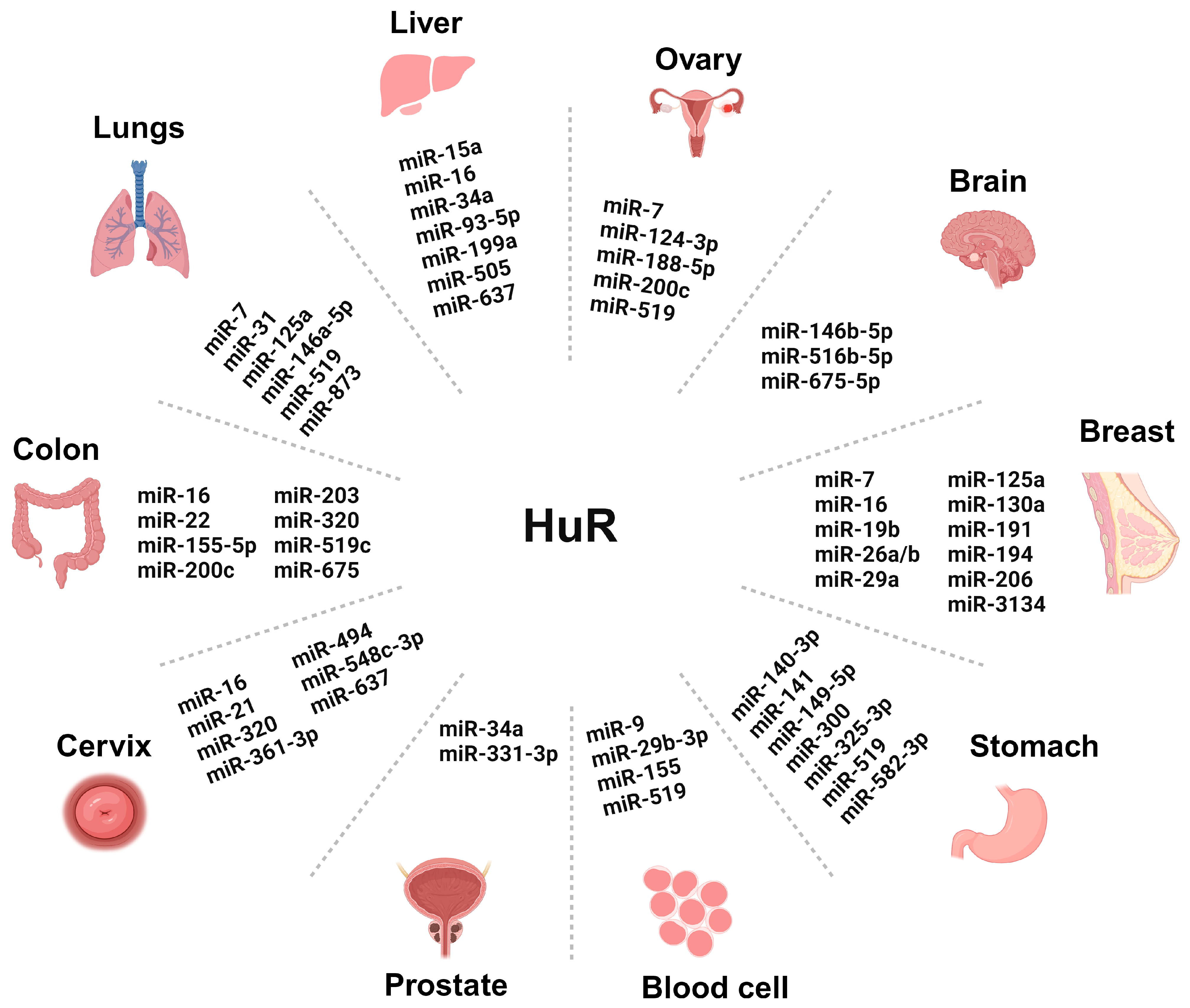
Figure 2. Graphics showing the role of different microRNAs (miRNAs) regulating human antigen R (HuR) in cancers of various organs. The names of the miRNAs are mentioned in the graphics. The miRNAs involved in gallbladder cancer (miR-502-3p), thyroid cancer (mir-31 and miR-19), sarcoma (mir-29), bladder cancer (miR-494), and kidney cancer (miR-519) are not mentioned in the graphics due to space restrictions. Each mentioned miRNA is either a tumor promoter or tumor suppressor in the specific cancer. The image was created with the help of Biorender.com.
Cancers 16 03183 g002
Figure 3. Schematics show the different functions of circular RNAs (circRNAs) involved in cancer. Briefly, the image shows how circRNAs are categorized and presented based on their function, ranging from transcriptional to translational regulation, miRNA sponging, alternative splicing, and RBP sponging. We have also shown examples of circRNA that are predicted to be probable biomarkers and signaling scaffolds. The black arrow shows the signaling pathway. The red arrow shows downregulation, and the green arrow shows upregulation. The other colors used in the graphics are only for better illustration and do not represent explicitly anything.
Cancers 16 03183 g003
Disclaimer/Publisher’s Note: The statements, opinions and data contained in all publications are solely those of the individual author(s) and contributor(s) and not of MDPI and/or the editor(s). MDPI and/or the editor(s) disclaim responsibility for any injury to people or property resulting from any ideas, methods, instructions or products referred to in the content.