Article

Isoform-Level Transcriptome Analysis of Peripheral Blood Mononuclear Cells from Breast Cancer Patients Identifies a Disease-Associated RASGEF1A Isoform

1
Faculty of Chemistry and Chemical Engineering, University of Maribor, Smetanova ulica 17, 2000 Maribor, Slovenia
2
Faculty of Medicine, University of Maribor, Taborska ulica 8, 2000 Maribor, Slovenia
3
Division of Gynecology and Perinatology, University Medical Center Maribor, Ljubljanska ulica 5, 2000 Maribor, Slovenia
4
Department for Science and Research, University Medical Centre Maribor, Ljubljanska ulica 5, 2000 Maribor, Slovenia
*
Author to whom correspondence should be addressed.
These authors contributed equally to this work.
Cancers 2024, 16(18), 3171; https://doi.org/10.3390/cancers16183171
Submission received: 15 August 2024 / Revised: 11 September 2024 / Accepted: 12 September 2024 / Published: 16 September 2024
(This article belongs to the Section Molecular Cancer Biology)

Abstract

:

Simple Summary

Peripheral blood analyses can offer a minimally invasive view into systemic immunity during cancer and can lead to the identification of biomarkers for cancer screening and therapeutic management. While a limited number of studies have reported blood transcriptome in breast cancer (BC) using RNA-seq analysis, our study is the first that aimed to identify potential BC biomarkers by analyzing transcriptome at an isoform level in peripheral blood mononuclear cells (PBMCs) from BC patients and healthy women. Our approach has led to the identification of an isoform of the RASGEF1A gene, the ENST00000374459 transcriptional variant, as a promising blood mRNA biomarker for distinguishing BC and healthy subjects. Additionally, our association analysis with clinicopathological characteristics revealed that lower ENST00000374459 expression in PBMCs of breast cancer patients was associated with higher proliferation and circulating tumor DNA (ctDNA) shedding, thereby linking expression of this isoform in blood immune cells to cancer progression and spreading.

Abstract

Background: Breast cancer (BC) comprises multiple subtypes with distinct molecular features, which differ in their interplay with host immunity, prognosis, and treatment. Non-invasive blood analyses can provide valuable insights into systemic immunity during cancer. The aim of this study was to analyze the expression of transcriptional isoforms in peripheral blood mononuclear cells (PBMCs) from BC patients and healthy women to identify potential BC immune biomarkers. Methods: RNA sequencing and isoform-level bioinformatics were performed on PBMCs from 12 triple-negative and 13 luminal A patients. Isoform expression validation by qRT-PCR and clinicopathological correlations were performed in a larger cohort (156 BC patients and 32 healthy women). Results: Transcriptional analyses showed a significant (p < 0.001) decrease in the ENST00000374459 RASGEF1A isoform in PBMCs of BC compared to healthy subjects, indicating disease-related expression changes. The decrease was associated with higher ctDNA and Ki-67 values. Conclusions: The levels of the RASGEF1A transcriptional isoform ENST00000374459 may have the potential to distinguish between BC and healthy subjects. The downregulation of ENST00000374459 in breast cancer is associated with higher proliferation and ctDNA shedding. Specialized bioinformatics analyses such as isoform analyses hold significant promise in the detection of biomarkers, since standard RNA sequencing analyses may overlook specific transcriptional changes that may be disease-associated and biologically important.

1. Introduction

Breast cancer (BC) is among the leading health concerns among women worldwide due to its high prevalence and mortality [1]. This heterogenous cancer comprises multiple subtypes that differ in prognosis and require different therapeutic regimens. The clinical subtypes are based on the expression of the estrogen receptor (ER) and the progesterone receptor (PR) and the amplification of the HER2 (ERBB2) receptor and include hormone receptor (HR)-positive (HR+/HER2−, also ER+/PR+/HER2−), triple-positive (HR+/HER2+, also ER+/PR+/HER2+), HER2-positive (ER−/PR−/HER2+, also HER2+), and triple-negative breast cancer (TNBC) (HR−/HER2−, also ER−/PR−/HER2−) [2,3,4]. In addition, the molecular expression profiling of BC tissues has led to the classification of several intrinsic BC subtypes with different prognoses and survival: Luminal A (ER+ and/or PR+, HER2−, proliferation marker Ki-67 < 14%), Luminal B (ER+ and/or PR+, HER2−, Ki-67 ≥ 14%; or ER+ and/or PR+, HER2+, any Ki-67), HER2(+)/HER2-enriched (ER-, PR-, HER2+, any Ki-67), and basal-like (triple-negative) (ER−, PR−, HER2−, any Ki-67) [4,5]. Intrinsic subtypes overlap with clinical subtypes to a great extent, though not completely [6].
Despite significant progress in tissue-based classifications, a substantial gap remains in understanding how blood immune-related biomarkers, such as transcriptome changes in peripheral blood, can be used to distinguish between BC subtypes. Additionally, while transcriptional isoforms have shown potential as powerful cancer biomarkers, their specific role in peripheral blood in BC patients is still largely unexplored, as further discussed below.
Studies of peripheral blood can provide valuable insights into the interplay between the host systemic immunity and cancer [7,8]. This may aid in identifying biomarkers related to immune responses in BC, enhancing our understanding of disease progression and supporting advancements in cancer screening, subtype characterization, diagnosis, prognosis, and therapy selection. Several research groups have reported that mRNA biomarkers from peripheral blood can be used as “surrogate” biomarkers for various solid tumors [9,10,11,12]. However, while transcript abundance changes in peripheral blood cells are very important in BC, basic gene expression analyses may not be able to fully explain the phenotypes observed during cancer development. The complex eukaryotic gene expression involves the generation of various coding mRNA variants or isoforms from single genes through mechanisms that include selection of transcription start sites, alternate UTR usage, intron retention, alternatively spliced exons, etc. [13,14,15]. Importantly, aberrant use of one isoform over another is frequently associated with cancer [14,16,17,18]. For instance, a specific 3′UTR variant of HNRNPA1 is downregulated in BC tissues in favor of a more stable isoform, from which more HNRNPA1 protein is produced. Importantly, this correlates with poorer survival [19]. Specific genes important in BC, including BRCA1, TP53, PTEN, and CD44, exhibit cancer-specific splice isoforms. Moreover, alternative splicing of CD44 and other genes in BC cell lines has been shown to play a role in epithelial-to-mesenchymal transition, a process associated with metastasis [20,21,22,23].
Notably, BC tumor tissues of individual cancer subtypes display distinct gene expression profiles, including specific transcriptional isoforms [14,24]. RNA sequencing of ER-positive and triple-negative BC tumors has identified specific sets of isoforms that differentiated these subtypes with even higher fidelity than standard mRNA expression profiles [14]. Additionally, dysregulation of splicing in BC subtypes has been shown to be influenced by specific RNA processing factors, since several RNA processing factors were differentially expressed between tumor subtypes and/or regulated by estrogen receptor [14].
Limited studies examining transcriptional isoforms in the peripheral blood cells of BC patients have revealed alterations in the splicing of BRCA1/2 and BRCA1-associated RING domain (BARD1) as significant disease-causing mechanisms [21,25,26]. Furthermore, high expression of the REST-N50 splice variant was observed in nucleated blood cells of locally advanced BC patients. Neoadjuvant therapy led to a decrease in REST-N50 levels, indicating a potential of this variant for the evaluation of therapy effectiveness [27]. Some alternatively spliced mRNA variants (ASVs) identified in the primary tumor have also been reported in the peripheral blood of cancer patients [27]. While isoform investigations in BC tissues have identified BC subtype-specific changes, there has been no study of transcriptional isoforms in blood cells that could specifically distinguish individual BC subtypes.
Together, the above-mentioned findings indicate that transcriptional isoforms have a promising potential to serve as cancer biomarkers [28]. A detailed characterization of the BC-associated mRNA variant repertoire may uncover new oncogenic mechanisms and host immune responses, which may in turn be relevant for the development of therapeutic strategies. Given the promising potential of transcriptional isoforms as cancer biomarkers, this study aimed to bridge the gap by characterizing mRNA isoform expression in peripheral blood mononuclear cells (PBMCs) from BC patients and healthy controls. Moreover, two BC subtypes (TNBC and luminal A), which are generally considered to differ significantly in immunogenicity [29,30], were investigated using RNA-seq to identify the underlying mechanisms leading to differences in systemic immunity between BC subtypes. While TNBC is known for its high immunogenicity due to its genomic instability and a high level of tumor-infiltrating lymphocytes [29,31], luminal A is considered less immunogenic [29,32]. Our RNA-seq analysis represents the first isoform-level transcriptome analysis in peripheral blood of BC patients.

2. Materials and Methods

2.1. The Study Population and Clinicopathological Characteristics

The pilot (discovery) cohort for RNA-seq comprised 12 TNBC and 13 luminal A female BC patients. The validation cohort for qRT-PCR analyses comprised 156 female BC patients (including the 26 patients from the discovery cohort) and 32 healthy females. All cancer patients had histologically confirmed BC and were treated at the University Medical Centre Maribor (UMC), Slovenia, where the clinicopathological characteristics (ER, PR, HER-2 status, histological type, localization, grade, tumor size, lymph node status, Ki-67) (Table 1) were determined by experienced BC pathologists according to the standard procedures of the pathology laboratory. The healthy controls were enrolled at the Faculty of Medicine at the University of Maribor. All participants provided written informed consent for their participation in this study. This study was approved by the UMC Maribor Medical Ethics Committee (reference number UKC-MB-KME-09/19) and was carried out according to the principles of the Declaration of Helsinki. The blood samples were acquired from BC patients prior to treatment.

2.2. Processing of Blood Samples

Whole blood (12 mL) was collected into BD Vacutainer EDTA vials (Becton Dickinson, Franklin Lakes, NJ, USA) and processed immediately after drawing. The samples were centrifuged (300× g, 20 min) to collect the plasma. The remainders of the samples were subjected to gradient centrifugation using Lympholyte-H (CL5020; Cedarlane, Burlington, ON, Canada) according to the manufacturer’s instructions to obtain PBMCs for expression analyses.
Circulating DNA was isolated from plasma by QIAamp Circulating nucleic acid kit (Qiagen, Hilden, Germany). The genome-wide Z score (Table 1) for estimation of ctDNA content in plasma was determined by the mFAST-SeqS method as previously described [33]. RNA was prepared from PBMCs using the innuPREP RNA Mini Kit (Analytik Jena, Jena, Germany) according to the manufacturer’s protocol.

2.3. Sequencing of RNA Isolated from PBMCs

RNA-seq was performed on 25 RNA samples extracted from PMBCs. Pair-end RNA-seq (oligo dT, stranded mRNA library, DNBseq platform, PE150, 20 M reads/6 Gb clean data) was performed at BGI (Hong Kong, China) using a NEBNext® Poly(A) mRNA Magnetic Isolation Module and BGI kit.

2.4. RNA-Seq Data Alignment and Identification of Differentially Expressed Isoforms

Raw .fastq files were first assessed for quality using FastQC software (0.11.9, Babraham Bioinformatics, Cambridge, UK) [https://www.bioinformatics.babraham.ac.uk/projects/fastqc/; accessed on 13 September 2024]. Subsequently, trimming of technical sequences was performed using the Trimmomatic tool (version 0.39, USADEL LAB, Aachen, Germany) [34]. Transcript-specific RNA-seq analysis was performed using the Kallisto pseudoalignment program (Pachter Lab, Berkeley, CA, USA) with the GRCh37 reference genome [35]. Furthermore, the R 4.2.1 environment (R Core Team 2020, Vienna, Austria) and a pipeline described elsewhere [36] were used for further processing of estimated counts and tpms. Statistically significant differential gene expression was identified using a false discovery rate (FDR) ≤ 0.1 and log2 count per million (logCPM) ≥ 1.

2.5. Preparation of cDNA and Isoform-Specific RT-qPCR Analysis

Total RNA (200 ng) was used to prepare cDNA by using the High-Capacity cDNA Reverse Transcription Kit (Applied Biosystems, Foster City, CA, USA) following the manufacturer’s instructions. Quantitative reverse transcription PCR (RT-qPCR) was performed on the LightCycler® 480 System (Roche, Basel, Switzerland) using the cDNA samples and the LightCycler®480 SYBRGreen I Master reaction mix (Roche, Basel, Switzerland). To amplify different RASGEF1A transcriptional variants, 374459-specific and 395810-specific forward primers and a common exon junction-spanning reverse primer were used. The optimal PCR conditions and efficiency were assessed for each primer pair. The primer sequences, final primer concentrations, and annealing temperatures were as follows: for ENST00000374459 (5′-CCGGCGGCCAGAATGTTCCTGGA-3′ and 5′-TACGTCCTATCGGGGTAATAGTCCACC-3′, 400 nmol, 52 °C); for ENST00000395810 (5′-AGCGACGCTGGCCCGGACCG-3′ and 5′-TACGTCCTATCGGGGTAATAGTCCACC-3′, 400 nmol, 62 °C). Target gene expression levels were calculated relative to the endogenous control 18S rRNA (5′-GCAATTATTCCCCATGAACG-3′ and 5′-GGGACTTAATCAACGCAAGC-3′) and described as relative expression levels using the 2−ΔΔCT method [37] for 374459 and the Pfaffl method [38] for 395810. Ct values > 40 were considered negative.
Statistical analyses of isoform expression were performed with SPSS Statistics 28 (IBM Corporation, Armonk, NY, USA) using the Mann–Whitney U test. The results were defined as significant when p < 0.05.

3. Results

3.1. RNA Sequencing Identified Differences in Isoform Expression between Luminal A and TNBC Patients

High-throughput RNA sequencing was performed using mRNA isolated from PBMCs of 13 luminal A and 12 triple-negative treatment-naïve BC patients in order to identify statistically significant differentially expressed isoforms (DEIs). The sequencing data were analyzed at the transcript isoform level by using the R 4.2.1 environment (R Core Team 2020, Vienna, Austria). The analysis rendered three transcriptional variants belonging to three different genes as significantly differentially expressed between TNBC and luminal A: the Ras-GEF Domain-Containing Family Member 1A (RASGEF1A) ENST00000374459 variant, the Tubulin Folding Cofactor B (TBCB) ENST00000589996 variant, and the Damage-Specific DNA Binding Protein 2 (DDB2) ENST00000378603 variant (Figure 1A and Table S1).
Function and pathway analyses were performed to gain insight into the biological context of these results. Analyses using the Reactome pathway database [39] (Figure 1B), UniProt [40] (Table 2), and the STRING database [41] (Figure S1) revealed that the three significantly differentially expressed genes do not share the same or similar pathways but are instead involved in distinct, non-overlapping cellular pathways. RASGEF1A participates in the Ras/Raf/MAP kinase signaling cascade, with gene ontology (GO) annotations related to this gene (Table 2) including guanyl-nucleotide exchange factor activity. DDB2 encodes a protein that participates in nucleotide excision repair, with GO annotations related to DDB2 including damaged DNA binding. TBCB encodes a protein involved in the regulation of tubulin heterodimer dissociation, with GO annotations related to TBCB including alpha-tubulin binding. Similarly, the Reactome analysis identified these genes in the signal transduction pathway (RASGEF1A), the DNA repair pathway (DDB2), and the protein folding pathway (TBCB) (Figure 1B), while the STRING analysis identified the RASGEF1A, DDB2, and TBCB protein interactors in these pathways (Figure S1).
In the continuation of our research, we focused on RASGEF1A due to its distinct association with the RAS pathway, a crucial signaling cascade implicated in cancer development and progression, with dysregulation of the Ras signaling pathway reported to lead to uncontrolled cell proliferation and resistance to apoptosis [42]. Furthermore, its close homolog, RASGEF1B, has been reported to play a functional role in macrophages and chemotaxis [43,44,45], which hinted at the possibility that RASGEF1 proteins assume multifaceted roles in both cancer-related signaling and immune modulation.
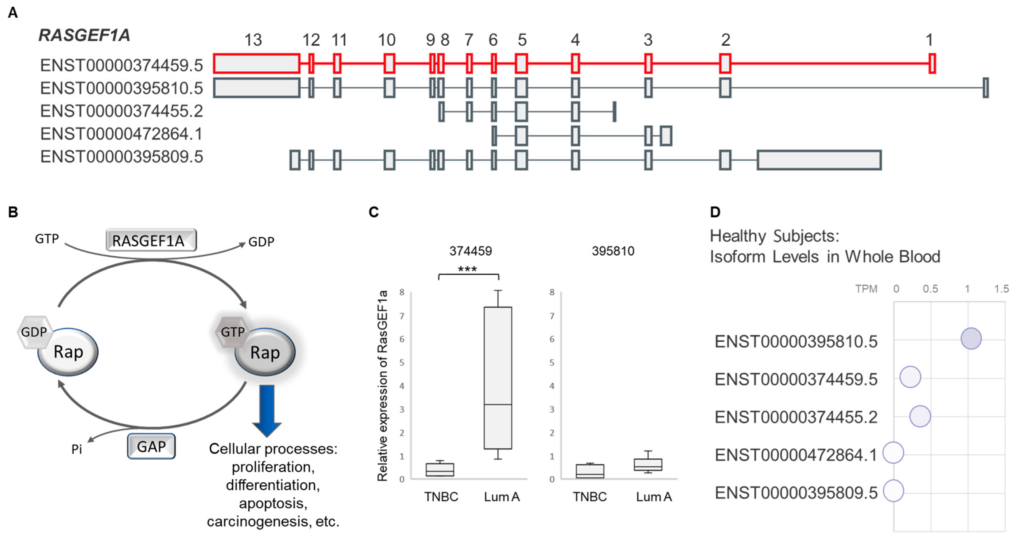
While five different isoforms have been reported for RASGEF1A (Figure 2A), RNA-seq returned only the ENST00000374459 variant (hereinafter 374459) as significantly downregulated in peripheral immune cells of TNBC patients. RASGEF1A is a member of the GEF (guanine nucleotide exchange factor) family of proteins, which mediate GDP release and GTP binding to the Rap proteins (Figure 2B). The Rap genes belong to the Ras family, known to be frequently mutated in cancer [46,47]. Considering that GEFs activate their targets through GTP exchange, not through transcriptional regulation, our RNA-seq analysis expectedly showed no significant differences in expression of the Rap genes between TNBC and luminal A.
The PBMC samples from RNA-seq were further subjected to qRT-PCR validation with isoform-specific primers capable of detecting two RASGEF1A variants that differ in the first exon and the first intron sequence: the 374459 and the ENST00000395810 (hereinafter 395810) isoforms (Figure 2A,C). In line with RNA-seq results, distinctive PBMC isoform expression was observed in validation analysis: while 374459 expression was significantly lower (p < 0.001) in TNBC samples, expression of 395810 was similar between the two BC subtypes (Figure 2C).
It may be worth noting that our initial isoform-indiscriminate (all-inclusive) whole-transcriptome RNA-seq analysis of PBMCs did not identify RASGEF1A as a differentially expressed gene (DEG) between TNBC and luminal A. This is not surprising considering that 395810 is the most represented RASGEF1A transcriptional variant in human whole blood, as seen in the publicly available GTEx expression data (Figure 2D), while 374459 is substantially less abundant. Taking into account the homogeneous 395810 expression among BC patient subtypes (Figure 2C), and the low overall presence of 374459 (Figure 2D), any potential expression changes for the less abundant 374459 transcriptional variant can easily be missed by the classical whole-transcriptome RNA-seq analysis, even though these alterations may be biologically important. However, the isoform-level RNA-seq analysis is able to pick up on such changes.

3.2. Expression of RASGEF1A Isoforms in a Larger Cohort Comprising BC Patients and Healthy Female Controls

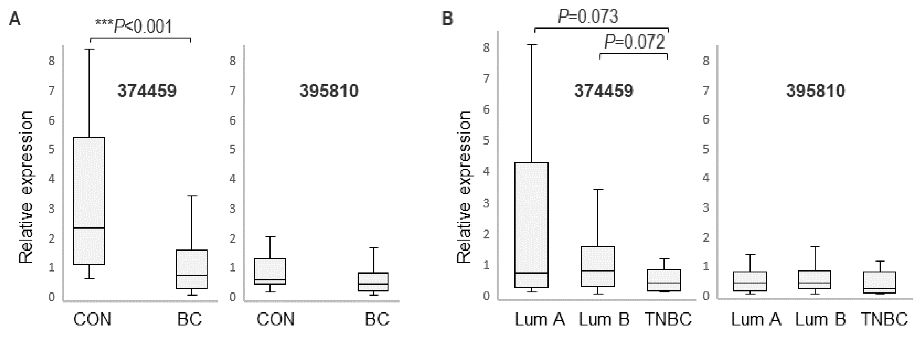
Isoform expression was further analyzed in a larger cohort that included 156 female BC patients (average age 60.7 ± 13.4 years) and 32 healthy women (average age 55.6 ± 9.2 years). Quantitative RT-PCR analysis revealed a significantly higher expression of the 374459 variant in PBMCs of healthy controls (p < 0.001) compared to BC patients (Figure 3A), while the levels of the 395810 isoform were similar between these groups. This suggested that expression of the 374459 variant is associated with BC and indicated a potential for 374459 as a disease-associated biomarker.
Variant expression was further compared between different subtypes of BC in the larger cohort. Here, the trend of lower 374459 expression in TNBC compared to luminal A persisted; however, it was short of reaching statistical significance (p = 0.073) (Figure 3B). A similar situation was observed when 374459 expression was compared between TNBC and luminal B (p = 0.072). Hence, despite the significant subtype-specific 374459 expression changes identified by RNA-seq in the pilot cohort, the targeted validation in the larger BC cohort indicated that 374459 expression changes were insufficient to serve for blood-based distinction of BC subtypes.
Consistent with the pilot cohort, the larger cohort showed similar expression of the 395810 isoform in healthy controls and BC patients as well as within the BC subtypes (Figure 3).

3.3. RASGEF1A Isoform Expression and Clinicopathological Characteristics of BC Patients

The expression of 374459 in PBMCs of BC patients was also analyzed with respect to the clinicopathological characteristics (Table 1) of BC patients.

3.3.1. Association with Ki-67 Proliferation Index

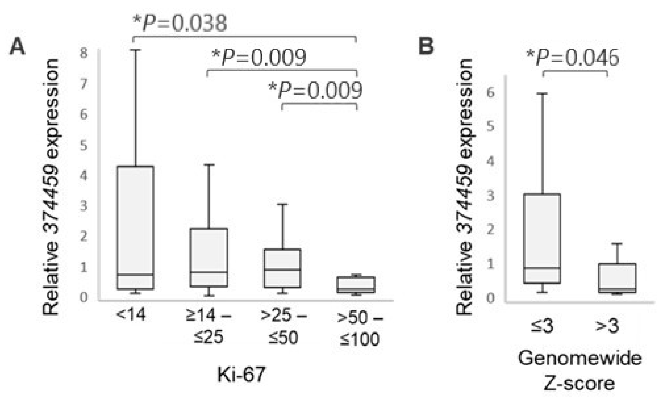
The prognostic proliferation marker Ki-67 is routinely evaluated by the clinical pathology labs to assist in BC subtyping and treatment decisions. Nonetheless, there is a lack of consensus regarding the cutoff points for high and low immunohistochemical Ki-67 values; therefore, these tend to vary between laboratories [49]. Taking into account the groupings in published BC studies [50,51,52,53,54], the BC patients in our cohort were stratified into groups with increasing Ki-67 values: group I (Ki-67 < 14%); group II (≥14 to ≤25%); group III (> 25 to ≤50%); and group IV (>50 to ≤100%) (Figure 4A). A significantly lower expression of the 374459 isoform was observed in group IV patients who are characterized by high immunohistochemical Ki-67 staining (>50%) compared to patients with Ki-67 ≤ 50% (Figure 4A), indicating an association between the downregulation of 374459 and tumor proliferation. While there was an incremental decrease in 374459 expression between groups I and II, and between groups II and III, it was not statistically significant (Figure 4A).

3.3.2. Association with Circulating Tumor DNA (ctDNA) Content

Circulating tumor DNAs are small pieces of extracellular DNA released by the dying tumor cells, which contain information on somatic mutations in the tumor cells [55]. We determined the ctDNA content in plasma of BC patients by employing the Modified Fast Aneuploidy Screening Test-Sequencing System (mFAST-SeqS). This method detects tumor-specific aneuploidy in circulating cell-free DNA (cfDNA). The acquired genome-wide mFAST-SeqS z-scores correlate with the tumor content in plasma [33,56,57]. The genome-wide z-scores were determined for 41 BC subjects (Table 1). The patients’ z-scores were <5, except in two subjects who had higher scores. While values below 5 indicate a generally low ctDNA content, we nonetheless observed that patients with z-scores >3 had significantly decreased 374459 expression (p = 0.046) compared to those with z-scores ≤3 (Figure 4B), suggesting that the levels of this RASGEF1A isoform are inversely correlated with ctDNA amount.
Together, the Ki-67 and ctDNA results indicate that the downregulation of 374459 is associated with tumor proliferation and ctDNA shedding.

3.3.3. Other Clinicopathological Characteristics

When other clinicopathological characteristics were evaluated with regard to 374459 expression, no statistically significant associations were observed (Figure S2).

4. Discussion

Our transcriptional analyses in mononuclear blood cells showed that the RASGEF1A 374459 transcriptional isoform is significantly downregulated in BC compared to healthy subjects, suggesting disease-associated expression changes, and indicating the ability of 374459 to distinguish between these two groups.

4.1. The Advantages of Blood Analyses over Standard Methods for Cancer Detection

We focused on peripheral blood cells because of the important advantages that blood-based cancer indicators offer over tissue markers. For one, collecting peripheral blood is simple, minimally invasive, and cost-efficient [58]. Additionally, evidence indicates that the analysis of blood can enable the detection of very early systemic changes, crucial for cancer screening [59,60,61,62]. In contrast, biopsies are invasive, carry a greater possibility of complications, and may not be suitable for screening purposes. While repeated tissue biopsies can be used to oversee the progression of cancer, single-site biopsies may have selection bias because of tumor heterogeneity and may not provide enough material [13]. On the other hand, peripheral blood is not prone to heterogeneity problems or selection bias and is readily available in sufficient quantities [58]. Analyzing blood may also offer advantages compared to current imaging methods. While mammography is important for screening, it has drawbacks like radiation exposure and physical discomfort. Furthermore, its capacity to identify the tumor in its early stages is hampered by the necessity for the tumor to reach a certain size to be detectable [63]. Also, due to high breast density, around 10% of cancers remain undetected on mammography [64,65].
While our investigation indicated that 374459 may have the capability to distinguish between healthy and breast cancer (BC) subjects, its potential utility as a biomarker needs to be established through replication. In pursuit of validation across different cohorts, we sought to identify an external RNA-seq cohort for further validation. However, we found only two articles reporting peripheral blood RNA-seq analyses in breast cancer, both without their data deposited in public repositories [66,67]. In contrast, available blood transcriptome studies in public repositories such as the GEO Database—NCBI and European Genome-Phenome Archive were performed using array profiling (e.g., GDS3952, GSE27562, GSE47862, EGAD00010001063), rendering them unsuitable for isoform analysis.

4.2. RASGEF1A Function

RASGEF1A is a member of the conserved RASGEF1 family of proteins, which includes RASGEF1A, RASGEF1B, and RASGEF1C. This family controls the activity of the Rap protein family (Figure 2B) [68,69]. RASGEF1B has a well-established role in immunity, where it is involved in macrophage signaling, chemotaxis, and cytokine response [43,44,70,71]. On the other hand, there is limited information available about RASGEF1A and RASGEF1C. Specifically, there are no reports yet suggesting a potential role in immunity for RASGEF1A. However, according to PBMC single-cell sequencing data from the Protein Atlas, RASGEF1A mRNA has been detected in T-cells, NK cells, and macrophages [72].
The Rap proteins (Rap1A, Rap1B, Rap2A, Rap2B, Rap2C) belong to the Ras superfamily and are found in nearly all tissues where they have regulatory roles in growth, differentiation, proliferation, carcinogenesis, cell adhesion, exocytosis, apoptosis, and phagocyte activity [70,71]. Individual Rap members have specific functions. For instance, Rap1 and Rap2 signal through distinct downstream pathways [73]. Moreover, Rap2C is the predominant Rap2 protein in circulating mononuclear leukocytes, but it is not present in platelets [71,73].
Interestingly, RASGEF1A and RASGEF1B proteins are highly specific guanine nucleotide exchange factors (GEFs) for Rap2 and do not act on Rap1 or other Ras members [47]. The Rap2 group comprises Rap2A, Rap2B, and Rap2C and has been implicated in carcinogenesis, regulation of cell adhesion, establishment of cell morphology, and modulation of synapses in neurons [70,74]. GEFs have been reported to have mixed roles in cancer, as they can act either as tumor suppressors or promoters [46]. The role of Rap2 proteins in cancer is also complex. For instance, in hepatocellular carcinoma cells, Rap2B has been described as an oncogene, which promotes proliferation and invasion [75]. The Rap2B gene has also been described in association with the p53 tumor suppressor involved in cell-cycle arrest, which is a protective mechanism that gives the damaged cell some time to repair the damage [76].
Considering the mixed roles of the GEF and Rap proteins in the regulation of oncogenic processes, the role of RASGEF1A in carcinogenesis may also be complex. Currently, the role of RASGEF1A and Rap2 proteins in PBMCs is not known. It is possible that they may be involved in immune responses to tumor, considering that a recent transcriptome analysis of PBMCs revealed a role of the Ras genes in host immune responses. More specifically, K-Ras and N-Ras were downregulated, while H-Ras was upregulated in PBMCs of patients with COVID-19 [77]. Interestingly, one study showed that the RASGEF1A protein had GEF activity for K-RAS, H-RAS, and N-RAS proteins in vitro [78], although this finding could not be replicated in a later study [47]. In breast cancer, it is possible that the observed downregulation of the 374459 RASGEF1A isoform in PBMCs of BC patients may affect the activity of these immune cells. By causing dysregulation of the Rap pathway, it may potentially reduce PBMC activity and contribute to the weakening of antitumor immunity. This would be in line with the observation that 374459 downregulation is most evident in TNBC, which is known as immunologically more evasive among the BC subtypes [79]. However, this postulation requires further experimentation and validation.

4.3. RASGEF1A 374459 Isoform and Cancer Proliferation and Shedding

Our results also revealed an association between the decreased 374459 expression and higher Ki-67 and ctDNA values. Ki-67 is a prognostic proliferation marker measured by the clinical pathology labs to assist in treatment decisions. While the margins delineating high and low Ki-67 values vary between laboratories [49], the Ki-67 cutoff point generally depends on the clinical objective: if Ki-67 is used to identify patients sensitive to chemotherapy protocols, it is preferred to set the cut-off at 25% [51,54]. If Ki-67 is used to exclude patients with slowly proliferating tumors from chemotherapeutic protocols, a cut-off of 10% can help avoid overtreatment [51]. Ki-67 is also used in BC classification, with Ki-67 < 14% best correlating with the gene-expression definition of Luminal A [50,52]. The Saint Gallen International Breast Cancer Conference (2011) Expert Panel designated tumors with a Ki-67 < 14% as “low proliferation” [80]. In our study, those BC patients who exhibited the highest Ki-67 values (>50%) had the lowest 374459 expression, suggesting an association between tumor proliferation and the 374459 RASGEF1A isoform. Interestingly, the Ki-67 values over 50% are most common in TNBC patients [49,53].
Additionally, in our study, the patients with lower 374459 expression had higher ctDNA content, as estimated by the mFAST-SeqS genome-wide z-scores. The mFAST-SeqS method detects tumor-specific aneuploidy in circulating cell-free DNA without the requirement for prior knowledge of specific aberrations of the primary tumor. The z-score of 5 predicts a mutant allele frequency (mAF) of 10.5% [56,57]. Overall, our findings that lower 374459 expression correlates with both higher Ki-67 proliferation index as well as ctDNA shedding are in agreement with the studies reporting an association between proliferation and ctDNA [81,82].

4.4. Advantages of Isoform-Level Bioinformatics Analysis of RNA-Seq Data

Our isoform-level RNA-seq analysis identified specific BC-associated changes in the RASGEF1A 374459 transcriptional isoform. It is interesting to note that when an all-inclusive RNA-seq analysis (i.e., including all isoforms for each gene) was performed, the expression of the RASGEF1A gene in blood immune cells from our two study groups was not statistically different. However, upon isoform-specific analysis, one of the RASGEF1A isoforms was detected as differentially expressed. This suggests that by using the standard RNA-seq bioinformatics analysis, specific isoforms may be missed, which can be disadvantageous in cases where changes in these isoforms are disease-associated and biologically important. Regarding the RASGEF1A gene, the 395810 isoform is generally more abundant in blood cells than 374459, which is a low-expression isoform (Figure 2D). Because of the quantitative preponderance of 395810, any changes in 374459 expression may fail to be detected in the all-inclusive RNA-seq analysis. Along the same line, biologically relevant gene expression changes could also potentially be missed in other situations, for instance, in the scenario where one of the isoforms of a given gene would be upregulated while another downregulated. Due to the “cancelling out” effect of expression changes (one variant up, one down), the given gene may not be detected as differentially expressed by all-inclusive RNA-seq analysis; the differences would only be observable by isoform analysis. Taking all this into account, it is clear that more specific bioinformatics analyses such as isoform analyses carry particular value in the detection of disease biomarkers.

4.5. Other Dysregulated Isoforms Identified in Our Study

Our study of PBMC-expressed isoforms identified two other transcriptional variants that were differentially expressed (i.e., elevated) in TNBC, the ENST00000589996 variant of the TBCB gene and the ENST00000378603 variant of the DDB2 gene. TBCB is involved in the folding of β-tubulin and the formation of α/β-tubulin [83], and localizes at spindle and midzone microtubules during mitosis [84]. Abnormal levels of TBCB and TBCE are associated with microtubule abnormalities [85]. Cancer cells are known to depend on their cytoskeleton (including microtubules) to proliferate, invade, and metastasize [86]. TBCB has previously been implicated in cancer [87,88], but only little is known about its involvement in BC. It has been reported that TBCB expression is elevated in BC tissues and that TBCB overexpression increases the degree of malignancy in BC cell lines [83,89].
On the other hand, many studies have linked DDB2 with cancer [90,91,92,93,94,95]. Due to its role in nucleotide excision repair (NER), it is not surprising that decreased DDB2 expression has been reported in various cancerous tissues, for instance, in prostate [91], skin [92], head and neck [93], and ovarian cancer [96]. However, DDB2 also has other functions, such as regulation of cell-cycle and transcription, and it appears to have a dual role in cancer. For example, upregulated DDB2 expression was detected in colorectal cancer tissues [94]. Moreover, introduction of the DDB2 gene into triple-negative MDA-MB231 cells stimulated growth and colony formation, while DDB2 knockdown in MCF-7 BC cells caused a decrease in cancer cell growth and colony formation [95].
While TBCB and DDB2 do not interact directly (Figure 1B and Figure S1), they are both crucial for maintaining cell cycle integrity—TBCB through its role in mitosis and DDB2 by ensuring genomic stability via DNA damage repair—and are thus functionally linked within broader cellular pathways. Both genomic integrity and proper microtubule dynamics (involved in cell division, intracellular transport, etc.) are essential for preserving the health and functionality of cells, including immune cells such as PBMCs. This, in turn, supports proper immune responses that are vital for preventing transformation toward BC, as disruptions in either process can be deleterious to immune cells and may impair the immune response to tumors.
Our isoform-level RNA-seq analysis highlighted a potentially interesting association between individual BC subtypes and specific TBCB and DDB2 isoforms in PBMCs. No study has yet investigated TBCB or DDB2 isoforms in connection with cancer. Thus, additional investigations are warranted to help unravel the potential utility of the here-identified TBCB and DDB2 isoforms as possible BC biomarkers.

4.6. Limitations and Future Perspectives

This case-control study identified a distinctively expressed transcriptional isoform (374459) among subject groups. It would be interesting to see how the 374459 and 395810 variants differ at the protein level. Analysis of their protein sequences reveals that they differ in the first exon, with 374459 encoding a protein that is eight amino acids longer at the N-terminus compared to 395810, making these isoforms “N-terminal proteoforms” [97]. While no functional studies have been conducted on RASGEF1A isoforms, making it difficult to determine the exact effect of the N-terminal amino acid difference, our RNA-seq and RT-qPCR results suggest that it is not inconsequential. Importantly, N-terminal proteoforms have been described in human disease [98]. The difference in the 374459 and 395810 isoforms does not affect the RAS-GEF functional domain, which is located away from the N-terminus. However, since UniProt indicates that RASGEF1A is membrane-associated, the additional N-terminal amino acids could potentially influence membrane localization signals [99], affecting isoform localization. Additionally, it is known that N-terminal proteoforms may engage in different protein complexes due to interactions with distinct molecules [97]. The extended N-terminal sequence could also influence the protein’s stability, N-terminal acetylation, folding, and function [97,98,100,101], potentially contributing to distinct properties between the isoforms.
Looking ahead, future mechanistic in vitro studies hold the potential to unveil the functions of RASGEF1A and the 374459 isoform in immune blood cells, which could provide insights into the differential expression in PBMCs of BC patients.

5. Conclusions

In conclusion, we performed the first isoform-level transcriptome analysis of PBMCs from breast cancer (BC) patients and identified the ENST00000374459 RASGEF1A isoform as BC-associated. The ENST00000374459 RASGEF1A isoform levels may have potential as a screening biomarker to differentiate BC patients from healthy subjects. In addition, we found that ENST00000374459 downregulation in BC was associated with increased Ki67 proliferation index and increased ctDNA shedding. Considering that analyzing the expression of this isoform is less work-intensive and financially more feasible than analyzing ctDNA, this expression analysis could prove useful as a surrogate indicator in the clinical setting for determining disease severity and prognosis. However, this will require further experimental validation.

Supplementary Materials

The following supporting information can be downloaded at: https://www.mdpi.com/article/10.3390/cancers16183171/s1. Figure S1: STRING interaction networks for RASGEF1A, DDB2, and TBCB; Figure S2: RASGEF1A 374459 isoform expression and clinicopathological parameters; Table S1: List of annotated isoforms identified as differentially expressed (DEIs) in PBMCs from TNBC compared to luminal A patients.

Author Contributions

Conceptualization, H.Č., M.G., D.A., I.T., M.S., B.C. and U.P.; methodology, B.C., M.S., H.Č., M.G. and M.K.; software, M.G.; validation, H.Č., M.G. and M.K.; formal analysis, M.G., H.Č. and M.K.; investigation, H.Č., M.S. and M.G.; resources, U.P.; data curation, M.G., H.Č. and M.K.; writing—original draft preparation, H.Č.; writing—review and editing, H.Č., M.G., M.S., M.K., I.T. and U.P.; visualization, H.Č., M.G. and M.K.; supervision, U.P.; project administration, U.P.; funding acquisition, D.A., I.T. and U.P. All authors have read and agreed to the published version of the manuscript.

Funding

This research was funded by the Slovenian Research and Innovation Agency, grant numbers P3-0427, P3-0067, J3-4523, J3-3069, I0-0029, J3-9272, and P3-0321; internal University Medical Centre Maribor research funding, grant numbers IRP-2019/01-05, IRP-2019/02-15, and IRP-2021/01-02; and the Republic of Slovenia Ministry of Higher Education, Science, and Innovation and the European Union through the European Regional Development Fund, grant RIUM.

Institutional Review Board Statement

This study was conducted in accordance with the Declaration of Helsinki and approved by the University Medical Center Maribor Medical Ethics Committee (protocol code UKC-MB-KME-09/19).

Informed Consent Statement

Informed consent was obtained from all subjects involved in this study.

Data Availability Statement

RNA-seq data from PBMCs of breast cancer patients were deposited in the NCBI-supported database Gene Expression Ominibus (GEO), accession record GSE270376 https://www.ncbi.nlm.nih.gov/geo/query/acc.cgi?acc=GSE270376; accessed on 13 September 2024.

Acknowledgments

We thank Katja Repnik for her contribution in detecting ctDNA in plasma by the mFAST-SeqS method.

Conflicts of Interest

The authors declare no conflicts of interest.

References

  1. Wilkinson, L.; Gathani, T. Understanding breast cancer as a global health concern. Br. J. Radiol. 2021, 95, 20211033. [Google Scholar] [CrossRef] [PubMed]
  2. Čelešnik, H.; Potočnik, U. Peripheral Blood Transcriptome in Breast Cancer Patients as a Source of Less Invasive Immune Biomarkers for Personalized Medicine, and Implications for Triple Negative Breast Cancer. Cancers 2022, 14, 591. [Google Scholar] [CrossRef] [PubMed]
  3. Skok, K.; Gradišnik, L.; Čelešnik, H.; Milojević, M.; Potočnik, U.; Jezernik, G.; Gorenjak, M.; Sobočan, M.; Takač, I.; Kavalar, R.; et al. MFUM-BrTNBC-1, a Newly Established Patient-Derived Triple-Negative Breast Cancer Cell Line: Molecular Characterisation, Genetic Stability, and Comprehensive Comparison with Commercial Breast Cancer Cell Lines. Cells 2021, 11, 117. [Google Scholar] [CrossRef] [PubMed]
  4. Szymiczek, A.; Lone, A.; Akbari, M.R. Molecular intrinsic versus clinical subtyping in breast cancer: A comprehensive review. Clin. Genet. 2021, 99, 613–637. [Google Scholar] [CrossRef]
  5. Park, S.; Koo, J.S.; Kim, M.S.; Park, H.S.; Lee, J.S.; Lee, J.S.; Kim, S.I.; Park, B.-W. Characteristics and outcomes according to molecular subtypes of breast cancer as classified by a panel of four biomarkers using immunohistochemistry. Breast 2012, 21, 50–57. [Google Scholar] [CrossRef]
  6. Prat, A.; Adamo, B.; Cheang, M.C.U.; Anders, C.K.; Carey, L.A.; Perou, C.M. Molecular characterization of basal-like and non-basal-like triple-negative breast cancer. Oncologist 2013, 18, 123–133. [Google Scholar] [CrossRef]
  7. Rajtak, A.; Ostrowska-Leśko, M.; Żak, K.; Tarkowski, R.; Kotarski, J.; Okła, K. Integration of local and systemic immunity in ovarian cancer: Implications for immunotherapy. Front. Immunol. 2022, 13, 1018256. [Google Scholar] [CrossRef]
  8. Xu, L.; Zou, C.; Zhang, S.; Chu, T.S.M.; Zhang, Y.; Chen, W.; Zhao, C.; Yang, L.; Xu, Z.; Dong, S.; et al. Reshaping the systemic tumor immune environment (STIE) and tumor immune microenvironment (TIME) to enhance immunotherapy efficacy in solid tumors. J. Hematol. Oncol. 2022, 15, 87. [Google Scholar] [CrossRef]
  9. Kalantari, S.; Kazemi, B.; Roudi, R.; Zali, H.; D’Angelo, A.; Mohamadkhani, A.; Madjd, Z.; Pourshams, A. RNA-sequencing for transcriptional profiling of whole blood in early stage and metastatic pancreatic cancer patients. Cell Biol. Int. 2023, 47, 238–249. [Google Scholar] [CrossRef]
  10. Kumar, S.; Das, A. Peripheral blood mononuclear cell derived biomarker detection using eXplainable Artificial Intelligence (XAI) provides better diagnosis of breast cancer. Comput. Biol. Chem. 2023, 104, 107867. [Google Scholar]
  11. Twine, N.C.; A Stover, J.; Marshall, B.; Dukart, G.; Hidalgo, M.; Stadler, W.; Logan, T.; Dutcher, J.; Hudes, G.; Dorner, A.J.; et al. Disease-associated expression profiles in peripheral blood mononuclear cells from patients with advanced renal cell carcinoma. Cancer Res. 2003, 63, 6069–6075. [Google Scholar] [PubMed]
  12. Yang, Y.; Zhang, T.; Xiao, R.; Hao, X.; Zhang, H.; Qu, H.; Xie, B.; Wang, T.; Fang, X. Platform-independent approach for cancer detection from gene expression profiles of peripheral blood cells. Brief. Bioinform. 2020, 21, 1006–1015. [Google Scholar] [CrossRef]
  13. de Fraipont, F.; Gazzeri, S.; William, C.C.; Eymin, B. Circular RNAs and RNA Splice Variants as Biomarkers for Prognosis and Therapeutic Response in the Liquid Biopsies of Lung Cancer Patients. Front. Genet. 2019, 10, 390. [Google Scholar] [CrossRef] [PubMed]
  14. Stricker, T.P.; Brown, C.D.; Bandlamudi, C.; McNerney, M.; Kittler, R.; Montoya, V.; Peterson, A.; Grossman, R.; White, K.P. Robust stratification of breast cancer subtypes using differential patterns of transcript isoform expression. PLoS Genet. 2017, 13, e1006589. [Google Scholar] [CrossRef]
  15. Weber, R.; Ghoshdastider, U.; Spies, D.; Duré, C.; Valdivia-Francia, F.; Forny, M.; Ormiston, M.; Renz, P.F.; Taborsky, D.; Yigit, M.; et al. Monitoring the 5′UTR landscape reveals isoform switches to drive translational efficiencies in cancer. Oncogene 2023, 42, 638–650. [Google Scholar] [CrossRef]
  16. Vitting-Seerup, K.; Sandelin, A. The Landscape of Isoform Switches in Human Cancers. Mol. Cancer Res. 2017, 15, 1206–1220. [Google Scholar] [CrossRef]
  17. Zhang, Y.; Qian, J.; Gu, C.; Yang, Y. Alternative splicing and cancer: A systematic review. Signal Transduct. Target. Ther. 2021, 6, 78. [Google Scholar] [CrossRef] [PubMed]
  18. Zhang, Z.; Pal, S.; Bi, Y.; Tchou, J.; Davuluri, R.V. Isoform level expression profiles provide better cancer signatures than gene level expression profiles. Genome Med. 2013, 5, 33. [Google Scholar] [CrossRef]
  19. Erdem, M.; Ozgul, I.; Dioken, D.N.; Gurcuoglu, I.; Ergun, S.G.; Cetin-Atalay, R.; Can, T.; Erson-Bensan, A.E. Identification of an mRNA isoform switch for HNRNPA1 in breast cancers. Sci. Rep. 2021, 11, 24444. [Google Scholar] [CrossRef]
  20. Brown, R.L.; Reinke, L.M.; Damerow, M.S.; Perez, D.; Chodosh, L.A.; Yang, J.; Cheng, C. CD44 splice isoform switching in human and mouse epithelium is essential for epithelial-mesenchymal transition and breast cancer progression. J. Clin. Investig. 2011, 121, 1064–1074. [Google Scholar] [CrossRef]
  21. Lombardi, G.; Falaschi, E.; Di Cristofano, C.; Naccarato, A.G.; Sensi, E.; Aretini, P.; Roncella, M.; Bevilacqua, G.; Caligo, M.A. Identification of novel alternatively spliced BRCA1-associated RING domain (BARD1) messenger RNAs in human peripheral blood lymphocytes and in sporadic breast cancer tissues. Genes Chromosomes Cancer 2007, 46, 791–795. [Google Scholar] [CrossRef]
  22. Okumura, N.; Yoshida, H.; Kitagishi, Y.; Nishimura, Y.; Matsuda, S. Alternative splicings on p53, BRCA1 and PTEN genes involved in breast cancer. Biochem. Biophys. Res. Commun. 2011, 413, 395–399. [Google Scholar] [CrossRef] [PubMed]
  23. Shapiro, I.M.; Cheng, A.W.; Flytzanis, N.C.; Balsamo, M.; Condeelis, J.S.; Oktay, M.H.; Burge, C.B.; Gertler, F.B. An EMT-driven alternative splicing program occurs in human breast cancer and modulates cellular phenotype. PLoS Genet. 2011, 7, e1002218. [Google Scholar] [CrossRef] [PubMed]
  24. Zhao, W.; Hoadley, K.A.; Parker, J.S.; Perou, C.M. Identification of mRNA isoform switching in breast cancer. BMC Genom. 2016, 17, 181. [Google Scholar] [CrossRef] [PubMed]
  25. Fackenthal, J.D.; Yoshimatsu, T.; Zhang, B.; de Garibay, G.R.; Colombo, M.; De Vecchi, G.; Ayoub, S.C.; Lal, K.; I Olopade, O.; Vega, A.; et al. Naturally occurring BRCA2 alternative mRNA splicing events in clinically relevant samples. J. Med. Genet. 2016, 53, 548–558. [Google Scholar] [CrossRef] [PubMed]
  26. Sanz, D.J.; Acedo, A.; Infante, M.; Durán, M.; Pérez-Cabornero, L.; Esteban-Cardeñosa, E.; Lastra, E.; Pagani, F.; Miner, C.; Velasco, E.A. A High Proportion of DNA Variants of BRCA1 and BRCA2 Is Associated with Aberrant Splicing in Breast/Ovarian Cancer Patients. Clin. Cancer Res. 2010, 16, 1957–1967. [Google Scholar] [CrossRef] [PubMed]
  27. Lend, A.K.; Kazantseva, A.; Kivil, A.; Valvere, V.; Palm, K. Diagnostic significance of alternative splice variants of REST and DOPEY1 in the peripheral blood of patients with breast cancer. Tumour Biol. 2015, 36, 2473–2480. [Google Scholar] [CrossRef]
  28. Dumeaux, V.; Fjukstad, B.; Fjosne, H.E.; Frantzen, J.-O.; Holmen, M.M.; Rodegerdts, E.; Schlichting, E.; Børresen-Dale, A.-L.; Bongo, L.A.; Lund, E.; et al. Interactions between the tumor and the blood systemic response of breast cancer patients. PLoS Comput. Biol. 2017, 13, e1005680. [Google Scholar] [CrossRef]
  29. Nicolini, A.; Ferrari, P.; Silvestri, R.; Gemignani, F. The breast cancer tumor microenvironment and precision medicine: Immunogenicity and conditions favoring response to immunotherapy. J. Natl. Cancer Cent. 2024, 4, 14–24. [Google Scholar] [CrossRef]
  30. Yao, J.; Li, S.; Wang, X. Identification of Breast Cancer Immune Subtypes by Analyzing Bulk Tumor and Single Cell Transcriptomes. Front. Cell Dev. Biol. 2021, 9, 781848. [Google Scholar]
  31. Loizides, S.; Constantinidou, A. Triple negative breast cancer: Immunogenicity, tumor microenvironment, and immunotherapy. Front. Genet. 2022, 13, 1095839. [Google Scholar] [CrossRef] [PubMed]
  32. Murazawa, C.; Hashimoto, N.; Kuraishi, K.; Motoyama, M.; Hashimoto, S.-I.; Ikeuchi, M.; Norimura, S.; Matsunaga, T.; Teramoto, K.; Haba, R.; et al. Status and prognostic value of immunological biomarkers of breast cancer. Oncol. Let. 2023, 25, 164. [Google Scholar] [CrossRef] [PubMed]
  33. Belic, J.; Koch, M.; Ulz, P.; Auer, M.; Gerhalter, T.; Mohan, S.; Fischereder, K.; Petru, E.; Bauernhofer, T.; Geigl, J.B.; et al. Rapid Identification of Plasma DNA Samples with Increased ctDNA Levels by a Modified FAST-SeqS Approach. Clin. Chem. 2015, 61, 838–849. [Google Scholar] [CrossRef] [PubMed]
  34. Bolger, A.M.; Lohse, M.; Usadel, B. Trimmomatic: A flexible trimmer for Illumina sequence data. Bioinformatics 2014, 30, 2114–2120. [Google Scholar] [CrossRef] [PubMed]
  35. Bray, N.L.; Pimentel, H.; Melsted, P.; Pachter, L. Near-optimal probabilistic RNA-seq quantification. Nat. Biotechnol. 2016, 34, 525–527. [Google Scholar] [CrossRef]
  36. Gorenjak, M.; Zupin, M.; Jezernik, G.; Skok, P.; Potočnik, U. Omics data integration identifies ELOVL7 and MMD gene regions as novel loci for adalimumab response in patients with Crohn’s disease. Sci. Rep. 2021, 11, 5449. [Google Scholar] [CrossRef]
  37. Livak, K.J.; Schmittgen, T.D. Analysis of relative gene expression data using real-time quantitative PCR and the 2(-Delta Delta C(T)) Method. Methods 2001, 25, 402–408. [Google Scholar]
  38. Pfaffl, M.W. A new mathematical model for relative quantification in real-time RT-PCR. Nucleic Acids Res. 2001, 29, e45. [Google Scholar] [CrossRef]
  39. Fabregat, A.; Sidiropoulos, K.; Viteri, G.; Marin-Garcia, P.; Ping, P.; Stein, L.; D’eustachio, P.; Hermjakob, H. Reactome diagram viewer: Data structures and strategies to boost performance. Bioinformatics 2018, 34, 1208–1214. [Google Scholar] [CrossRef]
  40. The UniProt Consortium. UniProt: The Universal Protein Knowledgebase in 2023. Nucleic Acids Res. 2023, 51, D523–D531. [Google Scholar] [CrossRef]
  41. Szklarczyk, D.; Kirsch, R.; Koutrouli, M.; Nastou, K.; Mehryary, F.; Hachilif, R.; Gable, A.L.; Fang, T.; Doncheva, N.T.; Pyysalo, S.; et al. The STRING database in 2023: Protein-protein association networks and functional enrichment analyses for any sequenced genome of interest. Nucleic Acids Res. 2023, 51, D638–D646. [Google Scholar] [CrossRef] [PubMed]
  42. Bahar, M.E.; Kim, H.J.; Kim, D.R. Targeting the RAS/RAF/MAPK pathway for cancer therapy: From mechanism to clinical studies. Signal Transduct. Target. Ther. 2023, 8, 455. [Google Scholar] [CrossRef] [PubMed]
  43. Andrade, W.A.; Silva, A.M.; Alves, V.S.; Salgado, A.P.C.; Melo, M.B.; Andrade, H.M.; Dall’Orto, F.V.; A Garcia, S.; Silveira, T.N.; Gazzinelli, R.T. Early endosome localization and activity of RasGEF1b, a toll-like receptor-inducible Ras guanine-nucleotide exchange factor. Genes Immun. 2010, 11, 447–457. [Google Scholar] [CrossRef] [PubMed]
  44. Fernandes, H.B.; de Oliveira, I.M.; Postler, T.S.; Lima, S.Q.; Santos, C.A.C.; Oliveira, M.S.; Leão, F.B.; Ghosh, S.; Souza, M.C.; Andrade, W.; et al. Transcriptomic analysis reveals that RasGEF1b deletion alters basal and LPS-induced expression of genes involved in chemotaxis and cytokine responses in macrophages. Sci. Rep. 2023, 13, 19614. [Google Scholar] [CrossRef]
  45. Leão, F.B.; Vaughn, L.S.; Bhatt, D.; Liao, W.; Maloney, D.; Carvalho, B.C.; Oliveira, L.; Ghosh, S.; Silva, A.M. Toll-like Receptor (TLR)-induced Rasgef1b expression in macrophages is regulated by NF-κB through its proximal promoter. Int. J. Biochem. Cell Biol. 2020, 127, 105840. [Google Scholar] [CrossRef]
  46. Sequera, C.; Manzano, S.; Guerrero, C.; Porras, A. How Rap and its GEFs control liver physiology and cancer development. C3G alterations in human hepatocarcinoma. Hepatic Oncol. 2018, 5, HEP05. [Google Scholar] [CrossRef]
  47. Yaman, E.; Gasper, R.; Koerner, C.; Wittinghofer, A.; Tazebay, U.H. RasGEF1A and RasGEF1B are guanine nucleotide exchange factors that discriminate between Rap GTP-binding proteins and mediate Rap2-specific nucleotide exchange. FEBS J. 2009, 276, 4607–4616. [Google Scholar] [CrossRef]
  48. GTEx Portal. 7/21/2023. Available online: www.gtexportal.org (accessed on 13 September 2024).
  49. Hashmi, A.A.; Hashmi, K.A.; Irfan, M.; Khan, S.M.; Edhi, M.M.; Ali, J.P.; Hashmi, S.K.; Asif, H.; Faridi, N.; Khan, A. Ki67 index in intrinsic breast cancer subtypes and its association with prognostic parameters. BMC Res. Notes 2019, 12, 605. [Google Scholar] [CrossRef]
  50. Cheang, M.C.U.; Chia, S.K.; Voduc, D.; Gao, D.; Leung, S.; Snider, J.; Watson, M.; Davies, S.; Bernard, P.S.; Parker, J.S.; et al. Ki67 Index, HER2 Status, and Prognosis of Patients with Luminal B Breast Cancer. JNCI J. Natl. Cancer Inst. 2009, 101, 736–750. [Google Scholar] [CrossRef]
  51. de Azambuja, E.; Cardoso, F.; de Castro, G.; Colozza, M.; Mano, M.S.; Durbecq, V.; Sotiriou, C.; Larsimont, D.; Piccart-Gebhart, M.J.; Paesmans, M. Ki-67 as prognostic marker in early breast cancer: A meta-analysis of published studies involving 12 155 patients. Br. J. Cancer 2007, 96, 1504–1513. [Google Scholar] [CrossRef]
  52. Goldhirsch, A.; Winer, E.P.; Coates, A.S.; Gelber, R.D.; Piccart-Gebhart, M.; Thürlimann, B.; Senn, H.-J. Personalizing the treatment of women with early breast cancer: Highlights of the St Gallen International Expert Consensus on the Primary Therapy of Early Breast Cancer 2013. Ann. Oncol. 2013, 24, 2206–2223. [Google Scholar] [CrossRef] [PubMed]
  53. Nishimura, R.; Osako, T.; Okumura, Y.; Hayashi, M.; Toyozumi, Y.; Arima, N. Ki-67 as a prognostic marker according to breast cancer subtype and a predictor of recurrence time in primary breast cancer. Exp. Ther. Med. 2010, 1, 747–754. [Google Scholar] [CrossRef] [PubMed]
  54. Spyratos, F.; Ferrero-Poüs, M.; Trassard, M.; Hacène, K.; Phillips, E.; Tubiana-Hulin, M.; Le Doussal, V. Correlation between MIB-1 and other proliferation markers: Clinical implications of the MIB-1 cutoff value. Cancer 2002, 94, 2151–2159. [Google Scholar] [CrossRef]
  55. Bettegowda, C.; Sausen, M.; Leary, R.J.; Kinde, I.; Wang, Y.; Agrawal, N.; Bartlett, B.R.; Wang, H.; Luber, B.; Alani, R.M.; et al. Detection of circulating tumor DNA in early- and late-stage human malignancies. Sci. Transl. Med. 2014, 6, 224ra24. [Google Scholar] [CrossRef]
  56. Belic, J.; Koch, M.; Ulz, P.; Auer, M.; Gerhalter, T.; Mohan, S.; Fischereder, K.; Petru, E.; Bauernhofer, T.; Geigl, J.B.; et al. mFast-SeqS as a Monitoring and Pre-screening Tool for Tumor-Specific Aneuploidy in Plasma DNA. Adv. Exp. Med. Biol. 2016, 924, 147–155. [Google Scholar]
  57. Mendelaar, P.A.J.; Robbrecht, D.G.J.; Rijnders, M.; de Wit, R.; de Weerd, V.; Deger, T.; Westgeest, H.M.; Aarts, M.J.B.; Voortman, J.; Martens, J.W.M.; et al. Genome-wide aneuploidy detected by mFast-SeqS in circulating cell-free DNA is associated with poor response to pembrolizumab in patients with advanced urothelial cancer. Mol. Oncol. 2022, 16, 2086–2097. [Google Scholar] [CrossRef] [PubMed]
  58. Čelešnik, H.; Potočnik, U. Blood-Based mRNA Tests as Emerging Diagnostic Tools for Personalised Medicine in Breast Cancer. Cancers 2023, 15, 1087. [Google Scholar] [CrossRef]
  59. Chen, S.; Liu, M.; Liang, B.; Ge, S.; Peng, J.; Huang, H.; Xu, Y.; Tang, X.; Deng, L. Identification of human peripheral blood monocyte gene markers for early screening of solid tumors. PLoS ONE 2020, 15, e0230905. [Google Scholar] [CrossRef]
  60. Holden, M.; Holden, L.; Olsen, K.S.; Lund, E. Local in Time Statistics for detecting weak gene expression signals in blood—illustrated for prediction of metastases in breast cancer in the NOWAC Post-genome Cohort. Adv. Genom. Genet. 2017, 7, 11–28. [Google Scholar] [CrossRef]
  61. Holsbø, E.; Olsen, K.S. Metastatic Breast Cancer and Pre-Diagnostic Blood Gene Expression Profiles-The Norwegian Women and Cancer (NOWAC) Post-Genome Cohort. Front. Oncol. 2020, 10, 575461. [Google Scholar] [CrossRef]
  62. Nøst, T.H.; Holden, M.; Dønnem, T.; Bøvelstad, H.; Rylander, C.; Lund, E.; Sandanger, T.M. Transcriptomic signals in blood prior to lung cancer focusing on time to diagnosis and metastasis. Sci. Rep. 2021, 11, 7406. [Google Scholar] [CrossRef] [PubMed]
  63. Weedon-Fekjær, H.; Lindqvist, B.H.; Vatten, L.J.; O Aalen, O.; Tretli, S. Breast cancer tumor growth estimated through mammography screening data. Breast Cancer Res. 2008, 10, R41. [Google Scholar] [CrossRef] [PubMed]
  64. Russo, C.; Wyld, L.; Aubreu, M.D.C.; Bury, C.S.; Heaton, C.; Cole, L.M.; Francese, S. Non-invasive screening of breast cancer from fingertip smears—A proof of concept study. Sci. Rep. 2023, 13, 1868. [Google Scholar] [CrossRef] [PubMed]
  65. Thigpen, D.; Kappler, A.; Brem, R. The Role of Ultrasound in Screening Dense Breasts—A Review of the Literature and Practical Solutions for Implementation. Diagnostics 2018, 8, 20. [Google Scholar] [CrossRef] [PubMed]
  66. Ming, W.; Xie, H.; Hu, Z.; Chen, Y.; Zhu, Y.; Bai, Y.; Liu, H.; Sun, X.; Liu, Y.; Gu, W. Two Distinct Subtypes Revealed in Blood Transcriptome of Breast Cancer Patients With an Unsupervised Analysis. Front. Oncol. 2019, 9, 985. [Google Scholar] [CrossRef] [PubMed]
  67. Suzuki, E.; Sugimoto, M.; Kawaguchi, K.; Pu, F.; Uozumi, R.; Yamaguchi, A.; Nishie, M.; Tsuda, M.; Kotake, T.; Morita, S.; et al. Gene expression profile of peripheral blood mononuclear cells may contribute to the identification and immunological classification of breast cancer patients. Breast Cancer 2019, 26, 282–289. [Google Scholar] [CrossRef]
  68. Cho, K.J.; Liang, J.R.; Crespo, P.; Aran, V. Editorial: Ras and Other GTPases in Cancer: From Basic to Applied Research. Front. Mol. Biosci. 2021, 8, 804818. [Google Scholar] [CrossRef]
  69. Yaman, E. Functional Identification of RASGEF1 Family of Exchange Factors as Activators of RAP2, and as Interacting Partners of CCDC124. Ph.D. Thesis, Department of Molecular Biology and Genetics and The Institute of Engineering and Science of Bilkent University, Ankara, Türkiye, 2009. [Google Scholar]
  70. Bokoch, G.M. Biology of the Rap proteins, members of the ras superfamily of GTP-binding proteins. Biochem. J. 1993, 289, 17–24. [Google Scholar] [CrossRef]
  71. Paganini, S.; Guidetti, G.F.; Catricalà, S.; Trionfini, P.; Panelli, S.; Balduini, C.; Torti, M. Identification and biochemical characterization of Rap2C, a new member of the Rap family of small GTP-binding proteins. Biochimie 2006, 88, 285–295. [Google Scholar] [CrossRef]
  72. The Human Protein Atlas. 7/21/2023. Available online: www.proteinatlas.org (accessed on 13 September 2024).
  73. Guo, X.-X.; An, S.; Yang, Y.; Liu, Y.; Hao, Q.; Xu, T.-R. Rap-Interacting Proteins are Key Players in the Rap Symphony Orchestra. Cell. Physiol. Biochem. 2016, 39, 137–156. [Google Scholar] [CrossRef]
  74. Qu, D.; Huang, H.; DI, J.; Gao, K.; Lu, Z.; Zheng, J. Structure, functional regulation and signaling properties of Rap2B. Oncol. Lett. 2016, 11, 2339–2346. [Google Scholar] [CrossRef] [PubMed]
  75. Zhang, L.; Duan, H.B.; Yang, Y.S. Knockdown of Rap2B Inhibits the Proliferation and Invasion in Hepatocellular Carcinoma Cells. Oncol. Res. 2017, 25, 19–27. [Google Scholar] [CrossRef]
  76. Zhang, X.; He, Y.; Lee, K.-H.; Dubois, W.; Li, Z.; Wu, X.; Kovalchuk, A.; Zhang, W.; Huang, J. Rap2b, a novel p53 target, regulates p53-mediated pro-survival function. Cell Cycle 2013, 12, 1279–1291. [Google Scholar] [CrossRef]
  77. Sciacchitano, S.; Sacconi, A.; De Vitis, C.; Blandino, G.; Piaggio, G.; Salvati, V.; Napoli, C.; Marchetti, P.; Taurelli, B.S.; Coluzzi, F.; et al. H-Ras gene takes part to the host immune response to COVID-19. Cell Death Discov. 2021, 7, 158. [Google Scholar] [CrossRef] [PubMed]
  78. Ura, K.; Obama, K.; Satoh, S.; Sakai, Y.; Nakamura, Y.; Furukawa, Y. Enhanced RASGEF1A Expression Is Involved in the Growth and Migration of Intrahepatic Cholangiocarcinoma. Clin. Cancer Res. 2006, 12, 6611–6616. [Google Scholar] [CrossRef]
  79. Bates, J.P.; Derakhshandeh, R.; Jones, L.; Webb, T.J. Mechanisms of immune evasion in breast cancer. BMC Cancer 2018, 18, 556. [Google Scholar] [CrossRef] [PubMed]
  80. Goldhirsch, A.; Wood, W.C.; Coates, A.S.; Gelber, R.D.; Thürlimann, B.; Senn, H.-J.; Panel members. Strategies for subtypes—Dealing with the diversity of breast cancer: Highlights of the St Gallen International Expert Consensus on the Primary Therapy of Early Breast Cancer 2011. Ann. Oncol. 2011, 22, 1736–1747. [Google Scholar] [CrossRef] [PubMed]
  81. Cailleux, F.; Agostinetto, E.; Lambertini, M.; Rothé, F.; Wu, H.-T.; Balcioglu, M.; Kalashnikova, E.; Vincent, D.; Viglietti, G.; Gombos, A.; et al. Circulating Tumor DNA After Neoadjuvant Chemotherapy in Breast Cancer Is Associated with Disease Relapse. JCO Precis. Oncol. 2022, 6, e2200148. [Google Scholar] [CrossRef]
  82. Magbanua, M.J.M.; Swigart, L.B.; Ahmed, Z.; Sayaman, R.W.; Renner, D.; Kalashnikova, E.; Hirst, G.L.; Yau, C.; Wolf, D.M.; Li, W.; et al. Clinical significance and biology of circulating tumor DNA in high-risk early-stage HER2-negative breast cancer receiving neoadjuvant chemotherapy. Cancer Cell 2023, 41, 1091–1102.e4. [Google Scholar] [CrossRef]
  83. Gong, J.; Wang, J.; Tian, Y.; Zhang, J.; Liang, W.; Li, Z.; Yu, J.; Tang, B.; He, S. Expression of tubulin folding cofactor B in mouse hepatic ischemia-reperfusion injury. Biomed. Rep. 2017, 6, 525–531. [Google Scholar] [CrossRef]
  84. Carranza, G.; Castaño, R.; Fanarraga, M.L.; Villegas, J.C.; Gonçalves, J.; Soares, H.; Avila, J.; Marenchino, M.; Campos-Olivas, R.; Montoya, G.; et al. Autoinhibition of TBCB regulates EB1-mediated microtubule dynamics. Cell. Mol. Life Sci. 2013, 70, 357–371. [Google Scholar] [CrossRef] [PubMed]
  85. Kortazar, D.; Fanarraga, M.; Carranza, G.; Bellido, J.; Villegas, J.; Avila, J.; Zabala, J. Role of cofactors B (TBCB) and E (TBCE) in tubulin heterodimer dissociation. Exp. Cell Res. 2007, 313, 425–436. [Google Scholar] [CrossRef] [PubMed]
  86. Schoumacher, M.; Goldman, R.D.; Louvard, D.; Vignjevic, D.M. Actin, microtubules, and vimentin intermediate filaments cooperate for elongation of invadopodia. J. Cell Biol. 2010, 189, 541–556. [Google Scholar] [CrossRef] [PubMed]
  87. Wang, B.; Wang, W.; Li, Q.; Guo, T.; Yang, S.; Shi, J.; Yuan, W.; Chu, Y. High Expression of Microtubule-associated Protein TBCB Predicts Adverse Outcome and Immunosuppression in Acute Myeloid Leukemia. J. Cancer 2023, 14, 1707–1724. [Google Scholar] [CrossRef] [PubMed]
  88. Tan, H.; Liao, H.; Zhao, L.; Lu, Y.; Jiang, S.; Tao, D.; Liu, Y.; Ma, Y. HILI destabilizes microtubules by suppressing phosphorylation and Gigaxonin-mediated degradation of TBCB. Sci. Rep. 2017, 7, 46376. [Google Scholar] [CrossRef]
  89. Hamel, E.; Sackett, D.L.; Vourloumis, D.; Nicolaou, K.C. The Coral-Derived Natural Products Eleutherobin and Sarcodictyins A and B:  Effects on the Assembly of Purified Tubulin with and without Microtubule-Associated Proteins and Binding at the Polymer Taxoid Site. Biochemistry 1999, 38, 5490–5498. [Google Scholar] [CrossRef]
  90. Gilson, P.; Drouot, G.; Witz, A.; Merlin, J.-L.; Becuwe, P.; Harlé, A. Emerging Roles of DDB2 in Cancer. Int. J. Mol. Sci. 2019, 20, 5168. [Google Scholar] [CrossRef]
  91. Chen, H.H.; Fan, P.; Chang, S.-W.; Tsao, Y.-P.; Huang, H.-P.; Chen, S.-L. NRIP/DCAF6 stabilizes the androgen receptor protein by displacing DDB2 from the CUL4A-DDB1 E3 ligase complex in prostate cancer. Oncotarget 2017, 8, 21501–21515. [Google Scholar] [CrossRef]
  92. Stoyanova, T.; Roy, N.; Bhattacharjee, S.; Kopanja, D.; Valli, T.; Bagchi, S.; Raychaudhuri, P. p21 Cooperates with DDB2 Protein in Suppression of Ultraviolet Ray-induced Skin Malignancies. J. Biol. Chem. 2012, 287, 3019–3028. [Google Scholar] [CrossRef]
  93. Bommi, P.V.; Ravindran, S.; Raychaudhuri, P.; Bagchi, S. DDB2 regulates Epithelial-to-Mesenchymal Transition (EMT) in Oral/Head and Neck Squamous Cell Carcinoma. Oncotarget 2018, 9, 34708–34718. [Google Scholar] [CrossRef]
  94. Liu, J.; Li, H.; Sun, L.; Feng, X.; Wang, Z.; Yuan, Y.; Xing, C. The Differential Expression of Core Genes in Nucleotide Excision Repair Pathway Indicates Colorectal Carcinogenesis and Prognosis. BioMed Res. Int. 2018, 2018, 9651320. [Google Scholar] [CrossRef] [PubMed]
  95. Kattan, Z.; Marchal, S.; Brunner, E.; Ramacci, C.; Leroux, A.; Merlin, J.L.; Domenjoud, L.; Dauça, M.; Becuwe, P. Damaged DNA Binding Protein 2 Plays a Role in Breast Cancer Cell Growth. PLoS ONE 2008, 3, e2002. [Google Scholar] [CrossRef] [PubMed]
  96. Han, C.; Zhao, R.; Liu, X.; Srivastava, A.; Gong, L.; Mao, H.; Qu, M.; Zhao, W.; Yu, J.; Wang, Q.-E. DDB2 suppresses tumorigenicity by limiting the cancer stem cell population in ovarian cancer. Mol. Cancer Res. 2014, 12, 784–794. [Google Scholar] [CrossRef] [PubMed]
  97. Bogaert, A.; Fijalkowska, D.; Staes, A.; Van de Steene, T.; Vuylsteke, M.; Stadler, C.; Eyckerman, S.; Spirohn, K.; Hao, T.; A Calderwood, M.; et al. N-terminal proteoforms may engage in different protein complexes. Life Sci. Alliance 2023, 6, e202301972. [Google Scholar] [CrossRef]
  98. Bogaert, A.; Fernandez, E.; Gevaert, K. N-Terminal Proteoforms in Human Disease. Trends Biochem. Sci. 2020, 45, 308–320. [Google Scholar] [CrossRef]
  99. Nakahara, K.; Shoun, H. N-Terminal Processing and Amino Acid Sequence of Two Isoforms of Nitric Oxide Reductase Cytochrome P450nor from Fusarium oxysporum1. J. Biochem. 1996, 120, 1082–1087. [Google Scholar] [CrossRef]
  100. Müntener, K.; Willimann, A.; Zwicky, R.; Svoboda, B.; Mach, L.; Baici, A. Folding Competence of N-terminally Truncated Forms of Human Procathepsin B. J. Biol. Chem. 2005, 280, 11973–11980. [Google Scholar] [CrossRef]
  101. Ree, R.; Varland, S.; Arnesen, T. Spotlight on protein N-terminal acetylation. Exp. Mol. Med. 2018, 50, 1–13. [Google Scholar] [CrossRef]
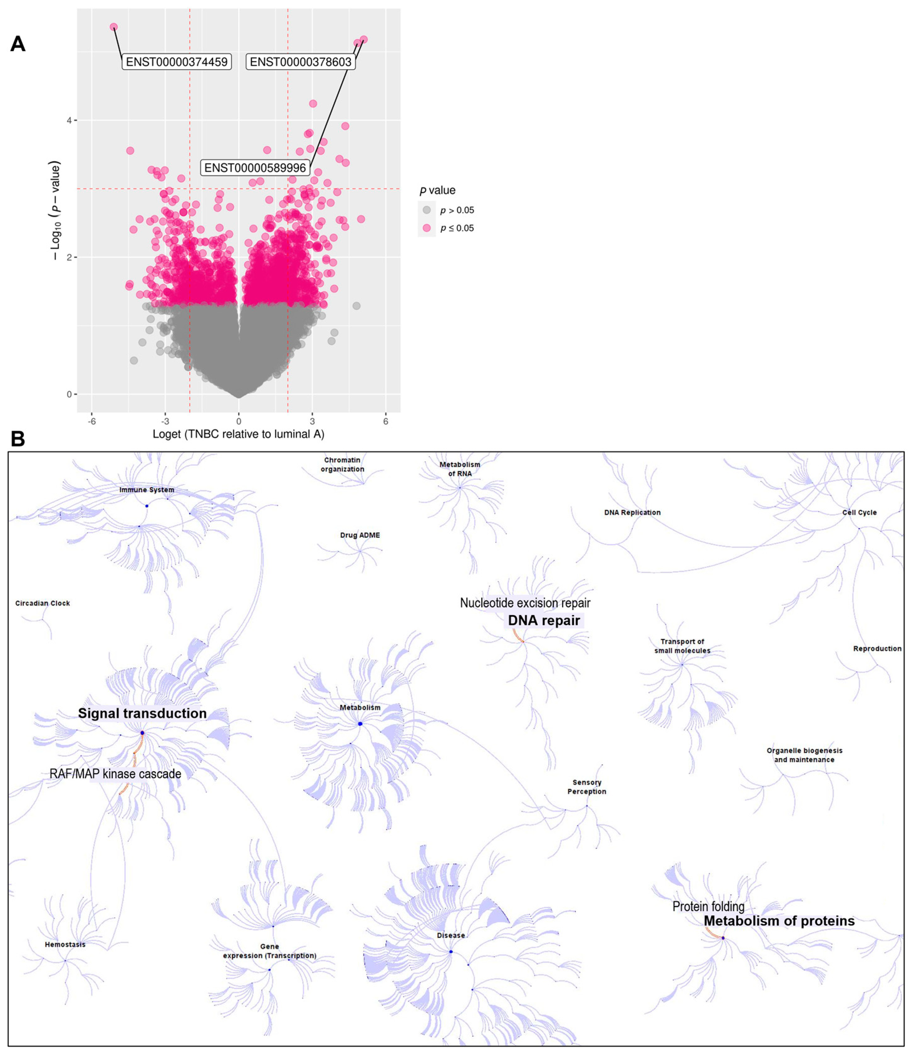
Figure 1. (A) Volcano plot showing differentially expressed isoforms (DEIs) between 13 luminal A and 12 triple−negative treatment−naïve BC patients. The top DEIs were the ENST00000374459 transcriptional variant of the RASGEF1A gene, which had lower PBMC expression in TNBC than luminal A; the ENST00000589996 transcriptional variant of the TBCB gene (with higher expression in TNBC); and the ENST00000378603 transcriptional variant of the DDB2 gene (with higher expression in TNBC). (B) Overview of Reactome pathways [39], highlighting the signal transduction (RAF/MAP kinase cascade) pathway involving RASGEF1A, the DNA repair (nucleotide excision repair) pathway involving DDB2, and the metabolism of proteins (protein folding) pathway involving TBCB.
Cancers 16 03171 g001
Figure 2. RNA-seq analysis of PBMCs from 12 TNBC and 13 luminal A female BC patients identified changes in expression of the RASGEF1A 374459 variant. (A) RASGEF1A variants. The isoform 374459 is depicted in red with exons numbered 1–13; adapted from the GTEx portal [48]; (B) RASGEF1A belongs to the GEF (guanine nucleotide exchange factor) family of proteins, which mediate GDP release and GTP binding to the Rap proteins, thereby activating them. Inactivation of the Rap proteins is initiated by their intrinsic GTPase activity, which is enhanced by the GTPase activating (GAP) proteins. The Rap proteins belong to the Ras family, whose members are known to participate in signaling pathways that control a diverse array of cellular processes (i.e., cell proliferation, differentiation, etc.); (C) the patient PBMC samples from RNA-seq analysis (i.e., the pilot cohort) were subjected to qRT-PCR validation with 374459-specific (left) and 395810-specific (right) primers; Mann–Whitney U test (*** p < 0.001). (D) The levels of RASGEF1A isoforms in whole blood of healthy subjects (from RNA-seq data) (adapted from the GTEx portal) [48].
Cancers 16 03171 g002
Figure 3. Isoform-specific RASGEF1A expression in a larger cohort comprising 32 healthy female subjects (CON) and 156 BC patients (45 luminal A, 90 luminal B, 5 HER2, 16 TNBC). (A) qRT-PCR analysis of PBMCs from healthy (CON) and BC subjects (comprising all BC subtypes) with 374459-specific (left) and 395810-specific (right) primers; (B) qRT-PCR analysis of PBMCs from different BC subtypes with 374459-specific (left) and 395810-specific (right) primers (the five HER2-positive samples were not included due to low number). Mann–Whitney U test, *** p < 0.001.
Cancers 16 03171 g003
Figure 4. Lower 374459 expression in PBMCs of BC patients showed association with higher Ki-67 proliferation index and ctDNA content. (A) qRT-PCR analysis of 374459 expression in PBMCs from BC patients stratified by increasing Ki-67 index; Mann–Whitney U test, * denotes p < 0.05. (B) qRT-PCR analysis of 374459-expression in PBMCs from BC subjects with lower (z-score ≤ 3) and higher (z-score > 3) ctDNA content; Mann–Whitney U test, * denotes p < 0.05.
Cancers 16 03171 g004
Table 1. Clinicopathological characteristics of BC patients.
Study Cohort: Female Breast Cancer Patients (n = 156)
BC subtypeLuminal ALuminal BHER2(+)TNBC
45 (28.85%)90 (57.69%)5 (3.21%)16 (10.26%)
Histological typeILCIDCIDC + DCIS* Other
18 (11.54%)57 (36.54%)73 (46.79%)8 (5.13%)
Localization/containmentLocalized/containedLocally advancedMetastaticNot known
124 (79.49%)17 (10.90%)8 (5.13%)7 (4.49%)
Grade123Not known
36 (23.08%)73 (46.79%)40 (25.64%)7 (4.49%)
Tumor sizeT1, ≤2 cmT2, >2 to ≤5 cmT3, >5 mmNot known
95 (60.90%)43 (27.56%)3 (1.92%)15 (9.61%)
Lymph nodesNegativeMicrometastasis ≤ 2 mmMacrometastasis > 2 mmNot known
98 (62.82%)7 (4.49%)23 (14.74%)28 (17.95%)
Ki-67 index<14%≥14 to ≤25%>25 to ≤50%>50 to ≤100%Not known
44 (28.21%)56 (35.90%)41 (26.28%)13 (8.33%)2 (1.28%)
Genome-wide z-score≤3%>3%Not determin.
30 (19.23%)11 (7.05%)115 (73.72%)
* Other histological types included mixed, tubular, and cribriform.
Table 2. Gene Ontology (GO) for RASGEF1A, DDB2, and TBCB [40].
GeneGO Term IDGO Term NameGO CategoryGO Term Description
RASGEF1AGO:0005085guanyl-nucleotide exchange factor activityMolecular FunctionStimulates the exchange of GDP to GTP on a signaling GTPase
RASGEF1AGO:0007265Ras protein signal transductionBiological ProcessInvolved in the transmission of signals through Ras proteins
DDB2GO:0003684Damaged DNA bindingMolecular FunctionThe ability to bind to DNA that has been damaged
DDB2GO:0006281DNA repairBiological ProcessCellular processes of restoring DNA after damage
TBCBGO:0043014Alpha-tubulin bindingMolecular FunctionBinding to the microtubule constituent protein alpha-tubulin
TBCBGO:0007021Tubulin complex assemblyBiological ProcessAssembly of alpha- and beta-tubulin to form a tubulin heterodimer
TBCBGO:0007023Post-chaperonin tubulin folding pathwayBiological ProcessCompletion of folding of alpha- and beta-tubulin after chaperonin-mediated partial folding
Disclaimer/Publisher’s Note: The statements, opinions and data contained in all publications are solely those of the individual author(s) and contributor(s) and not of MDPI and/or the editor(s). MDPI and/or the editor(s) disclaim responsibility for any injury to people or property resulting from any ideas, methods, instructions or products referred to in the content.