Canine Cancer Diagnostics by X-ray Diffraction of Claws
by
 and
and
Alexander Alekseev
1, 
Delvin Yuk
2,
Alexander Lazarev
2,
Daizie Labelle
2,
Lev Mourokh
3,* and
Pavel Lazarev
1,2 1
Matur UK Ltd., 5 New Street Square, London EC4A 3TW, UK
2
Arion Diagnostics, Inc., 911 Mustang Ct, Petaluma, CA 94954, USA
3
Physics Department, Queens College of the City University of New York, 65-30 Kissena Blvd, Flushing, NY 11367, USA
*
Author to whom correspondence should be addressed.
Cancers 2024, 16(13), 2422; https://doi.org/10.3390/cancers16132422
Submission received: 27 May 2024
/
Revised: 26 June 2024
/
Accepted: 28 June 2024
/
Published: 30 June 2024
(This article belongs to the Section Cancer Biomarkers)
Simple Summary
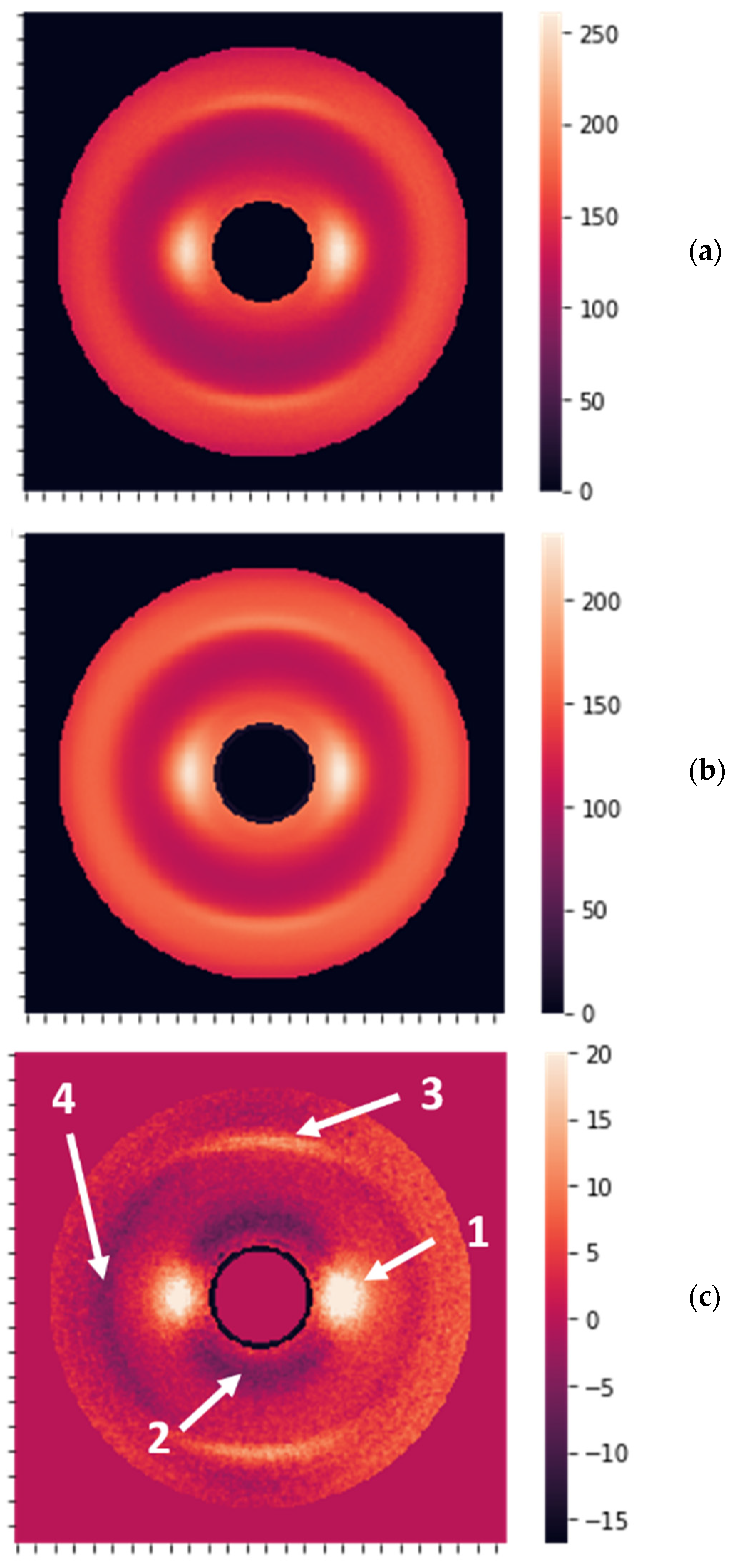
Canine cancer is a leading cause of dog mortality. In this study, we examine the hypothesis that the structure of keratin changes when cancer develops in the patient. We use X-ray diffraction of dog claws to detect these changes, finding that the modifications of the intermolecular distances are the most significant. Machine learning algorithms are utilized for cancer/non-cancer diagnostics, achieving a balanced accuracy of 85% for the blind group. Our research suggests that the changes in keratin structure can be tracked by X-ray diffraction, offering a potential tool for non-invasive cancer diagnostics. This could have significant implications for the early detection and treatment of canine cancer, potentially saving many lives. Moreover, this approach can be extended to human cancer detection.
Abstract
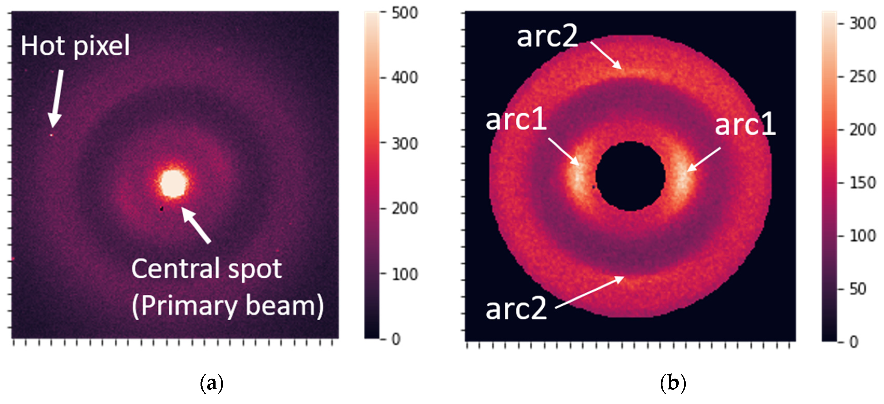
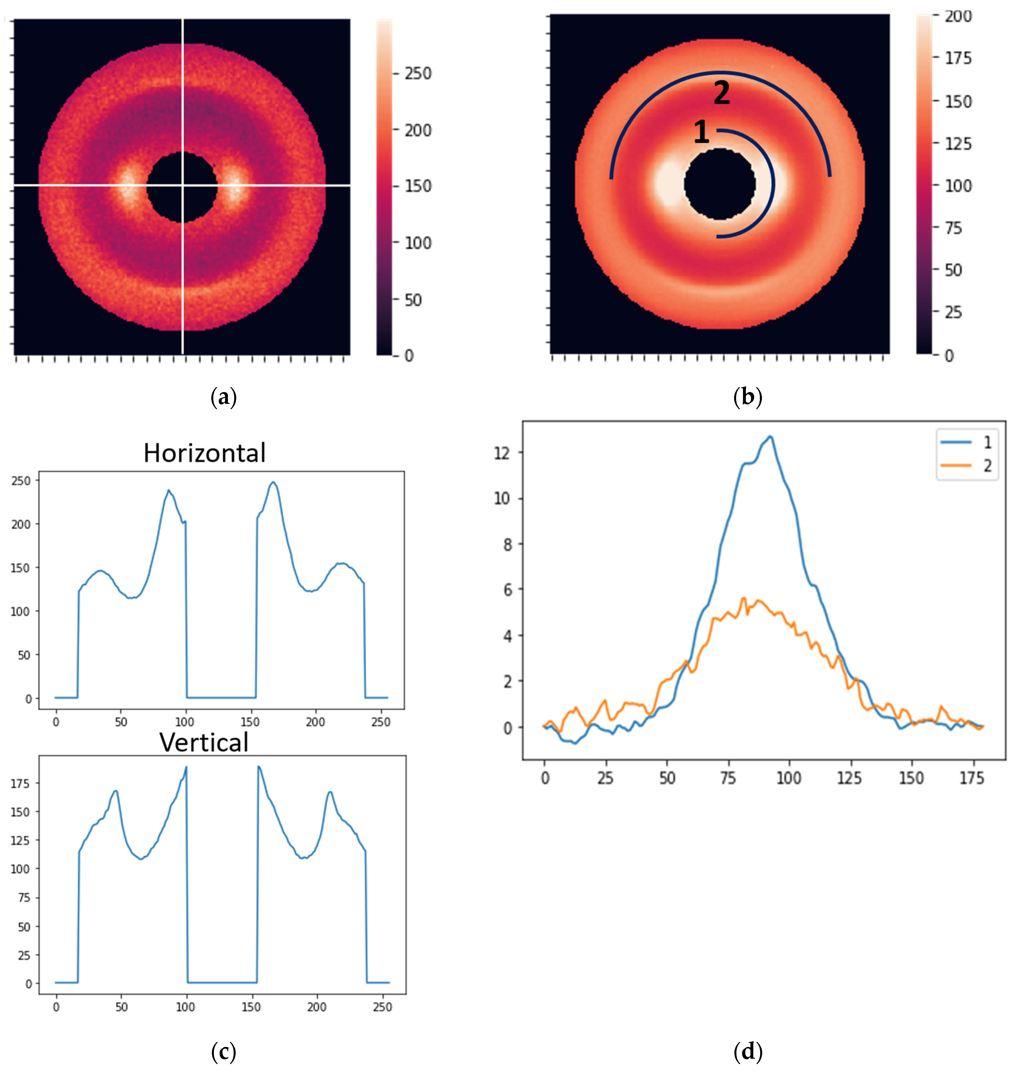
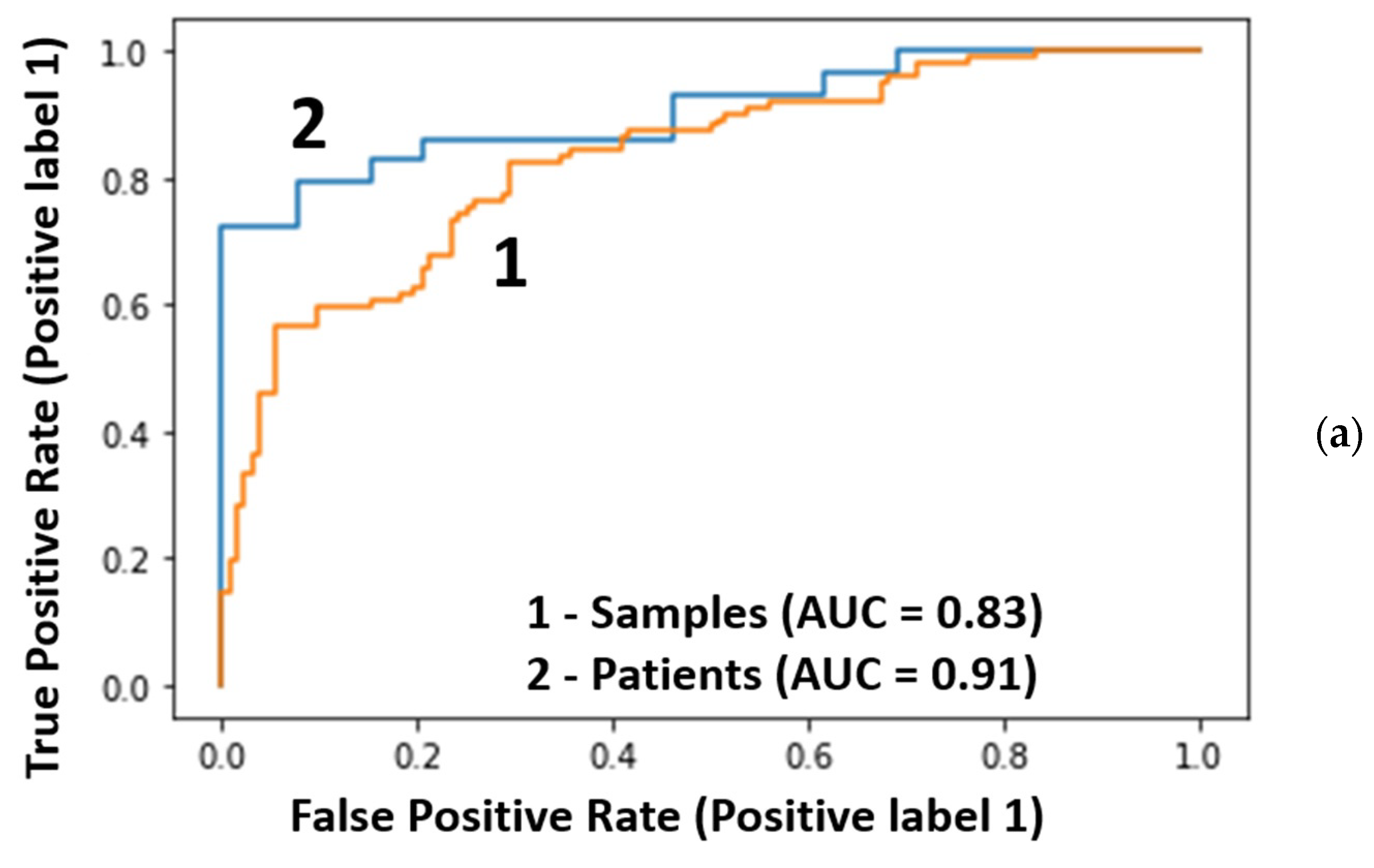
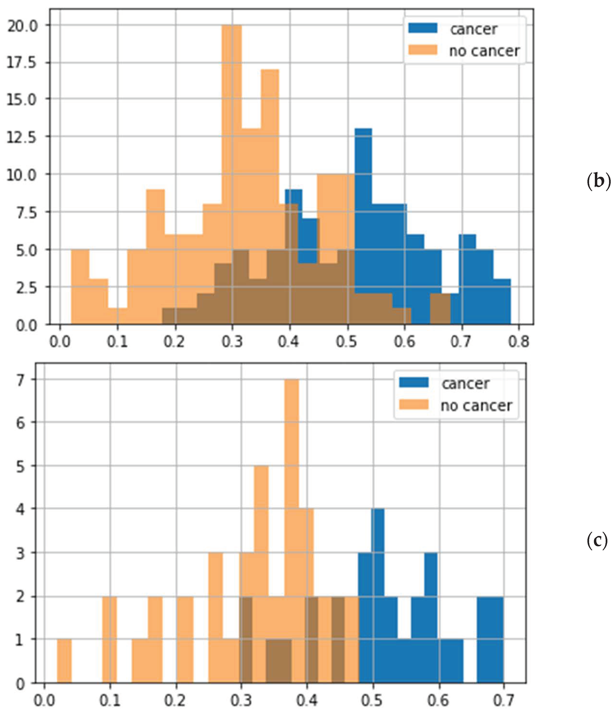
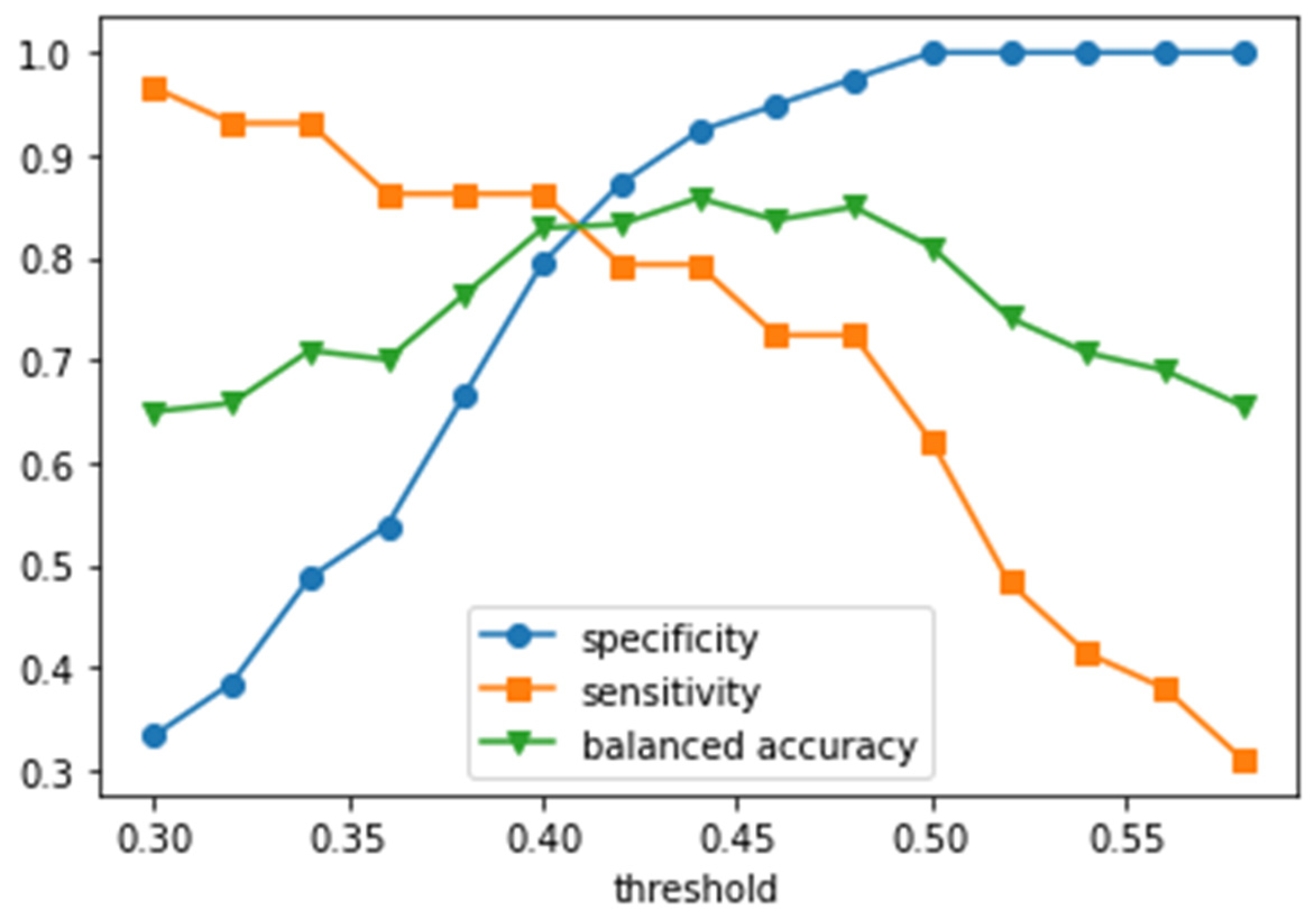
We report the results of X-ray diffraction (XRD) measurements of the dogs’ claws and show the feasibility of using this approach for early, non-invasive cancer detection. The obtained two-dimensional XRD patterns can be described by Fourier coefficients, which were calculated for the radial and circular (angular) directions. We analyzed these coefficients using the supervised learning algorithm, which implies optimization of the random forest classifier by using samples from the training group and following the calculation of mean cancer probability per patient for the blind dataset. The proposed algorithm achieved a balanced accuracy of 85% and ROC-AUC of 0.91 for a blind group of 68 dogs. The transition from samples to patients additionally improved the ROC-AUC by 10%. The best specificity and sensitivity values for 68 patients were 97.4% and 72.4%, respectively. We also found that the structural parameter (biomarker) most important for the diagnostics is the intermolecular distance.