Article

A Nomogram Incorporating Neutrophil-to-Lymphocyte Ratio and Squamous Cell Carcinoma Antigen Predicts the Prognosis of Oral Cancers

1
Department of Otorhinolaryngology-Head and Neck Surgery, Chang Gung Memorial Hospital, Chiayi 60040, Taiwan
2
College of Medicine, Chang Gung University, Taoyuan 330036, Taiwan
3
Department of Radiation Oncology, Chang Gung Memorial Hospital, Chiayi 60040, Taiwan
4
Department of Otorhinolaryngology-Head and Neck Surgery, Chang Gung Memorial Hospital, Taoyuan 333423, Taiwan
5
Department of Diagnostic Radiology, Chang Gung Memorial Hospital, Chiayi 60040, Taiwan
6
Department of Otorhinolaryngology-Head and Neck Surgery, Chang Gung Memorial Hospital, Keelung 20401, Taiwan
7
Department of Otorhinolaryngology-Head and Neck Surgery, Chang Gung Memorial Hospital, Kaohsiung 833253, Taiwan
*
Author to whom correspondence should be addressed.
Cancers 2023, 15(9), 2492; https://doi.org/10.3390/cancers15092492
Submission received: 27 March 2023 / Revised: 24 April 2023 / Accepted: 24 April 2023 / Published: 26 April 2023
(This article belongs to the Section Cancer Biomarkers)

Abstract

:

Simple Summary

We introduced a novel squamous cell carcinoma inflammatory index (SCI) derived by multiplying the serum squamous cell carcinoma antigen and neutrophil-to-lymphocyte ratio values for individuals with operable oral cavity squamous cell carcinomas (OSCCs). The prognostic value of SCI was explored by retrospectively analyzing data from 288 patients with a diagnosis of primary OSCC between January 2008 and December 2017. The current results demonstrated that patients with a high SCI (≥3.45) were associated with worse disease-free survival and overall survival than those with a low SCI (<3.45). An elevated preoperative SCI (≥3.45) predicted adverse overall survival (hazard ratio [HR] = 2.378; p < 0.002) and disease-free survival (HR = 2.219; p < 0.001) in a multivariable analysis. The constructed nomogram enables the clinical utility of the SCI and provides accurate OS predictions. Our findings indicate that SCI is a valuable and promising biomarker that is highly associated with patient survival outcomes in OSCC.

Abstract

We introduced a novel squamous cell carcinoma inflammatory index (SCI) and explored its prognostic utility for individuals with operable oral cavity squamous cell carcinomas (OSCCs). We retrospectively analyzed data from 288 patients who were given a diagnosis of primary OSCC from January 2008 to December 2017. The SCI value was derived by multiplying the serum squamous cell carcinoma antigen and neutrophil-to-lymphocyte ratio values. We appraised the associations of the SCI with survival outcomes by performing Cox proportional hazards and Kaplan–Meier analyses. We constructed a nomogram for survival predictions by incorporating independent prognostic factors in a multivariable analysis. By executing a receiver operating characteristic curve analysis, we identified the SCI cutoff to be 3.45, and 188 and 100 patients had SCI values of <3.45 and ≥3.45, respectively. The patients with a high SCI (≥3.45) were associated with worse disease-free survival and overall survival than those with a low SCI (<3.45). An elevated preoperative SCI (≥3.45) predicted adverse overall survival (hazard ratio [HR] = 2.378; p < 0.002) and disease-free survival (HR = 2.219; p < 0.001). The SCI-based nomogram accurately predicted overall survival (concordance index: 0.779). Our findings indicate that SCI is a valuable biomarker that is highly associated with patient survival outcomes in OSCC.

1. Introduction

Oral cavity cancer has the fourth-highest incidence in men in Taiwan [1]. Oral cavity squamous cell carcinoma (OSCC) was revealed to constitute over 90% of the cases of oral cavity cancer, which can typically be treated with radical resection with or without adjuvant therapy [2]. Despite advances in the treatment of OSCC, disease recurrence remains a significant problem. Studies have shown that up to 40–60% of patients with OSCC may experience disease recurrence within 5 years of treatment [3]. In addition to the tumor–node–metastasis (TNM) classification, several important prognostic factors for OSCC have been reported, such as lymphovascular invasion (LVI) [4], depth of invasion (DOI) [5], perineural invasion (PNI) [6], and extranodal extension (ENE) [2,7]. However, the aforementioned factors can be identified only after surgery. Accordingly, the identification of preoperative prognostic biomarkers may be helpful in predicting the prognosis of OSCC and selecting appropriate treatment strategies for individual patients.
The tumor-related protein squamous cell carcinoma antigen (SCC-Ag) was first identified in patients with cervical cancer by Kato et al. [8]. SCC-Ag belongs to a superfamily of serine proteinase inhibitors and plays a crucial role in tumorigenesis and cancer metastasis by increasing cell migration and inhibiting apoptosis [9]. Studies have reported SCC-Ag to be involved in the prognosis of numerous human squamous cell cancers, including esophageal [10], bladder [11], anal [12], lung [13], cervical [14], and OSCC [15]. Huang et al. included 142 patients with OSCC in their study and demonstrated that SCC-Ag at a level greater than or equal to 2.0 ng/mL has a significant association with the adverse pathological features and prognosis of OSCC [16]. In addition, cancer-related inflammatory responses have been reported to be closely associated with cancer development and progression [17,18]. Studies have indicated that several inflammatory biomarkers can be used as prognostic indicators for human cancers [19] and that a high neutrophil-to-lymphocyte ratio (NLR) can negatively influence the prognosis of individuals with OSCC [20]. However, the NLR is not a tumor-specific biomarker, which has prevented the development of a rationale supporting its use in cancer management. The results of the previously mentioned studies indicate that the combined evaluation of SCC-Ag and NLR may offer complementary information for OSCC prognostication. Nevertheless, to the best of our knowledge, the potential combined value of SCC-Ag and NLR as a prognostic biomarker has yet to be discussed in the literature. Accordingly, to address this gap, we herein present a novel squamous cell carcinoma inflammatory index (SCI) that we derived by multiplying serum SCC-Ag and NLR values. Given that the SCI may provide a comprehensive assessment of tumor–host interactions, we hypothesized that the SCI would have significant associations with survival outcomes and explored the SCI’s prognostic utility by retrospectively assessing patients that have been surgically treated for OSCC.

2. Materials and Methods

2.1. Patients

Our retrospective cohort study involved analyzing medical data derived from 313 consecutive patients who were pathologically determined to have primary OSCC between January 2008 and December 2017 at our hospital. All patients received radical surgery, which was followed by adjuvant therapy if indicated. The patients were excluded if they had a diagnosis of synchronous cancer or a cancer history (n = 7), had undergone neoadjuvant treatment before surgery (n = 6), had autoimmune diseases or acute infections prior to surgery (n = 1), or had missing laboratory or follow-up data (n = 11). We determined the data from 288 patients to be suitable for inclusion in the formal analysis. The study methods and procedures complied with the principles of the Declaration of Helsinki. In addition, this executed study protocol was granted approval by the Institutional Review Board of our hospital (number: 202300107B0).

2.2. Collection of Study Data

The medical personnel obtained the clinical data of the included patients by reviewing the electronic medical chart system of our hospital. Before the surgery, all patients had been subjected to a comprehensive cancer staging workup in accordance with our institutional guidelines. Clinicopathological features, including sex, age, primary tumor location, pathological cancer stage (American Joint Committee on Cancer Staging Manual, 8th edition, 2018), LVI, ENE status, PNI status, DOI, nearest surgical margin, and tumor differentiation, were extracted and analyzed as prognostic factors. For each patient, the internal tumor conference results and the institutional guidelines were used as the basis to determine whether they required adjuvant therapy within 6 weeks postsurgery [21]. Any underlying comorbidities were defined and quantified using the Charlson comorbidity index (CCI) [22]. The patients were considered to be consumers of alcohol, cigarettes, or areca nuts if they reported having >1 alcoholic beverage every week for >6 months, smoking ≥ 1 pack each day for >1 year, or chewing ≥ 2 areca nuts each day for >1 year, respectively [23].

2.3. Measurement of Serum Indices

This study used blood samples obtained one week before surgery. The serum SCC-Ag levels (reference cutoff value: 2.5 ng/mL) were obtained using a chemiluminescent microparticle immunoassay (Abbott Japan Co. Ltd., Tokyo, Japan). We obtained our patients’ blood biochemistry values by using a Cobas 8000 biochemistry analysis system, which is an automated system (Roche Hitachi, Rotkreuz, Switzerland). In addition, we employed a Sysmex SE-9000 hematology analysis system to derive the peripheral blood cell counts (Sysmex, Kobe, Japan). We defined the SCI by using the following equation: SCI = SCC-Ag × NLR.

2.4. Follow-Up and Study Endpoints

All patients had been scheduled for regular clinic visits (every 2 and 3 months during the first and second years, respectively, and every 6 months thereafter) and periodic imaging surveillance of the head, neck, and chest. The study endpoints were as follows: disease-free survival (DFS), which was specified as the period spanning from the surgery date to the date of receiving a diagnosis for distant metastasis or locoregional recurrence, death, being censored from the study, or the last follow-up appointment for the alive patients; and overall survival (OS), which was specified as the period spanning from the surgery date to that of all-cause mortality, being censored from the study, or the last follow-up appointment for the alive patients. Telephone interviews and electronic medical records were used to obtain the patients’ follow-up data. We determined our median (range) follow-up period to be 40.1 (3.5–122.4) months, with the last follow-up date being 31 December 2019.

2.5. Statistical Analysis

This study presents the categorical variables as percentages and absolute numbers for the study cohort. We determined the normality of the data by performing the Kolmogorov–Smirnov test. This study also presents the continuous variables as medians and interquartile ranges if the data were non-normally distributed. To derive the best serum index cutoff values, we executed a receiver operating characteristic (ROC) curve analysis along with a Youden’s index calculation. In addition, we estimated the areas under the ROC curves (AUCs) for OS predictions in order to compare the prognostic discriminatory ability of the SCI with that of other indices. The study patients were grouped in accordance with the optimal SCI cutoffs, and we compared the clinicopathological variables of the patients with low and high SCI values by performing the Mann–Whitney U test for the continuous variables and the chi-squared test for the categorical variables. We obtained the DFS and OS curves using the Kaplan–Meier method. Furthermore, we performed a log-rank test to determine the survival differences between the low- and high-SCI groups. To ascertain the independent prognostic factors for DFS and OS, we executed a Cox proportional hazards analysis. The variables were evaluated using the log-rank test in a univariable analysis, and those that reached statistical significance (p < 0.1) were included in a multivariable analysis. The stepwise selection method was executed using the R 4.2.0 software to select the optimal subset of independent factors. We performed the aforementioned statistical analyses using the SPSS (V21.0) software platform (SPSS, Chicago, IL, USA). We also considered the statistical significance to be represented by p < 0.05.
Using the rms package in R V5.1–0 (Vanderbilt University, Nashville, TN, USA) [24], we constructed a predictive nomogram by incorporating the independent prognostic factors identified in the multivariable analysis for predicting the individualized OS at 3 and 5 years. To evaluate our constructed nomogram’s accuracy in terms of predicting the OS, we derived its concordance index (c-index), in addition to deriving that of the conventional TNM staging system for comparison. To visually assess the degree of consistency between the actual OS outcomes and the nomogram-derived OS predictions, calibration plots were drawn.

3. Results

3.1. Characteristics of Included Patients

Table 1 lists the included patients’ baseline characteristics. Most of the enrollees were male (n = 262; 91.0%), and 190 (66.0%) patients were aged <65 years. The tongue was noted to constitute the most frequent tumor location (n = 110; 38.2%), and the buccal mucosa was noted to constitute the second most frequent location (n = 96; 33.4%). Stage I, II, III, and IV OSCC was given as a diagnosis for 61 (21.2%), 39 (13.5%), 40 (13.9%), and 148 (51.4%) patients, respectively. Regarding the pathological features, PNI, ENE, LVI, and poorly differentiated (P-D) OSCC were present in 72 (25.0%), 58 (20.1%), 20 (6.9%), and 35 (12.2%) patients, respectively. Regarding adjuvant therapy, 113 (39.2%) patients underwent adjuvant chemoradiotherapy (CRT), and 39 (13.5%) patients underwent adjuvant radiotherapy (RT).

3.2. Analysis of ROC Curves and AUCs of Serum Indices

By analyzing the ROC curves, we determined the optimal SCI cutoff value for predicting the OS to be 3.45 (AUC: 0.673; 95% confidence interval [CI]: 0.593–0.749; p < 0.001), and the sensitivity and specificity at this value were 58.2% and 74.2%, respectively (Figure 1). Similarly, the optimal cutoff values for NLR and SCC-Ag were determined to be 4.51 (AUC: 0.607; 95% CI: 0.528–0.687; p = 0.005) and 1.65 (AUC: 0.653; 95% CI: 0.578–0.728; p < 0.001), respectively. The AUCs for the enrolled indices and their combinations, including the NLR and SCI, were also compared in terms of the predictive performance of the OS (Table 2). Most indices could predict the OS (with the exception of neutrophils; p = 0.228), and the AUC value derived for the SCI (0.673) was higher than the values derived for the lymphocytes (0.608), SCC-Ag (0.653), and NLR (0.607). These findings indicate that the SCI had an ideal prognostic performance level in our study setting and that the prognostic value of the SCI should be more thoroughly investigated.

3.3. Associations between SCI and Clinicopathological Features

The differences in the clinicopathological features between the low-SCI (<3.45) and high-SCI (≥3.45) groups are presented in Table 3. The high- and low-SCI groups had 100 (34.7%) and 188 (65.3%) patients, respectively. We noted that the high-SCI group contained a higher proportion of individuals with stage III to IV OSCC (p < 0.001), PNI (p = 0.002), ENE (p < 0.001), late T and N statuses (both p < 0.001), LVI (p < 0.001), the need for adjuvant therapy (p < 0.001), higher CCI scores (p = 0.029), shorter median survival (p < 0.001), and DOI ≥10 mm (p < 0.001) compared to the low-SCI group.

3.4. Associations between SCI and OS

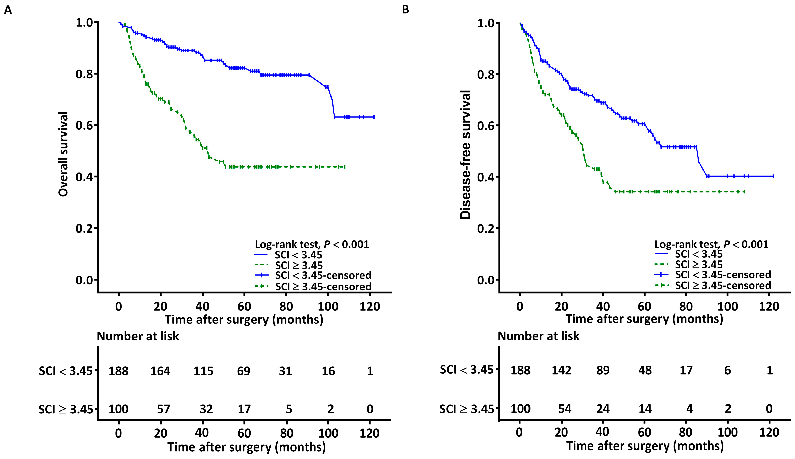
The Kaplan–Meier curves revealed the estimated median OS derived for the low-SCI (<3.45) and high-SCI (≥3.45) groups to be >178 and 43.2 months (95% CI: 29.1–57.3), respectively. The log-rank test revealed significant between-group differences with regard to survival (p < 0.001; Figure 2A). Our univariable analysis revealed LVI, stage IV disease, the need for CRT, PNI, CCI ≥ 2, NLR ≥ 4.51, P-D, SCC-Ag ≥ 1.65, and SCI ≥ 3.45 to have a significant association with adverse OS (Table 4). Since SCC-Ag and the NLR are components of the SCI, separate multivariable analysis models were employed to prevent multicollinearity. As shown in Table 5, the results of the multivariable analysis reveal that a high SCI of ≥3.45 had an independent association with adverse OS, with the corresponding hazard ratio (HR) being 2.378 (95% CI, 1.356–3.735; p = 0.002). We also identified additional independent prognostic factors for adverse OS, including stage IV disease, P-D, CCI ≥ 2, SCC-Ag ≥ 1.65, and NLR ≥ 4.51.

3.5. Associations between SCI and DFS

The Kaplan–Meier curves revealed the estimated median DFS derived for the low-SCI (<3.45) and high-SCI (≥3.45) groups to be 86.3 months (95% CI: 64.2–108.7) and 31.2 months (95% CI: 26.1–36.3), respectively. The log-rank test revealed significant between-group differences (p < 0.001; Figure 2B). Table 4 presents the significant predictors of a poor DFS as identified in the univariable analysis. The predictors included the following: stage IV OSCC, NLR ≥ 4.51, P-D, the need for adjuvant CRT, SCC-Ag ≥ 1.65, LVI, and SCI ≥ 3.45. In the multivariable analysis, an SCI value of ≥3.45 was independently associated with an unfavorable DFS, with the corresponding HR (95% CI) being 2.219 (1.437–3.425; p < 0.001; Table 5). We also determined additional independent risk factors for an unfavorable DFS, including stage IV disease, P-D, SCC-Ag ≥ 1.65, and NLR ≥ 4.51.

3.6. Stratified Analysis

We executed a stratified analysis of the SCI’s OS discriminatory ability (Figure 3). A high SCI value was consistently and significantly associated with adverse OS, regardless of whether the patient data were stratified by the tumor location (the tongue, buccal mucosa, and other locations), T status (T1 to T2 and T3 to T4; p = 0.004 and 0.001, respectively), N status (N0 and N1 to 3; p < 0.001 and 0.001, respectively), ENE status (absent or present; p < 0.001 and 0.009, respectively), PNI status (absent or present; p < 0.001 and 0.001, respectively), the need for adjuvant chemotherapy (necessary and not necessary; p = 0.009 and < 0.001, respectively), the closest margin (≥5 and <5 mm; both p < 0.001), or DOI status (<10 and ≥10 mm; p < 0.001 and 0.002, respectively).

3.7. Predictive Nomogram Construction

We constructed a nomogram by incorporating the significant prognostic factors derived from our multivariable analysis of the OS, including SCI, tumor differentiation, cancer stage, and CCI, and employed it for individualized OS predictions (Figure 4A). The SCI-based nomogram’s c-index value was 0.779 (95% CI: 0.744–0.813), whereas the c-index value for the nomogram constructed using only the TNM staging system was 0.697 (95% CI: 0.664–0.729). Calibration plots were also drawn to evaluate how close the nomogram-estimated OS was to the actual observed survival outcomes. The slopes of the predicted 3- and 5-year OS probability calibration curves (as displayed in Figure 4B,C, respectively) were noted to approximate the ideal 45° line. Accordingly, these findings verify the strong predictive performance of our constructed nomogram.

4. Discussion

In the present study, we introduce a novel SCI that we derived by integrating the NLR and SCC-Ag; thus, this unique cancer-related inflammatory index incorporates both host inflammatory responses and tumor-specific proteins. By enrolling 288 patients undergoing curative surgery for OSCC, we investigated the clinical role of the SCI in OSCC management and obtained several novel findings. First, the SCI had a significantly higher AUC compared to the SCC-Ag and NLR, suggesting that the SCI may be a better predictor of the OS than its individual components in OSCC prognostications. Second, our stepwise multivariable analysis demonstrated a high SCI value (≥3.45) to be an independent risk factor for unfavorable OS and DFS outcomes among individuals with OSCC. Furthermore, the SCI had consistent and significant prognostic values for OS in our stratified analyses. In summary, our hypothesis was verified, and the study results support that the novel SCI has promise as a prognostic biomarker for individuals with OSCC. Since the SCI can be easily obtained from preoperative laboratory tests, we propose that the SCI has considerable potential for use in everyday clinical practices and oncological research. Whether patients with a high SCI (≥3.45) before treatment may benefit from more personalized treatment planning warrants further investigation.
Several researchers have combined tumor markers with inflammatory indices and investigated their prognostic roles in individuals with cancer. In previous studies by our colleagues, high SCC-Ag and C-reactive protein levels can predict advanced cancer stages and adverse survival outcomes in patients with OSCC [16] and recurrent diseases [9]. Su et al. developed a novel prognostic index by multiplying carcinoembryonic antigen values and NLR values and determined that the derived index had a significant association with OS in patients undergoing regorafenib treatment for metastatic colorectal cancer [25]. Wang et al. conducted a retrospective analysis of data from 515 patients who had cervical cancer and observed that SCC-Ag had a higher predictive efficacy than the NLR for all tumor stages [26]. Our study further demonstrated that the AUC derived for the SCI was higher than that derived for the SCC-Ag and NLR. This finding suggests that the SCI has better predictive efficacy for patients with operable OSCC, presumably due to better reflecting host–tumor interactions and anti-tumor immune responses. Our study results support the idea that a comprehensive consideration of the tumor-specific factors and host inflammatory responses during prognostic evaluations can increase the accuracy of survival predictions for patients with OSCC. In the future, prospective studies should explore whether preoperative SCI values or changes in SCI values can be used to predict the prognosis of other human squamous cell cancers.
Our study findings support SCI as a unique and hopeful prognostic indicator for patients with OSCC. However, the exact mechanisms that govern the associations between an elevated SCI and adverse clinical outcomes remain unknown. An elevated SCI can be indicative of increased serum SCC-Ag levels and/or NLR values, which may provide insight into how the SCI predicts the prognosis of OSCC. Studies have reported that serum SCC-Ag exhibits an association with distant metastasis, tumor progression, and lymph node metastasis [27] and that the production of serum SCC-Ag in OSCC is attributable to lymphocytes surrounding cancer cells [28]. Lin et al. assessed 79 patients with OSCC and revealed an SCC-Ag value ≥ 2.0 ng/mL to be significantly associated with advanced tumor and nodal status and with poor OS and DFS [29]. Moreover, SCC-Ag has been reported to be able to predict the early recurrence of OSCC. In an evaluation of 100 patients’ medical records, Chen et al. demonstrated that a high SCC-Ag level could predict tumor recurrence and prognosis among individuals with recurrent OSCC [9]. This predictive effect may be explained by the involvement of SCC-Ag in tumor cell apoptosis inhibition at the molecular level [30]. Several studies have investigated the value of the NLR for predicting disease prognosis among patients who received treatment for OSCC, and this signifies that systemic inflammatory responses have an informative role in OSCC management. The mentioned studies have reported that a high NLR is associated with advanced cancer stages, a poor response to chemotherapy, and poor OS and DFS in patients with OSCC [20,31]. Furthermore, Yasumatsu et al. assessed the dynamic changes in the NLR values in 41 patients who had received nivolumab treatment for metastatic or recurrent head and neck cancer. They reported that monitoring changes in the NLR may enable the early detection of treatment failures during nivolumab monotherapies [32]. Our findings also reveal significant associations between a high SCI (≥3.45) and adverse clinicopathological features. The following mechanisms may govern the aforementioned associations: (1) a greater tumor burden (e.g., advanced tumor extension and lymph node metastasis) may be accompanied by higher cancer-related inflammation, leading to a high SCI; and (2) tumor invasion (e.g., PNI or LVI) may be promoted by SCC-Ag with the stimulation of matrix metalloprotease-9 production [33], contributing to a high SCI. The aforementioned findings provide evidence that may assist in identifying the mechanism underlying the prognostic effect of the SCI among patients with OSCC. Nevertheless, the exact mechanism warrants further exploration.
At present, the TNM staging system is the benchmark for OSCC treatment stratification and prognostication [34]. Nevertheless, the TNM staging system categorizes patients on the basis of their survival outcomes and anatomical extension, failing to account for crucial factors that govern the prognosis of OSCC, such as tumor differentiation and patients’ underlying comorbidity [35]. Given the limitations of the TNM staging system, nomograms have been proposed as an effective tool for determining disease prognoses in the era of personalized medicine [35]. Some of the main advantages of nomograms are as follows: (1) they offer pictorial presentations of clinical and pathological information with relevant weights for each variable; (2) they offer prognostic estimates that can assist in personalized treatment planning; and (3) they enable the estimation of the probability of a variety of clinical events that may affect the care of patients with cancer. The present study constructed an SCI-based nomogram that not only enables the realization of the clinical utility of the SCI but also incorporates independent prognostic factors to accurately predict individualized OSs. The SCI-based nomogram’s c-index values and calibration plots demonstrated our nomogram’s favorable performance and calibration in our OSCC cohorts. However, treatment selection for patients with OSCC should be based on clinical trials rather than nomogram data alone [35]. Accordingly, additional studies with a prospective design are warranted to verify whether the use of SCI-based nomograms for decision-making truly improves the prognosis of OSCC.
The key strength of this study is that it is the first to introduce a novel SCI that can help OSCC prognostication. Additionally, our constructed nomogram enables OS prediction that is both accurate and individualized; our nomogram also demonstrates the applicability of the SCI in clinical practice. The other strengths of the present study are its homogeneously treated cohorts and long follow-up period. However, the several limitations of our study should be acknowledged. First, the retrospective single-center study design has inherent bias. This bias can be overcome in future studies by employing national or multicenter databases. Second, we did not validate our derived study findings through the use of an independent dataset, which prevented the confirmation of the external validity of our findings. Finally, because a consensus has not been reached on the best cutoff level for the SCI, its use in clinical practice warrants further investigation. Therefore, well-designed, large, prospective, multi-institutional studies must be conducted to verify our findings and identify an ideal cutoff value for the SCI.

5. Conclusions

Our study results indicate that preoperative SCI is a promising biomarker with prognostic value for patients with operable OSCC, which may provide new insight into the development of cancer-related inflammatory indices. We additionally demonstrated the clinical utility of the SCI by creating an SCI-based nomogram that can provide personalized OS predictions. Future large-scale studies with prospective designs are warranted to verify our results and the utility of the SCI in predicting prognoses in other human squamous cell cancers.

Author Contributions

Conceptualization, Y.-T.T. and K.-H.F.; methodology, C.-H.L.; software, G.-H.C.; formal analysis, C.-M.H.; investigation, M.-S.T.; resources, C.-J.K.; data curation, C.-T.L.; writing—original draft preparation, Y.-T.T.; writing—review and editing, K.-H.F. and Y.-H.T.; visualization, M.-H.T. and Y.-C.L.; supervision, E.I.H.; funding acquisition, Y.-T.T. All authors have read and agreed to the published version of the manuscript.

Funding

The study was supported partly by a grant from the Chang Gung Medical Foundation, Taiwan (CMRPG6L0232).

Institutional Review Board Statement

The study was conducted according to the guidelines of the Declaration of Helsinki and approved by the Institutional Review Board of the Chang Gung Medical Foundation (number: 202300107B0, 31 January 2023).

Informed Consent Statement

Patient consent was waived due to the retrospective design of this study.

Data Availability Statement

The data presented in this study are available from the corresponding author upon request.

Acknowledgments

We acknowledge all of the invaluable contributions of the members of the HIE lab at the Chiayi Chang Gung Memorial Hospital.

Conflicts of Interest

The authors declare no conflict of interest.

References

  1. Kao, S.Y.; Lim, E. An Overview of Detection and Screening of Oral Cancer in Taiwan. Chin. J. Dent. Res. 2015, 18, 7–12. [Google Scholar] [PubMed]
  2. Ettinger, K.S.; Ganry, L.; Fernandes, R.P. Oral Cavity Cancer. Oral Maxillofac. Surg. Clin. N. Am. 2019, 31, 13–29. [Google Scholar] [CrossRef] [PubMed]
  3. Warnakulasuriya, S. Living with Oral Cancer: Epidemiology with Particular Reference to Prevalence and Life-Style Changes That Influence Survival. Oral Oncol. 2010, 46, 407–410. [Google Scholar] [CrossRef] [PubMed]
  4. Jardim, J.F.; Francisco, A.L.; Gondak, R.; Damascena, A.; Kowalski, L.P. Prognostic Impact of Perineural Invasion and Lymphovascular Invasion in Advanced Stage Oral Squamous Cell Carcinoma. Int. J. Oral Maxillofac. Surg. 2015, 44, 23–28. [Google Scholar] [CrossRef] [PubMed]
  5. den Toom, I.J.; Janssen, L.M.; van Es, R.J.J.; Karagozoglu, K.H.; de Keizer, B.; van Weert, S.; Willems, S.M.; Bloemena, E.; Leemans, C.R.; de Bree, R. Depth of Invasion in Patients with Early Stage Oral Cancer Staged by Sentinel Node Biopsy. Head Neck 2019, 41, 2100–2106. [Google Scholar] [CrossRef] [PubMed]
  6. Varsha, B.K.; Radhika, M.B.; Makarla, S.; Kuriakose, M.A.; Kiran, G.S.; Padmalatha, G.V. Perineural Invasion in Oral Squamous Cell Carcinoma: Case Series and Review of Literature. J. Oral Maxillofac. Pathol. 2015, 19, 335–341. [Google Scholar] [CrossRef] [PubMed]
  7. Wreesmann, V.B.; Katabi, N.; Palmer, F.L.; Montero, P.H.; Migliacci, J.C.; Gonen, M.; Carlson, D.; Ganly, I.; Shah, J.P.; Ghossein, R.; et al. Influence of Extracapsular Nodal Spread Extent on Prognosis of Oral Squamous Cell Carcinoma. Head Neck 2016, 38 (Suppl. S1), E1192–E1199. [Google Scholar] [CrossRef]
  8. Kato, H.; Torigoe, T. Radioimmunoassay for Tumor Antigen of Human Cervical Squamous Cell Carcinoma. Cancer 1977, 40, 1621–1628. [Google Scholar] [CrossRef]
  9. Chen, I.H.; Liao, C.T.; Wang, H.M.; Huang, J.J.; Kang, C.J.; Huang, S.F. Using Scc Antigen and Crp Levels as Prognostic Biomarkers in Recurrent Oral Cavity Squamous Cell Carcinoma. PLoS ONE 2014, 9, e103265. [Google Scholar] [CrossRef]
  10. Yin, N.; Liu, W. Clinical Value of Tumor Marker Index Based on Preoperative Cyfra 21-1 and Scc-Ag in the Evaluation of Prognosis and Treatment Effectiveness in Patients with Esophageal Squamous Cell Carcinoma. Onco Targets Ther. 2020, 13, 4135–4143. [Google Scholar] [CrossRef]
  11. Pectasides, D.; Bafaloucos, D.; Antoniou, F.; Gogou, L.; Economides, N.; Varthalitis, J.; Dimitriades, M.; Kosmidis, P.; Athanassiou, A. Tpa, Tati, Cea, Afp, Beta-Hcg, Psa, Scc, and Ca 19-9 for Monitoring Transitional Cell Carcinoma of the Bladder. Am. J. Clin. Oncol. 1996, 19, 271–277. [Google Scholar] [CrossRef] [PubMed]
  12. Henkenberens, C.; Toklu, H.; Tamme, C.; Bruns, F. Clinical Value of Squamous Cell Carcinoma Antigen (Sccag) in Anal Cancer—A Single-Center Retrospective Analysis. Anticancer Res. 2016, 36, 3173–3177. [Google Scholar] [PubMed]
  13. Vassilakopoulos, T.; Troupis, T.; Sotiropoulou, C.; Zacharatos, P.; Katsaounou, P.; Parthenis, D.; Noussia, O.; Troupis, G.; Papiris, S.; Kittas, C.; et al. Diagnostic and Prognostic Significance of Squamous Cell Carcinoma Antigen in Non-Small Cell Lung Cancer. Lung Cancer 2001, 32, 137–144. [Google Scholar] [CrossRef] [PubMed]
  14. Fu, J.; Wang, W.; Wang, Y.; Liu, C.; Wang, P. The Role of Squamous Cell Carcinoma Antigen (Scc Ag) in Outcome Prediction after Concurrent Chemoradiotherapy and Treatment Decisions for Patients with Cervical Cancer. Radiat. Oncol. 2019, 14, 146. [Google Scholar] [CrossRef]
  15. Dante, D.E.P.; Young, C.K.; Chien, H.T.; Tsao, C.K.; Fok, C.C.; Fan, K.H.; Liao, C.T.; Wang, H.M.; Kang, C.J.; Chang, J.T.; et al. Prognostic Roles of Scc Antigen, Crp and Cyfra 21-1 in Oral Cavity Squamous Cell Carcinoma. Anticancer Res. 2019, 39, 2025–2033. [Google Scholar]
  16. Huang, S.F.; Wei, F.C.; Liao, C.T.; Wang, H.M.; Lin, C.Y.; Lo, S.; Huang, J.J.; Chen, I.H.; Kang, C.J.; Chien, H.T.; et al. Risk Stratification in Oral Cavity Squamous Cell Carcinoma by Preoperative Crp and Scc Antigen Levels. Ann. Surg. Oncol. 2012, 19, 3856–3864. [Google Scholar] [CrossRef]
  17. Greten, F.R.; Grivennikov, S.I. Inflammation and Cancer: Triggers, Mechanisms, and Consequences. Immunity 2019, 51, 27–41. [Google Scholar] [CrossRef]
  18. Colotta, F.; Allavena, P.; Sica, A.; Garlanda, C.; Mantovani, A. Cancer-Related Inflammation, the Seventh Hallmark of Cancer: Links to Genetic Instability. Carcinogenesis 2009, 30, 1073–1081. [Google Scholar] [CrossRef]
  19. Brenner, D.R.; Scherer, D.; Muir, K.; Schildkraut, J.; Boffetta, P.; Spitz, M.R.; Le Marchand, L.; Chan, A.T.; Goode, E.L.; Ulrich, C.M.; et al. A Review of the Application of Inflammatory Biomarkers in Epidemiologic Cancer Research. Cancer Epidemiol. Biomarks Prev. 2014, 23, 1729–1751. [Google Scholar] [CrossRef]
  20. Nakashima, H.; Matsuoka, Y.; Yoshida, R.; Nagata, M.; Hirosue, A.; Kawahara, K.; Sakata, J.; Arita, H.; Hiraki, A.; Nakayama, H. Pre-Treatment Neutrophil to Lymphocyte Ratio Predicts the Chemoradiotherapy Outcome and Survival in Patients with Oral Squamous Cell Carcinoma: A Retrospective Study. BMC Cancer 2016, 16, 41. [Google Scholar] [CrossRef]
  21. Lin, C.Y.; Fan, K.H.; Lee, L.Y.; Hsueh, C.; Yang, L.Y.; Ng, S.H.; Wang, H.M.; Hsieh, C.H.; Lin, C.H.; Tsao, C.K.; et al. Precision Adjuvant Therapy Based on Detailed Pathologic Risk Factors for Resected Oral Cavity Squamous Cell Carcinoma: Long-Term Outcome Comparison of Cgmh and Nccn Guidelines. Int. J. Radiat. Oncol. Biol. Phys. 2020, 106, 916–925. [Google Scholar] [CrossRef] [PubMed]
  22. Brusselaers, N.; Lagergren, J. The Charlson Comorbidity Index in Registry-Based Research. Methods Inf. Med. 2017, 56, 401–406. [Google Scholar] [PubMed]
  23. Ko, C.A.; Fang, K.H.; Hsu, C.M.; Lee, Y.C.; Chang, G.H.; Huang, E.I.; Tsai, M.S.; Tsai, Y.T. The Preoperative C-Reactive Protein-Lymphocyte Ratio and the Prognosis of Oral Cavity Squamous Cell Carcinoma. Head Neck 2021, 43, 2740–2754. [Google Scholar] [CrossRef] [PubMed]
  24. Harrell, F.E., Jr.; Lee, K.L.; Mark, D.B. Multivariable Prognostic Models: Issues in Developing Models, Evaluating Assumptions and Adequacy, and Measuring and Reducing Errors. Stat. Med. 1996, 15, 361–387. [Google Scholar] [CrossRef]
  25. Su, Y.L.; Tsai, K.L.; Chiu, T.J.; Lin, Y.M.; Lee, K.C.; Lu, C.C.; Chen, H.H.; Wu, C.C.; Hsu, H.C. Development and Validation of a Novel Serum Prognostic Marker for Patients with Metastatic Colorectal Cancer on Regorafenib Treatment. Cancers 2021, 13, 5080. [Google Scholar] [CrossRef] [PubMed]
  26. Wang, L.; Jia, J.; Lin, L.; Guo, J.; Ye, X.; Zheng, X.; Chen, Y. Predictive Value of Hematological Markers of Systemic Inflammation for Managing Cervical Cancer. Oncotarget 2017, 8, 44824–44832. [Google Scholar] [CrossRef] [PubMed]
  27. Zhu, H. Squamous Cell Carcinoma Antigen: Clinical Application and Research Status. Diagnostics 2022, 12, 1065. [Google Scholar] [CrossRef]
  28. Yasumatsu, R.; Nakashima, T.; Azuma, K.; Hirakawa, N.; Kuratomi, Y.; Tomita, K.; Cataltepe, S.; Silverman, G.A.; Clayman, G.L.; Komiyama, S. Scca1 Expression in T-Lymphocytes Peripheral to Cancer Cells Is Associated with the Elevation of Serum Scc Antigen in Squamous Cell Carcinoma of the Tongue. Cancer Lett. 2001, 167, 205–213. [Google Scholar] [CrossRef]
  29. Lin, W.H.; Chen, I.H.; Wei, F.C.; Huang, J.J.; Kang, C.J.; Hsieh, L.L.; Wang, H.M.; Huang, S.F. Clinical Significance of Preoperative Squamous Cell Carcinoma Antigen in Oral-Cavity Squamous Cell Carcinoma. Laryngoscope 2011, 121, 971–977. [Google Scholar] [CrossRef]
  30. Takeda, A.; Kajiya, A.; Iwasawa, A.; Nakamura, Y.; Hibino, T. Aberrant Expression of Serpin Squamous Cell Carcinoma Antigen 2 in Human Tumor Tissues and Cell Lines: Evidence of Protection from Tumor Necrosis Factor-Mediated Apoptosis. Biol. Chem. 2002, 383, 1231–1236. [Google Scholar] [CrossRef]
  31. Lee, S.; Kim, D.W.; Kwon, S.; Kim, H.J.; Cha, I.H.; Nam, W. Prognostic Value of Systemic Inflammatory Markers for Oral Cancer Patients Based on the 8th Edition of Ajcc Staging System. Sci. Rep. 2020, 10, 12111. [Google Scholar] [CrossRef]
  32. Yasumatsu, R.; Wakasaki, T.; Hashimoto, K.; Nakashima, K.; Manako, T.; Taura, M.; Matsuo, M.; Nakagawa, T. Monitoring the Neutrophil-to-Lymphocyte Ratio May Be Useful for Predicting the Anticancer Effect of Nivolumab in Recurrent or Metastatic Head and Neck Cancer. Head Neck 2019, 41, 2610–2618. [Google Scholar] [CrossRef] [PubMed]
  33. Sueoka, K.; Nawata, S.; Nakagawa, T.; Murakami, A.; Takeda, O.; Suminami, Y.; Kato, H.; Sugino, N. Tumor-Associated Serpin, Squamous Cell Carcinoma Antigen Stimulates Matrix Metalloproteinase-9 Production in Cervical Squamous Cell Carcinoma Cell Lines. Int. J. Oncol. 2005, 27, 1345–1353. [Google Scholar] [CrossRef] [PubMed]
  34. Moeckelmann, N.; Ebrahimi, A.; Tou, Y.K.; Gupta, R.; Low, T.H.; Ashford, B.; Ch’ng, S.; Palme, C.E.; Clark, J.R. Prognostic Implications of the 8th Edition American Joint Committee on Cancer (Ajcc) Staging System in Oral Cavity Squamous Cell Carcinoma. Oral Oncol. 2018, 85, 82–86. [Google Scholar] [CrossRef]
  35. Balachandran, V.P.; Gonen, M.; Smith, J.J.; DeMatteo, R.P. Nomograms in Oncology: More Than Meets the Eye. Lancet Oncol. 2015, 16, e173–e180. [Google Scholar] [CrossRef] [PubMed]
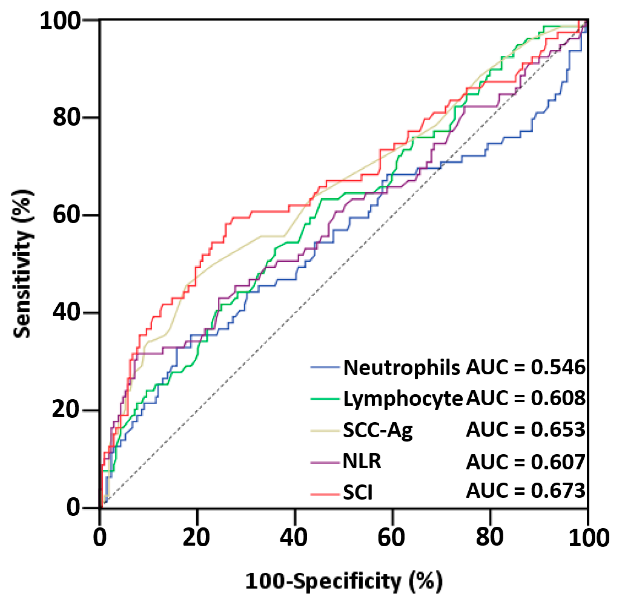
Figure 1. ROC curves for overall survival in patients with OSCC. Abbreviations: AUC, area under the curve; NLR, neutrophil/lymphocyte ratio; SCC-Ag, squamous cell carcinoma antigen; SCI, squamous cell carcinoma inflammatory index.
Cancers 15 02492 g001
Figure 2. Kaplan–Meier curves for (A) overall survival and (B) disease-free survival of patients with SCI of ≥3.45 and <3.45. Abbreviations: SCI, squamous cell carcinoma inflammatory index.
Cancers 15 02492 g002
Figure 3. Hazard ratios for SCI in stratified analysis. Abbreviations: DOI, depth of invasion; ENE, extranodal extension; HR, hazard ratio; PNI, perineural invasion; SCI, squamous cell carcinoma inflammatory index.
Cancers 15 02492 g003
Figure 4. (A) Nomogram for OS prediction based on independent prognostic factors in multivariable analysis. The corresponding points of each variable’s line segment indicate the degree of risk contributed by this variable. The sum of points from all variables yields the total points, which can be converted to the estimated 3- and 5-year OS probabilities by drawing a vertical line from the total points to the following survival axes. The nomogram’s calibration plots for the (B) 3- and (C) 5-year OS predictions. The 45° gray line indicates the perfect OS prediction, and the blue line indicates the nomogram’s predicted outcomes. Abbreviations: CCI, Charlson comorbidity index; M-D, moderately differentiated; P-D, poorly differentiated; SCI, squamous cell carcinoma inflammatory index; W-D, well differentiated.
Cancers 15 02492 g004
Table 1. Baseline characteristics of 288 patients with OSCC.
Characteristics
Age (years)
≥6598 (34.0%)
<65190 (66.0%)
Sex
Women26 (9.0%)
Men262 (91.0%)
Tumor location
Tongue110 (38.2%)
Buccal96 (33.4%)
Gingiva41 (14.2%)
Retromolar trigone15 (5.2%)
Mouth floor11 (3.8%)
Lip10 (3.5%)
Hard palate5 (1.7%)
AJCC stage
I61 (21.2%)
II39 (13.5%)
III40 (13.9%)
IV148 (51.4%)
T status
T179 (27.5%)
T253 (18.4%)
T339 (13.5%)
T4117 (40.6%)
N status
N0186 (64.6%)
N127 (9.4%)
N262 (21.5%)
N313 (4.5%)
PNI72 (25.0%)
ENE58 (20.1%)
LVI20 (6.9%)
Tumor differentiation
W-D and M-D253 (87.8%)
P-D35 (12.2%)
Closest margin
≥5 mm210 (72.9%)
<5 mm78 (27.1%)
DOI ≥ 10 mm133 (46.2%)
Treatment modality
Surgery only136 (47.3%)
Surgery + RT 39 (13.5%)
Surgery + CRT 113 (39.2%)
CCI
0157 (54.5%)
1 83 (28.8%)
≥248 (16.7%)
Personal Habits
Smoking241 (83.7%)
Alcohol drinking191 (66.3%)
Areca nut chewing233 (80.9%)
SCC-Ag (ng/mL), median (IQR)1.00 (0.70–1.70)
Neutrophil (×103/μL), median (IQR)5.01 (3.61–6.42)
Lymphocyte (×103/μL), median (IQR)2.01 (1.60–2.59)
SCI, median (IQR)2.45 (1.42–4.76)
Abbreviations: AJCC, American Joint Committee on Cancer; CCI, Charlson comorbidity index; CRT, chemoradiotherapy; DOI, depth of invasion; ENE, extranodal extension; IQR, interquartile range; LVI, lymphovascular invasion; M-D, moderately differentiated; OSCC, oral cavity squamous cell carcinoma; P-D, poorly differentiated; PNI, perineural invasion; RT, radiotherapy; SCC-Ag, squamous cell carcinoma antigen; SCI, squamous cell carcinoma inflammatory index; W-D, well differentiated.
Table 2. Comparison of the AUC values of SCI and its components.
IndexAUC95% CIpp a
Neutrophil0.546(0.464–0.628)0.228<0.001
Lymphocyte0.608(0.534–0.682)0.005<0.001
SCC-Ag0.653(0.578–0.728)<0.001<0.001
NLR0.607(0.528–0.687)0.005<0.001
SCI0.673(0.593–0.749)<0.001-
Abbreviations: AUC, area under the curve; CI, confidence interval; NLR, neutrophil-to-lymphocyte ratio; SCC-Ag, squamous cell carcinoma antigen; SCI, squamous cell carcinoma inflammatory index. a The AUC values between the SCI and the other factors were compared using the Z-test method. Mann–Whitney U test (Z-test: neutrophil: −6.798; lymphocyte: −6.285; SCC-Ag: −11.083; and NLR: −9.925).
Table 3. Clinicopathological features according to the cutoff value of SCI.
VariableNumber of Patients
SCI < 3.45, n = 188SCI ≥ 3.45, n =100p
Sex 0.191 a
Women20 (10.6%)6 (6.0%)
Men168 (89.4%)94 (94.0%)
Age 0.194 a
<65129 (68.6%)61 (61.0%)
≥6559 (31.4%)39 (39.0%)
AJCC stage <0.001 a
I–II91 (48.4%)9 (9.0%)
III–IV97 (51.6%)91 (91.0%)
T status <0.001 a
T1–T2113 (60.1%)19 (19.0%)
T3–T475 (39.9%)81 (81.0%)
N status
N0138 (73.4%)48 (48.0%)<0.001 a
N1–N350 (26.6%)52 (52.0%)
PNI
Absent152 (80.9%)64 (64.0%)0.002 a
Present36 (19.1%)36 (36.0%)
LVI
Absent183 (97.3%)85 (85.0%)<0.001 a
Present5 (2.7%)15 (15.0%)
ENE <0.001 a
Absent165 (87.8%)65 (65.0%)
Present23 (12.2%)35 (35.0%)
Tumor differentiation 0.145 a
W-D/M-D169 (89.9%)84 (84.0%)
P-D19 (10.1%)16 (16.0%)
Closest margin 0.562 a
≥5 mm 135 (71.8%)75 (75.0%)
<5 mm 53 (28.2%)25 (25.0%)
DOI ≥ 10 mm <0.001 a
No129 (68.6%)26 (26.0%)
Yes59 (31.4%)74 (74.0%)
Tumor location 0.215 a
Tongue76 (40.4%)34 (34.0%)
Buccal mucosa56 (29.8%)40 (40.0%)
Other56 (29.8%)26 (26.0%)
Personal habits 0.808 a
No exposure20 (10.6%)13 (13.0%)
One exposure10 (5.3%)6 (6.0%)
Two or all exposure158 (84.0%)81 (81.0%)
Treatment modality <0.001 a
Surgery111 (59.0%)25 (25.0%)
Surgery + RT26 (13.8%)13 (13.0%)
Surgery + CRT 51 (27.1%)62 (62.0%)
CCI 0.029 a
0 111 (59.0%)46 (46.0%)
1 53 (28.2%)30 (30.0%)
≥2 24 (12.8%)24 (24.0%)
Survival in months, median (IQR)49.00 (30.25–70.75)25.00 (13.00–48.00)<0.001 b
Abbreviations: AJCC, American Joint Committee on Cancer; CCI, Charlson comorbidity index; CRT, concurrent chemoradiotherapy; DOI, depth of invasion; ENE, extranodal extension; IQR, interquartile range; LVI, lymphovascular invasion; M-D, moderately differentiated squamous cell carcinoma; PNI, perineural invasion; P-D, poorly differentiated squamous cell carcinoma; RT, radiotherapy; SCI, squamous cell carcinoma inflammatory index; W-D, well differentiated squamous cell carcinoma. a chi-square test. b Mann–Whitney U test.
Table 4. Univariable analysis of the prognostic factors for OS and DFS.
VariableUnivariable Analysis (OS)Univariable Analysis (DFS)
HR (95% CI)pHR (95% CI)p
Sex
  WomenReference Reference
  Men1.584 (0.640–3.921)0.3201.316 (0.640–3.921)0.320
Age (years)
  <65Reference Reference
  ≥650.760 (0.468–1.236)0.2690.684 (0.465–1.006)0.053
AJCC stage
  IReference Reference
  II1.287 (0.392–4.219)0.6770.649 (0.305–1.379)0.261
  III1.010 (0.284–3.585)0.9880.938 (0.469–1.876)0.857
  IV5.831 (2.250–13.493)<0.0012.120 (1.313–3.422)0.002
Presence of PNI
  NoReference Reference
  Yes2.294 (1.453–3.622)<0.0011.336 (0.905–1.973)0.144
Presence of LVI
  NoReference Reference
  Yes3.707 (1.943–7.071)<0.0011.882 (1.010–3.505)0.046
Tumor differentiation
  W-D/M-DReference Reference
  P-D3.260 (1.935–5.492)<0.0012.224 (1.410–3.508)0.001
Treatment modality
  SurgeryReference Reference
  Surgery + RT1.400 (0.625–3.135)0.4140.854 (0.466–1.565)0.609
  Surgery + CRT3.300 (2.004–5.434)<0.0011.633 (1.131–2.357)0.009
Tumor location
  TongueReference Reference
  Buccal mucosa1.319 (0.776–2.240)0.3060.854 (0.466–1.565)0.609
  Other sites1.171 (0.672–2.040)0.5771.633 (0.531–2.357)0.439
Closest margin
  ≥ 5 mmReference Reference
  < 5 mm1.325 (0.826–2.124)0.2431.278 (0.881–1.854)0.196
Personal habits
  No exposureReference Reference
  One exposure2.213 (0.713–6.867)0.1692.220 (0.855–5.764)0.101
  Two or more exposure1.540 (0.668–3.551)0.3111.919 (0.972–3.791)0.060
CCI
  0Reference Reference
  11.057 (0.615–1.817)0.8410.682 (0.441–1.053)0.084
  ≥21.916 (1.121–3.273)0.0171.058 (0.672–1.667)0.808
SCC-Ag
  <1.65Reference Reference
  ≥1.653.521 (2.250–5.512)<0.0012.006 (1.390–2.893)<0.001
SCI
  <3.45Reference Reference
  ≥3.453.803 (2.419–5.980)<0.0011.900 (1.335–2.703)<0.001
NLR
  <4.51Reference Reference
  ≥4.514.465 (2.762–7.219)<0.0012.710 (1.783–4.118)<0.001
Abbreviations: AJCC, American Joint Committee on Cancer; CCI, Charlson comorbidity index; CI, confidence interval; CRT, chemoradiotherapy; DFS, disease-free survival; HR, hazard ratio; LVI, lymphovascular invasion; M-D, moderately differentiated; NPAR, neutrophil percentage-to-albumin ratio; OS, overall survival; P-D, poorly differentiated; PNI, perineural invasion; RT, radiotherapy; SCI, squamous cell carcinoma inflammatory index; W-D, well differentiated.
Table 5. Multivariable analysis of the prognostic factors for OS and DFS.
VariableSCC-Ag and NLR Model SCI Model
HR (95% CI)pHR (95% CI)p
Overall Survival
AJCC stage
IReference Reference
II
III
IV3.702 (2.028–6.758)<0.0013.935 (2.148–7.209)<0.001
Tumor differentiation
W-D/M-DReference Reference
P-D2.976 (1.738–5.095)<0.0012.855 (1.677–4.861)<0.001
CCI
0Reference Reference
1
≥21.265 (1.147–1.689)0.0211.309 (1.183–1.744)0.016
SCC-Ag
<1.65Reference
≥1.651.905 (1.164–3.118)0.011
SCI
<3.45 Reference
≥3.45 2.378 (1.356–3.735)0.002
NLR
<4.51Reference
≥4.512.611 (1.580–4.315)<0.001
Disease-free survival
AJCC stage
IReference Reference
II
III
IV2.326 (1.436–3.768)<0.0011.961 (1.320–2.913)<0.001
Tumor differentiation
W-D/M-DReference Reference
P-D2.238 (1.403–3.570)<0.0012.061 (1.302–3.262)0.002
SCC-Ag
<1.65Reference
≥1.651.546 (1.041–2.295)0.031
SCI
<3.45 Reference
≥3.45 2.219 (1.437–3.425)<0.001
NLR
<4.51Reference
≥4.512.051 (1.361–2.867)0.003
Abbreviations: AJCC, American Joint Committee on Cancer; CCI, Charlson comorbidity index; CI, confidence interval; HR, hazard ratio; M-D, moderately differentiated; NLR, neutrophil-to-lymphocyte ratio; P-D, poorly differentiated; SCC-Ag, squamous cell carcinoma antigen; SCI, squamous cell carcinoma inflammatory index; W-D, well differentiated.
Disclaimer/Publisher’s Note: The statements, opinions and data contained in all publications are solely those of the individual author(s) and contributor(s) and not of MDPI and/or the editor(s). MDPI and/or the editor(s) disclaim responsibility for any injury to people or property resulting from any ideas, methods, instructions or products referred to in the content.