Article

The Relationship between Oxidative Status and Radioiodine Treatment Qualification among Papillary Thyroid Cancer Patients

by
Angelika Buczyńska
1,*,
Iwona Sidorkiewicz
1,
Maria Kościuszko
2,
Agnieszka Adamska
2,
Katarzyna Siewko
2,
Janusz Dzięcioł
3,
Piotr Szumowski
4,
Janusz Myśliwiec
4,
Anna Popławska-Kita
2,*,† and
Adam Jacek Krętowski
1,2,†
1
Clinical Research Centre, Medical University of Bialystok, M. Skłodowskiej 24a, 15-276 Bialystok, Poland
2
Department of Endocrinology, Diabetology and Internal Medicine, Medical University of Bialystok, M. Skłodowskiej 24a, 15-276 Bialystok, Poland
3
Department of Human Anatomy, Medical University of Bialystok, ul. Mickiewicza 2A, 15-230 Bialystok, Poland
4
Nuclear Medicine, Medical University of Bialystok, M. Skłodowskiej 24a, 15-276 Bialystok, Poland
*
Authors to whom correspondence should be addressed.
These authors contributed equally to this work.
Cancers 2023, 15(9), 2436; https://doi.org/10.3390/cancers15092436
Submission received: 6 March 2023 / Revised: 13 April 2023 / Accepted: 21 April 2023 / Published: 24 April 2023
(This article belongs to the Special Issue Advances in Papillary Thyroid Cancer Research)

Abstract

:

Simple Summary

Studies analyzing the protein profile of thyroid tissue in patients with papillary thyroid cancer (PTC) have revealed disturbed metabolic pathways, including those related to oxidative status. This study aimed to assess the concentration of specific markers associated with oxidative homeostasis in PTC patients and their potential role as screening factors for radioiodine treatment (RAI) indication and further clinical management.

Abstract

Total oxidative status (TOS), total antioxidant capacity (TAC), tumor protein 53 (p53), nuclear factor kappa B (NF-κB), forkhead box protein O1 (FOXO), and sirtuin 1 (SIRT1) play crucial roles in oxidative homeostasis and the progression of papillary thyroid cancer (PTC), as previously demonstrated in the literature. Therefore, profiling these markers among PTC patients may be useful in determining their eligibility for radioiodine (RAI) treatment. Since treatment indications are based on multiple and dynamic recommendations, additional criteria for adjuvant RAI therapy are still needed. In our study, we evaluated the TOS, TAC, and serum concentrations of p53, NF-κB, FOXO, and SIRT1 to analyze the relationship between oxidative status and qualification for RAI treatment. For the purpose of this study, we enrolled 60 patients with PTC allocated for RAI treatment as the study group and 25 very low-risk PTC patients not allocated for RAI treatment as a reference group. The serum TOS and SIRT1 concentrations were significantly higher in the study group compared to the reference group (both p < 0.001), whereas the TAC and p53, NK-κB, and FOXO concentrations were significantly lower (all p < 0.05). We also demonstrated the diagnostic utility of TAC (AUC = 0.987), FOXO (AUC = 0.648), TOS (AUC = 0.664), SIRT1 (AUC = 0.709), p53 (AUC = 0.664), and NF-κB (AUC = 0.651) measurements as indications for RAI treatment based on American Thyroid Association recommendations. Our study revealed that oxidative status-related markers may become additional criteria for RAI treatment in PTC patients.

1. Introduction

Papillary thyroid cancer (PTC) is the most common malignant neoplasm arising from thyroid parenchymal cells [1,2]. According to the latest epidemiological data, the incidence rate of PTC is 16.1, and the mortality rate is 0.5 per 100,000 women and 0.3 per 100,000 men when adjusted for age [3,4]. In the United States, the incidence of PTC has more than tripled over the past three decades, with an annual percentage increase of approximately 4.4%. The incidence of PTC is higher in women than in men, with a female-to-male ratio of approximately 3:1. PTC is also more common in white individuals compared to other races [2,3,4].
Ultrasound-guided fine-needle aspiration biopsy (FNAB) is the gold standard for pre-operative assessment of PTC; nevertheless, up to 15% of the results are still inconsistent [5,6]. The clinical management of PTC patients is based on cancer progression, according to the American Thyroid Association (ATA) recommendations, and subsequent risk stratification [7]. A total thyroidectomy with radioactive iodine (RAI) treatment [8] is mostly recommended to eradicate potential residual disease and to improve prognosis [9,10,11,12]. However, several oxidative stress markers have been evaluated among patients who have been allocated for RAI, and significant disturbances in the oxidative status have been noted [13,14,15]. Assessing the relationship between oxidative status and PTC clinical management may reveal novel insights into RAI qualification [16]. The discrepancy between the production of free radicals and antioxidant defense has been shown to be associated with thyroid cancer. Muzza et al. demonstrated that the level of oxidative stress-related markers in PTC directly correlates with worse tumor presentation and higher tumor aggressiveness [17]. In our recent study on serum malondialdehyde concentration in PTC patients undergoing RAI, increased oxidative stress was demonstrated throughout the treatment procedure [18,19,20]. It has also been suggested that blood markers can predict early-stage RAI refractory PTC [21,22,23,24,25,26]. Our hypothesis was that there must be a relationship between oxidative stress and both the presence of PTC and distinct clinical management, including RAI.
Furthermore, tumor protein 53 (p53), nuclear factor kappa B (NF-κB), forkhead box protein O1 (FOXO), and sirtuin 1 (SIRT1) have all been shown to play important roles in regulating oxidative stress and the development of PTC [27,28,29,30,31,32]. Since many studies have demonstrated that increased oxidative stress in PTC is associated as a relevant risk factor, the level of oxidative stress-related markers in PTC has been shown to directly correlate with tumor aggressiveness [33]. Additionally, our recent study on serum malondialdehyde concentration in PTC patients undergoing radioactive iodine treatment (RAI) demonstrated increased oxidative stress throughout the treatment procedure [18]. Therefore, the measurement of parameters related to oxidative stress, including p53, NF-κB, FOXO, and SIRT1, may be useful for the clinical management of PTC patients, particularly during RAI qualification. These protein markers could be included in risk stratification criteria and used to develop personalized treatments that unify RAI recommendations.
In this study, the total oxidative status (TOS), total antioxidant capacity (TAC), and serum concentrations of p53, NF-κB, FOXO, and SIRT1 were measured to analyze the relationship between the oxidation–reduction status and qualification for RAI treatment. The study aimed to determine the potential diagnostic usefulness of these markers as indicative markers for RAI therapy.

2. Materials and Methods

2.1. Study Subjects

This research was conducted at the Department of Endocrinology, Diabetology, and Internal Medicine at the Medical University of Bialystok, Poland. All patients were diagnosed with PTC based on histopathological examinations, clinical laboratory tests, and ultrasound imaging. For this study, 60 patients who had been diagnosed with different stages of PTC, undergone total thyroidectomy, and demonstrated adjuvant indications for RAI therapy (multifocal carcinomas with angioinvasion and/or capsular infiltration, increased concentrations of thyroglobulin, Tg, and the thyroglobulin antibody, TgAb) were enrolled as the study group. Additionally, 25 PTC patients who had undergone total thyroidectomies but did not receive any recommendations for RAI therapy, qualified as very low-risk PTC patients (reference group), were also enrolled [9,34]. Therapeutic decisions in thyroid cancer were made within a multidisciplinary tumor board. The same team confirmed the pathological report in all patients to ensure consistency throughout the study. The exclusion criteria were as follows: any chronic diseases, ongoing inflammation, and additional treatment that may interfere with oxidative status.

2.2. Sample Collection and Measurement

Venous blood (5.5 mL) was obtained during the visit and centrifuged, which was followed by serum separation. The samples were then frozen at −80 °C.
The serum concentrations of TSH, free triiodothyronine (fT3), free thyroxine (fT4), 25-OH vitamin D (25-OH VIT D), Tg, and TgAb were measured using a Roche E411 device (Roche Diagnostics Ltd., Risch-Rotkreuz, Switzerland) via the electrochemiluminescence (ECLIA) method. The concentrations of triglyceride (TG), low-density lipoprotein (LDL), high-density lipoprotein (HDL), cholesterol (CHOL), C-reactive protein (CRP), and glucose were assayed using a Roche C111 device (Roche Diagnostics Ltd., Risch-Rotkreuz, Switzerland) via the enzymatic colorimetric method.
The TOS status was assessed using photometric immunodiagnostic assays (PerOx (TOS/TOC) Kit, KC5100, 64625 Bensheim, Germany), and the TAC status was determined using photometric assays (ImAnOx (TAS/TAC) Kit, KC5200, 64625 Bensheim, Germany). The concentrations of p53, NF-κB, FOXO, and SIRT1 were determined using enzyme-linked immunosorbent assays (ELISAs) (Enzyme-linked Immunosorbent Assay Kit, Cloud-Clone Corp., Wuhan, China; SEA928Hu, SEB824Hu, SEA764Hu and SEE912Hu, respectively), according to the manufacturer’s instructions. The samples and controls were measured in duplicate using a fully automated two-plate ELISA processing system (Dynex DS2, Chantilly, VA, USA) following the blind analysis method in the same run.

2.3. Statistical Analysis

Our statistical analyses were performed using the GraphPad Prism 9.0 software (GraphPad Software, Inc., San Diego, CA, USA). The preliminary statistical analysis (Shapiro–Wilk test) did not demonstrate a normal distribution for the data. Thus, nonparametric tests were used for the statistical analyses between the groups. In this paper, all the data are presented as the medians and quartiles. A Mann–Whitney U test for independent variables was used to examine the differences between the study and reference groups. Any correlations were determined using nonparametric Spearman’s tests. Values of p  <  0.05 were significant. In addition, the receiver operating characteristic (ROC) curves were determined using simultaneous sensitivity and specificity calculations.

2.4. Institutional Review Board Statement

The study was conducted according to the guidelines of the Declaration of Helsinki, and the procedures were approved by the Local Ethics Committee of the Medical University of Bialystok, Poland. Written informed consent was obtained from each participant (R-I-002/491/2019).

3. Results

3.1. Studied Population Characteristics

Sixty patients with incomplete resection of the thyroid gland, as confirmed by imaging tests, such as scintigraphy and/or ultrasound, were eligible for RAI based on ATA recommendations. These patients had multifocal carcinomas with angioinvasion and/or capsular infiltration, and increased concentrations of Tg and TgAb (study group).
The reference group consisted of 25 volunteers who had been diagnosed with very low-risk PTC and were not eligible for RAI treatment (Table 1).

3.2. Biochemical Profiling of the PTC Patients

First, the groups were compared in terms of lipid and thyroid hormone status, as well as other biochemical measurements. The concentrations of CHOL and LDL were found to be significantly higher among PTC patients who were allocated for RAI treatment, whereas the concentrations of 25-OH VIT D and HDL were lower compared to the reference group (all p < 0.05). Moreover, the concentrations of TG, CRP, glucose, TSH, fT3, fT4, Tg, and TgAb did not differ between the groups (all p > 0.05). Additionally, the TSH concentrations were suppressed in both groups due to the PTC treatments recommended (Table 2).

3.3. A Comparison of the Oxidative Status-Related Parameters between the Study and Reference Groups

In this study, we assessed TOC and TAC measurements as well as indirect oxidative status markers, such as p53, NF-κB, FOXO, and SIRT1, in PTC patients. The levels of oxidative status-related parameters differed significantly between the study and reference groups. The study group showed significantly increased TOC and SIRT1 concentrations compared to the reference group (p  <  0.05 and p  <  0.01, respectively). In addition, the study group demonstrated significant decreases in the TAC, p53, NF-κB, and FOXO concentrations compared to the reference group (all p  <  0.05) (Table 3, Figure 1).

3.4. The Association of the Oxidative Status-Related Parameters in PTC Patients

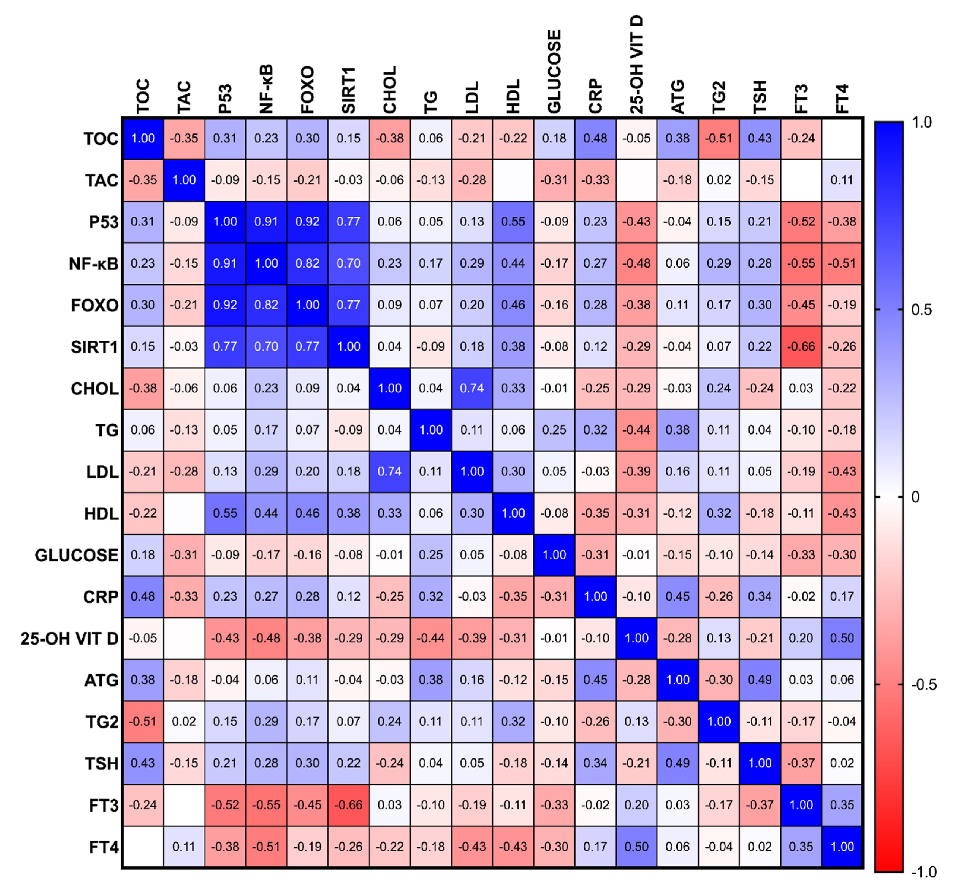
A Spearman regression was performed to study the relationship between the biochemical parameters. This study involved patients who had been diagnosed with different stages of PTC after thyroid resection; thus, a correlation assessment was performed on the total PTC group.
Interestingly, a positive correlation was demonstrated between the p53 and FOXO concentration and between the p53 and NF-κB concentration (r = 0.92 and r = 0.91, respectively; all p < 0.001). Furthermore, a positive correlation was observed between FOXO and NF-κB measurements (r = 0.82; p < 0.01). The SIRT1 concentrations were positively correlated with the p53, NF-κB, FOXO measurements, and negatively correlated with the fT3 concentration (r = 0.77, r = 0.70, r = 0.77, r = −0.66, all p < 0.01). Moreover, a positive correlation between the LDL and CHOL measurements was demonstrated (r = 0.74; p < 0.01). In our study, a negative correlation between the TOC and Tg levels was demonstrated (r = −0.51; p < 0.001). A positive correlation between p53 and HDL measurements, and a negative correlation between p53 and fT3, were found (r = 0.55, r = −0.52, respectively; all p < 0.01). The NF-κB measurement was found to be negatively correlated with fT3 and fT4 (r = -0.55; r = −0.51, respectively; all p < 0.05). A positive correlation between 25-OH vitamin D and fT4 was shown (r = 0.5; p < 0.01) (Figure 2).

3.5. Diagnostic Utility of the Studied Parameters for RAI Qualification, According to Current Recommendations

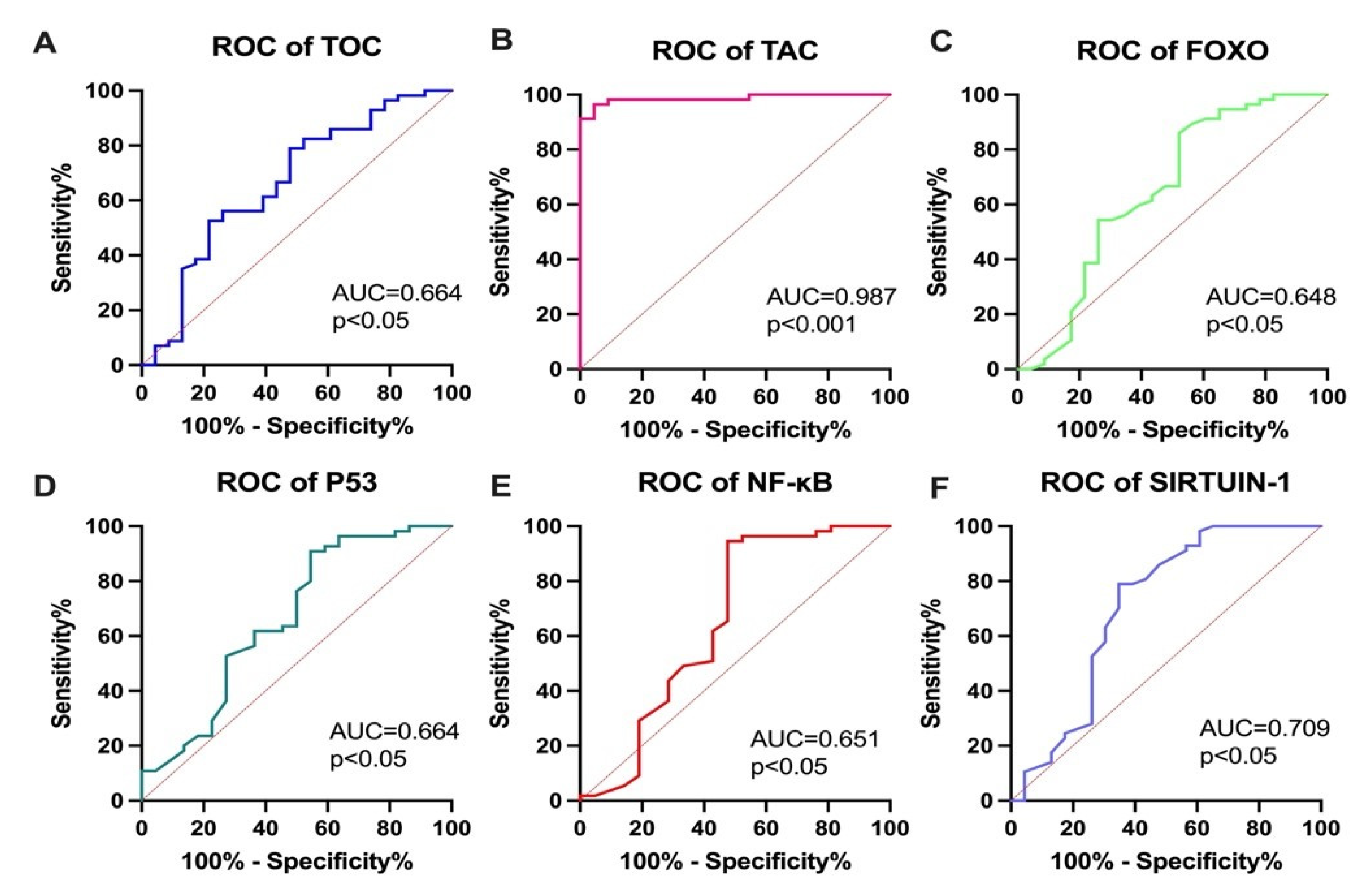
To assess the potential diagnostic utility of the parameters selected for RAI qualification, ROC curves were constructed. The highest diagnostic utility was demonstrated for the TAC measurement (AUC = 0.987; p < 0.001). Some of the parameters, such as FOXO (AUC = 0.648; p < 0.01), TOC (AUC = 0.664; p < 0.01), SIRT1 (AUC = 0.709; p < 0.01), p53 (AUC = 0.664; p < 0.05), and NF-κB (AUC = 0.651; p < 0.05), also demonstrated screening utility (p < 0.05) to support the current ATA recommendations (see Figure 3).

4. Discussion

The FNAB procedure plays a crucial role in the preoperative assessment of PTC. The TNM classification is based on the size of the primary tumor, the presence and number of lymph node metastases, and the number of distant metastases [7,35]. The PTC risk classification system consists of multiple stages and is based on a specific combination of criteria, including the size of the primary tumor, histological examinations, angioinvasion, the infiltration of tumor cells outside of the thyroid gland, tumor metastasis, and the age at the time of diagnosis. However, the choice of management should be made together with individual patients.
Furthermore, the postoperative management of PTC patients who undergo total thyroidectomy relies on the serial measurement of serum concentrations of Tg and TgAb. However, TgAb interference limits the utility of Tg as a tumor marker in TgAb-positive patients [36,37]. Therefore, incorporating novel biochemical determinations based on oxidative stress status profiling into further patient management procedures could lead to the simplification and clarification of guidelines for describing persistent or recurrent disease in PTC patients [38,39]. Thyrocytes require several protective systems against intracellular ROS to maintain thyroid hormone synthesis, and the imbalance of oxidative status in PTC cells is known to play a crucial role in PTC development and progression [40,41]. Additionally, evaluating circulating oxidative status-related markers can be used to fully elucidate the oxidative homeostasis in cancer patients [42]. Thus, serum TOS, TAC, and the concentrations of p53, NF-κB, FOXO, and SIRT1 were evaluated to analyze the relationship between oxidative status and qualification for RAI treatment.
Young et al. suggested that PTC tissue is characterized by an imbalanced oxidative status and increased lipid peroxidation [43]. Additionally, Song et al. demonstrated that downregulation of the FOXO pathway in PTC cell lines leads to enhanced proliferation and clonogenesis, as well as decreased apoptosis [44]. Moreover, the relationship between NF-κB activation and PTC development and progression has been confirmed [29,45]. Furthermore, an increased SIRT1 concentration in PTC tissues has been observed [46]. On the other hand, decreased p53 expression has been widely described in PTC [47,48]. Our study validated the serum concentration of selected oxidative stress-related markers in PTC patients to determine their potential utility in clinical management.
The patients allocated for adjuvant RAI treatment were characterized by decreased levels of FOXO, p53, and NF-κB, and increased concentrations of SIRT1 compared to the reference group. FOXO proteins are a family of transcription factors that play important roles in the regulation of gene expression involved in cell growth, proliferation, differentiation, longevity, DNA damage, and tumorigenesis [49]. FOXO also regulates mitochondrial function and adipocyte differentiation [50]. Interestingly, FOXO also acts as a tumor suppressor in cancer, which is in agreement with our results [51]. Despite the fact that patients allocated for RAI treatment presented increased TOC levels and decreased TAC levels, FOXO concentrations were also decreased, which could potentially be associated with PTC progression [52,53]. In future studies, the relationship between RAI and FOXO concentration should be determined [53]. Based on the protective effects of this protein in many processes, targeting its activity could lead to increased bioavailability for RAI and increased antioxidant protection [54].
The role of SIRT1 in cancer, including PTC patients, has been extensively studied over the past decade. Increased thyroid tissue SIRT1 expression has been found to be associated with cancer progression and worse prognosis for PTC patients [55]. In our study, increased SIRT1 concentrations were demonstrated in PTC patients allocated to RAI, which is consistent with the results presented by other authors [56]. Moreover, SIRT1 has been shown to reduce p53-mediated apoptosis, thus, promoting tumor development and progression [57]. The most important function of activated p53 is to induce cell cycle arrest, apoptosis, and DNA repair. In a study by Marcello et al., p53 expression was found to be higher in malignant tumors compared to benign thyroid lesions, indicating that the analysis of p53 activity could be useful for PTC clinical management [58]. Recent studies have also revealed that p53 can influence mitochondrial functions by changing from a normal to an abnormal state under different stress levels [59,60]. Deregulated p53 activity is particularly unfavorable when remnant thyroid tissue is found after PTC surgery [61]. Our study revealed decreased p53 concentrations among PTC patients who were allocated for RAI, suggesting that p53 could be considered an additional factor resulting from disturbed mitochondrial functionality and decreased capacity for DNA repair [62].
In addition, it has been demonstrated that NF-κB is involved in the regulation of many genes that are involved in inflammation, cell proliferation, and apoptosis, and its overactivation has been associated with cancer cell proliferation, invasion, and survival [63,64,65,66,67,68]. Moreover, several studies have reported a correlation between NF-κB activation and resistance to chemotherapy and radiotherapy in various cancers, including PTC [69,70]. Therefore, the evaluation of NF-κB concentrations could serve as a potential biomarker for RAI therapy response and may help in the selection of patients who could benefit from this treatment. However, further studies are required to elucidate the exact role of NF-κB in PTC and its potential utility in clinical practice.
Oxidative stress is a relevant risk factor linked to thyroid cancer development and progression; therefore, we hypothesized that measuring oxidative stress levels could be useful for qualifying patients for adjuvant cancer therapy. The TAC (AUC = 0.987; p < 0.001) and SIRT1 (AUC = 0.709; p < 0.01) measurements demonstrated the highest possible diagnostic utility, suggesting their usefulness in supporting clinical management and RAI qualification of PTC patients. Additionally, prior to RAI administration, the patients who were allocated for RAI treatment were characterized by increased TOC levels and decreased TAC levels. The oxidative–antioxidant status is related to RAI qualification and could be of significant clinical relevance. Moreover, positive correlations were demonstrated between the p53 and FOXO concentrations (r = 0.92; p < 0.001) and between the p53 and NF-κB concentrations (r = 0.91; p < 0.001). Furthermore, a very strong positive correlation was noticed between FOXO and NF-κB measurements (r = 0.82; p < 0.01). Since p53, FOXO, and NF-κB are involved in the PI3K/Akt pathway, their dysregulation may reflect cancer progression [68]. These data suggest a complex disorder of the oxidation–reduction status between PTC patients resulting from cellular and mitochondrial origin.
Interestingly, the TOC concentrations were also negatively correlated with Tg concentrations (r = −0.51; p < 0.001) in PTC patients. These outcomes emphasize the relationship between thyroid function markers and oxidative status [38,69]. Since oxidative stress has been found to be a valuable PTC risk factor, and RAI therapy is linked to increased oxidative status, the studied parameters could support recommendations in the case of unclear clinical features [70]. Further exploration of potential biomarkers and therapeutic targets could provide more detailed patient stratification and personalized treatments, which could improve the clinical management of PTC [67,71].
The integration of biochemical determinations could simplify and clarify the recommended guidelines for managing PTC patients. This study presents new directions in the diagnosis and treatment of PTC, but it also has several limitations. The small size of the study groups can be considered a weakness. Although the patients did not have any chronic diseases based on their medical history and laboratory measures, they were not thoroughly screened for diseases or medications that might affect the concentration of oxidative stress markers. Additionally, the presented results are preliminary, and multicenter cohort studies are needed to confirm the hypotheses. Furthermore, the study groups should be expanded to include an RAI-refractory group or benign thyroid lesions. Nonetheless, our study identified novel possibilities for further research, particularly as it demonstrated insufficient antioxidant properties among PTC patients.
Due to the fact that radiation not only eradicates cancer cells but also can affect nearby healthy cells, it can cause side effects, especially at higher activity levels of the administered 131I [66]. Therefore, assessing the oxidative status in postsurgical cancer restratification could enable a more personalized selection of the 131I doze activity. On the other hand, the risk stratification of PTC patients is a multistage process, and, thus, novel diagnostic tools that are useful in clinical management could lead to personalized treatment regimens [67]. Additional criteria that are useful in patient selection for RAI are needed because the ATA recurrence risk stratification can be challenging to apply in real-life practice [71].

5. Conclusions

Oxidative stress is a significant risk factor for PTC, and RAI therapy has been shown to disrupt the oxidative balance. Therefore, evaluating the oxidative status could help guide recommendations for adjuvant RAI treatment, particularly in cases with unclear clinical features. As PTC risk stratification is a multistep process, our study highlights the potential diagnostic value of TAC and SIRT1 measurements in guiding personalized adjuvant RAI treatments. Future research should focus on long-term follow-up studies with large cohort groups to enhance the clinical management of PTC patients.

Author Contributions

Conceptualization, A.B. and I.S.; methodology, A.B.; software, M.K. and A.A.; validation, A.B., P.S. and J.D.; formal analysis, I.S.; investigation, J.M.; resources, A.P.-K.; data curation, K.S.; writing—original draft preparation, A.B.; writing—review and editing, I.S. and A.J.K.; visualization, A.B.; supervision, A.P.-K.; project administration, A.P.-K.; funding acquisition, A.P.-K. All authors have read and agreed to the published version of the manuscript.

Funding

This research was funded by internal financing from the Medical University of Bialystok under grant number SUB/1/DN/22/002/1150.

Institutional Review Board Statement

This study was conducted in accordance with the Declaration of Helsinki and was approved by the Ethics Committee of the Medical University of Bialystok, Poland (R-I-002/491/2019). Informed consent was obtained from all the subjects who were involved in this study.

Informed Consent Statement

Informed consent was obtained from all subjects involved in the study.

Data Availability Statement

The data that support the findings of this study are available from the corresponding authors, upon reasonable request.

Conflicts of Interest

The authors declare no conflict of interest.

Abbreviations

25-OH VIT D25-OH vitamin D
AUCarea under the ROC curve
CHOLcholesterol
CRPC-reactive protein
FNABfine aspiration needle biopsy
fT3free triiodothyronine
fT4free thyroxine
FOXOforkhead box protein O1
HDLhigh-density lipoprotein
LDLlow-density lipoprotein
NF-κBnuclear factor kappa B
p53tumor protein 53
SIRT1sirtuin 1
PTCpapillary thyroid cancer
ROCreceived operatic characteristics
TACtotal antioxidant capacity
TOCtotal oxidative capacity
TGtriglyceride
Tgthyroglobulin
TgAbthyroglobulin antibody
TSHthyroid-stimulating hormone

References

  1. Miranda-Filho, A.; Lortet-Tieulent, J.; Bray, F.; Cao, B.; Franceschi, S.; Vaccarella, S.; Dal Maso, L. Thyroid cancer incidence trends by histology in 25 countries: A population-based study. Lancet Diabetes Endocrinol. 2021, 9, 225–234. [Google Scholar] [CrossRef] [PubMed]
  2. Rossi, E.D.; Pantanowitz, L.; Hornick, J.L. A worldwide journey of thyroid cancer incidence centred on tumour histology. Lancet Diabetes Endocrinol. 2021, 9, 193–194. [Google Scholar] [CrossRef] [PubMed]
  3. Sung, H.; Ferlay, J.; Siegel, R.L.; Laversanne, M.; Soerjomataram, I.; Jemal, A.; Bray, F. Global Cancer Statistics 2020: GLOBOCAN Estimates of Incidence and Mortality Worldwide for 36 Cancers in 185 Countries. CA Cancer J. Clin. 2021, 71, 209–249. [Google Scholar] [CrossRef] [PubMed]
  4. Li, Y.; Che, W.; Yu, Z.; Zheng, S.; Xie, S.; Chen, C.; Qiao, M.; Lyu, J. The Incidence Trend of Papillary Thyroid Carcinoma in the United States During 2003–2017. Cancer Control 2022, 29, 10732748221135447. [Google Scholar] [CrossRef] [PubMed]
  5. Rago, T.; Vitti, P. Risk Stratification of Thyroid Nodules: From Ultrasound Features to TIRADS. Cancers 2022, 14, 717. [Google Scholar] [CrossRef]
  6. Peng, S.; Liu, Y.; Lv, W.; Liu, L.; Zhou, Q.; Yang, H.; Ren, J.; Liu, G.; Wang, X.; Zhang, X.; et al. Deep learning-based artificial intelligence model to assist thyroid nodule diagnosis and management: A multicentre diagnostic study. Lancet Digit. Health 2021, 3, e250–e259. [Google Scholar] [CrossRef]
  7. Haugen, B.R.; Alexander, E.K.; Bible, K.C.; Doherty, G.M.; Mandel, S.J.; Nikiforov, Y.E.; Pacini, F.; Randolph, G.W.; Sawka, A.M.; Schlumberger, M.; et al. 2015 American Thyroid Association Management Guidelines for Adult Patients with Thyroid Nodules and Differentiated Thyroid Cancer The American Thyroid Association Guidelines Task Force on Thyroid Nodules and Differentiated Thyroid Cancer. THYROID 2016, 26, 1–133. [Google Scholar] [CrossRef]
  8. Wang, T.S.; Sosa, J.A. Thyroid surgery for differentiated thyroid cancer—Recent advances and future directions. Nat. Rev. Endocrinol. 2018, 14, 670–683. [Google Scholar] [CrossRef]
  9. Schlumberger, M.; Leboulleux, S. Current practice in patients with differentiated thyroid cancer. Nat. Rev. Endocrinol. 2020, 17, 176–188. [Google Scholar] [CrossRef]
  10. Li, M.; Zheng, R.; Dal Maso, L.; Zhang, S.; Wei, W.; Vaccarella, S. Mapping overdiagnosis of thyroid cancer in China. Lancet Diabetes Endocrinol. 2021, 9, 330–332. [Google Scholar] [CrossRef]
  11. Worden, F. Treatment strategies for radioactive iodine-refractory differentiated thyroid cancer. Ther. Adv. Med. Oncol. 2014, 6, 267–279. [Google Scholar] [CrossRef] [PubMed]
  12. Adamska, A.; Tomczuk-Bobik, P.; Popławska-Kita, A.B.; Siewko, K.; Buczyńska, A.; Szumowski, P.; Żukowski, Ł.; Myśliwiec, J.; Zbucka-Krętowska, M.; Adamski, M.; et al. Assessment of different markers of ovarian reserve in women with papillary thyroid cancer treated with radioactive iodine. Endocr. Connect. 2021, 10, 1283–1290. [Google Scholar] [CrossRef] [PubMed]
  13. Samimi, H.; Haghpanah, V. Molecular evidence reveals thyrotropin intervention enhances the risk of developing radioiodine-refractory differentiated thyroid carcinoma. Cancer Cell Int. 2022, 22, 61. [Google Scholar] [CrossRef] [PubMed]
  14. Pacini, F.; Fuhrer, D.; Elisei, R.; Handkiewicz-Junak, D.; Leboulleux, S.; Luster, M.; Schlumberger, M.; Smit, J.W. 2022 ETA Consensus Statement: What are the indications for post-surgical radioiodine therapy in differentiated thyroid cancer? Eur. Thyroid. J. 2022, 11, e210046. [Google Scholar] [CrossRef] [PubMed]
  15. Kim, J.; Gosnell, J.E.; Roman, S.A. Geographic influences in the global rise of thyroid cancer. Nat. Rev. Endocrinol. 2019, 16, 17–29. [Google Scholar] [CrossRef]
  16. Buczyńska, A.; Sidorkiewicz, I.; Krętowski, A.J.; Zbucka-Krętowska, M.; Adamska, A. Metformin Intervention-A Panacea for Cancer Treatment? Cancers 2022, 14, 1336. [Google Scholar] [CrossRef]
  17. Muzza, M.; Pogliaghi, G.; Colombo, C.; Carbone, E.; Cirello, V.; Palazzo, S.; Frattini, F.; Gentilini, D.; Gazzano, G.; Persani, L.; et al. Oxidative Stress Correlates with More Aggressive Features in Thyroid Cancer. Cancers 2022, 14, 5857. [Google Scholar] [CrossRef]
  18. Buczyńska, A.; Sidorkiewicz, I.; Rogucki, M.; Siewko, K.; Adamska, A.; Kościuszko, M.; Maliszewska, K.; Kozłowska, G.; Szumowski, P.; Myśliwiec, J.; et al. Oxidative stress and radioiodine treatment of differentiated thyroid cancer. Sci. Rep. 2021, 11, 17126. [Google Scholar] [CrossRef]
  19. Arfin, S.; Jha, N.K.; Jha, S.K.; Kesari, K.K.; Ruokolainen, J.; Roychoudhury, S.; Rathi, B.; Kumar, D. Oxidative Stress in Cancer Cell Metabolism. Antioxidants 2021, 10, 642. [Google Scholar] [CrossRef]
  20. Konukoǧlu, D.; Hüsrev Hatemi, H.; Arikan, S.; Demir, M.; Akçay, T. Radioiodine treatment and oxidative stress in thyroidectomised patients for differentiated thyroid cancers. Pharmacol. Res. 1998, 38, 311–315. [Google Scholar] [CrossRef]
  21. Xing, M. Oxidative stress: A new risk factor for thyroid cancer. Endocr. Relat. Cancer 2012, 19, C7–C11. [Google Scholar] [CrossRef] [PubMed]
  22. Liu, H.; Chen, Q.; Liu, B.; Wang, J.; Chen, C.; Sun, S. Blood Profiles in the Prediction of Radioiodine Refractory Papillary Thyroid Cancer: A Case-Control Study. J. Multidiscip. Healthc. 2023, 16, 535–546. [Google Scholar] [CrossRef] [PubMed]
  23. Rosário, P.W.; Batista, K.C.S.; Calsolari, M.R. Radioiodine-induced oxidative stress in patients with differentiated thyroid carcinoma and effect of supplementation with vitamins C and E and selenium (Antioxidants). Arch. Endocrinol. Metab. 2016, 60, 328–332. [Google Scholar] [CrossRef] [PubMed]
  24. Sengoz, T.; Kilic-Toprak, E.; Yaylali, O.; Kilic-Erkek, O.; Ozdemir, Y.; Oymak, B.; Senol, H.; Yuksel, D.; Kucukatay, V.; Bor-Kucukatay, M. Hemorheology and oxidative stress in patients with differentiated thyroid cancer following I-131 ablation/metastasis treatment. Clin. Hemorheol. Microcirc. 2019, 74, 209–221. [Google Scholar] [CrossRef]
  25. Klubo-Gwiezdzinska, J.; Jensen, K.; Bauer, A.; Patel, A.; Costello, J.; Burman, K.D.; Wartofsky, L.; Hardwick, M.J.; Vasko, V.V. The expression of translocator protein in human thyroid cancer and its role in the response of thyroid cancer cells to oxidative stress. J. Endocrinol. 2012, 214, 207–216. [Google Scholar] [CrossRef]
  26. Ameziane El Hassani, R.; Buffet, C.; Leboulleux, S.; Dupuy, C. Oxidative stress in thyroid carcinomas: Biological and clinical significance. Endocr. Relat. Cancer 2019, 26, R131–R143. [Google Scholar] [CrossRef]
  27. Brehar, A.C.; Brehar, F.M.; Bulgar, A.C.; Dumitrache, C. Genetic and epigenetic alterations in differentiatedthyroid carcinoma. J. Med. Life 2013, 6, 403. [Google Scholar]
  28. Kochman, J.; Jakubczyk, K.; Bargiel, P.; Janda-Milczarek, K. The Influence of Oxidative Stress on Thyroid Diseases. Antioxidants 2021, 10, 1442. [Google Scholar] [CrossRef]
  29. Li, W.; Ming, H.; Sun, D.; Li, W.; Wang, D.; Zhang, G.; Tan, J. The relationship between BRAFV600E, NF-κB and TgAb expression in papillary thyroid carcinoma. Pathol. Res. Pract. 2017, 213, 183–188. [Google Scholar] [CrossRef]
  30. Saljooghi, S.; Heidari, Z.; Saravani, M.; Rezaei, M.; Salimi, S. Association of FOXO1 Rs17592236 Polymorphism and Tumor Size in Papillary Thyroid Carcinoma. Rep. Biochem. Mol. Biol. 2022, 11, 216. [Google Scholar]
  31. Harb, O.A.; Kaf, R.M.; Taha, H.F.; Balata, S.A.; Hemeda, R.; Yehia, A.M.; Gertallah, L.M.; Embaby, A. Clinical, pathological and prognostic implications of USP22, SIRT1 and E-cadherin expression in papillary thyroid cancer (PTC) and adjacent non-neoplastic tissue. Surg. Exp. Pathol. 2019, 2, 22. [Google Scholar] [CrossRef]
  32. Morita, N.; Ikeda, Y.; Takami, H. Clinical significance of p53 protein expression in papillary thyroid carcinoma. World J. Surg. 2008, 32, 2617–2622. [Google Scholar] [CrossRef]
  33. Momesso, D.P.; Vaisman, F.; Yang, S.P.; Bulzico, D.A.; Corbo, R.; Vaisman, M.; Michael Tuttle, R. Dynamic Risk Stratification in Patients with Differentiated Thyroid Cancer Treated Without Radioactive Iodine. J. Clin. Endocrinol. Metab. 2016, 101, 2692. [Google Scholar] [CrossRef]
  34. Jarząb, B.; Dedecjus, M.; Lewiński, A.; Adamczewski, Z.; Bakuła-Zalewska, E.; Bałdys-Waligórska, A.; Barczyński, M.; Biskup-Frużyńska, M.; Bobek-Billewicz, B.; Bossowski, A.; et al. Diagnosis and treatment of thyroid cancer in adult patients—Recommendations of Polish Scientific Societies and the National Oncological Strategy. 2022 Update [Diagnostyka i leczenie raka tarczycy u chorych dorosłych—Rekomendacje Polskich Towarzystw Naukowych oraz Narodowej Strategii Onkologicznej. Aktualizacja na rok 2022]. Endokrynol. Pol. 2022, 73, 173–300. [Google Scholar] [CrossRef]
  35. Sapuppo, G.; Tavarelli, M.; Pellegriti, G. The new AJCC/TNM Staging System (VIII ed.) in papillary thyroid cancer: Clinical and molecular impact on overall and recurrence free survival. Ann. Transl. Med. 2020, 8, 838. [Google Scholar] [CrossRef]
  36. Spencer, C.; Petrovic, I.; Fatemi, S. Current Thyroglobulin Autoantibody (TgAb) Assays Often Fail to Detect Interfering TgAb that Can Result in the Reporting of Falsely Low/Undetectable Serum Tg IMA Values for Patients with Differentiated Thyroid Cancer. J. Clin. Endocrinol. Metab. 2011, 96, 1283–1291. [Google Scholar] [CrossRef]
  37. Spencer, C.A.; Petrovic, I.; Fatemi, S.; LoPresti, J. Serum Thyroglobulin (Tg) Monitoring of Patients with Differentiated Thyroid Cancer Using Sensitive (Second-Generation) Immunometric Assays Can Be Disrupted by False-Negative and False-Positive Serum Thyroglobulin Autoantibody Misclassifications. J. Clin. Endocrinol. Metab. 2014, 99, 4589. [Google Scholar] [CrossRef]
  38. Van Kinschot, C.M.J.; Peeters, R.P.; Van Den Berg, S.A.A.; Verburg, F.A.; Van Noord, C.; Van Ginhoven, T.M.; Visser, W.E. Thyroglobulin and thyroglobulin antibodies: Assay-dependent management consequences in patients with differentiated thyroid carcinoma. Clin. Chem. Lab. Med. 2022, 60, 756–765. [Google Scholar] [CrossRef]
  39. Algeciras-Schimnich, A. Thyroglobulin measurement in the management of patients with differentiated thyroid cancer. Crit. Rev. Clin. Lab. Sci. 2018, 55, 205–218. [Google Scholar] [CrossRef]
  40. Cazarin, J.; Dupuy, C.; Pires de Carvalho, D. Redox Homeostasis in Thyroid Cancer: Implications in Na+/I- Symporter (NIS) Regulation. Int. J. Mol. Sci. 2022, 23, 6129. [Google Scholar] [CrossRef]
  41. Kowaltowski, A.J. Strategies to detect mitochondrial oxidants. Redox Biol. 2019, 21, 101065. [Google Scholar] [CrossRef]
  42. Chan, X.C.Y.; Black, C.M.; Lin, A.J.; Ping, P.; Lau, E. Mitochondrial protein turnover: Methods to measure turnover rates on a large scale. J. Mol. Cell. Cardiol. 2015, 78, 54. [Google Scholar] [CrossRef]
  43. Young, O.; Crotty, T.; O’Connell, R.; O’Sullivan, J.; Curran, A.J. Levels of oxidative damage and lipid peroxidation in thyroid neoplasia. Head Neck 2010, 32, 750–756. [Google Scholar] [CrossRef]
  44. Song, H.M.; Song, J.L.; Li, D.F.; Hua, K.Y.; Zhao, B.K.; Fang, L. Inhibition of FOXO1 by small interfering RNA enhances proliferation and inhibits apoptosis of papillary thyroid carcinoma cells via Akt/FOXO1/Bim pathway. Onco. Targets. Ther. 2015, 8, 3565. [Google Scholar] [CrossRef]
  45. Palona, I.; Namba, H.; Mitsutake, N.; Starenki, D.; Podtcheko, A.; Sedliarou, I.; Ohtsuru, A.; Saenko, V.; Nagayama, Y.; Umezawa, K.; et al. BRAFV600E promotes invasiveness of thyroid cancer cells through nuclear factor kappaB activation. Endocrinology 2006, 147, 5699–5707. [Google Scholar] [CrossRef]
  46. Li, T.; Wang, G.; Li, Z.; Zhang, N.; Li, Y.; Zhao, Y.; Tian, X. SIRT1 Expression and BRAF V600E Mutation in Papillary Thyroid Cancer: Implications for Diagnosis and Prognosis. Res. Sq. 2020. [Google Scholar] [CrossRef]
  47. Wang, D.; Zheng, X.; Li, M. Correlation analysis between the pre-operative contrast-enhanced ultrasound parameters and biological characteristics of papillary thyroid carcinoma and associated risk factors for prognosis after radiofrequency ablation. Exp. Ther. Med. 2020, 20, 1575–1581. [Google Scholar] [CrossRef]
  48. McFadden, D.G.; Vernon, A.; Santiago, P.M.; Martinez-McFaline, R.; Bhutkar, A.; Crowley, D.M.; McMahon, M.; Sadow, P.M.; Jacks, T. p53 constrains progression to anaplastic thyroid carcinoma in a Braf-mutant mouse model of papillary thyroid cancer. Proc. Natl. Acad. Sci. USA 2014, 111, E1600–E1609. [Google Scholar] [CrossRef]
  49. Maiese, K. Forkhead transcription factors: New considerations for alzheimer’s disease and dementia. J. Transl. Sci. 2016, 2, 241. [Google Scholar] [CrossRef]
  50. Liu, L.; Tao, Z.; Zheng, L.D.; Brooke, J.P.; Smith, C.M.; Liu, D.; Long, Y.C.; Cheng, Z. FoxO1 interacts with transcription factor EB and differentially regulates mitochondrial uncoupling proteins via autophagy in adipocytes. Cell Death Discov. 2016, 2, 16066. [Google Scholar] [CrossRef]
  51. Liu, Y.; Ao, X.; Ding, W.; Ponnusamy, M.; Wu, W.; Hao, X.; Yu, W.; Wang, Y.; Li, P.; Wang, J. Critical role of FOXO3a in carcinogenesis. Mol. Cancer 2018, 17, 104. [Google Scholar] [CrossRef]
  52. Myatt, S.S.; Lam, E.W.F. The emerging roles of forkhead box (fox) proteins in cancer. Nat. Rev. Cancer 2007, 7, 847–859. [Google Scholar] [CrossRef]
  53. Lettieri-Barbato, D.; Ioannilli, L.; Aquilano, K.; Ciccarone, F.; Rosina, M.; Ciriolo, M.R. FoxO1 localizes to mitochondria of adipose tissue and is affected by nutrient stress. Metabolism 2019, 95, 84–92. [Google Scholar] [CrossRef]
  54. Jerome, M.S.; Kuthethur, R.; Kabekkodu, S.P.; Chakrabarty, S. Regulation of mitochondrial function by forkhead transcription factors. Biochimie 2022, 198, 96–108. [Google Scholar] [CrossRef]
  55. Ao, N.; Wang, L.; Liu, Y. Prognostic and clinicopathological significance of ubiquitin-specific protease 22 overexpression in cancers: Evidence from a meta-analysis. Onco. Targets. Ther. 2017, 10, 5533. [Google Scholar] [CrossRef]
  56. Yao, L.; Wang, Y. Bioinformatic Analysis of the Effect of the Sirtuin Family on Differentiated Thyroid Carcinoma. Biomed Res. Int. 2022, 2022, 5794118. [Google Scholar] [CrossRef]
  57. Lin, Z.; Yang, H.; Kong, Q.; Li, J.; Lee, S.M.; Gao, B.; Dong, H.; Wei, J.; Song, J.; Zhang, D.D.; et al. USP22 antagonizes p53 transcriptional activation by deubiquitinating Sirt1 to suppress cell apoptosis and is required for mouse embryonic development. Mol. Cell 2012, 46, 484–494. [Google Scholar] [CrossRef]
  58. Marcello, M.A.; Morari, E.C.; Cunha, L.L.; De Nadai Silva, A.C.; Carraro, D.M.; Carvalho, A.L.; Soares, F.A.; Vassallo, J.; Ward, L.S. P53 and expression of immunological markers may identify early stage thyroid tumors. Clin. Dev. Immunol. 2013, 2013, 846584. [Google Scholar] [CrossRef]
  59. Manzella, L.; Stella, S.; Pennisi, M.S.; Tirrò, E.; Massimino, M.; Romano, C.; Puma, A.; Tavarelli, M.; Vigneri, P. New Insights in Thyroid Cancer and p53 Family Proteins. Int. J. Mol. Sci. 2017, 18, 1325. [Google Scholar] [CrossRef]
  60. Dai, C.Q.; Luo, T.T.; Luo, S.C.; Wang, J.Q.; Wang, S.M.; Bai, Y.H.; Yang, Y.L.; Wang, Y.Y. p53 and mitochondrial dysfunction: Novel insight of neurodegenerative diseases. J. Bioenerg. Biomembr. 2016, 48, 337. [Google Scholar] [CrossRef]
  61. Madan, E.; Gogna, R.; Kuppusamy, P.; Bhatt, M.; Mahdi, A.A.; Pati, U. SCO2 Induces p53-Mediated Apoptosis by Thr845 Phosphorylation of ASK-1 and Dissociation of the ASK-1–Trx Complex. Mol. Cell. Biol. 2013, 33, 1285. [Google Scholar] [CrossRef] [PubMed]
  62. Omur, O.; Baran, Y. An update on molecular biology of thyroid cancers. Crit. Rev. Oncol. Hematol. 2014, 90, 233–252. [Google Scholar] [CrossRef] [PubMed]
  63. Chen, L.; Mu, Y.; Greene, W.C. Acetylation of RelA at discrete sites regulates distinct nuclear functions of NF-κB. EMBO J. 2002, 21, 6539. [Google Scholar] [CrossRef] [PubMed]
  64. Albensi, B.C. What is nuclear factor kappa B (NF-κB) doing in and to the mitochondrion? Front. Cell Dev. Biol. 2019, 7, 154. [Google Scholar] [CrossRef]
  65. Pacifico, F.; Mauro, C.; Barone, C.; Crescenzi, E.; Mellone, S.; Monaco, M.; Chiappetta, G.; Terrazzano, G.; Liguoro, D.; Vito, P.; et al. Oncogenic and anti-apoptotic activity of NF-kappa B in human thyroid carcinomas. J. Biol. Chem. 2004, 279, 54610–54619. [Google Scholar] [CrossRef]
  66. Shim, S.R.; Kitahara, C.M.; Cha, E.S.; Kim, S.J.; Bang, Y.J.; Lee, W.J. Cancer Risk After Radioactive Iodine Treatment for Hyperthyroidism: A Systematic Review and Meta-analysis. JAMA Netw. Open 2021, 4, e2125072. [Google Scholar] [CrossRef]
  67. Rogucki, M.; Buczyńska, A.; Krętowski, A.J.; Popławska-Kita, A. The Importance of miRNA in the Diagnosis and Prognosis of Papillary Thyroid Cancer. J. Clin. Med. 2021, 10, 4738. [Google Scholar] [CrossRef]
  68. Finnberg, N.; El-Deiry, W.S. Activating FOXO3a, NF-kappaB and p53 by targeting IKKs: An effective multi-faceted targeting of the tumor-cell phenotype? Cancer Biol. Ther. 2004, 3, 614–616. [Google Scholar] [CrossRef]
  69. Krajewska, J.; Jarząb, M.; Czarniecka, A.; Roskosz, J.; Kukulska, A.; Handkiewicz-Junak, D.; Puch, Z.; Wygoda, Z.; Paliczka-Cieślik, E.; Kropińska, A.; et al. Ongoing risk stratification for differentiated thyroid cancer (DTC)—Stimulated serum thyroglobulin (Tg) before radioiodine (RAI) ablation, the most potent risk factor of cancer recurrence in M0 patients. Endokrynol. Pol. 2016, 67, 2–11. [Google Scholar] [CrossRef]
  70. Dhama, K.; Latheef, S.K.; Dadar, M.; Samad, H.A.; Munjal, A.; Khandia, R.; Karthik, K.; Tiwari, R.; Yatoo, M.I.; Bhatt, P.; et al. Biomarkers in stress related diseases/disorders: Diagnostic, prognostic, and therapeutic values. Front. Media SA 2019, 6, 91. [Google Scholar] [CrossRef]
  71. Sparano, C.; Moog, S.; Hadoux, J.; Dupuy, C.; Al Ghuzlan, A.; Breuskin, I.; Guerlain, J.; Hartl, D.; Baudin, E.; Lamartina, L. Strategies for Radioiodine Treatment: What’s New. Cancers 2022, 14, 3800. [Google Scholar] [CrossRef] [PubMed]
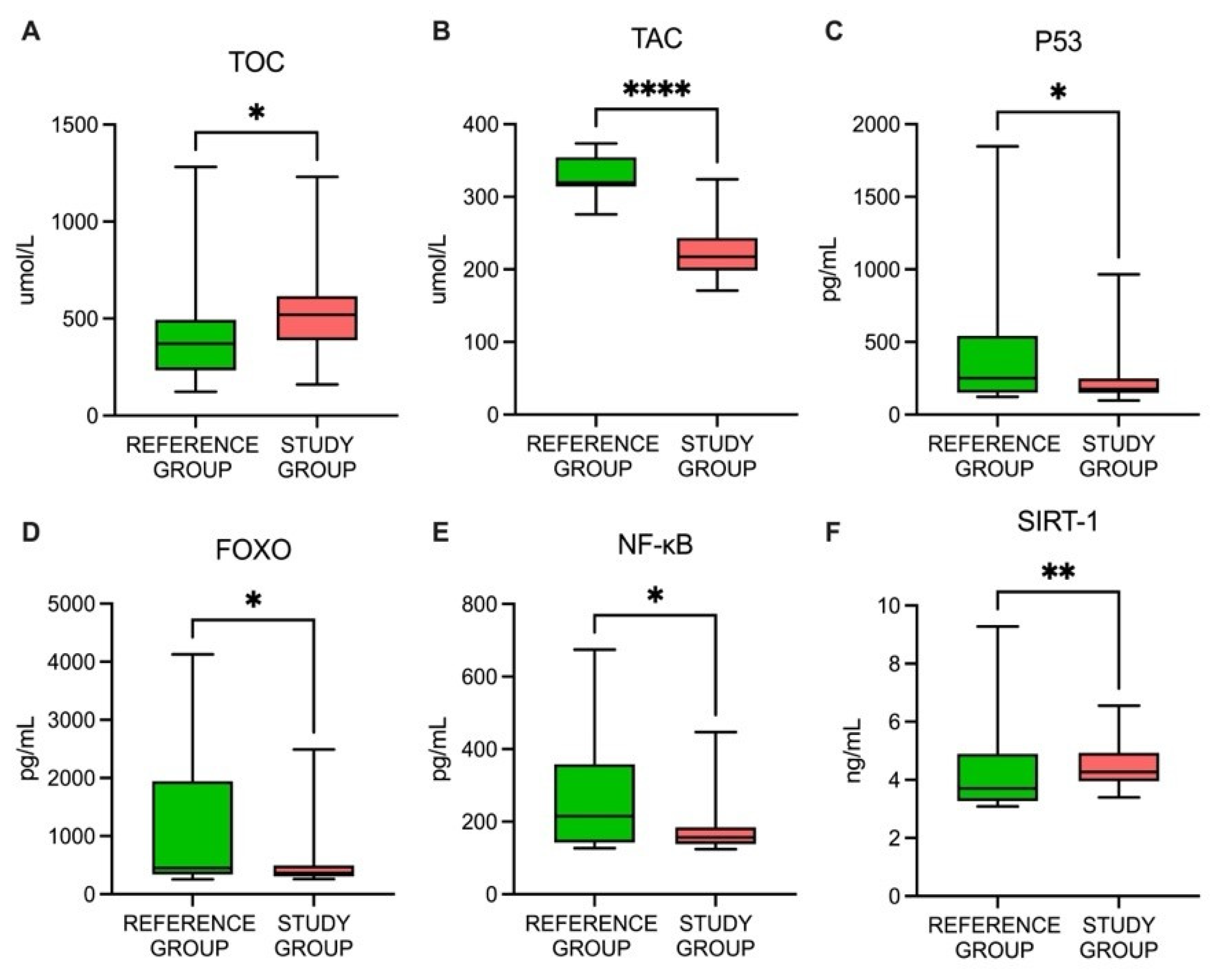
Figure 1. The profiles of the studied parameters: (A) TOC, total oxidative capacity; (B) TAC, total antioxidant capacity; (C) p53, tumor protein 53; (D) FOXO, forkhead box protein O1; (E) NF-κB, nuclear factor kappa B; (F) SIRT1, sirtuin 1; * p < 0.05; ** p < 0.01; **** p < 0.001.
Cancers 15 02436 g001
Figure 2. The correlation profiles of the parameters studied: FOXO, forkhead box protein O1; NF-κB, nuclear factor kappa B; p53, tumor protein 53; SIRT1, sirtuin 1; TAC, total antioxidant capacity; TOC, total oxidative capacity; CHOL, cholesterol; CRP, C-reactive protein; PTC, differentiated thyroid cancer; fT3, free triiodothyronine; fT4, free thyroxine; HDL, high-density lipoprotein; LDL, low-density lipoprotein; TG, triglyceride; TG, thyroglobulin; TGAb, thyroglobulin antibody; TSH, thyroid-stimulating hormone; 25-OH VIT D, 25-OH vitamin D.
Cancers 15 02436 g002
Figure 3. The ROC curves of the studied parameters: (A) TOC, total oxidative capacity; (B) TAC, total antioxidant capacity; (C) FOXO, forkhead box protein O1; (D); p53, tumor protein 53; (E) NF-κB, nuclear factor kappa B; (F) SIRT1, sirtuin 1.
Cancers 15 02436 g003
Table 1. The characteristics of the study and reference groups.
Study GroupReference Group p-Value
Number of patients6025
Median age (upper and lower quartiles)54 (51.41; 64.22)51 (50.21; 62.58)0.054
SexM: 18M: 80.052
F: 42F: 170.051
Menopausal status
Premenopausal950.064
Postmenopausal33120.082
Stage (TNM)pT1a(m): 11
pT1b: 15
pT1b(m): 6
pT2: 16
pT3/pT4: 12
pT1a: 25<0.001
F, female; M, male; (m), multifocal; p, pathological; TNM, the tumor, node, and metastasis classification of differentiated and anaplastic thyroid cancer (based on the characteristics of the primary tumor site (pT)); pT1a, the size of the largest thyroid tumor ≤ 1 cm; pT1b, the size of the largest thyroid tumor > 1 cm but ≤ 2 cm; pT2, the size of the largest thyroid tumor > 2 cm but ≤ 4 cm; pT3/pT4, the size of the largest thyroid tumor > 4 cm with gross extrathyroidal extensions.
Table 2. The biochemical parameter profiles of the study and reference groups.
Study GroupReference Group
Unit25% PercentileMedian75% PercentileRange25% PercentileMedian75% PercentileRangep-Value
CHOLmg/dL189.51216.00240.00142–463179.23142.05225.01101–2720.018
LDLmg/dL118.50138.04168.5270–31474.30107.92137.4255.7–164.8<0.001
TGmg/dL78.52103.07161.4540–49984.41107.91166.0045–4110.510
HDLmg/dL45.0653.0162.1431–11453.7358.9472.5739.26–102.80.048
CRPmg/L15.8522.4129.357.2–82.61.001.342.830.38–4.120.053
GLUCOSEmg/dL88.0794.1499.5369–24885.1292.2297.1577–1170.297
25-OH VIT Dng/mL15.8122.1729.357.2–82.623.3425.9236.8617.4–54.20.019
TSHµIU/mL0.150.602.320.1–68.550.120.360.790.075–1.550.318
fT3pg/mL2.212.563.011–6.272.472.632.861–3.270.754
fT4ng/dL0.961.191.370.4–2.141.051.191.230.4–1.80.991
Tgng/mL0.511.102.530.04–37.050.040.090.290.04–1.130.078
TgAbIU/mL0.852.026.670–1850.611.822.890.1–5.230.131
CHOL, cholesterol; CRP, C-reactive protein; PTC, differentiated thyroid cancer; fT3, free triiodothyronine; fT4, free thyroxine; HDL, high-density lipoprotein; LDL, low-density lipoprotein; TG, triglyceride; Tg, thyroglobulin; TgAb, thyroglobulin antibody; TSH, thyroid-stimulating hormone; 25-OH VIT D, 25-OH vitamin D.
Table 3. A comparison between the results from the study and reference groups for the studied parameters.
UnitStudy GroupReference Groupp-Value
25% PercentileMedian75% PercentileRange25% PercentileMedian75% PercentileRange
TOCUmol/L387.5519615.5161–1231233371493123–12820.020
TACUmol/L198.7217.4243.4170.8–324.3313.9317.2353.3271.4–373.3<0.001
p53pg/mL14917624998–966150.5250543.3123–18470.025
NF-κBpg/mL138157184124–447142215358127–6740.043
FOXOng/mL0.30.370.50.26–2.490.340.451.940.26–4.130.039
SIRT1ng/mL3.964.264.933.4–6.563.273.714.93.09–9.280.003
FOXO, forkhead box protein O1; NF-κB, nuclear factor kappa B; p53, tumor protein 53; SIRT1, sirtuin 1; TAC, total antioxidant capacity; TOC, total oxidative capacity.
Disclaimer/Publisher’s Note: The statements, opinions and data contained in all publications are solely those of the individual author(s) and contributor(s) and not of MDPI and/or the editor(s). MDPI and/or the editor(s) disclaim responsibility for any injury to people or property resulting from any ideas, methods, instructions or products referred to in the content.