
Endoscopic Ultrasound Advanced Techniques for Diagnosis of Gastrointestinal Stromal Tumours
 , ,
, , 
 ,
,  ,
,  , and
, and
Abstract
Simple Summary
Abstract
1. Introduction
2. Endoscopic and EUS-Based Findings
3. Contrast-Enhanced Harmonic EUS
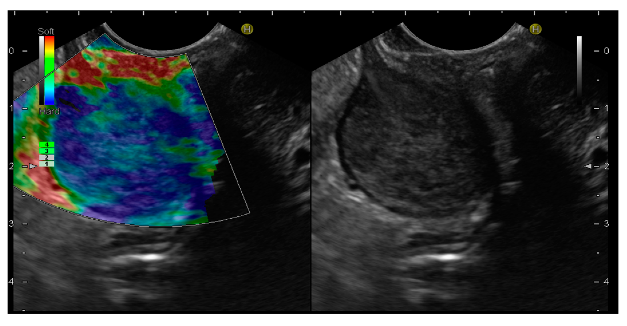
4. EUS-Elastography
5. EUS-Guided Fine-Needle Tissue Acquisition
6. Artificial Intelligence
7. Conclusions
Author Contributions
Funding
Conflicts of Interest
References
- Joensuu, H.; Hohenberger, P.; Corless, C.L. Gastrointestinal stromal tumour. Lancet 2013, 382, 973–983. [Google Scholar] [CrossRef] [PubMed]
- Sircar, K.; Hewlett, B.R.; Huizinga, J.D.; Chorneyko, K.; Berezin, I.; Riddel, R.H. Interstizial cells of Cajal as precursor of gastrointestinal stromal tumors. Am. J. Surg. Pathol. 1999, 23, 377–389. [Google Scholar] [PubMed]
- van Roggen, J.F.G.; van Velthunysen, M.L.; Hogendoorn, P.C. The Histopathological differential diagnosis of gastrointestinal stromal tumors. J. Clin. Pathol. 2001, 54, 96–102. [Google Scholar] [CrossRef] [PubMed]
- Wong, D.W.; Lupton, S.C.; Bhatt, L.; Gross, L.; Tanière, P.; Peake, D.R.; Spooner, D.; Geh, J.I. Use in imatinib mesylate in gastrointestinal stromal tumors: Pan-Birmingham Cancer Network experience. Clin. Oncol. 2008, 20, 517–522. [Google Scholar]
- Maleshwari, V.; Alam, K.; Varshney, M.; Jain, A.; Siddiqui, F.A.; Bhargava, S. Fine needle aspiration diagnosis of Gist: A diagnostic dilemma. Diagn. Cytopathol. 2012, 40, 834–838. [Google Scholar] [CrossRef]
- Todaro, P.; Crinò, S.F.; Pallio, S.; Fazzari, C.; Consolo, P.; Tuccari, G. Gastrointestinal stromal tumours of the stomach: Cytological and immunocytochemical diagnostic features of two cases diagnosed by endoscopic ultrasound-guided fine needle aspiration. Oncol. Lett. 2013, 5, 1862–1866. [Google Scholar] [CrossRef]
- Von Mehren, M.; Joensuu, H. Gastrointestinal Stromal Tumors. J. Clin. Oncol. 2018, 36, 136–143. [Google Scholar] [CrossRef]
- Søreide, K.; Sandvik, O.M.; Søreide, J.A.; Giljaca, V.; Jureckova, A.; Bulusu, V.R. Global epidemiology of gastrointestinal stromal tumours (GIST): A systematic review of population-based cohort studies. Cancer Epidemiol. 2016, 40, 39–46. [Google Scholar] [CrossRef]
- Miettinen, M.; Lasota, J. Gastrointestinal stromal tumour.Review on morphology, molecular pathology, prognosis, and differential diagnosis. Arch. Pathol. Lab. Med. 2006, 130, 1466–1478. [Google Scholar] [CrossRef]
- Miettinen, M.; Sobin, L.H.; Lasota, J. Gastrointestinal stromal tumours of the stomach: A clinicopathologic, immunoihistochemical, and molecular genetic study of 1765 cases with long-term follow up. Am. J. Surg. Pathol. 2005, 29, 52–68. [Google Scholar] [CrossRef]
- Joensuu, H. Risk stratification of patients diagnosed with gastrointestinal stromal tumor. Hum. Pathol. 2008, 39, 1411–1419. [Google Scholar] [CrossRef]
- Tran, T.; Davila, J.A.; El-Serag, H.B. The epidemiology of malignant gastrointestinal stromal tumors: An anlysis of 1458 cases from 1992 to 2000. Am. J. Gastroenterol. 2005, 100, 162–168. [Google Scholar] [CrossRef]
- Parab, T.M.; DeRogatis, M.J.; Boaz, A.M.; Grasso, S.A.; Issack, P.S.; Duarte, D.A.; Urayeneza, O.; Vahdat, S.; Qiao, J.H.; Hinika, G.S. Gastrointestinal stromal tumors: A comprehensive review. J. Gastrointest. Oncol. 2019, 10, 144–154. [Google Scholar] [CrossRef]
- Peng, C.Y.; Xu, G.F. Evaluation of preoperative endoscopic ultrasonography for diagnosis and invasive risk assessment of gastric gastrointestinal stromal tumors: A single center retrospective study. Chin. J. Digest. Endosc. 2015, 32, 361–366. [Google Scholar]
- Nilsson, B.; Bumming, P.; Meis-Kindblom, J.M.; Odén, A.; Dortok, A.; Gustavsson, B.; Sablinska, K.; Kindblom, L.G. Gastrointestinal Stromal tumors: The incidence, prevalence, clinical course, and prognostication in the preimatinib mesylate era-a population-based study in western Sweden. Cancer 2005, 103, 821–829. [Google Scholar] [CrossRef] [PubMed]
- West, R.B.; Corless, C.L.; Chen, X.; Rubin, B.P.; Subramanian, S.; Montgomery, K.; Zhu, S.; Ball, C.A.; Nielsen, T.O.; Patel, R.; et al. The novel marker, DOGI, is expressed ubiquitously in gastrointestinal stromal tumors irrespective of Kit or PDGFRA mutation status. Am. J. Pathol. 2004, 165, 107–113. [Google Scholar] [CrossRef] [PubMed]
- Nishida, T.; Sakai, Y.; Takagi, M.; Ozaka, M.; Kitagawa, Y.; Kurokawa, Y.; Masuzawa, T.; Naito, Y.; Kagimura, T.; Hirota, S.; et al. Adherence to the guidelines and the pathological diagnosis of high-risk gastrointestinal stromal tumors in the real world. Gastric. Cancer 2020, 23, 118–125. [Google Scholar] [CrossRef] [PubMed]
- Sepe, P.S.; Brugge, W.R. A guide for the diagnosis and management of gastrointestinal stromal tumors. Nat. Rev. Gastroenterol. Hepatol. 2009, 6, 363–371. [Google Scholar] [CrossRef] [PubMed]
- Akahoshi, K.; Oya, M. Gastrointestinal Stromal tumor of the stomach: How to manage? World. J. Gastrointest. Endosc. 2010, 2, 271–277. [Google Scholar] [CrossRef] [PubMed]
- Rubin, B.P.; Heinrich, M.C.; Corless, C.L. Gastrointestinal Stromal Tumour. Lancet 2007, 369, 1731–1741. [Google Scholar] [CrossRef] [PubMed]
- Plaat, B.E.; Hollema, H.; Molenar, W.M.; Torn Broers, G.H.; Pijpe, J.; Mastik, M.F.; Hoekstra, H.J.; van den Berg, E.; Scheper, R.J.; van der Graaf, W.T. Soft tissue leiomyosarcomas and malignant gastrointestinal stromal tumors: Differences in clinical outcome and expression of multidrug resistance proteins. J. Clin. Oncol. 2000, 18, 3220–3222. [Google Scholar]
- Facciorusso, A.; Di Maso, M.; Serviddio, G.; Vendemiale, G.; Spada, C.; Costamagna, G.; Muscatiello, N. Factors Associated with Recurrence of Advanced Colorectal Adenoma after Endoscopic Resection. Clin. Gastroenterol. Hepatol. 2016, 14, 1148–1154. [Google Scholar] [CrossRef] [PubMed]
- Standards of Practice Committee; Faulx, A.L.; Kothari, S.; Acosta, R.D.; Agrawal, D.; Bruining, D.H.; Chandrasekhara, V.; Eloubeidi, M.A.; Fanelli, R.D.; Gurudu, S.R.; et al. The role of endoscopy in subepithelial lesions of the GI tract. Gastrointest. Endosc. 2017, 85, 1117–1132. [Google Scholar] [CrossRef]
- Yoshinaga, S.; Hilmi, I.N.; Kwek, B.E.; Hara, K.; Goda, K. Current status of endoscopic ultrasound for the upper gastrointestinal tract in Asia. Dig. Endosc. 2015, 27, 2–10. [Google Scholar] [PubMed]
- Lisotti, A.; Napoleon, B.; Facciorusso, A.; Cominardi, A.; Crinò, S.F.; Brighi, N.; Gincul, R.; Kitano, M.; Yamashita, Y.; Marchegiani, G.; et al. Contrast-enhanced EUS for the characterization of mural nodules within pancreatic cystic neoplasms: Systematic review and meta-analysis. Gastrointest. Endosc. 2021, 94, 881–889. [Google Scholar] [CrossRef]
- Facciorusso, A.; Mohan, B.P.; Crinò, S.F.; Ofosu, A.; Ramai, D.; Lisotti, A.; Chandan, S.; Fusaroli, P. Contrast-enhanced harmonic endoscopic ultrasound-guided fine-needle aspiration versus standard fine-needle aspiration in pancreatic masses: A meta-analysis. Expert. Rev. Gastroenterol. Hepatol. 2021, 15, 821–828. [Google Scholar] [CrossRef]
- Crinó, S.F.; Brandolese, A.; Vieceli, F.; Paiella, S.; Bellocchi, M.C.C.; Manfrin, E.; Bernardoni, L.; Sina, S.; D‘Onofrio, M.; Marchegiani, G.; et al. Endoscopic Ultrasound Features Associated with Malignancy and Aggressiveness of Nonhypovascular Solid Pancreatic Lesions: Results from a Prospective Observational Study. Ultraschall. Med. 2021, 42, 167–177. [Google Scholar] [CrossRef]
- Facciorusso, A.; Crinò, S.F.; Ramai, D.; Ofosu, A.; Muscatiello, N.; Mangiavillano, B.; Lamonaca, L.; Lisotti, A.; Fusaroli, P.; Gkolfakis, P.; et al. Comparison between endoscopic ultrasound-guided fine-needle biopsy and bite-on-bite jumbo biopsy for sampling of subepithelial lesions. Dig. Liver Dis. 2022, 54, 676–683. [Google Scholar] [CrossRef]
- Gkolfakis, P.; Crinò, S.F.; Tziatzios, G.; Ramai, D.; Papaefthymiou, A.; Papanikolaou, I.S.; Triantafyllou, K.; Arvanitakis, M.; Lisotti, A.; Fusaroli, P.; et al. Comparative diagnostic performance of end-cutting fine-needle biopsy needles for EUS tissue sampling of solid pancreatic masses: A network meta-analysis. Gastrointest. Endosc. 2022, 95, 1067–1077. [Google Scholar] [CrossRef]
- Crinò, S.F.; Bernardoni, L.; Manfrin, E.; Parisi, A.; Gabbrielli, A. Endoscopic ultrasound features of pancreatic schwannoma. Endosc. Ultrasound. 2016, 5, 396–398. [Google Scholar] [CrossRef]
- Deprez, P.; Moons, L.; O’Toole, D.; Gincul, R.; Seicean, A.; Pimentel-Nunes, P.; Fernández-Esparrach, G.; Polkowski, M.; Vieth, M.; Borbath, I.; et al. Endoscopic management of subepithelial lesions including neuroendocrine neoplasms: European Society of Gastrointestinal Endoscopy (ESGE) Guideline. Endoscopy 2022, 54, 412–429. [Google Scholar] [CrossRef] [PubMed]
- Goto, O.; Kaise, M.; Iwakiri, K. Advancements in the Diagnosis of Gastric subepithelial Tumors. Gut Liver 2022, 16, 321–330. [Google Scholar] [CrossRef] [PubMed]
- Moon, K.S.; Young, K.E.; Woong, C.J. Predictive factors for differentiating gastrointestinal stromal tumors from leiomyomas on endoscopic ultrasonography finding in patients with Gastric subepitelial tumors: A Multicenter Retrospective Study. Clin. Endosc. 2021, 54, 872–880. [Google Scholar]
- Melita, G.; Pallio, S.; Tortora, A.; Crinò, S.F.; Macrì, A.; Dionigi, G. Diagnostic and Interventional Role of Endoscopic Ultrasonography for the Management of Pancreatic Neuroendocrine Neoplasms. J. Clin. Med. 2021, 10, 2638. [Google Scholar] [CrossRef] [PubMed]
- Futo, Y.; Saito, S.; Miyato, H.; Sadatomo, A.; Kaneko, Y.; Kono, Y.; Matsubara, D.; Horie, H.; Lefor, A.K.; Sata, N. Duodenal gastrointestinal stromal tumors appear similar to pancreatic neuroendocrine tumors: A case report. Int. J. Surg. Case Rep. 2018, 53, 358–361. [Google Scholar] [CrossRef] [PubMed]
- Buscaglia, J.M.; Nagula, S.; Jayaraman, V.; Robbins, D.H.; Vadada, D.; Gross, S.A.; DiMaio, C.J.; Pais, S.; Patel, K.; Sejpal, D.V.; et al. Diagnostic yield and safety of jumbo biopsy forceps in patients with subepithelial lesions of the upper and lower GI tract. Gastrointest. Endosc. 2012, 75, 1147–1152. [Google Scholar] [CrossRef]
- Cantor, M.J.; Davila, R.E.; Faigel, D.O. Yield of tissue sampling for subepithelial lesions evaluated by EUS: A comparison between forceps biopsies and endoscopic submucosal resection. Gastrointest. Endosc. 2006, 64, 29–34. [Google Scholar] [CrossRef]
- Minoda, Y.; Chinen, T.; Osoegawa, T.; Itaba, S.; Haraguchi, K.; Akiho, H.; Aso, A.; Sumida, Y.; Komori, K.; Ogino, H.; et al. Superiority of mucosal incision-assisted biopsy over ultrasound-guided fine needle aspiration biopsy in diagnosing small gastric subepithelial lesions: A propensity score matching analysis. BMC Gastroenterol. 2020, 20, 19. [Google Scholar] [CrossRef]
- Tacelli, M.; Bina, N.; Crinò, S.F.; Facciorusso, A.; Celsa, C.; Vanni, A.S.; Fantin, A.; Antonini, F.; Falconi, M.; Monica, F.; et al. Reliability of grading preoperative pancreatic neuroendocrine tumors on EUS specimens: A systematic review with meta-analysis of aggregate and individual data. Gastrointest. Endosc. 2022, 96, 898–908. [Google Scholar] [CrossRef]
- Crinò, S.F.; Bellocchi, M.C.C.; Di Mitri, R.; Inzani, F.; Rimbaș, M.; Lisotti, A.; Manfredi, G.; Teoh, A.Y.B.; Mangiavillano, B.; Sendino, O.; et al. Wet-suction versus slow-pull technique for endoscopic ultrasound-guided fine-needle biopsy: A multicenter, randomized, crossover trial. Endoscopy 2022, 27. [Google Scholar] [CrossRef]
- Mangiavillano, B.; Crinò, S.F.; Facciorusso, A.; Di Matteo, F.; Barbera, C.; Larghi, A.; Rizzatti, G.; Carrara, S.; Spadaccini, M.; Auriemma, F.; et al. Endoscopic ultrasound-guided fine-needle biopsy with or without macroscopic on-site evaluation: A randomized controlled noninferiority trial. Endoscopy 2022, 55, 129–137. [Google Scholar] [CrossRef] [PubMed]
- Meng, F.S.; Zhang, Z.H.; Ji, F. New endoscopic ultrasound techniques for digestive tract diseases: A comprehensive review. World. J. Gastroenterol. 2015, 21, 4809–4816. [Google Scholar] [CrossRef] [PubMed]
- Tamura, T.; Kitano, M. Contrast Enhanced Endoscopic Ultrasound Imaging for Gastroin-testinal Subepithelial Tumors. Clin. Endosc. 2019, 52, 306–313. [Google Scholar] [CrossRef]
- Tang, J.Y.; Tao, K.G.; Zhang, L.Y.; Wu, K.M.; Shi, J.; Zeng, X.; Lin, Y. Value of contrast-enhanced harmonic endoscopic ultrasonography in differentiating between gastrointestinal stromal tumors: A meta-analysis. J. Dig. Dis. 2019, 20, 127–134. [Google Scholar] [CrossRef] [PubMed]
- He, X.K.; Ding, Y.; Sun, L.M. Contrast-enhanced endoscopic ultrasound for differential di-agnosis of pancreatic cancer: An updated meta-analysis. Oncotarget 2017, 8, 66392–66401. [Google Scholar] [CrossRef] [PubMed]
- Pesenti, C.; Bories, E.; Caillol, F.; Ratone, J.P.; Godat, S.; Monges, G.; Poizat, F.; Raoul, J.L.; Ries, P.; Giovannini, M. Characterization of subepithelial lesions of the stomach and esophagus by contrast-enhanced EUS: A retrospective study. Endosc. Ultrasound. 2019, 8, 43–49. [Google Scholar] [CrossRef]
- Choi, J.H.; Seo, D.W. The Expanding Role of Contrast-Enhanced Endoscopic Ultrasound in Pancreatobiliary Disease. Gut Liver 2015, 9, 707–713. [Google Scholar] [CrossRef]
- Lisotti, A.; Ricci, C.; Serrani, M.; Calvanese, C.; Sferrazza, S.; Brighi, N.; Casadei, R.; Fusaroli, P. Contrast-enhanced endoscopic ultrasound for the differential diagnosis between benign and malignant lymph nodes: A meta-analysis. Endosc. Int. Open 2019, 7, E504–E513. [Google Scholar] [CrossRef]
- Sakamoto, H.; Kitano, M.; Matsui, S.; Kamata, K.; Komaki, T.; Imai, H.; Dote, K.; Kudo, M. Estimation of malignant potential of GI stromal tumors by contrast-enhanced harmonic EUS (with videos). Gastrointest. Endosc. 2011, 73, 227–237. [Google Scholar] [CrossRef]
- Ignee, A.; Jenssen, C.; Hocke, M.; Dong, Y.; Wang, W.P.; Cui, X.W.; Woenckhaus, M.; Iordache, S.; Saftoiu, A.; Schuessler, G.; et al. Contrast-enhanced (endoscopic) ultrasound and endoscopic ultrasound elastography in gastrointestinal stromal tumors. Endosc. Ultrasound. 2017, 6, 55–60. [Google Scholar] [CrossRef]
- Kannengiesser, K.; Mahlke, R.; Petersen, F.; Peters, A.; Ross, M.; Kucharzik, T.; Maaser, C. Contrast-enhanced harmonic endoscopic ultrasound is able to discriminate benign submucosal lesions from gastrointestinal stromal tumors. Scand. J. Gastroenterol. 2012, 47, 1515–1520. [Google Scholar] [CrossRef] [PubMed]
- Yamashita, Y.; Kato, J.; Ueda, K.; Nakamura, Y.; Abe, H.; Tamura, T.; Itonaga, M.; Yoshida, T.; Maeda, H.; Moribata, K.; et al. Contrast-enhanced endoscopic ultrasonography can predict a higher malignant potential of gastrointestinal stromal tumors by visualizing large newly formed vessels. J. Clin. Ultrasound 2015, 43, 89–97. [Google Scholar] [CrossRef] [PubMed]
- Bonavina, L.; Ariani, A.; Ficano, L.; Iannuzziello, D.; Pasquale, L.; Aragona, S.E.; Drago, L.; Ciprandi, G.; On Digestive Disorders ISG. Lactobacillus plantarum LP01, Lactobacillus lactis subspecies cremoris LLC02, and Lactobacillus delbrueckii LDD01 in patients undergoing bowel preparation. Acta Biomed. 2019, 90, 13–17. [Google Scholar] [PubMed]
- Kamata, K.; Takenaka, M.; Kitano, M.; Omoto, S.; Miyata, T.; Minaga, K.; Yamao, K.; Imai, H.; Sakurai, T.; Watanabe, T.; et al. Contrast-enhanced harmonic endoscopic ultrasonography for differential diagnosis of submucosal tumors of the upper gastrointestinal tract. J. Gastroenterol. Hepatol. 2017, 32, 1686–1692. [Google Scholar] [CrossRef] [PubMed]
- Park, H.Y.; Jeon, S.W.; Lee, H.S.; Cho, C.M.; Bae, H.I.; Seo, A.N.; Kweon, O.K. Can contrast-enhanced harmonic endosonography predict malignancy risk in gastrointestinal subepithelial tumors? Endosc. Ultrasound 2016, 5, 384–389. [Google Scholar] [PubMed]
- Cho, I.R.; Park, J.C.; Roh, Y.H.; Choi, S.I.; Lee, J.E.; Kim, E.H.; Shin, S.K.; Lee, S.K.; Lee, Y.C. Noninvasive prediction model for diagnosing gastrointestinal stromal tumors using contrast-enhanced harmonic endoscopic ultrasound. Dig. Liver Dis. 2019, 51, 985–992. [Google Scholar] [CrossRef] [PubMed]
- Lee, H.S.; Cho, C.M.; Kwon, Y.H.; Nam, S.Y. Predicting Malignancy Risk in Gastrointestinal Subepithelial Tumors with Contrast-Enhanced Harmonic Endoscopic Ultrasonography Using Perfusion Analysis Software. Gut Liver 2019, 13, 161–168. [Google Scholar] [CrossRef] [PubMed]
- Lefort, C.; Gupta, V.; Lisotti, A.; Palazzo, L.; Fusaroli, P.; Pujol, B.; Gincul, R.; Fumex, F.; Palazzo, M.; Napoléon, B. Diagnosis of gastric submucosal tumors and estimation of malignant risk of GIST by endoscopic ultrasound. Comparison between B mode and contrast-harmonic mode. Dig. Liver Dis. 2021, 53, 1486–1491. [Google Scholar] [CrossRef]
- Chantarojanasiri, T.; Kongkam, P. Endoscopic ultrasound elastography for solid pancreatic lesions. World. J. Gastrointest. Endosc. 2017, 9, 506–513. [Google Scholar] [CrossRef]
- Itokawa, F.; Itoi, T.; Sofuni, A.; Kurihara, T.; Tsuchiya, T.; Ishii, K.; Tsuji, S.; Ikeuchi, N.; Umeda, J.; Tanaka, R.; et al. EUS elastography combined with the strain ratio of tissue elasticity for diagnosis of solid pancreatic masses. J. Gastroenterol. 2011, 46, 843–853. [Google Scholar] [CrossRef]
- Nesje, L.B. Real-Time Elastography: Strain Ratio Measurements Are Influenced by the Position of the Reference Area. Echtzeit-Elastografie: Strainraten-Messungen sind ab-hängig von der Position des Referenzareals. Ultraschall. Med. 2012, 33, 559–568. [Google Scholar]
- Tsuji, Y.; Kusano, C.; Gotoda, T.; Itokawa, F.; Fukuzawa, M.; Sofuni, A.; Matsubayashi, J.; Nagao, T.; Itoi, T.; Moriyasu, F. Diagnostic potential of endoscopic ultrasonography-elastography for gastric submucosal tumors: A pilot study. Dig. Endosc. 2016, 28, 173–178. [Google Scholar] [CrossRef] [PubMed]
- Kim, S.H.; Yoo, I.K.; Kwon, C.I.; Hong, S.P.; Cho, J.Y. Utility of EUS elastography in the diagnosis of gastric subepithelial tumors: A pilot study (with video). Gastrointest. Endosc. 2020, 91, 172–177. [Google Scholar] [CrossRef] [PubMed]
- Dietrich, C.F.; Săftoiu, A.; Jenssen, C. Real time elastography endoscopic ultrasound (rte-eus), a comprehensive review. Eur. J. Radiol. 2014, 83, 405–414. [Google Scholar] [CrossRef] [PubMed]
- Kwon, S.J.; Jeong, M.K. Advances in ultrasound elasticity imaging. Biomed. Eng. Lett. 2017, 7, 71–79. [Google Scholar] [CrossRef] [PubMed]
- Guo, J.; Bai, T.; Ding, Z.; Du, F.; Liu, S. Efficacy of Endoscopic Ultrasound Elastography in Differential Diagnosis of Gastrointestinal Stromal Tumor Versus Gastrointestinal Leiomyoma. Med. Sci. Monit. 2021, 27, 927619. [Google Scholar] [CrossRef]
- Sigris, R.M.S.; Liau, J.; Kaffas, A.E.; Chammas, M.C.; Willmann, J.K. Ultrasound elastography: Review of techniques and clinical applicatios. Theranostics 2017, 7, 1303–1329. [Google Scholar] [CrossRef]
- Itoh, Y.; Itoh, A.; Kawashima, H.; Ohno, E.; Nakamura, Y.; Hiramatsu, T.; Sugimoto, H.; Sumi, H.; Hayashi, D.; Kuwahara, T.; et al. Quantitative analysis of diagnosing pancreatic fibrosis using eus-elastography (comparison with surgical specimens). J. Gastroenterol. 2014, 49, 1183–1192. [Google Scholar] [CrossRef]
- Săftoiu, A.; Vilmann, P.; Ciurea, T.; Popescu, G.L.; Iordache, A.; Hassan, H.; Gorunescu, F.; Iordache, S. Dynamic analysis of eus used for the differentiation of benign and malignant lymph nodes. Gastrointest. Endosc. 2007, 66, 291–300. [Google Scholar] [CrossRef]
- Schrader, H.; Wiese, M.; Ellrichmann, M.; Belyaev, O.; Uhl, W.; Tannapfel, A.; Schmidt, W.; Meier, J. Diagnostic value of quantitative eus elastography for malignant pancreatic tumors: Relationship with pancreatic fibrosis. Ultraschall. Med. 2012, 33, 196–201. [Google Scholar] [CrossRef]
- Xue, Y.; Yao, S.; Li, X.; Zhang, H. Value of shear wave elastography in discriminating malignant and benign breast lesions. Medicine 2017, 96, 7412. [Google Scholar] [CrossRef] [PubMed]
- Armellini, E.; Manfrin, E.; Trisolini, E.; Andorno, S.; Ballarè, M.; Bernardoni, L.; Boldorini, R.L.; Gabbrielli, A.; Frulloni, L.; Larghi, A.; et al. Histologic retrieval rate of a newly designed side-bevelled 20G needle for EUS-guided tissue acquisition of solid pancreatic lesions. United Eur. Gastroenterol. J. 2019, 7, 96–104. [Google Scholar] [CrossRef] [PubMed]
- Antonini, F.; Fusaroli, P.; Frazzoni, L.; Belfiori, V.; Auriemma, F.; Rahal, D.; Serrani, M.; Lisotti, A.; Giorgini, S.; Fuccio, L.; et al. EUS elastography strain ratio in the differential diagnosis of gastrointestinal subepithelial lesions: Preliminary results of a multicenter study. Endoscopy 2018, 50, OP170. [Google Scholar]
- Azuma, M.; Kusano, C.; Gotoda, T. Diagnostic potential of endoscopic ultrasonography-elastography for gastric submucosal tumors. Dig. Endosc. 2015, 27 (Suppl. S1), 23. [Google Scholar] [CrossRef]
- Rimbaş, M.; Horumbă, M.; Rizzatti, G.; Crinò, S.F.; Gasbarrini, A.; Costamagna, G.; Larghi, A. Interventional endoscopic ultrasound for pancreatic neuroendocrine neoplasms. Dig. Endosc. 2020, 32, 1031–1041. [Google Scholar] [CrossRef]
- Di Leo, M.; Crinò, S.F.; Bernardoni, L.; Rahal, D.; Auriemma, F.; Correale, L.; Donato, G.; Massidda, M.; Anderloni, A.; Manfrin, E.; et al. EUS-guided core biopsies of pancreatic solid masses using a new fork-tip needle: A multicenter prospective study. Dig. Liver Dis. 2019, 51, 1275–1280. [Google Scholar] [CrossRef]
- Crinò, S.F.; Larghi, A.; Bernardoni, L.; Parisi, A.; Frulloni, L.; Gabbrielli, A.; Parcesepe, P.; Scarpa, A.; Manfrin, E. Touch imprint cytology on endoscopic ultrasound fine-needle biopsy provides comparable sample quality and diagnostic yield to standard endoscopic ultrasound fine-needle aspiration specimens in the evaluation of solid pancreatic lesions. Cytopathology 2019, 30, 179–186. [Google Scholar] [CrossRef]
- Crinò, S.F.; Ammendola, S.; Meneghetti, A.; Bernardoni, L.; Conti Bellocchi, M.C.; Gabbrielli, A.; Landoni, L.; Paiella, S.; Pin, F.; Parisi, A.; et al. Comparison between EUS-guided fine-needle aspiration cytology and EUS-guided fine-needle biopsy histology for the evaluation of pancreatic neuroendocrine tumors. Pancreatology 2021, 21, 443–450. [Google Scholar] [CrossRef]
- Hoda, K.M.; Rodriguez, S.A.; Faigel, D.O. EUS-guided sampling of suspected GI stromal tumors. Gastrointest. Endosc. 2009, 69, 1218–1223. [Google Scholar] [CrossRef]
- Mekky, M.A.; Yamao, K.; Sawaki, A.; Mizuno, N.; Hara, K.; Nafeh, M.A.; Osman, A.M.; Koshikawa, T.; Yatabe, Y.; Bhatia, V. Diagnostic utility of EUS-guided FNA in patients with gastric submucosal tumors. Gastrointest. Endosc. 2010, 71, 913–919. [Google Scholar] [CrossRef]
- Eckardt, A.J.; Adler, A.; Gomes, E.M.; Jenssen, C.; Siebert, C.; Gottschalk, U.; Koch, M.; Röcken, C.; Rösch, T. Endosonographic large-core biopsy of gastric subepithelial tumors: A prospective multicenter study. Eur. J. Gastroenterol. Hepatol. 2012, 24, 1135–1144. [Google Scholar] [CrossRef] [PubMed]
- Polkowski, M.; Jenssen, C.; Kaye, P.; Carrara, S.; Deprez, P.; Gines, A.; Fernández-Esparrach, G.; Eisendrath, P.; Aithal, G.P.; Arcidiacono, P.; et al. Technical aspects of endoscopic ultrasound (EUS)-guided sampling in gastroenterology: European Society of Gastrointestinal En-doscopy (ESGE) Technical Guideline–March 2017. Endoscopy 2017, 49, 989–1006. [Google Scholar] [CrossRef] [PubMed]
- Facciorusso, A.; Bajwa, H.S.; Menon, K.; Buccino, V.R.; Muscatiello, N. Comparison between 22G aspiration and 22G biopsy needles for EUS-guided sampling of pancreatic lesions: A meta-analysis. Endosc. Ultrasound 2020, 9, 167–174. [Google Scholar] [CrossRef] [PubMed]
- Facciorusso, A.; Crinò, S.F.; Muscatiello, N.; Gkolfakis, P.; Samanta, J.; Castillo, J.L.; Cotsoglou, C.; Ramai, D. Endoscopic Ultrasound Fine-Needle Biopsy versus Fine-Needle Aspiration for Tissue Sampling of Abdominal Lymph Nodes: A Propensity Score Matched Multicenter Comparative Study. Cancers 2021, 13, 4298. [Google Scholar] [CrossRef] [PubMed]
- Hassan, G.M.; Laporte, L.; Paquin, S.C.; Menard, C.; Sahai, A.V.; Mâsse, B.; Trottier, H. Endoscopic Ultrasound Guided Fine Needle Aspiration versus Endoscopic Ultrasound Guided Fine Needle Biopsy for Pancreatic Cancer Diagnosis: A Systematic Review and Meta-Analysis. Diagnostics 2022, 12, 2951. [Google Scholar] [CrossRef]
- Crinò, S.F.; Di Mitri, R.; Nguyen, N.Q.; Tarantino, I.; de Nucci, G.; Deprez, P.H.; Carrara, S.; Kitano, M.; Shami, V.M.; Fernández-Esparrach, G.; et al. Endoscopic Ultrasound-guided Fine-needle Biopsy with or Without Rapid On-site Evaluation for Diagnosis of Solid Pancreatic Lesions: A Randomized Controlled Non-Inferiority Trial. Gastroenterology 2021, 161, 899–909. [Google Scholar] [CrossRef]
- Zhou, W.; Li, S.Y.; Jiang, H.; Gao, L.; Li, J.; Kong, X.Y.; Yang, L.; Fang, A.Q.; Jin, Z.D.; Wang, K.X. Optimal number of needle passes during EUS-guided fine-needle biopsy of solid pancreatic lesions with 22G ProCore needles and different suction techniques: A randomized controlled trial. Endosc. Ultrasound 2021, 10, 62–70. [Google Scholar]
- Yamashita, Y.; Ashida, R.; Yamazaki, H.; Kawaji, Y.; Shimokawa, T.; Tamura, T.; Hatamaru, K.; Itonaga, M.; Kitano, M. Comparison of 22G Fork-Tip and Franseen Needles and Usefulness of Contrast-Enhanced Endoscopic Ultrasound for Diagnosis of Upper Gastrointestinal Subepithelial Lesions. Diagnostics 2022, 12, 3122. [Google Scholar] [CrossRef]
- Hwang, J.H.; Rulyak, S.D.; Kimmey, M.B.; American Gastroenterological Association Institute. American Gastroenterological Association Institute technical review on the management of gastric subepithelial masses. Gastroenterology 2006, 130, 2217–2228. [Google Scholar] [CrossRef]
- Demetri, G.D.; von Mehren, M.; Antonescu, C.R.; DeMatteo, R.P.; Ganjoo, K.N.; Maki, R.G.; Pisters, P.W.; Raut, C.P.; Riedel, R.F.; Schuetze, S.; et al. NCCN Task Force report: Update on the management of patients with gastrointestinal stromal tumors. J. Natl. Compr. Cancer Netw. 2010, 8, 41–44. [Google Scholar] [CrossRef]
- ESMO/European Sarcoma Network Working Group. Gastrointestinal stromal tumors: ESMO clinical practice guidelines for diagnosis, treatment and follow up. Ann. Oncol. 2014, 25, 21–26. [Google Scholar] [CrossRef]
- Nishida, T.; Kawai, N.; Yamaguchy, S.; Nishida, Y. Submucosal tumors: A comprehensive guide for the diagnosis and therapy of gastrointestinal submucosa tumors. Dig. Endosc. 2013, 25, 479–489. [Google Scholar] [CrossRef]
- Nishida, T.; Blay, J.Y.; Hirota, S.; Kitagawa, Y.; Kang, Y.K. The standard diagnosis, treatment, and follow up of gastrointestinal stromal tumors based on guidelines. Gastric Cancer 2016, 19, 3–14. [Google Scholar] [CrossRef]
- Akahoshi, K.; Sumida, Y.; Matsui, N.; Oya, M.; Akinaga, R.; Kubokawa, M.; Motomura, Y.; Honda, K.; Watanabe, M.; Nagaie, T. Preoperative diagnosis of gastrointestinal stromal tumor by endoscopic ultrasound-guided fine needle aspiration. World J. Gastroenterol. 2007, 13, 2077–2082. [Google Scholar] [CrossRef]
- Larghi, A.; Fuccio, L.; Chiarello, G.; Attili, F.; Vanella, G.; Paliani, G.B.; Napoleone, M.; Rindi, G.; Larocca, L.M.; Costamagna, G.; et al. Fine-needle acquisition from subepithelial lesions using forward- viewing linear echoendoscope. Endoscopy 2014, 46, 39–45. [Google Scholar] [CrossRef] [PubMed]
- Uesato, M.; Tamachi, T.; Hanari, N.; Muto, Y.; Kagaya, A.; Urahama, R.; Ogura, Y.; Suito, H.; Nakano, A.; Aikawa, M.; et al. Drill needle aspiration biopsy for submucosal tumors in an experimental study. Gastric Cancer 2017, 20, 475–480. [Google Scholar] [CrossRef] [PubMed]
- Akahoshi, K.; Oya, M.; Koga, T.; Koga, H.; Motomura, Y.; Kubokawa, M.; Gibo, J.; Nakamura, K. Clinical usefulness of endoscopic ultrasound-fine needle aspiration for gastric subepithelial lesions smaller than 2 cm. J. Gastrointestin. Liver Dis. 2014, 23, 405–412. [Google Scholar] [CrossRef]
- Yamabe, A.; Irisawa, A.; Bhutani, M.S.; Shibukawa, G.; Abe, Y.; Saito, A.; Imbe, K.; Hoshi, K.; Igarashi, R. Usefulness of endoscopic ultrasound-guided fine-needle aspiration with a forward-viewing and curved linear-array echoendoscope for small gastrointestinal subepithelial lesions. Endosc. Int. Open. 2015, 3, 161–164. [Google Scholar] [CrossRef] [PubMed]
- Pyo, J.S.; Kang, G.; Sohn, J.H. Ki-67 Labeling Index can be used as a Prognostic Marker in Gastrointestinal Stromal Tumor: A Systematic Review and Meta-Analysis. Int. J. Biol. Markers 2016, 31, 204–210. [Google Scholar] [CrossRef] [PubMed]
- Ando, N.; Goto, H.; Niwa, Y.; Hirooka, Y.; Ohmiya, N.; Nagasaka, T.; Hayakawa, T. The diagnosis of GI stromal tumors with EUS-guided fine needle aspiration with immunohistochemical analysis. Gastrointest. Enosc. 2002, 55, 37–43. [Google Scholar] [CrossRef] [PubMed]
- Facciorusso, A.; Sunny, S.P.; Del Prete, V.; Antonino, M.; Muscatiello, N. Comparison between fine-needle sampling of sampling of subepithelial lesions: A meta-analysis. Gastrointest. Endosc. 2020, 91, 14–22. [Google Scholar] [CrossRef]
- Kim, Y.H.; Kim, G.H.; Kim, K.B.; Lee, M.W.; Lee, B.E.; Baek, D.H.; Kim, D.H.; Park, J.C. Application of A Convolutional Neural Network in The Diagnosis of Gastric Mesenchymal Tumors on Endoscopic Ultrasonography Images. J. Clin. Med. 2020, 9, 3162. [Google Scholar] [CrossRef] [PubMed]
- Oh, C.K.; Kim, T.; Cho, Y.K.; Cheung, D.Y.; Lee, B.I.; Cho, Y.S.; Kim, J.I.; Choi, M.G.; Lee, H.H.; Lee, S. Convolutional neural network-based object detection model to identify gastrointestinal stromal tumors in endoscopic ultrasound images. J. Gastroenterol. Hepatol. 2021, 36, 3387–3394. [Google Scholar] [CrossRef] [PubMed]
- Hirai, K.; Kuwahara, T.; Furukawa, K.; Kakushima, N.; Furune, S.; Yamamoto, H.; Marukawa, T.; Asai, H.; Matsui, K.; Sasaki, Y. Multimodal multipath artificial intelligence system for diagnosing gastric protruded lesions on endoscopic ultrasonography images. Artificial intelligence-based diagnosis of upper gastrointestinal subepithelial lesions on endoscopic ultrasonography images. Gastric Cancer 2022, 25, 382–391. [Google Scholar] [CrossRef]
- Yang, X.; Wang, H.; Dong, Q.; Xu, Y.; Liu, H.; Ma, X.; Yan, J.; Li, Q.; Yang, C.; Li, X. An Artificial Intelligence System for Distinguishing Between Gastrointestinal Stromal Tumors and Leiomyomas Using Endoscopic Ultrasonography (with video). Endoscopy 2022, 54, 251–261. [Google Scholar] [CrossRef]
- Tanaka, H.; Kamata, K.; Ishihara, R.; Handa, H.; Otsuka, Y.; Yoshida, A.; Yoshikawa, T.; Ishikawa, R.; Okamoto, A.; Yamazaki, T.; et al. Value of artificial intelligence with novel tumor tracking technology in the diagnosis of gastric submucosal tumors by contrast-enhanced harmonic endoscopic ultrasonography. J. Gastroenterol. Hepatol. 2022, 37, 841–846. [Google Scholar] [CrossRef]
- Liu, X.Y.; Song, W.; Mao, T.; Zhang, Q.; Zhang, C.; Li, X.Y. Application of artificial intelligence in the diagnosis of subepithelial lesions using endoscopic ultrasonography: A systematic review and meta-analysis. Front. Oncol. 2022, 12, 915481. [Google Scholar] [CrossRef] [PubMed]
- Seven, G.; Silahtaroglu, G.; Seven, O.O.; Senturk, H. Differentiating gastrointestinal stromal tumors from leiomyom, as using a neural network trained on endoscopic ultrasonography images. Dig. Dis. 2022, 40, 427–435. [Google Scholar] [CrossRef] [PubMed]
- Sinagra, E.; Badalamenti, M.; Maida, M.; Spadaccini, M.; Maselli, R.; Rossi, F.; Conoscenti, G.; Raimondo, D.; Pallio, S.; Repici, A.; et al. Use of artificial intelligence in improving adenoma detection rate during colonoscopy: Might both endoscopists and pathologists be further helped. World. J. Gastroenterol. 2020, 26, 5911–5918. [Google Scholar] [CrossRef]
- Mori, Y.; Kudo, S.E.; Mohmed, H.E.N.; Misawa, M.; Ogata, N.; Itoh, H.; Oda, M.; Mori, K. Artificial intelligence and upper gastrointestinal endoscopy: Current status and future perspective. Dig. Endosc. 2019, 31, 378–388. [Google Scholar] [CrossRef]
- Seo, S.W.; Hong, S.J.; Han, J.P.; Choi, M.H.; Song, J.Y.; Kim, H.K.; Lee, T.H.; Ko, B.M.; Cho, J.Y.; Lee, J.S.; et al. Accuracy of a scoring system for the differential diagnosis of common gastric subepithelial tumors based on endoscopic ultrasonography. J. Dig. Dis. 2013, 14, 647–653. [Google Scholar] [CrossRef] [PubMed]
- Lau, L.; Sung, J. Treatment of upper gastrointestinal bleeding in 2020: New techniques and outcomes. Dig. Endosc. 2021, 33, 83–94. [Google Scholar] [PubMed]
- Maimone, S.; Saffioti, S.; Filomia, R.; Caccamo, G.; Saitta, C.; Pallio, S.; Consolo, P.; Sabatini, S.; Sitajolo, K.; Franzè, M.S.; et al. Elective endoscopic variceal ligation is not a risk factor for bacterial infection in patients with liver cirrhosis. Dig. Liver Dis. 2018, 50, 366–369. [Google Scholar] [CrossRef] [PubMed]
- Ye, X.H.; Zhao, L.L.; Wang, L. Diagnostic accuracy of endoscopic ultrasound with artificial intelligence for gastrointestinal stromal tumors: A meta-analysis. J. Dig. Dis. 2022, 23, 253–261. [Google Scholar] [CrossRef] [PubMed]
- Minoda, Y.; Ihara, E.; Komori, K.; Ogino, H.; Otsuka, Y.; Chinen, T.; Tsuda, Y.; Ando, K.; Yamamoto, H.; Ogawa, Y. Efficacy of endoscopic ultrasound with artificial intelligence for the diagnosis of gastrointestinal stromal tumors. J. Gastroenterol. 2020, 55, 1119–1126. [Google Scholar] [CrossRef] [PubMed]
- Hunt, G.C.; Smith, P.P.; Faigel, D.O. Yield of tissue sampling for submucosal lesions evaluated by EUS. Gastrointest. Endosc. 2003, 57, 68–72. [Google Scholar] [CrossRef] [PubMed]
- Menon, L.; Buscaglia, J.M. Endoscopic approach to subepithelial lesions. Therap. Adv Gastroenterol. 2014, 7, 123–130. [Google Scholar] [CrossRef]
- Dumonceau, J.M.; Deprez, P.H.; Jenssen, C.; Iglesias-Garcia, J.; Larghi, A.; Vanbiervliet, G.; Aithal, G.P.; Arcidiacono, P.G.; Bastos, P.; Carrara, S.; et al. Indications, results, and clinical impact of endoscopic ultrasound (EUS)-guided sampling in gastroenterology: European Society of Gastrointestinal Endoscopy (ESGE) Clinical Guideline—Updated January 2017. Endoscopy 2017, 49, 695–714. [Google Scholar] [CrossRef]
- Antonini, F.; Delconte, G.; Fuccio, L.; De Nucci, G.; Fabbri, C.; Armellini, E.; Frazzoni, L.; Fornelli, A.; Magarotto, A.; Mandelli, E.; et al. EUS-guided tissue sampling with a 20-gauge core biopsy needle for the characterization of gastrointestinal subepithelial lesions: A multicenter study. Endosc. Ultrasound 2019, 8, 105–110. [Google Scholar] [CrossRef]
- Facciorusso, A.; Kovacevic, B.; Yang, D.; Vilas-Boas, F.; Martínez-Moreno, B.; Stigliano, S.; Rizzatti, G.; Sacco, M.; Arevalo-Mora, M.; Villarreal-Sanchez, L.; et al. Predictors of adverse events after endoscopic ultrasound-guided through-the-needle biopsy of pancreatic cysts: A recursive partitioning analysis. Endoscopy 2022, 54, 1158–1168. [Google Scholar] [CrossRef]
- Zhu, C.; Hua, Y.; Zhang, M.; Wang, Y.; Li, W.; Ding, Y.; She, Q.; Zhang, W.; Si, X.; Kong, Z.; et al. A multimodal multipath artificial intelligence system for diagnosing gastric protruded lesions on endoscopy and endoscopic ultrasonography images. Clin. Transl. Gastroenterol. 2022. [Google Scholar] [CrossRef] [PubMed]
- Zhao, Y.; Wang, Z.; Tian, J.; Ren, Y.; Li, M. Exploration of a new method for Photoshop-assisted endoscopic ultrasound to distinguish gastrointestinal stromal tumor and leiomyoma. Scand. J. Gastroenterol. 2022, 7, 1–5. [Google Scholar] [CrossRef] [PubMed]


| SEL Type | Originating Layer | Echogenicity | Size (mm) | Border | Location in Gastrointestinal Tract |
|---|---|---|---|---|---|
| Duplication cyst | 3rd | - | Sharp | Any | |
| Varices | 3rd | Anechoic, with doppler signal | - | Sharp, serpiginous shape | Any |
| Gastric inflammatory polyp | 2nd, 3rd | Hypoechoic, homogeneous, polypoid | 8–20 | Variable | Antrum Small bowel |
| Neuroendocrine tumor | 2nd, 3rd | Hypoechoic, intermediate hypoechogenicity, Hyperechoic | Variable | Sharp | Stomach Small bowel Rectum |
| Ectopic pancreas | 3rd, 4th | hypoechoic, heterogeneus echotexture, cyst or duct inside, central umbilication. | <5–20 | Variable | Antrum Gastric body Duodenum |
| Leyomioma | 2nd, 4th | Hypoechoic, homogeneus. | Variable | Sharp | Esophagus, Stomach, Anywhere in GI tract |
| GIST low risk | 2nd/4th | Hypoechoic, homogeneus, hypervascular. | <30 | Regular | Esophagus, Stomach, Small Intestine, Rectum |
| GIST high risk | 2nd/4th | Hypoecoic, heterogeneus cystic space, echogenic foci, calcifications, dimpling or ulcers. | >30 | Irregular | Esophagus, Stomach, Small Intestine, Rectum |
| Lymphoma | 2nd, 3rd, 4th | Hypoechoic | Variable | Irregular | Gastric, Small intestine |
| Schwannoma | 4th | Hypoechoic, homogeneous, marginal halo. | - | Sharp | Gastric body |
| Lipoma | 3rd | Hyperechoic, Homogeneous. | - | Irregular | Any |
| Author | Study | N. GISTs | Lesion Size mm | Echo Pattern | Sensitivity | Specificity | PPV | NPV | AUROC | Conclusion |
|---|---|---|---|---|---|---|---|---|---|---|
| Sakamoto et al., 2011 [49] | Prospective | 29 (n = 29 pts) | >30 mm (18/29, 62%) | Type I (regular vessels, homogeneous enhancement): Low-grade malignancy (n = 8); Type II (irregular vessels, heterogeneous enhancement): High-grade malignancy (n = 16), low-grade malignancy (n = 5) | 100% (malignancy prediction based on irregular vessels) | 63% (malignancy prediction based on irregular vessels) | NA | NA | 83% (malignancy prediction based on irregular vessels) | CH-EUS successfully visualized intratumoral vessels and may be useful in predicting GIST malignancy risk |
| Yamashita Y et al., 2015 [52] | Prospective | 13 (n = 13 pts) | 1.9–60 | Hyperenhancement (n = 13/13); vessel positive (n = 6): very low/ low-grade malignancy, 1 (17%); Intermediate/high-grade malignancy, 5 (83%)—vessel negative (n = 7): very low / low-grade malignancy, 7 (100%) | NA | The specificity of rich vascularity determined via CE-EUS for intermediate or high-risk GIST was high | NA | NA | NA | Intratumoral vessels identified using CE-EUS in GISTs are associated with a higher degree of angiogenesis, implying a higher malignant potential |
| Park HY et al., 2016 [55] | Retrospective | 35 | 32.5 ± 12.5 | Irregular vessels: high-grade malignancy (63.6%), low-grade malignancy (46.7%); Heterogeneous perfusion: high-grade malignancy (36.4%), low-grade malignancy (26.7%); Non-enhancing spots: high-grade malignancy (63.6%), low-grade malignancy (46.7%) | 53.8% | 66.7% [N. positive findings > 1 (benign vs. GIST)] | 86.4% [N. positive findings > 1 (benign vs. GIST)] | 46.2% [N. positive findings > 1 (benign vs. GIST)] | 71.4% [N. positive findings > 1 (benign vs. GIST)]; 63.6% (malignancy prediction) | CH-EUS had low sensitivity, specificity, and accuracy in predicting SEL malignancy risk |
| Ignee A et al., 2017 [50] | Prospective | 57 (SELs, n = 62) | 62.6 ± 42.1 (16–200) | Hyperenhancement: 56/57 (98%); avascular areas: 50/57 patients (88%) | 98% | 100% | 100% | 93% | 98% | CH-EUS reveals hyperenhancement and avascular areas in a high percentage of GISTs but not in leiomyoma. GISTs and leiomyoma can thus be distinguished precisely |
| Kannengiesser K et al., 2017 [51] | Prospective | 8 (n = 17 pts) | NA | Hyperenhancement (maximum intensity, 47.3 ± 11.6 db) (n = 8/8) | NA | NA | NA | NA | NA | CH-EUS can accurately distinguish GISTs from benign lesions |
| Kamata K et al., 2017 [54] | Retrospective | 58 (n = 73 pts) | 28 (10–90) | Hyperenhancement: 49/58 (84.5%); inhomogeneous: 21/58 (36.2%) | 84.5% | 73.3% | NA | NA | 82.2% | GISTs were discovered to have hyper-enhancement and inhomogeneous enhancement |
| Pesenti C et al., 2019 [46] | Retrospective | 5 (SELs, n = 14) | 35 | Hyperenhancement: 5/5 (100%) | 100% | NA | NA | NA | NA | CH-EUS could be used in conjunction with EUS to differentiate GISTs from other SELs (early and clear enhancement) |
| Cho IR et al., 2019 [56] | Retrospective | 37 (n = 176 pts) | 2.61 ± 1.71 | Hyperenhancement: 51.4%; positive vascularity: 81.1%; lower LSR: 1.3 | 81.1% (vascularity) | 84.8% (vascularity) | 85.8% (vascularity) | 80% (vascularity) | 82.9% (vascularity) | Upon conducting CH-EUS, the LSR and vascularity of SELs can be used as parameters for a noninvasive GIST prediction model |
| Tang JY et al., 2019 [44] | Meta-analysis | n = 187 pts | 25–63 | Hyperenhancement: 100% | 89% (95%CI 0.82–0.93) | 82% (95%CI 0.66–0.92) | NA | NA | 0.89 | CH-EUS is a noninvasive, safe method for differentiating GIST from benign SELs and, to a lesser extent, predicting their malignant potential |
| Lee HS et al., 2019 [57] | Retrospective | 32 (n = 44 pts) | Low-grade malignancy: 27 (16–50); High-grade malignancy: 34 (15–65) | Low-grade malignancy: irregular vessels 11 (55.0), heterogeneous perfusion 12 (60.0), hyperechoic foci 10 (50.0), non-enhancing spots 11 (55.0); High-grade malignancy: irregular vessels 8 (66.7), heterogeneous perfusion 5 (6.2), hyperechoic foci 8 (66.7), non-enhancing spots 8 (66.7) | 84.4% (perfusion) | 60% (perfusion) | 93.1% (perfusion) | 37.5% (perfusion) | NA | The combination of CH-EUS and perfusion analysis performed with perfusion analysis software may be a quantitative and independent method for predicting malignancy risk in gastrointestinal SELs |
| Lefort C et al., 2021 [58] | Retrospective | 40 (n = 54 pts) | 40 (15–150) | Hyperenhancement (NA) | Diagnostic (GIST): 85%; malignancy GISTs 100% | Diagnostic (GIST): 57.1%; malignancy prediction: 82.1% | NA | NA | Diagnostic (GIST): 77.8%; malignancy prediction: 86.1% | CH-EUS outperformed B-mode EUS with respect to differentiating leiomyomas and risk stratifying GIST. The addition of CH-EUS improved diagnostic accuracy in high-grade GISTs |
| Author | Study | N. GISTs | Lesion Size mm | Echo Pattern | SR/Elastic Scores | Sensitivity | Specificity | Conclusion |
|---|---|---|---|---|---|---|---|---|
| Tsuji Y et al., 2016 [62] | Prospective | 9 (SELs, n = 25) | <20 (36%)20–50 (56%) >50 (8%) | Homogeneous hypoechoic: 2/9 (22.2%); Heterogeneous: 7/9 (77.8%) | Giovannini elastic score 4: 6/9 pts (66.7%); score 5: 3/9 pts (33.3%) | NA | Low | EUS-E may be useful for differentiating GISTs from other SELs; GISTs are characterized as “hard” tissues in comparison to other SELs |
| Ignee A et al., 2017 [50] | Prospective | 57 (SELs, n = 62) | 62.6 ± 42.1 (16–200) | Blue pattern: 61/62 (98%; Homogenous: 48/61 (79%); Heterogeneous: 13/61 (21%) | No quantification techniques were employed (SR or histogram analysis) | Low | Low | EUS-E is ineffective for distinguishing GISTs from GI leiomyoma because both types of GI mesenchymal tumors are relatively hard lesions |
| Antonini F et al., 2018 [73] | Retrospective | 30 patients | NA | NA | NA | 81.8% | 85.7% | EUS-E, with a cut-off of 11.18, showed promise in distinguishing GISTs from leiomyomas |
| Kim SH et al., 2020 [63] | Prospective | 7 (SELs, n = 31) | 23 ± 7 | Homogeneous hypoechoic: 7/7 (100%) | SR: 51.1 (29.0–67.0) | 100% | 94.1% | EUS-E could be a useful diagnostic tool for evaluating gastric SELs, especially in differentiating GISTs from leiomyomas |
| Guo J et al., 2021 [66] | Retrospective | 47 | NA | NA | 4 channels’ mean hue values of RGB, R, G, and B: 20.25 ± 0.72, −0.79 ± 0.78, 20.79 ± 1.68, and 39.72 ±1.30 | 50% | 78.7% | There was insufficient evidence to support the use of quantitative EUS-E for the differential diagnosis of GIST and leiomyomas |
| Author | Study | N. EUS Images GISTs | N. GISTs | AI System | Lesion Size mm | Sensitivity | Specificity | AUROC | Conclusion |
|---|---|---|---|---|---|---|---|---|---|
| Kim YH et al., 2020 [102] | Retrospective | 905 images of gastric mesenchymal tumors (GIST, leiomyoma, and schwannoma): training dataset; 212 images of gastric mesenchymal tumors: valdation | Training dataset: 125 (69.8%); test dataset: 32 (46.4%) | CNN-CAD system | Training dataset: 3.6 ± 2.1; Test dataset: 3.2 ± 1.6 | 83.0 (77.4–87.5) | 75.5 (69.3–80.8) | 79.2 (73.3–84.2) | The CNN-CAD system performed exceptionally well with respect to detecting gastric mesenchymal tumors. |
| Oh CK et al., 2021 [103] | Retrospective | 376 images (n = 114 pts) | Training dataset: 85; validation dataset: 54 | CNN-based object | 25 (10–70) | 100% (per-patient) | 85.7% (per-patient) | 96.3% (per-patient) | High diagnostic ability for predicting gastric GISTs and outperformed human assessment. |
| Hirai K et al., 2022 [104] | Retrospective | 16,110 images (n = 631 pts) | Training dataset: 287 (68.5); validation dataset: 63 (70.0); test dataset: 85 (69.7) | AI—deep learning | Training: 25 (2.2–180); validation: 28 (6–130); test: 26.1 (3–180) | 98.8% | 67.6% | 89.3% | In terms of diagnostic performance, the AI system that classified SELs outperformed the experts and may help improve SEL diagnosis in clinical practice. |
| Yang X et al., 2022 [105] | Retrospective | 10,439 images (n = 752 pts) | 36 | AI-based system | Endosonographers’ accuracy in diagnosing GISTs or GI leiomyomas increased from 73.8% (95%CI 63.1–82.2%) to 88.8% (95%CI 79.8–94.2%; p = 0.01) | An AI-based EUS diagnostic system was developed that can effectively distinguish GISTs from GI leiomyomas and improve the diagnostic accuracy of SEL assessment. | |||
| Tanaka H et al., 2022 [106] | Retrospective | 10,600 images (n = 53 pts) | 42 | AI—deep learning involving a residual neural network and leave-one-out cross-validation | 26.4 | The sensitivity, specificity, and accuracy of AI for diagnosing GISTs were 90.5%, 90.9%, and 90.6%, which can be compared to 90.5%, 81.8%, and 88.7%, respectively, obtained for blind reading (p = 0.683) | The diagnostic ability of AI-evaluated CH-EUS results to distinguish between GISTs and leiomyomas was comparable to blind reading by expert endosonographers. | ||
| Liu XY et al., 2022 [107] | Meta-analysis (8 studies) | NA | 339 (training, validation, and test datasets) | Convolutional neural network (CNN) model | In terms of sensitivity (0.93 vs. 0.71), specificity (0.81 vs. 0.69), and AUC (0.94 vs. 0.75), AI-aided EUS outperformed expert-conducted EUS | AI-assisted EUS is a promising and dependable method for separating SELs with excellent diagnostic performance | |||
Disclaimer/Publisher’s Note: The statements, opinions and data contained in all publications are solely those of the individual author(s) and contributor(s) and not of MDPI and/or the editor(s). MDPI and/or the editor(s) disclaim responsibility for any injury to people or property resulting from any ideas, methods, instructions or products referred to in the content. |
© 2023 by the authors. Licensee MDPI, Basel, Switzerland. This article is an open access article distributed under the terms and conditions of the Creative Commons Attribution (CC BY) license (https://creativecommons.org/licenses/by/4.0/).
Share and Cite
Pallio, S.; Crinò, S.F.; Maida, M.; Sinagra, E.; Tripodi, V.F.; Facciorusso, A.; Ofosu, A.; Conti Bellocchi, M.C.; Shahini, E.; Melita, G. Endoscopic Ultrasound Advanced Techniques for Diagnosis of Gastrointestinal Stromal Tumours. Cancers 2023, 15, 1285. https://doi.org/10.3390/cancers15041285
Pallio S, Crinò SF, Maida M, Sinagra E, Tripodi VF, Facciorusso A, Ofosu A, Conti Bellocchi MC, Shahini E, Melita G. Endoscopic Ultrasound Advanced Techniques for Diagnosis of Gastrointestinal Stromal Tumours. Cancers. 2023; 15(4):1285. https://doi.org/10.3390/cancers15041285
Chicago/Turabian StylePallio, Socrate, Stefano Francesco Crinò, Marcello Maida, Emanuele Sinagra, Vincenzo Francesco Tripodi, Antonio Facciorusso, Andrew Ofosu, Maria Cristina Conti Bellocchi, Endrit Shahini, and Giuseppinella Melita. 2023. "Endoscopic Ultrasound Advanced Techniques for Diagnosis of Gastrointestinal Stromal Tumours" Cancers 15, no. 4: 1285. https://doi.org/10.3390/cancers15041285
APA StylePallio, S., Crinò, S. F., Maida, M., Sinagra, E., Tripodi, V. F., Facciorusso, A., Ofosu, A., Conti Bellocchi, M. C., Shahini, E., & Melita, G. (2023). Endoscopic Ultrasound Advanced Techniques for Diagnosis of Gastrointestinal Stromal Tumours. Cancers, 15(4), 1285. https://doi.org/10.3390/cancers15041285