Simple Summary
Exosomes are small (40–160 nanometer) extracellular vesicles with significant roles in cancer development and progression. Exosomes are abundantly produced by cancer cells, carry tumor-specific content, such as DNA, RNA, and proteins, and have the potential to serve as biomarkers and therapeutic targets. Since exosomes are present in various biofluids, such as blood, saliva, urine, and peritoneal fluid, they render themselves as a great platform for the development of liquid biopsies. This review offers a comprehensive summary of diagnostic, prognostic, and predictive exosomal biomarkers in colorectal and gastric cancers. We also discuss the challenges and limitations of exosomes in clinical application and future prospects.
Abstract
Exosomes are small, lipid-bilayer bound extracellular vesicles of 40–160 nanometers in size that carry important information for intercellular communication. Exosomes are produced more by tumor cells than normal cells and carry tumor-specific content, such as DNA, RNA, and proteins, which have been implicated in tumorigenesis, tumor progression, and treatment response. Due to the critical role of exosomes in cancer development and progression, they can be exploited to develop specific biomarkers and therapeutic targets. Since exosomes are present in various biofluids, such as blood, saliva, urine, and peritoneal fluid, they are ideally suited to be developed as liquid biopsy tools for early diagnosis, molecular profiling, disease surveillance, and treatment response monitoring. In the past decade, numerous studies have been published about the functional significance of exosomes in a wide variety of cancers, with a particular focus on exosome-derived RNAs and proteins as biomarkers. In this review, utilizing human studies on exosomes, we highlight their potential as diagnostic, prognostic, and predictive biomarkers in gastrointestinal cancers.
1. Introduction
Over the last decade, there has been a paradigm shift in cancer management with more customized and dynamically adjusted treatment designs based on the tumor status in an individual patient. This advanced approach is made possible by liquid biopsies capable of early detection, identifying somatic gene alterations and monitoring the treatment response and tumor progression. Liquid biopsies have distinct advantages over conventional tissue biopsies due to their less invasive nature, lower costs, and the feasibility to be repeated multiple times during treatment and surveillance. Furthermore, they can be performed on blood and other biofluids such as urine, saliva, and ascites [1,2].
Circulating tumor DNA (ctDNA) is the main liquid biopsy currently in clinical use for the management of gastrointestinal (GI) cancers. ctDNA is single- or double-stranded DNA released from necrotic or apoptotic tumor cells and carries molecular information that can be used to guide clinical decisions [1]. However, ctDNA has important limitations as its detection is influenced by several factors including disease burden, disease location, treatment, and tumor vascularization. Specifically in GI cancers, the detection of ctDNA is affected by the type of tumor and location of metastases. Recent studies by our group and others have demonstrated that among patients with stage IV GI cancers, those with peritoneal carcinomatosis (PC) either have undetectable or significantly lower ctDNA levels compared with other metastatic sites, highlighting the limitations of ctDNA [3,4,5]. Hence, further work to develop alternate liquid biopsy tools that are reliable and informative are necessary.
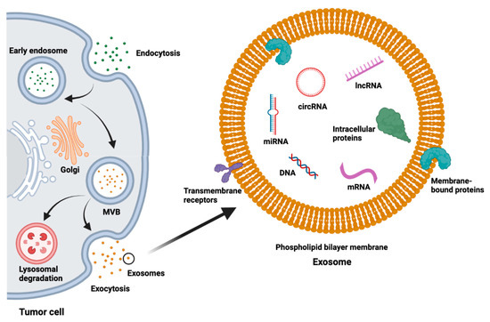
As the field of liquid biopsy continues to expand and refine, the value of exosomes as an important alternate platform is being increasingly recognized. Exosomes are extracellular vesicles (EVs) surrounded by a lipid bilayer membrane that range in size between 40 and 160 nanometers [6]. Exosome biogenesis begins with invagination of the cell membrane, then continues with a tightly regulated process with active sorting and packaging of exosomal content (Figure 1) [6,7]. Exosomes contain a variety of substances such as lipids, proteins, DNA, messenger RNA (mRNA), short single-stranded microRNAs (miRNAs, 18–25 nucleotides (nt)), long non-coding RNAs (lncRNAs, >200 nt), and novel circular RNAs (circRNA) (Figure 1) [6,8,9]. Besides the common cargo, some of the contents of exosomes are specific to their cell of origin and can be used to identify the source [10].

Figure 1.
Exosome biogenesis, structure, and contents. MVB = multivesicular body.
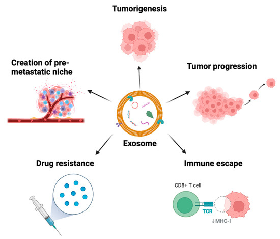
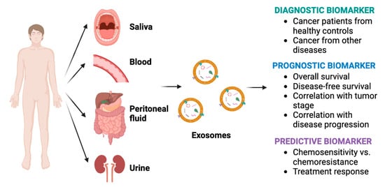
The function of exosomes was originally described by Pan and Johnstone as they tracked the loss of transferrin receptors via released vesicles during reticulocyte maturation [11]. Previously regarded as “garbage bins”, a plethora of preclinical and human studies have now demonstrated the active role of exosomes in cancer intercellular communication [12]. Exosomes play key roles in cancer, including creation of the premetastatic niche, tumorigenesis, tumor progression, immune escape, treatment resistance, and signaling between tumor cells and the surrounding tumor microenvironment (Figure 2) [13,14]. Hence, the content of exosomes could potentially be used as biomarkers, particularly since exosomes are readily found in a variety of biofluids and are produced more by malignant cells than normal cells (Figure 3) [8,13].

Figure 2.
The roles of exosomes in cancer. TCR = T cell receptor, MHC-I = major histocompatibility complex class I.

Figure 3.
Diagnostic, prognostic, and predictive roles of exosome biomarkers in cancer.
Furthermore, when compared with ctDNA, exosomes offer several advantages. First, exosomes are released by all living cells and may reveal information about living tumor cells, unlike ctDNA, which is released through apoptosis or necrosis [15]. Because of their abundance, less blood volume is required for exosomes compared with ctDNA. Exosomes are also very stable under different storage conditions due to their lipid bilayer, which protects their DNA, RNA, and protein contents [1,16]. ctDNA, on the other hand, is rapidly cleared from blood and susceptible to degradation in circulation due to DNase activity [2]. Given the potential of exosomes as liquid biopsies, many studies have been conducted, particularly evaluating RNAs and proteins from exosomes. In this review, we summarize human studies focused on biofluids such as blood and peritoneal fluid that have investigated the utility of exosomes as diagnostic, prognostic, and predictive biomarkers in colorectal and gastric cancers (GCs). All of the studies described below compare patients with cancer to healthy controls (HCs) unless otherwise specified.
2. Colorectal Cancer
Colorectal cancer (CRC) is the third most diagnosed cancer in both men and women in the United States (US), with an estimated 106,180 new cases and an estimated 52,580 deaths in 2022 [17]. Early detection is key as 5-year survival for early stages is 90% but 13% for stage IV disease [18]. Exosomes are being studied as a less invasive source of diagnostic, prognostic, and predictive biomarkers for CRC, as summarized below.
2.1. Diagnostic Biomarkers
The majority of the studies investigating exosome biomarkers for the diagnosis of CRC have predominantly focused on miRNAs. Table 1 provides a summary of the studies that have evaluated differentially expressed miRNAs, mRNAs, and lncRNAs between patients with CRC and HCs as potential diagnostic biomarkers. Ogata-Kawata et al. identified seven miRNAs (let-7a, miR-1229, miR-1246, miR-150, miR-21, miR-223, and miR-23a) in serum exosomes of CRC patients that were significantly overexpressed compared with HCs and downregulated following resection of the primary tumor, suggesting that the overexpressed miRNAs were of tumor origin [19]. Similarly, decreases in the levels of overexpressed RNAs following surgical resection of the tumor were observed by Ostenfeld et al. and Liang et al. [20,21]. Ostenfeld et al. in particular, attempted to evaluate tumor-derived EVs specifically by isolating epithelial cell adhesion molecular (EpCAM) positive EVs from the plasma of CRC patients and performing subsequent miRNA profiling. Thirteen miRNAs from these EpCAM+ EVs were overexpressed in CRC patients before surgical resection and eight of these miRNAs were downregulated after surgical resection, suggesting these miRNAs were of tumor origin [20]. Although the biomarkers were different between the three studies, the collective observations affirm that a portion of the overexpressed exosomal genes in the peripheral blood originate from the tumor and could provide meaningful insights about the tumor status.
Several miRNAs, including let-7a, miR-27a-3p, miR-383-5p, and miR-486-5p, that have well established roles in cancer have also been identified to be overexpressed in CRC patients [19,20,22,23]. Additionally, work has been performed to identify biomarkers that can be used to detect the early stages of CRC cancer. Wang et al. studied miR-125a-3p by comparing its expression in the plasma exosomes of patients with stage I and II colon cancer with HCs. mir-125a-3p expression was significantly increased in patients with early-stage disease. It is important to note that the predictive accuracy of miR-125a-3p to detect colon cancer improved from an area under the curve (AUC) of 0.685 to 0.855 when combined with the conventional diagnostic marker carcinoembryonic antigen (CEA) [24]. Similar observations of improved predictive accuracy when combining exosomal biomarkers with traditional tumor markers were observed in a study by Liu et al. In this study, exosomal lncRNA CRNDE-h was found to be higher in CRC patients compared with those with benign colon diseases and HCs (AUC = 0.892), and when combined with CEA, the diagnostic accuracy improved (AUC = 0.913) [25]. These observations open the possibility of utilizing exosome biomarkers as companion diagnostics with the existing modalities to improve diagnostic performance.
Other lncRNAs have also been identified as important diagnostic biomarkers of CRC. The lncRNA UCA1 has been shown to be downregulated in serum exosomes of patients with CRC, whereas the lncRNA TUG1 is upregulated compared with HCs. In combination, TUG1 and UCA1 had an AUC of 0.814 with a sensitivity of 93% and a specificity of 64% in distinguishing CRC patients from HCs [26]. Additionally, the lncRNA RPPH1 was found to have significantly higher expression in the plasma exosomes of CRC patients compared with HCs and the expression levels significantly decreased following surgical resection. The diagnostic power of RPPH1 to discriminate CRC patients from HCs (AUC = 0.856) was better than CEA (AUC = 0.790) [21].
Our group identified a gene signature that includes a combination of miRNA, mRNA and lncRNA. Plasma exosomes were isolated from patients with non-metastatic and metastatic (visceral metastases and PC) colon cancer and compared with HCs. Next-generation sequencing was used and after excluding highly prevalent and overlapping tRNA transcripts, 445 highly differentially expressed genes were identified. This gene signature, named ExoSig445, was able to fully discriminate colon cancer patients from HCs based on expression levels, suggesting gene panels can be developed as highly sensitive liquid biopsy tests (in press).

Table 1.
Summary of diagnostic exosomal biomarkers for colorectal cancer.
Table 1.
Summary of diagnostic exosomal biomarkers for colorectal cancer.
| Author(s) | Year | Biomarker(s) | Source | Findings |
|---|---|---|---|---|
| Ogata-Kawata et al. [19] | 2014 | 7 miRNAs: let-7a, miR-1229, miR-1246, miR-150, miR-21, miR-223, miR-23a | Serum |
|
| Ostenfeld et al. [20] | 2016 |
8 miRNAs: miR-16-5p, miR-23a-3p, miR-23b-3p,
miR-27a-3p, miR-27b-3p, miR-30b-5p, miR-30c-5p, miR-222-3p | Plasma |
|
| Dong et al. [27] | 2016 | mRNA KTTAP5-4, mRNA MEGEA3, lncRNA BCAR4 | Serum |
|
| Wang et al. [24] | 2017 | miR-125a-3p | Plasma |
|
| Liu et al. [23] | 2018 | miR-486-5p | Plasma |
|
| Barbagallo et al. [26] | 2018 | lncRNA UCA1, lncRNA TUG1 | Serum |
|
| Karimi et al. [28] | 2019 | miR-301a, miR-23a | Serum |
|
| Liang et al. [21] | 2019 | lncRNA RPPH1 | Plasma |
|
| Maminezhad et al. [29] | 2020 | 6 miRNA signature: let-7a, miR-150, miR-143, miR-145, miR-19a, miR-20a | Serum |
|
| Vallejos et al. (in press) | 2022 | 445 differently expressed genes from exosomal RNA (ExoSig445) | Plasma |
|
CRC = colorectal cancer, HC = healthy control, CEA = carcinoembryonic antigen, CA = cancer antigen, TCGA = The Cancer Genome Atlas.
2.2. Prognostic Biomarkers
Several of the biomarkers in CRC have both diagnostic and prognostic significance. The studies that have specifically reported the association of exosome biomarkers with prognosis have been summarized in Table 2. Some miRNA biomarkers have the potential to differentiate the early from late stages of CRC. In a study by Fu et al., overexpression of miR-17-5p and miR-92a-3p positively correlated with pathologic TNM stage, which allowed the authors to not only differentiate CRC from HCs but also non-metastatic disease from metastatic disease with high accuracy (AUC > 0.8) [30]. In a different study, plasma exosomal miR-21 was significantly higher in TNM stages III (n = 98) and IV (n = 67) compared with stages I (n = 51) and II (n = 110) [31]. Biomarkers that can aid in distinguishing early from late-stage CRC can be useful in guiding treatment, especially as neoadjuvant systemic therapy will often be considered for locally advanced and metastatic colon cancer [32].
Other studies have elucidated the role of miRNAs as prognostic biomarkers for recurrence. Liu et al. compared serum exosomes of patients with recurrent stage II/III disease with those without recurrent disease and found miR-4472-3p was upregulated in the recurrent disease group. Furthermore, miR-4772-3p was a predictor of recurrence with an OR of 11.3 (95% CI 2.38–53.2, p = 0.002) on multivariate logistic regression analysis and AUC of 0.72 based on the receiver operating characteristic (ROC) curve. Patients with higher miR-4772-3p expression had a shorter time to recurrence compared with lower miR-4772-3p expression (32.5 mean months vs. 77.0 mean months, p < 0.001) [33]. Matsumura et al. discovered another miRNA, miR-19a, as a prognostic biomarker for recurrence. Previously reported to promote proliferation and invasion of cancer cells [34], mir-19a was first identified by the authors to have increased expression in the exosomes of CRC patients with recurrence compared with patients without recurrence and increased expression in CRC patients compared with HCs across stages I–IV. Furthermore, high exosomal miR-19a expression in CRC patients was associated with worse overall survival (OS) and disease-free survival (DSF) than low expression and was an independent risk factor for OS and DFS [35].
Several other miRNAs have also been associated with poor survival and these include miR-21, miR-27a, miR-130a, miR-6803-5p, and miR-221 [31,36,37,38]. In all these studies, exosomal miRNA expression levels were higher in CRC compared with HCs, and when stratified into high and low exosomal expression groups, patients in the high-expression groups had worse OS for each of these individual miRNAs [31,36,37,38].
Although most studies have reported on the significance of overexpressed RNAs, few studies have focused on the significance of underexpressed RNAs. Downregulation of serum exosomal miR-150-5p was observed in CRC patients compared with HCs and exosomal expression of miR-150-5p is significantly higher following surgery compared with pre-resection levels. When survival was assessed, patients with low exosomal expression of miR-150-5p had worse DFS and OS. Furthermore, low serum exosomal miR-150-5p expression, along with lymph node metastasis and TNM stage, were independent prognostic factors for OS [39]. It is interesting to note that a high level of miR 150-5p in the tumor tissue is associated with aggressiveness in triple-negative breast cancer [40]. Gene expression levels in tumor tissue and peripheral blood exosomes may not be directly correlated and understanding these differences is essential to predict the results of exosome gene analysis. Additionally, miR-548c-5p is underexpressed in serum exosomes of CRC patients compared with HCs. On further analysis, patients with liver metastasis compared with no liver metastasis and patients with stage III/IV disease compared with stage I/II disease all had significantly lower miR-548-5p exosomal expression. Compared with patients with high levels of exosomal miR-548c-5p, low exosomal levels was independently associated with worse OS on multivariate analysis [41].
Along with the above-mentioned miRNAs, several lncRNAs have also been identified to have diagnostic and prognostic significance when underexpressed. GAS5 was identified by Liu et al. as a diagnostic and prognostic biomarker along with miR-221; however, unlike miR-221, GAS5 is downregulated in serum exosomes of CRC patients compared with HCs. GAS5 had an AUC of 0.964 for detecting CRC. Low exosomal GAS5 expression was associated with worse OS and was also an independent risk factor for OS [37]. HOTTIP is another lncRNA with significantly decreased expression in serum derived exosomes of CRC patients compared with HCs (AUC = 0.71). Moreover, OS was significantly decreased in patients with low/intermediate expression of HOTTIP (<75th percentile) compared with those with high (≥75th percentile) expression (47.0 months vs. 80.4 months, p = 0.0009). On multivariate analysis, low expression of HOTTIP was associated with worse OS [42]. However, it is important to note that there are studies of lncRNAs in which overexpression of lncRNAs was associated with poor outcomes. For example, a study by Liu et al. showed that patients with high expression of exosomal lncRNA CRNDE-h had lower OS rates than the low expression group (34.6% vs. 68.2%, p < 0.001) [25]. Collectively, these observations suggest that both over and underexpressed RNAs in the exosomal cargo bear diagnostic and prognostic potential. Identifying the right combinations of markers that provide the most actionable information and insights are essential to develop exosome liquid biopsy as a diagnostic tool for clinical applications.

Table 2.
Summary of prognostic exosomal biomarkers for colorectal cancer.
Table 2.
Summary of prognostic exosomal biomarkers for colorectal cancer.
| Author(s) | Year | Biomarker(s) | Source | Findings |
|---|---|---|---|---|
| Matsumura et al. [35] | 2015 | miR-19a | Serum |
|
| Liu et al. [33] | 2016 | miR-4772-3p | Serum |
|
| * Liu et al. [25] | 2016 | lncRNA CRNDE-h | Serum |
|
| * Tsukamoto et al. [31] | 2017 | miR-21 | Plasma |
|
| * Fu et al. [30] | 2018 | miR-17-5p, miR-92a-3p | Serum |
|
| * Yan et al. [36] | 2018 | miR-6803-5p | Serum |
|
| * Liu et al. [38] | 2018 | miR-27a, miR-130a | Plasma |
|
| * Liu et al. [37] | 2018 | lncRNA GAS5, miR-221 | Serum |
|
| * Peng et al. [41] | 2018 | miR-584c-5p | Serum |
|
| * Zou et al. [39] | 2019 | miR-150-5p | Serum |
|
| * Oehme et al. [42] | 2019 | lncRNA HOTTIP | Serum |
|
* These biomarkers were also found to have diagnostic roles. CRC = colorectal cancer, HC = healthy control, OS = overall survival, DFS = disease free survival, CEA = carcinoembryonic antigen.
2.3. Predictive Biomarkers
There have been several studies highlighting the potential of RNAs to predict chemoresistance (Table 3). In a study by Jin et al., a panel of four exosomal miRNAs were identified (miR-21-5p, miR-1246, miR-1229-5p, and miR-96-5p) that were upregulated in patients with unresectable stage III/IV CRC who had chemoresistance to 5-FU and oxaliplatin compared with patients who were chemosensitive. This miRNA panel was also able to distinguish chemoresistant CRC patients from their chemosensitive counterparts with an AUC of 0.804 [43].
In another study, Yagi et al. evaluated exosomal miR-125b expression in patients with advanced or recurrent CRC treated with modified fluorouracil, leucovorin, and oxaliplatin (mFOLFOX6)-based first-line therapy before treatment and at different time points during treatment. The response to chemotherapy was evaluated using Response Evaluation Criteria in Solid Tumors (RECIST) and patients were classified into complete response (CR), partial response (PR), stable disease (SD), and progressive disease (PD) groups. The authors found exosomal miR-125b expression post-treatment was significantly lower in patients with PR but significantly higher in patients with PD compared with pre-treatment levels. Patients with SD had no change in exosomal miR-125b expression with treatment. Furthermore, patients in the high miR-125b expression group had worse progression free survival (PFS) than the low expression group. On Cox multivariate regression analysis, KRAS mutation and exosomal miR-125b were found to be independent predictors of PFS. Taken together, these findings suggest miR-125b has the potential to be both a predictive and a prognostic biomarker of CRC [44]. The predictive capacity of exosomes to assess treatment resistance and tumor progression are of remarkable interest in the growing paradigm of utilizing liquid biopsies to dynamically adjust treatment strategies.

Table 3.
Summary of predictive exosomal biomarkers for colorectal cancer.
Table 3.
Summary of predictive exosomal biomarkers for colorectal cancer.
| Author(s) | Year | Biomarker(s) | Source | Findings |
|---|---|---|---|---|
| Jin et al. [43] | 2019 | 4 miRNA panel: miR-21-5p, miR-1246, miR-1229-5p, miR-96-5p | Serum |
|
| * Yagi et al. [44] | 2019 | miR-125b | Plasma |
|
* This biomarker was also found to have a prognostic role. CRC = colorectal cancer, HC = healthy control, PD = progressive disease, PR = partial response, SD = stable disease, PFS = progression free survival.
3. Gastric Cancer
The number of new GC cases in the US was estimated to be 26,380 in 2022 with 11,090 new deaths [17]. Early-stage GC is often asymptomatic, resulting in a delay in diagnosis. Even with advances in the medical and surgical management of GC over the past decade, late-stage GC is associated with a poor prognosis [12]. This highlights the need for novel noninvasive biomarkers for GC. Here, we discuss the research being conducted on exosomes and the diagnostic and prognostic roles they play in the management of GC (summarized in Table 4 and Table 5).

Table 4.
Summary of diagnostic exosomal biomarkers for gastric cancer.

Table 5.
Summary of prognostic exosomal biomarkers for gastric cancer.
3.1. Diagnostic Biomarkers
As most of the current diagnostic biomarkers for GC are not accurate enough to screen patients for GC, this raises the need to develop new and effective diagnostic biomarkers for the detection of this disease [10]. In 2017, Pan et al. carried out a study analyzing the sera of 94 GC patients and found a significant increase in ZFAS1 expression in GC patient serum exosomes compared with HCs (p < 0.001) [45]. Subsequently, another study found a significant increase (2.5×) in the expression of exosomal miR-221 in the peripheral blood of GC patients compared with HCs [52].
Certain miRNAs have been found to have increased diagnostic power when combined. Wang et al., for example, studied the serum of GC patients (n = 130) and HCs (n = 130) and found a significant increase in the exosomal expression levels of miR-106a-5p and miR-19b-3p in the serum of GC patients (p < 0.0001). The diagnostic accuracy was better when the two miRNAs were combined compared with either one of them individually and significantly outperformed the tumor markers alpha fetoprotein (AFP) and cancer antigen (CA) 19-9 [46]. In a study by Huang et al., the serum exosomal expression levels of miR10b-5p, miR132-3p, miR185-5p, miR195-5p, miR20a-3p, and miR296-5p were elevated in GC patients compared with HCs. Integrating the six miRNAs together led to improved accuracy in correctly identifying GC patients with an AUC of 0.703 [54].
Furthermore, as seen in CRC, exosomal biomarkers can be combined with current tumor markers to increase the overall accuracy in screening patients with GC. In the training phase of a study by Ge et al., there was an elevated expression of miR-1307-3p, piR-018569, piR-004918, and piR-019308 in the serum exosomes of GC patients. Evaluation with an additional cohort of patients confirmed significant exosomal overexpression of these miRNAs in GC patient serum (p < 0.0001). The diagnostic potential of miR-1307-3p, piR-019308, piR-004918, and piR-018569 was shown with AUCs of 0.845, 0.820, 0.754, and 0.732, respectively. After integration of tumor markers such as CEA and CA 19-9 into the previous biomarkers, their AUCs improved to 0.902, 0.914, 0.859, and 0.868, respectively [59]. Other studies have identified miRNA biomarkers that have lower expression in GC compared with HCs, such as miR-23b [56].
Aside from miRNAs, lncRNAs and circRNAs have also been implicated as diagnostic biomarkers for GC. Cai et al. studied the blood samples of GC and healthy patients and found a significantly lower expression of the lncRNA PCSK2-2:1 in the serum exosomes of GC patients (p = 0.006). Serum exosome levels of lncRNA PCSK2-2:1 was additionally found to be accurate in detecting GC patients with an AUC of 0.896 [50]. Zhao et al. studied the serum of 246 GC and healthy patients and found an upregulation in the expression of exosomal lncRNA HOTTIP in GC patients (p < 0.001). ROC curve analysis was performed for HOTTIP in patient serum, showing an AUC of 0.827, which was higher than the AUC of other tumor markers (CEA and CA 19-9) combined (p < 0.001) [55]. Xie et al. showed the presence of increased expression of circSHKBP1 in the serum of GC patients compared with HCs. These findings were supported with sub-analysis that showed a decrease in the expression of circSHKBP1 after tumor removal in 12 patients in this study [58]. Shao et al. studied the plasma of GC patients, which showed a significant downregulation of Hsa_circ_0065149 in early GC (stage I and II) patient plasma exosomes compared with HCs (p < 0.001), with an AUC of 0.640 (p = 0.031) by ROC [51]. This, along with previous studies, shows the possibility of increased accuracy of GC detection with the use of exosomal biomarkers compared with other noninvasive biomarkers and tumor markers.
Few studies have specifically evaluated exosomal biomarkers to discriminate GC from other non-malignant conditions of the stomach such as chronic atrophic gastritis (CAG) and intestinal metaplasia (IM). In a study by Lin et al. that evaluated patients with early-stage GC (stage I and II), exosomal lncUEGC1 and lncUEGC2 levels were significantly increased in early-stage GC compared with HCs (p < 0.0001). Additionally, plasma exosomal lncUEGC1 expression was significantly increased in stage 1 GC patients compared with CAG patients [49]. A subsequent study that looked at the serum of 862 patients showed significantly elevated circulating levels of exosomal lncRNA-GC1 in GC patients. This biomarker was highly accurate in differentiating GC patients from HCs with an AUC of 0.9033, which was higher than that of current tumor markers such as CEA and CA 19-9. In the verification phase of this study, the circulating exosomal lncRNA-GC1 levels were significantly higher in GC patients compared with those with CAG, IM, and Helicobacter pylori positivity or negativity [57].
3.2. Prognostic Biomarkers
Recurrence and metastasis are associated with progression of disease and carry a worse prognosis in GC patients, highlighting the need for prognostic biomarkers that can be used in the monitoring and surveillance of GC patients. Studies that have shown the diagnostic potential of exosomes have given us insights into the prognostic possibilities, as well. In the 2017 study by Pan et al., GC patients were separated into high and low expression of ZFAS1 in patient serum exosomes and the high-expression group was positively associated with lymphatic metastasis (p = 0.005) and advanced TNM stage (p = 0.010) [45]. Around this time, Ma et al. showed that the serum expression level of miR-221 was associated with TNM stage and overall poor clinical prognosis in GC patients [52]. In the same year, Yen et al. studied the exosome profile in the peripheral blood of 61 GC patients, which showed a positive association between the expression of TGF-β1 and TNM stage (p = 0.03) and lymph node metastasis (p = 0.01). Additionally, there was a higher level of exosomal TGF-β1 in late-stage GC compared with stage I GC patients and a two-fold increase in patients with lymph node metastasis versus those without [53]. In studies by Wang et al. and Huang et al., the expression levels of different miRNAs (Table 5) were found to be significantly higher in the serum of GC patients with lymphatic metastasis (p = 0.001, p = 0.008, respectively) and late-stage GC (III and IV) compared with early-stage GC (I and II) (p = 0.048, p = 0.031, respectively) [46,54].
Studies have also shown the value of exosomal prognostic biomarkers in differentiating between early- and late-stage GC, particularly metastatic disease from non-metastatic disease. Kumata et al. found that exosomal miR-23b levels decreased with the progression of GC with a significant decrease in the expression of miR-23b in stage IV GC compared with earlier stages (p < 0.05). After separating patients into high and low miR-23b expression groups, they found an association between miR-23b expression and tumor size, invasion depth, liver metastasis, and TNM stage. Low expression of miR-23b was associated with worse DFS in patients undergoing curative surgery and correlated with recurrence and poor prognosis across all stages of GC [56].
Utilizing a separate set of miRNAs, Zhang et al. found that the expression levels of miR-10b-5p, miR 143-5p, and miR 101-3p were significantly elevated in GC patients with lymph node, liver, and ovarian metastasis, respectively (p < 0.05). Additionally, after ROC analysis, the AUCs were calculated to be 0.8919, 0.8247, and 0.8905, respectively (p < 0.05) [60].
lncRNAs and circRNAs, have prognostic value as exosomal biomarkers for GC, as well. Although the lncRNA PCSK2-2:1 was significantly lower in the serum exosomes of GC patients, as previously described, it was also correlated with tumor size (p = 0.0441), tumor stage (p = 0.0061), and the degree of venous invasion (p = 0.0367) [50]. Guo et al. were able to show a significant difference in the exosomal levels of lncRNA-GC1 between the four clinical stages of GC with an incremental increase in the exosomal levels of lncRNA-GC1 from stages T1 to T4 and N0 to N3 [57]. Furthermore, Xie et al. showed that the expression of exosomal circSHKBP1 was correlated with advanced TNM stage, vascular invasion, and overall poor prognosis [58]. Studies investigating the field of exosomes have shown promise in the use of exosomes as prognostic biomarkers in the management of GC. Although the wide variation of biomarkers between studies requires further refinement and standardization, the current evidence has shown the potential of exosome cargo to assess disease severity and prognosis in GC.
4. Peritoneal Fluid Exosomes in Colon and Gastric Cancer
Besides blood, peritoneal fluid is of significant interest in GI cancers both due to the propensity of these cancers to metastasize to the peritoneum and the presence of exosomes in the peritoneal space that could be diagnostic of cancer. Moreover, exosomes can play a significant role in creating a premetastatic niche in the peritoneal cavity [61]. The current diagnostic modality to identify microscopic peritoneal disease, namely peritoneal cytology, has a sensitivity as low as 11% to 80% in GC [62]. Hence, exosomal analysis of the peritoneal fluid could be of major importance to fill a critical gap in diagnosing peritoneal disease.
In CRC, Roman-Canal et al. identified exosomal miRNAs from peritoneal lavage of patients with CRC that could potentially have diagnostic significance. Compared with ascites fluid from non-cancer patients, 210 miRNAs were significantly dysregulated in peritoneal lavage fluid from CRC patients. Ten miRNAs were significantly overexpressed in CRC patients with an AUC > 0.95 (Table 6) [22].
In GC, Ohzawa et al. evaluated the miRNA expressions in the peritoneal fluid of patients with and without peritoneal metastasis (PM). Compared with those without PM, the authors found significant upregulation of four miRNAs (miR-21-5p, miR-92a-3p, miR-223-3p, and miR-342-3p) in patients with PM (p < 0.05), whereas the miR-29 family, especially miR-29b-3p and miR-29c-3p, was downregulated in all the patients with PM. Additionally, among patients with PM, the expression levels of miR-21-5p, miR-92a-3p, miR-223-3p, and miR-342-3p were positively correlated with the peritoneal carcinomatosis index (PCI) [63]. A follow-up study by Ohzawa et al. confirmed the previous findings, showing decreased expression of the miR-29s (miR-29a-3p, miR-29b-3p, and miR-29c-3p) in the peritoneal lavage fluid/ascites of patients with PM (p < 0.001). When patients with T4 tumors status post curative gastrectomy were separated based on low and high expression of miR-29b-3p in the exosomes of the peritoneal fluid, those with low miR-29b-3p expression exhibited worse peritoneal recurrence free survival (RFS) (p < 0.05). Furthermore, low expression was associated with significantly worse OS for all three miR-29s (Table 6) [64]. Thus, exosomes isolated from the peritoneal fluid/ascites of GC patients have the potential to be a more accurate alternative to cytology in diagnosing GC patients with PC. These studies highlight the fact that exosomes exist in a variety of biofluids and showcase peritoneal fluid as another possible source of biomarkers for GI cancers.

Table 6.
Summary of exosomal biomarkers from peritoneal fluid for colorectal and gastric cancer.
Table 6.
Summary of exosomal biomarkers from peritoneal fluid for colorectal and gastric cancer.
| Author(s) | Year | Biomarker(s) | Source | Findings |
|---|---|---|---|---|
| Roman-Canal et al. [22] | 2019 | miRNA-199b-5p, miRNA-150-5p, miRNA-29c-5p, miRNA-218-5p, miRNA-99a-3p, miRNA-383-5p, miRNA-199a-3p, miRNA-193a-5p, miRNA-10b-5p, miRNA-181c-5p | Peritoneal lavage |
|
| Ohzawa et al. [63] | 2019 | miR-21-5p, miR-92a-3p, miR-233-3p, miR-342-3p | Peritoneal lavage |
|
| Ohzawa et al. [64] | 2020 | miR-29a-3p, miR-29b-3p, miR-29c-3p | Peritoneal lavage |
|
CRC = colorectal cancer, GC = gastric cancer, PM = peritoneal metastasis, PCI = peritoneal carcinomatosis index, RFS = recurrence free survival, OS = overall survival.
5. Clinical Challenges and Future Prospects
This review highlights the role of exosomes as diagnostic, prognostic, and predictive biomarkers in various GI cancers. However, there are challenges and limitations that need to be overcome before the widespread uptake of exosomal liquid biopsy in clinical practice. First, there are various methods for exosome isolation, each with their own advantages and disadvantages. The two common methods are ultracentrifugation (UC) and precipitation. UC separates exosomes from other components based on size and density differences with high-speed centrifugation [7]. Although UC is considered the gold standard for exosome extraction and separation, UC requires costly instrumentation and is time consuming and more suitable for large volumes, which may not be appropriate in a clinical setting [7]. Precipitation, on the other hand, relies on reducing the solubility of the exosomes, then separating them from other contents using low-speed centrifugation [7]. Compared with UC, the precipitation method requires less time, does not require costly equipment, and can be used with smaller sample volumes. However, other proteins and lipoproteins may be isolated as well, compromising the purity of the exosomes [7]. Current studies often use differing exosome isolation methods, making it difficult to compare studies [8]. Once isolated, EVs should be confirmed as exosomes by examining the morphology, protein expression, size, and concentration using methods proposed by the International Society of Extracellular Vesicles (ISEV) [65], which takes additional time and expertise. Given these challenges, exosome isolation and characterization methods need improved efficiency and throughput to be successfully transitioned to clinical application.
Second, the study of exosomal gene expression requires standardization of its methods and reference genes. In our review, for example, we present many different miRNAs, lncRNAs, circRNAs, and proteins that have been identified as having diagnostic, prognostic, and predictive potential with limited overlap for the same cancer types. There are also contrasting findings between cancer types. For example, low expression of the exosomal lncRNA HOTTIP, as described above, was found to be an independent predictor of worse OS in CRC [42]. These findings contrast with that of a recent meta-analysis by Fan et al. in which HOTTIP tumor tissue overexpression in multiple cancers was associated with increased tumor stage, lymph node metastasis, distant metastasis, and poor OS [66]. Although, the differences between the tumor expression levels of genes and exosomal levels could be different and may account for these observed differences, an exosomal study in GC showed high levels of expression of HOTTIP to be diagnostic of GC [55]. These contradicting results are a prime example of issues in exosome research and are one of the major barriers to clinical translation. Furthermore, standardization of the control group is needed to compare differences in gene expression among the experimental groups. HCs are often used in the studies we describe but it is important to note variability in gene expression can also exist among healthy individuals. Therefore, an accepted set of reference genes with expected expression levels should be developed.
Future work can be conducted to identify universal exosomal genes with known functions in cancer or gene panels that may have more diagnostic, prognostic, or predictive power than individual biomarkers. Another area of exciting application is the use of exosomes in therapeutics as drug delivery systems [67,68]. Therapeutic applications of exosomes are beyond the scope of this review; however, the readers are directed to cited references for detailed information. Exosomal genes with known functions in cancers can also be targeted for therapy in the future.
6. Conclusions
In short, exosomes are an exciting and promising source of biomarkers with the potential for clinical application as liquid biopsies for various GI cancers. Exosomes carry a variety of RNAs and proteins, many of which have shown diagnostic, prognostic, and predictive potential. More work is needed to overcome the challenges of applying exosomes to clinical practice. However, the abundance of information that can be garnered from exosomes is apparent and exciting.
Author Contributions
Conceptualization, F.D. and M.S.; Writing—Original Draft Preparation, J.Y., A.O., A.G., K.M. and M.S.; Writing—Review and Editing: C.C.W.H., F.D. and M.S. All authors have read and agreed to the published version of the manuscript.
Funding
This research received no external funding.
Acknowledgments
All figures were created with BioRender.com.
Conflicts of Interest
F.D. has received honoraria from Astrazeneca, Genentech, Eisai, Exelixis, Deciphera, Ipsen, Sirtex, Natera, and Servier and research support from the institution from Amgen, Astrazeneca, Exelixis, BMS, Merck, Trishula, Natera, Taiho, and Ipsen. C.C.W.H. is the founder and Chief Scientific Officer of Aracari Bioscience. The other authors declare no conflict of interest.
References
- Pinzani, P.; D’Argenio, V.; Del Re, M.; Pellegrini, C.; Cucchiara, F.; Salvianti, F.; Galbiati, S. Updates on liquid biopsy: Current trends and future perspectives for clinical application in solid tumors. Clin. Chem. Lab. Med. 2021, 59, 1181–1200. [Google Scholar] [CrossRef] [PubMed]
- Lopez, A.; Harada, K.; Kaya, D.M.; Dong, X.; Song, S.; Ajani, J.A. Liquid biopsies in gastrointestinal malignancies: When is the big day? Expert Rev. Anticancer. Ther. 2017, 18, 19–38. [Google Scholar] [CrossRef] [PubMed]
- Sullivan, B.G.; Lo, A.; Yu, J.; Gonda, A.; Dehkordi-Vakil, F.; Dayyani, F.; Senthil, M. Circulating Tumor DNA is Unreliable to Detect Somatic Gene Alterations in Gastrointestinal Peritoneal Carcinomatosis. Ann. Surg. Oncol. 2022, 30, 278–284. [Google Scholar] [CrossRef] [PubMed]
- Maron, S.B.; Chase, L.M.; Lomnicki, S.; Kochanny, S.; Moore, K.L.; Joshi, S.S.; Landron, S.; Johnson, J.; Kiedrowski, L.A.; Nagy, R.J.; et al. Circulating Tumor DNA Sequencing Analysis of Gastroesophageal Adenocarcinoma. Clin. Cancer Res. 2019, 25, 7098–7112. [Google Scholar] [CrossRef] [PubMed]
- Bando, H.; Nakamura, Y.; Taniguchi, H.; Shiozawa, M.; Yasui, H.; Esaki, T.; Kagawa, Y.; Denda, T.; Satoh, T.; Yamazaki, K.; et al. Effects of Metastatic Sites on Circulating Tumor DNA in Patients With Metastatic Colorectal Cancer. JCO Precis. Oncol. 2022. [Google Scholar] [CrossRef] [PubMed]
- Dai, J.; Su, Y.; Zhong, S.; Cong, L.; Liu, B.; Yang, J.; Tao, Y.; He, Z.; Chen, C.; Jiang, Y. Exosomes: Key players in cancer and potential therapeutic strategy. Signal Transduct. Target. Ther. 2020, 5, 145. [Google Scholar] [CrossRef] [PubMed]
- Zhang, Y.; Bi, J.; Huang, J.; Tang, Y.; Du, S.; Li, P. Exosome: A Review of Its Classification, Isolation Techniques, Storage, Diagnostic and Targeted Therapy Applications. Int. J. Nanomed. 2020, 15, 6917–6934. [Google Scholar] [CrossRef]
- Xiao, Y.; Zhong, J.; Zhong, B.; Huang, J.; Jiang, L.; Jiang, Y.; Yuan, J.; Sun, J.; Dai, L.; Yang, C.; et al. Exosomes as potential sources of biomarkers in colorectal cancer. Cancer Lett. 2020, 476, 13–22. [Google Scholar] [CrossRef]
- Vahabi, A.; Rezaie, J.; Hassanpour, M.; Panahi, Y.; Nemati, M.; Rasmi, Y.; Nemati, M. Tumor Cells-derived exosomal CircRNAs: Novel cancer drivers, molecular mechanisms, and clinical opportunities. Biochem. Pharmacol. 2022, 200. [Google Scholar] [CrossRef]
- Tang, X.-H.; Guo, T.; Gao, X.-Y.; Wu, X.-L.; Xing, X.-F.; Ji, J.-F.; Li, Z.-Y. Exosome-derived noncoding RNAs in gastric cancer: Functions and clinical applications. Mol. Cancer 2021, 20, 1–15. [Google Scholar] [CrossRef]
- Pan, B.-T.; Johnstone, R.M. Fate of the transferrin receptor during maturation of sheep reticulocytes in vitro: Selective externalization of the receptor. Cell 1983, 33, 967–978. [Google Scholar] [CrossRef] [PubMed]
- Liu, Y.; Wang, Y.; Lv, Q.; Li, X. Exosomes: From garbage bins to translational medicine. Int. J. Pharm. 2020, 583, 119333. [Google Scholar] [CrossRef] [PubMed]
- Cao, J.; Zhang, M.; Xie, F.; Lou, J.; Zhou, X.; Zhang, L.; Fang, M.; Zhou, F. Exosomes in head and neck cancer: Roles, mechanisms and applications. Cancer Lett. 2020, 494, 7–16. [Google Scholar] [CrossRef] [PubMed]
- Fabbri, M.; Paone, A.; Calore, F.; Galli, R.; Gaudio, E.; Santhanam, R.; Lovat, F.; Fadda, P.; Mao, C.; Nuovo, G.J.; et al. MicroRNAs bind to Toll-like receptors to induce prometastatic inflammatory response. Proc. Natl. Acad. Sci. 2012, 109, E2110–E2116. [Google Scholar] [CrossRef] [PubMed]
- Yu, W.; Hurley, J.; Roberts, D.; Chakrabortty, S.; Enderle, D.; Noerholm, M.; Breakefield, X.; Skog, J. Exosome-based liquid biopsies in cancer: Opportunities and challenges. Ann. Oncol. 2021, 32, 466–477. [Google Scholar] [CrossRef] [PubMed]
- Keller, S.; Ridinger, J.; Rupp, A.-K.; Janssen, J.W.G.; Altevogt, P. Body fluid derived exosomes as a novel template for clinical diagnostics. J. Transl. Med. 2011, 9, 86. [Google Scholar] [CrossRef]
- Siegel, R.L.; Miller, K.D.; Fuchs, H.E.; Jemal, A. Cancer statistics. CA Cancer J. Clin. 2022, 72, 7–33. [Google Scholar] [CrossRef]
- Baassiri, A.; Nassar, F.; Mukherji, D.; Shamseddine, A.; Nasr, R.; Temraz, S. Exosomal Non Coding RNA in LIQUID Biopsies as a Promising Biomarker for Colorectal Cancer. Int. J. Mol. Sci. 2020, 21, 1398. [Google Scholar] [CrossRef]
- Ogata-Kawata, H.; Izumiya, M.; Kurioka, D.; Honma, Y.; Yamada, Y.; Furuta, K.; Gunji, T.; Ohta, H.; Okamoto, H.; Sonoda, H.; et al. Circulating Exosomal microRNAs as Biomarkers of Colon Cancer. PLoS ONE 2014, 9, e92921. [Google Scholar] [CrossRef]
- Ostenfeld, M.S.; Jensen, S.G.; Jeppesen, D.K.; Christensen, L.-L.; Thorsen, S.B.; Stenvang, J.; Hvam, M.L.; Thomsen, A.; Mouritzen, P.; Rasmussen, M.H.; et al. miRNA profiling of circulating EpCAM+extracellular vesicles: Promising biomarkers of colorectal cancer. J. Extracell. Vesicles 2016, 5, 31488. [Google Scholar] [CrossRef]
- Liang, Z.X.; Liu, H.S.; Wang, F.W.; Xiong, L.; Zhou, C.; Hu, T.; He, X.W.; Wu, X.J.; Xie, D.; Wu, X.R.; et al. LncRNA RPPH1 promotes colorectal cancer metastasis by interacting with TUBB3 and by promoting exosomes-mediated macrophage M2 polarization. Cell Death Dis. 2019, 10, 1–17. [Google Scholar] [CrossRef]
- Roman-Canal, B.; Tarragona, J.; Moiola, C.P.; Gatius, S.; Bonnin, S.; Ruiz-Miró, M.; Sierra, J.E.; Rufas, M.; González, E.; Porcel, J.M.; et al. EV-associated miRNAs from peritoneal lavage as potential diagnostic biomarkers in colorectal cancer. J. Transl. Med. 2019, 17, 1–14. [Google Scholar] [CrossRef] [PubMed]
- Liu, X.; Chen, X.; Zeng, K.; Xu, M.; He, B.; Pan, Y.; Sun, H.; Pan, B.; Xu, X.; Xu, T.; et al. DNA-methylation-mediated silencing of miR-486-5p promotes colorectal cancer proliferation and migration through activation of PLAGL2/IGF2/β-catenin signal pathways. Cell Death Dis. 2018, 9, 1–17. [Google Scholar] [CrossRef] [PubMed]
- Wang, J.; Yan, F.; Zhao, Q.; Zhan, F.; Wang, R.; Wang, L.; Zhang, Y.; Huang, X. Circulating exosomal miR-125a-3p as a novel biomarker for early-stage colon cancer. Sci. Rep. 2017, 7, 4150. [Google Scholar] [CrossRef] [PubMed]
- Liu, T.; Zhang, X.; Gao, S.; Jing, F.; Yang, Y.; Du, L.; Zheng, G.; Li, P.; Li, C.; Wang, C. Exosomal long noncoding RNA CRNDE-h as a novel serum-based biomarker for diagnosis and prognosis of colorectal cancer. Oncotarget 2016, 7, 85551–85563. [Google Scholar] [CrossRef] [PubMed]
- Barbagallo, C.; Brex, D.; Caponnetto, A.; Cirnigliaro, M.; Scalia, M.; Magnano, A.; Caltabiano, R.; Barbagallo, D.; Biondi, A.; Cappellani, A.; et al. LncRNA UCA1, Upregulated in CRC Biopsies and Downregulated in Serum Exosomes, Controls mRNA Expression by RNA-RNA Interactions. Mol. Ther.-Nucleic Acids 2018, 12, 229–241. [Google Scholar] [CrossRef] [PubMed]
- Dong, L.; Lin, W.; Qi, P.; Xu, M.-D.; Wu, X.; Ni, S.; Huang, D.; Weng, W.-W.; Tan, C.; Sheng, W.; et al. Circulating Long RNAs in Serum Extracellular Vesicles: Their Characterization and Potential Application as Biomarkers for Diagnosis of Colorectal Cancer. Cancer Epidemiology Biomarkers Prev. 2016, 25, 1158–1166. [Google Scholar] [CrossRef] [PubMed]
- Karimi, N.; Feizi, M.A.H.; Safaralizadeh, R.; Hashemzadeh, S.; Baradaran, B.; Shokouhi, B.; Teimourian, S. Serum overexpression of miR-301a and miR-23a in patients with colorectal cancer. J. Chin. Med Assoc. 2019, 82, 215–220. [Google Scholar] [CrossRef]
- Maminezhad, H.; Ghanadian, S.; Pakravan, K.; Razmara, E.; Rouhollah, F.; Mossahebi-Mohammadi, M.; Babashah, S. A panel of six-circulating miRNA signature in serum and its potential diagnostic value in colorectal cancer. Life Sci. 2020, 258, 118226. [Google Scholar] [CrossRef]
- Fu, F.; Jiang, W.; Zhou, L.; Chen, Z. Circulating Exosomal miR-17-5p and miR-92a-3p Predict Pathologic Stage and Grade of Colorectal Cancer. Transl. Oncol. 2018, 11, 221–232. [Google Scholar] [CrossRef]
- Tsukamoto, M.; Iinuma, H.; Yagi, T.; Matsuda, K.; Hashiguchi, Y. Circulating Exosomal MicroRNA-21 as a Biomarker in Each Tumor Stage of Colorectal Cancer. Oncology 2017, 92, 360–370. [Google Scholar] [CrossRef] [PubMed]
- Gosavi, R.; Chia, C.; Michael, M.; Heriot, A.G.; Warrier, S.K.; Kong, J.C. Neoadjuvant chemotherapy in locally advanced colon cancer: A systematic review and meta-analysis. Int. J. Color. Dis. 2021, 36, 2063–2070. [Google Scholar] [CrossRef]
- Liu, C.; Eng, C.; Shen, J.; Lu, Y.; Takata, Y.; Mehdizadeh, A.; Chang, G.J.; Rodriguez-Bigas, M.A.; Li, Y.; Chang, P.; et al. Serum exosomal miR-4772-3p is a predictor of tumor recurrence in stage II and III colon cancer. Oncotarget 2016, 7, 76250–76260. [Google Scholar] [CrossRef] [PubMed]
- Zhang, J.; Xiao, Z.; Lai, D.; Sun, J.; He, C.; Chu, Z.; Ye, H.; Chen, S.; Wang, J. miR-21, miR-17 and miR-19a induced by phosphatase of regenerating liver-3 promote the proliferation and metastasis of colon cancer. Br. J. Cancer 2012, 107, 352–359. [Google Scholar] [CrossRef]
- Matsumura, T.; Sugimachi, K.; Iinuma, H.; Takahashi, Y.; Kurashige, J.; Sawada, G.; Ueda, M.; Uchi, R.; Ueo, H.; Takano, Y.; et al. Exosomal microRNA in serum is a novel biomarker of recurrence in human colorectal cancer. Br. J. Cancer 2015, 113, 275–281. [Google Scholar] [CrossRef] [PubMed]
- Yan, S.; Jiang, Y.; Liang, C.; Cheng, M.; Jin, C.; Duan, Q.; Xu, D.; Yang, L.; Zhang, X.; Ren, B.; et al. Exosomal miR-6803-5p as potential diagnostic and prognostic marker in colorectal cancer. J. Cell. Biochem. 2018, 119, 4113–4119. [Google Scholar] [CrossRef] [PubMed]
- Liu, L.; Meng, T.; Yang, X.-H.; Sayim, P.; Lei, C.; Jin, B.; Ge, L.; Wang, H.-J. Prognostic and predictive value of long non-coding RNA GAS5 and mircoRNA-221 in colorectal cancer and their effects on colorectal cancer cell proliferation, migration and invasion. Cancer Biomarkers 2018, 22, 283–299. [Google Scholar] [CrossRef] [PubMed]
- Liu, X.; Pan, B.; Sun, L.; Chen, X.; Zeng, K.; Hu, X.; Xu, T.; Xu, M.; Wang, S. Circulating Exosomal miR-27a and miR-130a Act as Novel Diagnostic and Prognostic Biomarkers of Colorectal Cancer. Cancer Epidemiology Biomarkers Prev. 2018, 27, 746–754. [Google Scholar] [CrossRef]
- Zou, S.-L.; Chen, Y.-L.; Ge, Z.-Z.; Qu, Y.-Y.; Cao, Y.; Kang, Z.-X. Downregulation of serum exosomal miR-150-5p is associated with poor prognosis in patients with colorectal cancer. Cancer Biomarkers 2019, 26, 69–77. [Google Scholar] [CrossRef]
- Sugita, B.M.; Rodriguez, Y.; Fonseca, A.S.; Souza, E.N.; Kallakury, B.; Cavalli, I.J.; Ribeiro, E.M.S.F.; Aneja, R.; Cavalli, L.R. MiR-150-5p Overexpression in Triple-Negative Breast Cancer Contributes to the In Vitro Aggressiveness of This Breast Cancer Subtype. Cancers 2022, 14, 2156. [Google Scholar] [CrossRef]
- Peng, Z.; Gu, R.; Yan, B. Downregulation of exosome-encapsulated miR-548c-5p is associated with poor prognosis in colorectal cancer. J. Cell. Biochem. 2018, 120, 1457–1463. [Google Scholar] [CrossRef] [PubMed]
- Oehme, F.; Krahl, S.; Gyorffy, B.; Muessle, B.; Rao, V.; Greif, H.; Ziegler, N.; Lin, K.; Thepkaysone, M.-L.; Polster, H.; et al. Low level of exosomal long non-coding RNA HOTTIP is a prognostic biomarker in colorectal cancer. RNA Biol. 2019, 16, 1339–1345. [Google Scholar] [CrossRef] [PubMed]
- Jin, G.; Liu, Y.; Zhang, J.; Bian, Z.; Yao, S.; Fei, B.; Zhou, L.; Yin, Y.; Huang, Z. A panel of serum exosomal microRNAs as predictive markers for chemoresistance in advanced colorectal cancer. Cancer Chemother. Pharmacol. 2019, 84, 315–325. [Google Scholar] [CrossRef] [PubMed]
- Yagi, T.; Iinuma, H.; Hayama, T.; Matsuda, K.; Nozawa, K.; Tsukamoto, M.; Shimada, R.; Akahane, T.; Tsuchiya, T.; Ozawa, T.; et al. Plasma exosomal microRNA-125b as a monitoring biomarker of resistance to mFOLFOX6-based chemotherapy in advanced and recurrent colorectal cancer patients. Mol. Clin. Oncol. 2019, 11, 416–424. [Google Scholar] [CrossRef] [PubMed]
- Pan, L.; Liang, W.; Fu, M.; Huang, Z.-H.; Li, X.; Zhang, W.; Zhang, P.; Qian, H.; Jiang, P.-C.; Xu, W.-R.; et al. Exosomes-mediated transfer of long noncoding RNA ZFAS1 promotes gastric cancer progression. J. Cancer Res. Clin. Oncol. 2017, 143, 991–1004. [Google Scholar] [CrossRef] [PubMed]
- Wang, N.; Wang, L.; Yang, Y.; Gong, L.; Xiao, B.; Liu, X. A serum exosomal microRNA panel as a potential biomarker test for gastric cancer. Biochem. Biophys. Res. Commun. 2017, 493, 1322–1328. [Google Scholar] [CrossRef]
- Li, W.; Gao, Y.-Q. MiR-217 is involved in the carcinogenesis of gastric cancer by down-regulating CDH1 expression. Kaohsiung J. Med Sci. 2018, 34, 377–384. [Google Scholar] [CrossRef]
- Fu, H.; Yang, H.; Zhang, X.; Wang, B.; Mao, J.; Li, X.; Wang, M.; Zhang, B.; Sun, Z.; Qian, H.; et al. Exosomal TRIM3 is a novel marker and therapy target for gastric cancer. J. Exp. Clin. Cancer Res. 2018, 37, 162. [Google Scholar] [CrossRef]
- Lin, L.-Y.; Yang, L.; Zeng, Q.; Wang, L.; Chen, M.-L.; Zhao, Z.-H.; Ye, G.-D.; Luo, Q.-C.; Lv, P.-Y.; Guo, Q.-W.; et al. Tumor-originated exosomal lncUEGC1 as a circulating biomarker for early-stage gastric cancer. Mol. Cancer 2018, 17, 84. [Google Scholar] [CrossRef]
- Cai, C.; Zhang, H.; Zhu, Y.; Zheng, P.; Xu, Y.; Sun, J.; Zhang, M.; Lan, T.; Gu, B.; Li, S.; et al. Serum Exosomal Long Noncoding RNA pcsk2-2:1 As A Potential Novel Diagnostic Biomarker For Gastric Cancer. OncoTargets Ther. 2019, 12, 10035–10041. [Google Scholar] [CrossRef]
- Shao, Y.; Tao, X.; Lu, R.; Zhang, H.; Ge, J.; Xiao, B.; Ye, G.; Guo, J. Hsa_circ_0065149 is an Indicator for Early Gastric Cancer Screening and Prognosis Prediction. Pathol. Oncol. Res. 2019, 26, 1475–1482. [Google Scholar] [CrossRef]
- Ma, M.; Chen, S.; Liu, Z.; Xie, H.; Deng, H.; Shang, S.; Wang, X.; Xia, M.; Zuo, C. miRNA-221 of exosomes originating from bone marrow mesenchymal stem cells promotes oncogenic activity in gastric cancer. OncoTargets Ther. 2017, 10, 4161–4171. [Google Scholar] [CrossRef] [PubMed]
- Yen, E.-Y.; Miaw, S.-C.; Yu, J.-S.; Lai, I.-R. Exosomal TGF-β1 is correlated with lymphatic metastasis of gastric cancers. Am. J. Cancer Res. 2017, 7, 2199–2208. [Google Scholar]
- Huang, Z.; Zhu, D.; Wu, L.; He, M.; Zhou, X.; Zhang, L.; Zhang, H.; Wang, W.; Zhu, J.; Cheng, W.; et al. Six Serum-Based miRNAs as Potential Diagnostic Biomarkers for Gastric Cancer. Cancer Epidemiol. Biomark. Prev. 2017, 26, 188–196. [Google Scholar] [CrossRef] [PubMed]
- Zhao, R.; Zhang, Y.; Zhang, X.; Yang, Y.; Zheng, X.; Li, X.; Liu, Y.; Zhang, Y. Exosomal long noncoding RNA HOTTIP as potential novel diagnostic and prognostic biomarker test for gastric cancer. Mol. Cancer 2018, 17, 68. [Google Scholar] [CrossRef] [PubMed]
- Kumata, Y.; Iinuma, H.; Suzuki, Y.; Tsukahara, D.; Midorikawa, H.; Igarashi, Y.; Soeda, N.; Kiyokawa, T.; Horikawa, M.; Fukushima, R. Exosome-encapsulated microRNA-23b as a minimally invasive liquid biomarker for the prediction of recurrence and prognosis of gastric cancer patients in each tumor stage. Oncol. Rep. 2018. [Google Scholar] [CrossRef]
- Guo, X.; Lv, X.; Ru, Y.; Zhou, F.; Wang, N.; Xi, H.; Zhang, K.; Li, J.; Chang, R.; Xie, T.; et al. Circulating Exosomal Gastric Cancer–Associated Long Noncoding RNA1 as a Biomarker for Early Detection and Monitoring Progression of Gastric Cancer. JAMA Surg. 2020, 155, 572–579. [Google Scholar] [CrossRef]
- Xie, M.; Yu, T.; Jing, X.; Ma, L.; Fan, Y.; Yang, F.; Ma, P.; Jiang, H.; Wu, X.; Shu, Y.; et al. Exosomal circSHKBP1 promotes gastric cancer progression via regulating the miR-582-3p/HUR/VEGF axis and suppressing HSP90 degradation. Mol. Cancer 2020, 19, 1–22. [Google Scholar] [CrossRef]
- Ge, L.; Zhang, N.; Li, D.; Wu, Y.; Wang, H.; Wang, J. Circulating exosomal small RNAs are promising non-invasive diagnostic biomarkers for gastric cancer. J. Cell. Mol. Med. 2020, 24, 14502–14513. [Google Scholar] [CrossRef]
- Zhang, Y.; Han, T.; Feng, D.; Li, J.; Wu, M.; Peng, X.; Wang, B.; Zhan, X.; Fu, P. Screening of non-invasive miRNA biomarker candidates for metastasis of gastric cancer by small RNA sequencing of plasma exosomes. Carcinog. 2019, 41, 582–590. [Google Scholar] [CrossRef]
- Serratì, S.; Porcelli, L.; Fragassi, F.; Garofoli, M.; Di Fonte, R.; Fucci, L.; Iacobazzi, R.; Palazzo, A.; Margheri, F.; Cristiani, G.; et al. The Interaction between Reactive Peritoneal Mesothelial Cells and Tumor Cells via Extracellular Vesicles Facilitates Colorectal Cancer Dissemination. Cancers 2021, 13, 2505. [Google Scholar] [CrossRef] [PubMed]
- Leake, P.-A.; Cardoso, R.; Seevaratnam, R.; Lourenco, L.; Helyer, L.; Mahar, A.; Rowsell, C.; Coburn, N.G. A systematic review of the accuracy and utility of peritoneal cytology in patients with gastric cancer. Gastric Cancer 2011, 15, 27–37. [Google Scholar] [CrossRef] [PubMed]
- Ohzawa, H.; Kumagai, Y.; Yamaguchi, H.; Miyato, H.; Sakuma, Y.; Horie, H.; Hosoya, Y.; Lefor, A.K.; Sata, N.; Kitayama, J. Exosomal microRNA in peritoneal fluid as a biomarker of peritoneal metastases from gastric cancer. Ann. Gastroenterol. Surg. 2019, 4, 84–93. [Google Scholar] [CrossRef] [PubMed]
- Ohzawa, H.; Saito, A.; Kumagai, Y.; Kimura, Y.; Yamaguchi, H.; Hosoya, Y.; Lefor, A.K.; Sata, N.; Kitayama, J. Reduced expression of exosomal miR-29s in peritoneal fluid is a useful predictor of peritoneal recurrence after curative resection of gastric cancer with serosal involvement. Oncol. Rep. 2020, 43, 1081–1088. [Google Scholar] [CrossRef] [PubMed]
- Théry, C.; Witwer, K.W.; Aikawa, E.; Alcaraz, M.J.; Anderson, J.D.; Andriantsitohaina, R.; Antoniou, A.; Arab, T.; Archer, F.; Atkin-Smith, G.K.; et al. Minimal information for studies of extracellular vesicles 2018 (MISEV2018): A position statement of the International Society for Extracellular Vesicles and update of the MISEV2014 guidelines. J. Extracell. Vesicles 2018, 7, 1535750. [Google Scholar] [CrossRef]
- Fan, Y.; Yan, T.; Chai, Y.; Jiang, Y.; Zhu, X. Long noncoding RNA HOTTIP as an independent prognostic marker in cancer. Clin. Chim. Acta 2018, 482, 224–230. [Google Scholar] [CrossRef]
- Xu, Z.; Chen, Y.; Ma, L.; Chen, Y.; Liu, J.; Guo, Y.; Yu, T.; Zhang, L.; Zhu, L.; Shu, Y. Role of exosomal non-coding RNAs from tumor cells and tumor-associated macrophages in the tumor microenvironment. Mol. Ther. 2022, 30, 3133–3154. [Google Scholar] [CrossRef]
- Xu, Z.; Zeng, S.; Gong, Z.; Yan, Y. Exosome-based immunotherapy: A promising approach for cancer treatment. Mol. Cancer 2020, 19, 1–16. [Google Scholar] [CrossRef]
Disclaimer/Publisher’s Note: The statements, opinions and data contained in all publications are solely those of the individual author(s) and contributor(s) and not of MDPI and/or the editor(s). MDPI and/or the editor(s) disclaim responsibility for any injury to people or property resulting from any ideas, methods, instructions or products referred to in the content. |
© 2023 by the authors. Licensee MDPI, Basel, Switzerland. This article is an open access article distributed under the terms and conditions of the Creative Commons Attribution (CC BY) license (https://creativecommons.org/licenses/by/4.0/).