Review

Recent Progress in Modulation of WD40-Repeat Domain 5 Protein (WDR5): Inhibitors and Degraders

1
College of Pharmacy, Gachon University, 191 Hambakmoe-ro, Yeonsu-gu, Incheon 21936, Republic of Korea
2
College of Pharmacy, Chungbuk National University, 194-31 Osongsaengmyeong 1-ro, Heungdeok-gu, Cheongju-si 28160, Republic of Korea
3
Gachon Institute of Pharmaceutical Science, Gachon University, 191 Hambakmoe-ro, Yeonsu-gu, Incheon 21936, Republic of Korea
*
Authors to whom correspondence should be addressed.
Cancers 2023, 15(15), 3910; https://doi.org/10.3390/cancers15153910
Submission received: 1 July 2023 / Revised: 29 July 2023 / Accepted: 29 July 2023 / Published: 1 August 2023
(This article belongs to the Section Cancer Drug Development)

Abstract

:

Simple Summary

WD40-repeat (WDR) domain proteins play a crucial role in mediating protein–protein interactions that sustain oncogenesis in human cancers. WDR5 has two protein interaction sites, the “WDR5-binding motif” (WBM) site for MYC interaction and the histone methyltransferases SET1 recognition motif “WDR5-interacting” (WIN) site forming MLL complex. Significant efforts have been dedicated to the discovery of inhibitors that target the WDR5 protein. In this review, we address the recent progress made in targeting WDR5 to inhibit MDR5–MYC and MDR5–MLL1 interactions, including its targeted protein degradation and their potential impact on anticancer drug discovery.

Abstract

WD40-repeat (WDR) domain proteins play a crucial role in mediating protein–protein interactions that sustain oncogenesis in human cancers. One prominent example is the interaction between the transcription factor MYC and its chromatin co-factor, WD40-repeat domain protein 5 (WDR5), which is essential for oncogenic processes. The MYC family of proteins is frequently overexpressed in various cancers and has been validated as a promising target for anticancer therapies. The recruitment of MYC to chromatin is facilitated by WDR5, highlighting the significance of their interaction. Consequently, inhibiting the MYC–WDR5 interaction has been shown to induce the regression of malignant tumors, offering an alternative approach to targeting MYC in the development of anticancer drugs. WDR5 has two protein interaction sites, the “WDR5-binding motif” (WBM) site for MYC interaction and the histone methyltransferases SET1 recognition motif “WDR5-interacting” (WIN) site forming MLL complex. Significant efforts have been dedicated to the discovery of inhibitors that target the WDR5 protein. More recently, the successful application of targeted protein degradation technology has enabled the removal of WDR5. This breakthrough has opened up new avenues for inhibiting the interaction between WDR5 and the binding partners. In this review, we address the recent progress made in targeting WDR5 to inhibit MDR5–MYC and MDR5–MLL1 interactions, including its targeted protein degradation and their potential impact on anticancer drug discovery.

1. Introduction

WD40-repeat-containing protein 5 (WDR5) belongs to the WD repeat protein family, characterized by the presence of repeated WD40 motifs. WD repeats consist of approximately 40 amino acids forming conserved regions that contain the Gly-His and Trp-Asp (GH-WD) repeat motif. These promote the formation of multimeric protein complexes [1]. WDR5, in particular, contains seven WD repeats and interacts with related proteins to be involved in various cellular processes, including gene expression regulation, cell cycle progression, signal transduction, and epigenetic modifications (Figure 1) [1,2,3]. WDR5 has been found to be overexpressed in various cancers, including leukemia [4], breast cancer [5], lung cancer [6], and bladder cancer [7], suggesting its potential as a therapeutic target for cancer treatment [8]. Its interaction with key oncogenes and tumor suppressor proteins affects their function, contributing to cancer development and metastasis. The primary function of WDR5 is its involvement in the assembly and maintenance of histone methyltransferase complexes. Specifically, it is a core component of the MLL (Mixed-Lineage Leukemia) complex (also known as complex of proteins associated with SET1 (COMPASS) or MLL core complex with WDR5, RbBP5, Ash2L, and two copies of DPY-30 (MWRAD2)) [9] and MLL1 is a member of the SET1 family of H3K4 methyltransferases [10]. H3K4 methylation depends on the level of assembly of the complex [11]. Thus, MLL1-WRAD2 is a well-known contributor to cancer, especially in poor-prognosis acute leukemias [12]. Additionally, several of these complex members, including WDR5, have recently been implicated in glioblastoma [13]. Recently, WDR5 was also reported to be involved in lymphatic metastasis in bladder cancer through heat shock factor 1 (HSF1)–protein arginine methyltransferase 5 (PRMT5)–WDR5 axis [7]. HSF1 interacts with PRMT5, and finally, the interaction results in an increase in H3K4me3 by the recruitment of WDR5.
WDR5 also interacts with MYC, a well-known oncogene involved in cell growth and proliferation (Figure 1) [14]. This interaction enhances MYC’s transcriptional activity, promoting MYC-driven tumorigenesis. WDR5 interacts with MYC as a major determinant of MYC recruitment to chromatin. Thus, WDR5 induces transcriptional activation of key oncogenes. MYC protein is tightly controlled through multiple checkpoint mechanisms in normal cells, such as apoptosis, cellular senescence, and proliferative arrest [15]. However, the loss of regulatory mechanisms of MYC initiates cancer by modulating the expression of genes related to cell growth and proliferation, as well as inducing changes in the cancer microenvironment [15,16]. Targeted therapy, which aims to control proteins involved in cancer cell survival, is an effective approach in many human cancers. Although the MYC oncoprotein is considered a promising target for anticancer therapy, it is poorly defined pocket has made it challenging to design structure-based inhibitors. Recently, WD-repeat-containing protein 5 (WDR5) has been identified as a specific binding partner of the conserved region of MYC [16]. This discovery has led to alternative strategies for targeting the MYC protein [14,17,18,19,20,21,22]. For example, specific inhibition of the WDR5–MYC interaction has shown a reduction in tumor malignancy, as the function of the MYC transcription factor relies on WDR5 for gene recognition. By targeting WDR5, it is possible to disrupt the activation of MYC and potentially limit cancer progression [23].

2. WDR5 Inhibitors

2.1. WDR5-MYC Interaction Inhibitors

Fragment-based screening by the NMR spectroscopy method suggested small molecules, including sulfonamide derivatives, as inhibitors of WDR5-MYC interactions [19,20]. In a recent study by J. D. Macdonald and colleagues, it was reported that small-molecule biaryl sulfonamides disrupt WDR5–MYC interaction [19]. Further modification process was performed to enhance the drug-like properties for in vivo studies by the same group [20]. S. C. Simon and coworkers obtained the NMR spectrum of 15N-labeled WDR5 in complex with unlabeled MYC “MbIIIb” peptide. A screening of approximately 14,000 compound fragments from an in-house library was conducted against WDR5 using NMR spectroscopy. Finally, they identified compound 12, a sulfonamide merged fragment hit compound with a potent binding affinity (Kd = 0.1 μM, Figure 2). Binding experiments confirmed the robust disruption of the WDR5–MYC interaction in cell lysates. Co-IP studies revealed an approximate fourfold reduction in WDR5–MYC interaction. As a result, compound 12 was demonstrated to decrease the localization of MYC to chromatin at loci where the recruitment of MYC depends on WDR5. Consequently, the authors propose that compound 12 could be further studied for the development of therapeutic agents targeting MYC-driven cancers.
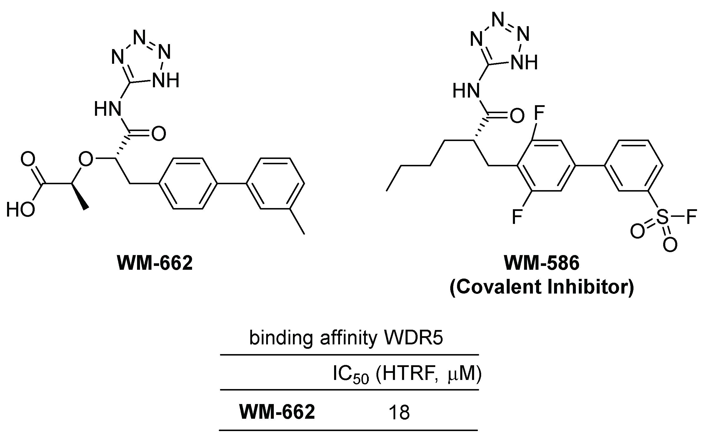
In a recent publication, Ding et al. identified WM-662 as a compound that disrupts the WDR5-MYC interaction by binding the WDR5 binding motif (WBM) of WDR5 (Figure 3) [18]. Using a Novartis in-house library of approximately 330,000 compounds, a high throughput screening was conducted using a competitive homogeneous time-resolved fluorescence (HTRF) assay to identify WM-662, which exhibited moderate activity with an IC50 value of 18 μM. The crystallographic structural study of the WDR5-WM-662 complex led to the development of WM-586, the first covalent inhibitor of WDR5, through a structure–activity relationship (SAR) study and optimization process (Figure 3). A sulfonyl fluoride-based probe was utilized by introducing a sulfonyl fluoride (SF) group to the phenyl moiety, enabling the generation of covalent adducts with the target protein WDR5 [24]. HTRF measurements of WM-586′s IC50 values demonstrated time-dependent inhibition, indicating efficient covalent bond formation. The covalent bond formation between WM-586 and WDR5 was confirmed through mass spectrometry (MS) and liquid chromatography–tandem mass spectrometry (LC-MS/MS) peptide mapping, which revealed that the primary binding residue is mostly Lys250 of WDR5 [18].

2.2. WDR5-MLL1 Protein–Protein Inhibitors

2.2.1. Peptide and Peptidomimetic Inhibitors of WDR5–MLL1 Interactions

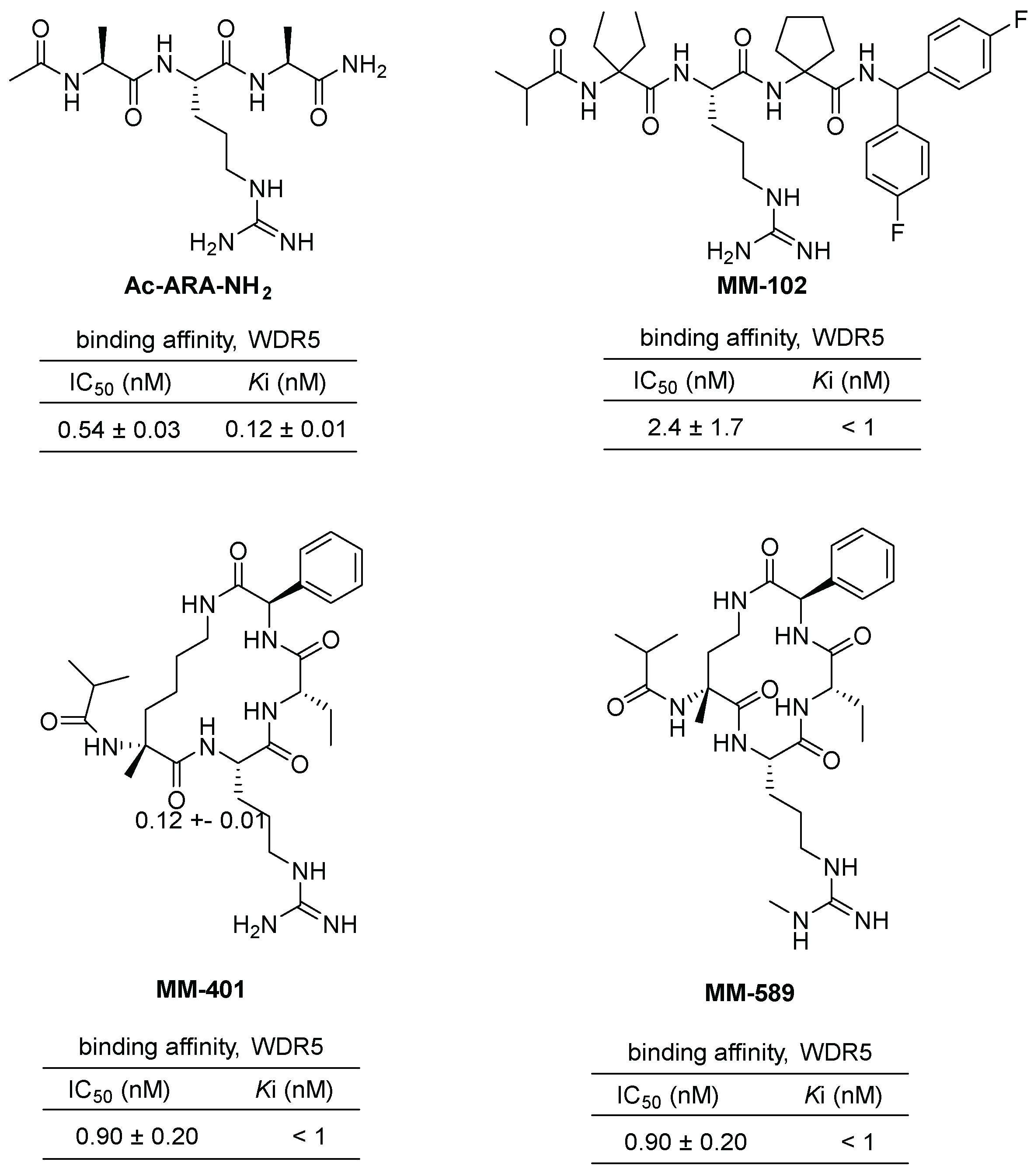
The protein–protein interaction between WDR5 and MLL1 plays a critical role in acute leukemia. Consequently, several inhibitors have been developed to mimic this interaction. The first among these is the tripeptide Ac-ARA-NH2 (Figure 4), designed by Shaomeng Wang’s group, with a Ki value of 120 nM [25]. Through further optimization, the peptidomimetic MM-102 (Figure 4) was identified, exhibiting an IC50 value of 2.4 nM. MM-102 specifically inhibits cell growth in leukemia cells expressing MLL1 fusion proteins. Analysis of the cocrystal structure of MM-102 in complex with WDR5 revealed a stronger hydrophobic interaction than its natural ligand, the MLL1 Win peptide. Utilizing this structure, the cyclized peptidomimetic inhibitor MM-401 (Figure 4) was designed, demonstrating potent inhibition of MLL1 H3K4 methyltransferase (HMT) activity with an IC50 value of 0.9 nM and high specificity [26]. It also exhibited specific cytotoxicity against MLL1-dependent leukemia cells. Notably, the inhibition of MLL1 with MM-401 was shown to play a predominant role in transcriptome regulation in MLL1-dependent leukemia. Subsequently, MM-589 (Figure 4), a peptidomimetic inhibitor containing monomethylated guanidine, was developed and exhibited cellular potency more than 40 times greater than MM-401 [27,28,29]. The cocrystal structure MM-589 with WDR5 further supports its strong inhibition activity. Overall, these inhibitors have demonstrated the potential to mimic the WDR5–MLL1 interaction and inhibit cell growth in leukemia cells. Further research is necessary to evaluate their potential as therapeutic agents.

2.2.2. Benzamide Scaffold Small Molecule Inhibitors of WDR5–MLL1 Interactions

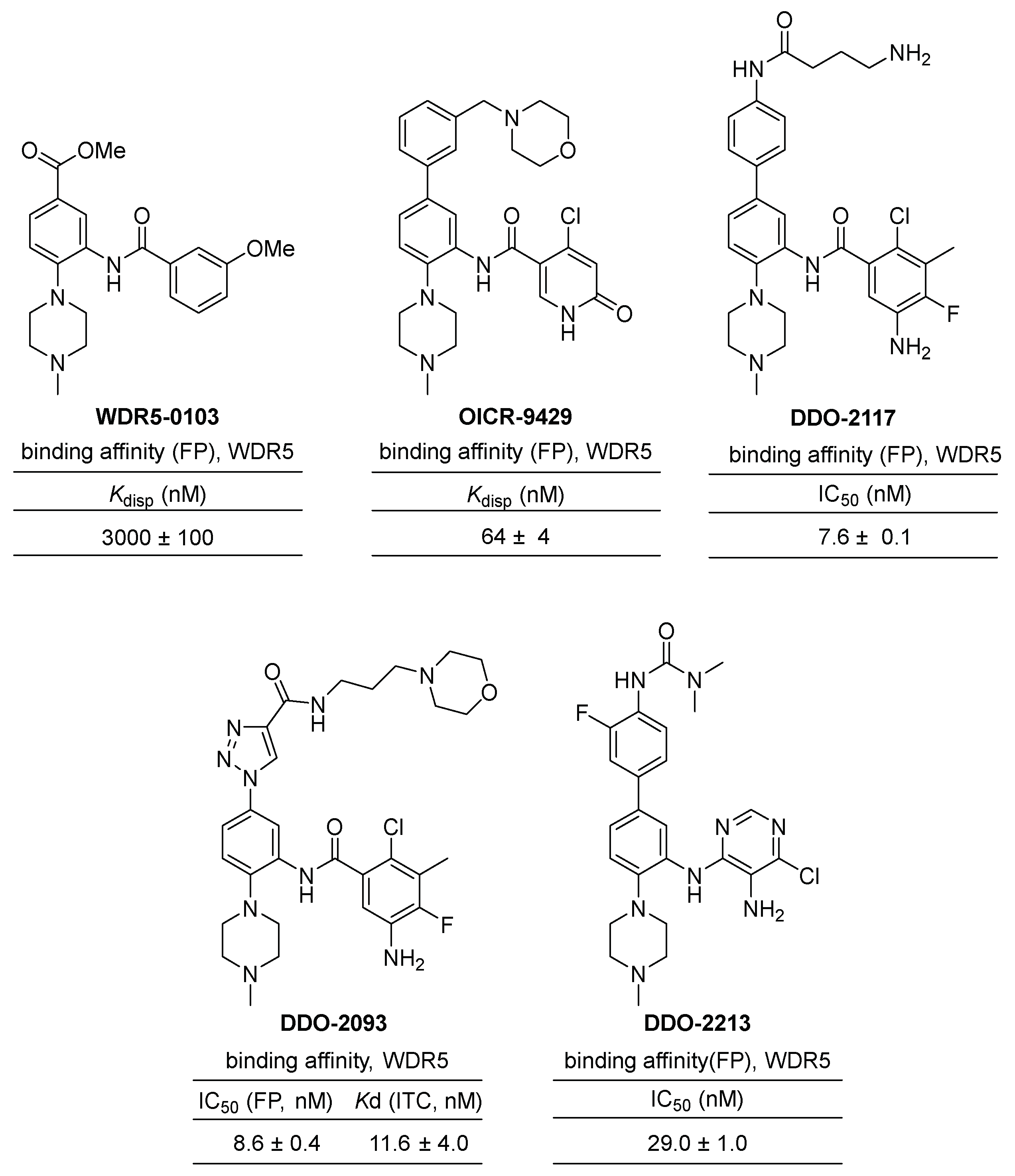
The discovery of small-molecule inhibitors targeting WDR5–MLL interactions has made significant progress in recent years [30,31,32]. A high-throughput screening of a 16,000-molecule library using fluorescence polarization (FP) identified only one strong competitor of the WIN peptide, WDR5-0101, which bears a benzamide scaffold. Further screenings of databases containing 6 million similar chemical structures led to the identification of two additional compounds, WDR5-0102 and 0103, both sharing a benzamide scaffold (Figure 5). WDR5-0102 exhibited a binding affinity of a Kd = 4.0 μM to WDR5, while WDR5-0103 showed a higher affinity with Kd = 0.45 μM. In an inhibition assay of MLL1 HMT activity, WDR5-0103 specifically and dose-dependently decreases the catalytic activity of both the trimeric and tetrameric complexes of MLL1. Crystal structures of WDR5-0103 in complex with WDR5 (PDB code: 3SMR and 3UR4) revealed that the inhibitor directly competes with the MLL1 peptide, disrupting the WDR5–MLL1 interaction by occupying the central cavity (P2 pocket) that normally accommodates an arginine side chain of interacting peptides. This structural information explained the improved potency of WDR5-0103 and provided a basis for the further development of the inhibitors targeting WDR5–MLL1 protein–protein interactions.
OICR-9429 is a potent small-molecule inhibitor of the WDR5–MLL1 interaction, discovered through further optimization of an initial phenyl benzamide hit, WDR5-0102 (Figure 3). OICR-9429 has been utilized as a high-quality chemical probe in various studies investigating the mechanism of WDR5–MLL1 interaction. For example, OICR-9429 selectively inhibits proliferation and induces differentiation of human acute myeloid leukemia (AML) cells expressing C/EBPα p30, a mutation present in 9% of AML patients [32]. Furthermore, it selectively inhibits the proliferation of tumor cells expressing gain-of-function (GOF) mutant p53 resulting in increased histone methylation [33]. It has also been shown to suppress metastasis and PDL1-based immune evasion and increase the chemosensitivity of cisplatin in muscle-invasive bladder cancer [34] and advanced prostate cancer [35]. Additionally, it reduces neuroblastoma cell proliferation by blocking the formation of the WDR5–MYCN complex, which is another isomeric family of MYC [36]. The complex crystal structure of WDR5 with OICR-9429 reveals van der Waals contacts with A47, L321, and I305, as well as a parallel π-stacking interaction of the phenyl ring of the C-5 substituent with the side chains of F133 and Y191 [32,37]. These interactions make OICR-9429 a powerful inhibitor and an ideal chemical probe for further understanding of WDR5–MLL1 interaction and a potential therapeutic for MLL1-driven acute myeloid leukemias.
The Guo group also investigated the structure–activity relationship based on W-26 (Kd = 206 nM), a 4-amino-biphenyl inhibitor [31]. Focused optimization of biphenyl inhibitors led to the development of a potent WDR5-MLL1 inhibitor, DDO-2117, with a Kd value of 13.6 nM and an IC50 value of 0.19 μM in HMT screening [38].
In 2021, the Guo group reported a linear scaffold hopping approach for the discovery of new phenylalkynyl inhibitors. They synthesized a series of phenyltriazoles through further derivatization using click chemistry and found that compound DDO-2093 exhibited the strongest binding affinity with a Kd value of 11.6 nM and an IC50 value of 8.6 nM [39]. Remarkably, DDO-2093 demonstrated selectivity against leukemia cell lines. Additionally, it showed a well-balanced water solubility and cell permeability, and no obvious toxicity was found in normal mice. In vivo assays in the MV4-11 xenograft mouse model further demonstrated that DDO-2093 significantly reduced tumor volume by inhibiting MLL1 HMT activity and downstream signaling gene expression.
The same research group discovered DDO-2213, an orally active and potent inhibitor of WDR5–MLL1 protein–protein interaction [40]. Through a scaffold-hopping strategy from the initial benzamide inhibitor WDR5-0103, they conducted a comprehensive structure–activity relationship study was carried out to identify DDO-221 as a strong inhibitor. DDO-221 exhibited an IC50 of 29 nM in a competitive fluorescence polarization assay and a Kd value of 72.9 nM for the WDR5 protein. It selectively inhibited MLL histone methyltransferase activity and the proliferation of cells harboring MLL translocation. Furthermore, DDO-2213 demonstrated optimal metabolic stability in mouse and human microsomes, along with sufficient pharmacokinetic properties for oral administration. The inhibitor effectively suppressed the growth of MV4-11 xenograft tumors in mice following oral administration. Importantly, this study represents the first verification of the in vivo efficacy of a small molecule targeting the WDR5–MLL1 protein–protein interaction.

2.2.3. Acridine-1,8-Dione Scaffold Small Molecule Inhibitors of WDR5–MLL1 Interactions

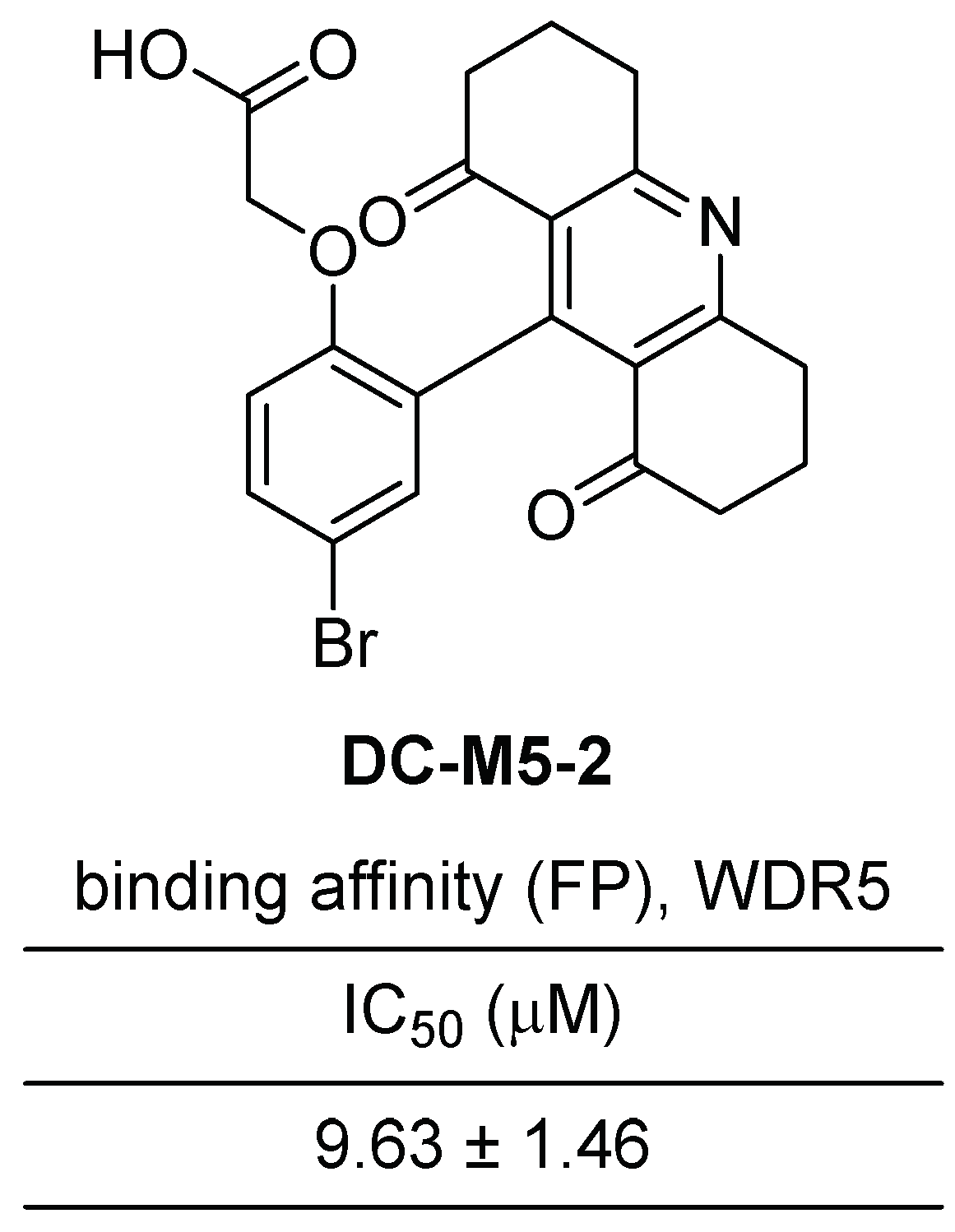
Ye et al. recently reported the discovery of novel acridine-1,8-diones as inhibitors of WDR5–MLL1 interaction [41]. They employed an FP-based high throughput screening to discover small-molecule inhibitors that effectively target the WDR5–MLL1 interaction. Subsequently, nuclear magnetic resonance (NMR) assays were conducted to validate the direct binding between the identified compound and WDR5 and resulting in the development of DC-M5-2 as an inhibitor of WDR5–MLL1 interaction.
DC-M5-2 is characterized as a 9-phenoxy-decahydroacridine-1,8-dione compound, featuring an acid functionality at the ortho position of the phenyl group (Figure 6). The inhibitory activity of DC-M5-2 was demonstrated with an IC50 value of 9.63 μM. To gain further insights into the binding modes and interaction mechanisms between DC-M5-2 and WDR5, a molecular docking study was performed. The results revealed that DC-M5-2 establishes several hydrophobic contacts with surrounding residues, including S49, D107, Y131, Y260, F133, I305, and L321. Additionally, the carboxyl group of DC-M5-2 participates in the formation of a hydrogen bond network with Y191 and C261.

2.2.4. Benzyl Benzamides Scaffold Small Molecule Inhibitors of WDR5–MLL1 Interactions

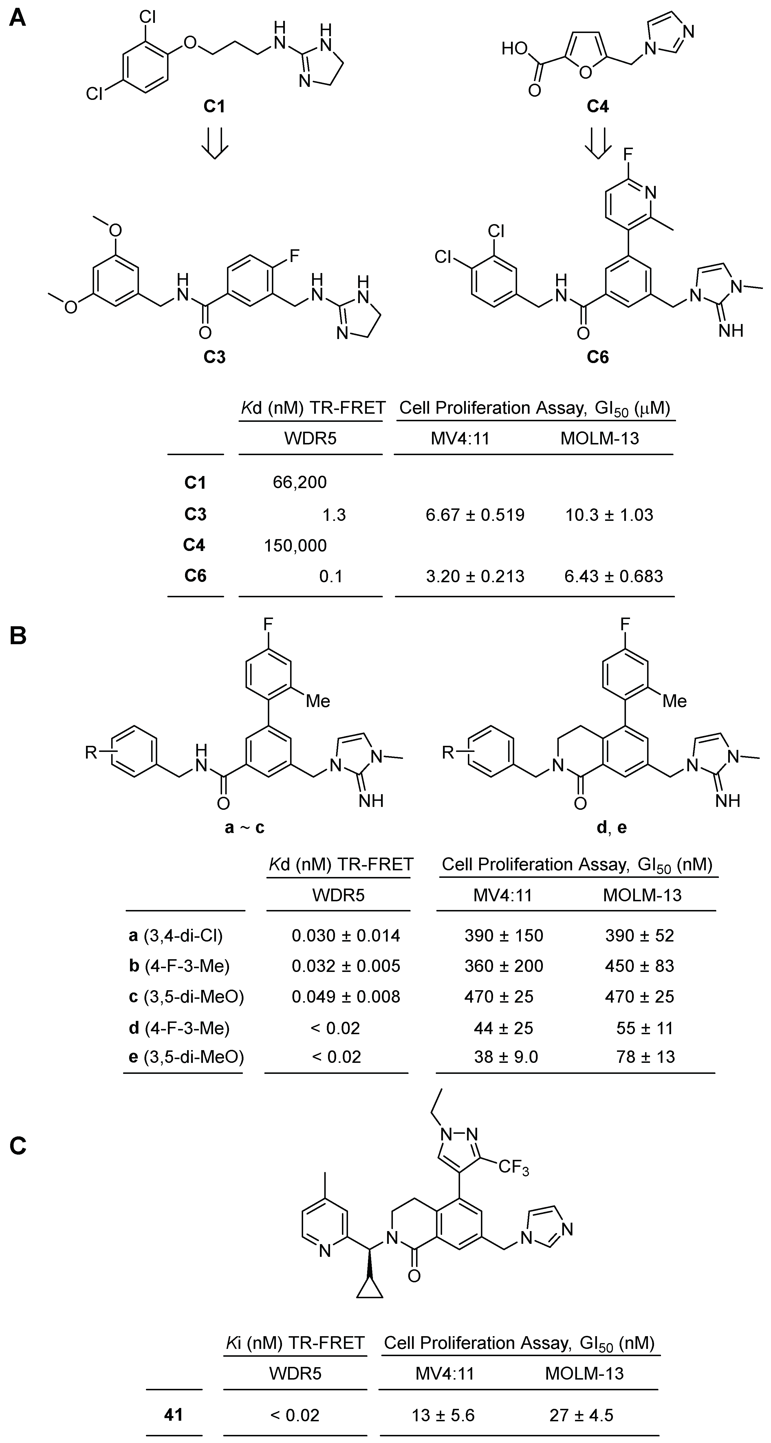
In 2019, novel benzyl benzamides were reported as inhibitors of the WIN site of WDR5 with picomolar affinity [42]. Aho, E. R. et al. conducted a fragment library screening using the HMQC NMR method, screening approximately 13,800 compounds to identify small molecule hits that bind the WIN site. Among the fragment hits, compound C1, a 2- alkylaminoimidazoline, exhibited a Kd value of 66 μM. Further optimization led to the development of benzyl benzamide C3, which displayed improved binding affinity with a Kd value of 1.3 nM (Figure 7A). Additionally, another fragment hit, C4, initially showed a Kd value of 150,000 nM but underwent structural evolution to become the second-generation WIN site inhibitor C6, exhibiting picomolar binding affinity (Kd~100 pM), representing a remarkable 1,500,000-fold improvement in binding affinity.
In vitro evaluations of C3 and C6 focused on their ability to inhibit cell growth against two representative leukemia cell lines, MV4:11 and MOLM-13. Compound C3 demonstrated moderate growth inhibition, with IC50 values of 6.67 μM against MV4:11 and 10.3 μM against MOLM-13. Compound C6 also exhibited moderate growth inhibition, with IC50 values of 3.20 μM against MV4:11 and 6.43 μM against MOLM-13 (Figure 7A).
In a subsequent article in 2020, a structure-based optimization study was conducted on inhibitor C6 to discover more potent and selective WDR5 inhibitors [43]. Tian, J. et al. made modifications to the compound, starting by replacing the upper 4-fluoro-2-methylphenyl with 4-fluoro-4-methylphenyl. They then introduced diverse substituents to the benzyl group and found that 3,4-chlorobenzyl, 4-fluoro-3-methylbenzyl, and 3,5-dimethoxy derivatives displayed potent binding affinities with 0.030, 0.032, 0.049 nM, respectively (Figure 7B).
To further enhance potency and improve drug-like properties, the authors employed a ring-closing strategy, converting the benzamide into dihydroisoquinoline. This modification significantly increased both binding affinity and cell growth inhibitory activity. The dihydroisoquinoline derivatives compounds d and e exhibited binding affinities of less than 0.02 nM and half-maximal growth inhibition (GI50) values of 44 and 38 nM against MV4:11 cell lines, demonstrating potency approximately ten times greater than that of the benzamide derivatives (Figure 7B).
However, despite its robust binding and inhibitory activity of compound e towards the WDR5 WIN-site, the most promising candidate exhibited certain limitations, including low solubility, poor permeability, and insufficient pharmacokinetic profiles [44]. In order to gain further understanding of the binding mechanisms of WDR5 and compound e, Teuscher, K. B. et al. obtained the crystal structure of the WDR5-compound e complex. This structural analysis revealed that the 4-fluoro-2-methylphenyl group binds to hydrophobic subsites and exhibits potential tolerance toward heterocycles. Therefore, they substituted the 4-fluoro-2-methylphenyl group with various six- and five-membered heterocycles, such as substituted pyridines, isoxazoles, and pyrazoles. The derivatives displayed binding affinities comparable to compound e. Notably, 2-methylpyridin-3-yl, 2,5-dimethylpyridin-4-yl, 1-methyl-3-(trifluoromethyl)pyrazol-4-yl, and 1-ethyl-3-(trifluoromethyl)pyrazol-4-yl exhibited good binding affinities of less than 0.02 nM.
Furthermore, the optimization of the 2-iminoimidazole moiety involved the introduction of other nitrogen-containing five-membered heterocycles, including substituted imidazoles, pyrazoles, and triazoles. Among the derivatives, 10midazole-1-yl and 2-methylimidazol-1-yl analogs showed binding affinities of less than 0.02 nM. Finally, the benzyl group was further optimized by transforming it into a substituted pyridine with a benzylic alkyl group, resulting in compound 41 (Figure 7C). This compound exhibited a cLogP of 3.6, a kinetic solubility of 66 μM, a binding affinity of less than 0.02 nM, and a GI50 value of 17 nM against MV4:11.

3. WDR5 Degraders

Targeted protein degradation represents a burgeoning class of therapeutic interventions, with the PROTAC (PROteolysis Targeting Chimeras) technology occupying a prominent position in drug discovery [45]. PROTAC is a dual-function molecule consisting of a small molecule binder for a protein of interest (POI) and a ligand for the E3 ubiquitin ligase. Upon binding to POI, PROTAC can recruit E3 ligase for POI ubiquitination, which is proteolytically degraded by the ubiquitin-proteasome system (UPS). Recently, numerous biotechnology companies are focusing their efforts on the exploration and development of PROTAC-based drugs. Thus, the pivotal role of WDR5 as a scaffolding protein has attracted considerable interest in pursuing targeted degradation strategies targeting WDR5. As some reports have illuminated pharmacologic and expression-level suppression of WDR5, it was explored whether selective functional protein degradation is more efficient compared to the generation of non-functional proteins [32,46]. In 2021, Dölle et al. reported the pioneering design and synthesis of WDR5 degraders based on PROTACs [47]. These PROTACs were designed by incorporating known WDR5 inhibitors, namely OICR-9429 and pyrroloimidazole scaffold inhibitors [32,37,48], as binders for the protein of interest (POI), and employing representative E3 ligases, CRBN, VHL, and MDM229, as the degradation platforms. In the initial series of WDR5-targeting PROTACs, a combination of OICR-9429 as a WDR5 binder and pomalidomide as a ligand for CRBN was synthesized, incorporating PEG and aromatic linkers (Figure 8A). However, in the degradation assay, none of the PROTAC compounds resulted in the elimination of WDR5, while compound 7a showed 12 nM of Kd value.
Subsequently, a new series of PROTAC compounds were designed and synthesized by combining OICR-9429 as a WDR5 binder with a VHL ligand, utilizing aliphatic, PEG, and aromatic linkers (Figure 8B). A total of nine PROTAC compounds were prepared. The aliphatic and aromatic linked PROTACs exhibited moderate to good binding affinity, with compound 8g, featuring a four-carbon linker, displaying the highest degradation activity (DC50 = 53 nM). However, the maximum degradation efficacy achieved was relatively low, with 58% of Dmax (Figure 8B). Additionally, three PROTAC compounds were designed and synthesized by combining OICR-9429 as a WDR5 binder with an MDM ligand, incorporating PEG and aromatic linkers (Figure 8C). Unfortunately, these compounds did not exhibit significant binding affinity towards WDR5, determined by the NanoBRET assay.
A series of WDR5-PROTAC was designed and synthesized, incorporating a pyrroloimidolylbenzene scaffold as the WDR5 binder and a VHL ligand attached by PEG linkers (Figure 8D). Among the seven PROTAC compounds tested, only one PROTAC with two PEG linkers, compound 17b, exhibited moderate binding affinity and mild degradation activity (DC50 = 1.24 nM).
Although various WDR5-PROTAC degraders were developed by employing different WDR5 binders and E3 ligase ligands, it was observed that the degradation activity is highly influenced by the type and length of the linker, as well as the choice of E3 ligase ligand. PROTACs based on the VHL E3 ligase platform demonstrated relatively better degradation activity. However, limited structural variations were explored regarding the exit vectors of WDR5 binders, E3 ligase ligands, and linkers.
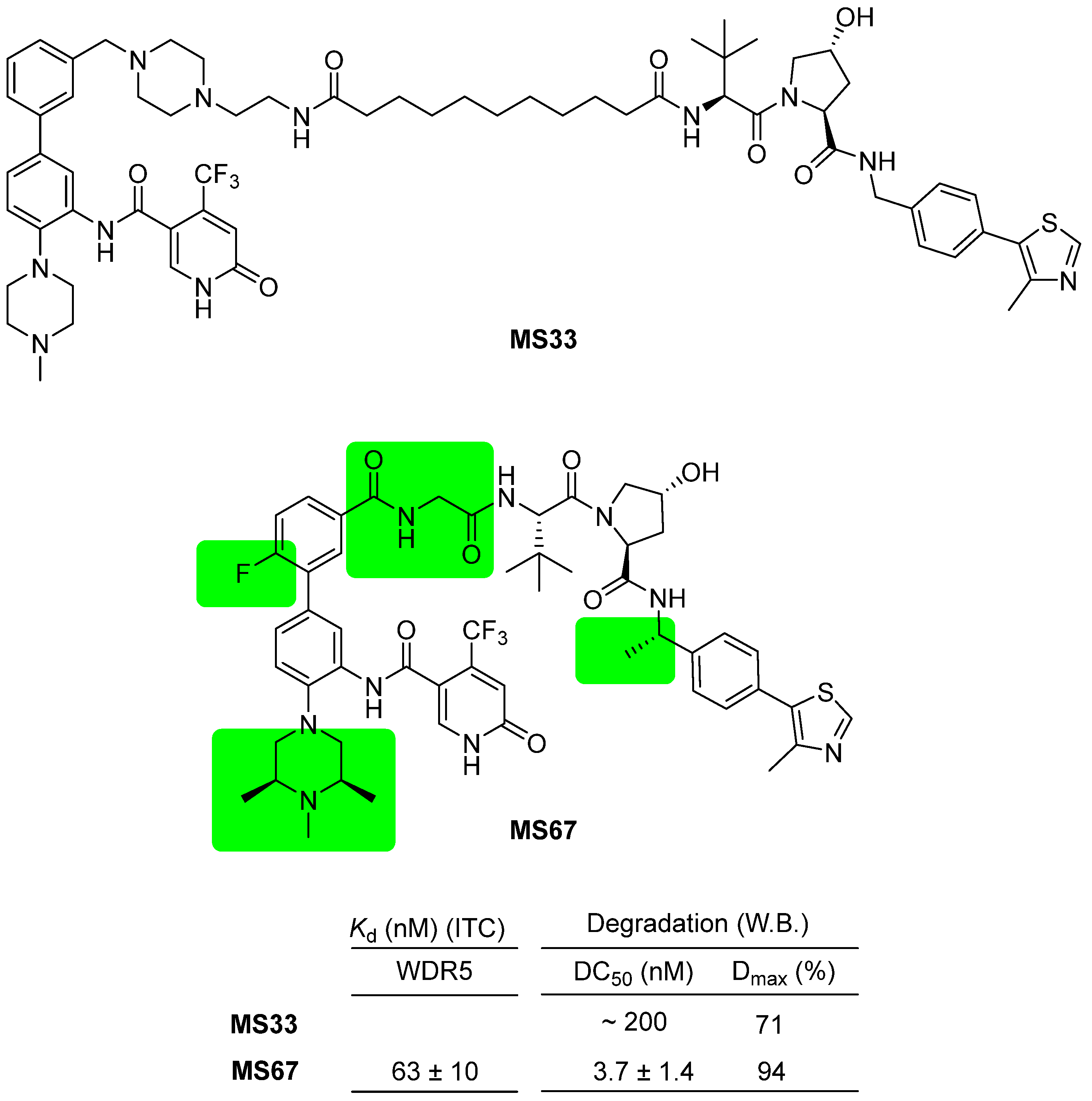
Selective PROTAC degraders targeting WDR5 have been reported and demonstrated to effectively suppress tumor growth in vivo and improve survival rates in an AML-PDX xenograft model. Two PROTAC degraders, namely MS33 and MS67, composed of phenylbenzamide-scaffolded WDR5 binders and VHL ligands, were introduced (Figure 9) [39]. These degraders exhibit structural variations compared to previous WDR5 PROTACs, specifically in the exit vector of the phenylbenzamide-scaffolded WDR5 binder, which is positioned meta to the phenyl group. MS33 and MS67 display distinct structural differences as well. The linking chemistry between the two PROTACs differs; MS33 employs an alkyl linker, while MS67 utilizes an amide linker. Additionally, the WDR5 binder was optimized from N-methylpiperazine in MS33 to 2,6-dimethyl-N-methylpiperazine in MS67. Importantly, the two PROTACs are distinguished by their linkers; MS33 possesses a relatively long piperazinylalkyl linker, while MS67 has a very short or no linker. X-ray crystallographic data of the two PROTACs revealed that the structures of the ternary complexes differ completely based on the linker length. Ultimately, the short-linked MS67 exhibited superior binding affinity and higher degradation efficacy. MS67 demonstrated degradation values of 3.7 nM of DC50 value and 94% for Dmax against wild-type WDR5, while it exhibited no degradation activity against WDR5 D172A mutant and reduced activity against WDR5 Y191A mutant. MS67 exhibited potent growth suppression of AML cells from deidentified patients in vitro and displayed efficacy in an MLL-AF9+ AML PDX-xenograft model, leading to prolonged survival rates without any significant loss of body weight.
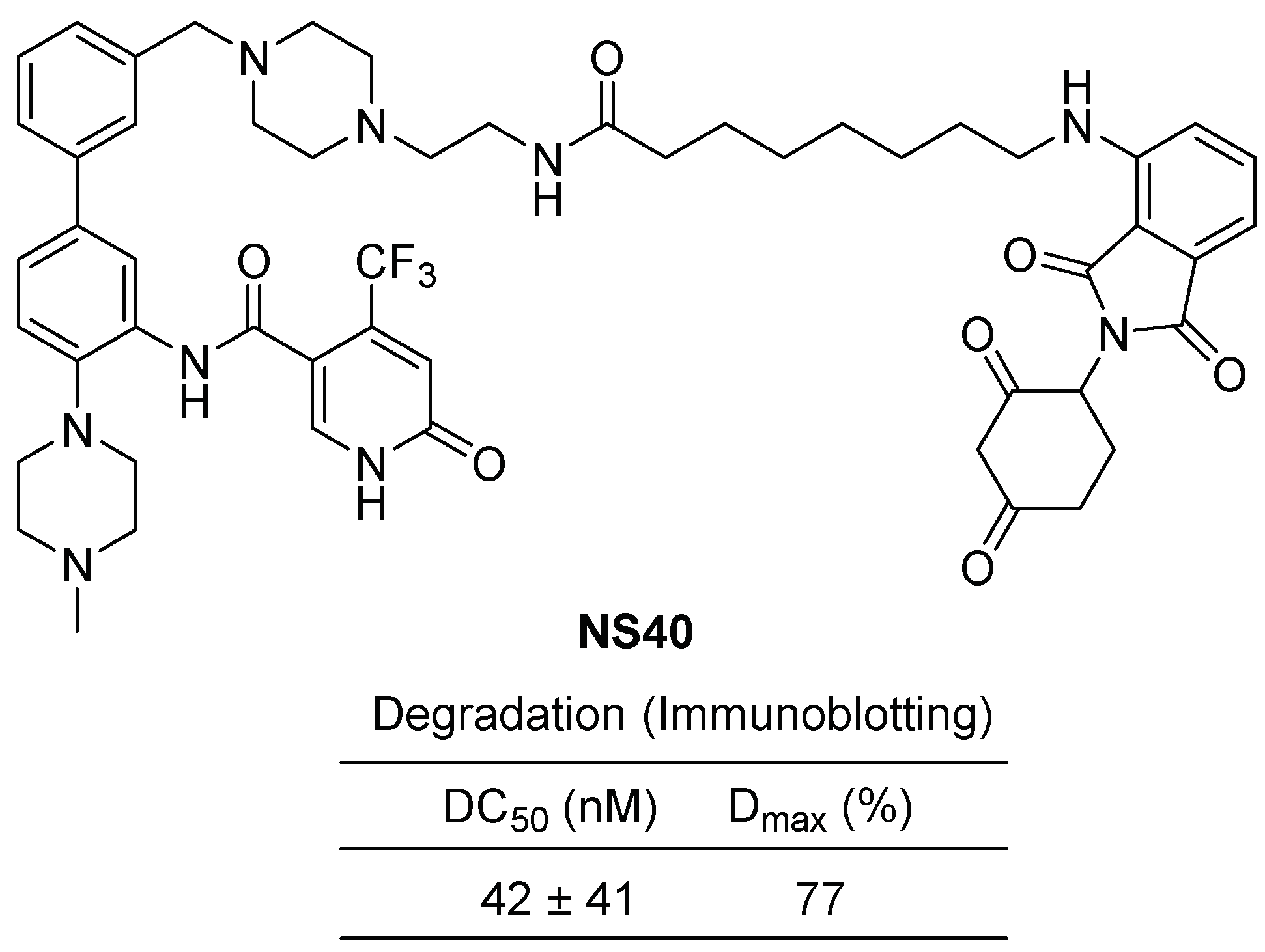
In 2022, the same research group published a study on a dual degrader targeting both WDR5 and CRBN neo-substrates [49]. Thalidomide and its derivatives, such as lenalidomide and pomalidomide, have been demonstrated as effective degraders against ikaros and aiolos, leading to their utilization as anticancer drugs for multiple myeloma. The dual degrader NS40 was designed by linking phenylbenzamide as a WDR5 binder and lenalidomide as a CRBN ligand using a piperazine-containing alkylamide linker (Figure 10). NS40 was found to induce the degradation of WDR5 through the PROTAC mechanism and ikaros1/3 through the molecular glue mechanism. NS40 exhibited successful inhibition of tumor cell growth and demonstrated efficacy in a PDX mouse xenograft model.

4. Conclusions

Until now, peptidomimetics and small molecules targeting the WDR5 protein–protein interaction (PPI) have been developed as anticancer strategies. Those WDR5 inhibitors reduce the proliferation of cancer cells. However, their evaluation has primarily been limited to cellular studies due to their non-optimized drug-like properties. Some in vivo animal studies of WDR5-MLL1 inhibitors have been performed, and no clinical studies have been reported so far. The efficacies and affinities of these inhibitors still require validation in animal models and subsequent clinical trials. Furthermore, the inhibition of the WDR5 PPI may potentially lead to cytotoxicity and side effects due to the nonselective interference with normal cellular functions. It has also been reported that excessive targeted protein degradation can lead to on-target-mediated side effects similar to gene knockdown approaches. Thus, it is imperative to assess the safety profiles of these inhibitors in future investigations. However, it is still noteworthy that a few WDR5 targeting compounds were reported as efficient with no toxicity. For example, OICR-9429, a small molecule antagonist, has low toxicity to normal cells and selectively inhibits only AML cells with upregulated C/EBPα-mutant p30 isoform. Some WDR5 inhibitors have shown dose-dependent in vivo antitumor efficacy without toxicity by oral dosing. WDR5 PROTACs have recently shown in vivo efficacy in a mouse MV4:11 xenograft model.
Finally, the development of WDR5 inhibitors and degraders is at an early stage but still with great potential to target several cancers.

Author Contributions

Conceptualization, S.H. and D.S.; methodology, S.H. and D.S.; writing—original draft preparation, R.G., D.O., R.P., S.H. and D.S.; writing—review and editing, S.H. and D.S. All authors have read and agreed to the published version of the manuscript.

Funding

This research was funded by the National Research Foundation of Korea (NRF-2020R1A6A1A03043708 NRF-2021R1A2C1012280, NRF-2021R1I1A1A01059834 and MRC 2017R1A5A2015541), and supported by “Regional Innovation Strategy (RIS)” through the National Research Foundation of Korea (NRF) funded by the Ministry of Education (MOE) (2021RIS-001). This work was supported by a research grant from the Chungbuk National University in 2021.

Institutional Review Board Statement

Not applicable.

Informed Consent Statement

Not applicable.

Data Availability Statement

Not applicable.

Conflicts of Interest

The authors declare that there are no conflict of interest.

References

  1. Li, D.; Roberts, R. Human Genome and Diseases: WD-repeat proteins: Structure characteristics, biological function, and their involvement in human diseases. Cell. Mol. Life Sci. 2001, 58, 2085–2097. [Google Scholar] [CrossRef]
  2. Chen, X.; Xu, J.J.; Wang, X.H.; Long, G.L.; You, Q.D.; Guo, X.K. Targeting WD Repeat-Containing Protein 5 (WDR5): A Medicinal Chemistry Perspective. J. Med. Chem. 2021, 64, 10537–10556. [Google Scholar] [CrossRef]
  3. Teuscher, K.B.; Chowdhury, S.; Meyers, K.M.; Tian, J.; Sai, J.; Van Meveren, M.; South, T.M.; Sensintaffar, J.L.; Rietz, T.A.; Goswami, S.; et al. Structure-based discovery of potent WD repeat domain 5 inhibitors that demonstrate efficacy and safety in preclinical animal models. Proc. Natl. Acad. Sci. USA 2023, 120, e2211297120. [Google Scholar] [CrossRef]
  4. Ge, Z.; Song, E.; Li, J.; Dovat, S.; Song, C. Clinical Significance of WDR5 High Expression and Its Effect on Tumorigenesis in Adult Leukemia. Blood 2015, 126, 3657. [Google Scholar] [CrossRef]
  5. Chen, X.; Xie, W.; Gu, P.; Cai, Q.; Wang, B.; Xie, Y.; Dong, W.; He, W.; Zhong, G.; Lin, T.; et al. Upregulated WDR5 promotes proliferation, self-renewal and chemoresistance in bladder cancer via mediating H3K4 trimethylation. Sci. Rep. 2015, 5, 8293. [Google Scholar] [CrossRef] [Green Version]
  6. Xie, Q.; Li, Z.; Chen, J. WDR5 positively regulates p53 stability by inhibiting p53 ubiquitination. Biochem. Biophys. Res. Commun. 2017, 487, 333–338. [Google Scholar] [CrossRef] [Green Version]
  7. Huang, M.; Dong, W.; Xie, R.H.; Wu, J.L.; Su, Q.; Li, W.G.; Yao, K.; Chen, Y.L.; Zhou, Q.H.; Zhang, Q.; et al. HSF1 facilitates the multistep process of lymphatic metastasis in bladder cancer via a novel PRMT5-WDR5-dependent transcriptional program. Cancer Commun. 2022, 42, 447–470. [Google Scholar] [CrossRef]
  8. Aho, E.R.; Weissmiller, A.M.; Fesik, S.W.; Tansey, W.P. Targeting WDR5: A WINning Anti-Cancer Strategy? Epigenet. Insights 2019, 12, 2516865719865282. [Google Scholar] [CrossRef] [PubMed] [Green Version]
  9. Southall, S.M.; Wong, P.-S.; Odho, Z.; Roe, S.M.; Wilson, J.R. Structural Basis for the Requirement of Additional Factors for MLL1 SET Domain Activity and Recognition of Epigenetic Marks. Mol. Cell 2009, 33, 181–191. [Google Scholar] [CrossRef] [PubMed]
  10. Dharmarajan, V.; Lee, J.-H.; Patel, A.; Skalnik, D.G.; Cosgrove, M.S. Structural Basis for WDR5 Interaction (Win) Motif Recognition in Human SET1 Family Histone Methyltransferases. J. Biol. Chem. 2012, 287, 27275–27289. [Google Scholar] [CrossRef] [Green Version]
  11. Namitz, K.E.W.; Tan, S.; Cosgrove, M.S. Hierarchical assembly of the MLL1 core complex regulates H3K4 methylation and Hierarchical assembly of the MLL1 core complex regis dependent on temperature and component concentration. J. Biol. Chem. 2023, 299, 102874. [Google Scholar] [CrossRef] [PubMed]
  12. Ziemin-van der Poel, S.; McCabe, N.R.; Gill, H.J.; Espinosa, R.; Patel, Y.; Harden, A.; Rubinelli, P.; Smith, S.D.; LeBeau, M.M.; Rowley, J.D. Identification of a gene, MLL, that spans the breakpoint in 11q23 translocations associated with human leukemias. Proc. Natl. Acad. Sci. USA 1991, 88, 10735–10739. [Google Scholar] [CrossRef] [PubMed]
  13. Mitchell, K.; Sprowls, S.A.; Arora, S.; Shakya, S.; Silver, D.J.; Goins, C.M.; Wallace, L.; Roversi, G.; Schafer, R.E.; Kay, K.; et al. WDR5 represents a therapeutically exploitable target for cancer stem cells in glioblastoma. Genes Dev. 2023, 37, 86–102. [Google Scholar] [CrossRef] [PubMed]
  14. Thomas, L.R.; Adams, C.M.; Wang, J.; Weissmiller, A.M.; Creighton, J.; Lorey, S.L.; Liu, Q.; Fesik, S.W.; Eischen, C.M.; Tansey, W.P. Interaction of the oncoprotein transcription factor MYC with its chromatin cofactor WDR5 is essential for tumor maintenance. Proc. Natl. Acad. Sci. USA 2019, 116, 25260–25268. [Google Scholar] [CrossRef] [PubMed] [Green Version]
  15. Gabay, M.; Li, Y.; Felsher, D.W. MYC activation is a hallmark of cancer initiation and maintenance. Cold Spring Harb. Perspect. Med. 2014, 4, a014241. [Google Scholar] [CrossRef] [Green Version]
  16. Dang, C.V. MYC on the Path to Cancer. Cell 2012, 149, 22–35. [Google Scholar] [CrossRef] [Green Version]
  17. Thomas, L.R.; Wang, Q.; Grieb, B.; Phan, J.; Foshage, A.M.; Sun, Q.; Olejniczak, E.T.; Clark, T.; Dey, S.; Lorey, S.; et al. Interaction with WDR5 Promotes Target Gene Recognition and Tumorigenesis by MYC. Mol. Cell 2015, 58, 440–452. [Google Scholar] [CrossRef] [Green Version]
  18. Ding, J.; Li, G.; Liu, H.; Liu, L.; Lin, Y.; Gao, J.; Zhou, G.; Shen, L.; Zhao, M.; Yu, Y.; et al. Discovery of Potent Small-Molecule Inhibitors of WDR5-MYC Interaction. ACS Chem. Biol. 2023, 18, 34–40. [Google Scholar] [CrossRef]
  19. Macdonald, J.D.; Simon, S.C.; Han, C.; Wang, F.; Shaw, J.G.; Howes, J.E.; Sai, J.; Yuh, J.P.; Camper, D.; Alicie, B.M.; et al. Discovery and Optimization of Salicylic Acid-Derived Sulfonamide Inhibitors of the WD Repeat-Containing Protein 5–MYC Protein–Protein Interaction. J. Med. Chem. 2019, 62, 11232–11259. [Google Scholar] [CrossRef]
  20. Simon, S.C.; Wang, F.; Thomas, L.R.; Phan, J.; Zhao, B.; Olejniczak, E.T.; Macdonald, J.D.; Shaw, J.G.; Schlund, C.; Payne, W.; et al. Discovery of WD Repeat-Containing Protein 5 (WDR5)-MYC Inhibitors Using Fragment-Based Methods and Structure-Based Design. J. Med. Chem. 2020, 63, 4315–4333. [Google Scholar] [CrossRef]
  21. Thomas, L.R.; Adams, C.M.; Fesik, S.W.; Eischen, C.M.; Tansey, W.P. Targeting MYC through WDR5. Mol. Cell. Oncol. 2020, 7, 1709388. [Google Scholar] [CrossRef] [PubMed] [Green Version]
  22. Thomas, L.R.; Foshage, A.M.; Weissmiller, A.M.; Tansey, W.P. The MYC-WDR5 Nexus and Cancer. Cancer Res. 2015, 75, 4012–4015. [Google Scholar] [CrossRef] [PubMed] [Green Version]
  23. Carugo, A.; Genovese, G.; Seth, S.; Nezi, L.; Rose, J.L.; Bossi, D.; Cicalese, A.; Shah, P.K.; Viale, A.; Pettazzoni, P.F.; et al. In Vivo Functional Platform Targeting Patient-Derived Xenografts Identifies WDR5-Myc Association as a Critical Determinant of Pancreatic Cancer. Cell Rep. 2016, 16, 133–147. [Google Scholar] [CrossRef] [PubMed] [Green Version]
  24. Narayanan, A.; Jones, L.H. Sulfonyl fluorides as privileged warheads in chemical biology. Chem. Sci. 2015, 6, 2650–2659. [Google Scholar] [CrossRef] [PubMed] [Green Version]
  25. Karatas, H.; Townsend, E.C.; Cao, F.; Chen, Y.; Bernard, D.; Liu, L.; Lei, M.; Dou, Y.L.; Wang, S.M. High-Affinity, Small-Molecule Peptidomimetic Inhibitors of MLL1/WDR5 Protein-Protein Interaction. J. Am. Chem. Soc. 2013, 135, 669–682. [Google Scholar] [CrossRef] [Green Version]
  26. Cao, F.; Townsend, E.C.; Karatas, H.; Xu, J.; Li, L.; Lee, S.; Liu, L.; Chen, Y.; Ouillette, P.; Zhu, J.D.; et al. Targeting MLL1 H3K4 Methyltransferase Activity in Mixed-Lineage Leukemia. Mol. Cell 2014, 53, 247–261. [Google Scholar] [CrossRef] [Green Version]
  27. Karatas, H.; Li, Y.B.; Liu, L.; Ji, J.; Lee, S.; Chen, Y.; Yang, J.L.; Huang, L.Y.; Bernard, D.; Xu, J.; et al. Discovery of a Highly Potent, Cell-Permeable Macrocyclic Peptidomimetic (MM-589) Targeting the WD Repeat Domain 5 Protein (WDR5)-Mixed Lineage Leukemia (MLL) Protein-Protein Interaction. J. Med. Chem. 2017, 60, 4818–4839. [Google Scholar] [CrossRef]
  28. Senisterra, G.; Wu, H.; Allali-Hassani, A.; Wasney, G.A.; Barsyte-Loyejoy, D.; Dombrovski, L.; Dong, A.P.; Nguyen, K.T.; Smil, D.; Bolshan, Y.; et al. Small-molecule inhibition of MLL activity by disruption of its interaction with WDR5. Biochem. J. 2013, 449, 151–159. [Google Scholar] [CrossRef]
  29. Li, D.D.; Wang, Z.H.; Chen, W.L.; Xie, Y.Y.; You, Q.D.; Guo, X.K. Structure-based design of ester compounds to inhibit MLL complex catalytic activity by targeting mixed lineage leukemia 1 (MLL1)-WDR5 interaction. Biorg. Med. Chem. 2016, 24, 6109–6118. [Google Scholar] [CrossRef]
  30. Bolshan, Y.; Getlik, M.; Kuznetsova, E.; Wasney, G.A.; Hajian, T.; Poda, G.; Nguyen, K.T.; Wu, H.; Dombrovski, L.; Dong, A.; et al. Synthesis, Optimization, and Evaluation of Novel Small Molecules as Antagonists of WDR5-MLL Interaction. ACS Med. Chem. Lett. 2013, 4, 353–357. [Google Scholar] [CrossRef] [Green Version]
  31. Li, D.D.; Chen, W.L.; Xu, X.L.; Jiang, F.; Wang, L.; Xie, Y.Y.; Zhang, X.J.; Guo, X.K.; You, Q.D.; Sun, H.P. Structure-based design and synthesis of small molecular inhibitors disturbing the interaction of MLL1-WDR5. Eur. J. Med. Chem. 2016, 118, 1–8. [Google Scholar] [CrossRef] [PubMed]
  32. Grebien, F.; Vedadi, M.; Getlik, M.; Giambruno, R.; Grover, A.; Avellino, R.; Skucha, A.; Vittori, S.; Kuznetsova, E.; Smil, D.; et al. Pharmacological targeting of the Wdr5-MLL interaction in C/EBPα N-terminal leukemia. Nat. Chem. Biol. 2015, 11, 571–578. [Google Scholar] [CrossRef]
  33. Zhu, J.; Sammons, M.A.; Donahue, G.; Dou, Z.; Vedadi, M.; Getlik, M.; Barsyte-Lovejoy, D.; Al-awar, R.; Katona, B.W.; Shilatifard, A.; et al. Gain-of-function p53 mutants co-opt chromatin pathways to drive cancer growth. Nature 2015, 525, 206–211. [Google Scholar] [CrossRef] [Green Version]
  34. Zhang, J.T.; Zhou, Q.H.; Xie, K.J.; Cheng, L.; Peng, S.M.; Xie, R.H.; Liu, L.X.; Zhang, Y.J.; Dong, W.; Han, J.L.; et al. Targeting WD repeat domain 5 enhances chemosensitivity and inhibits proliferation and programmed death-ligand 1 expression in bladder cancer. J. Exp. Clin. Cancer Res. 2021, 40, 203. [Google Scholar] [CrossRef] [PubMed]
  35. Zhou, Q.; Chen, X.; He, H.; Peng, S.; Zhang, Y.; Zhang, J.; Cheng, L.; Liu, S.; Huang, M.; Xie, R.; et al. WD repeat domain 5 promotes chemoresistance and Programmed Death-Ligand 1 expression in prostate cancer. Theranostics 2021, 11, 4809–4824. [Google Scholar] [CrossRef]
  36. Sun, Y.; Bell, J.L.; Carter, D.; Gherardi, S.; Poulos, R.C.; Milazzo, G.; Wong, J.W.H.; Al-Awar, R.; Tee, A.E.; Liu, P.Y.; et al. WDR5 Supports an N-Myc Transcriptional Complex That Drives a Protumorigenic Gene Expression Signature in Neuroblastoma. Cancer Res. 2015, 75, 5143–5154. [Google Scholar] [CrossRef] [PubMed] [Green Version]
  37. Getlik, M.; Smil, D.; Zepeda-Velazquez, C.; Bolshan, Y.; Poda, G.; Wu, H.; Dong, A.P.; Kuznetsova, E.; Marcellus, R.; Senisterra, G.; et al. Structure-Based Optimization of a Small Molecule Antagonist of the Interaction Between WD Repeat-Containing Protein 5 (WDR5) and Mixed-Lineage Leukemia 1 (MLL1). J. Med. Chem. 2016, 59, 2478–2496. [Google Scholar] [CrossRef]
  38. Li, D.D.; Chen, W.L.; Wang, Z.H.; Xie, Y.Y.; Xu, X.L.; Jiang, Z.Y.; Zhang, X.J.; You, Q.D.; Guo, X.K. High-affinity small molecular blockers of mixed lineage leukemia 1 (MLL1)-WDR5 interaction inhibit MLL1 complex H3K4 methyltransferase activity. Eur. J. Med. Chem. 2016, 124, 480–489. [Google Scholar] [CrossRef] [PubMed]
  39. Chen, W.L.; Chen, X.; Li, D.D.; Wang, X.H.; Long, G.L.; Jiang, Z.Y.; You, Q.D.; Guo, X.K. Discovery of a potent MLL1 and WDR5 protein-protein interaction inhibitor with in vivo antitumor activity. Eur. J. Med. Chem. 2021, 223, 113677. [Google Scholar] [CrossRef]
  40. Chen, W.L.; Chen, X.; Li, D.D.; Zhou, J.R.; Jiang, Z.Y.; You, Q.D.; Guo, X.K. Discovery of DDO-2213 as a Potent and Orally Bioavailable Inhibitor of the WDR5-Mixed Lineage Leukemia 1 Protein-Protein Interaction for the Treatment of MLL Fusion Leukemia. J. Med. Chem. 2021, 64, 8221–8245. [Google Scholar] [CrossRef]
  41. Ye, X.Q.; Zhang, R.K.; Lian, F.L.; Zhang, W.Y.; Lu, W.C.; Han, J.; Zhang, N.X.; Jin, J.; Luo, C.; Chen, K.X.; et al. The identification of novel small-molecule inhibitors targeting WDR5-MLL1 interaction through fluorescence polarization based high-throughput screening. Bioorg. Med. Chem. Lett. 2019, 29, 638–645. [Google Scholar] [CrossRef] [PubMed]
  42. Aho, E.R.; Wang, J.; Gogliotti, R.D.; Howard, G.C.; Phan, J.; Acharya, P.; Macdonald, J.D.; Cheng, K.; Lorey, S.L.; Lu, B.; et al. Displacement of WDR5 from Chromatin by a WIN Site Inhibitor with Picomolar Affinity. Cell Rep. 2019, 26, 2916–2928.e13. [Google Scholar] [CrossRef] [PubMed] [Green Version]
  43. Tian, J.; Teuscher, K.B.; Aho, E.R.; Alvarado, J.R.; Mills, J.J.; Meyers, K.M.; Gogliotti, R.D.; Han, C.H.; Macdonald, J.D.; Sai, J.Q.; et al. Discovery and Structure-Based Optimization of Potent and Selective WD Repeat Domain 5 (WDR5) Inhibitors Containing a Dihydroisoquinolinone Bicyclic Core. J. Med. Chem. 2020, 63, 656–675. [Google Scholar] [CrossRef] [PubMed]
  44. Teuscher, K.B.; Meyers, K.M.; Wei, Q.Q.; Mills, J.J.; Tian, J.H.; Alvarado, J.; Sai, J.Q.; Van Meveren, M.; South, T.M.; Rietz, T.A.; et al. Discovery of Potent Orally Bioavailable WD Repeat Domain 5(WDR5) Inhibitors Using a Pharmacophore-Based Optimization. J. Med. Chem. 2022, 65, 6287–6312. [Google Scholar] [CrossRef] [PubMed]
  45. Békés, M.; Langley, D.R.; Crews, C.M. PROTAC targeted protein degraders: The past is prologue. Nat. Rev. Drug Discov. 2022, 21, 181–200. [Google Scholar] [CrossRef]
  46. Yu, X.F.; Li, D.X.; Kottur, J.; Shen, Y.D.; Kim, H.S.; Park, K.S.; Tsai, Y.H.; Gong, W.D.; Wang, J.; Suzuki, K.; et al. A selective WDR5 degrader inhibits acute myeloid leukemia in patient-derived mouse models. Sci. Transl. Med. 2021, 13, eabj1578. [Google Scholar] [CrossRef]
  47. Dölle, A.; Adhikari, B.; Krämer, A.; Weckesser, J.; Berner, N.; Berger, L.-M.; Diebold, M.; Szewczyk, M.M.; Barsyte-Lovejoy, D.; Arrowsmith, C.H.; et al. Design, Synthesis, and Evaluation of WD-Repeat-Containing Protein 5 (WDR5) Degraders. J. Med. Chem. 2021, 64, 10682–10710. [Google Scholar] [CrossRef]
  48. Wang, F.; Jeon, K.O.; Salovich, J.M.; Macdonald, J.D.; Alvarado, J.H.; Gogliotti, R.D.; Phan, J.; Olejniczak, E.T.; Sun, Q.; Wang, S.D.; et al. Discovery of Potent 2-Aryl-6,7-dihydro-5H-pyrrolo 1,2-a imidazoles as WDR5-WIN-Site Inhibitors Using Fragment-Based Methods and Structure-Based Design. J. Med. Chem. 2018, 61, 5623–5642. [Google Scholar] [CrossRef]
  49. Li, D.X.; Yu, X.F.; Kottur, J.; Gong, W.D.; Zhang, Z.; Storey, A.J.; Tsai, Y.H.; Uryu, H.; Shen, Y.D.; Byrum, S.D.; et al. Discovery of a dual WDR5 and Ikaros PROTAC degrader as an anti-cancer therapeutic. Oncogene 2022, 41, 3328–3340. [Google Scholar] [CrossRef]
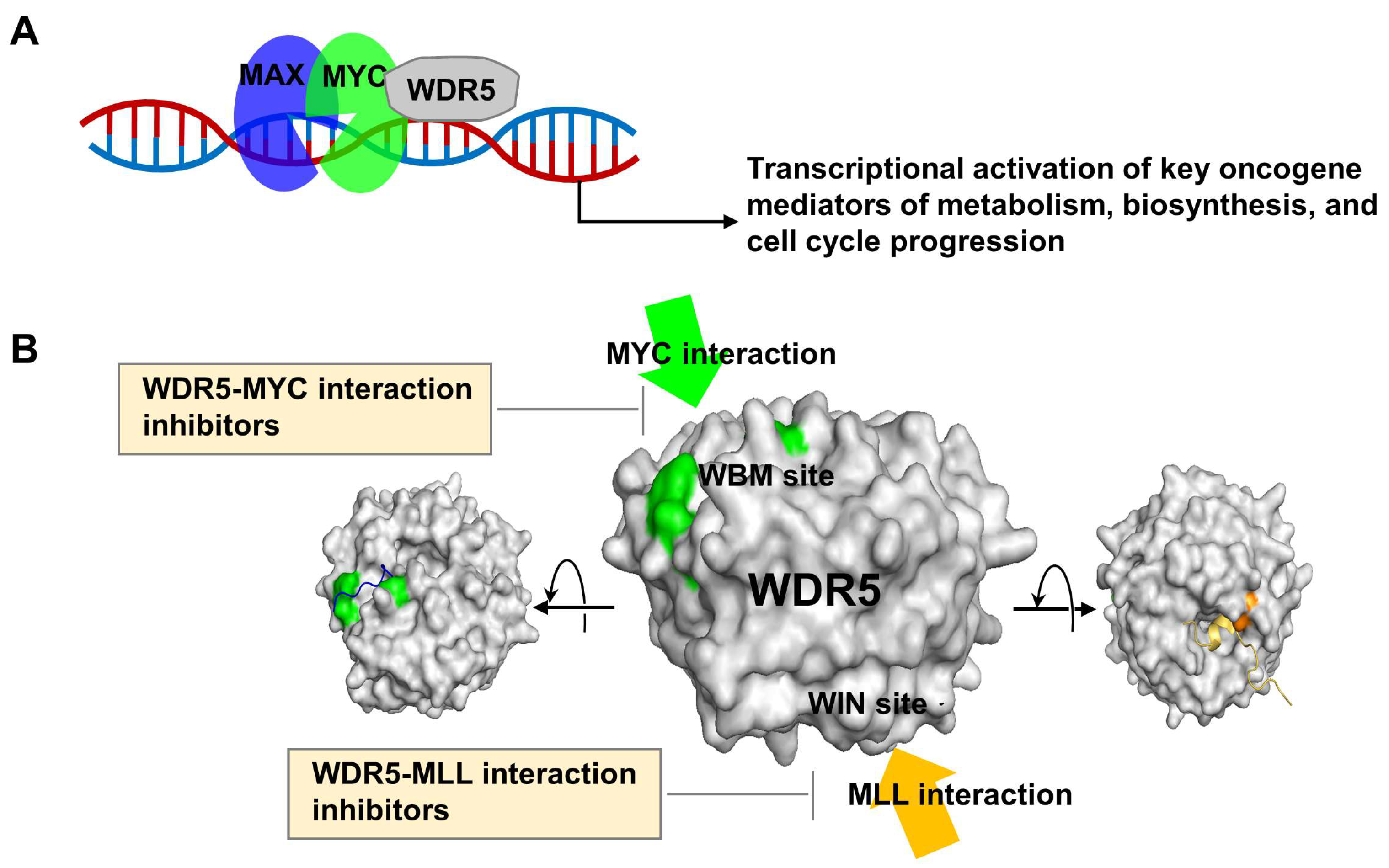
Figure 1. Role of WDR5 in tumorigenesis and its protein interaction partners. (A) WDR5 induces transcriptional activation of key oncogenes. (B) WDR5 interacts with MYC and MLL proteins on a distinct interface, presenting a promising avenue for the development of therapeutic targets in cancer treatment. Modified X-ray cocrystal structures of WDR5 with MYC peptide (green, PDB: 4Y7R) and WDR5 with Win peptide (orange, PDB: 4ESG).
Cancers 15 03910 g001
Figure 2. Compound 12 as a WDR5–MYC interaction inhibitor. (A) Combining the HTS sulfonamide hit 1 with fragment-based hit F2 results in merged compounds. Optimization of the merged compounds by incorporation of the phenol and chlorine sulfonyl ring of previously reported best-in-class compounds piece results in improved compounds. Incorporation of methyl sulfone group onto C-2 of the imidazole leads to the strongest binder, compound 12. (B) X-ray cocrystal structure of compound 12 (green, PDB: 6UOZ) bound to WDR5.
Cancers 15 03910 g002
Figure 3. Chemical structure and inhibitory efficiency of WM-662 and the structure of WM-586 as WDR5–MYC interaction inhibitors. The IC50 value of WM-662 was determined to be 18 μM using HTRF assay. WM-586, the first covalent inhibitor, was developed through a structural optimization process of WM-662.
Cancers 15 03910 g003
Figure 4. Chemical structures and their inhibitory efficiencies of Ac-ARA-NH2, MM-102, MM-401, and MM-589 as peptide (or peptidomimetic) inhibitors of WDR5–MLL1 interactions. Inhibitory efficiencies, IC50 values, against histone H3 lysine 4 (H3K4) methyltransferases (HMT) of reconstituted MLL1 core complex were measured with scintillation counter assay. The fully reconstituted in vitro HMT assay is composed of recombinant MLL1, WDR5, RbBP5, and ASH2L proteins.
Cancers 15 03910 g004
Figure 5. Chemical structures and binding affinities (or inhibitory efficiencies) of WDR5-0103, OICR-09429, DDO-2217, DDO-2093, DDO-2213 as benzamide scaffold small molecule inhibitors of WDR5–MLL1 interactions. Binding affinities were measured by competitive fluorescence polarization assays or ITC experiments.
Cancers 15 03910 g005
Figure 6. Chemical structure and binding affinity of DC-M5-2 as acridine-1,8-dione scaffold of WDR5–MLL1 interaction.
Cancers 15 03910 g006
Figure 7. Benzyl benzamides scaffold as WDR5–MLL1 interaction inhibitors. (A) Optimization of inhibitor using a fragment library screening using the HMQC NMR method. (B) Further modifications of C6. (C) Inhibitory binding affinity Ki value using TR-FRET competition assay and half-maximal growth inhibition (GI50) value of compound 41.
Cancers 15 03910 g007
Figure 8. Dual WDR5 and Ikaros degraders. (A) A combination of OICR-9429 and pomalidomide was synthesized, incorporating PEG and aromatic linkers. (B) A combination of OICR-9429 with a VHL ligand, utilizing aliphatic, PEG, and aromatic linkers. (C) A combination of OICR-9429 with an MDM ligand, incorporating PEG and aromatic linkers. (D) a combination of a pyrroloimidolylbenzene scaffold and a VHL ligand attached by a PEG linker.
Cancers 15 03910 g008
Figure 9. WDR5 PROTAC degraders, MS33 and MS67, composed of phenylbenzamide-scaffolded WDR5 binders and VHL ligands. The difference between MS33 and MS67 is highlighted in green.
Cancers 15 03910 g009
Figure 10. Dual WDR5 and Ikaros 1/3 Degrader, NS40, linking phenylbenzamide as a WDR5 binder and lenalidomide as a CRBN ligand using a piperazine-containing alkylamide linker.
Cancers 15 03910 g010
Disclaimer/Publisher’s Note: The statements, opinions and data contained in all publications are solely those of the individual author(s) and contributor(s) and not of MDPI and/or the editor(s). MDPI and/or the editor(s) disclaim responsibility for any injury to people or property resulting from any ideas, methods, instructions or products referred to in the content.