Article

Radioisotope-Guided Excision of Mediastinal Lymph Nodes in Patients with Non-Small Cell Lung Carcinoma: Feasibility and Clinical Impact

1
Department of Biomedical Sciences, Humanitas University, Via Rita Levi Montalcini 4, Pieve Emanuele, 20072 Milan, Italy
2
Diagnostic Imaging Department, IRCCS Humanitas Research Hospital, Rozzano, 20089 Milan, Italy
3
Division of Thoracic Surgery, IRCCS Humanitas Research Hospital, Via Manzoni 56, 20089 Rozzano, Italy
4
Nuclear Medicine Unit, Department of Diagnostic Imaging, Ente Ospedaliero “Ospedali Galliera”, 16128 Genoa, Italy
5
Department of Nuclear Medicine and Clinical Molecular Imaging, University Hospital Tübingen, 72076 Tübingen, Germany
*
Author to whom correspondence should be addressed.
These authors contributed equally to this work.
Current address: School of Medicine and Surgery, University of Milano-Bicocca, 20900 Monza, Italy.
§
Current address: Faculty of Medicine, Università Vita-Salute San Raffaele, 20132 Milan, Italy.
Current address: Department of Nuclear Medicine, IRCCS San Raffaele, 20132 Milan, Italy.
Cancers 2023, 15(13), 3320; https://doi.org/10.3390/cancers15133320
Submission received: 21 May 2023 / Revised: 19 June 2023 / Accepted: 21 June 2023 / Published: 24 June 2023
(This article belongs to the Special Issue PET/CT and Conventional Imaging in Cancers)

Abstract

:

Simple Summary

We tried to use a small amount of radioactivity, injected around non-small cell lung cancers, to see if this technique can identify the lymph node receiving the flow (and thus potentially the metastases) from the tumour. Our results show that it is possible to do so; this technique could be used to obtain a personalised and potentially safer approach in lung cancer surgery.

Abstract

Background: Intraoperative localisation of nodal disease in non-small cell lung cancer (NSCLC) can be challenging. Lymph node localisation via radiopharmaceuticals is used in many conditions; we tested the feasibility of this approach in NSCLC. Methods: NSCLC patients were prospectively recruited. Intraoperative peri-tumoral injections of [99mTc]Tc-albumin nanocolloids were performed, followed by removing the tumour and locoregional lymph nodes. These were examined ex vivo with a gamma probe and labelled sentinel lymph nodes (SLNs) if they showed any activity or non-sentinel lymph nodes (nSLNs) if they did not. Thereafter, the surgical field was scanned with the probe; any further radioactive lymph node was removed and labelled as “extra” SLNs (eSLNs). All specimens were sent to histology, and metastatic status was recorded. Results: 48 patients were enrolled, and 290 nodal stations were identified: 179 SLNs, 87 nSLNs, and 24 eSLNs. A total of 44 nodal metastases were identified in 22 patients, with 36 of them (82%) located within SLNs. Patients with nSLNs metastases had at least a co-existing positive SLN. No metastases were found in eSLNs. Conclusions: The technique shows high sensitivity for intraoperative nodal metastases identification. This information could allow selective lymphadenectomies in low-risk patients or more aggressive approaches in high-risk patients.

Graphical Abstract

1. Introduction

Non-small cell lung carcinoma (NSCLC) is among the most common and lethal neoplasms, representing the leading cause of cancer-related death in Western countries [1]. NSCLC is characterised by an early asymptomatic phase, which hinders disease identification in its earliest stages [2]. Consequently, NSCLC is often diagnosed at a clinical stage, when the potentially curative surgical approach is challenging [3]. The main tools for the NSCLC workup are contrast-enhanced CT, optimal in defining the extension of the primary tumour, and [18F]Fluorodeoxygluocose ([18F]FDG) PET/CT, characterised by a great sensitivity in detecting nodal metastases and in ruling out remote localisations [4,5,6]. In general, NSCLCs up to stage IIIA are routinely considered for surgical treatment; in particular, nodal localisations in the homolateral hilus (N1) and underneath the carina (N2) are considered surgically manageable [7]. However, higher stages can also be considered for surgery, in high-volume centres and after a thorough multidisciplinary case-by-case evaluation [8,9,10]. Regrettably, despite aggressive approaches, the prognosis of NSCLC patients with nodal disease remains dismal [11].
The effectiveness of the surgical approach relies on the accurate identification of all disease foci and the subsequent attainability of a radical result. However, pre-operative staging based on medical imaging does not guarantee perfect sensitivity; in particular, [18F]FDG PET/CT is limited by the intrinsic spatial resolution of the technique and cannot detect micro-metastases reliably [12]. Moreover, smaller lymph nodes might be challenging to identify during the intervention [13]. Due to these limitations, current standards of care guidelines prudentially recommend extensive mediastinal lymph node dissections even in early-stage cancers; this procedure can be technically demanding and may increase the rate of surgical complications and prolong hospitalisation.
It should be borne in mind that the process of nodal spread is not of random nature. Tumour cells are collected by the local lymphatic vessels, and then transported to the closest mediastinal nodes. This process can be tracked using specific radiopharmaceuticals, such as radioisotope-labelled colloids which, following peritumoral injection, quickly travel and become trapped within the local lymph nodes. By employing a radioisotope probe during surgery, these first-line nodes (also called sentinel lymph nodes, SLNs) can be identified [14].
The intraoperative identification of SLNs via radiopharmaceuticals has been employed in several tumour types successfully, most commonly in breast cancer and melanoma, where this technique revolutionised the standard-of-care and surgical management [15,16]. However, this technique has yet to gain traction in the NSCLC setting due to the higher complexity of the mediastinal node network, and the technical challenge associated with intra-operative radiopharmaceutical injection. Moreover, most attempts so far only included early-stage forms, so our knowledge of the sentinel node pathing in the advanced disease is limited. In this context, we set up a prospective, single-centre study in which we tested the radiopharmaceutical-guided SLN approach in patients eligible for surgical treatment of NSCLC of all surgically amenable stages, with the aim to evaluate the feasibility and accuracy of this technique.

2. Materials and Methods

2.1. Study Design and Population

Patients referring to IRCCS Humanitas Research Hospital for surgery in NSCLC were prospectively and consecutively recruited between May 2021 and April 2022. The enrolment requirements included a histologically confirmed diagnosis of NSCLC and the completion of the pre-operatory staging with CT and [18F]FDG PET/CT. All patients signed a study-specific informed consent module, and their enrolment was subject to approval from the local ethical committee (approval number 336/21, 20 April 2021). For all patients, data relative to demographics, clinical history, and tumour stage according to the eighth version of the American Joint Committee on Cancer TNM system and pathology, were collected from the electronic clinical medical records.

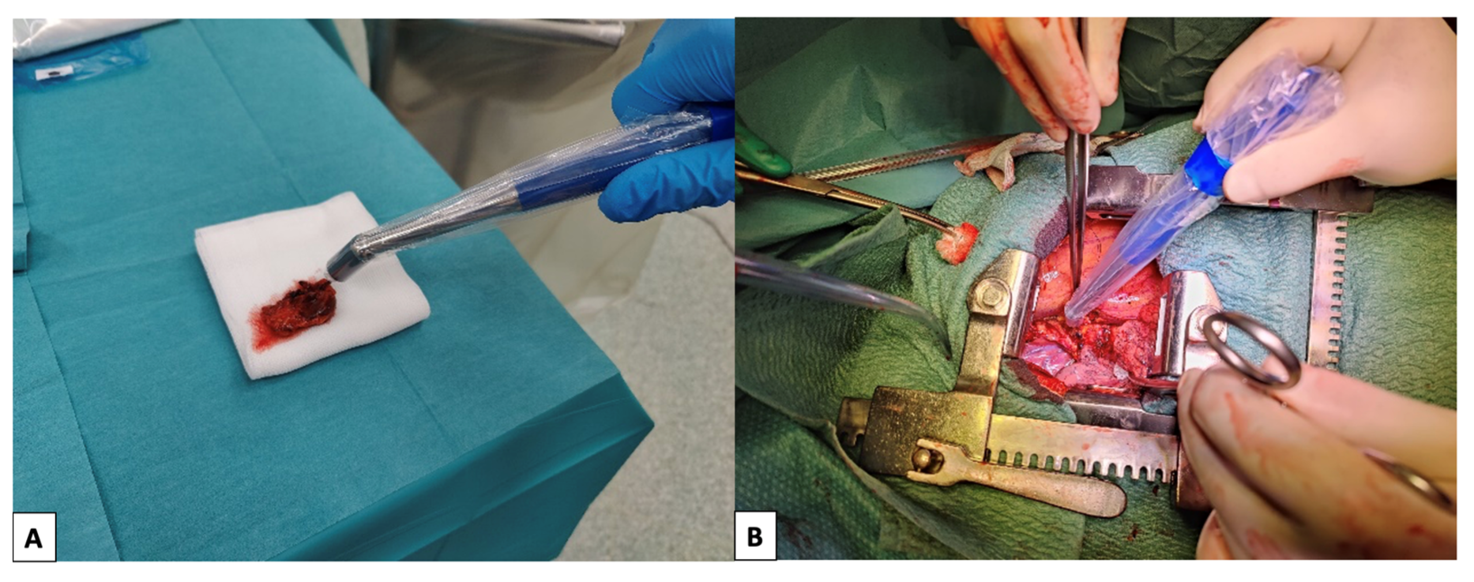
2.2. Lymphoscintigraphy and Surgery

During surgery, immediately after the identification of the primary tumour site, four intraparenchymal peri-tumoral injections of [99mTc]Tc-Albumin Nanocolloids (Nanotop®, ROTOP Pharmaka AG, Dresden, Germany) were performed. Starting from 10 min after the injection, the surgical removal of the primary tumour and locoregional lymph nodes was performed according to the best clinical practices.
All the excised lymph nodes were examined with a gamma probe (NeoProbe® Gamma Detection System, Devicor Medical Products, Inc., Cincinnati, OH, USA or Gamma Finder II®, World of Medicine GmbH, Berlin, Germany) ex vivo (Figure 1A) and considered as sentinel lymph nodes (SLNs) if they showed a significant activity; in particular, the activity was deemed significant when the uptake count of the specimen was higher than the background. The remaining ones, showing no significant activity, were considered non-sentinel lymph nodes (nSLN). Thereafter, at the end of the standard-of-care surgical excision of the primary tumour and locoregional lymph nodes, the surgical field was scanned with the gamma probe (Figure 1B), and any further radioactive lymph node was removed; these were labelled as “extra” SLNs (eSLNs). All uptake counts, and timings of the measurements, were recorded. All nSLNs, SLNs and eSLNs were sent to definitive histology, and the presence or absence of metastases was documented.

2.3. Statistical Analysis

Data are presented as mean and interquartile range (IQR), unless otherwise specified. Predictors of metastases in SLNs, nSLNs, and eSLNs were analysed via a univariate binary logistic regression model; further multinomial logistic regression analysis was conducted on selected factors based on significance level at the univariate testing and clinical reasoning. SPSS v. 24 (IBM, Armonk, NY, USA) was used for the analyses. A p-value of <0.05 was considered significant.

3. Results

3.1. Patients’ Population

A total of 48 patients were prospectively enrolled. In higher prevalence, they were males (65%), with smoking habits (77%), and a histologically confirmed diagnosis of lung adenocarcinoma (71%). Baseline patients’ characteristics and histopathological staging details are described in Table 1.

3.2. Distribution of Metastases in SLNs and nSLNs

During surgery, a total of 290 lymph-nodal stations were dissected, including 179 SLNs, 87 nSLNs, and 24 eSLNs. A total of 44 metastatic lymph-nodal stations were identified in 22 patients. A total of 36 out of these affected stations (82%) were located within SLNs. In three patients, however, there was a co-existence of metastases within and without SLNs. Particularly, one of these patients had a single nSLN metastasis, one had a mixture of SLNs and nSLNs metastases, while the last one had a single positive SLN and evidence of diffuse neoplastic lymphangitis, which probably hindered the tracer diffusion to the proper sentinel localisations (Table 2). Five, nine, and thirty metastases were identified in patients with cN0/cN1/cN2 status, respectively. In two cases, nodal metastases were identified in a mediastinal localisation with no evidence of hilar/intrapulmonary disease; in both cases, the nodal localisations were within SLNs. See Table 3 for details. Finally, there was no significant correlation between count intensity and the presence or absence of metastasis within SLNs.

3.3. Lymphatic Pathing

In typical upper lobe resections (26 procedures), only in 2 cases were metastases found in sub-carinal lymph nodes, and in both cases they were SLNs; in 6 cases, sub-carinal SLNs were pathologically negative; in 18 cases, pathologically negative sub-carinal nodes were nSLNs. In typical lower lobe resections (10 procedures), no metastases were located within higher para-tracheal lymph nodes; in 5 cases, pathologically negative high para-tracheal nodes were located within SLNs areas, and in the remaining 5 cases in nSLNs.

3.4. Predictors of Lymph Nodes Localisations and of eSLNs

Predictors of the presence of positive sentinel lymph nodes are depicted in Supplemental Table S1 (univariate analysis) and Table 4 (multivariate model): male gender and history of neoadjuvant chemotherapy showed a trend for increasing the risk, yet the presence of clinically positive lymph node in N2 localisations were the only definite predictor of this occurrence.
Given the rarity of its occurrence, no single parameter was correlated with presence of metastases within non-sentinel lymph nodes, even if cN positivity showed some degree of association (Supplemental Table S2). Therefore, no multivariate analysis was attempted.
Finally, a longer uptake time (greater than the median value) was the sole predictor inversely correlated with the presence of eSLNs on both univariate (Supplemental Table S3) and multivariate analysis (Table 5).

4. Discussion

The present study allowed for the gaining of a number of insights into the potential role of the sentinel lymph node technique in non-small-cell lung cancer. This technique resulted in safe, easily implementable, and accurate identification of nodal disease. In fact, disease localisations were found within sentinel nodes or, in rare instances, in non-sentinel lymph node stations located farther down the line of positive lymph nodes. These data support the use of the technique to identify nodal localisations reliably, or to prompt further exploration in case of positive sentinel localisations. Some guidelines advocate for lobe-specific mediastinal lymph node dissection, with the rationale being that there is an obvious correlation between the site of lymph-nodal metastasis and the location of the primary tumour [17]. In particular, in upper-lobe tumours, there is a significantly higher incidence of metastasis within superior mediastinal lymph nodes; conversely, lower-lobe tumours tend to spread to the inferior and sub-carinal nodal stations. This reasoning could justify a targeted approach to lymph-nodal dissection omitting the complete sub-carinal nodal resection in the surgical treatment of upper-lobe NSCLC [18]. Nonetheless, in some records, nearly 6% of patients treated according to the lobe-specific dissection might have had their metastatic lymph nodes missed by sampling stations not included in the lobe-specific protocol [17]. In any case, most guidelines agree that sub-carinal lymph nodes should at least always be sampled, and the European Society of Thoracic Surgeons (ESTS) guidelines have defined systematic nodal dissection as requiring the dissection and removal of all mediastinal tissue containing lymph nodes within precise anatomical landmarks [19]. Our analysis further highlighted how the clinical N+ status on the staging exams affects the likelihood of nodal metastasization. Combining the information from the pre-surgical staging imaging with that of sentinel node biopsy might help in guiding the surgical decision-making as to whether or not to sample the more remote lymph nodes. Patients treated with neo-adjuvant therapy might also be more likely to bear nodal localisations, given their more advanced disease.
In our series, retrieving “extra” sentinel nodes, i.e., those that the surgeon would not have removed if they had not had the sentinel node technique available, did not change the staging or improve the radicality of the intervention. However, such sentinel nodes were mostly present in patients with no evidence of nodal metastases. It might be interesting to further test the concept of additional lymph node identification in higher stages NSCLC, with a higher likelihood of node spread. Moreover, the current study was carried out in a high-volume referral centre; it might be speculated that using the sentinel node method to detect more lymph nodes than those identified by the surgeon might benefit operators still in the earlier phases of the learning curve. On the other hand, it must also be considered that the main factor inversely correlating to the presence of “extra” lymph nodes was the time elapsed between the tracer injection and the conclusion of the regular surgical procedure. In this sense, it could be hypothesised that a longer surgical procedure might allow the operators to perform a more complete lymphadenectomy, comprising all nodal stations reached by the radiopharmaceutical.
Our results about the intraoperative detection rate are in line with those presented in the literature, even if the large majority of them analysed early-stage NSCLC [20,21,22]; nonetheless, some smaller reports included higher-stage tumours as well [23]. The novelty of our work also resides in the fact that we analysed a wide range of NSCLC staging, including those with higher stages. We found that, especially in patients with clinically evident metastatic spread, the SLN technique can be of value in identifying patients with diffuse nodal disease, or in which the lymphatic flow does not follow the expected pattern. Our data suggest that more advanced and aggressive tumours might present unique challenges; in particular, massive metastastization or neoplastic lymphangitis might prevent an accurate nodal mapping with the pharmaceutical. In these cases, the method might underestimate the disease extent, and more aggressive surgical excision should be carried out.
In general, however, the information gathered thanks to this method could inform more selective lymphadenectomies, especially in early-stage lung cancers, reducing the surgical burden for operators and patients and driving a paradigm shift towards less invasive approaches [24]. Conversely, the added sensitivity provided by the intraoperative mapping of the locoregional lymphatic system can improve the identification of hard-to-spot nodes, especially in more advanced diseases, empowering more radical interventions, reducing the rate of relapses and improving survival. Finally, it has been suggested that employing the sentinel lymph node techniques might reduce the surgery time and curb its overall costs [25].
These results might prompt further targeted investigations. On the one hand, higher-stage tumours, which are those more prone to relapse and progression, might benefit from the sentinel lymph node approach the most. Studying the behaviour of the nodal spread in these patients might improve our capability to offer a radical treatment, even in these aggressive forms. On the other hand, prospective investigations assessing the possibility of more limited nodal resection in early-stage NSCLC might help reduce the rate of complications and postoperative morbidity [26,27]. Finally, even higher detection rates could be reached by switching from the one-dimensional exploration capability afforded by the gamma-probe to the 2D imaging offered by portable gamma cameras [28] or even to 3D, in vivo representation of sentinel nodes following pre-operative SPECT imaging, which has been suggested in many surgical fields [29,30].
From the feasibility point of view, we found that this technique can be implemented easily, with a very fast learning curve for the surgical team. An important advantage of this method is that it does not entail novel or hard-to-setup radiopharmaceutical: the lymphatic mapping can be prepared in minutes with minimal notice beforehand. Moreover, the carrier molecule is cheap and widely available; the dose absorbed by the patients as well as the surgical team is negligible.
Our study is not without limitations. The studied population is relatively small, even if the sample size is in line with or larger than the existing literature. We recruited patients with all NSCLC stages amenable to surgery. Thus, our results might not always be representative of every single stage. Nonetheless, this study was conceived to shed light on the lymphatic pathing of all lung cancer stages; the consistency of our results with those of the existing literature suggests that the sentinel node mapping can be applied safely to higher stages as well. The role of this method in patients with a clinical N2 status could be limited, since these patients should undergo a full excision in all cases; however, performing a sentinel node mapping could be helpful in those cases were the cN2 staging is conflicting across methods or unclear.
Finally, follow-up data of the studied patients are not available yet; the correlation between the sentinel node status and the long-term outcome should be assessed as soon as this information becomes available.

5. Conclusions

Sentinel node mapping with radiolabelled colloids in NSCLC is a safe procedure in all stages, able to identify nodal metastases in most cases. The identification of metastasization in a sentinel lymph node should, however, prompt a radical excision of all the nearby nodal stations, especially when an obstacle in the lymphatic drainage is suspected. On the other hand, identifying sentinel lymph nodes outside of the standard operating field does not seem to bring advantages in metastases identification.

Supplementary Materials

The following supporting information can be downloaded at: https://www.mdpi.com/article/10.3390/cancers15133320/s1, Supplemental Table S1: Univariate predictors of SLN metastases; Table S2: Univariate predictors of nSLN metastases; Table S3. Univariate predictors of eSLN presence.

Author Contributions

All authors contributed to the study conception and design. Material preparation, data collection and analysis were performed by C.P., L.A. and F.F. The first draft of the manuscript was written by C.P. and F.F.; all authors commented on previous versions of the manuscript. All authors have read and agreed to the published version of the manuscript.

Funding

This research received no external funding.

Institutional Review Board Statement

This study was performed in line with the principles of the Declaration of Helsinki. Approval was granted by the Ethics Committee of IRCCS Humanitas Research Hospital (April 20 2021/No. 336/21).

Informed Consent Statement

Informed consent was obtained from all individual participants included in the study.

Data Availability Statement

The datasets generated during and/or analysed during the current study are available from the corresponding author upon reasonable request.

Acknowledgments

We thank all clinicians involved in patient management and image reporting. We also thank Valeria Albano for her contribution to the production of the figures.

Conflicts of Interest

Arturo Chiti reports a fellowship grant from Sanofi, and personal fees from AAA, Blue Earth Diagnostics, and General Electric Healthcare outside of the submitted work. The other authors do not report any conflict of interest.

References

  1. Duma, N.; Santana-Davila, R.; Molina, J.R. Non–Small Cell Lung Cancer: Epidemiology, Screening, Diagnosis, and Treatment. Mayo Clin. Proc. 2019, 94, 1623–1640. [Google Scholar] [CrossRef] [PubMed]
  2. Balata, H.; Fong, K.M.; Hendriks, L.E.; Lam, S.; Ostroff, J.S.; Peled, N.; Wu, N.; Aggarwal, C. Prevention and Early Detection for NSCLC: Advances in Thoracic Oncology 2018. J. Thorac. Oncol. 2019, 14, 1513–1527. [Google Scholar] [CrossRef]
  3. Bao, F.; Yuan, P.; Yuan, X.; Lv, X.; Wang, Z.; Hu, J. Predictive Risk Factors for Lymph Node Metastasis in Patients with Small Size Non-Small Cell Lung Cancer. J. Thorac. Dis. 2014, 6, 1697. [Google Scholar] [CrossRef]
  4. Ost, D.E.; Yeung, S.C.J.; Tanoue, L.T.; Gould, M.K. Clinical and Organizational Factors in the Initial Evaluation of Patients With Lung Cancer: Diagnosis and Management of Lung Cancer, 3rd Ed: American College of Chest Physicians Evidence-Based Clinical Practice Guidelines. Chest 2013, 143, e121S–e141S. [Google Scholar] [CrossRef] [PubMed] [Green Version]
  5. Kirchner, J.; Sawicki, L.M.; Nensa, F.; Schaarschmidt, B.M.; Reis, H.; Ingenwerth, M.; Bogner, S.; Aigner, C.; Buchbender, C.; Umutlu, L.; et al. Prospective Comparison of 18 F-FDG PET/MRI and 18 F-FDG PET/CT for Thoracic Staging of Non-Small Cell Lung Cancer. Eur. J. Nucl. Med. Mol. Imaging 2019, 46, 437–445. [Google Scholar] [CrossRef] [PubMed]
  6. Wu, Y.; Li, P.; Zhang, H.; Shi, Y.; Wu, H.; Zhang, J.; Qian, Y.; Li, C.; Yang, J. Diagnostic Value of Fluorine 18 Fluorodeoxyglucose Positron Emission Tomography/Computed Tomography for the Detection of Metastases in Non-Small-Cell Lung Cancer Patients. Int. J. Cancer 2012, 132, E37–E47. [Google Scholar] [CrossRef]
  7. Akhurst, T. Staging of Non–Small-Cell Lung Cancer. PET Clin. 2018, 13, 1–10. [Google Scholar] [CrossRef]
  8. Yang, C.F.J.; Gu, L.; Shah, S.A.; Yerokun, B.A.; D’Amico, T.A.; Hartwig, M.G.; Berry, M.F. Long-Term Outcomes of Surgical Resection for Stage IV Non-Small-Cell Lung Cancer: A National Analysis. Lung Cancer 2018, 115, 75–83. [Google Scholar] [CrossRef]
  9. Raman, V.; Jawitz, O.K.; Yang, C.F.J.; Voigt, S.L.; Wang, H.; D’Amico, T.A.; Harpole, D.H.; Tong, B.C. Outcomes of Surgery versus Chemoradiotherapy in Patients with Clinical or Pathologic Stage N3 Non–Small Cell Lung Cancer. J. Thorac. Cardiovasc. Surg. 2019, 158, 1680–1692. [Google Scholar] [CrossRef]
  10. Dartevelle, P.G.; Mitilian, D.; Fadel, E. Extended Surgery for T4 Lung Cancer: A 30 Years’ Experience. Gen. Thorac. Cardiovasc. Surg. 2017, 65, 321–328. [Google Scholar] [CrossRef]
  11. Tabchi, S.; Kassouf, E.; Rassy, E.; El Kourie, H.R.; Martin, J.; Campeau, M.P.; Tehfe, M.; Blais, N. Management of Stage III Non–Small Cell Lung Cancer. Semin. Oncol. 2017, 44, 163–177. [Google Scholar] [CrossRef]
  12. Fischer, B.M.; Olsen, M.W.B.; Ley, C.D.; Klausen, T.L.; Mortensen, J.; Højgaard, L.; Kristjansen, P.E.G. How Few Cancer Cells Can Be Detected by Positron Emission Tomography? A Frequent Question Addressed by an in Vitro Study. Eur. J. Nucl. Med. 2006, 33, 697–702. [Google Scholar] [CrossRef]
  13. Passlick, B.; Kubuschock, B.; Sienel, W.; Thetter, O.; Pantel, K.; Izbicki, J.R. Mediastinal Lymphadenectomy in Non-Small Cell Lung Cancer: Effectiveness in Patients with or without Nodal Micrometastases-Results of a Preliminary Study. Eur. J. Cardiothorac. Surg. 2002, 21, 520–526. [Google Scholar] [CrossRef] [Green Version]
  14. Taghizadeh Kermani, A.; Bagheri, R.; Tehranian, S.; Shojaee, P.; Sadeghi, R.; Krag, D.N. Accuracy of Sentinel Node Biopsy in the Staging of Non-Small Cell Lung Carcinomas: Systematic Review and Meta-Analysis of the Literature. Lung Cancer 2013, 80, 5–14. [Google Scholar] [CrossRef] [PubMed]
  15. Lyman, G.H.; Somerfield, M.R.; Bosserman, L.D.; Perkins, C.L.; Weaver, D.L.; Giuliano, A.E. Sentinel Lymph Node Biopsy for Patients with Early-Stage Breast Cancer: American Society of Clinical Oncology Clinical Practice Guideline Update. J. Clin. Oncol. 2017, 35, 561–564. [Google Scholar] [CrossRef] [Green Version]
  16. Swetter, S.M.; Tsao, H.; Bichakjian, C.K.; Curiel-Lewandrowski, C.; Elder, D.E.; Gershenwald, J.E.; Guild, V.; Grant-Kels, J.M.; Halpern, A.C.; Johnson, T.M.; et al. Guidelines of Care for the Management of Primary Cutaneous Melanoma. J. Am. Acad. Derm. 2019, 80, 208–250. [Google Scholar] [CrossRef] [Green Version]
  17. Handa, Y.; Tsutani, Y.; Mimae, T.; Miyata, Y.; Ito, H.; Shimada, Y.; Nakayama, H.; Ikeda, N.; Okada, M.; Okada, M.; et al. Systematic Versus Lobe-Specific Mediastinal Lymphadenectomy for Hypermetabolic Lung Cancer. Ann. Surg. Oncol. 2021, 28, 7162–7171. [Google Scholar] [CrossRef]
  18. Aokage, K.; Yoshida, J.; Ishii, G.; Hishida, T.; Nishimura, M.; Nagai, K. Subcarinal Lymph Node in Upper Lobe Non-Small Cell Lung Cancer Patients: Is Selective Lymph Node Dissection Valid? Lung Cancer 2010, 70, 163–167. [Google Scholar] [CrossRef] [PubMed]
  19. Lardinois, D.; De Leyn, P.; Van Schil, P.; Rami Porta, R.; Waller, D.; Passlick, B.; Zielinski, M.; Junker, K.; Rendina, A.; Ris, H.-B.; et al. Invited Paper ESTS Guidelines for Intraoperative Lymph Node Staging in Non-Small Cell Lung Cancer. Eur. J. Cardio-Thoracic Surg. 2006, 30, 787–792. [Google Scholar] [CrossRef] [PubMed] [Green Version]
  20. Sugi, K.; Kaneda, Y.; Sudoh, M.; Sakano, H.; Hamano, K. Effect of Radioisotope Sentinel Node Mapping in Patients with CT1 N0 M0 Lung Cancer. J. Thorac. Cardiovasc. Surg. 2003, 126, 568–573. [Google Scholar] [CrossRef] [PubMed] [Green Version]
  21. Liptay, M.J.; Masters, G.A.; Winchester, D.J.; Edelman, B.L.; Garrido, B.J.; Hirschtritt, T.R.; Perlman, R.M.; Fry, W.A. Intraoperative Radioisotope Sentinel Lymph Node Mapping in Non–Small Cell Lung Cancer. Ann. Thorac. Surg. 2000, 70, 384–389. [Google Scholar] [CrossRef]
  22. Faries, M.B.; Bleicher, R.J.; Ye, X.; Essner, R.; Morton, D.L. Lymphatic Mapping and Sentinel Lymphadenectomy for Primary and Metastatic Pulmonary Malignant Neoplasms. Arch. Surg. 2004, 139, 870–877. [Google Scholar] [CrossRef] [Green Version]
  23. Lardinois, D.; Brack, T.; Gaspert, A.; Spahr, T.; Schneiter, D.; Steinert, H.C.; Weder, W. Bronchoscopic Radioisotope Injection for Sentinel Lymph-Node Mapping in Potentially Resectable Non-Small-Cell Lung Cancer. Eur. J. Cardio-Thoracic Surg. 2003, 23, 824–827. [Google Scholar] [CrossRef] [PubMed] [Green Version]
  24. Nomori, H.; Watanabe, K.; Ohtsuka, T.; Naruke, T.; Suemasu, K. In Vivo Identification of Sentinel Lymph Nodes for Clinical Stage I Non-Small Cell Lung Cancer for Abbreviation of Mediastinal Lymph Node Dissection. Lung Cancer 2004, 46, 49–55. [Google Scholar] [CrossRef]
  25. Kim, S.; Kim, H.K.; Kang, D.-Y.; Jeong, J.M.; Choi, Y.H. Intra-Operative Sentinel Lymph Node Identification Using a Novel Receptor-Binding Agent (Technetium-99m Neomannosyl Human Serum Albumin, 99mTc-MSA) in Stage I Non-Small Cell Lung Cancer. Eur. J. Cardio-Thorac. Surg. 2010, 37, 1450–1456. [Google Scholar] [CrossRef]
  26. Suzuki, K.; Saji, H.; Aokage, K.; Watanabe, S.; Okada, M.; Mizusawa, J.; Nakajima, R.; Tsuboi, M.; Nakamura, S.; Nakamura, K.; et al. Comparison of Pulmonary Segmentectomy and Lobectomy: Safety Results of a Randomized Trial. J. Thorac. Cardiovasc. Surg. 2019, 158, 895–907. [Google Scholar] [CrossRef]
  27. Kanzaki, R.; Nagoya, A.; Kanou, T.; Ose, N.; Funaki, S.; Minami, M.; Okamoto, Y.; Tabuchi, H.; Hoshino, T.; Tajima, T.; et al. Risk Factors for Non-Cancer Death after Surgery in Patients with Stage I Non-Small-Cell Lung Cancer. Eur. J. Cardio-Thorac. Surg. 2021, 59, 633–640. [Google Scholar] [CrossRef] [PubMed]
  28. Vollmer, I.; Sánchez-Izquierdo, N.; Martínez, D.; Sánchez-Lorente, D.; Casanueva-Eliceiry, S.; Boada, M.; Guirao, Á.; Romero-Zayas, I.; Vidal-Sicart, S.; Paredes, P. Role of a Portable Gamma-Camera with Optical View for Margins Assessment of Pulmonary Nodules Resected by Radioguided Surgery. Eur. J. Nucl. Med. Mol. Imaging 2021, 49, 361–370. [Google Scholar] [CrossRef] [PubMed]
  29. Profeta, A.C.; Schilling, C.; McGurk, M. Augmented Reality Visualization in Head and Neck Surgery: An Overview of Recent Findings in Sentinel Node Biopsy and Future Perspectives. Br. J. Oral. Maxillofac. Surg. 2016, 54, 694–696. [Google Scholar] [CrossRef]
  30. Lecointre, L.; Verde, J.; Goffin, L.; Venkatasamy, A.; Seeliger, B.; Lodi, M.; Swanström, L.L.; Akladios, C.; Gallix, B. Robotically Assisted Augmented Reality System for Identification of Targeted Lymph Nodes in Laparoscopic Gynecological Surgery: A First Step toward the Identification of Sentinel Node. Surg. Endosc. 2022, 36, 9224–9233. [Google Scholar] [CrossRef] [PubMed]
Figure 1. Counting of lymph nodal activity ex vivo (A) and control of the surgical field with gamma-probe (B).
Cancers 15 03320 g001
Table 1. Characteristics of the patients included in the study.
Patients, n48Mean injected activity, MBq (IQR)31.5 (5)
Sex, n17 F (35%)31 M (65%)
Mean age, years (IQR)69 (12)Dissected lymph nodes, nTotal290
SLNs179 (62%)
Smoking habit, nYes37 (77%)nSLNs87 (30%)
No11 (23%)eSLNs24 (8%)
Histoype, nAdenocarcinoma34 (71%)Metastatic lymph nodes, nTotal44
Squamous cells14 (29%)SLNs36 (82%)
Neoadjuvant treatment, nYes10 (21%)nSLNs 8 (18%)
No38 (79%)eSLNs0 (0%)
Clinical T (cT) staging, ncT114 (29%)Pathological T (pT) staging, npT115 (31%)
cT216 (33%)pT218 (38%)
cT315 (31%)pT39 (19%)
cT43 (6%)pT46 (12%)
Clinical N (cN) staging, ncN025 (52%)Pathological N (pN) staging, npN026 (54%)
cN110 (21%)pN19 (19%)
cN213 (27%)pN213 (27%)
IQR: interquartile range.
Table 2. Uptake patterns in patients with metastases in both SLNs and nSLNs.
Patient #Primary
Localization
HistotypecTNM, yTNMPositive SLN
Stations
Positive nSLN
Stations
Negative SLN
Stations
31Left lungSquamous cellscT2N2Within the left lungSubaorticLeft hilus, subcarinal, paraesophageal
37Left upper lobeAdenocarcinomacT2N1SubaorticPara-aortic, left hilus, subcarinal, paraesophagealWithin the left upper lobe
47Right upper lobeAdenocarcinomacT3N2, yT2N2Within the right upper lobe, upper/lower tracheal right, subcarinalAnterior mediastinum, upper tracheal right, upper tracheal cornerLower tracheal left
# consecutive patient number.
Table 3. Distribution of nodal metastases according to clinical staging.
Nodal StagingNumber of PatientsNumber of Metastases in SLNsNumber of Metastases in nSLNsSkip Metastases
cN0255None1 *
cN11054None
cN2132641 *
* found in an SLN.
Table 4. Multivariate analysis of positive SLN predictors.
ParameterORSignificanceLBUB
Sex (male)8.2730.0650.87578.204
Neoadjuvant chemotherapy12.8820.0780.753220.279
cN0Reference
cN19.6650.0830.746125.195
cN217.5060.0211.548198.031
OR: odds ratio; LB: lower bound; UB: upper bound.
Table 5. Multivariate analysis of predictors of “extra” sentinel lymph nodes.
ParameterORSignificanceLBUB
Neoadjuvant chemotherapy0.170.1590.0151.996
Uptake time (>median)0.2270.0290.060.862
cN0Reference
cN10.7370.7830.0846.439
cN20.6530.6450.1074.1
OR: odds ratio; LB: lower bound; UB: upper bound.
Disclaimer/Publisher’s Note: The statements, opinions and data contained in all publications are solely those of the individual author(s) and contributor(s) and not of MDPI and/or the editor(s). MDPI and/or the editor(s) disclaim responsibility for any injury to people or property resulting from any ideas, methods, instructions or products referred to in the content.

