Article

Comparative Analyses of the Clinicopathologic Features of Short-Term and Long-Term Survivors of Patients with Pancreatic Ductal Adenocarcinoma Who Received Neoadjuvant Therapy and Pancreatoduodenectomy

1
Department of Pathology, The University of Texas MD Anderson Cancer Center, Houston, TX 77030, USA
2
Department of Surgical Oncology, The University of Texas MD Anderson Cancer Center, Houston, TX 77030, USA
3
Department of Gastrointestinal Medical Oncology, The University of Texas MD Anderson Cancer Center, Houston, TX 77030, USA
4
Department of Radiation Oncology, The University of Texas MD Anderson Cancer Center, Houston, TX 77030, USA
5
Department of Translational Molecular Pathology, The University of Texas MD Anderson Cancer Center, Houston, TX 77030, USA
6
Department of Biostatistics, The University of Texas MD Anderson Cancer Center, Houston, TX 77030, USA
*
Author to whom correspondence should be addressed.
Cancers 2023, 15(12), 3231; https://doi.org/10.3390/cancers15123231
Submission received: 28 April 2023 / Revised: 31 May 2023 / Accepted: 15 June 2023 / Published: 18 June 2023
(This article belongs to the Special Issue Neoadjuvant Therapies in Pancreatic Cancer)

Abstract

:

Simple Summary

In this study, we systematically examined the clinicopathologic characteristics of 60 short-term survivors and 149 long-term survivors and compared them to 352 intermediate-term survivors of pancreatic ductal adenocarcinoma (PDAC) who received NAT and pancreatoduodenectomy. We found that lymph node stage (ypN) was an independent predictor of both short-term and long-term survivors. In addition, tumor differentiation was an independent predictor for short-term survivors, and tumor response grading and perineural invasion were independent predictors for long-term survivors. Our results may help to plan and select post-operative adjuvant therapy for patients with PDAC who received NAT and pancreatoduodenectomy based on the pathologic data.

Abstract

Neoadjuvant therapy (NAT) is increasingly used to treat patients with pancreatic ductal adenocarcinoma (PDAC). Patients with PDAC often show heterogenous responses to NAT with variable clinical outcomes, and the clinicopathologic parameters associated with these variable outcomes remain unclear. In this study, we systematically examined the clinicopathologic characteristics of 60 short-term survivors (overall survival < 15 months) and 149 long-term survivors (overall survival > 60 months) and compared them to 352 intermediate-term survivors (overall survival: 15–60 months) of PDAC who received NAT and pancreatoduodenectomy. We found that the short-term survivor group was associated with male gender (p = 0.03), tumor resectability prior to NAT (p = 0.04), poorly differentiated tumor histology (p = 0.006), more positive lymph nodes (p = 0.04), higher ypN stage (p = 0.002), and higher positive lymph node ratio (p = 0.03). The long-term survivor group had smaller tumor size (p = 0.001), lower ypT stage (p = 0.001), fewer positive lymph nodes (p < 0.001), lower ypN stage (p < 0.001), lower positive lymph node ratio (p < 0.001), lower rate of lymphovascular invasion (p = 0.001) and perineural invasion (p < 0.001), better tumor response grading (p < 0.001), and less frequent recurrence/metastasis (p < 0.001). The ypN stage is an independent predictor of both short-term and long-term survivors by multivariate logistic regression analyses. In addition, tumor differentiation was also an independent predictor for short-term survivors, and tumor response grading and perineural invasion were independent predictors for long-term survivors. Our results may help to plan and select post-operative adjuvant therapy for patients with PDAC who received NAT and pancreatoduodenectomy based on the pathologic data.

1. Introduction

Pancreatic ductal adenocarcinoma (PDAC) is a highly aggressive and lethal malignancy. It is the third leading cause of cancer-related deaths in the United States with a poor 5-year survival rate of approximately 11% [1]. The incidence of pancreatic cancer has increased worldwide and is projected to be the second-most-common cause of cancer-related deaths in the United States by 2030 [2,3]. Despite recent advancements in the field of oncology including immunotherapy and targeted therapy, the survival for patients with PDAC has only improved slightly over the past few decades [3]. Surgical resection continues to provide the only potential cure for PDAC.
The neoadjuvant therapy (NAT) approach is more commonly used to treat patients with potentially resectable PDAC and is currently the standard of care for those with borderline resectable and high-risk resectable disease according to the current National Comprehensive Cancer Network (NCCN) and the American Society of Clinical Oncology (ASCO) guidelines [4]. For patients with locally advanced disease, NAT can help select those most suitable for resection. Several neoadjuvant chemotherapy regimens with or without radiation therapy have been used to treat patients with potentially resectable, borderline resectable, or locally advanced PDAC [5]. These regimens include gemcitabine-based regimens, such as gemcitabine monotherapy, gemcitabine plus capecitabine, nab-paclitaxel, or S1, and fluoropyrimidine-based regimens, such as folinic acid, 5-FU, irinotecan and oxaliplatin (FOLFIRINOX), and modified FOLFIRINOX [5,6,7,8]. A recent meta-analysis of 38 studies suggested that NAT may be offered as an acceptable standard of care for patients with potentially resectable PDAC [9].
Patients with PDAC often show heterogenous responses to NAT and variable clinical outcomes. Previous studies of post-therapy pancreatectomy specimens have shown that the majority of patients (>80%) demonstrated moderate or minimal responses to NAT, which is associated with poor survival. Only a minority (12.6–18.6%) of PDAC patients demonstrate a complete or near-complete response to NAT, which is associated with better survival [10,11,12,13]. In addition to tumor response grading (TRG), several other pathologic parameters, including tumor differentiation, pathologic primary tumor stage (ypT), lymph node stage (ypN), perineural invasion, vascular invasion, resection margins, tumor involvement of superior mesenteric vein/portal vein, and integrated pathologic score, have also been shown to affect the clinical outcomes and survival in PDAC patients who received NAT [10,13,14,15,16,17,18,19]. In our previous studies of PDAC patients who received NAT [10,13,14,15,16,17,18,19], we noticed that there were a significant number of patients who survived longer than 5 years (long-term survivors) and a smaller percentage of patients who survived less than 15 months (short-term survivors). However, the clinicopathologic parameters that are associated with long-term and short-term survivors in PDAC patients who received NAT have not been examined.
In this retrospective study, we systematically examined and compared clinicopathologic characteristics of short-term survivors (<15 months) and long-term survivors (>60 months) with the intermediate-term survivors (15–60 months) in a large cohort of 561 patients who received NAT and pancreatoduodenectomy (PD) for PDAC at our institution.

2. Materials and Methods

2.1. Patient Population

This study was approved by the institutional review board of the University of Texas MD Anderson Cancer Center. Our study population consisted of 561 PDAC patients (313 male and 248 female) who were diagnosed between January 1999 and December 2017, completed NAT, and underwent pancreatoduodenectomy at our institution. The pretreatment diagnosis of PDAC was confirmed in all cases by reviewing the biopsies and/or fine needle aspiration cytology. We excluded patients who had undergone distal pancreatectomy and those who had undergone pancreatoduodenectomy for other neoplasms. The median age for our study population was 64.2 years (range: 34.5–85.4 years). Tumor resectability prior to NAT was evaluated based on the computed tomographic (CT) scan optimized for pancreatic imaging as previously described [20].

2.2. Pathologic Evaluation

All pancreatoduodenectomy specimens were evaluated and reported using a standardized protocol at our institution, which include histologic type, differentiation/histologic grade, tumor location, tumor size, presence or absence of lymphovascular or perineural invasion, number of positive and total lymph nodes, TRG, tumor involvement of an extrapancreatic tissue/organ, and margin status [18,21]. The uncinate/superior mesenteric artery margin was considered as positive if the tumor extended to ≤1.0 mm to the inked margin. All other margins were routinely evaluated by en face section(s), and the margin was considered as positive if carcinoma was present. The ypTNM was grouped according to the American Joint committee on Cancer (AJCC) Cancer Staging Manual 8th edition [22]. Tumor response grading was evaluated using the MD Anderson (MDA) or the College of American Pathologists (CAP) grading systems. Given the small number of patients with ypT0 or MDA grade 0, we combined ypT0 and ypT1 as one group (ypT0/ypT1) and MDA grade 0 and 1 as one tumor response group for the multifactorial logistic analysis.

2.3. Clinical and Follow-Up Data

The demographic, clinical, pathologic, and follow-up data of our study population were retrieved from a prospectively maintained multidisciplinary pancreatic cancer database and verified by reviewing patients’ medical records and reviewing the U.S. Social Security Index when necessary. Overall survival was calculated from the date of first diagnosis to the date of death or the last follow-up date (if death did not occur). Based on the overall survival, we grouped our patients into three groups: the short-term survivors (overall survival < 15 months), the intermediate-term survivors (overall survival: 15–60 months), and the long-term survivors (overall survival > 60 months).

2.4. Statistical Analysis

Correlations between the survivor groups with different clinicopathologic parameters were performed using chi-square analyses or Fischer’s exact tests for categorical data. The independent-samples t test or one-way ANOVA was used to compare the means among different survivor groups for continuous variables. Multivariate logistic regression analyses were performed to identify the independent clinicopathologic parameters that are predictive of the short-term and long-term survivor groups. All statistical analyses were performed using Statistical Package for Social Sciences software for Windows (Version 26, SPSS, Inc., Chicago, IL, USA). A 2-sided significance level of <0.05 was used for all statistical analyses.

3. Results

3.1. Patient Characteristics

Among 561 patients, 386 (68.8%) were potential resectable, 136 (24.2%) were borderline resectable, and 39 (7.7%) had locally advanced disease. A total of 182 (32.4%) received neoadjuvant fluoropyrimidine-based chemotherapy with radiation (182), 328 (58.5%) received gemcitabine-based chemotherapy with radiation, 36 (6.4%) received gemcitabine-based chemotherapy, and 15 (2.7%) received FOLFIRINOX. Totals of 16 (2.9%), 194 (34.6%), 284 (50.6%), and 67 (11.9%) were ypT0, ypT1, ypT2, and ypT3, respectively, and 259 (46.2%), 194 (34.6%), and 108 (19.3%) were ypN0, ypN1, and ypN2, respectively. A total of 350 (62.4%) had a well to moderately differentiated histology and 211 (37.6%) had a poorly differentiated tumor. Totals of 16 (2.9%), 69 (12.3%), and 476 (84.8%) had MD Anderson grade 0, 1, and 2 responses, respectively, and 16 (2.9%), 69 (12.3%), 304 (54.2%), and 172 (30.6%) had CAP grade 0, 1, 2, and 3 responses, respectively. R0 (margin-negative microscopically) and R1 resection (margin-positive microscopically) were achieved in 483 (86.1%) and 78 (13.9%), respectively. The median overall survival was 35.1 months (range: 7.6 months to 257.5 months). There were 60 (10.7%) short-term survivors, 352 (62.7%) intermediate-term survivors, and 149 (26.6%) long-term survivors, respectively.

3.2. The Clinicopathologic Features of Short-Term Survivor Group

Comparisons of clinicopathologic parameters between the short-term survivor group and the intermediate-term survivor group are shown in Table 1. The short-term survivor group was associated with male gender (p = 0.03), poor differentiated histology (p = 0.006), more positive lymph nodes (p = 0.04), higher ypN stage (p = 0.002), and higher positive lymph node ratio (p = 0.03). Interestingly, compared to the intermediate-term survivor group, more patients in the short-term survival group were classified as potentially resectable before the initiation of NAT (83.3% vs. 66.8%, p =0.01). There were no significant differences between these two groups in age, NAT regimens, tumor size, ypT stage, total number of lymph nodes examined, lymphovascular and perineural invasion, margin status, tumor response grading using either the MDA or CAP grading system, and recurrence/metastasis (p > 0.05, Table 1). The mean duration of NAT was 3.0 ± 1.9 months for the short-term survivor group compared to 4.4 ± 2.7 months for the intermediate-term survivors (p < 0.001).

3.3. The Clinicopathologic Features of Long-Term Survivor Group

Comparisons of the clinicopathological parameters between the long-term survivor group and the intermediate-term survivor group are shown in Table 2. The long-term survivor group had smaller tumor size (p = 0.001), lower ypT stage (p = 0.001), fewer positive lymph nodes (p < 0.001), lower ypN stage (p < 0.001), lower positive lymph node ratio (p < 0.001), lower rate of lymphovascular invasion (p = 0.001) and perineural invasion (p < 0.001), better tumor response grading using either the MDA or CAP grading system (p < 0.001), and less frequent recurrence/metastasis (p < 0.001). There were no significant differences in age, gender, NAT regimens, tumor resectability prior to NAT, total number of lymph nodes examined, tumor differentiation, and margin status (p > 0.05). The mean duration of NAT was 4.1 ± 3.9 months for the long-term survivor group compared to 4.4 ± 2.7 months for the intermediate-term survivors (p = 0.28).

3.4. Comparison of the Clinicopathologic Features of Long-Term Survivor Group with Short-Term Survivor Group

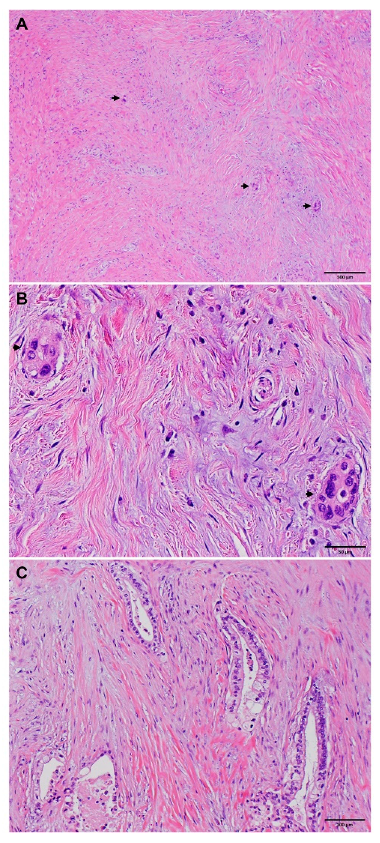
Comparisons of clinicopathologic parameters between the long-term survivor group and the short-term survivor group are shown in Table 3. Compared to the short-term survivor group, the long-term survivor group was associated with younger age (p = 0.04) and female gender (p = 0.02), had smaller tumor size (p = 0.002), lower ypT stage (p = 0.04), better tumor differentiation (p = 0.001), lower rate of lymphovascular invasion (p < 0.001) and perineural invasion (p < 0.001), better tumor response grading using the MDA or CAP grading system (p = 0.04), fewer positive lymph nodes (p < 0.001), lower positive lymph node ratio (p < 0.001), lower ypN stage (p < 0.001), and less frequent recurrence/metastasis (p < 0.001). Representative histologic features that are associated with short- and long-term survivors are shown in Figure 1 and Figure 2. No significant differences in NAT regimens, tumor resectability prior to NAT, total number of lymph nodes examined, and margin status (p > 0.05) were found.

3.5. Multivariate Logistic Regression Analyses

Using the binary multivariate logistic regression analysis to predict short-term versus intermediate-term survivor groups, we found that ypN stage (p = 0.006) and tumor differentiation (p = 0.01) were independent factors that differentiated the two groups (Table 4 and Figure 1). Pretreatment tumor resectability (p = 0.07), TRG (p = 0.06), perineural invasion (p = 0.06), and gender (p = 0.15) were not statistically significant. On the other hand, the logistic regression analyses to differentiate long- vs. intermediate-term survivor groups discovered ypN stage (p = 0.003), tumor response grading (p = 0.04), and perineural invasion (p = 0.04) as independent factors that are associated with the long-term survivor group (Table 5). In particular, we found that better tumor response grading, lower ypN stage, and the absence of perineural invasion were associated with long- vs. intermediate-term survival (Figure 2). These logistic regression results present a consistent picture regarding the important clinicopathological parameters that were predictive of length of survival, which can be used clinically as prognostic markers.

4. Discussion

In this study, we examined clinicopathologic characteristics of short- and long-term survivor groups and compared them to an intermediate-term survivor group in a cohort of 561 patients with PDAC who underwent neoadjuvant therapy prior to pancreatoduodenectomy. We found that ypN stage and tumor differentiation were independent factors that were associated with the short-term survivor group. The independent factors that were associated with the long-term survivor group were ypN stage, tumor response grading, and perineural invasion. Our results highlight the importance of accurate pathologic evaluation in pancreatoduodenectomy specimens from PDAC patients who received neoadjuvant therapy.
The reported long-term survivor (survival ≥ 5 year) rate in patients with PDAC who received up-front surgical resection and adjuvant therapy ranges from 12.1% to 27.0% [23,24,25,26,27,28,29]. Consistent with previous studies, we showed that the long-term survivor rate was (26.6%) in PDAC patients who received NAT and pancreatoduodenectomy. However, the clinicopathologic parameters associated with long-term survivors of PDAC after NAT and pancreatoduodenectomy have not been studied in a large cohort of patients. In this study, we demonstrated that the long-term survivor group had a significantly lower ypN stage, fewer positive lymph nodes, lower positive lymph node ratio, smaller tumor size, lower ypT stage, lower rates of lymphovascular invasion and perineural invasion, better tumor response grading, and less frequent recurrence/metastasis compared to the intermediate-term survivor group. In multifactorial logistic regression analyses, ypN stage and perineural invasion were independent factors that are associated with the long-term survivor group. In addition, we showed for the first time that tumor response grading was associated with long-term survivors in PDAC patients who received NAT and pancreatoduodenectomy. Our results are consistent with previous studies showing that ypN stage and perineural invasion are associated with long-term survivors in PDAC patients who received up-front surgical resection [22,24,29,30,31,32]. Patient age and tumor differentiation were not associated with long-term survivors in this study.
Very few studies have examined the clinicopathologic parameters that are associated with short-term survivors for PDAC patients who underwent pancreatectomy. Takeuchi et al. examined the pre-operative predictors of short-term survivors of PDAC patients who underwent surgical resection. They found that the short-term survivors of PDAC patients (overall survival < 12 months) was negatively associated with patient age, tumor size of ≥3 cm based on a computerized tomography (CT) scan, and the level of serum carcinoembryonic antigen (CEA) [33]. A meta-analysis by Agalianos et al. showed that positive para-aortic lymph nodes following pancreatectomy was associated with a lower one-year survival rate [34]. In our cohort of patients with PDAC who received NAT and pancreatoduodenectomy, only 24 of 561 patients (4.3%) survived < 12 months. To increase the number of short-term survivors and the representation of this study group, we used the overall survival time of 15 months as the cut off for the short-term survivor group and identified 60 (10.7%) short-term survivors in this study. We demonstrated that male gender, poor differentiated histology, higher number of positive lymph nodes, higher ypN stage, and higher positive lymph node ratio were associated the short-term survivor group compared to the intermediate-term survivor group. In multivariate logistic regression analyses, ypN stage and tumor differentiation were independent predictors for short-term survivors. We did not observe a significant association between the short-term survivor group and age, tumor size, ypT stage, lymphovascular invasion, or margin status.
Accurate lymph node staging plays a critical role in patient prognosis in both PDAC patients who underwent upfront surgical resection and those who received NAT followed by resection with curative intent [13,35]. In this study, we found that ypN stage was an independent factor associated with both short-term and long-term survivor groups by multivariate logistic regression analyses. This result is consistent with the findings of our previous studies showing that the number of positive lymph nodes, ypN stage, and the positive lymph node ratio are independent prognostic factors for both disease free survival (DFS) and overall survival (OS) in patients with PDAC who received NAT and pancreatectomy [13,16,18,21].
A recent study by Tong et al. showed that patients with PDAC who received neoadjuvant FOLFIRINOX were younger and had higher rates of borderline resectable and locally advanced disease, but had higher rates of complete or near-complete pathologic responses and lower ypN stage compared to those who received neoadjuvant gemcitabine/nab-paclitaxel [6]. They also showed that FOLFIRINOX plus radiation was associated with decreased lymph node metastasis and lower ypN stage than those who received FOLFIRINOX alone. However, they did not observe a significant difference in survival between the FOLFIRINOX and gemcitabine/nab-paclitaxel groups [6]. Similar results were also reported from a Phase II study, which compared neoadjuvant FOLFIRINOX versus gemcitabine/nab-paclitaxel for borderline-resectable PDAC (NUPAT-01) and showed no significant difference in the 3-year survival between these two regimens. [36] Consistent with these results, we did not find a significant association of different NAT regimens with either short-term or long-term survivors. It should be noted that the majority (90.9%) of PDAC patients in this study received neoadjuvant fluoropyrimidine-based or gemcitabine-based chemotherapy with radiation, only 6.4% received gemcitabine-based chemotherapy alone, and 2.7% received FOLFIRINOX alone. Prospective randomized clinical trials are needed to compare the efficacy of different NAT regimens with or without radiation.
Limitations of this study include that this is a single-institution retrospective study at a tertiary cancer center, which may have potential bias in the patient population. In addition, our study population does not include any patients who underwent neoadjuvant therapy but did not undergo surgical resection, due to tumor progression during treatment. Another limitation of this study is that our patient population spanned a period of 18 years between 1999 and 2017, and neoadjuvant regimens have evolved over this time period. Therefore, our study results should be interpreted cautiously. Despite the stated limitations, our study is the first large study to examine clinicopathologic characteristics of short-term, intermediate-term, or long-term survivors of PDAC patients treated with neoadjuvant therapy followed by pancreatoduodenectomy.

5. Conclusions

In summary, our study demonstrates that ypN stage is an independent predictor of both short- and long-term survivors in patients with PDAC who received NAT and pancreatoduodenectomy. In addition, we demonstrated that tumor differentiation is an important predictor of short-term survivors and that tumor response grading and perineural invasion are important predictors of long-term survivors. Our results may help to select and plan post-operative adjuvant therapy for patients with PDAC who received NAT and pancreatoduodenectomy based on the pathologic data.

Author Contributions

Conceptualization, T.Z.L., M.H.G.K. and H.W. (Huamin Wang); methodology, T.Z.L., M.H.G.K., L.R.P. and H.W. (Huamin Wang); formal analysis, S.K. and H.W. (Huamin Wang); investigation, T.Z.L., M.H.G.K., L.R.P. and H.W. (Huamin Wang); resources, M.H.G.K. and H.W. (Huamin Wang); data curation, H.W. (Hua Wang), D.C., M.K., C.-W.D.T., N.I., R.A.W., D.Z., E.J.K. and A.M. writing—original draft preparation, T.Z.L. and H.W. (Huamin Wang); writing—review and editing, T.Z.L., M.H.G.K., L.R.P., H.W. (Hua Wang), D.C., M.K., C.-W.D.T., N.I., R.A.W., D.Z., E.J.K. and A.M.; supervision, H.W. (Huamin Wang); project administration, H.W. (Huamin Wang); funding acquisition, H.W. (Huamin Wang). All authors have read and agreed to the published version of the manuscript.

Funding

Supported by the National Institutes of Health grants (1R01CA196941, 1R01CA195651, U01CA196403, P01CA117969, and P50CA221707).

Institutional Review Board Statement

This study was approved by the Institutional Review Board of The University of Texas MD Anderson Cancer Center (LAB05-0854).

Informed Consent Statement

Patient consent was waived based on our approved protocol.

Data Availability Statement

The data are unavailable due to privacy or ethical restrictions.

Conflicts of Interest

The authors declare no conflict of interest.

References

  1. Siegel, R.L.; Miller, K.D.; Fuchs, H.E.; Jemal, A. Cancer statistics, 2022. CA Cancer J. Clin. 2022, 72, 7–33. [Google Scholar] [CrossRef] [PubMed]
  2. Sung, H.; Ferlay, J.; Siegel, R.L.; Laversanne, M.; Soerjomataram, I.; Jemal, A.; Bray, F. Global Cancer Statistics 2020: GLOBOCAN Estimates of Incidence and Mortality Worldwide for 36 Cancers in 185 Countries. CA Cancer J. Clin. 2021, 71, 209–249. [Google Scholar] [CrossRef] [PubMed]
  3. Rahib, L.; Wehner, M.R.; Matrisian, L.M.; Nead, K.T. Estimated Projection of US Cancer Incidence and Death to 2040. JAMA Netw. Open 2021, 4, e214708. [Google Scholar] [CrossRef]
  4. Tempero, M.A.; Malafa, M.P.; Al-Hawary, M.; Behrman, S.W.; Benson, A.B.; Cardin, D.B.; Chiorean, E.G.; Chung, V.; Czito, B.; Del Chiaro, M.; et al. Pancreatic Adenocarcinoma, Version 2.2021, NCCN Clinical Practice Guidelines in Oncology. J. Natl. Compr. Cancer Netw. 2021, 19, 439–457. [Google Scholar] [CrossRef]
  5. Springfeld, C.; Ferrone, C.R.; Katz, M.H.G.; Philip, P.A.; Hong, T.S.; Hackert, T.; Buchler, M.W.; Neoptolemos, J. Neoadjuvant therapy for pancreatic cancer. Nat. Rev. Clin. Oncol. 2023, 20, 318–337. [Google Scholar] [CrossRef]
  6. Tong, Y.T.; Lai, Z.; Katz, M.H.G.; Prakash, L.R.; Wang, H.; Chatterjee, D.; Kim, M.; Tzeng, C.D.; Lee, J.E.; Ikoma, N.; et al. Prognosticators for Patients with Pancreatic Ductal Adenocarcinoma Who Received Neoadjuvant FOLFIRINOX or Gemcitabine/Nab-Paclitaxel Therapy and Pancreatectomy. Cancers 2023, 15, 2608. [Google Scholar] [CrossRef] [PubMed]
  7. Janssen, Q.P.; van Dam, J.L.; Kivits, I.G.; Besselink, M.G.; van Eijck, C.H.J.; Homs, M.Y.V.; Nuyttens, J.; Qi, H.; van Santvoort, H.J.; Wei, A.C.; et al. Added Value of Radiotherapy Following Neoadjuvant FOLFIRINOX for Resectable and Borderline Resectable Pancreatic Cancer: A Systematic Review and Meta-Analysis. Ann. Surg. Oncol. 2021, 28, 8297–8308. [Google Scholar] [CrossRef]
  8. Wolfe, A.R.; Prabhakar, D.; Yildiz, V.O.; Cloyd, J.M.; Dillhoff, M.; Abushahin, L.; Alexandra Diaz, D.; Miller, E.D.; Chen, W.; Frankel, W.L.; et al. Neoadjuvant-modified FOLFIRINOX vs nab-paclitaxel plus gemcitabine for borderline resectable or locally advanced pancreatic cancer patients who achieved surgical resection. Cancer Med. 2020, 9, 4711–4723. [Google Scholar] [CrossRef]
  9. Versteijne, E.; Vogel, J.A.; Besselink, M.G.; Busch, O.R.C.; Wilmink, J.W.; Daams, J.G.; van Eijck, C.H.J.; Groot Koerkamp, B.; Rasch, C.R.N.; van Tienhoven, G.; et al. Meta-analysis comparing upfront surgery with neoadjuvant treatment in patients with resectable or borderline resectable pancreatic cancer. Br. J. Surg. 2018, 105, 946–958. [Google Scholar] [CrossRef] [Green Version]
  10. Chatterjee, D.; Katz, M.H.; Rashid, A.; Varadhachary, G.R.; Wolff, R.A.; Wang, H.; Lee, J.E.; Pisters, P.W.; Vauthey, J.N.; Crane, C.; et al. Histologic grading of the extent of residual carcinoma following neoadjuvant chemoradiation in pancreatic ductal adenocarcinoma: A predictor for patient outcome. Cancer 2012, 118, 3182–3190. [Google Scholar] [CrossRef] [Green Version]
  11. Chou, A.; Ahadi, M.; Arena, J.; Sioson, L.; Sheen, A.; Fuchs, T.L.; Pavlakis, N.; Clarke, S.; Kneebone, A.; Hruby, G.; et al. A Critical Assessment of Postneoadjuvant Therapy Pancreatic Cancer Regression Grading Schemes With a Proposal for a Novel Approach. Am. J. Surg. Pathol. 2021, 45, 394–404. [Google Scholar] [CrossRef]
  12. Lee, S.M.; Katz, M.H.; Liu, L.; Sundar, M.; Wang, H.; Varadhachary, G.R.; Wolff, R.A.; Lee, J.E.; Maitra, A.; Fleming, J.B.; et al. Validation of a Proposed Tumor Regression Grading Scheme for Pancreatic Ductal Adenocarcinoma After Neoadjuvant Therapy as a Prognostic Indicator for Survival. Am. J. Surg. Pathol. 2016, 40, 1653–1660. [Google Scholar] [CrossRef] [PubMed]
  13. Nagaria, T.S.; Wang, H.; Chatterjee, D. Pathology of Treated Pancreatic Ductal Adenocarcinoma and Its Clinical Implications. Arch. Pathol. Lab. Med. 2020, 144, 838–845. [Google Scholar] [CrossRef] [Green Version]
  14. Chatterjee, D.; Katz, M.H.; Rashid, A.; Wang, H.; Iuga, A.C.; Varadhachary, G.R.; Wolff, R.A.; Lee, J.E.; Pisters, P.W.; Crane, C.H.; et al. Perineural and intraneural invasion in posttherapy pancreaticoduodenectomy specimens predicts poor prognosis in patients with pancreatic ductal adenocarcinoma. Am. J. Surg. Pathol. 2012, 36, 409–417. [Google Scholar] [CrossRef] [PubMed] [Green Version]
  15. Chatterjee, D.; Rashid, A.; Wang, H.; Katz, M.H.; Wolff, R.A.; Varadhachary, G.R.; Lee, J.E.; Pisters, P.W.; Gomez, H.F.; Abbruzzese, J.L.; et al. Tumor invasion of muscular vessels predicts poor prognosis in patients with pancreatic ductal adenocarcinoma who have received neoadjuvant therapy and pancreaticoduodenectomy. Am. J. Surg. Pathol. 2012, 36, 552–559. [Google Scholar] [CrossRef] [PubMed] [Green Version]
  16. Fischer, L.K.; Katz, M.H.; Lee, S.M.; Liu, L.; Wang, H.; Varadhachary, G.R.; Wolff, R.A.; Lee, J.E.; Maitra, A.; Roland, C.L.; et al. The number and ratio of positive lymph nodes affect pancreatic cancer patient survival after neoadjuvant therapy and pancreaticoduodenectomy. Histopathology 2016, 68, 210–220. [Google Scholar] [CrossRef] [Green Version]
  17. Liu, L.; Katz, M.H.; Lee, S.M.; Fischer, L.K.; Prakash, L.; Parker, N.; Wang, H.; Varadhachary, G.R.; Wolff, R.A.; Lee, J.E.; et al. Superior Mesenteric Artery Margin of Posttherapy Pancreaticoduodenectomy and Prognosis in Patients With Pancreatic Ductal Adenocarcinoma. Am. J. Surg. Pathol. 2015, 39, 1395–1403. [Google Scholar] [CrossRef]
  18. Estrella, J.S.; Rashid, A.; Fleming, J.B.; Katz, M.H.; Lee, J.E.; Wolf, R.A.; Varadhachary, G.R.; Pisters, P.W.; Abdalla, E.K.; Vauthey, J.N.; et al. Post-therapy pathologic stage and survival in patients with pancreatic ductal adenocarcinoma treated with neoadjuvant chemoradiation. Cancer 2012, 118, 268–277. [Google Scholar] [CrossRef] [Green Version]
  19. Sohn, A.J.; Taherian, M.; Katz, M.H.G.; Prakash, L.R.; Chatterjee, D.; Wang, H.; Kim, M.; Tzeng, C.D.; Lee, J.E.; Ikoma, N.; et al. Integrated Pathologic Score Effectively Stratifies Patients With Pancreatic Ductal Adenocarcinoma Who Received Neoadjuvant Therapy and Pancreaticoduodenectomy. Am. J. Surg. Pathol. 2023, 47, 421–430. [Google Scholar] [CrossRef]
  20. Varadhachary, G.R.; Tamm, E.P.; Abbruzzese, J.L.; Xiong, H.Q.; Crane, C.H.; Wang, H.; Lee, J.E.; Pisters, P.W.; Evans, D.B.; Wolff, R.A. Borderline resectable pancreatic cancer: Definitions, management, and role of preoperative therapy. Ann. Surg. Oncol. 2006, 13, 1035–1046. [Google Scholar] [CrossRef]
  21. Chatterjee, D.; Katz, M.H.; Foo, W.C.; Sundar, M.; Wang, H.; Varadhachary, G.R.; Wolff, R.A.; Lee, J.E.; Maitra, A.; Fleming, J.B.; et al. Prognostic Significance of New AJCC Tumor Stage in Patients With Pancreatic Ductal Adenocarcinoma Treated With Neoadjuvant Therapy. Am. J. Surg. Pathol. 2017, 41, 1097–1104. [Google Scholar] [CrossRef]
  22. Amin, M.B.; Greene, F.L.; Edge, S.B.; Compton, C.C.; Gershenwald, J.E.; Brookland, R.K.; Meyer, L.; Gress, D.M.; Byrd, D.R.; Winchester, D.P. The Eighth Edition AJCC Cancer Staging Manual: Continuing to build a bridge from a population-based to a more "personalized" approach to cancer staging. CA Cancer J. Clin. 2017, 67, 93–99. [Google Scholar] [CrossRef]
  23. Belfiori, G.; Crippa, S.; Francesca, A.; Pagnanelli, M.; Tamburrino, D.; Gasparini, G.; Partelli, S.; Andreasi, V.; Rubini, C.; Zamboni, G.; et al. Long-Term Survivors after Upfront Resection for Pancreatic Ductal Adenocarcinoma: An Actual 5-Year Analysis of Disease-Specific and Post-Recurrence Survival. Ann. Surg. Oncol. 2021, 28, 8249–8260. [Google Scholar] [CrossRef] [PubMed]
  24. Cleary, S.P.; Gryfe, R.; Guindi, M.; Greig, P.; Smith, L.; Mackenzie, R.; Strasberg, S.; Hanna, S.; Taylor, B.; Langer, B.; et al. Prognostic factors in resected pancreatic adenocarcinoma: Analysis of actual 5-year survivors. J. Am. Coll. Surg. 2004, 198, 722–731. [Google Scholar] [CrossRef]
  25. Conlon, K.C.; Klimstra, D.S.; Brennan, M.F. Long-term survival after curative resection for pancreatic ductal adenocarcinoma. Clinicopathologic analysis of 5-year survivors. Ann. Surg. 1996, 223, 273–279. [Google Scholar] [CrossRef] [PubMed]
  26. Ferrone, C.R.; Pieretti-Vanmarcke, R.; Bloom, J.P.; Zheng, H.; Szymonifka, J.; Wargo, J.A.; Thayer, S.P.; Lauwers, G.Y.; Deshpande, V.; Mino-Kenudson, M.; et al. Pancreatic ductal adenocarcinoma: Long-term survival does not equal cure. Surgery 2012, 152, S43–S49. [Google Scholar] [CrossRef] [Green Version]
  27. Han, S.S.; Jang, J.Y.; Kim, S.W.; Kim, W.H.; Lee, K.U.; Park, Y.H. Analysis of long-term survivors after surgical resection for pancreatic cancer. Pancreas 2006, 32, 271–275. [Google Scholar] [CrossRef]
  28. Schnelldorfer, T.; Ware, A.L.; Sarr, M.G.; Smyrk, T.C.; Zhang, L.; Qin, R.; Gullerud, R.E.; Donohue, J.H.; Nagorney, D.M.; Farnell, M.B. Long-term survival after pancreatoduodenectomy for pancreatic adenocarcinoma: Is cure possible? Ann. Surg. 2008, 247, 456–462. [Google Scholar] [CrossRef] [PubMed]
  29. Strobel, O.; Lorenz, P.; Hinz, U.; Gaida, M.; Konig, A.K.; Hank, T.; Niesen, W.; Kaiser, J.O.R.; Al-Saeedi, M.; Bergmann, F.; et al. Actual Five-year Survival After Upfront Resection for Pancreatic Ductal Adenocarcinoma: Who Beats the Odds? Ann. Surg. 2022, 275, 962–971. [Google Scholar] [CrossRef]
  30. Yamamoto, T.; Yagi, S.; Kinoshita, H.; Sakamoto, Y.; Okada, K.; Uryuhara, K.; Morimoto, T.; Kaihara, S.; Hosotani, R. Long-term survival after resection of pancreatic cancer: A single-center retrospective analysis. World J. Gastroenterol. 2015, 21, 262–268. [Google Scholar] [CrossRef]
  31. Paniccia, A.; Hosokawa, P.; Henderson, W.; Schulick, R.D.; Edil, B.H.; McCarter, M.D.; Gajdos, C. Characteristics of 10-Year Survivors of Pancreatic Ductal Adenocarcinoma. JAMA Surg. 2015, 150, 701–710. [Google Scholar] [CrossRef] [PubMed] [Green Version]
  32. Kimura, K.; Amano, R.; Nakata, B.; Yamazoe, S.; Hirata, K.; Murata, A.; Miura, K.; Nishio, K.; Hirakawa, T.; Ohira, M.; et al. Clinical and pathological features of five-year survivors after pancreatectomy for pancreatic adenocarcinoma. World J. Surg. Oncol. 2014, 12, 360. [Google Scholar] [CrossRef] [PubMed] [Green Version]
  33. Takeuchi, M.; Kondo, S.; Sugiura, H.; Katoh, H. Pre-operative predictors of short-term survival after pancreatic cancer resection. Hepatogastroenterology 1998, 45, 2399–2403. [Google Scholar] [PubMed]
  34. Agalianos, C.; Gouvas, N.; Papaparaskeva, K.; Dervenis, C. Positive para-aortic lymph nodes following pancreatectomy for pancreatic cancer. Systematic review and meta-analysis of impact on short term survival and association with clinicopathologic features. HPB 2016, 18, 633–641. [Google Scholar] [CrossRef] [Green Version]
  35. Wang, H.; Chetty, R.; Hosseini, M.; Allende, D.S.; Esposito, I.; Matsuda, Y.; Deshpande, V.; Shi, J.; Dhall, D.; Jang, K.T.; et al. Pathologic Examination of Pancreatic Specimens Resected for Treated Pancreatic Ductal Adenocarcinoma: Recommendations From the Pancreatobiliary Pathology Society. Am. J. Surg. Pathol. 2022, 46, 754–764. [Google Scholar] [CrossRef]
  36. Yamaguchi, J.; Yokoyama, Y.; Fujii, T.; Yamada, S.; Takami, H.; Kawashima, H.; Ohno, E.; Ishikawa, T.; Maeda, O.; Ogawa, H.; et al. Results of a Phase II Study on the Use of Neoadjuvant Chemotherapy (FOLFIRINOX or GEM/nab-PTX) for Borderline-resectable Pancreatic Cancer (NUPAT-01). Ann. Surg. 2022, 275, 1043–1049. [Google Scholar] [CrossRef] [PubMed]
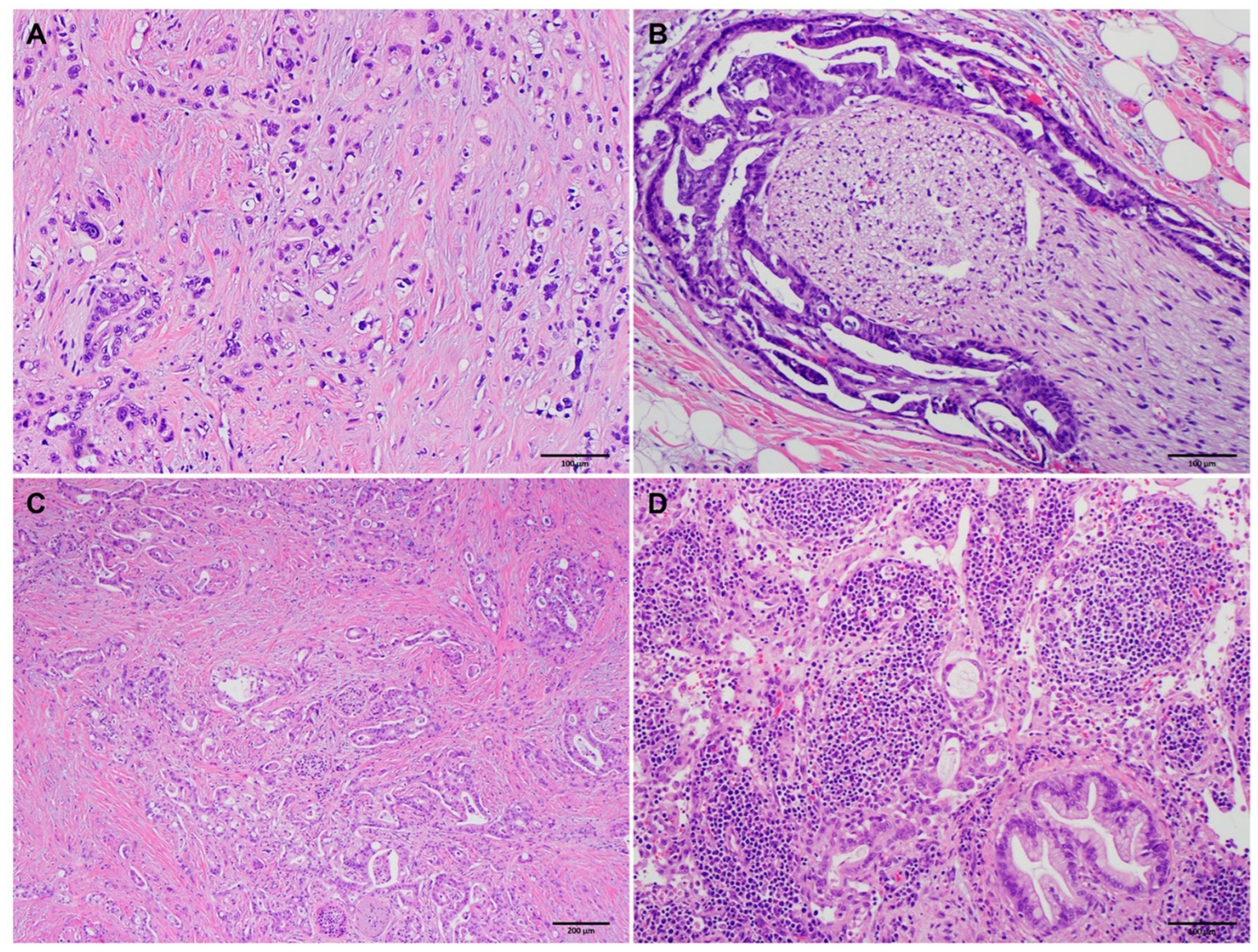
Figure 1. Representative micrographs showing poorly differentiated histology ((A), original magnification 100×), perineural invasion ((B), original magnification 100×), minimal response to neoadjuvant therapy ((C), original magnification 40×), and metastatic adenocarcinoma in a lymph node ((D), original magnification 100×) in four different short-term survivors.
Cancers 15 03231 g001
Figure 2. Representative micrographs showing near-complete response to neoadjuvant therapy ((A,B) in a long-term survivor). (A) Extensive fibrosis with microscopic foci of residual viable tumor (marked with arrows, original magnification 20×). (B) Higher magnification showing the microscopic foci of residual viable adenocarcinoma cells (marked with arrows, original magnification 200×). (C) Representative micrographs showing well-differentiated histology (original magnification 100×) in a long-term survivor.
Cancers 15 03231 g002
Table 1. Comparison of the clinicopathologic factors between short-term and intermediate-term survivors.
CharacteristicsShort-TermIntermediate-Termp Value
Mean age ± SD (years)66.0 ± 9.863.5 ± 9.30.06
Sex 0.03
Female18159
Male42193
Neoadjuvant therapy 0.214
FP-based chemo + radiation16119
Gem-based chemo + radiation41193
Gem-based chemo230
FOLFIRINOX110
Resectability 0.04
Potentially resectable50235
Borderline resectable892
Locally advanced225
Differentiation 0.006
Well-moderate26221
Poor34131
Mean tumor size ± SD (cm)2.9 ± 1.22.7 ± 1.30.13
ypT stage 0.36
ypT0/ypT121115
ypT228193
ypT31144
Lymphovascular invasion 0.12
Negative22169
Positive38183
Perineural invasion 0.052
Negative673
Positive54279
ypN stage 0.002
ypN012153
ypN131119
ypN21780
Mean number of positive nodes ± SD3.1 ± 3.72.1 ± 3.30.04
Mean total number of nodes ± SD23.8 ± 9.625.9 ± 10.30.14
Mean positive lymph node ratio ± SD0.12 ± 0.130.09 ± 0.120.03
MDA Tumor Response Grade 0.46
MDA grade 007
MDA grade 1732
MDA grade 253313
CAP Tumor Response Grade 0.50
Grade 007
Grade 1732
Grade 231203
Grade 322110
Margin status 0.70
Negative52296
Positive856
Recurrence/metastasis 0.15
None863
Local877
Distant44212
Abbreviations: SD, standard deviation; FP, fluoropyrimidine; Gem, gemcitabine; chemo, chemotherapy.
Table 2. Comparison of the clinicopathologic factors between long-term and intermediate-term survivors.
Long-TermIntermediate-Termp Value
Mean age ± SD (years)63.0 ± 9.663.5 ± 9.30.59
Sex 0.63
Female71159
Male78193
Neoadjuvant therapy 0.08
FP-based chemo + radiation47119
Gem-based chemo + radiation94193
Gem-based chemo430
FOLFIRINOX410
Resectability 0.86
Potentially resectable101235
Borderline resectable3692
Locally advanced1225
Differentiation 0.19
Well-moderate103221
Poor46131
Mean tumor size ± SD (cm)2.24 ± 1.522.67 ± 1.270.001
ypT stage 0.001
ypT0/ypT174115
ypT263193
ypT31244
Lymphovascular invasion 0.001
Negative96169
Positive53183
Perineural invasion <0.001
Negative5973
Positive90279
ypN stage <0.001
ypN094153
ypN144119
ypN21180
Mean number of positive nodes ± SD1.04 ± 2.532.14 ± 3.30<0.001
Mean total number of nodes ± SD25.58 ± 10.4925.88 ± 10.290.77
Mean positive lymph node ratio ± SD0.036 ± 0.0680.087 ± 0.124<0.001
MDA Tumor Response Grade <0.001
MDA grade 097
MDA grade 13032
MDA grade 2110313
CAP Tumor Response Grade <0.001
Grade 097
Grade 13032
Grade 270203
Grade 340110
Margin status 0.07
Negative135296
Positive1456
Recurrence/metastasis <0.001
None9763
Local1377
Distant39212
Abbreviations: SD, standard deviation; FP, fluoropyrimidine; Gem, gemcitabine; chemo, chemotherapy.
Table 3. Comparison of the clinicopathologic factors between short-term and long-term survivors.
CharacteristicsShort-TermLong-Termp Value
Mean age ± SD (years)66.0 ± 9.863.0 ± 9.60.04
Sex 0.02
Female1871
Male4278
Neoadjuvant therapy 0.86
FP-based chemo + radiation1647
Gem-based chemo + radiation4194
Gem-based chemo24
FOLFIRINOX14
Resectability 0.07
Potentially resectable50101
Borderline resectable836
Locally advanced212
Differentiation 0.001
Well-moderate26103
Poor3446
Mean tumor size ± SD (cm)2.93 ± 1.232.24 ± 1.520.002
ypT stage 0.04
ypT0/ypT12174
ypT22863
ypT31112
Lymphovascular invasion <0.001
Negative2296
Positive3853
Perineural invasion <0.001
Negative659
Positive5490
ypN stage <0.001
ypN01294
ypN13144
ypN21711
Mean number of positive nodes ± SD3.12 ± 3.651.04 ± 2.53<0.001
Mean total number of nodes ± SD23.75 ± 9.6225.58 ± 10.490.24
Mean positive lymph node ratio ± SD0.124 ± 0.1340.036 ± 0.068<0.001
MDA Tumor Response Grade 0.04
MDA grade 009
MDA grade 1730
MDA grade 253110
CAP Tumor Response Grade 0.07
Grade 009
Grade 1730
Grade 23170
Grade 32240
Margin status 0.46
Negative52135
Positive814
Recurrence <0.001
None897
Local813
Distant4439
Abbreviations: SD, standard deviation; FP, fluoropyrimidine; Gem, gemcitabine; chemo, chemotherapy.
Table 4. Multivariate logistic regression analysis of short-term vs. intermediate-term survivors.
CharacteristicsNumber of PatientsOR (95% CI)p Value
Sex
Female1771.00
Male2351.57 (0.85–2.90)0.15
Resectability 0.07
Potentially resectable2851.00
Borderline resectable1003.22 (0.72–14.49)0.13
Locally advanced271.48 (0.29–7.61)0.64
Tumor differentiation
Well-moderate 2471.00
Poor1652.09 (1.18–3.72)0.01
ypN stage 0.006
ypN01651.00
ypN11503.33 (1.59–6.97)0.001
ypN2972.67 (1.15–6.18)0.02
Tumor response grading
MDA grade 0 or 1 461.00
MDA grade 23663.02 (0.97–9.45)0.06
Perineural Invasion
Negative791.00
Present3332.87 (0.95–8.64)0.06
Abbreviations: OR, Odds ratio; CI, confidence interval.
Table 5. Multivariate logistic regression analysis of long-term vs. intermediate-term survivors.
CharacteristicsNumber of PatientsOR (95% CI)p Value
Neoadjuvant therapy 0.41
FP-based chemo + radiation1661.00
Gem-based chemo + radiation2871.21 (0.78–1.86)0.40
Gem-based chemo340.50 (0.16–1.53)0.22
FOLFIRINOX141.02 (0.29–3.62)0.97
ypT stage 0.44
ypT0/ypT11891.00
ypT22560.76 (0.48–1.20)0.24
ypT3560.71 (0.33–1.51)0.37
ypN stage 0.003
ypN02471.00
ypN11630.72 (0.46–1.12)0.14
ypN2910.30 (0.15–0.60)0.001
Tumor Response Grading
MDA grade 0 or 1 781.00
MDA grade 24230.55 (0.32–0.97)0.04
Lymphovascular invasion
Negative2651.00
Positive2360.86 (0.54–1.37)0.52
Perineural invasion
Negative1321.00
Positive3690.61 (0.38–0.98)0.04
Margin
Negative4311.00
Positive700.92 (0.47–1.81)0.82
Abbreviations: FP, fluoropyrimidine; Gem, gemcitabine; chemo, chemotherapy; OR, Odds ratio; CI, confidence interval.
Disclaimer/Publisher’s Note: The statements, opinions and data contained in all publications are solely those of the individual author(s) and contributor(s) and not of MDPI and/or the editor(s). MDPI and/or the editor(s) disclaim responsibility for any injury to people or property resulting from any ideas, methods, instructions or products referred to in the content.