Article

TP53 Mutations in AML Patients Are Associated with Dismal Clinical Outcome Irrespective of Frontline Induction Regimen and Allogeneic Hematopoietic Cell Transplantation

1
Department of Laboratory Medicine and Pathobiology, University of Toronto, Toronto, ON M5S 1A8, Canada
2
Department of Laboratory Hematology, Laboratory Medicine Program, University Health Network, Toronto, ON M5G 2C4, Canada
3
Department of Medical Oncology and Hematology, Princess Margaret Cancer Centre, University Health Network, Toronto, ON M5G 2M9, Canada
4
Department of Biostatistics, University Health Network, Toronto, ON M5G 2C4, Canada
5
Hans Messner Allogeneic Blood and Marrow Transplantation Program, Division of Medical Oncology and Hematology, Princess Margaret Cancer Centre, University Health Network, Toronto, ON M5G 2M9, Canada
*
Author to whom correspondence should be addressed.
Cancers 2023, 15(12), 3210; https://doi.org/10.3390/cancers15123210
Submission received: 2 May 2023 / Revised: 8 June 2023 / Accepted: 12 June 2023 / Published: 16 June 2023
(This article belongs to the Special Issue Hematologic Malignancies: Challenges from Diagnosis to Treatment)

Abstract

:

Simple Summary

TP53 mutations are adverse-risk genetic aberrations in acute myeloid leukemia (AML). The optimal treatment approach in patients with TP53-mutated (TP53MUT) AML remains unclear. We aimed to evaluate the prognostic implications of different frontline treatment strategies and transplantation for patients with TP53MUT AML. Patients treated with intensive induction or azacitidine-venetoclax induction had no significant improvement in survival compared to patients treated with other HMA regimens despite having higher complete remission rates. Transplantation was not significantly associated with improved outcomes in time-dependent or landmark analysis, however, transplanted patients with lower TP53MUT variant allele frequency (VAF) at the time of diagnosis had superior outcomes compared to transplanted patients with higher TP53 VAF. Current therapeutic strategies remain ineffective for TP53MUT AML patients, which highlights the urgent need for new treatment strategies for this high-risk population.

Abstract

TP53 mutations are associated with extremely poor outcomes in acute myeloid leukemia (AML). The outcomes of patients with TP53-mutated (TP53MUT) AML after different frontline treatment modalities are not well established. Allogeneic hematopoietic cell transplantation (allo-HCT) is a potentially curative procedure for AML; however, long-term outcomes among patients with TP53MUT AML after allo-HCT are dismal, and the benefit of allo-HCT remains controversial. We sought to evaluate the outcomes of patients with TP53MUT AML after treatment with different frontline induction therapies and allo-HCT. A total of 113 patients with TP53MUT AML were retrospectively evaluated. Patients with TP53MUT AML who received intensive or azacitidine-venetoclax induction had higher complete remission rates compared to patients treated with other hypomethylating-agent-based induction regimens. However, OS and EFS were not significantly different among the induction regimen groups. Allo-HCT was associated with improved OS and EFS among patients with TP53MUT AML; however, allo-HCT was not significantly associated with improved OS or EFS in time-dependent or landmark analysis. While the outcomes of all patients were generally poor irrespective of therapeutic strategy, transplanted patients with lower TP53MUT variant allele frequency (VAF) at the time of diagnosis had superior outcomes compared to transplanted patients with higher TP53 VAF. Our study provides further evidence that the current standards of care for AML confer limited therapeutic benefit to patients with TP53 mutations.

1. Introduction

The TP53 gene, located on chromosome 17p, plays a central role in anti-tumour response by regulating the cell cycle and apoptosis [1]. Mutations in TP53 occur in approximately 10–20% of acute myeloid leukemia (AML) patients and are associated with an extremely poor prognosis [2]. TP53 mutation is an adverse-risk genetic abnormality according to the 2022 ELN risk classification of AML due to its association with complex karyotype, advanced age, and resistance to standard therapies [3]. TP53 mutation defines a distinct standalone entity within the International Consensus Classification of Myeloid Neoplasms and Acute Leukemia [4].
In fit patients with high-risk AML, the standard treatment approach is induction therapy, followed by allogeneic hematopoietic cell transplantation (allo-HCT) as a curative modality for those in remission [3]. Patients with TP53-mutated (TP53MUT) AML have a lower probability of achieving remission when treated with intensive chemotherapy and, consequently, have poor outcomes; for those who achieve remission, allo-HCT provides a relatively small improvement in long-term survival [2,5,6,7,8,9].
Given that there is limited data comparing the outcomes after different treatment modalities for patients with TP53MUT AML, we retrospectively reviewed our institution’s experience with TP53MUT AML. We assessed the prognostic implications of different frontline treatment strategies and transplantation for patients with TP53MUT AML.

2. Materials and Methods

2.1. Patients and Treatment

We screened all adult University Health Network (UHN) patients who were diagnosed with AML with blasts ≥20% from February 2015 to December 2021 and who had molecular testing results available. Patients’ medical records were retrospectively assessed with respect to demographic and clinicopathological data, treatment modalities, and survival outcomes. The study was approved by the University Health Network Research Ethics Board.
Fifty-seven patients with TP53MUT AML received intensive induction therapy, with regimens including fludarabine, cytarabine, idarubicin, and granulocyte-colony stimulating factor (FLAG-IDA) (n = 32); cytarabine and daunorubicin (3 + 7) (n = 23); and liposomal cytarabine and daunorubicin (CPX-351) (n = 2). The dosages of these induction regimens are provided in the Supplementary Materials. Thirty-one patients received HMA-based induction therapy, with regimens including azacitidine and venetoclax (AZA-VEN) (n = 10), azacitidine alone (n = 18), guadecitabine (n = 2), and azacitidine and enasidenib (n = 1). The latter three treatment regimens were referred to as “other HMA-based regimens”. Patients treated with HMA-based regimens received a median of 3 (range, 1–12) cycles of HMA. Patients treated with FLAG-IDA induction who achieved complete remission were subsequently treated with FLAG-IDA consolidation. Patients treated with cytarabine and daunorubicin who achieved complete remission proceeded to be treated with cytarabine and daunorubicin consolidation or high-dose cytarabine (HiDAC) consolidation.
According to the American Society of Transplantation and Cellular Therapy recommendations for unfavourable-risk AML, allo-HCT was offered to patients with TP53MUT AML in CR if a suitable donor was identified [10,11]. Patients received 1–2 cycles of consolidation chemotherapy before proceeding to treatment with allo-HCT.

2.2. Mutational Analysis and Determination of TP53 Allelic Status

Next-generation sequencing (NGS) was performed using a custom gene panel of 49 myeloid genes (Oxford Gene Technologies, Oxford, UK) or 54 myeloid genes (Illumina, San Diego, CA, USA) and run on the MiSeq platform (Illumina), as previously described [12,13,14]. The limit of detection for variant calling was 2%. For both panels, for 13/41 genes, the complete coding regions were analyzed, and for 28/41 genes, the same exonic hotspots were analyzed (Tables S1 and S2). All consensus-coding TP53 exons were interrogated. Interpretation and classification of variants was performed as previously described [12]. Briefly, pathogenic (AMP/ASCO Tier 1) variants consisted of (a) loss-of-function truncating variants (nonsense, frameshift, and splicing) and (b) missense or in-frame variants (typically, within the DNA-binding domain) demonstrating loss-of-function (e.g., p.R110P) [15,16]. Likely pathogenic (AMP/ASCO Tier 2) variants consisted of (a) missense or in-frame variants involving the same residue (e.g., R110) as an established pathogenic variant and (b) missense or in-frame variants (typically, within the DNA binding domain) classified as disruptive in the TP53 IARC database [17]. Variants of uncertain significance (VUS, AMP/ASCO Tier 3) included variants classified as partially or fully transcriptionally active according to the TP53 IARC database as well as variants with no functional evidence of deleterious potential. VUS and benign polymorphisms were excluded from analysis. When patients had multiple mutations in the same gene, the higher variant allele frequency (VAF) was used for analysis. TP53 mutations were visualized using cBioPortal [18,19].
Patients were considered to be double/multi-hit TP53 when (A) at least two TP53 variants were detected via NGS, (B) one TP53 variant detected via NGS co-occurred with cytogenetic loss of TP53, or(C) one TP53 variant was detected via NGS with a VAF of ≥55%, as previously described [20].

2.3. Karyotype Analysis

In accordance with the International System for Human Cytogenetic Nomenclature’s guidelines, karyotypes were obtained from diagnostic bone marrow samples and described as appropriate. The cytogenetic loss of TP53 was determined as previously described [21].

2.4. Statistical Analysis

Categorical variables were summarized as counts and percentages. Continuous variables were summarized as medians and ranges. Evaluation of patient outcome including overall survival (OS) and event-free survival (EFS) was carried out through retrospective analysis of patient records. OS was calculated from the date of diagnosis to last follow-up or death. EFS was calculated from the date of diagnosis to last follow-up, relapse, or death. Log-rank test was used to compare Kaplan–Meier survival curves. Univariate Cox proportional-hazard regression models were fitted to identify prognostic factors for OS and EFS. Cox proportional-hazard regression was used to evaluate the prognostic impact of transplantation as a time-dependent variable, as transplant (if any) can occur during the follow-up period. Differences in categorical variables were assessed using the Fisher’s exact test. Differences in numerical variables across different groups were assessed using the Kruskal–Wallis and Wilcoxon rank-sum tests. Statistical analysis was performed using R version 4.0.5 (R Core Team (2020). R: A language and environment for statistical computing, Vienna, Austria).

3. Results

3.1. Patient Characteristics

We identified 113 patients with TP53MUT AML from our institution. Patient characteristics of the cohort after being stratified by frontline treatment modality are summarized and compared in Table 1. Fifty-seven (50%) patients received intensive chemotherapy, ten (9%) patients received AZA-VEN, twenty-one (19%) patients received other HMA-based regimens, and twenty-five (22%) patients received best supportive care. Gender ratio, baseline hematological parameters, and clinical ontogeny did not differ among the different treatment groups. However, patients who were treated with intensive chemotherapy were younger than those who were treated with AZA-VEN, other HMA-based regimens, and best supportive care (p = 0.0050, p < 0.0001, and p < 0.0001, respectively). In addition, the complete remission rate in the intensive chemotherapy and AZA-VEN groups were higher compared to the other HMA-based regimens group (p = 0.0013 and p = 0.022, respectively). A total of seventeen patients received allo-HCT after achieving remission; fifteen of the transplanted patients were treated with intensive induction chemotherapy, and two of the transplanted patients were treated with AZA-VEN induction. Eight patients had a matched unrelated donor transplant, five patients had a matched related donor transplant, and four patients had a haploidentical donor transplant. The median time-to-transplantation from diagnosis was 4.11 months (range 2.1–6.7 months).
Additional baseline characteristics of the cohort are summarized in Table S3. According to the WHO 2016 classification, the majority of patients were classified as either AML with myelodysplasia-related changes (n = 92, 81%) or therapy-related AML (n = 13, 12%). The most frequent co-mutations were detected in DNMT3A (n = 17, 15%), IDH1 (n = 11, 10%), TET2 (n = 8, 7%), and JAK2 (n = 5, 4%). The most frequent cytogenetic abnormalities were complex karyotype (n = 95, 84%), −5/del(5q) (n = 69, 61%), −7/del(7q) (n = 52, 46%), and -17/del(17p)/dic(17p) (n = 49, 43%).
A total of 144 TP53 mutations were detected, including 102 (71%) missense mutations, 18 (13%) frameshift mutations, 11 (8%) nonsense mutations, 11 (8%) splice site mutations, and 2 (1%) in-frame amino acid insertions/deletions (Figure 1).

3.2. Outcomes of Patients According to Treatment and Molecular Characteristics

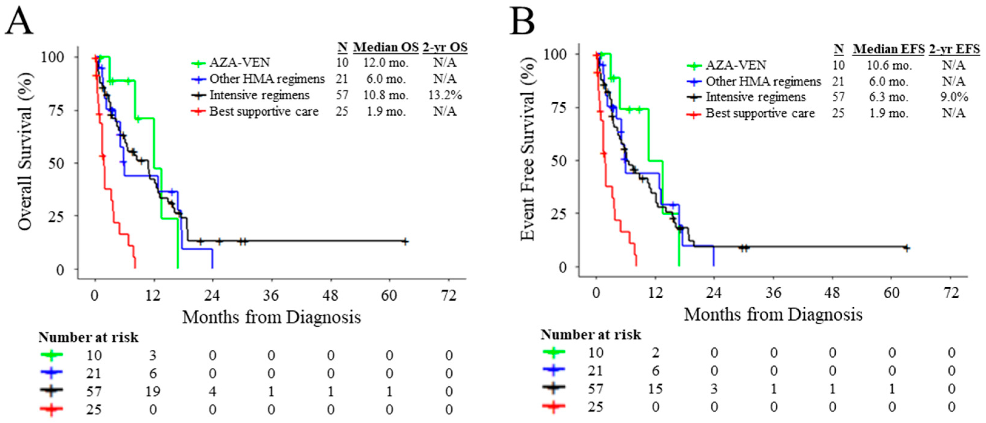
Compared to patients with TP53MUT AML receiving best supportive care, patients who were treated with intensive induction, AZA-VEN, or other HMA-based regimens had superior OS (HR: 0.23, 95% CI: 0.12–0.42, p < 0.0001; HR: 0.07, 95% CI: 0.02–0.34, p = 0.0009; and HR: 0.25, 95% CI: 0.12–0.55, p = 0.0005, respectively) and EFS (HR: 0.28, 95% CI: 0.15–0.49, p < 0.0001; HR: 0.10, 95% CI: 0.02–0.42, p = 0.0020; and HR: 0.25, 95% CI: 0.12–0.55, p = 0.0005, respectively) (Figure 2A,B). Patients with TP53MUT AML who received intensive regimens did not have significantly different OS or EFS compared to patients treated with AZA-VEN (HR: 1.20, 95% CI: 0.47–3.07, p = 0.70; and HR: 1.39, 95% CI: 0.55–3.53, p = 0.48, respectively) or other HMA-based regimens (HR: 0.84, 95% CI: 0.47–1.53, p = 0.58; and HR: 1.01, 95% CI: 0.56–1.82, p = 0.96, respectively) (Figure 2A,B). Patients with TP53MUT AML treated with AZA-VEN did not have significantly different OS or EFS compared to patients treated with other HMA-based regimens (HR: 0.82, 95% CI: 0.29–2.35, p = 0.71; and HR: 0.79, 95% CI: 0.28–2.22, p = 0.65, respectively) (Figure 2A,B). Overall, while patients benefited from treatment compared to best supportive care, no treatment strategy conferred significantly superior outcomes in our cohort.
Patients treated with HMA-based and intensive induction regimens were included in all downstream survival analyses. No significant difference in OS or EFS was observed when stratifying patients by the presence or absence of mutation in the DNA binding domain of TP53 (HR: 0.94, 95% CI: 0.43–2.06, p = 0.869; and HR: 0.94, 95% CI: 0.43–2.06, p = 0.873, respectively) (Figure S1A,B). We further evaluated if any of the frequently co-mutated genes were associated with differences in OS or EFS. Among the treated patients, DNMT3A and TET2 mutations were associated with marginally inferior OS (HR: 2.02, 95% CI: 0.99–4.13, p = 0.055; and HR: 2.40, 95% CI: 0.94–6.10, p = 0.066 respectively) and EFS (HR: 1.81, 95% CI: 0.89–3.68, p = 0.103; and HR: 2.32, 95% CI: 0.92–5.87, p = 0.076, respectively). The presence of an IDH1 mutation was not associated with significantly different OS or EFS (HR: 0.73, 95% CI: 0.29–1.84, p = 0.508; and HR: 0.61, 95% CI: 0.24–1.54, p = 0.298, respectively).
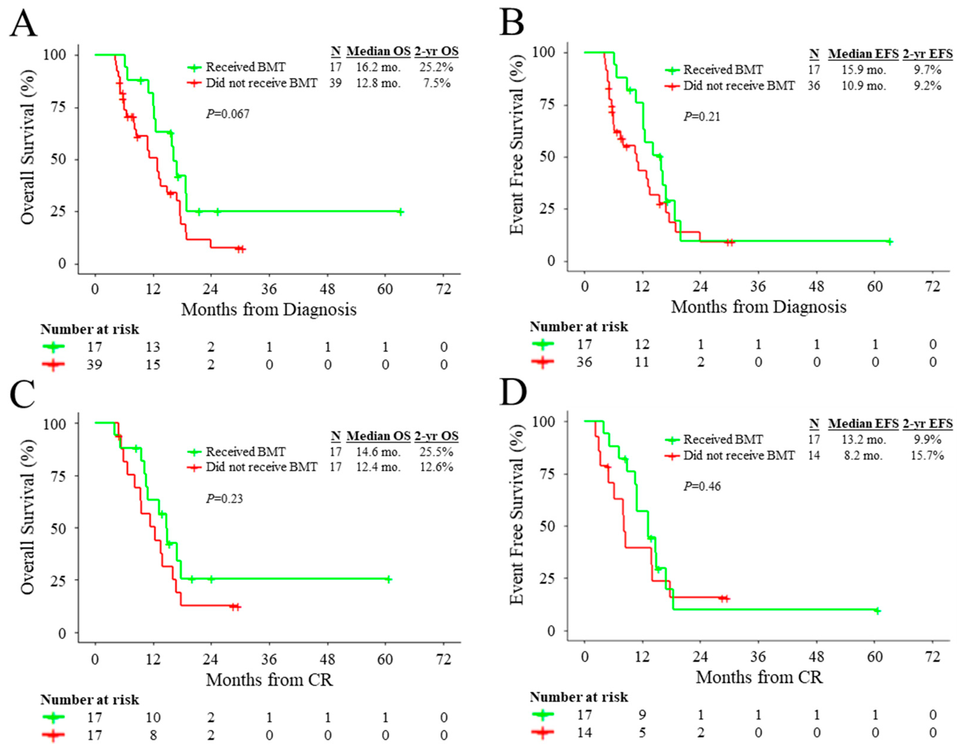
In patients treated with HMA-based and intensive induction regimens, allo-HCT was associated with improved OS (HR: 0.39, 95% CI: 0.20–0.75, p = 0.0047) and EFS (HR: 0.44, 95% CI: 0.24–0.82, p = 0.010) compared to non-transplanted patients (Figure S2A,B). However, when evaluating the impact of transplantation as a time-dependent variable, transplantation was not significantly associated with improved OS (HR: 0.61, 95% CI: 0.30–1.25, p = 0.18) or EFS (HR: 0.74, 95% CI: 0.38–1.47, p = 0.39). Landmark analysis for OS that excluded patients who died or were lost to follow-up before 4.11 months following diagnosis (corresponding to the median time from diagnosis to allo-HCT) indicated that allo-HCT marginally improved OS (HR: 0.53, 95% CI: 0.26–1.06, p = 0.072) (Figure 3A). Similarly, landmark analysis for EFS that excluded patients who died, were lost to follow-up, or relapsed before 4.11 months following diagnosis indicated that allo-HCT did not significantly improve EFS (HR: 0.65, 95% CI: 0.33–1.27, p = 0.21) (Figure 3B).
To evaluate whether patients benefited from allo-HCT after the achievement of CR, we performed a separate landmark analysis of patients that achieved CR. Landmark analysis for OS, which excluded patients who died or were lost to follow-up before 2.34 months following achievement of CR (corresponding to the median time from achievement of CR to allo-HCT), indicated that allo-HCT did not significantly improve OS (HR: 0.62, 95% CI: 0.28–1.37, p = 0.24) in remission (Figure 3C). Similarly, landmark analysis for EFS that excluded patients who died, were lost to follow-up, or relapsed before 2.34 months following the achievement of CR indicated that allo-HCT did not significantly improve EFS (HR: 0.74, 95% CI: 0.33–1.67, p = 0.47) in remission (Figure 3D).
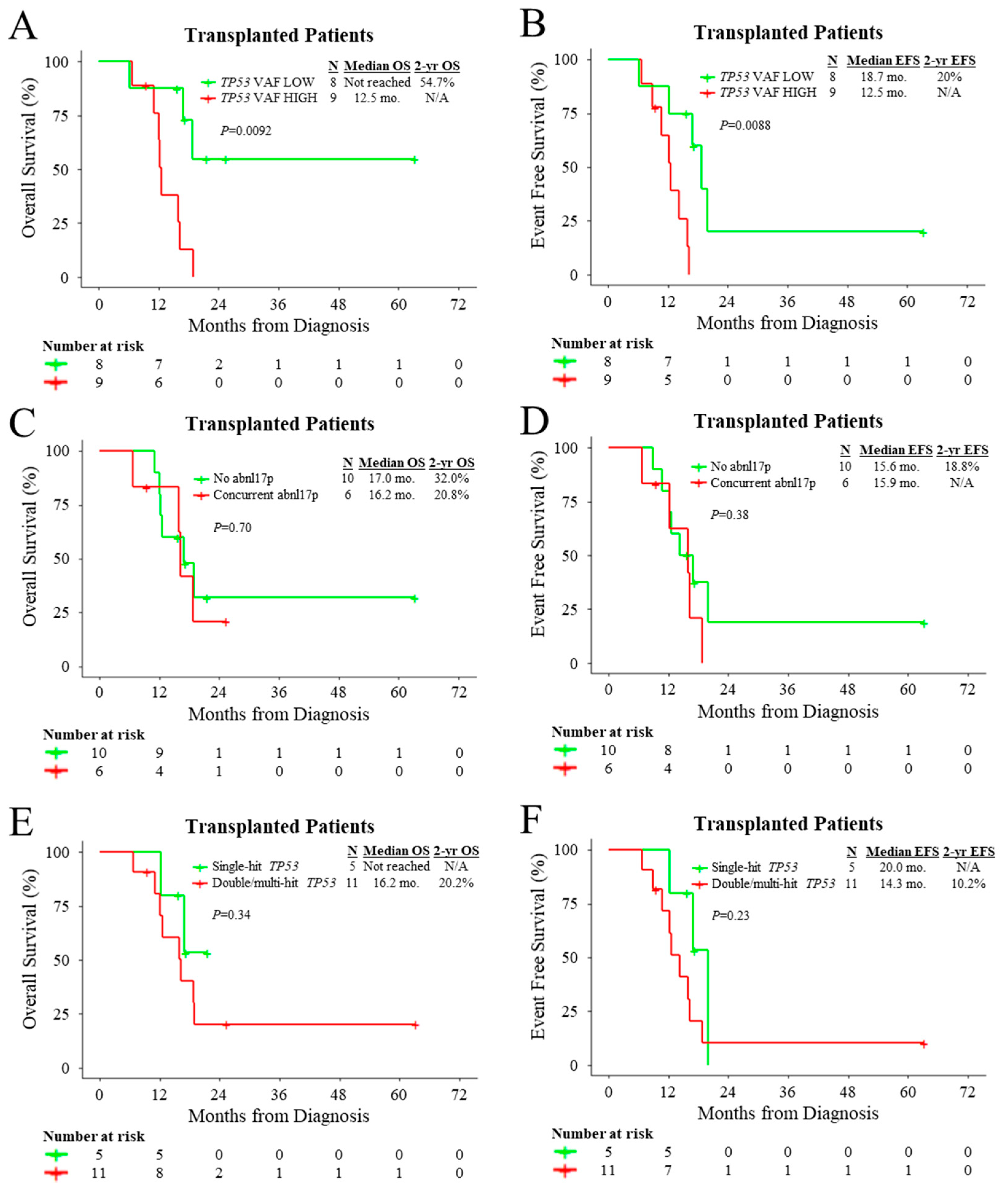
Given the dismal outcomes of patients with TP53MUT AML treated with allo-HCT, we sought to identify factors that may predict favourable outcomes with respect to transplantation. When using the median TP53 VAF of transplanted patients (45%) as the cut-off, transplanted patients with low TP53 VAF at the time of diagnosis had superior OS (HR: 0.19, 95% CI: 0.05–0.74, p = 0.017) and EFS (HR: 0.15, 95% CI: 0.03–0.75, p = 0.020) compared to those with high TP53 VAF (Figure 4A,B). Among transplanted patients, concurrent cytogenetic abnormality in 17p did not modify OS (HR: 1.29, 95% CI: 0.36–4.61, p = 0.70) or EFS (HR: 1.70, 95% CI: 0.51–5.61, p = 0.39) (Figure 4C,D). In addition, double/multi-hit TP53 status did not significantly influence OS (HR: 2.11, 95% CI: 0.44–10.03, p = 0.35) or EFS (HR: 2.20, 95% CI: 0.59–8.24, p = 0.24) among transplanted patients (Figure 4E,F).
To further confirm that the favourable outcome associated with transplantation may be confined to patients with low TP53 mutational burden, we performed subgroup landmark analyses for treated patients stratified by the median TP53 VAF of the transplanted patients (45%). For patients with low TP53 VAF, landmark analysis for OS that excluded patients who died or were lost to follow-up before 4.11 months following diagnosis (corresponding to the median time from diagnosis to allo-HCT) indicated that allo-HCT significantly improved OS (HR: 0.21, 95% CI: 0.06–0.73, p = 0.014) (Figure S3A). Similarly, landmark analysis for EFS that excluded patients who died, were lost to follow-up, or relapsed before 4.11 months following diagnosis indicated that allo-HCT marginally improved EFS (HR: 0.36, 95% CI: 0.13–1.01, p = 0.053) among patients with low TP53 VAF (Figure S3B). In contrast, for patients with high TP53 VAF, landmark analysis for OS that excluded patients who died or were lost to follow-up before 4.11 months following diagnosis indicated that allo-HCT did not significantly improve OS (HR: 1.10, 95% CI: 0.44–2.71, p = 0.84) (Figure S3C). Similarly, landmark analysis for EFS that excluded patients who died, were lost to follow-up, or relapsed before 4.11 months following diagnosis indicated that allo-HCT did not significantly improve EFS (HR: 1.06, 95% CI: 0.41–2.71, p = 0.91) among patients with high TP53 VAF (Figure S3D).

4. Discussion

In this study, we report a single-institution evaluation of different treatment modalities for patients with TP53MUT AML. While the use of any treatment was associated with better outcomes than best supportive care, intensive induction chemotherapy was not associated with a better outcome than HMA therapy. Furthermore, while transplanted patients had significantly superior outcomes compared to non-transplanted patients, this association was lost in time-dependent analysis and in landmark analysis.
Anti-cancer responses to cytotoxic chemotherapies are critically dependent on wild-type TP53, as the activation of p53 by chemotherapy-induced DNA damage induces apoptosis and cancer cell death [22,23]. As such, standard induction treatments using DNA-damaging drugs may be a suboptimal therapeutic approach for patients with TP53MUT AML. Prior clinical studies have demonstrated that patients with TP53MUT AML have poor outcomes and response rates when treated with intensive chemotherapy regimens [24,25,26,27,28,29]. In addition, other less-intensive therapies such as hypomethylating agents and venetoclax have shown varying levels of efficacy among TP53MUT AML patients [30,31,32,33]. In our cohort, patients with TP53MUT AML had no significant differences in outcomes after treatment with intensive and non-intensive frontline regimens despite the younger age of the intensively treated patients. While the addition of venetoclax significantly improved complete remission rates among patients with TP53MUT AML treated with HMA-based regimens, survival was not significantly improved with venetoclax, which is in line with prior reports [34,35,36]. Indeed, TP53 mutations have been shown to confer resistance to BCL-2 inhibition in preclinical studies [37,38,39]. Clinical trials for novel targeted agents and immunotherapeutic approaches such as TP53MUT re-activators and anti-CD47 blockade have shown potential in the frontline setting of TP53MUT AML and should be explored further in this high-risk population [40].
Allo-HCT is considered to be a potentially curative option for high-risk AML patients; however, whether transplantation confers a beneficial impact among patients with TP53MUT AML is controversial. Prior studies have largely supported the use of allo-HCT in these patients as they have reported superior outcomes in transplanted patients compared to non-transplanted patients [27,35,41,42].Conversely, there is evidence in the literature that patients with TP53MUT AML do not significantly benefit from allo-HCT [34]. We previously evaluated the impact of allo-HCT among patients with AML with myelodysplasia-related changes and reported that allo-HCT had a significant benefit among TP53MUT patients [43]. However, consistent with our data presented herein, TP53MUT patients did not experience sustained and plateauing long-term outcomes and survival rates after allo-HCT. Since the outcomes of transplanted and non-transplanted patients may be confounded due to guarantee-time bias, we performed a prognostic evaluation of allo-HCT using time-dependent and landmark analyses [44]. In these analyses, TP53MUT patients who received allo-HCT did not have significantly superior outcomes compared to non-transplant patients, suggesting that the differences in outcome according to allo-HCT status are confounded by guarantee-time bias. Despite this, among transplanted patients, low TP53 VAF at the time of diagnosis was associated with superior outcomes. Further landmark analysis of patient subgroups revealed that the favourable outcome associated with transplant is confined to patients with low TP53 VAF, who had significantly improved outcomes with allo-HCT compared to non-transplanted patients with low TP53 VAF. Our findings are consistent with previous reports demonstrating associations between low TP53 VAF and superior outcomes in transplanted patients with TP53MUT AML [35,45]. As such, TP53 VAF may have clinical utility for guiding the decision-making process of whether TP53MUT patients should undergo transplantation. While other studies have shown an adverse prognostic impact of cytogenetic loss of TP53 among transplanted patients with TP53MUT AML, we did not observe a significant difference in our cohort [46]. Future studies are needed to optimize transplant approaches and post-transplant monitoring practices and to explore the prognostic benefit of allo-HCT for TP53MUT AML patients.
The updated 2022 ELN guidelines for the management of AML recommend for TP53 mutational analysis to be performed within the first cycle of chemotherapy [3]. At our institution, TP53 sequencing is performed via NGS, which has a relatively longer turnaround time compared to other diagnostic tests. The lack of benefit for intensive chemotherapy among patients with TP53MUT AML suggests that genetic results for TP53 should be obtained before beginning treatment to avoid ineffective and aggressive therapy. Rapid and cost-effective methods such as immunohistochemistry have shown potential for predicting TP53 mutational status and should be further explored in the clinical setting [47,48].
We acknowledge that our study has several limitations. First, our study is retrospective in nature and includes limited patient numbers in its subgroup analyses. Future multi-center prospective studies are needed to confirm our data. Second, transplanted patient outcomes may be influenced by factors such as patient performance status, conditioning regimen intensity, and post-transplant graft-versus-host disease. Further studies are needed to evaluate the impact of these factors on patient outcomes and to determine the optimal allo-HCT approach for patients with TP53MUT AML. Third, this study only focused on patients with TP53MUT AML; future prospective studies should include patients with and without TP53 mutations to evaluate the clinical impact of TP53 mutations across different treatment groups.

5. Conclusions

In conclusion, outcomes among patients with TP53MUT AML after intensive therapy and allo-HCT remain poor; however, patients with low TP53 VAF at the time of diagnosis may still benefit from transplantation. Our study provides further evidence that patients with TP53MUT AML derive limited benefit from current treatments and highlights the urgent need for new treatment strategies.

Supplementary Materials

The following supporting information can be downloaded at: https://www.mdpi.com/article/10.3390/cancers15123210/s1, Figure S1: Outcomes according to presence/absence of TP53 mutations in the DNA binding domain. Figure S2: Outcomes according to transplant status. Figure S3: Outcomes according to TP53 VAF and transplant status; Table S1: Gene panel for targeted sequencing; Table S2: Exon coverage for hotspot genes; Table S3: Summary of additional clinicopathological features.

Author Contributions

D.Z. analyzed data, performed statistical analysis, and wrote the manuscript. D.Z., M.Z., Q.Z., J.-M.C.-C., A.S., M.D.M. and R.K. collected data for analysis. E.G.A. performed statistical analysis. H.C. designed the study, analyzed data, supervised the project, and wrote the manuscript. All authors have read and agreed to the published version of the manuscript.

Funding

This study received no external funding.

Institutional Review Board Statement

The study was approved by the UHN Research Ethics Board and was conducted according to the principles of the Declaration of Helsinki.

Informed Consent Statement

Patient consent was not required because of the retrospective nature of the study.

Data Availability Statement

Data are available on request from the corresponding author.

Acknowledgments

We would like to acknowledge the physicians and nurses of the UHN leukemia programme for their outstanding patient care and the support of the PMH cancer registry. We also thank Entsar Eladl for providing helpful suggestions.

Conflicts of Interest

The authors declare no conflict of interest.

References

  1. Chen, J. The Cell-Cycle Arrest and Apoptotic Functions of p53 in Tumor Initiation and Progression. Cold Spring Harb. Perspect. Med. 2016, 6, a026104. [Google Scholar] [CrossRef] [PubMed] [Green Version]
  2. Molica, M.; Mazzone, C.; Niscola, P.; de Fabritiis, P. TP53 Mutations in Acute Myeloid Leukemia: Still a Daunting Challenge? Front. Oncol. 2021, 10, 3368. Available online: https://pubmed.ncbi.nlm.nih.gov/33628731/ (accessed on 26 March 2022). [CrossRef] [PubMed]
  3. Döhner, H.; Wei, A.H.; Appelbaum, F.R.; Craddock, C.; DiNardo, C.D.; Dombret, H.; Ebert, B.L.; Fenaux, P.; Godley, L.A.; Hasserjian, R.P.; et al. Diagnosis and management of AML in adults: 2022 recommendations from an international expert panel on behalf of the ELN. Blood 2022, 140, 1345–1377. [Google Scholar] [CrossRef] [PubMed]
  4. Arber, D.A.; Orazi, A.; Hasserjian, R.P.; Borowitz, M.J.; Calvo, K.R.; Kvasnicka, H.M.; Wang, S.A.; Bagg, A.; Barbui, T.; Branford, S.; et al. International Consensus Classification of Myeloid Neoplasms and Acute Leukemias: Integrating morphologic, clinical, and genomic data. Blood 2022, 140, 1200–1228. [Google Scholar] [CrossRef]
  5. Ciurea, S.O.; Chilkulwar, A.; Saliba, R.M.; Chen, J.; Rondon, G.; Patel, K.P.; Khogeer, H.; Shah, A.R.; Randolph, B.V.; Ramos Perez, J.M.; et al. Prognostic factors influencing survival after allogeneic transplantation for AML/MDS patients with TP53 mutations. Blood 2018, 131, 2989–2992. [Google Scholar] [CrossRef] [PubMed]
  6. Poiré, X.; Labopin, M.; Maertens, J.; Yakoub-Agha, I.; Blaise, D.; Ifrah, N.; Socié, G.; Gedde-Dhal, T.; Schaap, N.; Cornelissen, J.J.; et al. Allogeneic stem cell transplantation in adult patients with acute myeloid leukaemia and 17p abnormalities in first complete remission: A study from the Acute Leukemia Working Party (ALWP) of the European Society for Blood and Marrow Transplantation (EBMT). J. Hematol. Oncol. 2017, 10, 1–10. [Google Scholar] [CrossRef] [Green Version]
  7. Daher-Reyes, G.; Kim, T.H.; Novitzky-Basso, I.; Kim, K.H.; Smith, A.; Stockley, T.; Capochichi, J.M.; Al-Shaibani, Z.; Pasic, I.; Law, A.; et al. Prognostic impact of the adverse molecular-genetic profile on long-term outcomes following allogeneic hematopoietic stem cell transplantation in acute myeloid leukemia. Bone Marrow Transplant. 2021, 56, 1908–1918. [Google Scholar] [CrossRef]
  8. Middeke, J.M.; Herold, S.; Rücker-Braun, E.; Berdel, W.E.; Stelljes, M.; Kaufmann, M.; Schäfer-Eckart, K.; Baldus, C.D.; Stuhlmann, R.; Ho, A.D.; et al. TP53 mutation in patients with high-risk acute myeloid leukaemia treated with allogeneic haematopoietic stem cell transplantation. Br. J. Haematol. 2016, 172, 914–922. [Google Scholar] [CrossRef]
  9. Grimm, J.; Jentzsch, M.; Bill, M.; Goldmann, K.; Schulz, J.; Niederwieser, D.; Platzbecker, U.; Schwind, S. Prognostic impact of the ELN2017 risk classification in patients with AML receiving allogeneic transplantation. Blood Adv. 2020, 4, 3864–3874. [Google Scholar] [CrossRef]
  10. Kanate, A.S.; Majhail, N.S.; Savani, B.N.; Bredeson, C.; Champlin, R.E.; Crawford, S.; Giralt, S.A.; LeMaistre, C.F.; Marks, D.I.; Omel, J.L.; et al. Indications for Hematopoietic Cell Transplantation and Immune Effector Cell Therapy: Guidelines from the American Society for Transplantation and Cellular Therapy. Biol. Blood Marrow Transplant. 2020, 26, 1247–1256. [Google Scholar] [CrossRef]
  11. Dholaria, B.; Savani, B.N.; Hamilton, B.K.; Oran, B.; Liu, H.D.; Tallman, M.S.; Ciurea, S.O.; Holtzman, N.G.; II, G.L.P.; Devine, S.M.; et al. Hematopoietic Cell Transplantation in the Treatment of Newly Diagnosed Adult Acute Myeloid Leukemia: An Evidence-Based Review from the American Society of Transplantation and Cellular Therapy. Transplant. Cell. Ther. 2021, 27, 6–20. [Google Scholar] [CrossRef] [PubMed]
  12. Gupta, V.; Kennedy, J.A.; Capo-Chichi, J.M.; Kim, S.; Hu, Z.H.; Alyea, E.P.; Popat, U.R.; Sobecks, R.M.; Scott, B.L.; Gerds, A.T.; et al. Genetic factors rather than blast reduction determine outcomes of allogeneic HCT in BCR-ABL-negative MPN in blast phase. Blood Adv. 2020, 4, 5562–5573. [Google Scholar] [CrossRef] [PubMed]
  13. Alduaij, W.; McNamara, C.J.; Schuh, A.; Arruda, A.; Sukhai, M.; Kanwar, N.; Thomas, M.; Spiegel, J.; Kennedy, J.A.; Stockley, T.; et al. Clinical Utility of Next-generation Sequencing in the Management of Myeloproliferative Neoplasms: A Single-Center Experience. HemaSphere 2018, 2, e44. [Google Scholar] [CrossRef] [PubMed]
  14. Thomas, M.; Sukhai, M.A.; Zhang, T.; Dolatshahi, R.; Harbi, D.; Garg, S.; Misyura, M.; Pugh, T.; Stockley, T.L.; Kamel-Reid, S. Integration of Technical, Bioinformatic, and Variant Assessment Approaches in the Validation of a Targeted Next-Generation Sequencing Panel for Myeloid Malignancies. Arch. Pathol. Lab. Med. 2017, 141, 759–775. [Google Scholar] [CrossRef] [PubMed] [Green Version]
  15. Xu, J.; Reumers, J.; Couceiro, J.R.; De Smet, F.; Gallardo, R.; Rudyak, S.; Cornelis, A.; Rozenski, J.; Zwolinska, A.; Marine, J.C.; et al. Gain of function of mutant p53 by coaggregation with multiple tumor suppressors. Nat. Chem. Biol. 2011, 7, 285–295. [Google Scholar] [CrossRef] [Green Version]
  16. Wang, B.; Niu, D.; Lam, T.H.; Xiao, Z.; Ren, E.C. Mapping the p53 transcriptome universe using p53 natural polymorphs. Cell Death Differ. 2014, 21, 521–532. [Google Scholar] [CrossRef]
  17. Bouaoun, L.; Sonkin, D.; Ardin, M.; Hollstein, M.; Byrnes, G.; Zavadil, J.; Olivier, M. TP53 Variations in Human Cancers: New Lessons from the IARC TP53 Database and Genomics Data. Hum. Mutat. 2016, 37, 865–876. [Google Scholar] [CrossRef]
  18. Cerami, E.; Gao, J.; Dogrusoz, U.; Gross, B.E.; Sumer, S.O.; Aksoy, B.A.; Jacobsen, A.; Byrne, C.J.; Heuer, M.L.; Larsson, E.; et al. The cBio cancer genomics portal: An open platform for exploring multidimensional cancer genomics data. Cancer Discov. 2012, 2, 401–404. [Google Scholar] [CrossRef] [Green Version]
  19. Gao, J.; Aksoy, B.A.; Dogrusoz, U.; Dresdner, G.; Gross, B.; Sumer, S.O.; Sun, Y.; Jacobsen, A.; Sinha, R.; Larsson, E.; et al. Integrative analysis of complex cancer genomics and clinical profiles using the cBioPortal. Sci. Signal. 2013, 6, pl1. [Google Scholar] [CrossRef] [Green Version]
  20. Grob, T.; Al Hinai, A.S.A.; Sanders, M.A.; Kavelaars, F.G.; Rijken, M.; Gradowska, P.L.; Biemond, B.J.; Breems, D.A.; Maertens, J.; van Marwijk Kooy, M.; et al. Molecular characterization of mutant TP53 acute myeloid leukemia and high-risk myelodysplastic syndrome. Blood 2022, 139, 2347–2354. [Google Scholar] [CrossRef]
  21. Montalban-Bravo, G.; Kanagal-Shamanna, R.; Benton, C.B.; Class, C.A.; Chien, K.S.; Sasaki, K.; Naqvi, K.; Alvarado, Y.; Kadia, T.M.; Ravandi, F.; et al. Genomic context and TP53 allele frequency define clinical outcomes in TP53-mutated myelodysplastic syndromes. Blood Adv. 2020, 4, 482–495. [Google Scholar] [CrossRef] [PubMed] [Green Version]
  22. Hientz, K.; Mohr, A.; Bhakta-Guha, D.; Efferth, T. The role of p53 in cancer drug resistance and targeted chemotherapy. Oncotarget 2017, 8, 8921–8946. [Google Scholar] [CrossRef] [PubMed] [Green Version]
  23. Karran, P. Mechanisms of tolerance to DNA damaging therapeutic drugs. Carcinogenesis 2001, 22, 1931–1937. [Google Scholar] [CrossRef] [PubMed] [Green Version]
  24. Przespolewski, A.; Goldberg, A.D.; Talati, C.; Fazal, S.; Vachhani, P.; Sanikommu, S.R.R.; Thota, S.; Waksal, J.; Ball, B.; Famulare, C.; et al. Safety and Efficacy of CPX-351 in Younger Patients <60 Years Old with Secondary Acute Myeloid Leukemia: An Updated Analysis. Blood 2021, 138, 1264. [Google Scholar]
  25. Goldberg, A.D.; Talati, C.; Desai, P.; Famulare, C.; Devlin, S.M.; Farnoud, N.; Sallman, D.A.; Lancet, J.E.; Roboz, G.J.; Sweet, K.L.; et al. TP53 Mutations Predict Poorer Responses to CPX-351 in Acute Myeloid Leukemia. Blood 2018, 132, 1433. [Google Scholar] [CrossRef]
  26. Chiche, E.; Rahme, R.; Bertoli, S.; Dumas, P.Y.; Micol, J.B.; Hicheri, Y.; Pasquier, F.; Peterlin, P.; Chevallier, P.; Thomas, X.; et al. Real-life experience with CPX-351 and impact on the outcome of high-risk AML patients: A multicentric French cohort. Blood Adv. 2021, 5, 176–184. [Google Scholar] [CrossRef]
  27. Kadia, T.M.; Jain, P.; Ravandi, F.; Garcia-Manero, G.; Andreef, M.; Takahashi, K.; Borthakur, G.; Jabbour, E.; Konopleva, M.; Daver, N.G.; et al. TP53 mutations in newly diagnosed acute myeloid leukemia: Clinicomolecular characteristics, response to therapy, and outcomes. Cancer 2016, 122, 3484–3491. [Google Scholar] [CrossRef] [Green Version]
  28. Grossmann, V.; Schnittger, S.; Kohlmann, A.; Eder, C.; Roller, A.; Dicker, F.; Schmid, C.; Wendtner, C.M.; Staib, P.; Serve, H.; et al. A novel hierarchical prognostic model of AML solely based on molecular mutations. Blood 2012, 120, 2963–2972. [Google Scholar] [CrossRef]
  29. Lindsley, R.C.; Gibson, C.J.; Murdock, H.M.; Stone, R.M.; Cortes, J.E.; Uy, G.L.; Lin, T.L.; Ritchie, E.K.; Prebet, T.; Ryan, R.J.; et al. Genetic Characteristics and Outcomes By Mutation Status in a Phase 3 Study of CPX-351 Versus 7 + 3 in Older Adults with Newly Diagnosed, High-Risk/Secondary Acute Myeloid Leukemia (AML). Blood 2019, 134, 15. [Google Scholar] [CrossRef]
  30. Kim, K.; Maiti, A.; Loghavi, S.; Pourebrahim, R.; Kadia, T.M.; Rausch, C.R.; Furudate, K.; Daver, N.G.; Alvarado, Y.; Ohanian, M.; et al. Outcomes of TP53-mutant acute myeloid leukemia with decitabine and venetoclax. Cancer 2021, 127, 3772–3781. [Google Scholar] [CrossRef]
  31. Bally, C.; Adès, L.; Renneville, A.; Sebert, M.; Eclache, V.; Preudhomme, C.; Mozziconacci, M.J.; de The, H.; Lehmann-Che, J.; Fenaux, P. Prognostic value of TP53 gene mutations in myelodysplastic syndromes and acute myeloid leukemia treated with azacitidine. Leuk. Res. 2014, 38, 751–755. [Google Scholar] [CrossRef] [PubMed]
  32. Pollyea, D.A.; Pratz, K.W.; Wei, A.H.; Pullarkat, V.; Jonas, B.A.; Recher, C.; Babu, S.; Schuh, A.C.; Dail, M.; Sun, Y.; et al. Outcomes in Patients with Poor-risk Cytogenetics with or without TP53 Mutations Treated with Venetoclax and Azacitidine. Clin. Cancer Res. 2022, 28, 5272–5279. [Google Scholar] [CrossRef] [PubMed]
  33. Welch, J.S.; Petti, A.A.; Miller, C.A.; Fronick, C.C.; O’Laughlin, M.; Fulton, R.S.; Wilson, R.K.; Baty, J.D.; Duncavage, E.J.; Tandon, B.; et al. TP53 and Decitabine in Acute Myeloid Leukemia and Myelodysplastic Syndromes. N. Engl. J. Med. 2016, 375, 2023–2036. [Google Scholar] [CrossRef] [PubMed]
  34. Venugopal, S.; Shoukier, M.; Konopleva, M.; Dinardo, C.D.; Ravandi, F.; Short, N.J.; Andreeff, M.; Borthakur, G.; Daver, N.; Pemmaraju, N.; et al. Outcomes in patients with newly diagnosed TP53-mutated acute myeloid leukemia with or without venetoclax-based therapy. Cancer 2021, 127, 3541–3551. [Google Scholar] [CrossRef] [PubMed]
  35. Short, N.J.; Montalban-Bravo, G.; Hwang, H.; Ning, J.; Franquiz, M.J.; Kanagal-Shamanna, R.; Patel, K.P.; DiNardo, C.D.; Ravandi, F.; Garcia-Manero, G.; et al. Prognostic and therapeutic impacts of mutant TP53 variant allelic frequency in newly diagnosed acute myeloid leukemia. Blood Adv. 2020, 4, 5681–5689. [Google Scholar] [CrossRef] [PubMed]
  36. DiNardo, C.D.; Jonas, B.A.; Pullarkat, V.; Thirman, M.J.; Garcia, J.S.; Wei, A.H.; Konopleva, M.; Döhner, H.; Letai, A.; Fenaux, P.; et al. Azacitidine and Venetoclax in Previously Untreated Acute Myeloid Leukemia. N. Engl. J. Med. 2020, 383, 617–629. [Google Scholar] [CrossRef]
  37. Thijssen, R.; Diepstraten, S.T.; Moujalled, D.; Chew, E.; Flensburg, C.; Shi, M.X.; Dengler, M.A.; Litalien, V.; MacRaild, S.; Chen, M.; et al. Intact TP-53 function is essential for sustaining durable responses to BH3-mimetic drugs in leukemias. Blood 2021, 137, 2721–2735. [Google Scholar] [CrossRef]
  38. Schimmer, R.R.; Kovtonyuk, L.V.; Klemm, N.; Fullin, J.; Stolz, S.M.; Mueller, J.; Caiado, F.; Kurppa, K.J.; Ebert, B.L.; Manz, M.G.; et al. TP53 mutations confer resistance to hypomethylating agents and BCL-2 inhibition in myeloid neoplasms. Blood Adv. 2022, 6, 3201–3206. [Google Scholar] [CrossRef]
  39. Nechiporuk, T.; Kurtz, S.E.; Nikolova, O.; Liu, T.; Jones, C.L.; D’alessandro, A.; Culp-Hill, R.; D’almeida, A.; Joshi, S.K.; Rosenberg, M.; et al. The TP53 Apoptotic Network Is a Primary Mediator of Resistance to BCL2 Inhibition in AML Cells. Cancer Discov. 2019, 9, 910–925. [Google Scholar] [CrossRef]
  40. Daver, N.G.; Maiti, A.; Kadia, T.M.; Vyas, P.; Majeti, R.; Wei, A.H.; Garcia-Manero, G.; Craddock, C.; Sallman, D.A.; Kantarjian, H.M. TP53-Mutated Myelodysplastic Syndrome and Acute Myeloid Leukemia: Biology, Current Therapy, and Future Directions. Cancer Discov. 2022, 12, 2516–2529. [Google Scholar] [CrossRef]
  41. Badar, T.; Atallah, E.; Shallis, R.M.; Goldberg, A.D.; Patel, A.; Abaza, Y.; Bewersdorf, J.P.; Saliba, A.N.; Correia, G.S.D.C.; Murthy, G.; et al. Outcomes of TP53-mutated AML with evolving frontline therapies: Impact of allogeneic stem cell transplantation on survival. Am. J. Hematol. 2022, 97, E232–E235. [Google Scholar] [CrossRef]
  42. Shahzad, M.; Tariq, E.; Chaudhary, S.G.; Anwar, I.; Iqbal, Q.; Fatima, H.; Abdelhakim, H.; Ahmed, N.; Balusu, R.; Hematti, P.; et al. Outcomes with allogeneic hematopoietic stem cell transplantation in TP53-mutated acute myeloid leukemia: A systematic review and meta-analysis. Leuk. Lymphoma 2022, 63, 3409–3417. [Google Scholar] [CrossRef] [PubMed]
  43. Zhao, D.; Eladl, E.; Zarif, M.; Capo-Chichi, J.M.; Schuh, A.; Atenafu, E.; Minden, M.; Chang, H. Molecular characterization of AML-MRC reveals TP53 mutation as an adverse prognostic factor irrespective of MRC-defining criteria, TP53 allelic state, or TP53 variant allele frequency. Cancer Med. 2022, 12, 6511–6522. [Google Scholar] [CrossRef] [PubMed]
  44. Giobbie-Hurder, A.; Gelber, R.D.; Regan, M.M. Challenges of guarantee-time bias. J. Clin. Oncol. 2013, 31, 2963–2969. [Google Scholar] [CrossRef]
  45. Najima, Y.; Sadato, D.; Harada, Y.; Oboki, K.; Hirama, C.; Toya, T.; Doki, N.; Haraguchi, K.; Yoshifuji, K.; Akiyama, M.; et al. Prognostic impact of TP53 mutation, monosomal karyotype, and prior myeloid disorder in nonremission acute myeloid leukemia at allo-HSCT. Bone Marrow Transplant. 2021, 56, 334–346. [Google Scholar] [CrossRef] [PubMed]
  46. Loke, J.; Labopin, M.; Craddock, C.; Cornelissen, J.J.; Labussière-Wallet, H.; Wagner-Drouet, E.M.; Van Gorkom, G.; Schaap, N.P.M.; Kröger, N.M.; Veelken, J.H.; et al. Additional cytogenetic features determine outcome in patients allografted for TP53 mutant acute myeloid leukemia. Cancer 2022, 128, 2922–2931. [Google Scholar] [CrossRef] [PubMed]
  47. Tashakori, M.; Kadia, T.; Loghavi, S.; Daver, N.; Kanagal-Shamanna, R.; Pierce, S.; Sui, D.; Wei, P.; Khodakarami, F.; Tang, Z.; et al. TP53 copy number and protein expression inform mutation status across risk categories in acute myeloid leukemia. Blood 2022, 140, 58–72. [Google Scholar] [CrossRef]
  48. Fitzpatrick, M.J.; Boiocchi, L.; Fathi, A.T.; Brunner, A.M.; Hasserjian, R.P.; Nardi, V. Correlation of p53 immunohistochemistry with TP53 mutational status and overall survival in newly diagnosed acute myeloid leukaemia. Histopathology 2022, 81, 496–510. [Google Scholar] [CrossRef] [PubMed]
Figure 1. Distribution and frequency of TP53 mutations in the cohort. Lolliplot indicates the distribution and frequency of TP53 missense, nonsense, frameshift, in-frame deletions/insertions, and splice site mutations.
Cancers 15 03210 g001
Figure 2. Outcomes according to frontline treatment modality. Kaplan–Meier plot for (A) OS and (B) EFS of patients with TP53MUT AML stratified by frontline treatment regimen.
Cancers 15 03210 g002
Figure 3. Outcomes according to allo-HCT status in landmark analysis. (A,B) Landmark analysis for OS and EFS from diagnosis stratified by allo-HCT status. (C,D) Landmark analysis for OS and EFS from achievement of CR stratified by allo-HCT status.
Cancers 15 03210 g003
Figure 4. Outcomes of transplanted patients according to molecular and cytogenetic characteristics. Kaplan–Meier plot for OS and EFS of transplanted patients with TP53MUT AML stratified by (A,B) median TP53 variant allele frequency (45%), (C,D) presence or absence of concurrent cytogenetic loss of TP53, and (E,F) TP53 allelic status.
Cancers 15 03210 g004
Table 1. Baseline characteristics of the TP53MUT AML cohort.
Clinical FeatureTotal (n = 113)Intensive Induction (n = 57)AZA-VEN (n = 10)Other HMA Regimens (n = 21)Supportive Care (n = 25)p-Value
Age (y), median [range]70.7 [34.3–91.7]63.7 [34.3–76.7]72.0 [55.2–83.1]78.8 [60.2–91.7]77.6 [53.8–91.6]<0.0001 a
Male/Female gender52:6132:256:46:158:170.052 b
WBC count ×109/L, median [range]2.9 [0.3–76.9]2.9 [0.3–76.9]2.6 [0.3–11.2]2.6 [1.1–16.8]4.0 [0.8–72.4]0.322 a
Platelets ×109/L, median [range]46 [5–782]46 [7–782]48 [10–162]68 [5–290]40 [6–338]0.981 a
Hemoglobin, ×1012/L, median [range]82 [57–125]85 [60–125]86 [68–118]82 [67–121]79 [57–106]0.358 a
BM blasts %, median [range]37 [20–90]44 [20–90]50 [23–90]32 [20–79]32 [20–83]0.285 a
LDH, IU/L, median [range]320 [128–3391]336 [128–2790]309 [157–770]285 [148–841]371 [199–3391]0.081 a
Clinical ontogeny
  de novo AML83 (73)46 (81)7 (70)15 (71)15 (60)0.054 b
  s-AML13 (12)7 (12)1 (10)0 (0)5 (20)
  t-AML17 (15)4 (7)2 (20)6 (29)5 (20)
Complete remission36 (41)29 (51)5 (50)2 (10)n/a0.0022 b
Abbreviations: WBC, white blood cell; BM, bone marrow; LDH, lactate dehydrogenase; s-AML, secondary AML evolving from antecedent hematological disorder; t-AML, therapy-related AML. a Kruskal–Wallis test; b Fisher’s exact test.
Disclaimer/Publisher’s Note: The statements, opinions and data contained in all publications are solely those of the individual author(s) and contributor(s) and not of MDPI and/or the editor(s). MDPI and/or the editor(s) disclaim responsibility for any injury to people or property resulting from any ideas, methods, instructions or products referred to in the content.