Article

Preclinical Validation of an Advanced Therapy Medicinal Product Based on Cytotoxic T Lymphocytes Specific for Mutated Nucleophosmin (NPM1mut) for the Treatment of NPM1mut-Acute Myeloid Leukemia

1
SSD Cell Factory e Center for Advanced Therapies, Department of Woman and Child Health, Fondazione IRCCS Policlinico San Matteo, 27100 Pavia, Italy
2
Section of Hematology, Department of Medical and Surgical Sciences, University of Modena and Reggio Emilia, AOU Modena, 41124 Modena, Italy
3
SC Pediatric Hematology/Oncology, Department of Woman and Child Health, Fondazione IRCCS Policlinico San Matteo, 27100 Pavia, Italy
4
Department of Laboratory Medicine and Pathology, Unità Sanitaria Locale, 41126 Modena, Italy
5
Center for the Study of Myelofibrosis, General Medicine 2, Fondazione IRCCS Policlinico San Matteo, 27100 Pavia, Italy
*
Author to whom correspondence should be addressed.
These authors contributed equally to this work.
Cancers 2023, 15(10), 2731; https://doi.org/10.3390/cancers15102731
Submission received: 27 March 2023 / Revised: 5 May 2023 / Accepted: 10 May 2023 / Published: 12 May 2023

Abstract

:

Simple Summary

Nearly 30% of adult acute myeloid leukemias (AML) harbor mutations of the nucleophosmin (NPM1) gene. These forms have a favorable outcome but, despite notable treatment advances, about 50% of patients still die of progressive disease. Thus, identification of new therapeutic opportunities is important to improve the prognosis. The aim of our study was to assess the feasibility of obtaining a cell therapy medicinal product specific for the mutated NPM1 protein from patients or healthy donors that could be employed to control leukemia and prevent hematologic relapse. We demonstrated that cytotoxic T cells specific for the mutated antigen can be reproducibly expanded, and these cells efficiently recognize and lyse leukemia blasts or other cell types carrying the NPM1-mutated antigen, without causing damage to normal hematopoietic cells. We believe that these T cells may integrate other therapy options in the treatment of patients with refractory or relapsed AML.

Abstract

Acute myeloid leukemia (AML) with nucleophosmin (NPM1) genetic mutations is the most common subtype in adult patients. Refractory or relapsed disease in unfit patients or after allogeneic hematopoietic stem cell transplantation (allo-HSCT) has a poor prognosis. NPM1-mutated protein, stably expressed on tumor cells but not on normal tissues, may serve as an ideal target for NPM1-mutated AML immunotherapy. The study aim was to investigate the feasibility of producing mutated-NPM1-specific cytotoxic T cells (CTLs) suitable for somatic cell therapy to prevent or treat hematologic relapse in patients with NPM1-mutated AML. T cells were expanded or primed from patient or donor peripheral blood mononuclear cells by NPM1-mutated protein-derived peptides, and tested for leukemia antigen-targeted cytotoxic activity, cytokine production and hematopoietic precursor inhibitory effect. We found that mutated-NPM1-specific CTLs, displaying specific cytokine production and high-level cytotoxicity against patients’ leukemia blasts, and limited inhibitory activity in clonogenic assays, could be obtained from both patients and donors. The polyfunctional mutated-NPM1-specific CTLs included both CD8+ and CD4+ T cells endowed with strong lytic capacity. Our results suggest that mutated-NPM1-targeted CTLs may be a useful therapeutic option to control low-tumor burden relapse following conventional chemotherapy in older NPM1-mutated AML patients or eradicate persistent MRD after HSCT.

1. Introduction

The outcome of acute myeloid leukemia (AML), an heterogeneous hematologic malignancy characterized by the clonal expansion of myeloid blasts, has improved over recent decades, especially in children and young adults who can tolerate intensified treatment strategies, including hematopoietic stem cell transplantation (HSCT) [1,2]. However, relapse is still the major cause of treatment failure in patients who undergo standard induction and consolidation chemotherapy and HSCT [1,2]. Moreover, the management of elderly AML patients, often presenting with comorbidities, remains a major clinical challenge, although in the past few years a high rate of complete remissions were observed in this cohort with the use of low-toxicity agents, such as combined bcl-2 inhibitor venetoclax and hypomethylating agent azacytidine [1,2]. In recent years, the association between the presence of minimal residual disease (MRD) at the end of consolidation therapy and development of hematologic relapse has pointed to the importance of obtaining MRD clearance [1,3]. Hence, novel, low-toxicity strategies to improve post-remission management of AML are being tested, and among those, cell therapy may have a role in complementing existing therapies.
Immunological interventions, such as the use of leukemia-targeted monoclonal antibodies that engage patients’ lymphocytes, or transfer of cancer-specific immune cells, are promising approaches to overcome leukemia resistance to chemotherapy and induce durable remissions [4,5]. Cellular therapies, including T lymphocytes genetically modified to express chimeric antigen receptors (CAR-T), have shown high efficacy in the control of B cell hematologic malignancies [6,7], but major challenges remain to be overcome to limit toxicity and to safely apply CAR-T cell therapy to patients with acute myeloid leukemia [8]. T cell therapies that do not involve genetic modification, such as leukemia antigen-specific cytotoxic T lymphocytes (CTLs), have been developed for AML [9,10,11]. This approach may have the advantage of being more physiologic than CAR-T cell therapy, and thus potentially endowed with fewer side effects, although its efficacy remains to be assessed in controlled clinical trials.
Ideally, AML-specific neoantigens, selectively expressed on malignant myeloid cells and not on normal tissue, would guarantee lower toxicity. Nucleophosmin (NPM1) mutations are among the most common recurring genetic abnormalities seen in AML, accounting for 30–35% of adult cases [12,13]. These mutations are usually expressed in the entire leukemic population and result in structural changes of the C-terminal of the NPM1 protein, leading to aberrant cytoplasmic delocalization [14]. Mutated NPM1 protein (NPM1mut) may be considered a neoantigen, and it is able to elicit a T cell response that has been found to contribute to the maintenance of long-lasting complete remission (CR) in patients treated with conventional chemotherapy [15,16]. Thence, an NPM1mut-targeted treatment, based on the use of a patient-derived somatic advanced therapy medicinal product (ATMP) delivered upon the finding of MRD or hematologic relapse after conventional or low-toxicity therapy, may help promote (re)induction of remission in patients with NPM1-mutated AML not amenable to HSCT, and contribute to obtain pre-transplant MRD negativity in candidates for allo-HSCT [17]. Moreover, NPM1mut-specific T cells may be expanded from HSCT donors, and used in a targeted donor lymphocyte infusion (DLI) protocol to control hematologic relapse after HSCT [17,18].
In this study, we validated a protocol for the expansion of NPM1mut-specific cytotoxic T lymphocytes (CTLs) from both patients and healthy individual candidates donating hematopoietic stem cells for transplantation, and tested their potency and in vitro safety.

2. Materials and Methods

2.1. Patients and Samples

Bone marrow-derived mononuclear cells (BMMCs) from patients with NPM1-mutated AML at diagnosis or relapse and peripheral blood mononuclear cells (PBMCs) from the same patients at remission, or from healthy HSCT donors, were isolated by density gradient centrifugation, and cryopreserved [16]. Written informed consent was obtained from both patients and donors, according to the Declaration of Helsinki.

2.2. Production of Cytotoxic T Cell Lines Specific for NPM1mut

Dendritic cells (DCs) were obtained from PBMCs by CD14+ cell selection using CD14 Microbeads (Miltenyi Biotec GmbH, Bergisch Gladbach, Germany); positive selected cells were resuspended in supplemented medium adding interferon-α2b (Miltenyi Biotec GmbH), and granulocyte-monocyte colony-stimulating factor (GM-CSF, CellGenix, Freiburg, Germany) and cultured at 37 °C for 3 days.
CTL-NPM1mut lines were obtained by culturing PBMCs from patients or donors with autologous dendritic cells pulsed with an NPMmut peptide pool [17]. In detail, at day +5 of culture, autologous DCs were pulsed with the NPMmut peptide pool at a final concentration of 5 µg/mL and irradiated (3000 rad). Donor or patient PBMCs were cocultured with DCs at a responder:stimulator ratio of 40:1 in RPMI 1640 medium supplemented with 5% autologous or healthy human serum pool and incubated at 37 °C in a humidified atmosphere at 5% CO2 for 7 days. At day +7–+9 and +14–+16, cultures were restimulated with a suspension of irradiated autologous feeder PBMCs (1 × 10⁶/well) pulsed with the NPM1mut peptide pool, in the presence of 20 IU/mL recombinant human interleukin-2 (r-IL2) (Novartis, Basel, Switzerland), and 10 ng/mL recombinant human interleukin-15 (r-IL15, Miltenyi Biotec). On days +10–+12 and +17–+19, r-IL2 (20 IU/mL) and r-IL15 (10 ng/mL) were added to the cultures. At day +21/+23, the T cells obtained were collected, characterized for immune phenotype, and tested for potency in a standard 51Cr release cytotoxicity assay using P815 cell line, autologous or allogeneic phytohemagglutinin (PHA) blasts [16] pulsed or not with the NPM1mut peptide pool or irrelevant peptides, and autologous or allogeneic AML blasts.
Furthermore, to test the product’s in vitro safety, growth inhibition of nonleukemic bone marrow-derived clonogenic progenitor cells derived from patients’ bone marrow cells at remission was also performed [19].

2.3. Immunophenotyping

NPM1mut-specific CTL products were characterized for phenotype by monoclonal antibody staining and flow cytometry. Anti-CD3 FITC, anti-CD4 PE, anti-HLA-DR PE, anti-CD8 APC, CDγδ FITC, anti-CD56 Pc5.5, anti-CD14 FITC, anti-CD56 PE, anti-CD3 Pc5.5, anti-CD19+ CD20 APC, anti-CCR7 FITC, and anti-CD45RA PE (Becton Dickinson, Franklin Lakes, NJ, USA) were employed.

2.4. NPM1-Specific T-Cytotoxic Activity Assessed as 51Cr Release

NPM1mut-specific T cells were tested for lytic activity towards different target cells, including autologous or allogeneic phytohemagglutinin (PHA) blasts [16] pulsed or not with the NPM1mut peptide pool or irrelevant peptides, and autologous or allogeneic AML blasts. For the cytotoxicity assay, effector cells were incubated with target cells at effector/target (E:T) ratios from 20:1 to 0.01:1. Results are reported as percentage specific lysis at different E:T ratios [16]. The cytotoxic activity of the ATMPs was also assessed in a cytotoxicity assay against the NK-resistant cell line P815 in the presence and absence of anti-CD3 agonist antibody (OKT3), in order to assess the general lytic potential [17].

2.5. Cytokine Secretion Assessment by Enzyme-Linked Immunospot (ELISPOT) Assay or Flow Cytometry

The ability of NPM1mut-specific T cells to secrete a Th1 cytokine, i.e., interferon-γ (IFN-γ), was assessed in an ELISPOT assay [19]. A total of 1 × 10⁵ cells/well were stimulated for 20 h with the NPM1mut peptide pool (final concentration of 50 μg/mL) [16]. Unstimulated ATMPs were used as negative controls. In detail, 96-well multiscreen filter plates (MAIPS 4510, Millipore, Bedford, MA, USA) were coated with 100 μL of primary antibody (IFN-γ, Mabtech, Nacka, Sweden) at 2.5 μg/mL, and incubated overnight at 4 °C. Cultured T cells were seeded in the absence or presence of the NPM1mut peptide pool (final concentration of 50 μg/mL) [16]. After incubation for 24 h at 37 °C, 100 μL of biotinylated secondary antibody (Mabtech, 0.5 μg/mL) was added, and plates were then processed according to standard procedure. IFN-γ spots were counted using an ELISPOT reader (Bioline, Torino, Italy). The number of spots per well was calculated after subtracting assay background, defined as an average of the number of spots in 24 wells containing only complete medium, and specific background, defined as the number of spots in wells with responder alone.
Flow cytometry cytokine secretion assays for IFNγ, IL-2, and TNFα (Miltenyi Biotec) were also employed to functionally characterize the products, according to the manufacturer’s instructions. In detail, 106 total cells were washed in cold buffer (phosphate buffered saline containing 0.5% bovine serum albumin and 2 mM EDTSA), centrifuged at 4–8 °C and after removing supernatant, were resuspended in RPMI 1640 medium (Life Technologies, Carlsbad, CA, USA) containing 5% human serum. IFNγ, IL-2, and TNFα Catch Reagent (Fitc, PE and APC conjugated, respectively) was added, and the sample was incubated on ice. After dilution with warm medium and incubations at 37 °C, and on ice, IFNγ, IL-2, and TNFα detection antibodies were added, together with anti-CD4 and -CD8 mAb. At the end of incubation, the cells were washed by cold buffer and analyzed by flow cytometry.

2.6. Clonogenic Assay

NPM1mut-specific T cells were incubated with marrow mononuclear cells in Iscove’s Modified Dulbecco’s Medium (IMDM, Gibco, Life Technologies) supplemented with 10% FBS at 37 °C in a 5% CO2 humidified atmosphere for 4 or 24 h. The cells were then collected, centrifuged, resuspended in 200 µL of IMDM and plated in a clonogenic assay for the growth of erythroid (BFU-E), and granulocytic-macrophagic (CFU-GM) colonies [20]. Cells were plated in 30 mm Petri dishes in 0.9% methylcellulose with 30% FCS, 10 ng/mL interleukin 3 (IL-3) (Miltenyi Biotec), 50 ng/mL granulocyte-macrophage colony-stimulating factor (GM-CSF) (CellGenix), and 3 IU of erythropoietin (rhEpo). After 14 days, colonies were counted using standard criteria.

2.7. Statistical Analysis

Data were described as the median and range if continuous and as count and percentage if categorical. To determine differences among patient groups, categorical variables were compared by chi-squared analysis, continuous variables with t-tests, and, if skewed, with non-parametric tests (Mann–Whitney U test); p-values < 0.05 were considered statistically significant. NCSS System (NCSS, Cary, NC, USA) was used for computation.

3. Results

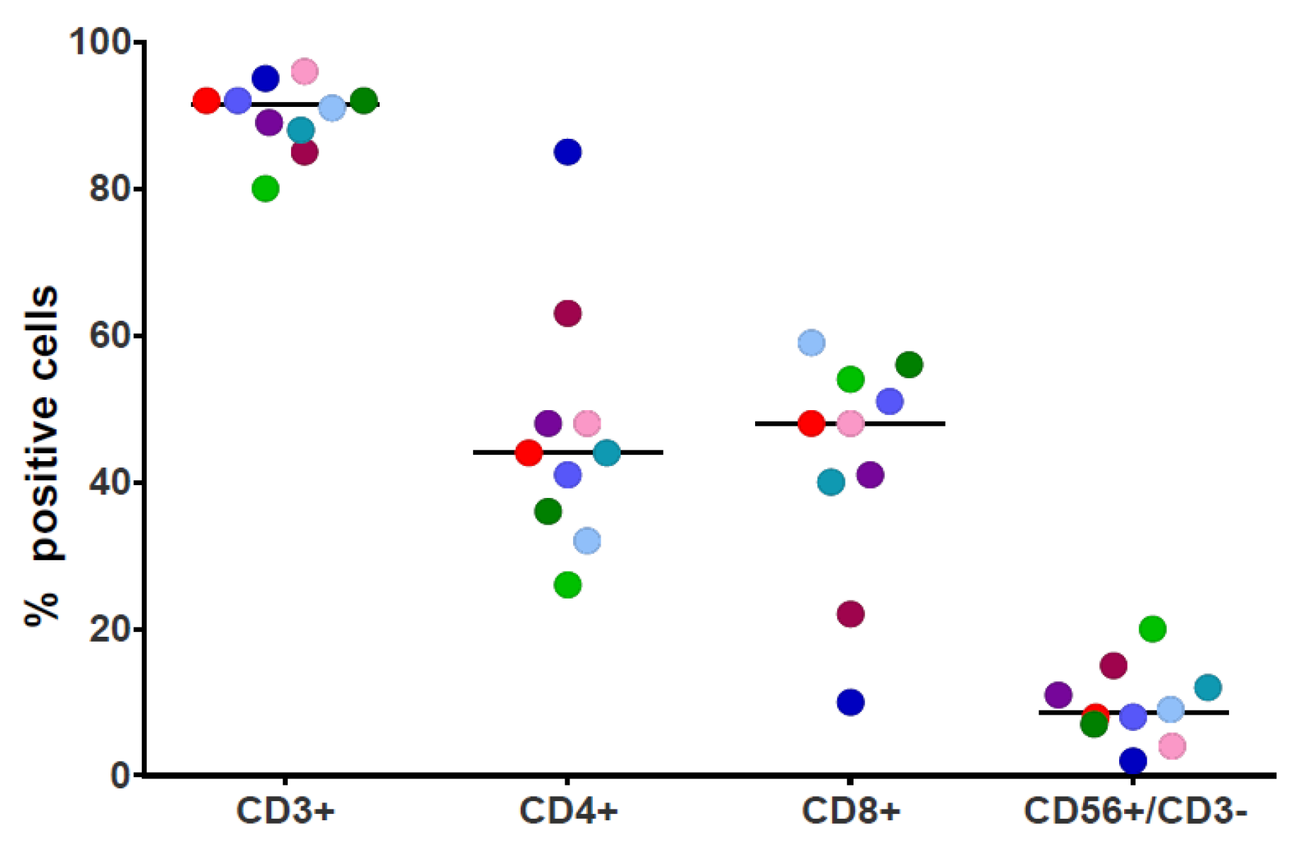
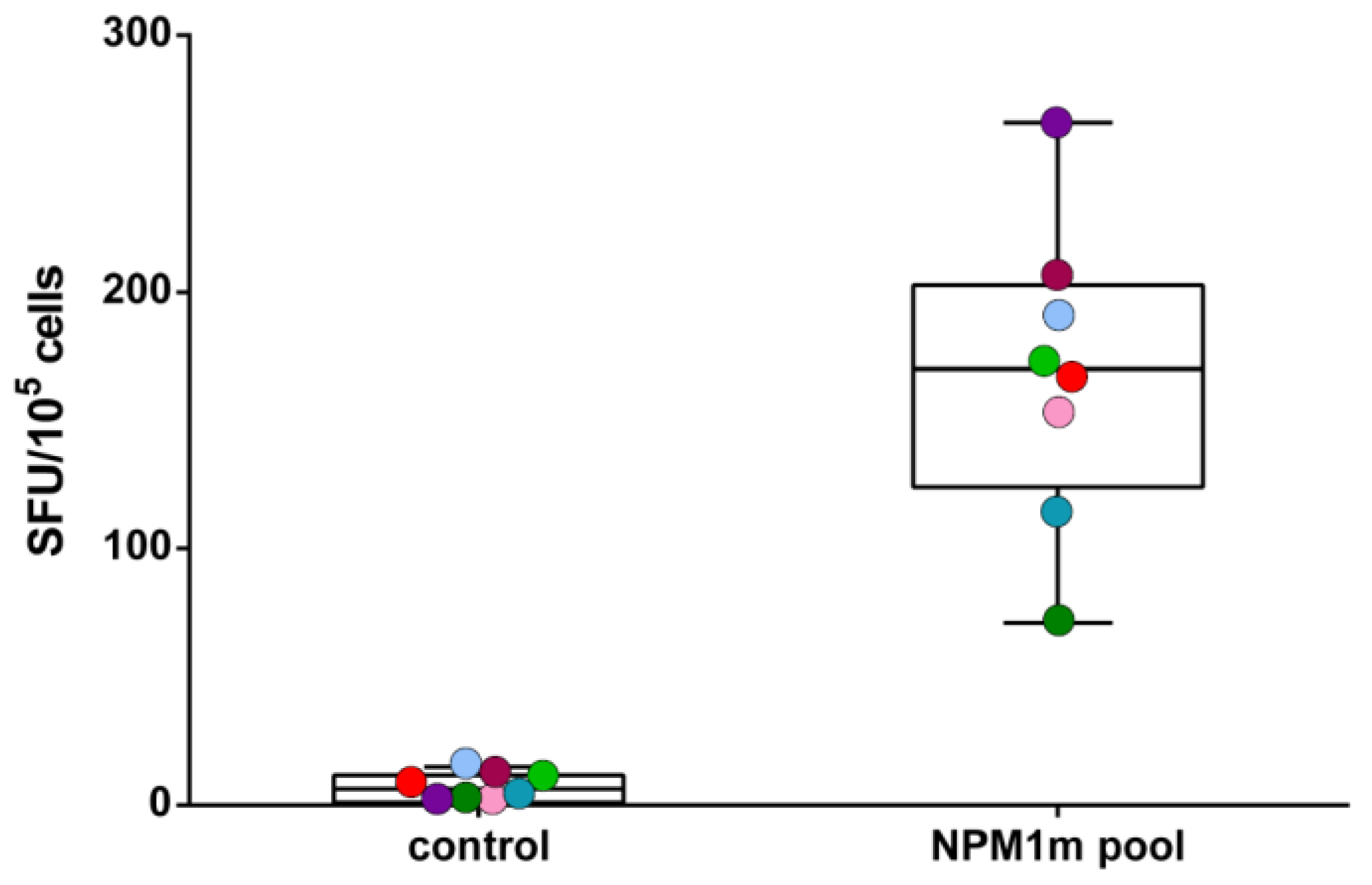
3.1. NPM1mut-CTLs Can Be Expanded from AML Patients and Healthy Donors by Stimulation with NPM1mut-Peptide Pools

To evaluate whether NPM1mut-CTLs could be expanded from the peripheral blood of AML patients in remission, and potential stem cell donors, PBMC were stimulated or primed with autologous DC pulsed with the NPM1mut-peptide pool in the presence of IL2 and IL15. After one round of re-stimulation, T cells expanded from 3 patients and 7 donors showed a mean expansion of 11.1-fold (range 5.7–31).
The ATMP were tested for microbiological safety by sterility testing in an automated blood culture system, and by evaluation of endotoxin levels and mycoplasma contamination. The T cell products were found to be sterile, and with endotoxin levels within the acceptable range.
The recovered cells were predominantly CD3+ (mean 92%) with balanced predominance of CD4+ and CD8+ T cells (median 44%, range 26–63% and mean 48%, range 10–59%, respectively). CTLs contained variable low numbers of natural killer CD3-CD56+ cells (median 9%, range 2–20%) (Figure 1). Analysis of memory phenotype showed a slight prevalence of CD45RO+/CCR7− T cells in accordance with an effector memory phenotype, but central memory and naïve T cells were also present.
To demonstrate the presence of cytotoxic T cells within the product, we employed a CD3-redirected cytotoxicity assay. The T cell lines from both patients and donors mediated killing of P815 cell line induced by binding through OKT3 monoclonal antibody. The median percentage lysis at an effector to target (E:T) ratio of 5:1 was 52.5% (range 14–97%) (Figure 2).
We then proceeded to evaluate specific activity of the CTLs against the NPM1mutantigen. All T cell lines from patients and donors showed specific lysis against PHA blasts pulsed with the NPM1mut peptide pool. In detail, we observed a median percentage lysis at the 5:1 effector to target (E:T) ratio of 35% (range 15–52), with median percentage lysis against control-pulsed PHA blasts of 3 (range 0–11) (Figure 2). To confirm the ability of NPM1mut-CTLs to recognize and kill leukemic cells, lytic activity against autologous (in the case of patients) or HLA partially-matched NPM1mut-positive leukemic blasts was also tested. Both patient-derived and donor-derived NPM1mut-CTLs mediated a measurable cytotoxic activity against their target population, with a median percentage lysis of 22% (range 10–59) at E:T ratio of 5:1 (Figure 2). We hypothesize that the activity we observed against allogeneic leukemia blasts in NPM1mut-CTLs from healthy donors was directed against leukemia, and not against alloantigens, as median cytotoxicity against the allogeneic non-pulsed PHA blast counterpart was 2% (range 0–9) (Figure 2 and Figure 3).
In order to verify that the response was directed to the mutated form of NPM1 antigen, rather than the wild-type form, we performed in vitro experiments using NPM1mut-CTLs from 3 donors, and testing cytotoxicity against PHA blasts pulsed with the NPM1mut peptide pool and the HL-60 cell line that is positive for NPM1-wt. We observed a median percentage lysis at the 5:1 effector-to-target (E:T) ratio of 25% (range 14–49) for NPM1mut peptide-pulsed targets, with median percentage lysis against HL-60 cells of 8 (range 0–12) (p < 0.05) (Figure 4). These results indicate a specificity of the CTL lines for the mutated form, although low lysis to NPM1wt antigen could be observed for some CTL lines.
We subsequently analyzed whether there was a difference in the specific response of patients’ and donors’ CTLs. We observed comparable leukemia-specific activity in the two cohorts, as median lysis at 5:1 E:T ratio against PHA blasts pulsed with the NPM1mut peptide pool was 32% for donor CTLs compared with 38% for patients’ products (p = ns), and median cytotoxicity against autologous (in the case of patients) or HLA partially matched LB (donor CTLs) was 22 and 21%, respectively (p = ns). Only for CD3-redirected cytotoxicity, CTLs expanded from donors exhibited a higher, although not statistically significant, lytic potential than patients’ CTLs (median lysis of 74 vs. 41%, respectively, p = 0.11).
Consistent with the cytotoxicity results, NPM1mut-directed cytokine production, measured in a ELISpot assay, confirmed specificity of the CTLs. Antigen recognition in IFNγ-ELISpot showed a mean of 170 SFU/105 cells (range 71–266) (Figure 5). The CTLs had a polyfunctional profile, as among CD8+ T cells, a median of 21% cells (range 2–30) were triple-positive for IFNγ, IL-2, and TNFα, while 15% CD4+ T cells (range 2–22) were triple-positive. Additionally in this case, we did not observe significant differences in the IFNγ-secreting activity of patients’ CTLs compared with donor T cell lines (median SFU/105 cells 176 vs. 162, respectively).

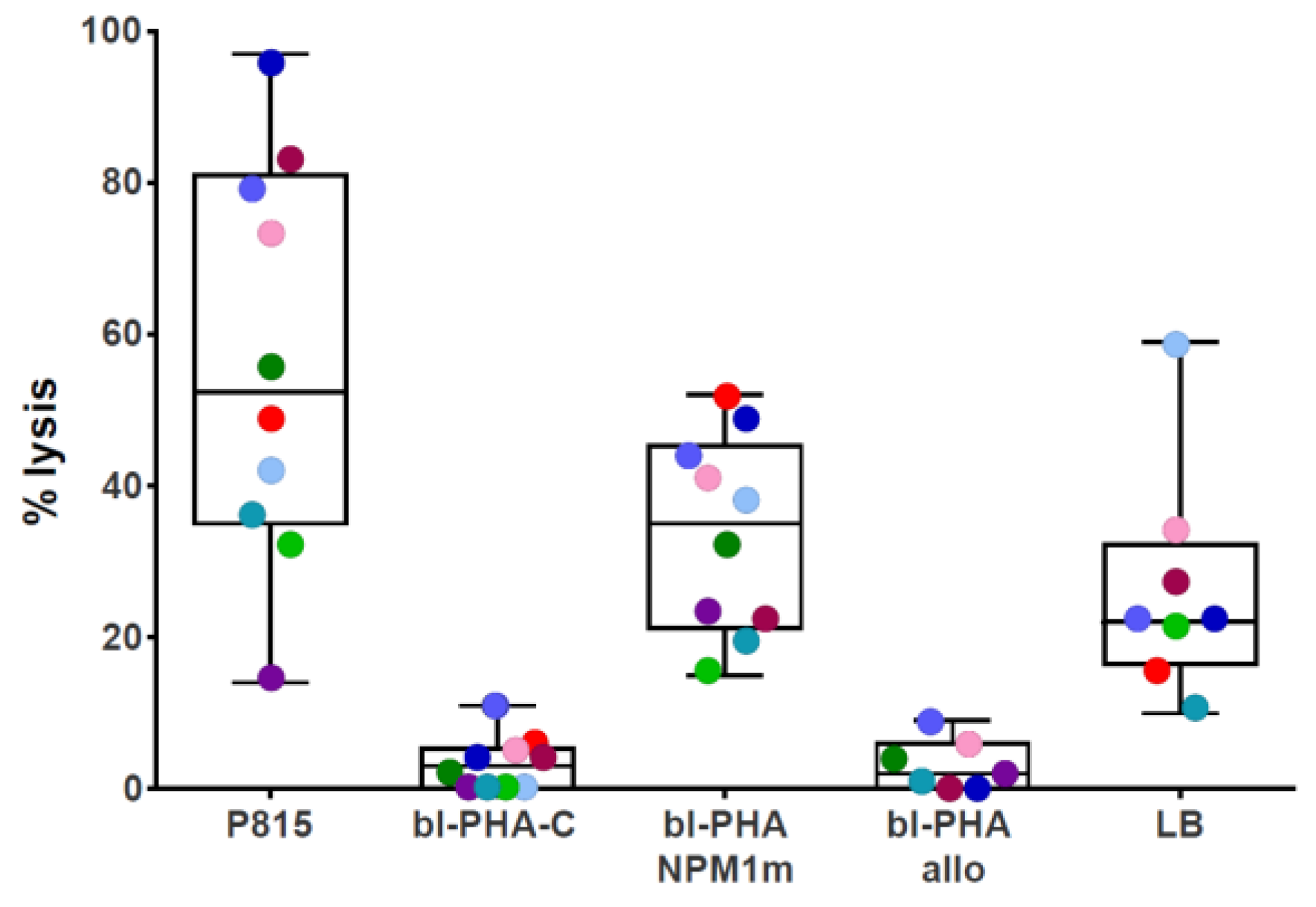
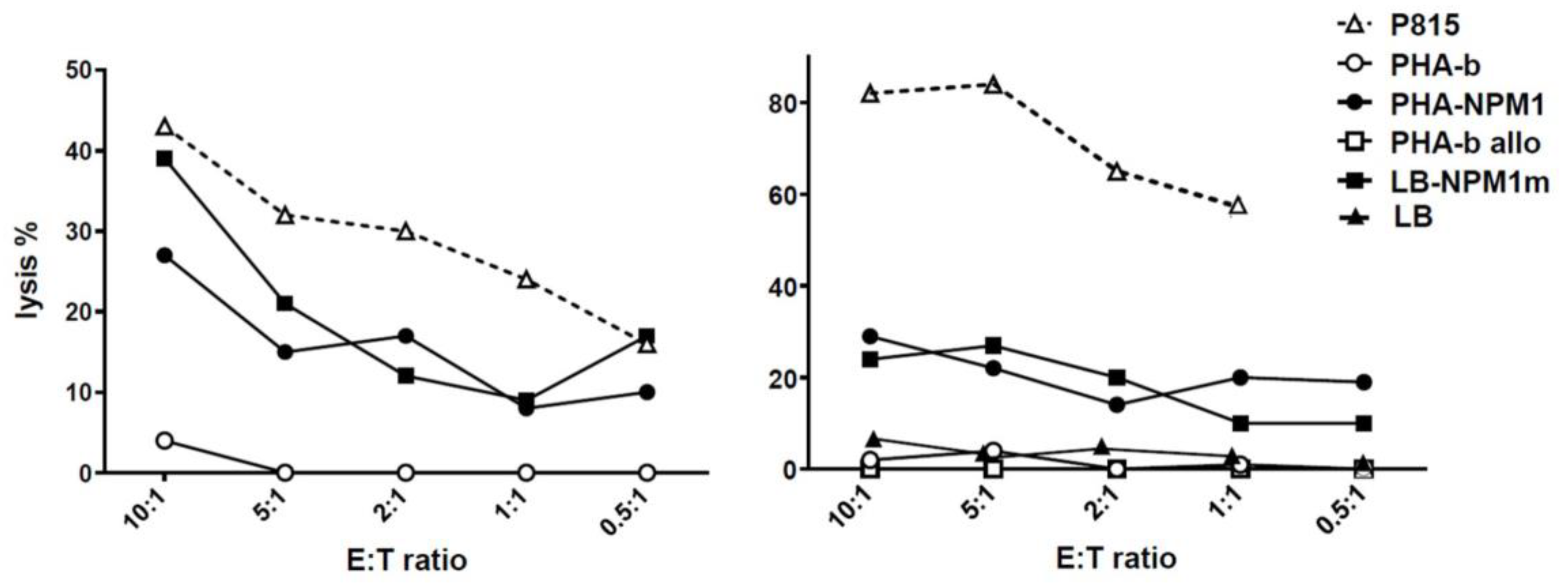
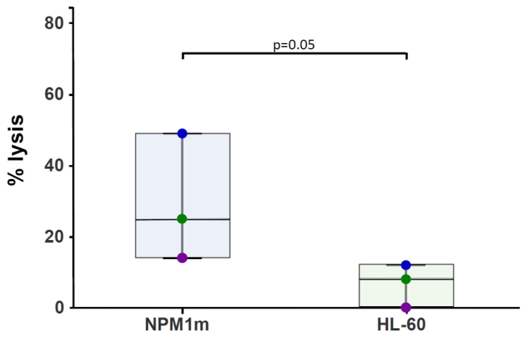
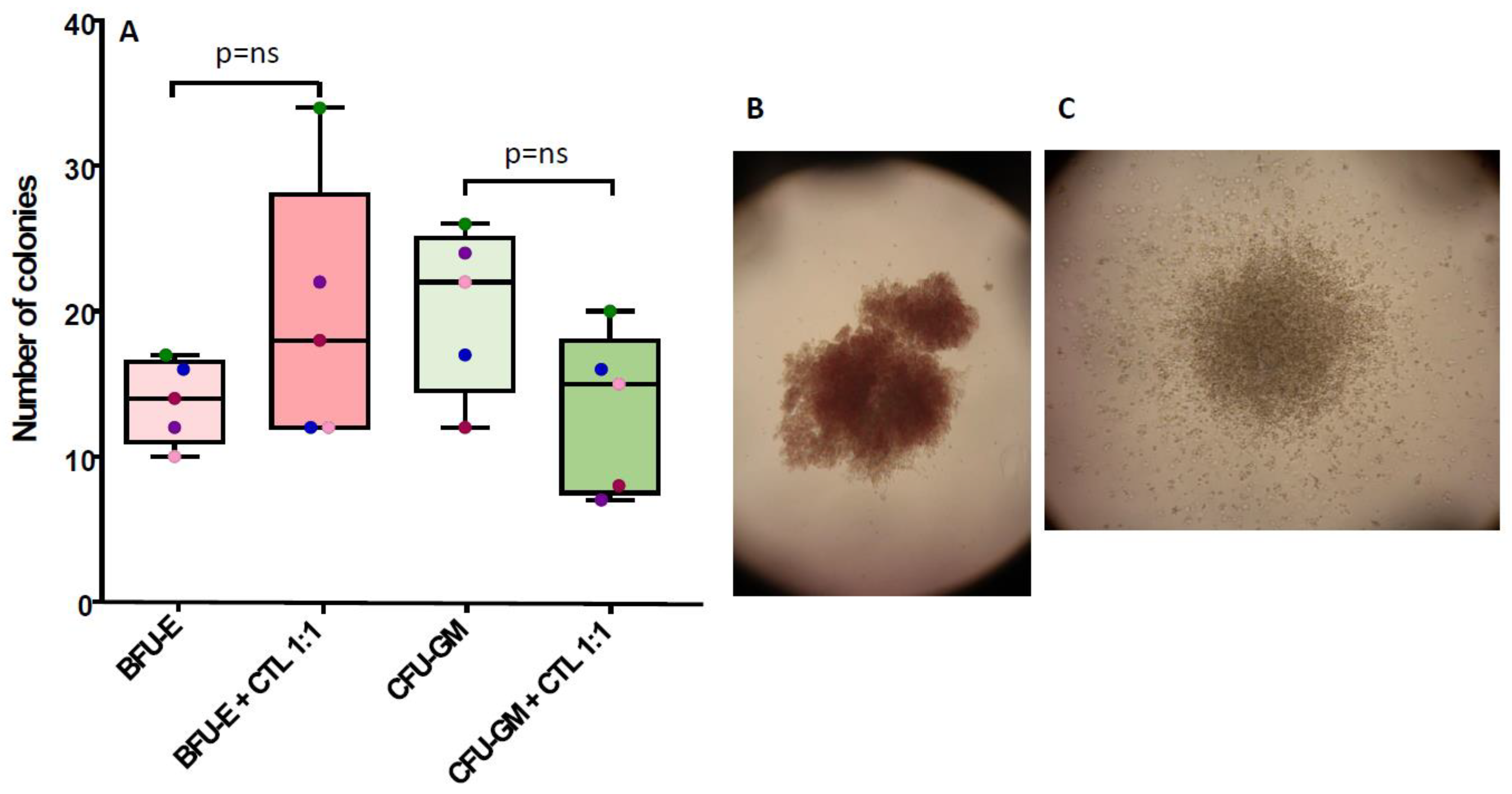
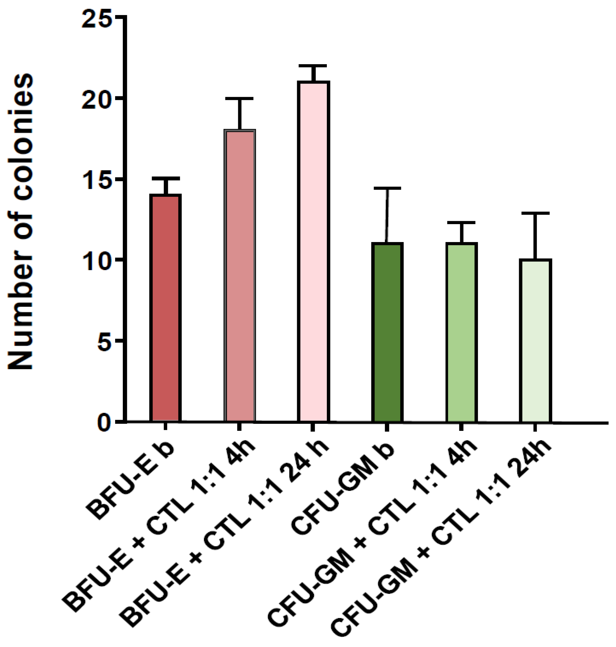
3.2. NPM1mut-CTLs Show Limited Inhibition of CFU-GM, but Not of BFU-E, When Cocultured with BM Progenitors

NPM1 antigen is present in its non-mutated wild-type (WT) form in healthy myeloid precursors, and although peptides derived from the mutated protein have been used to stimulate CTL expansion, it is theoretically possible that specific NPMmut CTLs could recognize peptides of WT protein on BM precursors, and that this mechanism leads to bone marrow suppression and toxicities. To evaluate the bone marrow suppressive effect of NPM1mut-CTLs in vitro, CTLs were cocultured with bone marrow samples from AML patients in remission, matched in at least one HLA antigen, for 4 or 24 h. As control conditions, BM cells were incubated with unmanipulated PBMCs from the same individuals.
At the end of coculture, cells were recovered, resuspended in the appropriate media, and plated in clonogenic assays for BFU-E and CFU-GM for 14 days. Colonies were then counted.
The results show that an inhibitory effect was observed mainly after 24 h incubation, and it concerned CFU-GM (median number of colonies, baseline: 20 vs. 13 after incubation with CTL, p = 0.35), while no effect was observed on BFU-E, which were increased in number after CTL co-incubation (median number of colonies, baseline: 14 vs. 20 after incubation with CTL, p = 0.24) (Figure 6).
Figure 7 shows the effect of CTL coculture time on BM progenitor colony growth. At 4 h, the inhibition on CFU-GM is less prominent.

4. Discussion

Despite remarkable advances in the treatment of NPM1-mutated AML, due to optimization of conventional induction chemotherapy and risk-stratified consolidation with cytarabine and HSCT in first remission in younger patients, and development of novel therapeutic approaches including BCL-2 inhibitor venetoclax and hypomethylating agent association, immune checkpoint inhibitors and AML-targeted monoclonal antibodies in older unfit patients, about 50% of patients still die of progressive disease [12,21,22]. Thus, there is a need for new therapies. Beyond novel agents such as menin inhibitors or the nucleolar stress triggerer dactinomycin [23,24,25,26], which have shown anti-leukemic activity in preclinical models and are being investigated in clinical trials, cell therapy approaches targeting NPM1-mutated protein on AML cells could represent a useful complementary approach to the other available treatment options. Our study shows the feasibility and reproducibility to expand NPM1mut-specific CTLs on a GMP scale from AML patients in hematologic remission, but also from healthy donors, by stimulation with DCs pulsed with a peptide pool derived from the mutated NPM1 antigen, irrespective of the donor HLA type. These CTLs, which include both CD8+ and CD4+ T cells, have high cytotoxic potential, as they are able to recognize and lyse target cells pulsed with NPM1mut peptides and, more importantly, they efficiently kill autologous or, in the case of donor, HLA partially matched NPM1mut-positive leukemia blasts.
In the past, a number of studies have demonstrated a correlation between the emergence of tumor antigen-targeted T cells in the peripheral blood and/or bone marrow of leukemia patients after induction and maintenance chemotherapy, or after allogeneic HSCT, and long-term disease remission [17,27,28,29,30,31]. In patients with NPM1-mutated AML, a better overall survival was documented in the patients that developed autologous NPM1mut-specific T cell responses [15]. In line with this observation, our group was able to demonstrate, through sequential monitoring of IFNγ-producing NPM1mut-specific T cells coupled with molecular MRD [32,33,34], that the kinetics of leukemia-specific T cells inversely correlated with molecular or morphologic leukemia status, having detected increased and sustained specific immune responses in patients with persistent molecular CR, in some cases years after completion of leukemia treatments [16]. These data suggest the potential for an autologous NPM1mut-specific CTL product to promote leukemia control by eradicating persistent MRD or low-tumor burden relapse following conventional chemotherapy in older NPM1-mutated AML patients not eligible for allogeneic HSCT, as we observed in a patient treated for Ph+ALL with B-cell receptor-ABL (BCR-ABL) p190-directed CTLs [35].
The feasibility to expand leukemia-specific CTLs from healthy donors by leukemia antigen-derived peptide stimulation had been first shown in the setting of chronic myeloid leukemia (CML) by using the B-cell receptor-ABL (BCR-ABL) p210 fusion protein, proteinase 3 (Pr3) and Wilms’ tumor antigen 1 (WT1) antigens [36], and subsequently replicated for BCR-ABL p190 [35], WT1 alone [37,38] or combined with multiple antigens including Pr3, human neutrophil elastase (NE), melanoma-associated antigen A3 (MAGE-A3), preferentially expressed antigen in melanoma (PRAME) and survivin [11,18,39]. In the case of NPM1-mutated AML, we were successful in priming leukemia-specific CTLs able to kill efficiently partially HLA-matched primary myeloid leukemia blasts from all donors tested. This finding reflects previous observations from our and other groups on the high frequency of IFNγ-producing NPM1mut-specific T cells in the peripheral blood of healthy volunteers or AML patients after HSCT [16,40], possibly due to a cross-reactive immune response induced by short amino acid sequences from the C-terminal of NPM1-mutated protein homologous with several common viral and bacterial antigens [16]. Successful expansion of CTLs from individuals with varied HLA types was obtained by employing a pool of fifteen 9- and 11-mer NPM1mut peptides, but also three 18-mer peptides that could contain multiple T cell epitopes, possibly cross-presented through various HLA class I and II alleles [41]. In addition, the peptides included epitopes representative of the most common NPM1 gene mutations, in order to allow broad antigen targeting [16].
The use of a donor-derived NPM1mut-specific product could be usefully employed as targeted DLI in MRD-positive patients to prevent hematologic relapse after allogeneic HSCT. Indeed, some of these products were infused prophylactically [18,35,39] or pre-emptively [18,36] in patients with CML or acute leukemia who were at high risk of relapse or had positive MRD after allogeneic HSCT, demonstrating safety and in vivo antileukemia effects. Although the design of these early phase trials did not allow assessing with certainty the role of leukemia antigen-targeted DLIs in patient outcomes, as patients generally received associated therapies, it is undeniable that these cohorts had at least comparable efficacy to chemotherapy and unmanipulated DLI, with a reduced risk of GVHD as compared with DLI [18,39].
Although a preemptive strategy guided by MRD monitoring could, more successfully, guarantee leukemia control due to the low tumor burden, a curative treatment approach in patients with hematologic relapse after HSCT may also be pursued. Indeed, in some of the early phase studies, responses were registered in patients with active disease [35,39]. In this latter setting, it is unlikely that the use of cell therapy alone, as observed for hypomethylating agent (HMA)-based salvage therapy [42,43,44], will be sufficient to control the outgrowth of leukemia blasts long-term, except perhaps in a few patients with late relapse and low tumor burden [44]. However, the use of targeted cell therapies that include a higher number of leukemia-specific T cells compared with unmanipulated DLIs, combined with azacytidine and/or the BCL-2 inhibitor venetoclax, or FLT3 inhibitors such as gilteritinib or sorafenib [45], may provide an added advantage and be able to increase the response rate in patients with hematologic relapse after HSCT. Indeed, HMA have been shown to induce HLA class I and costimulatory molecule expression on leukemia blasts, and favor their susceptibility to T cell-mediated cytotoxicity, while venetoclax was able to directly enhance cytotoxicity against AML cells both in vitro and in vivo [46,47].
So far, no specific T cell therapy for NPM1-mutated AML has been employed in vivo. Van der Lee et al. have been able to clone and transfer a NPM1-mutated, HLA-A2-restricted, T cell receptor with efficient in vitro specificity against NPM1-mutated primary leukemic blasts and in vivo activity in immunodeficient mice engrafted with a human NPM1-mutated AML cell line [10]. The advantages of gene therapy are the relatively short time required for T cell production, and the possibility to introduce the TCR into different T cell subsets with higher in vivo persistence and antitumor efficacy, such as central memory or stem cell memory T cells. On the other hand, a gene therapy product could induce more severe toxicity than somatic cell therapy [48], and be difficult to modulate in older, unfit patients, or in the early post-transplant setting.
One other problem of ATMP therapy, which has emerged in the past few years in connection with CAR-T cell therapy, is the development of tumor immune escape by several mechanisms, including loss of antigen expression [49]. In the case of NPM1mut-specific products, immune escape is more unlikely, as NPM1 mutation is a driver of genetic lesion, critical for leukemia cell survival. The use of associated therapies could provide the means to further broaden the response to leukemia, and avoid development of immune escape from antigen loss, by lysing leukemia cells in vivo and inducing antigen spreading [35]. This, in turn, could stimulate either recruited endogenous T cells emerging from the graft, or some T cells with low NPM1mut fitness present in the bulk ATMP product. Stimulation with long peptides presented by DCs allowed for expansion of CD4+ T cell populations alongside CD8+ CTLs. CD4+ T cells in the products, in addition to providing T cell help to CD8+ CTLs, were also endowed with lytic activity, as we could observe cytotoxicity mediated by this subset. This characteristic of the NPM1mut-specific CTL product could be of advantage to counteract immune escape mechanisms developed by leukemia cells, in particular downregulation of surface HLA class I molecules, necessary for CD8+ CTL activity. Virus-specific CD4+ CTLs have been proven to play a protective role in antiviral immunity, when pathogens such as herpesviruses escaped from CD8-mediated cellular immunity by downregulating the expression of HLA class I on the surface of infected cells through inhibition of the TAP transporter and/or proteasome degradation pathways [50]. A similar mechanism has been suggested also in the setting of tumor immune surveillance [51,52].
The wild-type form of NPM1 (wt-NPM1) is expressed on all cells, including BM-resident CD34+ hematopoietic stem cells. Although the mutated form is a neoantigen almost exclusively found in AML [14], and generally expressed in the entire leukemic population, while not detectable in clonal hematopoiesis, there is a theoretical concern that NPMmut CTLs, once administered to patients, could recognize wt-NPM1 on non-leukemic hematopoietic progenitors and mediate BM toxicity. To analyze this potential cross-reactivity, inhibition assays of BM precursor colony formation were performed by co-incubation of patients’ bone marrow cells with scaled ratios of NPMmut CTLs, and subsequent culture. The results of clonogenic assays showed that NPMmut- CTLs did not significantly affect the growth of patient-derived normal progenitor cells. These data are in line with a recent report that described a high response to wt-NPM1 by CD8+ CTLs in an NPM1-mutated AML patient post-HSCT [53]. In this specific patient, the emergence of a cellular immune response to wt-NPM1 was not accompanied by side effects, such as GVHD or BM aplasia, and coincided with leukemia control, further suggesting the potential role and relative safety of NPM1-directed immunity after HSCT.

5. Conclusions

Patients with relapsed or refractory NPM1-mutated AML after allogeneic HSCT or unfit patients relapsing after chemotherapy are an unmet medical need. Novel targeted therapies are emerging, and may change the prognosis of these cohorts. However, additional, complementary strategies, such as mutated-NPM1 specific cellular therapy, could represent a tool to prevent tumor outgrowth and escape, and further increase the probability of leukemia control.
Our findings indicate that NPM1mut-CTLs, obtained from patients or healthy donors and endowed with leukemia-specific activity, may constitute a safe and effective option in the treatment of patients affected by refractory or relapsed NPM1-mutated AML.

Author Contributions

Conceptualization, M.D.C., I.L., S.B., F.F., M.Z., M.L. and P.C.; investigation, M.D.C., I.L., S.M., A.B., S.D.V., C.P. and M.S.; data curation, D.S.; writing—original draft preparation, M.D.C., I.L., S.B. and D.S.; writing—review and editing, S.B., A.C., G.R., M.Z., F.F., M.L. and P.C.; supervision, S.B., P.B., F.F., V.R. and A.C. All authors have read and agreed to the published version of the manuscript.

Funding

This research was funded by Regione Lombardia, Project POR FESR 2526393 FORCE-4-CURE; Fondazione Regionale per la Ricerca Biomedica (FRRB, Regione Lombardia), Project N° CP2_10/2018; Ministry of Health, Ricerca Corrente Fondazione IRCCS Policlinico San Matteo, (08069119 to PC and 08045818 to MZ), Fondazione Just Italia to PC. This work was also supported by a grant to M.L. from the Associazione Italiana per la Ricerca sul Cancro (AIRC) (IG project #20624); PNRR CN3 Terapia Genica-Spoke 2 (Project #CN00000041); Progetto Dipartimenti di Eccellenza MIUR 2017; “Charity Dinner Initiative” in memory of Alberto Fontana for Associazione Italiana Lotta alle Leucemie, Linfoma e Mieloma (AIL)—Sezione “Luciano Pavarotti”—Modena-ONLUS; Fondazione IRIS CERAMICA GROUP. This work was also supported by a fellowship to I.L. from the Italian Society of Experimental Hematology (SIES Fellowship 2018–2019).

Institutional Review Board Statement

The study was conducted in accordance with the Declaration of Helsinki, and approved by the local Institutional Review Board (Comitato Etico Provinciale di Modena–Protocol 4745/13).

Informed Consent Statement

Written informed consent was obtained from all subjects involved in the study.

Data Availability Statement

The clinico-laboratory data of this study are available on reasonable request from the corresponding authors, according to privacy and ethical restrictions.

Conflicts of Interest

The authors declare no conflict of interest.

References

  1. Döhner, H.; Wei, A.H.; Appelbaum, F.R.; Craddock, C.; DiNardo, C.D.; Dombret, H.; Ebert, B.L.; Fenaux, P.; Godley, L.A.; Hasserjian, R.P.; et al. Diagnosis and Management of AML in Adults: 2022 Recommendations from an International Expert Panel on Behalf of the ELN. Blood 2022, 140, 1345–1377. [Google Scholar] [CrossRef]
  2. Shimony, S.; Stahl, M.; Stone, R.M. Acute Myeloid Leukemia: 2023 Update on Diagnosis, Risk-stratification, and Management. Am. J. Hematol. 2023, 98, 502–526. [Google Scholar] [CrossRef]
  3. Heuser, M.; Freeman, S.D.; Ossenkoppele, G.J.; Buccisano, F.; Hourigan, C.S.; Ngai, L.L.; Tettero, J.M.; Bachas, C.; Baer, C.; Béné, M.-C.; et al. 2021 Update on MRD in Acute Myeloid Leukemia: A Consensus Document from the European LeukemiaNet MRD Working Party. Blood 2021, 138, 2753–2767. [Google Scholar] [CrossRef]
  4. Lachowiez, C.A.; DiNardo, C.D.; Loghavi, S. Molecularly Targeted Therapy in Acute Myeloid Leukemia: Current Treatment Landscape and Mechanisms of Response and Resistance. Cancers 2023, 15, 1617. [Google Scholar] [CrossRef]
  5. Daver, N.; Alotaibi, A.S.; Bücklein, V.; Subklewe, M. T-Cell-Based Immunotherapy of Acute Myeloid Leukemia: Current Concepts and Future Developments. Leukemia 2021, 35, 1843–1863. [Google Scholar] [CrossRef]
  6. Laetsch, T.W.; Maude, S.L.; Rives, S.; Hiramatsu, H.; Bittencourt, H.; Bader, P.; Baruchel, A.; Boyer, M.; De Moerloose, B.; Qayed, M.; et al. Three-Year Update of Tisagenlecleucel in Pediatric and Young Adult Patients With Relapsed/Refractory Acute Lymphoblastic Leukemia in the ELIANA Trial. J. Clin. Oncol. 2022, 41, 1664–1669. [Google Scholar] [CrossRef]
  7. Locke, F.L.; Ghobadi, A.; Jacobson, C.A.; Miklos, D.B.; Lekakis, L.J.; Oluwole, O.O.; Lin, Y.; Braunschweig, I.; Hill, B.T.; Timmerman, J.M.; et al. Long-Term Safety and Activity of Axicabtagene Ciloleucel in Refractory Large B-Cell Lymphoma (ZUMA-1): A Single-Arm, Multicentre, Phase 1–2 Trial. Lancet Oncol. 2019, 20, 31–42. [Google Scholar] [CrossRef]
  8. Koedam, J.; Wermke, M.; Ehninger, A.; Cartellieri, M.; Ehninger, G. Chimeric Antigen Receptor T-Cell Therapy in Acute Myeloid Leukemia. Curr. Opin. Hematol. 2022, 29, 74–83. [Google Scholar] [CrossRef]
  9. Montagna, D.; Maccario, R.; Montini, E.; Tonelli, R.; Lisini, D.; Pagani, S.; Comoli, P.; Moretta, A.; Assirelli, E.; Basso, S.; et al. Generation and Ex Vivo Expansion of Cytotoxic T Lymphocytes Directed toward Different Types of Leukemia or Myelodysplastic Cells Using Both HLA-Matched and Partially Matched Donors. Exp. Hematol. 2003, 31, 1031–1038. [Google Scholar] [CrossRef]
  10. Van Der Lee, D.I.; Reijmers, R.M.; Honders, M.W.; Hagedoorn, R.S.; De Jong, R.C.M.; Kester, M.G.D.; Van Der Steen, D.M.; De Ru, A.H.; Kweekel, C.; Bijen, H.M.; et al. Mutated Nucleophosmin 1 as Immunotherapy Target in Acute Myeloid Leukemia. J. Clin. Investig. 2019, 129, 774–785. [Google Scholar] [CrossRef]
  11. Weber, G.; Gerdemann, U.; Caruana, I.; Savoldo, B.; Hensel, N.F.; Rabin, K.R.; Shpall, E.J.; Melenhorst, J.J.; Leen, A.M.; Barrett, A.J.; et al. Generation of Multi-Leukemia Antigen-Specific T Cells to Enhance the Graft-versus-Leukemia Effect after Allogeneic Stem Cell Transplant. Leukemia 2013, 27, 1538–1547. [Google Scholar] [CrossRef]
  12. Falini, B.; Brunetti, L.; Martelli, M.P. How I Diagnose and Treat NPM1-Mutated AML. Blood 2021, 137, 589–599. [Google Scholar] [CrossRef]
  13. Falini, B.; Martelli, M.P.; Bolli, N.; Sportoletti, P.; Liso, A.; Tiacci, E.; Haferlach, T. Acute Myeloid Leukemia with Mutated Nucleophosmin (NPM1): Is It a Distinct Entity? Blood 2011, 117, 1109–1120. [Google Scholar] [CrossRef]
  14. Falini, B.; Mecucci, C.; Tiacci, E.; Alcalay, M.; Rosati, R.; Pasqualucci, L.; La Starza, R.; Diverio, D.; Colombo, E.; Santucci, A.; et al. Cytoplasmic Nucleophosmin in Acute Myelogenous Leukemia with a Normal Karyotype. N. Engl. J. Med. 2005, 352, 254–266. [Google Scholar] [CrossRef]
  15. Greiner, J.; Schneider, V.; Schmitt, M.; Götz, M.; Döhner, K.; Wiesneth, M.; Döhner, H.; Hofmann, S. Immune Responses against the Mutated Region of Cytoplasmatic NPM1 Might Contribute to the Favorable Clinical Outcome of AML Patients with NPM1 Mutations (NPM1mut). Blood 2013, 122, 1087–1088. [Google Scholar] [CrossRef]
  16. Forghieri, F.; Riva, G.; Lagreca, I.; Barozzi, P.; Vallerini, D.; Morselli, M.; Paolini, A.; Bresciani, P.; Colaci, E.; Maccaferri, M.; et al. Characterization and Dynamics of Specific T Cells against Nucleophosmin-1 (NPM1)-Mutated Peptides in Patients with NPM1-Mutated Acute Myeloid Leukemia. Oncotarget 2019, 10, 869–882. [Google Scholar] [CrossRef]
  17. Riva, G.; Luppi, M.; Barozzi, P.; Quadrelli, C.; Basso, S.; Vallerini, D.; Zanetti, E.; Morselli, M.; Forghieri, F.; Maccaferri, M.; et al. Emergence of BCR-ABL–Specific Cytotoxic T Cells in the Bone Marrow of Patients with Ph+ Acute Lymphoblastic Leukemia during Long-Term Imatinib Mesylate Treatment. Blood 2010, 115, 1512–1518. [Google Scholar] [CrossRef]
  18. Kinoshita, H.; Cooke, K.R.; Grant, M.; Stanojevic, M.; Cruz, C.R.; Keller, M.; Fortiz, M.F.; Hoq, F.; Lang, H.; Barrett, A.J.; et al. Outcome of Donor-Derived TAA-T Cell Therapy in Patients with High-Risk or Relapsed Acute Leukemia Post Allogeneic BMT. Blood Adv. 2022, 6, 2520–2534. [Google Scholar] [CrossRef]
  19. Janetzki, S.; Price, L.; Schroeder, H.; Britten, C.M.; Welters, M.J.P.; Hoos, A. Guidelines for the Automated Evaluation of Elispot Assays. Nat. Protoc. 2015, 10, 1098–1115. [Google Scholar] [CrossRef]
  20. Massa, M.; Rosti, V.; Ferrario, M.; Campanelli, R.; Ramajoli, I.; Rosso, R.; De Ferrari, G.M.; Ferlini, M.; Goffredo, L.; Bertoletti, A.; et al. Increased Circulating Hematopoietic and Endothelial Progenitor Cells in the Early Phase of Acute Myocardial Infarction. Blood 2005, 105, 199–206. [Google Scholar] [CrossRef]
  21. Isidori, A.; Cerchione, C.; Daver, N.; DiNardo, C.; Garcia-Manero, G.; Konopleva, M.; Jabbour, E.; Ravandi, F.; Kadia, T.; de la Fuente Burguera, A.; et al. Immunotherapy in Acute Myeloid Leukemia: Where We Stand. Front. Oncol. 2021, 11, 656218. [Google Scholar] [CrossRef]
  22. Forghieri, F.; Riva, G.; Lagreca, I.; Barozzi, P.; Bettelli, F.; Paolini, A.; Nasillo, V.; Lusenti, B.; Pioli, V.; Giusti, D.; et al. Neoantigen-Specific T-Cell Immune Responses: The Paradigm of NPM1-Mutated Acute Myeloid Leukemia. Int. J. Mol. Sci. 2021, 22, 9159. [Google Scholar] [CrossRef]
  23. Klossowski, S.; Miao, H.; Kempinska, K.; Wu, T.; Purohit, T.; Kim, E.; Linhares, B.M.; Chen, D.; Jih, G.; Perkey, E.; et al. Menin Inhibitor MI-3454 Induces Remission in MLL1-Rearranged and NPM1-Mutated Models of Leukemia. J. Clin. Investig. 2020, 130, 981–997. [Google Scholar] [CrossRef] [PubMed]
  24. Dzama, M.M.; Steiner, M.; Rausch, J.; Sasca, D.; Schönfeld, J.; Kunz, K.; Taubert, M.C.; McGeehan, G.M.; Chen, C.-W.; Mupo, A.; et al. Synergistic Targeting of FLT3 Mutations in AML via Combined Menin-MLL and FLT3 Inhibition. Blood 2020, 136, 2442–2456. [Google Scholar] [CrossRef] [PubMed]
  25. Gionfriddo, I.; Brunetti, L.; Mezzasoma, F.; Milano, F.; Cardinali, V.; Ranieri, R.; Venanzi, A.; Pierangeli, S.; Vetro, C.; Spinozzi, G.; et al. Dactinomycin Induces Complete Remission Associated with Nucleolar Stress Response in Relapsed/Refractory NPM1-Mutated AML. Leukemia 2021, 35, 2552–2562. [Google Scholar] [CrossRef] [PubMed]
  26. Issa, G.C.; Aldoss, I.; DiPersio, J.; Cuglievan, B.; Stone, R.; Arellano, M.; Thirman, M.J.; Patel, M.R.; Dickens, D.S.; Shenoy, S.; et al. The Menin Inhibitor Revumenib in KMT2A-Rearranged or NPM1-Mutant Leukaemia. Nature 2023, 615, 920–924. [Google Scholar] [CrossRef] [PubMed]
  27. Rezvani, K.; Brenchley, J.M.; Price, D.A.; Kilical, Y.; Gostick, E.; Sewell, A.K.; Li, J.; Mielke, S.; Douek, D.C.; Barrett, A.J. T-Cell Responses Directed against Multiple HLA-A*0201-Restricted Epitopes Derived from Wilms’ Tumor 1 Protein in Patients with Leukemia and Healthy Donors: Identification, Quantification, and Characterization. Clin. Cancer Res. 2005, 11, 8799–8807. [Google Scholar] [CrossRef]
  28. Montagna, D.; Maccario, R.; Locatelli, F.; Montini, E.; Pagani, S.; Bonetti, F.; Daudt, L.; Turin, I.; Lisini, D.; Garavaglia, C.; et al. Emergence of Antitumor Cytolytic T Cells Is Associated with Maintenance of Hematologic Remission in Children with Acute Myeloid Leukemia. Blood 2006, 108, 3843–3850. [Google Scholar] [CrossRef] [PubMed]
  29. Weber, G.; Caruana, I.; Rouce, R.H.; Barrett, A.J.; Gerdemann, U.; Leen, A.M.; Rabin, K.R.; Bollard, C.M. Generation of Tumor Antigen-Specific T Cell Lines from Pediatric Patients with Acute Lymphoblastic Leukemia—Implications for Immunotherapy. Clin. Cancer Res. 2013, 19, 5079–5091. [Google Scholar] [CrossRef] [PubMed]
  30. Scheibenbogen, C.; Letsch, A.; Thiel, E.; Schmittel, A.; Mailaender, V.; Baerwolf, S.; Nagorsen, D.; Keilholz, U. CD8 T-Cell Responses to Wilms Tumor Gene Product WT1 and Proteinase 3 in Patients with Acute Myeloid Leukemia. Blood 2002, 100, 2132–2137. [Google Scholar] [CrossRef]
  31. Rezvani, K.; Yong, A.S.M.; Mielke, S.; Savani, B.N.; Musse, L.; Superata, J.; Jafarpour, B.; Boss, C.; Barrett, A.J. Leukemia-Associated Antigen-Specific T-Cell Responses Following Combined PR1 and WT1 Peptide Vaccination in Patients with Myeloid Malignancies. Blood 2008, 111, 236–242. [Google Scholar] [CrossRef] [PubMed]
  32. Schnittger, S.; Kern, W.; Tschulik, C.; Weiss, T.; Dicker, F.; Falini, B.; Haferlach, C.; Haferlach, T. Minimal Residual Disease Levels Assessed by NPM1 Mutation–Specific RQ-PCR Provide Important Prognostic Information in AML. Blood 2009, 114, 2220–2231. [Google Scholar] [CrossRef] [PubMed]
  33. Zhou, Y.; Othus, M.; Walter, R.B.; Estey, E.H.; Wu, D.; Wood, B.L. Deep NPM1 Sequencing Following Allogeneic Hematopoietic Cell Transplantation Improves Risk Assessment in Adults with NPM1-Mutated AML. Biol. Blood Marrow Transplant. 2018, 24, 1615–1620. [Google Scholar] [CrossRef]
  34. Forghieri, F.; Comoli, P.; Marasca, R.; Potenza, L.; Luppi, M. Minimal/Measurable Residual Disease Monitoring in NPM1-Mutated Acute Myeloid Leukemia: A Clinical Viewpoint and Perspectives. Int. J. Mol. Sci. 2018, 19, 3492. [Google Scholar] [CrossRef]
  35. Comoli, P.; Basso, S.; Riva, G.; Barozzi, P.; Guido, I.; Gurrado, A.; Quartuccio, G.; Rubert, L.; Lagreca, I.; Vallerini, D.; et al. BCR-ABL–Specific T-Cell Therapy in Ph+ ALL Patients on Tyrosine-Kinase Inhibitors. Blood 2017, 129, 582–586. [Google Scholar] [CrossRef]
  36. Bornhäuser, M.; Thiede, C.; Platzbecker, U.; Kiani, A.; Oelschlaegel, U.; Babatz, J.; Lehmann, D.; Hölig, K.; Radke, J.; Tuve, S.; et al. Prophylactic Transfer of BCR-ABL–, PR1-, and WT1-Reactive Donor T Cells after T Cell–Depleted Allogeneic Hematopoietic Cell Transplantation in Patients with Chronic Myeloid Leukemia. Blood 2011, 117, 7174–7184. [Google Scholar] [CrossRef]
  37. Weber, G.; Karbach, J.; Kuçi, S.; Kreyenberg, H.; Willasch, A.; Koscielniak, E.; Tonn, T.; Klingebiel, T.; Wels, W.S.; Jäger, E.; et al. WT1 Peptide-Specific T Cells Generated from Peripheral Blood of Healthy Donors: Possible Implications for Adoptive Immunotherapy after Allogeneic Stem Cell Transplantation. Leukemia 2009, 23, 1634–1642. [Google Scholar] [CrossRef] [PubMed]
  38. Ferulli, F.; Tanzi, M.; Turin, I.; Montini, E.; Rosti, V.; Acquafredda, G.; Lisini, D.; Compagno, F.; Boghen, S.; Licari, A.; et al. Generation of Donor-Derived Wilms Tumor Antigen 1–Specific Cytotoxic T Lymphocytes with Potent Anti-Leukemia Activity for Somatic Cell Therapy in Children given Haploidentical Stem Cell Transplantation: A Feasibility Pre-Clinical Study. Cytotherapy 2019, 21, 958–972. [Google Scholar] [CrossRef] [PubMed]
  39. Lulla, P.D.; Naik, S.; Vasileiou, S.; Tzannou, I.; Watanabe, A.; Kuvalekar, M.; Lulla, S.; Carrum, G.; Ramos, C.A.; Kamble, R.; et al. Clinical Effects of Administering Leukemia-Specific Donor T Cells to Patients with AML/MDS after Allogeneic Transplant. Blood 2021, 137, 2585–2597. [Google Scholar] [CrossRef]
  40. Greiner, J.; Ono, Y.; Hofmann, S.; Schmitt, A.; Mehring, E.; Götz, M.; Guillaume, P.; Döhner, K.; Mytilineos, J.; Döhner, H.; et al. Mutated Regions of Nucleophosmin 1 Elicit Both CD4+ and CD8+ T-Cell Responses in Patients with Acute Myeloid Leukemia. Blood 2012, 120, 1282–1289. [Google Scholar] [CrossRef]
  41. Comoli, P.; Schilham, M.W.; Basso, S.; Van Vreeswijk, T.; Bernardo, M.E.; Maccario, R.; Van Tol, M.J.D.; Locatelli, F.; Veltrop-Duits, L.A. T-Cell Lines Specific for Peptides of Adenovirus Hexon Protein and Devoid of Alloreactivity Against Recipient Cells Can Be Obtained From HLA-Haploidentical Donors. J. Immunother. 2008, 31, 529–536. [Google Scholar] [CrossRef]
  42. Craddock, C.; Labopin, M.; Robin, M.; Finke, J.; Chevallier, P.; Yakoub-Agha, I.; Bourhis, J.H.; Sengelov, H.; Blaise, D.; Luft, T.; et al. Clinical Activity of Azacitidine in Patients Who Relapse after Allogeneic Stem Cell Transplantation for Acute Myeloid Leukemia. Haematologica 2016, 101, 879–883. [Google Scholar] [CrossRef] [PubMed]
  43. Liberatore, C.; Stanghellini, M.T.L.; Lorentino, F.; Vago, L.; Carrabba, M.G.; Greco, R.; Marktel, S.; Assanelli, A.; Farina, F.; Corti, C.; et al. Azacitidine and Donor Lymphocytes Infusions in Acute Myeloid Leukemia and Myelodysplastic Syndrome Relapsed after Allogeneic Hematopoietic Stem Cell Transplantation from Alternative Donors. Ther. Adv. Hematol. 2022, 13, 1–20. [Google Scholar] [CrossRef]
  44. Rautenberg, C.; Bergmann, A.; Germing, U.; Fischermanns, C.; Pechtel, S.; Kaivers, J.; Jäger, P.; Schuler, E.; Haas, R.; Kobbe, G.; et al. Prediction of Response and Survival Following Treatment with Azacitidine for Relapse of Acute Myeloid Leukemia and Myelodysplastic Syndromes after Allogeneic Hematopoietic Stem Cell Transplantation. Cancers 2020, 12, 2255. [Google Scholar] [CrossRef] [PubMed]
  45. Bazarbachi, A.; Bug, G.; Baron, F.; Brissot, E.; Ciceri, F.; Dalle, I.A.; Döhner, H.; Esteve, J.; Floisand, Y.; Giebel, S.; et al. Clinical Practice Recommendation on Hematopoietic Stem Cell Transplantation for Acute Myeloid Leukemia Patients with FLT3 -Internal Tandem Duplication: A Position Statement from the Acute Leukemia Working Party of the European Society for Blood and Marrow Transplantation. Haematologica 2020, 105, 1507–1516. [Google Scholar] [CrossRef]
  46. Nahas, M.R.; Stroopinsky, D.; Rosenblatt, J.; Cole, L.; Pyzer, A.R.; Anastasiadou, E.; Sergeeva, A.; Ephraim, A.; Washington, A.; Orr, S.; et al. Hypomethylating Agent Alters the Immune Microenvironment in Acute Myeloid Leukaemia (AML) and Enhances the Immunogenicity of a Dendritic Cell/AML Vaccine. Br. J. Haematol. 2019, 185, 679–690. [Google Scholar] [CrossRef]
  47. Lee, J.B.; Khan, D.H.; Hurren, R.; Xu, M.; Na, Y.; Kang, H.; Mirali, S.; Wang, X.; Gronda, M.; Jitkova, Y.; et al. Venetoclax Enhances T Cell–Mediated Antileukemic Activity by Increasing ROS Production. Blood 2021, 138, 234–245. [Google Scholar] [CrossRef] [PubMed]
  48. Van Loenen, M.M.; De Boer, R.; Amir, A.L.; Hagedoorn, R.S.; Volbeda, G.L.; Willemze, R.; Van Rood, J.J.; Falkenburg, J.H.F.; Heemskerk, M.H.M. Mixed T Cell Receptor Dimers Harbor Potentially Harmful Neoreactivity. Proc. Natl. Acad. Sci. USA 2010, 107, 10972–10977. [Google Scholar] [CrossRef]
  49. Beatty, G.L.; Gladney, W.L. Immune Escape Mechanisms as a Guide for Cancer Immunotherapy. Clin. Cancer Res. 2015, 21, 687–692. [Google Scholar] [CrossRef]
  50. Levitskaya, J.; Sharipo, A.; Leonchiks, A.; Ciechanover, A.; Masucci, M.G. Inhibition of Ubiquitin/Proteasome-Dependent Protein Degradation by the Gly-Ala Repeat Domain of the Epstein–Barr Virus Nuclear Antigen 1. Proc. Natl. Acad. Sci. USA 1997, 94, 12616–12621. [Google Scholar] [CrossRef]
  51. Disis, M.L.; Wallace, D.R.; Gooley, T.A.; Dang, Y.; Slota, M.; Lu, H.; Coveler, A.L.; Childs, J.S.; Higgins, D.M.; Fintak, P.A.; et al. Concurrent Trastuzumab and HER2/Neu -Specific Vaccination in Patients With Metastatic Breast Cancer. J. Clin. Oncol. 2009, 27, 4685–4692. [Google Scholar] [CrossRef] [PubMed]
  52. Ohkuri, T.; Sato, M.; Abe, H.; Tsuji, K.; Yamagishi, Y.; Ikeda, H.; Matsubara, N.; Kitamura, H.; Nishimura, T. Identification of a Novel NY-ESO-1 Promiscuous Helper Epitope Presented by Multiple MHC Class II Molecules Found Frequently in the Japanese Population. Cancer Sci. 2007, 98, 1092–1098. [Google Scholar] [CrossRef] [PubMed]
  53. Nemeckova, S.; Alexova-Zurkova, K.; Hainz, P.; Krystofova, J.; Mackova, J.; Roubalova, K.; Stastna-Markova, M.; Vrana, M.; Vydra, J. Non-Mutated Nucleophosmin 1 Is Recognized by the CD8+ T Lymphocytes of an AML Patient after the Transplantation of Hematopoietic Stem Cells from an HLA-Haploidentical Donor. Curr. Oncol. 2022, 29, 2928–2934. [Google Scholar] [CrossRef] [PubMed]
Figure 1. Surface phenotype of NPM1mut-CTLs. The results are reported as % positive cells. The 3 patients are indicated with light green, light blue, and red dots.
Cancers 15 02731 g001
Figure 2. Potency of NPM1mut-CTLs: cytotoxic activity. The lytic activity of NPM1mut-CTLs against P815 cell line (CD3-redirected cytotoxicity), against control (bl-PHA-C) or NPM1mut peptide pool-pulsed autologous PHA blasts (bl-PHA-NPM1m), against allogeneic PHA blasts (bl-PHA-allo), and against autologous or allogeneic leukemia blasts (LB) is shown. The results are reported as % lysis at an E:T ratio of 5:1. The 3 patients are indicated with light green, light blue, and red dots.
Cancers 15 02731 g002
Figure 3. Potency of NPM1mut-CTLs: cytotoxic activity. Exemplificative cytotoxicity experiments of NPM1mut-CTLs from an AML patient (left) panel and a healthy donor (right) panel are shown. Lysis against P815 cell line (CD3-redirected cytotoxicity, white triangles), against control (PHA-b, white circles) or NPM1mut peptide pool-pulsed autologous PHA blasts (PHA-NPM1, black circles), against allogeneic PHA blasts (PHA-b allo, white squares), and against autologous or allogeneic NPM1m-positive leukemia blasts (LB-NPM1m, black squares) or allogeneic NPM1m-negative LB (LB, black triangles), is reported as % lysis at different E:T ratios.
Cancers 15 02731 g003
Figure 4. Potency of NPM1mut-CTLs: cytotoxic activity. The lytic activity of NPM1mut-CTLs against NPM1mut peptide pool-pulsed autologous PHA blasts (bl-PHA-NPM1m), and against the NPM1wt-positive HL-60 cell line is shown. The results are reported as % lysis at a E:T ratio of 5:1.
Cancers 15 02731 g004
Figure 5. Potency of NPM1mut-CTLs: IFNγ secreting activity. IFNγ production by NPM1mut-CTLs in the absence of specific stimulation (control) or after stimulation with NPM1mut peptide pool (NPM1m pool) is shown. The results are reported as spot-forming units (SFU)/105 CTLs. The 3 patients are indicated with light green, light blue, and red dots.
Cancers 15 02731 g005
Figure 6. In vitro safety of NPM1mut-CTLs: effects of CTL co-incubation on BM progenitor cells. (A) The number of BFU-E (pink boxes), and CFU-GM (green boxes), at baseline (lighter color boxes) or after 24 h co-incubation with CTLs (darker boxes) are shown. In (B,C), examples of BFU-E and CFU-GM are shown.
Cancers 15 02731 g006
Figure 7. In vitro safety of NPM1mut-CTLs: effects of CTL co-incubation duration on BM progenitor cells. The number of BFU-E (pink columns), CFU-GM (green columns) at baseline (darker color column), or after 4 h (medium color column) and 24 h (lighter color column) co-incubation with CTLs, are shown for an exemplificative CTL line.
Cancers 15 02731 g007
Disclaimer/Publisher’s Note: The statements, opinions and data contained in all publications are solely those of the individual author(s) and contributor(s) and not of MDPI and/or the editor(s). MDPI and/or the editor(s) disclaim responsibility for any injury to people or property resulting from any ideas, methods, instructions or products referred to in the content.

