
Evaluation of Temozolomide Treatment for Glioblastoma Using Amide Proton Transfer Imaging and Diffusion MRI


Abstract
:Simple Summary
Abstract
1. Introduction
2. Materials and Methods
2.1. Cell Culture
2.2. Animal Preparation
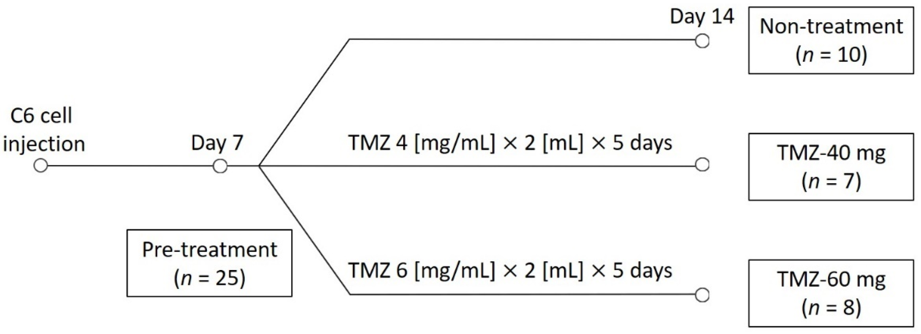
2.3. TMZ Treatment
2.4. Rat MRI
2.5. Image Analysis
2.6. Histological Studies
2.7. Statistical Analysis
3. Results
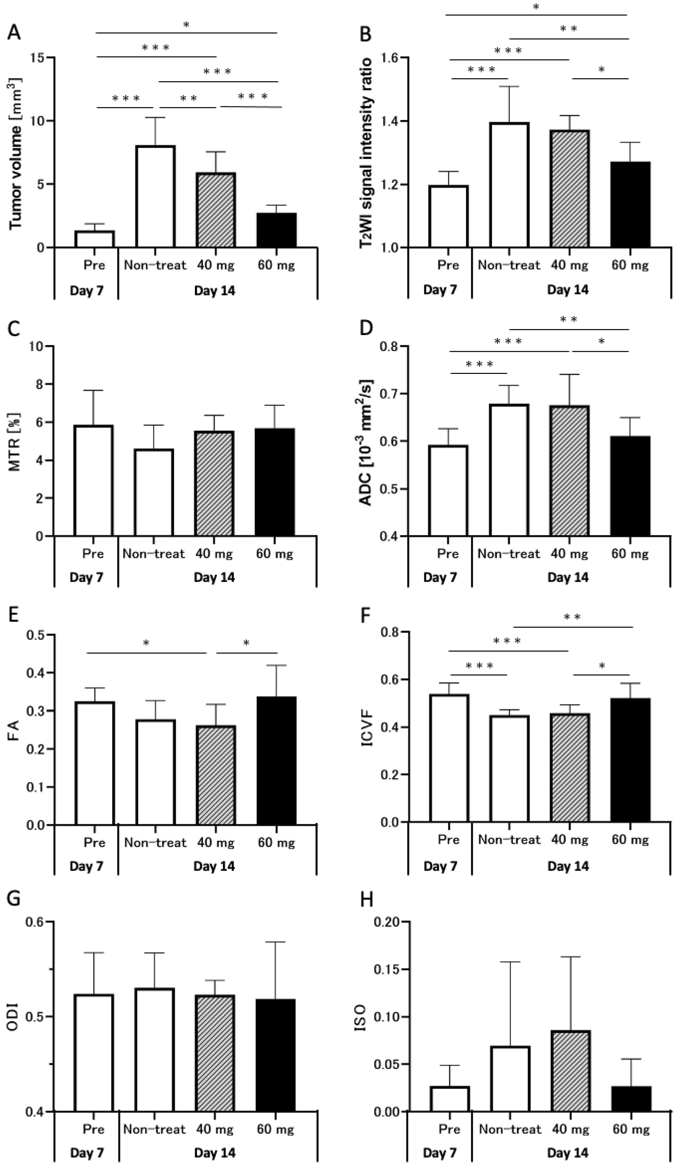
3.1. Effect of TMZ Treatment on Tumor Size
3.2. Effects of TMZ Treatment on T2WI
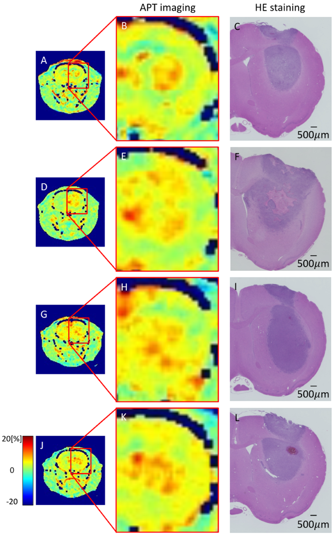
3.3. Effects of TMZ Treatment on APT Imaging
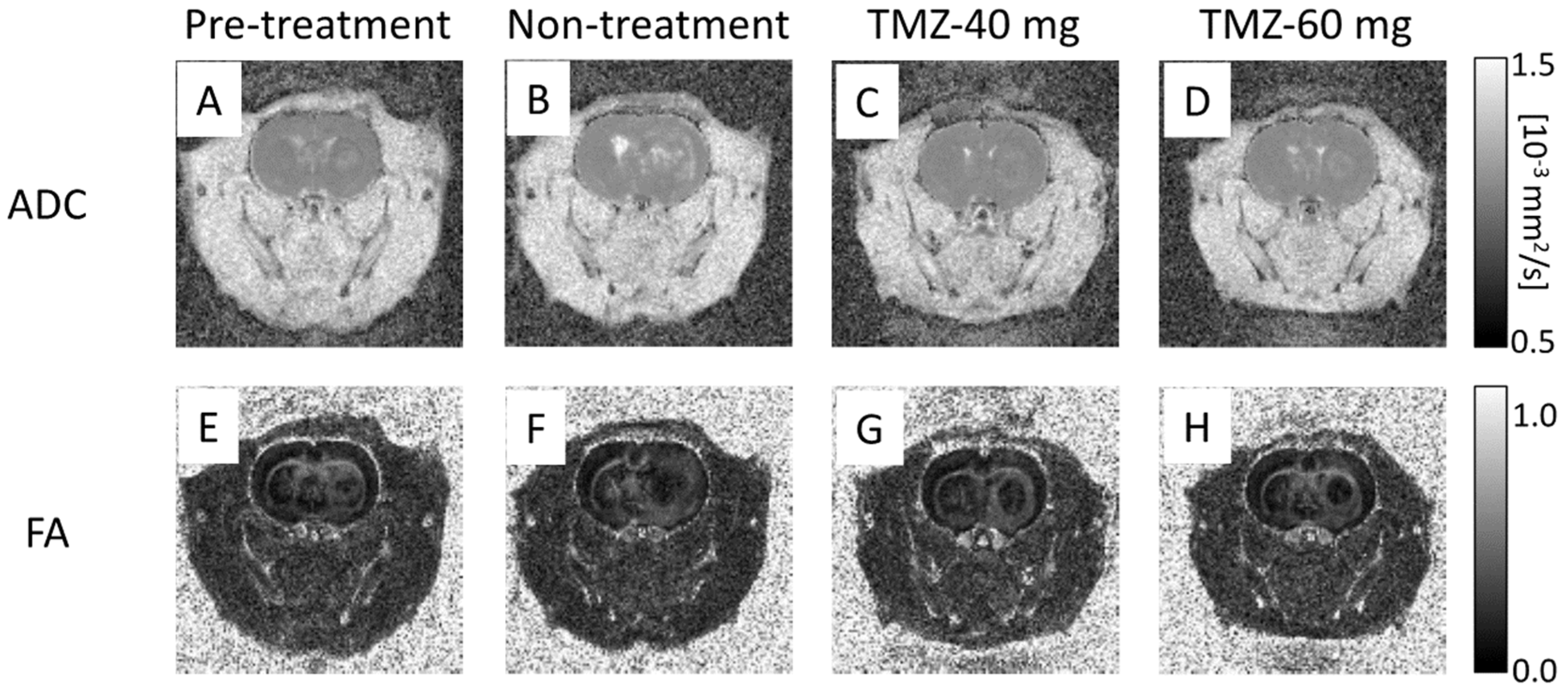
3.4. Effect of TMZ Treatment on DTI
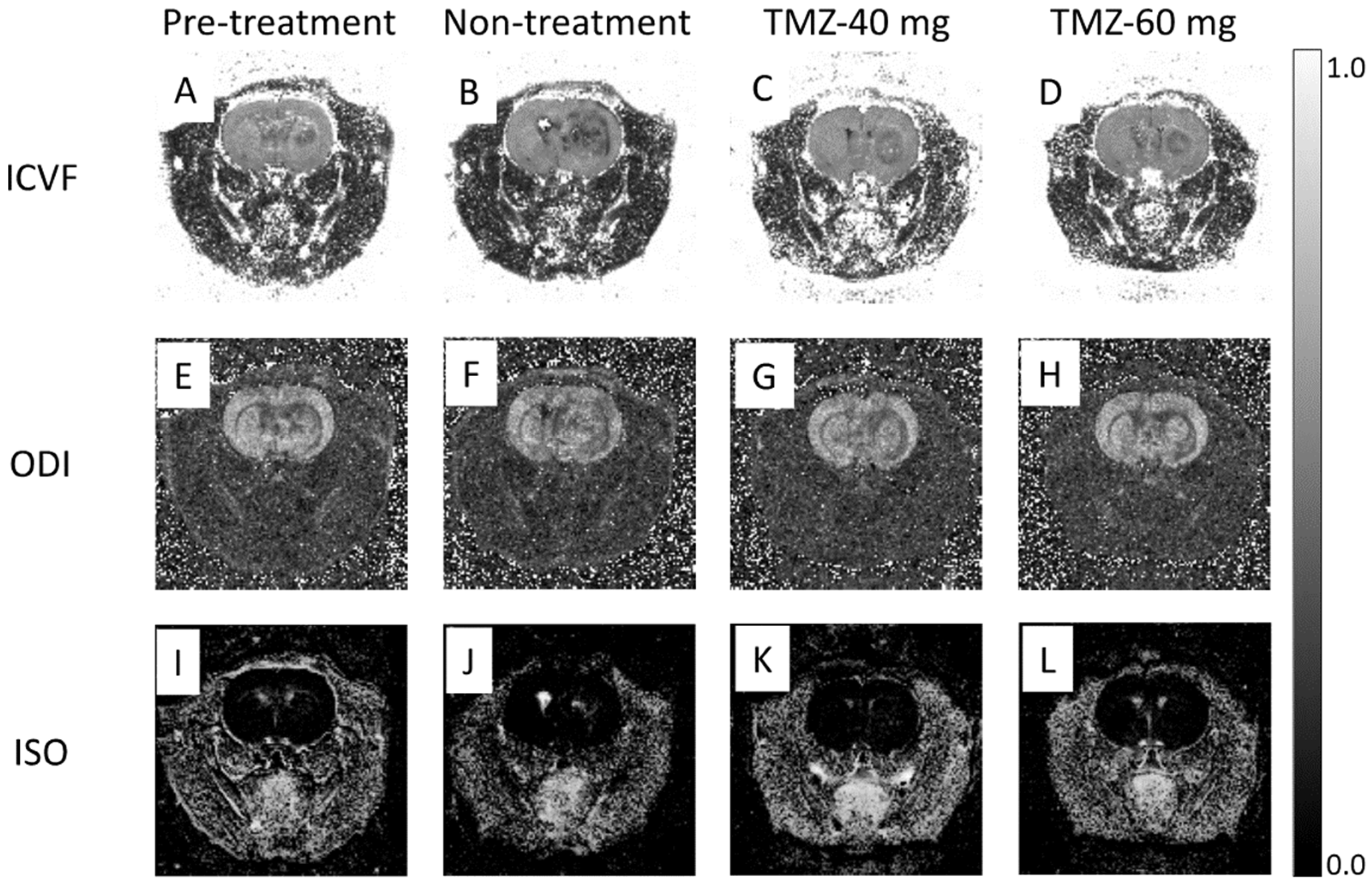
3.5. Effect of TMZ Treatment on NODDI
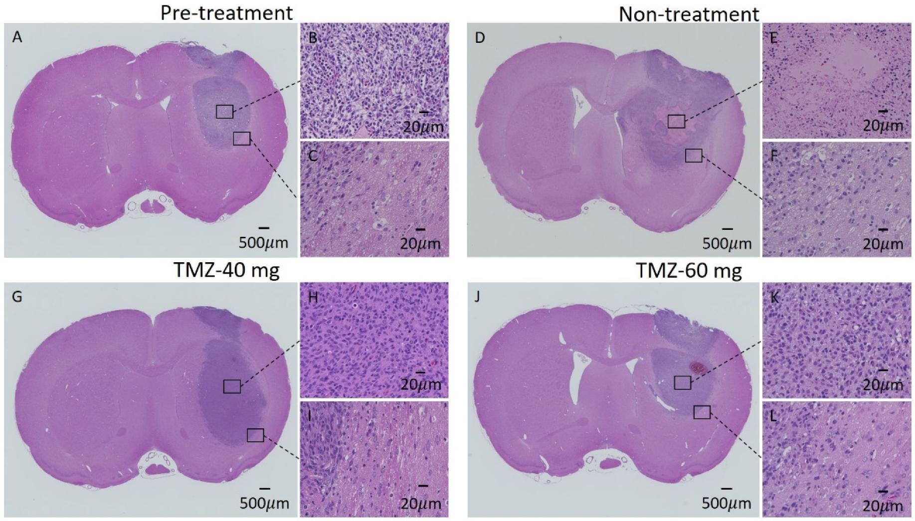
3.6. HE Staining of GBM Model
3.7. Body Weight Changes in Rats Treated with TMZ
4. Discussion
5. Conclusions
Author Contributions
Funding
Institutional Review Board Statement
Informed Consent Statement
Data Availability Statement
Conflicts of Interest
References
- Towner, R.A.; Smith, N.; Saunders, D.; Brown, C.A.; Cai, X.; Ziegler, J.; Mallory, S.; Dozmorov, M.G.; Coutinho De Souza, P.; Wiley, G.; et al. Okn-007 Increases Temozolomide (Tmz) Sensitivity and Suppresses Tmz-Resistant Glioblastoma (Gbm) Tumor Growth. Transl. Oncol. 2019, 12, 320–335. [Google Scholar] [CrossRef] [PubMed]
- Zacher, A.; Kaulich, K.; Stepanow, S.; Wolter, M.; Kohrer, K.; Felsberg, J.; Malzkorn, B.; Reifenberger, G. Molecular Diagnostics of Gliomas Using Next Generation Sequencing of a Glioma-Tailored Gene Panel. Brain Pathol. 2017, 27, 146–159. [Google Scholar] [CrossRef] [PubMed]
- Ostrom, Q.T.; Gittleman, H.; Farah, P.; Ondracek, A.; Chen, Y.; Wolinsky, Y.; Stroup, N.E.; Kruchko, C.; Barnholtz-Sloan, J.S. Cbtrus Statistical Report: Primary Brain and Central Nervous System Tumors Diagnosed in the United States in 2006–2010. Neuro Oncol. 2013, 15 (Suppl. 2), ii1–ii56. [Google Scholar] [CrossRef] [PubMed] [Green Version]
- Claes, A.; Idema, A.J.; Wesseling, P. Diffuse Glioma Growth: A Guerilla War. Acta Neuropathol. 2007, 114, 443–458. [Google Scholar] [CrossRef] [PubMed] [Green Version]
- Stupp, R.; Mason, W.P.; van den Bent, M.J.; Weller, M.; Fisher, B.; Taphoorn, M.J.; Belanger, K.; Research European Organisation for Tumor Treatment of Cancer Brain; Groups Radiotherapy; Group National Cancer Institute of Canada Clinical Trials; et al. Radiotherapy Plus Concomitant and Adjuvant Temozolomide for Glioblastoma. N. Engl. J. Med. 2005, 352, 987–996. [Google Scholar] [CrossRef]
- Meissner, J.E.; Korzowski, A.; Regnery, S.; Goerke, S.; Breitling, J.; Floca, R.O.; Debus, J.; Schlemmer, H.P.; Ladd, M.E.; Bachert, P.; et al. Early Response Assessment of Glioma Patients to Definitive Chemoradiotherapy Using Chemical Exchange Saturation Transfer Imaging at 7 T. J. Magn. Reson. Imaging 2019, 50, 1268–1277. [Google Scholar] [CrossRef] [PubMed]
- Ostrom, Q.T.; Patil, N.; Cioffi, G.; Waite, K.; Kruchko, C.; Barnholtz-Sloan, J.S. Cbtrus Statistical Report: Primary Brain and Other Central Nervous System Tumors Diagnosed in the United States in 2013–2017. Neuro Oncol. 2020, 22 (Suppl. 2), iv1–iv96. [Google Scholar] [CrossRef]
- Yamahara, T.; Numa, Y.; Oishi, T.; Kawaguchi, T.; Seno, T.; Asai, A.; Kawamoto, K. Morphological and Flow Cytometric Analysis of Cell Infiltration in Glioblastoma: A Comparison of Autopsy Brain and Neuroimaging. Brain Tumor Pathol. 2010, 27, 81–87. [Google Scholar] [CrossRef]
- Hong, X.; Liu, L.; Wang, M.; Ding, K.; Fan, Y.; Ma, B.; Lal, B.; Tyler, B.; Mangraviti, A.; Wang, S.; et al. Quantitative Multiparametric Mri Assessment of Glioma Response to Radiotherapy in a Rat Model. Neuro Oncol. 2014, 16, 856–867. [Google Scholar] [CrossRef] [Green Version]
- Doblas, S.; He, T.; Saunders, D.; Pearson, J.; Hoyle, J.; Smith, N.; Lerner, M.; Towner, R.A. Glioma Morphology and Tumor-Induced Vascular Alterations Revealed in Seven Rodent Glioma Models by in Vivo Magnetic Resonance Imaging and Angiography. J Magn. Reson. Imaging 2010, 32, 267–275. [Google Scholar] [CrossRef] [Green Version]
- Zhang, W.D.; Liang, B.L.; Huang, S.Q.; Ye, R.X. Preliminary Study of Astrocytic Tumors by Diffusion Tensor Imaging. Ai Zheng 2004, 23, 317–321. [Google Scholar] [PubMed]
- Server, A.; Graff, B.A.; Josefsen, R.; Orheim, T.E.; Schellhorn, T.; Nordhoy, W.; Nakstad, P.H. Analysis of Diffusion Tensor Imaging Metrics for Gliomas Grading at 3 T. Eur. J. Radiol. 2014, 83, e156–e165. [Google Scholar] [CrossRef] [PubMed]
- De Belder, F.E.; Oot, A.R.; van Hecke, W.; Venstermans, C.; Menovsky, T.; van Marck, V.; van Goethem, J.; van den Hauwe, L.; Vandekerckhove, M.; Parizel, P.M. Diffusion Tensor Imaging Provides an Insight into the Microstructure of Meningiomas, High-Grade Gliomas, and Peritumoral Edema. J. Comput. Assist. Tomogr. 2012, 36, 577–582. [Google Scholar] [CrossRef]
- Soffietti, R.; Abacioglu, U.; Baumert, B.; Combs, S.E.; Kinhult, S.; Kros, J.M.; Marosi, C.; Metellus, P.; Radbruch, A.; Freixa, S.S.V.; et al. Diagnosis and Treatment of Brain Metastases from Solid Tumors: Guidelines from the European Association of Neuro-Oncology (Eano). Neuro Oncol. 2017, 19, 162–174. [Google Scholar] [CrossRef] [PubMed] [Green Version]
- Ahmed, R.; Oborski, M.J.; Hwang, M.; Lieberman, F.S.; Mountz, J.M. Malignant Gliomas: Current Perspectives in Diagnosis, Treatment, and Early Response Assessment Using Advanced Quantitative Imaging Methods. Cancer Manag. Res. 2014, 6, 149–170. [Google Scholar] [PubMed] [Green Version]
- Zhang, H.; Schneider, T.; Wheeler-Kingshott, C.A.; Alexander, D.C. Noddi: Practical in Vivo Neurite Orientation Dispersion and Density Imaging of the Human Brain. Neuroimage 2012, 61, 1000–1016. [Google Scholar] [CrossRef] [PubMed]
- Ohki, A.; Saito, S.; Hata, J.; Okano, H.J.; Higuchi, T.; Fukuchi, K. Neurite Orientation Dispersion and Density Imaging for Evaluating the Severity of Neonatal Hypoxic-Ischemic Encephalopathy in Rats. Magn. Reson. Imaging 2019, 62, 214–219. [Google Scholar] [CrossRef] [PubMed]
- Ozarslan, E.; Koay, C.G.; Shepherd, T.M.; Komlosh, M.E.; Irfanoglu, M.O.; Pierpaoli, C.; Basser, P.J. Mean Apparent Propagator (Map) Mri: A Novel Diffusion Imaging Method for Mapping Tissue Microstructure. Neuroimage 2013, 78, 16–32. [Google Scholar] [CrossRef] [Green Version]
- Mao, J.; Zeng, W.; Zhang, Q.; Yang, Z.; Yan, X.; Zhang, H.; Wang, M.; Yang, G.; Zhou, M.; Shen, J. Differentiation between High-Grade Gliomas and Solitary Brain Metastases: A Comparison of Five Diffusion-Weighted Mri Models. BMC Med. Imaging 2020, 20, 124. [Google Scholar] [CrossRef]
- Kadota, Y.; Hirai, T.; Azuma, M.; Hattori, Y.; Khant, Z.A.; Hori, M.; Saito, K.; Yokogami, K.; Takeshima, H. Differentiation between Glioblastoma and Solitary Brain Metastasis Using Neurite Orientation Dispersion and Density Imaging. J. Neuroradiol. 2020, 47, 197–202. [Google Scholar] [CrossRef]
- Caverzasi, E.; Papinutto, N.; Castellano, A.; Zhu, A.H.; Scifo, P.; Riva, M.; Bello, L.; Falini, A.; Bharatha, A.; Henry, R.G. Neurite Orientation Dispersion and Density Imaging Color Maps to Characterize Brain Diffusion in Neurologic Disorders. J. Neuroimaging 2016, 26, 494–498. [Google Scholar] [CrossRef] [PubMed]
- Masjoodi, S.; Hashemi, H.; Oghabian, M.A.; Sharifi, G. Differentiation of Edematous, Tumoral and Normal Areas of Brain Using Diffusion Tensor and Neurite Orientation Dispersion and Density Imaging. J. Biomed. Phys. Eng. 2018, 8, 251–260. [Google Scholar] [CrossRef] [PubMed]
- Zhao, J.; Li, J.B.; Wang, J.Y.; Wang, Y.L.; Liu, D.W.; Li, X.B.; Song, Y.K.; Tian, Y.S.; Yan, X.; Li, Z.H.; et al. Quantitative Analysis of Neurite Orientation Dispersion and Density Imaging in Grading Gliomas and Detecting Idh-1 Gene Mutation Status. Neuroimage Clin. 2018, 19, 174–181. [Google Scholar] [CrossRef] [PubMed]
- Wen, Q.; Kelley, D.A.; Banerjee, S.; Lupo, J.M.; Chang, S.M.; Xu, D.; Hess, C.P.; Nelson, S.J. Clinically Feasible Noddi Characterization of Glioma Using Multiband Epi at 7 T. Neuroimage Clin. 2015, 9, 291–299. [Google Scholar] [CrossRef] [PubMed] [Green Version]
- Maximov, I.I.; Tonoyan, A.S.; Pronin, I.N. Differentiation of Glioma Malignancy Grade Using Diffusion Mri. Phys. Med. 2017, 40, 24–32. [Google Scholar] [CrossRef] [Green Version]
- Figini, M.; Riva, M.; Graham, M.; Castelli, G.M.; Fernandes, B.; Grimaldi, M.; Baselli, G.; Pessina, F.; Bello, L.; Zhang, H.; et al. Prediction of Isocitrate Dehydrogenase Genotype in Brain Gliomas with Mri: Single-Shell Versus Multishell Diffusion Models. Radiology 2018, 289, 788–796. [Google Scholar] [CrossRef]
- Tanoue, M.; Saito, S.; Takahashi, Y.; Araki, R.; Hashido, T.; Kioka, H.; Sakata, Y.; Yoshioka, Y. Amide Proton Transfer Imaging of Glioblastoma, Neuroblastoma, and Breast Cancer Cells on a 11.7t Magnetic Resonance Imaging System. Magn. Reson. Imaging 2019, 62, 181–190. [Google Scholar] [CrossRef]
- Cui, J.; Zu, Z. Towards the Molecular Origin of Glutamate Cest (Glucest) Imaging in Rat Brain. Magn. Reson. Med. 2020, 83, 1405–1417. [Google Scholar] [CrossRef] [Green Version]
- Takahashi, Y.; Kioka, H.; Fukuhara, S.; Kuribayashi, S.; Saito, S.; Asano, Y.; Takashima, S.; Yoshioka, Y.; Sakata, Y. Visualization of Spatial Distribution of Spermatogenesis in Mouse Testes Using Creatine Chemical Exchange Saturation Transfer Imaging. J. Magn. Reson. Imaging 2021, 54, 1457–1465. [Google Scholar] [CrossRef]
- Saito, S.; Takahashi, Y.; Ohki, A.; Shintani, Y.; Higuchi, T. Early Detection of Elevated Lactate Levels in a Mitochondrial Disease Model Using Chemical Exchange Saturation Transfer (Cest) and Magnetic Resonance Spectroscopy (Mrs) at 7t-Mri. Radiol. Phys. Technol. 2019, 12, 46–54. [Google Scholar] [CrossRef]
- Saito, S.; Mori, Y.; Tanki, N.; Yoshioka, Y.; Murase, K. Factors Affecting the Chemical Exchange Saturation Transfer of Creatine as Assessed by 11.7 T Mri. Radiol. Phys. Technol. 2015, 8, 146–152. [Google Scholar] [CrossRef]
- Zhou, J.; Heo, H.Y.; Knutsson, L.; van Zijl, P.C.M.; Jiang, S. Apt-Weighted Mri: Techniques, Current Neuro Applications, and Challenging Issues. J. Magn. Reson. Imaging 2019, 50, 347–364. [Google Scholar] [CrossRef] [PubMed]
- Kamimura, K.; Nakajo, M.; Yoneyama, T.; Takumi, K.; Kumagae, Y.; Fukukura, Y.; Yoshiura, T. Amide Proton Transfer Imaging of Tumors: Theory, Clinical Applications, Pitfalls, and Future Directions. Jpn. J. Radiol. 2019, 37, 109–116. [Google Scholar] [CrossRef] [PubMed]
- Togao, O.; Yoshiura, T.; Keupp, J.; Hiwatashi, A.; Yamashita, K.; Kikuchi, K.; Suzuki, Y.; Suzuki, S.O.; Iwaki, T.; Hata, N.; et al. Amide Proton Transfer Imaging of Adult Diffuse Gliomas: Correlation with Histopathological Grades. Neuro Oncol. 2014, 16, 441–448. [Google Scholar] [CrossRef] [PubMed] [Green Version]
- Suh, C.H.; Park, J.E.; Jung, S.C.; Choi, C.G.; Kim, S.J.; Kim, H.S. Amide Proton Transfer-Weighted Mri in Distinguishing High- and Low-Grade Gliomas: A Systematic Review and Meta-Analysis. Neuroradiology 2019, 61, 525–534. [Google Scholar] [CrossRef] [PubMed]
- Sakata, A.; Fushimi, Y.; Okada, T.; Arakawa, Y.; Kunieda, T.; Minamiguchi, S.; Kido, A.; Sakashita, N.; Miyamoto, S.; Togashi, K. Diagnostic Performance between Contrast Enhancement, Proton Mr Spectroscopy, and Amide Proton Transfer Imaging in Patients with Brain Tumors. J. Magn. Reson. Imaging 2017, 46, 732–739. [Google Scholar] [CrossRef] [PubMed]
- Choi, Y.S.; Ahn, S.S.; Lee, S.K.; Chang, J.H.; Kang, S.G.; Kim, S.H.; Zhou, J. Amide Proton Transfer Imaging to Discriminate between Low- and High-Grade Gliomas: Added Value to Apparent Diffusion Coefficient and Relative Cerebral Blood Volume. Eur. Radiol. 2017, 27, 3181–3189. [Google Scholar] [CrossRef]
- Jiang, S.; Zou, T.; Eberhart, C.G.; Villalobos, M.A.V.; Heo, H.Y.; Zhang, Y.; Wang, Y.; Wang, X.; Yu, H.; Du, Y.; et al. Predicting Idh Mutation Status in Grade Ii Gliomas Using Amide Proton Transfer-Weighted (Aptw) Mri. Magn. Reson. Med. 2017, 78, 1100–1109. [Google Scholar] [CrossRef]
- Zhou, J.; Tryggestad, E.; Wen, Z.; Lal, B.; Zhou, T.; Grossman, R.; Wang, S.; Yan, K.; Fu, D.X.; Ford, E.; et al. Differentiation between Glioma and Radiation Necrosis Using Molecular Magnetic Resonance Imaging of Endogenous Proteins and Peptides. Nat. Med. 2011, 17, 130–134. [Google Scholar] [CrossRef] [Green Version]
- Zaiss, M.; Windschuh, J.; Goerke, S.; Paech, D.; Meissner, J.E.; Burth, S.; Kickingereder, P.; Wick, W.; Bendszus, M.; Schlemmer, H.P.; et al. Downfield-Noe-Suppressed Amide-Cest-Mri at 7 Tesla Provides a Unique Contrast in Human Glioblastoma. Magn. Reson. Med. 2017, 77, 196–208. [Google Scholar] [CrossRef]
- Zaiss, M.; Bachert, P. Chemical Exchange Saturation Transfer (Cest) and Mr Z-Spectroscopy in Vivo: A Review of Theoretical Approaches and Methods. Phys. Med. Biol. 2013, 58, R221–R269. [Google Scholar] [CrossRef] [PubMed]
- Kang, S.G.; Kim, J.S.; Park, K.; Kim, J.S.; Groves, M.D.; Nam, D.H. Combination Celecoxib and Temozolomide in C6 Rat Glioma Orthotopic Model. Oncol. Rep. 2006, 15, 7–13. [Google Scholar] [CrossRef] [PubMed] [Green Version]
- Yoshimura, J.; Siu, I.M.; Thomale, U.W.; Jallo, G.I. The Effects of Temozolomide Delivered by Prolonged Intracerebral Microinfusion against the Rat Brainstem Gbm Allograft Model. Childs Nerv. Syst. 2012, 28, 707–713. [Google Scholar] [CrossRef]
- Rao, J.U.; Coman, D.; Walsh, J.J.; Ali, M.M.; Huang, Y.; Hyder, F. Temozolomide Arrests Glioma Growth and Normalizes Intratumoral Extracellular Ph. Sci. Rep. 2017, 7, 7865. [Google Scholar] [CrossRef] [Green Version]
- Taphoorn, M.J.; Stupp, R.; Coens, C.; Osoba, D.; Kortmann, R.; van den Bent, M.J.; Mason, W.; Research European Organisation for Group Treatment of Cancer Brain Tumour; Eortc Radiotherapy Group; Group National Cancer Institute of Canada Clinical Trials. Health-Related Quality of Life in Patients with Glioblastoma: A Randomised Controlled Trial. Lancet Oncol. 2005, 6, 937–944. [Google Scholar] [CrossRef]
- Schreck, K.C.; Grossman, S.A. Role of Temozolomide in the Treatment of Cancers Involving the Central Nervous System. Oncology 2018, 32, 555–560. [Google Scholar] [PubMed]
- Gilbert, M.R.; Wang, M.; Aldape, K.D.; Stupp, R.; Hegi, M.E.; Jaeckle, K.A.; Armstrong, T.S.; Wefel, J.S.; Won, M.; Blumenthal, D.T.; et al. Dose-Dense Temozolomide for Newly Diagnosed Glioblastoma: A Randomized Phase Iii Clinical Trial. J. Clin. Oncol. 2013, 31, 4085–4091. [Google Scholar] [CrossRef] [Green Version]
- Yu, Y.; Lee, D.H.; Peng, S.L.; Zhang, K.; Zhang, Y.; Jiang, S.; Zhao, X.; Heo, H.Y.; Wang, X.; Chen, M.; et al. Assessment of Glioma Response to Radiotherapy Using Multiple Mri Biomarkers with Manual and Semiautomated Segmentation Algorithms. J. Neuroimaging 2016, 26, 626–634. [Google Scholar] [CrossRef] [Green Version]
- Zhou, J.; Zhu, H.; Lim, M.; Blair, L.; Quinones-Hinojosa, A.; Messina, S.A.; Eberhart, C.G.; Pomper, M.G.; Laterra, J.; Barker, P.B.; et al. Three-Dimensional Amide Proton Transfer Mr Imaging of Gliomas: Initial Experience and Comparison with Gadolinium Enhancement. J. Magn. Reson. Imaging 2013, 38, 1119–1128. [Google Scholar] [CrossRef] [Green Version]
- Wang, S.; Tryggestad, E.; Zhou, T.; Armour, M.; Wen, Z.; Fu, D.X.; Ford, E.; van Zijl, P.C.; Zhou, J. Assessment of Mri Parameters as Imaging Biomarkers for Radiation Necrosis in the Rat Brain. Int. J. Radiat. Oncol. Biol. Phys. 2012, 83, e431–e436. [Google Scholar] [CrossRef] [Green Version]
- Sagiyama, K.; Mashimo, T.; Togao, O.; Vemireddy, V.; Hatanpaa, K.J.; Maher, E.A.; Mickey, B.E.; Pan, E.; Sherry, A.D.; Bachoo, R.M.; et al. In Vivo Chemical Exchange Saturation Transfer Imaging Allows Early Detection of a Therapeutic Response in Glioblastoma. Proc. Natl. Acad. Sci. USA 2014, 111, 4542–4547. [Google Scholar] [CrossRef] [PubMed] [Green Version]
- Park, J.E.; Kim, H.S.; Park, S.Y.; Jung, S.C.; Kim, J.H.; Heo, H.Y. Identification of Early Response to Anti-Angiogenic Therapy in Recurrent Glioblastoma: Amide Proton Transfer-Weighted and Perfusion-Weighted Mri Compared with Diffusion-Weighted Mri. Radiology 2020, 295, 397–406. [Google Scholar] [CrossRef] [PubMed]
- Kumari, N.; Thakur, N.; Cho, H.R.; Choi, S.H. Assessment of Early Therapeutic Response to Nitroxoline in Temozolomide-Resistant Glioblastoma by Amide Proton Transfer Imaging: A Preliminary Comparative Study with Diffusion-Weighted Imaging. Sci. Rep. 2019, 9, 5585. [Google Scholar] [CrossRef] [PubMed] [Green Version]
- Won, Y.I.; Chung, C.K.; Kim, C.H.; Park, C.K.; Koo, B.B.; Lee, J.M.; Jung, H.W. White Matter Change Revealed by Diffusion Tensor Imaging in Gliomas. Brain Tumor Res. Treat. 2016, 4, 100–106. [Google Scholar] [CrossRef] [PubMed]
- Salehi Ravesh, M.; Huhndorf, M.; Moussavi, A. Non-Contrast Enhanced Molecular Characterization of C6 Rat Glioma Tumor at 7t. Magn. Reson. Imaging 2019, 61, 175–186. [Google Scholar] [CrossRef] [PubMed]


| Average Body Weight (g) | |||
|---|---|---|---|
| Group | Non-treatment | TMZ-40 mg | TMZ-60 mg |
| Day 7 | 198.2 ± 14.9 | 194.1 ± 12.1 | 211.9 ± 7.4 |
| Day 14 | 224.1 ± 13.0 | 189.7 ± 16.4 | 204.3 ± 10.4 |
| Average body weight ratio | |||
| Day 14/Day 7 | 1.13 | 0.98 | 0.96 |
| Compared groups | p | ||
| Non-treatment | TMZ-40 mg | <0.0001 | |
| Non-treatment | TMZ-60 mg | <0.0001 | |
| TMZ-40 mg | TMZ-60 mg | ns | |
Publisher’s Note: MDPI stays neutral with regard to jurisdictional claims in published maps and institutional affiliations. |
© 2022 by the authors. Licensee MDPI, Basel, Switzerland. This article is an open access article distributed under the terms and conditions of the Creative Commons Attribution (CC BY) license (https://creativecommons.org/licenses/by/4.0/).
Share and Cite
Onishi, R.; Sawaya, R.; Tsuji, K.; Arihara, N.; Ohki, A.; Ueda, J.; Hata, J.; Saito, S. Evaluation of Temozolomide Treatment for Glioblastoma Using Amide Proton Transfer Imaging and Diffusion MRI. Cancers 2022, 14, 1907. https://doi.org/10.3390/cancers14081907
Onishi R, Sawaya R, Tsuji K, Arihara N, Ohki A, Ueda J, Hata J, Saito S. Evaluation of Temozolomide Treatment for Glioblastoma Using Amide Proton Transfer Imaging and Diffusion MRI. Cancers. 2022; 14(8):1907. https://doi.org/10.3390/cancers14081907
Chicago/Turabian StyleOnishi, Ryutarou, Reika Sawaya, Keiho Tsuji, Narumi Arihara, Akiko Ohki, Junpei Ueda, Junichi Hata, and Shigeyoshi Saito. 2022. "Evaluation of Temozolomide Treatment for Glioblastoma Using Amide Proton Transfer Imaging and Diffusion MRI" Cancers 14, no. 8: 1907. https://doi.org/10.3390/cancers14081907
APA StyleOnishi, R., Sawaya, R., Tsuji, K., Arihara, N., Ohki, A., Ueda, J., Hata, J., & Saito, S. (2022). Evaluation of Temozolomide Treatment for Glioblastoma Using Amide Proton Transfer Imaging and Diffusion MRI. Cancers, 14(8), 1907. https://doi.org/10.3390/cancers14081907