Article

CHEK2p.I157T Mutation Is Associated with Increased Risk of Adult-Type Ovarian Granulosa Cell Tumors

1
Cytopathos s. r. o., 831 03 Bratislava, Slovakia
2
Agel Nový Jíčín, a.s., 741 01 Nový Jíčín, Czech Republic
3
Šikl’s Department of Pathology, Charles University in Prague, Faculty of Medicine and Faculty Hospital in Pilsen, 301 00 Pilsen, Czech Republic
4
Bioptická Laboratoř s. r. o., 326 00 Pilsen, Czech Republic
5
National Cancer Institute, 833 10 Bratislava, Slovakia
6
Georgia Institute of Technology, School of Biological Sciences, Atlanta, GA 30332, USA
*
Authors to whom correspondence should be addressed.
Cancers 2022, 14(5), 1208; https://doi.org/10.3390/cancers14051208
Submission received: 20 January 2022 / Revised: 15 February 2022 / Accepted: 18 February 2022 / Published: 25 February 2022
(This article belongs to the Special Issue Functional Genomics of Cancer)

Abstract

:

Simple Summary

Granulosa cell tumors of the ovary represent a distinct subset of ovarian cancers typically characterized by hormonal disbalance, slow disease progression, and late recurrence years after surgical removal of the primary tumor. Risk factors associated with development of these rare tumors have not yet been established. In this study, we identified an association between increased risk of developing adult-type granulosa cell tumors (AGCTs) and a specific germline mutation in the CHEK2 gene. Our findings further support the relevance of this deleterious mutation in the increased risk of various cancer types, and opens a new avenue that can be exploited for future development of CHEK2-targeted preventive and therapeutic interventions directed at AGCTs.

Abstract

Pathogenic germline mutations c.1100delC and p.I157T in the CHEK2 gene have been associated with increased risk of breast, colon, kidney, prostate, and thyroid cancers; however, no associations have yet been identified between these two most common European founder mutations of the CHEK2 gene and ovarian cancers of any type. Our review of 78 female heterozygous carriers of these mutations (age > 18 years) found strikingly higher proportion of adult-type granulosa cell tumors of the ovary (AGCTs) among ovarian cancers that developed in these women (~36%) compared to women from the general population (1.3%). Based on this finding, we performed a cross-sectional study that included 93 cases previously diagnosed with granulosa cell tumors, refined and validated their AGCT diagnosis through an IHC study, determined their status for the two CHEK2 mutations, and compared the prevalence of these mutations in the AGCT cases and reference populations. The prevalence ratios for the p.I157T mutation in the AGCT group relative to the global (PR = 26.52; CI95: 12.55–56.03) and European non-Finnish populations (PR = 24.55; CI95: 11.60–51.97) support an association between the CHEK2p.I157T mutation and AGCTs. These rare gynecologic tumors have not been previously associated with known risk factors and genetic predispositions. Furthermore, our results support the importance of the determination of the FOXL2p.C134W somatic mutation for accurate diagnosis of AGCTs and suggest a combination of IHC markers that can serve as a surrogate diagnostic marker to infer the mutational status of this FOXL2 allele.

1. Introduction

Checkpoint kinase 2 is a serine/threonine protein kinase encoded by the CHEK2 gene involved in cellular responses to genotoxic stress. Depending on the cell context and the DNA damage extent, responses mediated by the CHEK2 kinase can include cell cycle checkpoint activation, DNA repair, DNA damage tolerance, cell senescence, or apoptosis (reviewed in [1]).
Pathogenic germline mutations in the CHEK2 gene have been consistently associated with mildly to moderately increased risk of developing cancers of the female breast, prostate, and kidney, and some evidence also supports increased risk of developing colorectal cancers, papillary thyroid carcinoma, melanoma, endometrial, testicular, and male breast cancers, as well as some leukemias and lymphomas (reviewed in [2]). Because of numerous recognized CHEK2 sequence variants with different phenotypic effects, and due to its expression in a variety of tissues, the full scope of cancers associated with inactivating mutations of CHEK2 has yet to be determined [3].
Truncating c.1100delC (p.T367fs*15) and missense p.I157T (c.470T>C) are the two most common European founder mutations of CHEK2. Depending on the populations studied, they have been shown to increase risk for breast and colorectal cancers twofold (c.1100delC) and the risk of breast, colon, kidney, prostate, and thyroid cancers 1.5–4.5-fold (p.I157T) [4]. In addition, c.100delC heterozygosity has been associated with even higher risk of breast cancer in women with a family history of breast cancer [5], but also with a younger age of breast cancer onset [5], increased risk of bilateral breast cancers [6,7], and worse prognosis among women with estrogen receptor (ER)-positive cases [6]. In contrast, however, the missense p.I157T mutation has not been associated with significantly lower age at onset or worse prognosis of breast cancer patients, which implies a possible difference between these two mutations in their functional consequences [8].
Germline testing for selected CHEK2 mutations has been included in most breast cancer, colorectal cancer, and pan-cancer panels from major commercial genetic testing laboratories [9].
We have previously reviewed list of 78 female patients (age ≥ 18 years) who received genetic counseling at the Department of Medical Genetics, National Cancer Institute, Bratislava, Slovakia, during the period of 2009–2019 due to personal or family cancer history, who were found to be heterozygous carriers of either the p.I157T (N = 71) or the c.1100delC (N = 7) germline CHEK2 mutation. Twelve of these patients were diagnosed with ovarian cancer: seven patients had epithelial ovarian cancer (age at diagnosis: 50.5 years), four patients had adult granulosa cell tumor (AGCT) (median age at diagnosis: 47.5 years), and in one case the histological type was unknown (age at diagnosis: 23 years). Since AGCT reportedly represents only 3–5% of all ovarian cancers in the general population [10], the high proportion (36.4%) of this malignancy in this patient cohort is striking. Based on these observations, we hypothesized a positive association between the two most common European founder mutations of CHEK2 and the risk of developing AGCT.
In this report we present the results of our study that further support the implied association between the CHEK2 germline mutation p.I157T and the risk of developing AGCTs. In addition, we present the results of our evaluation of four immunohistochemical markers used for the diagnosis of GCTs. We report that FOXL2 is the most sensitive single IHC marker for the diagnosis of AGCTs that also shows a fair concordance with the FOXL2p.C134W somatic mutation, which is pathognomonic for AGCTs. We suggest a combination of three IHC markers as a surrogate marker for this mutation.

2. Materials and Methods

Cases diagnosed with ovarian granulosa cell tumors (GCTs) between 1994 and 2018 in women aged 18 or more years at diagnosis were retrieved from the Tumor Registry at Bioptical Laboratory Pilsen, and the registry of the Šikl’s Department of Pathology in Pilsen, Czech Republic. The most representative paraffin blocks representing primary or metastatic lesions were selected; 4 µm sections were produced and stained by hematoxylin–eosin or processed for evaluation of expression of four selected GCT markers by IHC.
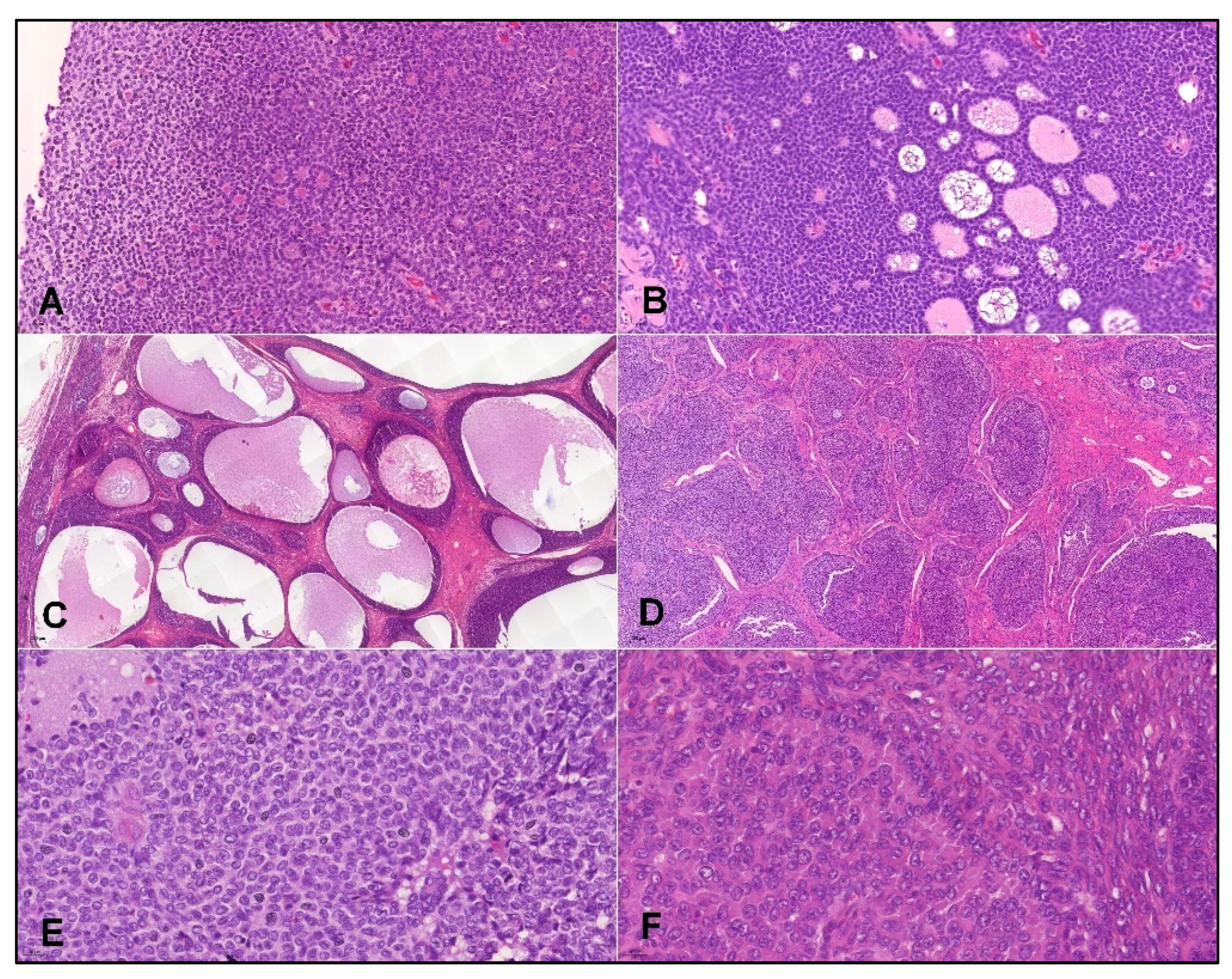
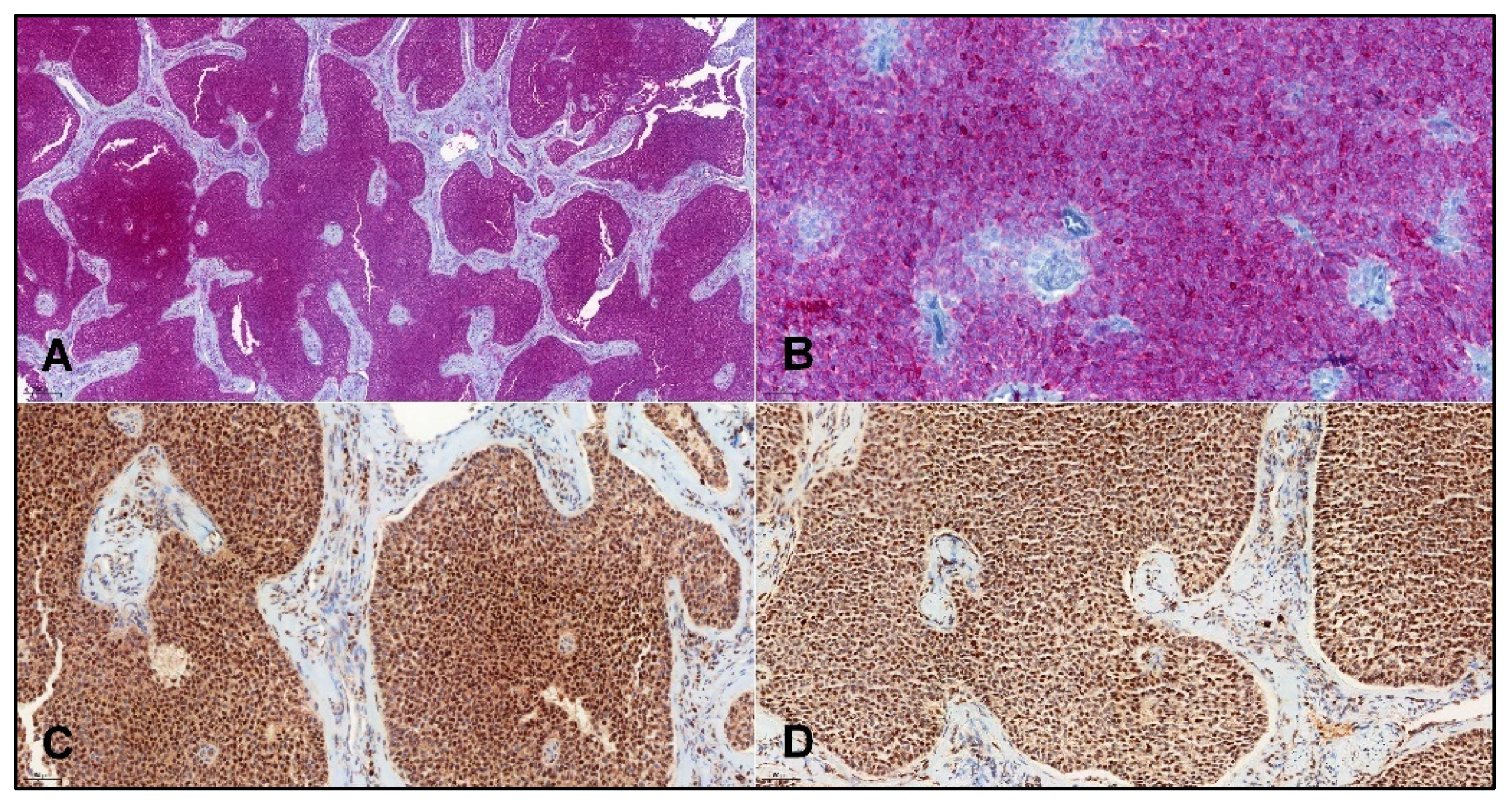
  • Histopathology and IHC:
Hematoxylin–eosin slides from all cases were reviewed to confirm the diagnosis of AGCT. The tumors showed a variety of histology patterns. Diffuse growth pattern was the most common, but other patterns, including trabecular, insular, microfollicular (with the presence of rare Call–Exner bodies), and macrofollicular patterns were also present among these cases. Tumor cells featured scant cytoplasm and uniform oval nuclei with irregular nuclear membranes and nuclear grooves. Luteinization was present in rare cases (Figure 1). In some cases, AGCTs were distinguished from fibromas/thecomas using reticulin stain.
The immunohistochemical study was performed using the Ventana Benchmark XT automated stainer (Ventana Medical System, Inc., Tucson, AZ, USA). The following primary antibodies were used: Anti-FOXL2 (polyclonal, Invitrogen, Waltham, MA, USA, dilution 1:100), Anti-inhibin alpha (clone R1, CellMarque, Rocklin, CA, USA, ready-to-use), Anti-calretinin (SP65, Ventana, Tucson, AZ, USA, ready-to-use), and anti-SF1 (EPR19744, AbCam, Cambridge, UK, dilution 1:100), using either DAB/HRP (anti-FOXL2, anti-SF1), or Fast Red/ALP (anti-inhibin α, anti-calretinin) as a chromogen/reporter enzyme combination. Appropriate positive and negative controls were used. Immunostaining in >5% of cells was considered positive, and immunostaining in ≤5% of cells was considered negative. Positive results were recorded as focal (staining in >5% and ≤50% of tumor cells) or diffuse (>50%) patterns (Figure 2).
  • DNA extraction
DNA from formalin-fixed paraffin-embedded (FFPE) tumor tissue was extracted using QIAsymphony DSP DNA Mini Kit (Qiagen, Hilden, Germany) on an automated extraction system (QIAsymphony SP, Qiagen) according to the manufacturer’s supplementary protocol for FFPE samples (Purification of genomic DNA from FFPE tissue using the QIAamp DNA FFPE Tissue Kit and Deparaffinization Solution). Concentration and purity of isolated DNA were determined using NanoDrop ND 1000 (NanoDrop Technologies Inc., Wilmington, DE, USA). DNA integrity was examined by amplification of control genes in a multiplex PCR [11].
  • Analysis of FOXL2 and CHEK2 mutations
The analyses of hot spot mutation c.402C>G, (p.C134W) of the FOXL2 gene (NCBI RefSeq: NM_023067.4) and two hotspot mutations c.470T>C, (p.I157T) and c.1100delC, (p.T367fs) of the CHEK2 gene (NCBI RefSeq: NM_007194.4) were performed using PCR and Sanger sequencing. PCR reactions were used for amplification of relevant regions of CHEK2 (part of exon 4 containing c.470T>C; part of exon 11 with c.1100delC) and FOXL2 (part of exon 1 containing c.402C>G). Briefly, 100 ng DNA was added to a reaction mixture consisting of 12.5 µL of FastStart PCR Master (Roche Diagnostic, Mannheim, Germany), 10 pmol of forward and reverse primers (Appendix A, Table A1) and distilled water up to 25 µL. The amplification program: initial denaturation (95 °C for 9 min), followed by 40 cycles of denaturation (95 °C for 1 min), annealing (56 °C (CHEK2 c.470) or 60 °C (FOXL2 and CHEK2 c.1100) for 1 min) and extension (72 °C for 1 min). The program was terminated by incubation at 72 °C for 7 min. The PCR products were separated by electrophoresis on a 2% agarose gel. Successfully amplified PCR products selected for sequencing analysis were purified with magnetic particles Agencourt® AMPure® (Agencourt Bioscience Corporation, A Beckman Coulter Company, Beverly, MA, USA), according to the manufacturer’s protocol, and both sides were sequenced using the Big Dye Terminator Sequencing kit (Applied Biosystems, Waltham, MA, USA) on an automated sequencer ABI Prism 3130xl (Applied Biosystems) at a constant voltage of 13.2 kV for 20 min. The results were analyzed using Geneious 6.1.6 analysis software (Geneious) or visually inspected. DNA sequences were compared to the reference sequence by the online program BLAST [12].
  • Data analysis and statistics
Cases with focal or diffuse expression of FOXL2, inhibin, calretinin, or SF1 were classified as positive for these IHC markers. The Genome Aggregation Database (gnomAD) [13] was used for retrieval of prevalence of the CHEK2p.I157T mutation in three different reference populations. This database aggregates and harmonizes exome and genome sequencing data for 141,456 (v2.1) or 76,156 (v3.1) unrelated individuals from various disease-specific and population genetic studies [14]. Confidence intervals of 95% for proportions were determined using Wilson’s procedure with a correction for continuity implemented in VassarStats [15]. Association between status of the two germline CHEK2 mutations and group membership (AGCT vs. population reference groups) was assessed from prevalence ratios (PRs) determined using the OpenEpi tool version 3.01 updated 4 June 2013 [16,17]. Confidence intervals of 95% for PRs were approximated by a Taylor series, and the significance of difference from PR = 1 was tested using the Mid-p exact method. The Kaplan––Meier method implemented in GraphPad Prism version 8.0.1 for Windows (GraphPad Software, LaJolla, CA, USA) was used to analyze the time to event (age at AGCT diagnosis) data. Difference between groups of CHEK2 mutation carriers and non-carriers were assessed using the Gehan–Breslow–Wilcoxon test, which does not require a consistent hazard ratio and gives more weight to the diagnosis of AGCT at an earlier age. Performance of IHC markers for diagnosis of FOXL2p.C134W AGCTs was characterized by sensitivity, specificity, and Youden’s J index that summarizes the performance of markers giving equal weight to false positive and false negative values. The 95% confidence intervals for sensitivity and specificity were computed by the Wilson–Brown method using GraphPad Prism version 8.0.1. Youden’s J indices, including CI95 intervals, were calculated using the Two-Way Contingency Table Analysis implemented in StatPages [18].

3. Results

We identified 93 cases diagnosed with ovarian granulosa cell tumors from primary (N = 74) or metastatic (N = 19) biopsy specimens. Age at diagnosis was 18–83 years (median = 58 years).
The FOXL2 mutation p.C134W (c.402C>G) was determined in 69 cases and found in 58 cases (57 cases with heterozygous and one with homozygous genotype), which account for 84.1% (CI95: 72.8–91.4%) of cases with determined status of this mutation. A total of 11 cases were negative and the status of the FOXL2 could not be determined in 24 cases (Table 1).

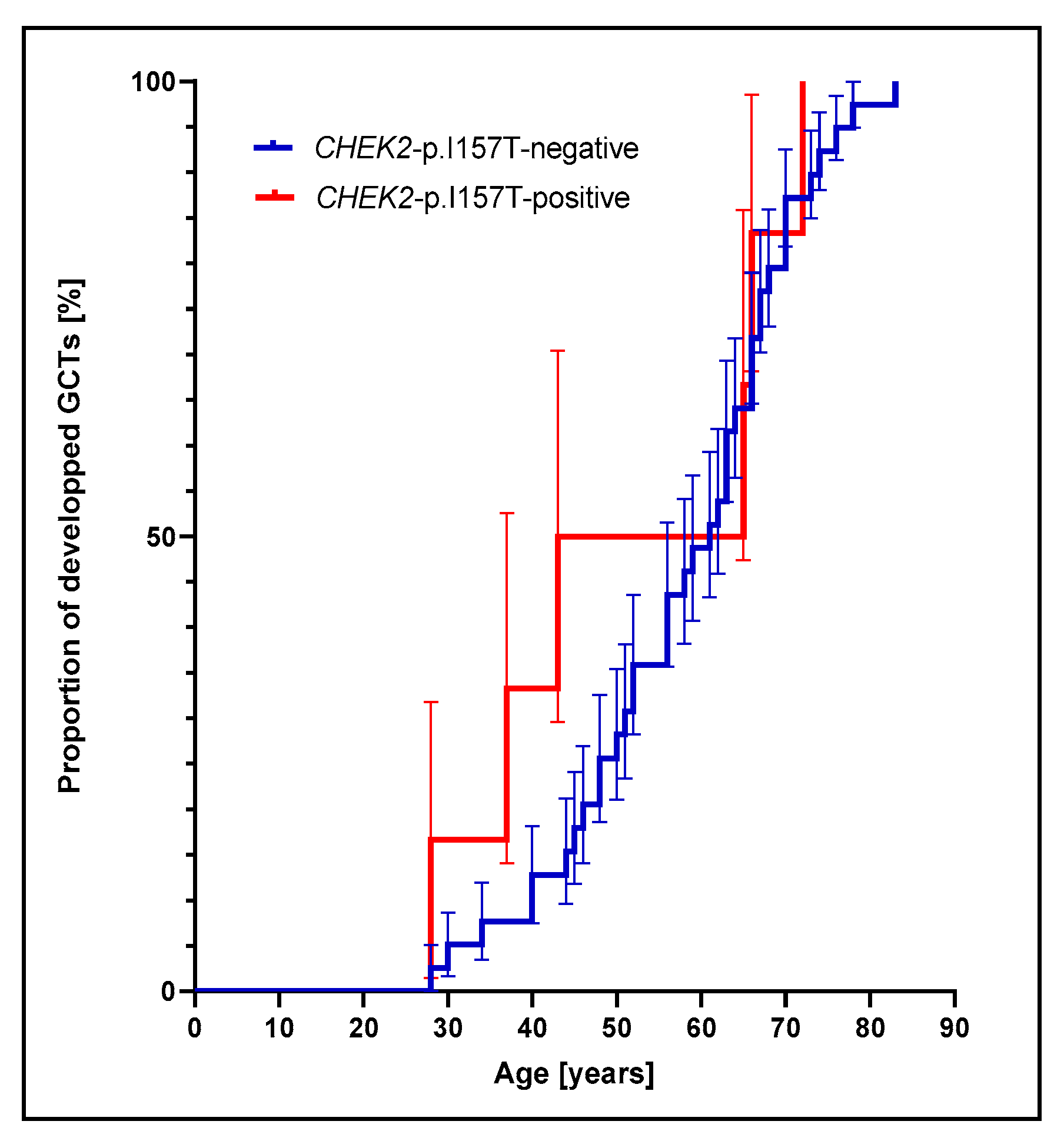
3.1. Prevalence of CHEK2p.I157T Mutation Is Increased among Adult GCT Patients

The CHEK2 c.1100delC mutation was found in a single case of a primary ovarian tumor with the FOXL2p.C134W somatic mutation, morphologically consistent with AGCT and positive by IHC for inhibin, FOXL2, calretinin, and SF1. However, this mutation was not further considered in the context of its association with AGCTs due to an insufficient number of cases in our group.
For the analysis of the association of CHEK2 founder mutations with GCTs, we only included in the analysis 58 cases with tumors positive for the FOXL2 mutation p.C134W, which is pathognomonic for adult-type GCTs (Table 1).
The group with the FOXL2p.C134W mutation displays higher median age at diagnosis than the group negative for this mutation (FOXL2 wild-type). In addition, our FOXL2p.C134W-positive group includes only cases with a minimum age at diagnosis of 28 years, while the FOXL2 wild-type group includes also women diagnosed at a younger age. As a result, the FOXL2p.C134W-positive group largely represents cases of bona fide adult-type ovarian granulosa cell tumor (AGCTs).
Among the 46 cases with known status for the CHEK2p.I157T germline mutation, six patients were found to be carriers of this mutation (Table 1), which indicates the prevalence of 13.0% (CI95: 5.4–27.0%). The Genome Aggregation Databases v.2.1.1 and v.3.1.1 indicate global prevalence of the CHEK2p.I157T mutation as 1391/282,816 (0.49%; CI95: 0.46–0.52%) and 615/152,156 (0.40%; CI95: 0.37–0.43%), respectively. Prevalence for the European non-Finnish populations was found to be 686/129140 (0.53%; CI95: 0.49–0.57%) and 362/68,034 (0.53%; CI95: 0.48–0.59), respectively. The highest prevalence of the CHEK2p.I157T mutation was found for the Finnish population 627/25,118 (2.50%; CI95: 2.31–2.70%) and 245/10,606 (2.31%; CI95: 2.04–2.62%). Our results indicate significantly higher prevalence of the CHEK2p.I157T mutation among patients with AGCTs than in the global population (p = 1.2 × 10⁻⁷), the European non-Finnish population (p = 1.9 × 10⁻⁷), or the European Finnish population (p = 0.0011). Prevalence ratios for the p.I157T mutation in the AGCT group were PR = 26.52 (CI95:12.55–56.03), PR = 24.55 (CI95: 11.60–51.97), and PR = 5.23 (CI95: 2.47–11.06) relative to the global, European non-Finnish, and European Finnish populations, respectively. Of note, this analysis compared the p.I157 mutation prevalence in our GCT group with different reference populations from the gnomAD database v2.1.
Carriers of the CHEK2p.I157T mutation also displayed a lower median age at diagnosis of the AGCT compared to non-carriers (54 years vs. 60 years). Nevertheless, the difference between the groups was not significant (Gehan–Breslow–Wilcoxon test p = 0.4288). However, the log-rank hazard ratio HR = 1.40 (CI95: 0.52–3.78) between the groups with and without the CHEK2p.I157T mutation is inconclusive, and the Kaplan–Meier analysis does not support the proportional hazards model (Figure 3). When both CHEK2 founder mutations are considered, mutations carriers’ median age at diagnosis is 43 years and thus lower than the median age at diagnosis of 58.5 years in patients with confirmed absence of both founder mutations; however, the difference did not reach statistical significance (Gehan–Breslow–Wilcoxon test p = 0.2067).

3.2. Performance of IHC Markers for Detection of Adult GCTs

The four selected IHC markers displayed positive or focally positive staining in the following proportions of biopsy specimens: FOXL2: 83/93 cases (89.3%; CI95: 80.7–94.4%); SF1: 77/93 cases (82.8%; 73.3–89.6%); inhibin: 76/93 cases (81.7%; CI95: 72.1–88.7%), and calretinin: 76/93 cases (81.7%; CI95: 72.1–88.7%) (Table 1).
Since the FOXL2p.C134W (c.402C>G) somatic mutation is pathognomonic for adult-type ovarian granulosa cell tumors (AGCTs), among which it reportedly displays a prevalence of 97% [19], we were interested in exploring the potential of the four typically used IHC markers for GCTs to serve as surrogate markers predicting the presence of this mutation. Positivity for all the four IHC markers was found in 37/58 (63.8%, CI95: 50.1–75.7%) cases with the FOXL2p.C134W mutation, but only in 3/11 (27.3%, CI95: 7.3–60.7%) cases without this mutation (Fisher’s exact test p = 0.0427). In contrast, negative IHC for two or more markers was found in 5/11 (45.5%, CI95: 18.1–75.4%) tumors without the FOXL2 mutation, but only in 5/58 (8.6%, CI95: 3.2–19.7%) tumors with the FOXL2 mutation (Fisher’s exact p = 0.0068).
The potential of the four IHC markers to serve as surrogate markers for the FOXL2p.C134W mutation was assessed individually across 69 specimens with a known mutation status of this FOXL2 allele. The highest sensitivity was found for FoxL2 immunoexpression (~95%) and the highest specificity for inhibin and calretinin (~46%) (Table 2). FOXL2 immunoexpression was found to be in fair agreement with FOXL2p.C134W mutation status (~84% agreement; Cohen’s kappa = 0.271; CI95: −0.037–0.579).
Association between the expression of all the four IHC markers and FOXL2p.C134W mutational status was also examined by binary logistic regression. The strongest predictor of FOXL2 mutation status was FOXL2 immunoexpression, recording an odds ratio of 12.44, controlling for immunoexpression of inhibin, calretinin, and SF1 (Table 3). The logistic model with all four markers (Model 1) displays better data fit than an alternative model with no IHC markers (omnibus test of model coefficients p = 0.009); however, its estimated sensitivity 96.6% (CI95: 88.3–99.4%), specificity 27.3% (CI95: 9.7–56.6%) and Youden’s J index (0.238; CI95: 0.010–0.414) are not significantly better than the sensitivity, specificity and Youden’s J index of a single FOXL2 IHC marker (Table 2).
A logistic regression model was also built with three IHC markers FOXL2, inhibin, and calretinin (Model 2, Table 3). These IHC markers were included in Model 2, because they were found to be the most sensitive or most specific predictors of CHEK2 mutation status as single IHC markers, and their Wald p-values in Model 1 were p < 0.1. In Model 2, FOXL2 expression remained the strongest predictor of FOXL2p.C134W mutation with an odds ratio of 10.61, controlling for inhibin and calretinin. This model with three predictors displayed sensitivity 94.8% (CI95: 90.1–98.4%), specificity 45.5% (CI95: 20.4–64.5%), and Youden’s J index 0.403 (0.104–0.629) (Table 3), and it performed better than the logistic Model 1 that included all four IHC markers, although the difference in classification was not statistically significant for our dataset (McNemar’s test p = 0.25). The model with three predictors performed better than any single IHC marker (Table 2), but the differences were also not statistically significant (McNemar’s test p > 0.05).
When the analysis was limited to 58 cases positive for FOXL2p.C134W mutation, FOXL2 expression was positive in 94.8% cases (CI95: 85.9–98.6%), inhibin in 86.2% (CI95: 75.1–92.8%), calretinin in 86.2% (CI95: 75.1–92.8%), and SF1 in 84.5% (CI95: 73.1–91.6%) (Table 2). No agreement above chance was found between immunostaining by FOXL2 and calretinin (Cohen’s kappa = −0.068; CI95: −0.126–(−0.009)).
The highest agreement was found between immunostaining by inhibin and SF1 (Cohen’s kappa = 0.262; CI95: −0.055–0.580), even though the degree of agreement was inconclusive due to the small sample size and a wide 95% confidence interval. Taken together, SF1 appears to be a redundant IHC marker providing lower performance as well as limited additional information compared to inhibin for the prediction of FOXL2 mutation status.
For the remaining pairs of IHC markers, low values of estimated Cohen’s kappa imply only slight agreement between these markers; however, wide confidence intervals do not allow for drawing confident conclusions: calretinin and inhibin (Cohen’s kappa = 0.130; CI95: −0.180–0.440); FOXL2 and inhibin (Cohen’s kappa = 0.115; CI95: −0.196–0.426); calretinin and SF1 (Cohen’s kappa: 0.105; CI95: −0.193–0.402); and FOXL2 and SF1 (Cohen’s kappa = 0.0966; CI95: −0.190–0.329).

4. Discussion

Ovarian granulosa cell tumors (GCTs) are rare gynecologic tumors that represent less than 5% of all ovarian tumors [20]. They account for about 70% of all sex cord-stromal tumors (SCST) that arise from the gonadal primitive sex cords or stromal cells.
Adult-type ovarian granulosa cell tumors (AGCTs) and juvenile-type ovarian granulosa cell tumors (JGCTs) are epidemiologically, clinically, and histopathologically distinct entities that account for 95% and 5% of GCTs, respectively [21]. AGCTs are typically diagnosed in perimenopausal or early postmenopausal women with a median age at diagnosis of 50–54 years, although they can occasionally also be diagnosed in children [22]. In contrast, the average age at diagnosis of JGCT is reportedly 13 years, although 21% of cases were still diagnosed in women over 21 years of age [23]. Of note, these cases were typically classified as AGCTs or JGCTs based on morphological criteria.
Consistent with previous reports [22], we found the median age at diagnosis of ovarian GCTs to be 58 years among all patients and 59 years in a subgroup with the FOXL2p.C134W mutation. On the other hand, a Korean case-series study of 91 patients with adult-type ovarian GCT reported the median age at diagnosis to be 42 years (range 7–85 years) [24], which is considerably lower than was found in our study or reported by other investigators. This disagreement may be caused by different definitions of AGCT, which is molecular in our study and morphological/clinical in the Korean study. Relatively non-specific histopathologic features of AGCTs have been recognized as a source of misdiagnosis of other cancers as AGCTs and imply the importance of molecularly defined diagnosis of these tumors [25].
The presence of the somatic mutation FOXL2p.C134W and the expression of FOXL2 protein are characteristic of the adult-type GCT. In contrast, juvenile-type GCTs virtually never display this mutation, and the expression of FOXL2 may be variable, and even reduced in aggressive phenotypes and advanced stages of JGCTs (reviewed in [26]). Ovarian granulosa tumors with the FOXL2p.C134W mutation reportedly displayed higher expression of FOXL2 on mRNA level than those with wild-type FOXL2, which in turn correlated with the intensity of FOXL2 IHC staining among AGCTs and JGCTs [21]. Consistent with these findings, our results show more prevalent positivity of FOXL2 IHC among cases with the FOXL2p.C134W mutation than in cases without this mutation (PR = 1.3; CI95:0.90–1.88). We found this association between the FOXL2p.C134W mutation status and FOXL2 positivity by IHC to be marginally statistically significant (mid-p-value = 0.052).
Consequently, the presence of somatic mutation FOXL2p.C134W can serve as a defining feature of the AGCTs; therefore, in this study we delineated the AGCTs as cases previously diagnosed with ovarian granulosa cell tumors or granulosa cell-like tumors, in which we additionally found the somatic mutation FOXL2p.C134W. This approach is in line with that of other investigators, who reappraised FOXL2 wild-type AGCT cases, which were previously diagnosed entirely by morphology, as most likely representing thecomas or fibromas [19,26,27]. It should be noted, however, that FOXL2p.C134W was also detected in a subset of thecomas [19].
To streamline the diagnosis of AGCTs, we evaluated the performance of selected IHC markers to distinguish ovarian granulosa cell tumors with the FOXL2p.C134W mutation from granulosa cell tumors with wild-type FOXL2, as the latter cases presumably represent JGCTs and possibly some misdiagnosed SCSTs of other types. Our results show a generally appreciable sensitivity but relatively low specificity of individual IHC markers.
The best diagnostic performance was achieved using a logistic regression model that integrated IHC expression of FOXL2, inhibin, and calretinin. However, due to a relatively small sample size, diagnostic performances were not found to be significantly different across several classifiers discussed in this study. The problem of small sample size affects most studies reporting research on this rare type of ovarian cancer. Nevertheless, research on rare cancers, such as gastrointestinal stromal tumors, acute myeloid leukemia, seminomas, and others, produced fundamental insights translatable into innovative therapeutic strategies for these rare malignancies, as well as in better a molecular understanding of more common types of cancers [28]. This is because rare cancers are usually homogeneous entities that (i) tend to result from a single identifiable genetic cause or exposure to a single identifiable environmental carcinogen, (ii) can be characterized by a small number of mutations, and (iii) typically deviate from normal cells only in a small number of pathways that are amenable to therapeutic targeting.
Thus far, no reproductive, occupational, environmental, or general lifestyle risk factors have been consistently associated with the risk of developing AGCTs [29], and no inherited predispositions have been identified for the development of these tumors. For these reasons, AGCTs have been considered sporadic and unrelated to exposure.
The study presented in this report was motivated by our finding of an unexpectedly high prevalence of AGCTs among female carriers of the CHEK2p.I157T mutations diagnosed with ovarian cancer. AGCT reportedly represents only 3–5% of all ovarian cancers in the general population [10]. Among the 75,024 ovarian cancers (ICD-O-3 site code C56.9) registered in the US SEER 13 cancer registries over 1992–2018, only 974 cases represented GCTs (ICD-O-3 code 8620/3), which accounted for 1.3% of all ovarian cancers [30]. By contrast, however, GCTs represented 4 of 11 (~36.4%) histologically characterized ovarian cancers in a group of the CHEK2p.I157T mutation carriers.
Our results further demonstrated a higher prevalence of this germline mutation among AGCT patients relative to three reference populations, which provided support to our hypothesis of the positive association between the CHEK2p.I157T mutation and adult-type ovarian granulosa cell tumors. Since GCTs are rare tumors that represent no more than 5% of all ovarian malignancies and display a similar occurrence across various populations [10], the risk of bias potentially generated by the selection of reference populations is low. Notably, we also found a higher prevalence of the CHEK2p.I157T mutation in the group of AGCT patients than in the European Finnish population that displays the highest prevalence of this mutation (Ensembl, release 105 [31]; rs17879961), which further supports our conclusion.
Our study implicated for the first time the CHEK2p.I157T mutation in granulosa cell carcinogenesis and suggested a specific genetic predisposition in adult-type ovarian granulosa cell tumors. The CHEK2p.I157T mutation has been previously shown to impede Chek2 protein homodimerization, which is required for its activation, and to interfere with wild-type Chek2 protein in heterozygous cells in a dominant-negative manner [8]. This mutation is considered to confer multi-organ tumor susceptibility through its probable synergism with other genetic factors or environmental exposures [3]; however, no conclusive association has been reported between this mutation and any type of ovarian cancer thus far. A small case-control study of patients with GCT suggested an association with family history of breast (OR = 2.13; 1.19–3.80) or ovarian cancer (OR = 2.89; 1.08–7.72), which implies a possible existence of shared genetic predispositions between these cancers [32].
An underlying mechanism behind the role of the CHEK2p.I157T germline mutation in the potentially increased risk of AGCTs has yet to be determined. One possible explanation can be that this germline CHEK2 mutation, which affects cell cycle regulation and DNA damage response pathways, can predispose to unique patterns of subsequent somatic mutations, including FoxL2p.C134W, which is pathognomonic for AGCTs. A similar mechanism has been previously suggested for prostate cancer, since patients with pathogenic germline CHEK2 mutations displayed a significantly higher prevalence of the somatic CDK12 mutation than unselected prostate cancer patients from the TCGA cohort. Consequently, CHEK2 germline mutations have already been associated with increased occurrence of a specific somatic mutation in a cancer-relevant gene [33].
Besides this suggested role of the germline CHEK2p.I157T mutation in the risk of developing a subsequent FOXL2p.C134W mutation in granulosa cells, other mechanisms can also be operative in the AGCT-CHEK2p.I157T association identified in our study. For instance, a wild-type FOXL2 was shown to modulate cell cycle regulators and promote cell cycle arrest in the G1 phase and in the repair of oxidatively damaged DNA [34]. This implies that the presence of the germline CHEK2 mutation impairing the cell cycle and DNA damage response may synergize with the FOXL2p.C134W mutation that may develop in granulosa cells as a “second hit” and further aggravate the impairment of the cell cycle and DNA damage repair processes, paving the way for ovarian granulosa carcinogenesis. Nevertheless, these suggested mechanisms are not mutually exclusive and can both contribute to the development of granulosa cell tumors.

5. Conclusions

The CHEK2 missense p.I157T (c.470T>C) germline mutation, which was previously reported to increase the risk of breast, colon, kidney, prostate, and thyroid cancers [4], is also associated with adult granulosa cell tumors (AGCTs). This association suggests that CHEK2p.I157T can be a predisposing genetic factor for AGCTs.
The FOXL2p.C134W mutation, which is a pathognomonic defining feature of AGCTs, was detected among ~84% of cases previously diagnosed as AGCTs based on clinical and histopathological findings. This finding supports the necessity of including detection of the FOXL2p.C134W mutation in the diagnosis of AGCT. The presence of this mutation is in fair agreement with IHC positivity of the tumor cells for FOXL2 expression, which allows for application of the FOXL2 expression as a surrogate IHC marker for the FOXL2p.C134W mutation.
Combination of IHC markers FOXL2, inhibin, and calretinin displayed the best performance for the prediction of FOXL2p.C134W mutational status among the AGCT cases diagnosed by clinical and histopathological findings.

Author Contributions

Conceptualization: R.M. and V.U.; data curation: R.M. and M.Š. (Marián Švajdler); formal analysis: R.M., P.Š., M.Š. (Marián Švajdler), M.Š. (Monika Šedivcová) and P.V.; investigation: P.Š., M.Š. (Marián Švajdler), M.Š. (Monika Šedivcová) and V.U.; methodology: R.M., P.Š. and M.Š. (Marián Švajdler); visualization: R.M., P.Š. and M.Š. (Marián Švajdler); resources: M.M.; writing—original draft: R.M., P.Š., M.Š. (Marián Švajdler) and M.Š. (Monika Šedivcová); writing—review and editing: R.M., V.U., P.V. and M.M. All authors have read and agreed to the published version of the manuscript.

Funding

This research received no external funding.

Institutional Review Board Statement

The study was conducted in accordance with the Declaration of Helsinki, and the protocol was approved by the Ethics Committee of Bioptická laboratoř (Project identification code 010/20).

Informed Consent Statement

The informed consent was waived due to the retrospective nature of the study performed on archived and strictly anonymized biospecimens, which did not lead to the change of a previously rendered diagnosis and had no clinical consequences on the patients’ management. For these studies, local ethical committees and other authorities in the Czech Republic do not require patient consent for the use of an archived specimen.

Data Availability Statement

Publicly available datasets analyzed in this study can be found here: gnomAD: [https://gnomad.broadinstitute.org, accessed on 22 October 2021]; SEER*Stat: [https://seer.cancer.gov/data-software/documentation/seerstat/nov2020/, accessed on 22 October 2021]. Other data available on request from the corresponding authors.

Acknowledgments

For Kay A. Mereish, in memoriam.

Conflicts of Interest

The authors declare no conflict of interest.

Appendix A

Table A1. Primers used for PCR amplification and sequencing of the FOXL2 and CHEK2 genes.
Primer NameSequence 5′-3′
FOXL2-402-F1CCTCAACGAGTGCTTCATCA
FOXL2-402-R2GCCGGTAGTTGCCCTTCT
CHEK2 470 F2CTCTATTTTAGGAAGTGGGTCC
CHEK2 470 R2TAGTGACAGTGCAATTTCAGAA
CHEK2 1100FTGTCTTCTTGGACTGGCAGA
CHEK2 1100RGGGGTTCCACATAAGGTTCTC

References

  1. Zannini, L.; Delia, D.; Buscemi, G. CHK2 kinase in the DNA damage response and beyond. J. Mol. Cell Biol. 2014, 6, 442–457. [Google Scholar] [CrossRef] [PubMed] [Green Version]
  2. Stolarova, L.; Kleiblova, P.; Janatova, M.; Soukupova, J.; Zemankova, P.; Macurek, L.; Kleibl, Z. CHEK2 germline variants in cancer predisposition: Stalemate rather than checkmate. Cells 2020, 9, 2675. [Google Scholar] [CrossRef]
  3. Cybulski, C.; Górski, B.; Huzarski, T.; Masojć, B.; Mierzejewski, M.; Dębniak, T.; Teodorczyk, U.; Byrski, T.; Gronwald, J.; Matyjasik, J.; et al. CHEK2 is a multiorgan cancer susceptibility gene. Am. J. Hum. Genet. 2004, 75, 1131–1135. [Google Scholar] [CrossRef] [Green Version]
  4. Leedom, T.P.; LaDuca, H.; McFarland, R.; Li, S.; Dolinsky, J.S.; Chao, E.C. Breast cancer risk is similar for CHEK2 founder and non-founder mutation carriers. Cancer Genet. 2016, 209, 403–407. [Google Scholar] [CrossRef] [PubMed] [Green Version]
  5. Weischer, M.; Bojesen, S.E.; Ellervik, C.; Tybjærg-Hansen, A.; Nordestgaard, B.G. CHEK2*1100delC genotyping for clinical assessment of breast cancer risk: Meta-analyses of 26,000 patient cases and 27,000 controls. J. Clin. Oncol. 2008, 26, 542–548. [Google Scholar] [CrossRef] [PubMed] [Green Version]
  6. Weischer, M.; Nordestgaard, B.G.; Pharoah, P.; Bolla, M.K.; Nevanlinna, H.; Veer, L.J.V.; Garcia-Closas, M.; Hopper, J.L.; Hall, P.; Andrulis, I.L.; et al. CHEK2*1100delC heterozygosity in women with breast cancer associated with early death, breast cancer–specific death, and increased risk of a second breast cancer. J. Clin. Oncol. 2012, 30, 4308–4316. [Google Scholar] [CrossRef] [Green Version]
  7. Fletcher, O.; Johnson, N.; dos Santos Silva, I.; Kilpivaara, O.; Aittomäki, K.; Blomqvist, C.; Nevanlinna, H.; Wasielewski, M.; Meijers-Heijerboer, H.; Broeks, A.; et al. Family history, genetic testing, and clinical risk prediction: Pooled analysis of CHEK2*1100delC in 1828 bilateral breast cancers and 7030 controls. Cancer Epidemiol. Biomark. Prev. 2009, 18, 230–234. [Google Scholar] [CrossRef] [Green Version]
  8. Muranen, T.A.; Blomqvist, C.; Dörk, T.; Jakubowska, A.; Heikkilä, P.; Fagerholm, R.; Greco, D.; Aittomäki, K.; Bojesen, S.E.; Shah, M.; et al. Patient survival and tumor characteristics associated with CHEK2: p.I157T—Findings from the Breast Cancer Association Consortium. Breast Cancer Res. 2016, 18, 98. [Google Scholar] [CrossRef] [Green Version]
  9. Sutcliffe, E.G.; Stettner, A.R.; Miller, S.A.; Solomon, S.R.; Marshall, M.L.; Roberts, M.E.; Susswein, L.R.; Arvai, K.J.; Klein, R.T.; Murphy, P.D.; et al. Differences in cancer prevalence among CHEK2 carriers identified via multi-gene panel testing. Cancer Genet. 2020, 246–247, 12–17. [Google Scholar] [CrossRef]
  10. Alexiadis, M.; Rowley, S.M.; Chu, S.; Leung, D.T.; Stewart, C.J.; Amarasinghe, K.C.; Campbell, I.G.; Fuller, P.J. Mutational landscape of ovarian adult granulosa cell tumors from whole exome and targeted TERT promoter sequencing. Mol. Cancer Res. 2018, 17, 177–185. [Google Scholar] [CrossRef] [Green Version]
  11. Van Dongen, J.J.M.; Langerak, A.; Brüggemann, M.; Evans, P.A.S.; Hummel, M.; Lavender, F.; Delabesse, E.; Davi, F.; Schuuring, E.; Garcia-Sanz, R.; et al. Design and standardization of PCR primers and protocols for detection of clonal immunoglobulin and T-cell receptor gene recombinations in suspect lymphoproliferations: Report of the BIOMED-2 concerted action BMH4-CT98-3936. Leukemia 2003, 17, 2257–2317. [Google Scholar] [CrossRef] [PubMed] [Green Version]
  12. National Center for Biotechnology Information. Basic Local Alignment Search Tool (BLAST); National Center for Biotechnology Information: Bethesda, MD, USA, 1990. [Google Scholar]
  13. The Genome Aggregation Database. Available online: https://gnomad.broadinstitute.org/about (accessed on 22 October 2021).
  14. Karczewski, K.J.; Francioli, L.C.; Tiao, G.; Cummings, B.B.; Alfoldi, J.; Wang, Q.; Collins, R.L.; Laricchia, K.M.; Ganna, A.; Birnbaum, D.P.; et al. The mutational constraint spectrum quantified from variation in 141,456 humans. Nature 2020, 581, 434–443. [Google Scholar] [CrossRef] [PubMed]
  15. VassarStats. Website for Statistical Computation. Available online: http://vassarstats.net/ (accessed on 22 October 2021).
  16. Sullivan, K.M.; Dean, A.; Soe, M.M. OpenEpi: A web-based epidemiologic and statistical calculator for public health. Public Health Rep. 2009, 124, 471–474. [Google Scholar] [CrossRef] [Green Version]
  17. OpenEpi. Open-Source Epidemiologic Statistics for Public Health, Version 3.01, Updated. 6 April 2013. Available online: https://www.openepi.com/Menu/OE_Menu.htm (accessed on 22 October 2021).
  18. StatPages. The Interactive Statistical Pages. Available online: https://statpages.info/index.html (accessed on 22 October 2021).
  19. Shah, S.P.; Köbel, M.; Senz, J.; Morin, R.D.; Clarke, B.A.; Wiegand, K.C.; Leung, G.; Zayed, A.; Mehl, E.; Kalloger, S.E.; et al. Mutation of FOXL2 in granulosa-cell tumors of the ovary. N. Engl. J. Med. 2009, 360, 2719–2729. [Google Scholar] [CrossRef]
  20. Kottarathil, V.D.; Antony, M.A.; Nair, I.R.; Pavithran, K. Recent advances in granulosa cell tumor ovary: A review. Indian J. Surg. Oncol. 2012, 4, 37–47. [Google Scholar] [CrossRef] [PubMed] [Green Version]
  21. D’Angelo, E.; Mozos, A.; Nakayama, D.; Espinosa, I.; Catasus, L.; Muñoz, J.; Prat, J.; Mu, J. Prognostic significance of FOXL2 mutation and mRNA expression in adult and juvenile granulosa cell tumors of the ovary. Mod. Pathol. 2011, 24, 1360–1367. [Google Scholar] [CrossRef]
  22. Schumer, S.T.; Cannistra, S.A. Granulosa cell tumor of the ovary. J. Clin. Oncol. 2003, 21, 1180–1189. [Google Scholar] [CrossRef]
  23. Young, R.H.; Dickersin, G.R.; Scully, R.E. Juvenile granulosa cell tumor of the ovary: A clinicopathological analysis of 125 cases. Am. J. Surg. Pathol. 1984, 8, 575–596. [Google Scholar] [CrossRef]
  24. Shim, S.-H.; Lee, S.J.; Kim, D.-Y.; Kim, J.; Kim, S.-N.; Kang, S.-B.; Kim, J.-H.; Kim, Y.-M.; Kim, Y.-T.; Nam, J.-H. A long-term follow-up study of 91 cases with ovarian granulosa cell tumors. Anticancer Res. 2014, 34, 1001–1010. [Google Scholar]
  25. McConechy, M.K.; Färkkilä, A.; Horlings, H.M.; Talhouk, A.; Unkila-Kallio, L.; van Meurs, H.S.; Yang, W.; Rozenberg, N.; Andersson, N.; Zaby, K.; et al. Molecularly defined adult granulosa cell tumor of the ovary: The clinical phenotype. JNCI J. Natl. Cancer Inst. 2016, 108, djw134. [Google Scholar] [CrossRef] [Green Version]
  26. Jamieson, S.; Fuller, P.J. Molecular pathogenesis of granulosa cell tumors of the ovary. Endocr. Rev. 2012, 33, 109–144. [Google Scholar] [CrossRef] [PubMed]
  27. Kim, M.S.; Hur, S.Y.; Yoo, N.J.; Lee, S.H. Mutational analysis of FOXL2 codon 134 in granulosa cell tumour of ovary and other human cancers. J. Pathol. 2010, 221, 147–152. [Google Scholar] [CrossRef] [PubMed]
  28. Berman, J.J. Neoplasms: Principles of Development and Diversity; Jones & Bartlett Learning: Burlington, MA, USA, 2009. [Google Scholar]
  29. Bryk, S.; Pukkala, E.; Martinsen, J.I.; Unkila-Kallio, L.; Tryggvadottir, L.; Sparén, P.; Kjærheim, K.; Weiderpass, E.; Riska, A. Incidence and occupational variation of ovarian granulosa cell tumours in Finland, Iceland, Norway and Sweden during 1953–2012: A longitudinal cohort study. BJOG 2016, 124, 143–149. [Google Scholar] [CrossRef] [Green Version]
  30. SEER Program. Incidence—SEER Research Data, 13 Registries, November 2020 Sub (1992–2018)—Linked to County Attributes—Time Dependent (1990–2018) Income/Rurality, 1969–2019 Counties, National Cancer Institute, DCCPS, Surveillance Research Program, Released April 2021, Based on the November 2020 Submission. Available online: www.seer.cancer.gov (accessed on 18 January 2022).
  31. Howe, K.L.; Achuthan, P.; Allen, J.; Alvarez-Jarreta, J.; Ridwan Amode, M.; Armean, I.M.; Azov, A.G.; Bennet, R.; Bhai, J.; Billis, K.; et al. Ensembl 2021. Nucleic Acids Res. 2021, 49, D884–D891. [Google Scholar] [CrossRef] [PubMed]
  32. Boyce, E.; Costaggini, I.; Vitonis, A.; Feltmate, C.; Muto, M.; Berkowitz, R.; Cramer, D.; Horowitz, N. The epidemiology of ovarian granulosa cell tumors: A case-control study. Gynecol. Oncol. 2009, 115, 221–225. [Google Scholar] [CrossRef] [PubMed]
  33. Nizialek, E.; Lotan, T.L.; Isaacs, W.B.; Yegnasubramanian, S.; Paller, C.J.; Antonarakis, E.S. The somatic mutation landscape of germline CHEK2-altered prostate cancer. J. Clin. Oncol. 2021, 39, 5084. [Google Scholar] [CrossRef]
  34. Benayoun, B.A.; Georges, A.B.; L’Hôte, D.; Andersson, N.; Dipietromaria, A.; Todeschini, A.-L.; Caburet, S.; Bazin, C.; Anttonen, M.; Veitia, R.A. Transcription factor FOXL2 protects granulosa cells from stress and delays cell cycle: Role of its regulation by the SIRT1 deacetylase. Hum. Mol. Genet. 2011, 20, 1673–1686. [Google Scholar] [CrossRef] [Green Version]
Figure 1. Representative images of HE staining in adult granulosa cell tumors. AGCTs show a variety of histology patterns, including solid, with the presence of: Call–Exner bodies (A), original magnification ×200; Microfollicular (B), original magnification ×200; Macrofollicular (C), original magnification ×50; or insular (D), original magnification ×50. Tumor cells have irregular nuclear membranes, oval nuclei, and nuclear grooves (E), original magnification ×400. Luteinized granulosa cell tumor cells have abundant eosinophilic cytoplasm and less conspicuous grooving of the nuclei (F), original magnification ×400.
Cancers 14 01208 g001
Figure 2. Representative images of IHC staining in adult granulosa cell tumors: Calretinin (A), original magnification ×50; inhibin alpha (B), original magnification ×200; SF1 (C), original magnification ×200; and FOXL-2 (D), original magnification ×200.
Cancers 14 01208 g002
Figure 3. Kaplan–Meier estimate for age at diagnosis of GCT for CHEK2p.I157T-positive and -negative groups. Percent ovarian GCT cases developed by specific age. Error bars: standard errors (asymmetrical).
Cancers 14 01208 g003
Table 1. Characteristics of cases diagnosed with ovarian granulosa cell tumors classified per status of the FOXL2p.C134W mutation.
FOXL2 Status
FOXL2p.C134WFOXL2 Wild-TypeUnknownALL
Number of cases58112493
Age at diagnosis (years)
Median (range)59 (28–83)52 (18–81)59.5 (32–75)-
CHEK2 mutation p.I157T
Positive6107
Negative406046
Unknown1242440
CHEK2 mutation c.1100delC
Positive1001
Negative398855
Unknown1831637
FOXL2 by IHC
Positive5582083
Negative33410
Inhibin by IHC
Positive5062076
Negative85417
Calretinin by IHC
Positive5062076
Negative85417
SF1 by IHC
Positive4982077
Negative93416
Table 2. Performance of IHC markers to predict FOXL2p.C134W or FOXL2 wild-type status in tumors diagnosed as GCTs/likely GCTs.
FOXL2InhibinCalretininSF1
Sensitivity
(CI95)
94.8% 86.2%86.2%84.5%
(85.9–98.6%)(75.1–92.8%)(75.1–92.8%)(73.1–91.6%)
Specificity
(CI95)
27.3%45.5%45.5%27.3%
(9.7–56.6%)(21.3–72.0%)(21.3–72.0%)(9.7–56.6%)
Youden’s J
(CI95)
0.2210.3170.3170.118
(−0.009–0.448)(0.008–0.631)(0.008–0.631)(−0.116–0.455)
Table 3. Logistic regression analysis of association between FOXL2 mutational status and four IHC markers.
Model Variables (Model 1 and Model 2)
B *SEWaldDfp-ValueOR95% CI for OR
LowerUpper
Constant
Model 1−2.241.223.3810.066---
Model 2−2.411.213.9710.046---
Inhibin
Model 11.610.863.4710.0625.000.9227.13
Model 21.440.823.0310.0824.200.8421.15
FOXL2
Model 12.521.026.1710.01312.441.7090.94
Model 22.361.005.6310.01810.611.5174.63
SF1
Model 1−0.671.050.4210.5190.5100.0663.956
Model 2NANANANANANANANA
Calretinin
Model 11.610.863.4710.0625.000.92027.131
Model 21.440.823.0310.0824.200.83521.149
Model fit and classification performance
Omnibus test of model coefficientsChi-squaredDfp-value
Model 113.5140.009
Model 213.0730.004
HL testChi-squaredDfp-value
Model 11.1330.771
Model 21.7420.419
-2 Log likelihoodChi-squaredDfp-value
Model 147.03NANA
Model 247.47NANA
Classification resultsFOXL2p.C134WFOXL2wtFOXL2p.C134W/FOXL2wt
Model 1Correct56 (96.6%)3 (27.3%)59 (85.5%)
Incorrect2810
Youden’s J0.238 (CI95: 0.010–0.414)
Model 2Correct55 (94.8%)5 (45.5%)60 (87.0%)
Incorrect369
Youden’s J0.403 (CI95: 0.104–0.629)
* Regression coefficient.
Publisher’s Note: MDPI stays neutral with regard to jurisdictional claims in published maps and institutional affiliations.

