Systematic Review

Molecular Systems Architecture of Interactome in the Acute Myeloid Leukemia Microenvironment

by
V. A. Shiva Ayyadurai
1,*,†,
Prabhakar Deonikar
1,
Kevin G. McLure
2 and
Kathleen M. Sakamoto
3,†
1
Systems Biology Group, International Center for Integrative Systems, Cambridge, MA 02138, USA
2
Ermaris Bio, Inc., Oakland, CA 94618, USA
3
Division of Hematology/Oncology, Department of Pediatrics, Stanford University, Stanford, CA 94305, USA
*
Author to whom correspondence should be addressed.
These authors contributed equally to this work.
Cancers 2022, 14(3), 756; https://doi.org/10.3390/cancers14030756
Submission received: 22 December 2021 / Accepted: 29 January 2022 / Published: 1 February 2022
(This article belongs to the Section Systematic Review or Meta-Analysis in Cancer Research)

Abstract

:

Simple Summary

Acute myeloid leukemia (AML) is a cancer of blood and bone marrow that causes rapid production of abnormal red and white blood cells. Once established, the cancer cells communicate through a complex set of molecular interactions with neighboring cells in order to survive, spread rapidly, and evade detection and destruction by the body’s immune system. In this study, a systematic review produced a comprehensive set of critical molecular interactions that was then organized into molecular “systems architecture” to map the communications between cancer cells and neighboring cells. This systems architecture may aid in identifying effective targets that disrupt communication between the cancer cells and the neighboring environment, leading to effective treatment strategies.

Abstract

A molecular systems architecture is presented for acute myeloid leukemia (AML) to provide a framework for organizing the complexity of biomolecular interactions. AML is a multifactorial disease resulting from impaired differentiation and increased proliferation of hematopoietic precursor cells involving genetic mutations, signaling pathways related to the cancer cell genetics, and molecular interactions between the cancer cell and the tumor microenvironment, including endothelial cells, fibroblasts, myeloid-derived suppressor cells, bone marrow stromal cells, and immune cells (e.g., T-regs, T-helper 1 cells, T-helper 17 cells, T-effector cells, natural killer cells, and dendritic cells). This molecular systems architecture provides a layered understanding of intra- and inter-cellular interactions in the AML cancer cell and the cells in the stromal microenvironment. The molecular systems architecture may be utilized for target identification and the discovery of single and combination therapeutics and strategies to treat AML.

1. Introduction

Acute Myeloid Leukemia (AML) is characterized by uncontrolled proliferation, increased survival, and impaired differentiation of hematopoietic progenitor cells [1]. Increased proliferation and apoptosis resistance, as well as the inhibition of differentiation and/or aberrant activation of growth factor receptor signaling pathways, are central to AML pathogenesis [2,3]. Aberrant and constitutive activation of signal transduction molecules are found in about 50% of primary AML bone marrow samples, enhancing the survival and proliferation of hematopoietic progenitor cells via the RAF/MEK/ERK cascade and the PI3K/AKT pathways that are dysregulated by mutations in receptor tyrosine kinases (RTK), Fms related receptor tyrosine kinase 3 (FLT3), N-Ras and K-Ras, and Kit [1,4].
Specific surface biomarkers characterize the subpopulations of AML cells. For example, leukemic stem cells are characterized by CD34+/CD38 surface markers, megakaryocyte-erythroid progenitors (MEPs) are characterized by CD34+/CD38+/CD45RA surface markers, and granulocytic-monocytic progenitors (GMPs) are characterized by CD34+/CD38+/CD45RA+ surface markers [5,6,7]. Aberrant multipotent progenitor cells give rise to myeloid lineage-committed cells showing further phenotypical as well as functional changes. AML patients present with a significantly expanded population of granulocytic-monocytic progenitor cells (GMP), while the megakaryocytic-erythroid progenitor (MEP) population is severely reduced [8]. In addition, there is a significant depletion of HSC numbers in AML as a result of a differentiation block at the HSC–progenitor transition [9]. The increased constitutive activation of GMP clusters in AML has been attributed to insufficient production of cytokines such as TGFβ 1 and CXCL4—factors that promote the quiescence of the GMP clusters [10].
Gene mutations precipitate key events in AML pathogenesis [11,12]. The gene mutations common in AML are well documented elsewhere [7] and some of the key genetic factors are summarized in Table 1. Class I mutations lead to uncontrolled cellular proliferation and evasion of apoptosis and include mutation conferring constitutive activity to tyrosine kinases or dysregulation of downstream signaling molecules (in genes such as BCR-ABL, LLT3, c-KIT, and RAS) [13]. Class II mutations are associated with inhibition of differentiation, including key transcription factors, such as CBF and retinoic acid receptor alpha (RARα), and proteins that are involved in transcriptional regulation, such as p300, CBP, MOX, TIF2, and MLL [14]. Class III mutations are involved in epigenetic regulation and include genes such as TET2, IDH1/2, DNMT3A, ASXK1, Cohesin, NPM1 and EZH2 [11,14]. Additionally, genes such as WT1 and TP53 are implicated in tumor suppression activity [14]. Oncogenes common in AML include PML-RARa, FLT3-ITD, AML-ETO, and CBFB-MYH11 (oncogene for Inversion 16 cytogenetic alteration) [14,15].
Recent reviews have discussed in detail the cytogenetic targets for potential treatments of AML [14,16,17]. In addition to cytogenetic factors, the interactions in the tumor microenvironment that promote suppression of immune response, cancer cell proliferation, and inhibition of apoptosis also contribute significantly to the pathogenesis of AML [2]. This research employs a systems biology approach to provide not only a systematic review of the current understanding of AML tumor microenvironment (TME) but also a molecular systems representation, i.e., the interactome of the molecular interactions within cancer cells and across the cells in the stromal microenvironment. The insights from this review aim to provide the AML research community an integrative molecular systems approach to understanding the complexity of the biomolecular interactions involved in AML pathogenesis. The results of this investigation may be used to support identification of potential targets for therapeutic interventions.

2. Literature Review

The scientific literature was searched to identify journal papers that contain research on AML, molecular pathways of AML, cells in the AML microenvironment, interactions between AML cells and the cells of AML microenvironment, and the molecular pathways involved in the cellular crosstalk in the AML microenvironment. CytoSolve® is an established systems biology tool that enables a systematic bioinformatics literature review process, as well as providing scalable computational modeling of molecular pathways [18,19,20,21,22,23,24]. In this study, CytoSolve has been employed to perform a systematic review as well as to support the curation and development of the molecular systems architecture of AML pathogenesis.
The systematic review process for this study involved the following four steps:
  • creating a list of Medical Subject Headings (MeSH) keywords to optimize the recall and precision of peer-reviewed articles (listed in Table 2);
  • searching and retrieving the relevant peer-reviewed articles published between January 1980 to June 2021 from PubMed, Medline, and Google Scholar, which were stored as an “Initial Set” repository;
  • screening the titles and abstracts of the articles in the Initial Set repository to identify the most relevant articles based on our inclusion criteria, which were stored as the “Final Set” repository; and
  • performing a full-length review of the Final Set repository using the domain experts.

The Inclusion Criteria

The full text of the articles, not only the abstracts, were reviewed completely by the authors. An article was deemed relevant only if the body of the article contained the keywords set out in Table 2 (e.g., CXCR4, TGF-β, MDSC, etc.), with specific relation to AML pathogenesis. In the screening process, abstracts and unpublished literature were not sought, as they had not been peer-reviewed adequately to authenticate their results. The List of Medical Subject Headings (MeSH) keywords to optimize recall and precision of peer-reviewed articles is provided in Table 2 below.
The CytoSolve systematic bioinformatics literature review process and categorization are represented in Figure 1 as per the PRISMA guidelines [25]. We registered the systematic review with Research Registry. The unique identifying number assigned to our systematic review is: reviewregistry1290. Here is the link to the registry file: https://www.researchregistry.com/browse-the-registry#registryofsystematicreviewsmeta-analyses/registryofsystematicreviewsmeta-analysesdetails/61f5ba59a8ef6574c8e0f142/ (accessed on 18 April 2021).

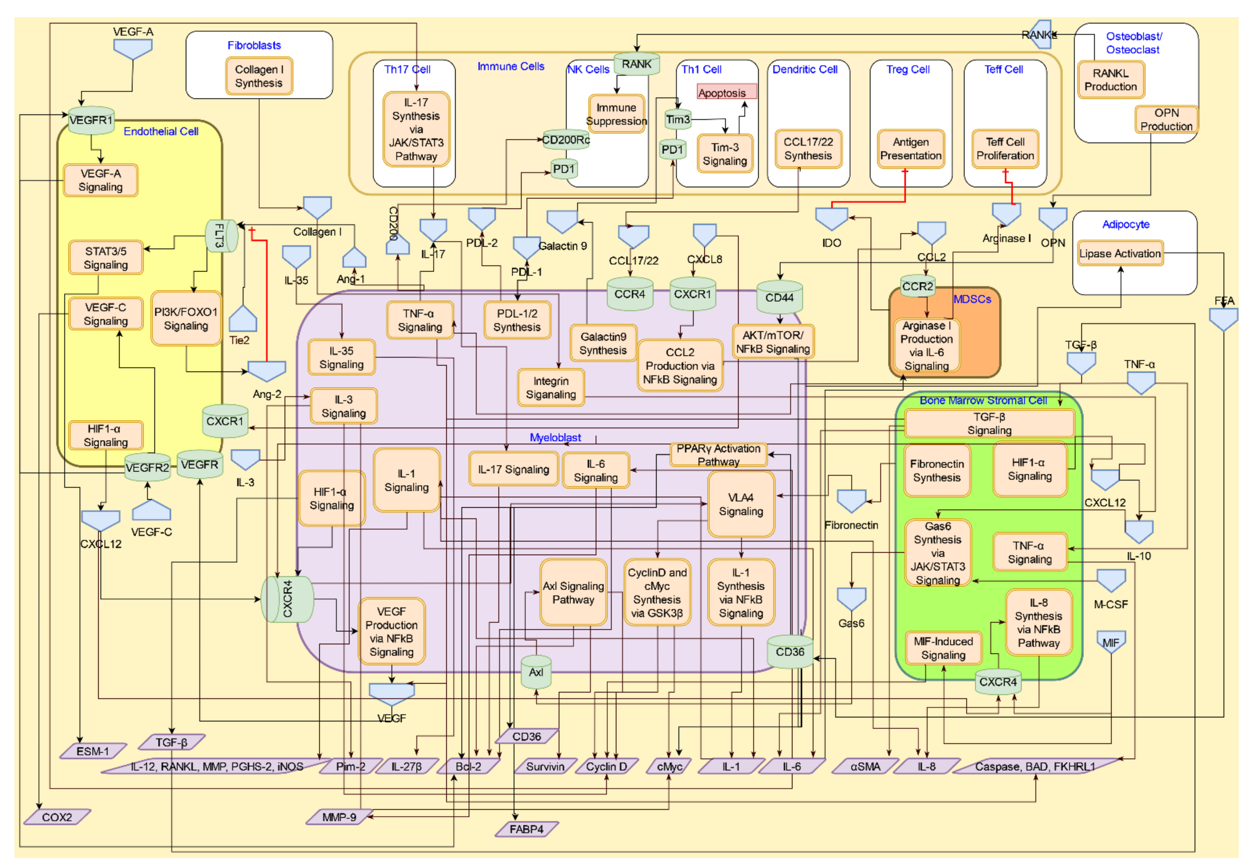
3. Molecular Systems Architecture of AML

From the systems biology perspective, living organisms can be viewed as being comprised of dynamic networks of biochemical reactions [20]. The origin of disease is characterized by the disruption of one or more signaling cascades, which may arise due to defects at the molecular level and may ultimately result in the symptomatic manifestation of disease, due to gain or loss in the usual functions of the cascades involved [26]. The integration of molecular pathways acts as a backbone for the development of a molecular systems architecture for a disease [21]. In complex diseases, there are numerous cells involving different signaling cascades. In such cases, an integration of molecular pathway systems affecting these cell types results in a systems view of the disease or biological process.
In Figure 2, we schematically illustrate a multilayered architecture of the AML microenvironment, with (i) an interconnected system of pathways in immune cells, endothelial cells, bone marrow stromal cells (BMSC), and myeloid-derived suppressor cells (MDSCs); (ii) converging points of key signaling pathways in the microenvironment, among AML cells, endothelial cells, BMSCs, MDSCs, and immune cells (interactive signaling layer); and (iii) the potential impact of such convergent pathways on the progression of AML (disease layer).
In AML, mutated leukemia stem cells (LSC) exploit the normal microenvironment and alter it to maintain their survival [27]. Alterations in the AML microenvironment can lead to AML relapse due to anti-apoptotic, anti-differentiation, and proliferative effects [28]. Stromal cells have a primary role in initiating AML, resulting in AML cells altering the normal localization and differentiation of HSCs as well as rapid leukemia growth expanding the intrinsically hypoxic microenvironment [29]. The microenvironment in AML consists of immune cells, stromal cells, and stem cells. Growth factors and cytokines released in the bone marrow (BM), thymus, and other immune tissue microenvironments provide paracrine and autocrine signals for long-term hematopoietic regulation of stem cells [30] and protect the AML cells from chemotherapeutic agents to promote drug resistance [31,32,33]. AML cells evade the immune cells by arresting the cell cycle of cytotoxic T cells, inducing cytotoxicity in NK cells and Th1 cells via tryptophan starvation [34,35].

4. Interactive Signaling in the AML Microenvironment

The AML cells interact with the stromal cells to effect immunosuppression, immunoevasion, and survival/proliferation through promotion of inflammatory phenotypes in T cells, suppression of anti-inflammatory T cell phenotypes, and enhanced angiogenesis via a myriad of signaling transduction mechanisms. The signaling molecules that affect these processes can originate either from the leukemic cell or from the proinflammatory immune cells and other stromal cells; hence, they are important in developing a molecular systems architecture. CytoSolve’s bioinformatics process yields the schematic of the interactive signaling in the tumor microenvironment, as shown in Figure 3. Table 3 provides the legend describing the various graphical components of the systems architecture.

4.1. Interactive Crosstalk between AML Cells, Bone Marrow Stromal Cells, Endothelial Cells, Osteoblasts, and Adipocytes

4.1.1. CXCR4/CXCL12 Signaling

The CXCR4/CXCL12 axis regulates retention of HSC quiescence, survival, and the size of the HSC pool in the marrow. It is also implicated in cellular migration, mobilization, and homing of LSCs during the initiation and progression of AML [36]. CXCR4 is a G protein-coupled chemokine receptor expressed on the surface of HSC and AML cells [29]. CXCR4 is essential for metastatic spread to organs and thereby allows tumor cells to access cellular niches, such as the bone marrow, that favor tumor-cell survival and growth. CXCL12 produced by the BMSCs, endothelial cells, osteoblasts, osteoclasts, and MSCs is a homeostatic chemokine that signals through CXCR4 and plays an important role in hematopoiesis and the development and organization of the immune system [37].
High levels of CXCL12 in hypoxic condition in the bone marrow niche indicate a regulator for the transcription factor, hypoxia inducibleg factor-1 (HIF-1) [38]. HIF-1α, in particular, is responsible for creating a concentration gradient of CXCL12 that guides malignant cell to the bone marrow niche and has been shown to upregulate the expression of CXCR4 on malignant cells [39]. Under normoxic conditions, HIF-1α is hydrolyzed by the prolyl hydroxylase domain protein (PHD), which leads to its ubiquitination [40]. The function of PHD is catalyzed by IDH and mutations in IHD have been shown to increase the accumulation of HIF-1α [40,41]. These results indicate that mutation in IHD may cause diminishing activity of IHD, leading to downregulation of PHD activity and higher levels of HIF-α. Another mutated gene in AML, FLT3-ITD, also has been shown to upregulate the translation of HIF-1α [41]. Mutation in FLT3-ITD leads to activation of FLT3 signaling [40], which upregulates the PI3K/AKT/mTOR pathway responsible for translation of HIF-1α [41,42]. These observations indicate a link between AML oncogenes IDH and FLT3-IDT and upregulation of the HIF-1α-induced CXCR4/CXCL12 axis signaling.
Secretion of functional CXCL12 from human BMSCs is a contact-dependent event mediated by connexin-43 and connexin-45 gap junctions [43]. The binding of CXCL12 to CXCR4 leads to activation of the PI3K/Akt and MAPK pathways that mediate the survival and proliferation of AML cells. CXCL12 also activates the NF-κB pathway, which induces the production of soluble factors, such as matrix metalloproteinases (MMPs), IL-8, and VEGF, leading to the angiogenesis promoted by MMPs and VEGF, and drug resistance initiated by IL-8 [32]. These soluble factors help degrade the extracellular matrix and induce blood vessel formation [37].
CXCL12 derived from MSCs has been shown to induce production of autophagy proteins such as ATG1, ATG5, and LC3 in the AML cells, which allows the AML cell to survive under stress [34]. MSCs-derived CXCL12 also upregulated the expression of the drug resistance protein P-glycoprotein (P-gp) via the PI3K/Akt/p38-MAPK pathway in the AML cells [44]. The ubiquitous nature of the CXCL12/CXCR4 axis in the AML microenvironment makes it a prime target for anticancer therapies [45]. Several therapeutics are under development, including those that inhibit or downregulate the expression of CXCR4 [46], inhibit the binding of CXCL12 to CXCR4 [47], and prevent the binding of CXCL12 to CXCR4 [48].
In the bone marrow niche, osteoblasts and osteoclasts lining the endosteum regulate bone formation and resorption [49]. During leukemogenesis, AML cells migrate to the bone marrow niche, due to the CXCL12 gradient created by osteoblasts and osteoclasts, [50] and evade detection [49].
The CXCR4/CXCL12 signaling pathway’s interactions across the AML cell, endothelial cell, osteoblasts/osteoclast, MSC, and the BMSC are shown in Figure 4.

4.1.2. TGF-β Signaling

The multifunctional TGF-β regulates cell proliferation, survival, and apoptosis [51]. The three major mammalian TGF-β isoforms are TGF-β1, TGF-β2, and TGF-β3. TGF-β1 is the most abundant, universally expressed isoform. Once activated, the TGF-β ligands regulate cellular processes by binding to two ubiquitously expressed, high-affinity cell-surface receptors—type I receptor (TβRI) and type II receptor (TβRII)—both of which contain a serine/threonine protein kinase in their intracellular domains. Once bound to TGF-β, TβRII recruits, binds, and phosphorylates TβRI, thereby stimulating its protein kinase activity [52]. The activated TβRI then recruits and phosphorylates the receptor-activated transcription factors, Smad2/3, which then bind to the common Smad4, translocate into the nucleus, and interact in a cell-specific manner with transcription factors, coactivators, and corepressors to regulate the transcription of TGF-β-responsive genes [53]. The TGF-β signaling interactions across the AML cell, endothelial cell, and the BMSC, are shown in Figure 5.
TGF-β1 stimulates the secretion of IL-6 by BMSC and VEGF by AML cells, which in turn promotes the survival of AML cells and angiogenesis, respectively [54]. The TGF-β–Smad pathway is also known to induce the production of the extracellular matrix component fibronectin and the expression of integrin receptors in tumor cells, which facilitate cell adhesion and the cell-to-cell interaction of tumor cells with the extracellular matrix of BMSCs [51]. TGF-β1 induces expression of the chemokine receptor CXCR4 through activation of Smad2/3 [55]. CXCR4 is highly expressed in AML, and the interactions between CXCR4 and its ligand CXCL12, constitutively secreted by BMSCs and MSCs, promote the proliferation, survival, migration, and homing of cancer cells [36]. TGF-β1-triggered nuclear translocation of Smad2/3 regulates IL-6 and αSMA transcription, whereas HIF-1α translocation regulates VEGF and TGF-β1 transcription [56,57]. BMSC-derived TGF-β1 also induces the expression of aldehyde dehydrogenase-2 (ALDH2) via the non-canonical TGF-β-p38-ALDH2 pathway [58]. ALDH2 is implicated in conferring AML cells with drug resistance to chemotherapy [58].
The role of AML cells and the pro-angiogenic factor VEGF secreted by AML cells in promoting angiogenesis is dependent on the microenvironmental niche (e.g., the bone marrow niche, the vascular niche, etc.) and the progression of the disease. In the bone marrow niche, even though angiogenesis occurred, the resulting blood vessels were shown to be abnormal, leading to toxic levels of nitric oxide (NO)/reaction oxygen species (ROS) production and resulting in vascular regression [59]. In addition, as the disease progresses, the failed vasculature allows the AML cells to maintain low oxygen levels and to evade the chemotherapeutics, both of which are carried through blood [59]. Thus, unlike the solid tumors, the presence of VEGF in AML may not lead to angiogenesis of a functional vasculature.

4.1.3. RANK/RANKL and Osteopontin Signaling in Osteoblasts/Osteoclasts

The RANK/RANKL pathway governs bone remodeling. The receptor RANK is on the surface of osteoclasts, and its ligand RANKL is expressed on the membranes of osteoblasts, and also secreted by activated lymphocytes and AML cells [60]. The binding of RANKL to RANK initiates osteoclastogenesis and increases the survival of osteoclasts [61]. RANK is also expressed on natural killer (NK) cells. RANKL expressed in AML cells binds with RANK on NK cells to compromise their anti-leukemic activity [60].
Osteopontin (OPN), an extracellular matrix protein expressed on osteoblasts and osteoclasts, was found to be increased in the serum of patients’ AML [62]. OPN promotes the survival and proliferation of AML blasts through its binding to CD44 on the AML cell surface, which subsequently initiates the AKT/mTOR/NF-κB signaling pathways [62,63]. The RANK/RANKL and OPN/CD44 pathways are shown in Figure 6.
Adipose tissue, which accounts for up to 70% of the bone marrow, acts as a reservoir for HSCs and progenitor cells [64]. Bone marrow adipocytes have been shown to support the survival and proliferation of AML cells in vivo and in vitro [65]. AML blasts induce the activation of lipolysis in the adipocytes by promoting phosphorylation of the lipase, leading to free fatty acid release [65]. Lipolysis is initiated by activation of the β-adrenergic receptor [65], leading to stimulation of hormone-sensitive lipase (HSL) in the presence of AML blasts [65,66].
The free fatty acids released by adipocytes are internalized by the CD36 receptor on the AML cells and subsequently transferred to the nucleus and mitochondria by the intracellular lipid chaperone fatty acid binding protein 4 (FABP4). In the nucleus, the free fatty acid activates the transcription factor PPARγ, which induces the transcription of fat transport-associated genes, such as CD36 and FABP4, and the anti-apoptotic gene BCL2 [67]. In the mitochondria, fatty acids are used as a source of energy via the metabolic activation of fatty acid oxidation (FAO) and oxidative phosphorylation [67]. The interactions between adipocytes and AML cell are illustrated in Figure 7.

4.2. Interactive Crosstalk Signaling between AML Cells and Endothelial Cells via Adhesion Molecules

Interactions between adhesion molecules, such as VCAM-1 and E-selectin, on endothelial cells and their ligands, expressed on HSCs/AML cells and the marrow niche, mediate the retention of HSCs and AML cells within the bone marrow niche [68]. Very late antigen-4 (VLA-4), also known as integrin α4β1, is a heterodimer expressed on leukocytes and variably on AML blasts [69]. In addition, In addition, VCAM-1, which is a ligand for VLA-4 on AML cells, is also expressed by osteoblasts and endothelial cells [70]. Under normal circumstances, CXCL12 stimulation results in the activation of VLA-4 on HSCs, leading to activation of the VLA-4/VCAM-1 signaling pathway, and enhancement of HSC adhesion to the endothelial cells followed by their trans-endothelial migration [71]. Interactions of AML cells with endothelial cells and their subsequent integration and proliferation in the vascular niche are facilitated by the VLA-4/VCAM-1 axis [29].
The VCAM1-VLA-4 signaling, as illustrated in Figure 8, promotes cell survival and proliferation by interfering with the activation of receptor tyrosine kinase (RTK) [72]. A main structural and signaling protein, integrin-linked kinase (ILK), binds with VLA-4. ILK forms multiprotein complexes with several key components involved in intracellular signaling cascades [73]. ILK kinase activity is dependent on PI3K and requires binding of phosphatidylinositol (3,4,5)-trisphosphate (PIP3). Next, glycogen synthase kinase-3β (GSK3β) is phosphorylated by ILK at serine 9 residue, leading to the activation of the activator protein 1 (AP-1), which then upregulates cyclin D1 and Myc-1 [74]. Thus, the VCAM1-VLA-4 signaling pathway plays a critical role in the survival and proliferation of leukemic cells.
CXCL12/CXCR4 signaling in AML cells activates the NF-κB pathway, which induces the production of MMPs and VEGF, leading to angiogenesis [32]. These soluble factors help degrade the extracellular matrix and induce blood vessel formation [37]. In the endothelial cells, VEGF signaling leads to glycolysis-mediated vascular remodeling [58]. Targeting VEGF receptor-2 with a tyrosine kinase inhibitor resulted in AML cytotoxicity, due to inhibition of VEGF-induced survival signaling and vascular remodeling in the tumor microenvironment [34].
E-selectin, a cell adhesion molecule, regulates the rolling of leukocytes over endothelial cells [75]. Its ligand, E-selectin ligand-1 (ESL-1), is found on HSCs as well as AML blasts [76]. E-selectin directly regulates disease progression and chemoresistance in AML [77]. AML blasts’ survival is enhanced by their adhesion to the vascular niche via ESL-1, which activates Wnt signaling [78]. Inhibition of E-selectin binding to AML blasts augmented chemotherapeutic effect and lowered the vascular niche-mediated survival of AML blasts [76]. Recently, CD162 has emerged as another E-selectin ligand that is implicated in the chemoresistance of AML [77].

4.3. Interactive Signaling with Myeloid-derived Suppressor Cells (MDSCs) in AML

MDSCs are critical to the immunosuppressive characteristic of the tumor microenvironment and are involved in promoting immune tolerance and disease growth. AML patients show significant increase in MDSCs in the circulation, and leukemic blasts directly induce the expansion of MDSCs [79]. MDSCs are the nonmalignant immature myeloid cells, whereas AML blasts are a malignant expansion of immature myeloid cells; both have the ability to suppress immune cells [80]. A key factor that characterizes the interactions between an AML cell and MDSC is the oncogene MUC1. The expression of MUC1 on the leukemic blasts and leukemia-initiating cells induces MDSC expansion in the microenvironment. The silencing of MUC1 has been shown to reduce the capacity of AML blasts to induce MDSC expansion in the tumor microenvironment [79]. Interactions of MDSC with the AML blasts and immune cells are illustrated in Figure 9.
Immunosuppression by MDSCs and AML blasts is regulated through several mechanisms. One of the key mechanisms includes immunosuppression by the enzyme arginase present in both MDSCs and AML blasts [81]. MDSCs express arginase I and AML blasts express arginase II. The two isoforms of arginase are likely to have resulted from a gene duplication event during evolution, with arginase I located in the cytosol and arginase II in the mitochondria [82]. Both arginase isoforms convert arginine into urea. The two enzymes catalyze the same reaction, converting arginine into ornithine, with urea as a byproduct. In healthy individuals, arginase I is expressed predominantly by hepatocytes, whereas arginase II is expressed in a more diverse range of organs [82].
AML blasts express and release arginase II to suppress T-cell proliferation via depletion of L-arginine in the microenvironment [83]. The combination of increased intracellular arginase activity and plasma arginase activity halts T-cell proliferation and contributes to the lymphopenia. In addition, arginase II levels and activity serve as important biomarkers in patients with AML. The measurement of arginase II levels acts as a biomarker for minimal residual disease. AML blasts polarize neighboring monocytes to an immunosuppressive M2-like phenotype [81].
MDSCs also synthesize indoleamine-pyrrole 2,3-dioxygenase (IDO), a tryptophan-degrading enzyme, and contribute to immune tolerance by mediating T cell suppression. IDO locally depletes tryptophan and generates tryptophan metabolites, including kynurenine, resulting in reduced proliferation of CD4+ T cells, CD8+ T cells, and natural killer (NK) cells [84]. ROS secretion also contributes to the immunosuppressive action of MDSC and is caused by the increase in NADPH oxidase activity in granulocytic MDSC [85]. A subset of MDSCs deplete cysteine as an alternate mechanism of immune suppression. MDSCs also modulate the surrounding macrophages and dendritic cells [86].
The post-translational addition of a palmitate to a protein creates greater affinity for non-polar structures, such as lipid bilayers, and are critical to the functioning of normal as well as cancer cells [87]. In AML cell-derived extracellular vesicles, carrying palmitoylated proteins leads to monocytes differentiating into MDSCs. This process is regulated by TLR-2 signaling, leading to upregulation of cEBPβ and IL-10 expression [34].

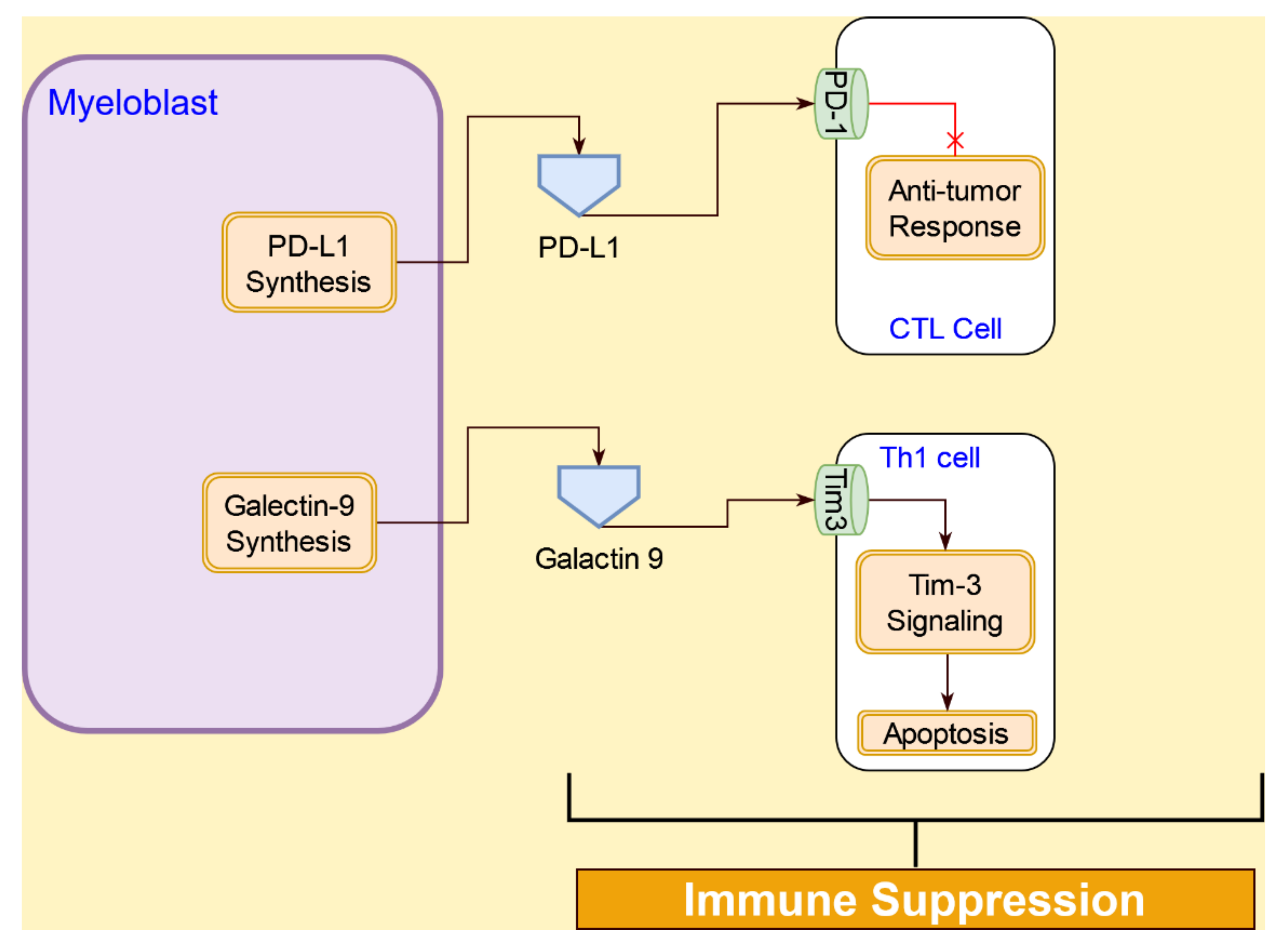
4.4. Interactive Signaling AML Cells and Immune Cells

In the AML microenvironment, the AML cells directly interact with several immune cells, including Treg cells, NK cells, Th-1 cells, dendritic cells (DC), and T effector (Teff) cells [88]. The interactions between AML cells and the immune cells are described below.

4.4.1. Interferon α Signaling

Interferon-α (IFN-α), a type I IFN, has been previously used as an antitumor agent [89]. Type I IFNs exert direct antitumor effects on AML cells by multiple mechanisms modulated via the expression of interferon-inducible genes. IFN-α inhibits the production of pro-proliferative cytokines such, as IL-1 and IL-6, and pro-angiogenic cytokine IL-8 [89]. IFN-α also promotes the expression of FasL in AML cells, which initiates apoptotic signaling via caspase-8 [90]. IFN-α is also implicated in the activation of DCs, NK cells, and T cells, which in turn play major roles in antitumor immune responses [91].
Mutation in cohesin complex proteins, seen in AML cells, downregulates Type I IFNs in macrophages [92]. Type I IFNs are critical to the initiation of antitumor immunity through direct actions on DCs. IFNα induces DCs to exert direct cytotoxic activity against AML cells [93]. IFN-α has an important role in modulating NK cell function [91]. IFN-α upregulates the expression of immunomodulatory cytokines, such as IFN-γ in the NK cells, promoting their “helper” function. The helper NK cells induce the DCs with Th-1-polarizing capacity, which is necessary for antitumor immunity [89]. Activation of the immune system by interferons has been shown to undermine AML cell growth [11,89,93] The IFN-α signaling interactions are illustrated in Figure 10.

4.4.2. Immunosuppressive Interactions of Tregs in AML Tumor Microenvironment

Tregs play a pivotal role in maintaining peripheral immunological tolerance by preventing autoimmunity and chronic inflammation. There are two subtypes of Tregs: naturally occurring Tregs (nTregs) and induced Tregs (iTregs) [94]. iTregs start out as CD4+ cells and acquire CD25 and FoxP3 expression following adequate antigenic stimulation in a specific tolerogenic microenvironment [95]. AML patients with a higher expression of IDO critically induce a de novo population of Foxp3+ Tregs [96]. AML cells have also been shown to actively recruit and program Treg to suppress antitumor immune responses [97].
AML cells promote the expansion of Tregs via the inducible T-cell costimulator ligand (ICOSL). TNF-α signaling induces ICOSL expression in AML cells [98]. The accumulation of Tregs in the AML microenvironment is driven by the chemotactic effect of CCL2 [88]. Once established in the microenvironment, Tregs actively prevent or downregulate antitumor responses from the immune cells in the tumor microenvironment. Tregs suppress the immune response from Teffs through two mechanisms: a contact-dependent manner, and a contact-independent manner. While nTregs use both mechanisms, iTregs induce immunosuppression in a contact-independent manner that involves cytokines, such as IL-4, IL-10, or TGF-β [97,98]. In addition to suppressing APCs, IL-10 also promotes AML cell proliferation via the ERK/p38/STAT3 pathway [98]. Treatment with an FLT3 inhibitor, midostaurin, showed a significant decrease in the Treg population, reduction in the FOX3p mRNA expression in AML cells, and reduction in IL-10 levels, indicating a role for IL-10 as a potential biomarker for AML cancer treatment [99].
Direct cell-to-cell interactions between Tregs and Teffs result in apoptosis and/or suppression of Teffs. The direct transfer of cAMP from Tregs to Teff through the gap junctions leads to downregulation of IL-2 production and subsequent proliferations of Teff [100]. On contact, the formation of gap junctions occurs between Tregs and Teffs. cAMP transferred through the gap from Tregs to Teffs suppresses the proliferation of Teffs by decreasing IL-2 production.
Apoptosis of CD4+CD25+ Teffs is caused by Tregs through the granzyme B-dependent and perforin-independent mechanisms [101]. Tregs also suppress NK-cell proliferation via depletion of IL-2 [101]. Tregs block the maturation of DCs. The immature DCs express IDO, which depletes tryptophan needed for T-cell proliferation [102]. Metabolites resulting from IDO depletion of tryptophan, such as kynurenines, actively promote T-cell apoptosis [97]. The immunosuppression by AML cells through Tregs is illustrated in Figure 11.

4.4.3. Immunosuppression Interactions of AML Cells with T Cells

The programmed death-1 (PD-1) receptor is expressed on various cell types, including T cells [103]. In AML, an increased expression of PD-1 receptor is observed in cytotoxic T lymphocytes (CTL). PD-L1, a ligand for the PD-1 receptor, is present on cancer cells, including AML [104]. PD-1 and PD-L1 interaction suppresses CTL response to AML blasts [97]. PD-L1 expression was found to be higher in patients undergoing chemotherapy or those who have a relapse, suggesting a refractory role for PD-1/PD-L1 interaction [105].
AML blasts also participate in the suppression of Th cells through Tim-3 and galactin-9 (gal-9) interactions [106]. A type I membrane glycoprotein, Tim-3 is expressed on Th1 cells and innate immune cells [107]. Gal-9 is expressed on AML cells and participates in the Tim-3/gal-9 pathway that leads to apoptosis of Th1 cells [108]. In addition, the Tim-3/gal-9 pathway, along with the PD-1/PD-1 ligand pathway, is involved in regulating CD8+ CTL responses [109]. The interactions between AML blasts and Th1, as well as CD8+ CTLs, are illustrated in Figure 12.

4.4.4. Interactive Signaling with Natural Killer (NK) Cells in AML

NK cells are lymphocytes from the innate immune system. NK cells can directly eliminate tumor cells via their cytotoxic and cytokine-secreting capacity and indirectly contribute to tumor control by communicating with DC and other immune cells, supporting the development of an efficient adaptive antitumor immune response [110].
NK cells mediate their antitumor activity through the expression of several chemokine receptors, such as CCR1, CCR4, CCR6, CCR7, CXCR1, CXCR3, CXCR4, CXCR6, and CX3CR1 [111]. Many of the ligands for these NK cell receptors are constitutively released by AML cells, including the chemokines within the CCL2–4/CXCL1/8 cluster found in most AML patients, indicating an expected migration of NK cells towards the AML blasts [112]. However, AML cells are able to evade NK cell immune surveillance through a number of mechanisms. The interactions between AML cells and NK cells are illustrated in Figure 13.
A dysfunctional antitumor immune response by the NK cell could result in NK cell abnormalities, immuno-suppressive and immuno-evasive properties of AML target cells, and preferential interactions with other immune cells rather than AML blasts [113]. In AML, changes in the expression of both receptors and ligands are commonly found, which substantially impair NK cell-mediated killing. The majority of AML patients have a downregulated NK cell surface expression of the activating natural cytotoxicity receptors; thus, AML cells evade NK cells’ mediated killing by the lowered or absent expression of surface ligands (e.g., CD48, NKG2DL, etc.) for various NK cell activating receptors [114]. NK cells can also be inactivated by soluble inhibitory factors, such as TGF-β, and reactive oxygen species secreted by AML blasts [115,116]. However, patients with AML-ETO-positive AML have a better prognosis, as AML-ETO has been shown to upregulate the NK cell ligand CD48. This allows NK cells to perform cell-mediated killing of AML cells [117].
Another mechanism for AML cells to escape from NK cells is the activation of the aryl hydrocarbon receptor (AHR) pathway in the NK cells [118]. IDO, which is highly expressed in AML blasts compared to normal cells [91], initiates the AHR activation by kynurenine. The active AHR binds to the AHR receptor on naïve NK cells and leads to the expression of miR-29/b1, which blocks the NK cell differentiation [118], thereby allowing the AML cells to escape from the NK cells. Inhibition of AHR has been shown to restore the NK cell-mediated killing of AML cells [118], indicating a potential role for AHR as a therapeutic target.

5. Discussion

The CytoSolve systematic bioinformatics review identified critical molecular systems components of AML pathogenesis. The organization of these components into a molecular systems architecture is presented in Figure 14. The first layer, starting from the bottom of Figure 14, represents cellular components of the stromal microenvironment: fibroblast, MSCs, endothelial cells, BMSCs, MDSCs, and immune cells. The second layer, in the middle of Figure 14, represents the key molecular interactions implicated in the pathogenesis of AML: collagen synthesis and TGF-β signaling in fibroblasts; CXCL12 signaling and IL-8 signaling in MSCs; VLA4 signaling, VEGF signaling, and hypoxia signaling in endothelial cells; CXCL12 signaling in BMSCs; CD36/FABP4/PPARγ signaling and the FAO energy metabolic pathway in adipocytes; the OPN production pathway and the RANKL production pathway in osteoblast/osteoclast; arginase signaling and CCL2 signaling in MDSCs; and PD-L1 signaling, IDO signaling, Tim-3 signaling, and IL-17 signaling in immune cells. The third layer, shown at the top of Figure 14, represents the biological processes implicated in the pathogenesis of AML: angiogenesis; cell proliferation, cell survival (inhibition of apoptosis), and immune suppression.
The molecular systems architecture in Figure 14 provides a consolidated guide to understanding the overall AML pathogenesis. Interactions among the nine cell types in the bottom layer give rise to the sixteen molecular systems presented in the middle layer. Of these sixteen molecular systems components, eight of them (collagen synthesis, TGF-β signaling, CXCL12 signaling, IL-8 signaling, the OPN production pathway, VLA4 signaling, VEGF signaling, and hypoxia signaling) contribute to AML pathogenesis by promoting cell proliferation and cell survival/inhibition of apoptosis. The remaining eight molecular systems components (arginase signaling, CCL2 signaling, PD-L1 signaling, IDO signaling, Tim-3 signaling, TLR-2 signaling, the AHR pathway, and RANK/RANKL signaling) contribute to AML pathogenesis by promoting immune evasion and suppression. The integrated processes of cell proliferation, cell survival, and immune suppression, driven by the molecular subsystems and the respective cellular interactions, give rise to AML.
The architecture also may offer a vehicle for new insights and discovery. For example, we have identified several targets across different cell types in the microenvironment that can potentially be used to develop therapeutic interventions to inhibit suppression of immune response, inhibit cell proliferation, and promote cancer cell apoptosis, as listed in Table 4.
Efforts are already underway to develop antibody conjugates for cell surface markers, such as CD44, CLL-1, CD34, and Tim-3 [119,120], and insights from this architecture can advance such efforts by the identification of new targets and understanding the mechanisms of action of new single and combination therapies based on their interactions with the targets.

6. Future Directions

Mechanistic in silico modeling is emerging as a valuable pre-clinical drug discovery tool. Molecular systems architecture, as presented in this study, provides a starting point for such mechanistic in silico modeling efforts. The computational capabilities of CytoSolve can be employed to create an integrative computational model for the AML stromal microenvironment. The resulting in silico AML stromal microenvironment model can then be used as a testing and validation platform to identify new targets and novel combination therapies.

7. Conclusions

The molecular systems architecture developed in this review provides a blueprint for understanding the complex interactions occurring in the AML microenvironment. This understanding will enable the identification of targets in the interactive signaling pathways that may be used to develop novel combination therapies and synthetic approaches that may be more effective than the current therapeutic options and may potentially mitigate undesirable side effects. The molecular systems architecture provides a versatile tool in identifying how targeting a particular mechanism in a stromal cell can a have either a positive or negative cascading effect on the rest of the stromal microenvironment, thereby providing a much better drug development paradigm that can minimize side effects and maximize efficacy of treatment. This architecture may also be converted to an open science interactive web-based tool to enable ongoing collaborative development by the AML research community. Such efforts have been done before in the field of human knee osteoarthritis [121].

Author Contributions

Conceptualization, V.A.S.A. and K.M.S.; methodology, V.A.S.A. and P.D.; software, V.A.S.A. and P.D.; validation, V.A.S.A., K.M.S., K.G.M. and P.D.; formal analysis, V.A.S.A. and P.D.; investigation, V.A.S.A., K.M.S., K.G.M. and P.D.; resources, V.A.S.A. and K.M.S.; data curation, V.A.S.A. and P.D.; writing—original draft preparation, V.A.S.A. and P.D.; writing—review and editing, V.A.S.A., K.M.S., K.G.M. and P.D.; visualization, V.A.S.A. and P.D.; supervision, V.A.S.A., K.M.S. and K.G.M.; project administration, V.A.S.A. and P.D.; funding acquisition, K.M.S. All authors have read and agreed to the published version of the manuscript.

Funding

This research received no external funding.

Acknowledgments

K.M.S. is supported by the Leukemia and Lymphoma Society grant number 6587-20, Pediatric Cancer Research Foundation grant number 597630, Hyundai Hope on Wheels grant number 587744, and Cure Childhood Cancer grant number 641095.

Conflicts of Interest

K. McLure is an employee of Ermaris Bio; V.A.S. Ayyadurai, and P. Deonikar are employees of CytoSolve, Inc.; K. Sakamoto has no potential conflict of interests.

References

  1. Scholl, C.; Gilliland, D.G.; Fröhling, S. Deregulation of signaling pathways in acute myeloid leukemia. Semin. Oncol. 2008, 35, 336–345. [Google Scholar] [CrossRef] [PubMed]
  2. Sakamoto, K.M.; Grant, S.; Saleiro, D.; Crispino, J.D.; Hijiya, N.; Giles, F.; Platanias, L.; Eklund, E.A. Targeting novel signaling pathways for resistant acute myeloid leukemia. Mol. Genet. Metab. 2015, 114, 397–402. [Google Scholar] [CrossRef] [PubMed] [Green Version]
  3. Abboud, C.N. Another nail in the AML coffin. Blood 2009, 113, 6045–6046. [Google Scholar] [CrossRef] [PubMed]
  4. Steffen, B.; Müller-Tidow, C.; Schwäble, J.; Berdel, W.E.; Serve, H. The molecular pathogenesis of acute myeloid leukemia. Crit. Rev. Oncol. Hematol. 2005, 56, 195–221. [Google Scholar] [CrossRef] [PubMed]
  5. Thomas, D.; Majeti, R. Biology and relevance of human acute myeloid leukemia stem cells. Blood 2017, 129, 1577–1585. [Google Scholar] [CrossRef]
  6. Hanekamp, D.; Cloos, J.; Schuurhuis, G.J. Leukemic stem cells: Identification and clinical application. Int. J. Hematol. 2017, 105, 549–557. [Google Scholar] [CrossRef]
  7. Schonherz, A.A.; Bødker, J.S.; Schmitz, A.; Brøndum, R.F.; Jakobsen, L.H.; Roug, A.S.; Severinsen, M.T.; El-Galaly, T.C.; Jensen, P.; Johnsen, H.E.; et al. Normal myeloid progenitor cell subset-associated gene signatures for acute myeloid leukaemia subtyping with prognostic impact. PLoS ONE 2020, 15, e0229593. [Google Scholar] [CrossRef]
  8. Boulais, P.E.; Frenette, P.S. Making sense of hematopoietic stem cell niches. Blood 2015, 125, 2621–2629. [Google Scholar] [CrossRef] [Green Version]
  9. Miraki-Moud, F.; Anjos-Afonso, F.; Hodby, K.A.; Griessinger, E.; Rosignoli, G.; Lillington, D.; Jia, L.; Davies, J.K.; Cavenagh, J.; Smith, M.; et al. Acute myeloid leukemia does not deplete normal hematopoietic stem cells but induces cytopenias by impeding their differentiation. Proc. Natl. Acad. Sci. USA 2013, 110, 13576–13581. [Google Scholar] [CrossRef] [Green Version]
  10. Hérault, A.; Binnewies, M.; Leong, S.; Calero-Nieto, F.J.; Zhang, S.Y.; Kang, Y.A.; Wang, X.; Pietras, E.M.; Chu, S.H.; Barry-Holson, K.; et al. Myeloid progenitor cluster formation drives emergency and leukaemic myelopoiesis. Nature 2017, 544, 53–58. [Google Scholar] [CrossRef]
  11. Cuartero, S.; Innes, A.J.; Merkenschlager, M. Towards a better understanding of cohesin mutations in AML. Front. Oncol. 2019, 9, 867. [Google Scholar] [CrossRef] [PubMed] [Green Version]
  12. Renneville, A.; Roumier, C.; Biggio, V.; Nibourel, O.; Boissel, N.; Fenaux, P.; Preudhomme, C. Cooperating gene mutations in acute myeloid leukemia: A review of the literature. Leukemia 2008, 22, 915–931. [Google Scholar] [CrossRef] [PubMed] [Green Version]
  13. Conway O’Brien, E.; Prideaux, S.; Chevassut, T. The epigenetic landscape of acute myeloid leukemia. Adv. Hematol. 2014, 2014, 103175. [Google Scholar] [CrossRef] [PubMed] [Green Version]
  14. Dombret, H. Gene mutation and AML pathogenesis. Blood 2011, 118, 5366–5367. [Google Scholar] [CrossRef]
  15. Kavianpour, M.; Ahmadzadeh, A.; Shahrabi, S.; Saki, N. Significance of oncogenes and tumor suppressor genes in AML prognosis. Tumor Biol. 2016, 37, 10041–10052. [Google Scholar] [CrossRef]
  16. Staudt, D.; Murray, H.C.; McLachlan, T.; Alvaro, F.; Enjeti, A.K.; Verrills, N.M.; Dun, M.D. Targeting oncogenic signaling in mutant FLT3 acute myeloid leukemia: The path to least resistance. Int. J. Mol. Sci. 2018, 19, 3198. [Google Scholar] [CrossRef] [Green Version]
  17. Walasek, A. The new perspectives of targeted therapy in acute myeloid leukemia. Adv. Clin. Exp. Med. 2019, 28, 271–276. [Google Scholar] [CrossRef]
  18. Ayyadurai, V.A.S.; Deonikar, P. Bioactive compounds in green tea may improve transplant tolerance: A computational systems biology analysis. Clin. Nutr. ESPEN 2021, 46, 439–452. [Google Scholar] [CrossRef]
  19. Oketch-Rabah, H.A.; Hardy, M.L.; Patton, A.P.; Chung, M.; Sarma, N.D.; Yoe, C.; Ayyadurai, V.A.S.; Fox, M.A.; Jordan, S.A.; Mwamburi, M.; et al. Multi-Criteria decision analysis model for assessing the risk from multi-ingredient dietary supplements (MIDS). J. Diet. Suppl. 2021, 18, 293–315. [Google Scholar] [CrossRef] [Green Version]
  20. Ayyadurai, V.A.S.; Dewey, C.F. CytoSolve: A scalable computational method for dynamic integration of multiple molecular pathway models. Cell. Mol. Bioeng. 2011, 4, 28–45. [Google Scholar] [CrossRef] [Green Version]
  21. Sweeney, M.D.; Ayyadurai, S.; Zlokovic, B.V. Pericytes of the neurovascular unit: Key functions and signaling pathways. Nat. Neurosci. 2016, 19, 771–783. [Google Scholar] [CrossRef] [PubMed]
  22. Koo, A.; Nordsletten, D.; Umeton, R.; Yankama, B.; Ayyadurai, S.; García-Cardeña, G.; Dewey, C.F. In silico modeling of shear-stress-induced nitric oxide production in endothelial cells through systems biology. Biophys. J. 2013, 104, 2295–2306. [Google Scholar] [CrossRef] [PubMed] [Green Version]
  23. Nordsletten, D.A.; Yankama, B.; Umeton, R.; Ayyadurai, V.A.S.; Dewey, C.F. Multiscale mathematical modeling to support drug development. IEEE Trans. Biomed. Eng. 2011, 58, 3508–3512. [Google Scholar] [CrossRef] [PubMed]
  24. Al-Lazikani, B.; Banerji, U.; Workman, P. Combinatorial drug therapy for cancer in the post-genomic era. Nat. Biotechnol. 2012, 30, 679–692. [Google Scholar] [CrossRef]
  25. Moher, D.; Liberati, A.; Tetzlaff, J.; Altman, D.G. Preferred reporting items for systematic reviews and meta-analyses: The PRISMA statement. BMJ 2009, 339, 332–336. [Google Scholar] [CrossRef] [Green Version]
  26. Berg, J.M.; Tymoczko, J.L.; Stryer, L. Defects in Signaling Pathways Can Lead to Cancer and Other Diseases. In Biochemistry; W H Freeman: New York, NY, USA, 2002. [Google Scholar]
  27. Schepers, K.; Campbell, T.B.; Passegué, E. Normal and leukemic stem cell niches: Insights and therapeutic opportunities. Cell Stem Cell 2015, 16, 254–267. [Google Scholar] [CrossRef] [Green Version]
  28. Cogle, C.R.; Saki, N.; Khodadi, E.; Li, J.; Shahjahani, M.; Azizidoost, S. Bone marrow niche in the myelodysplastic syndromes. Leuk. Res. 2015, 39, 1020–1027. [Google Scholar] [CrossRef]
  29. Rashidi, A.; DiPersio, J.F. Targeting the leukemia–stroma interaction in acute myeloid leukemia: Rationale and latest evidence. Ther. Adv. Hematol. 2016, 7, 40–51. [Google Scholar] [CrossRef]
  30. Su, Y.C.; Li, S.C.; Wu, Y.C.; Wang, L.M.; Chao, K.S.C.; Liao, H.F. Resveratrol downregulates interleukin-6-stimulated sonic hedgehog signaling in human acute myeloid leukemia. Evid.-Based Complement. Altern. Med. 2013, 2013, 547430. [Google Scholar] [CrossRef]
  31. Chang, Y.-T.; Hernandez, D.; Alonso, S.; Gao, M.; Su, M.; Ghiaur, G.; Levis, M.J.; Jones, R.J. Role of CYP3A4 in bone marrow microenvironment–mediated protection of FLT3/ITD AML from tyrosine kinase inhibitors. Blood Adv. 2019, 3, 908. [Google Scholar] [CrossRef]
  32. Chen, T.; Zhang, G.; Kong, L.; Xu, S.; Wang, Y.; Dong, M. Leukemia-Derived exosomes induced IL-8 production in bone marrow stromal cells to protect the leukemia cells against chemotherapy. Life Sci. 2019, 221, 187–195. [Google Scholar] [CrossRef] [PubMed]
  33. Thomas, C.M.; Campbell, P. FLT3 inhibitors in acute myeloid leukemia: Current and future. J. Oncol. Pharm. Pract. 2019, 25, 163–171. [Google Scholar] [CrossRef] [PubMed]
  34. Tohumeken, S.; Baur, R.; Böttcher, M.; Stoll, A.; Loschinski, R.; Panagiotidis, K.; Braun, M.; Saul, D.; Völkl, S.; Baur, A.S.; et al. Palmitoylated proteins on AML-derived extracellular vesicles promote myeloid-derived suppressor cell differentiation via TLR2/Akt/mTOR signaling. Cancer Res. 2020, 80, 3663–3676. [Google Scholar] [CrossRef] [PubMed]
  35. Leisch, M.; Greil, R.; Pleyer, L. IDO in MDS/AML disease progression and its role in resistance to azacitidine: A potential new drug target? Br. J. Haematol. 2020, 190, 314. [Google Scholar] [CrossRef]
  36. Zhang, Y.; Dépond, M.; He, L.; Foudi, A.; Kwarteng, E.O.; Lauret, E.; Plo, I.; Desterke, C.; Dessen, P.; Fujii, N.; et al. CXCR4/CXCL12 axis counteracts hematopoietic stem cell exhaustion through selective protection against oxidative stress. Sci. Rep. 2016, 6, 37827. [Google Scholar] [CrossRef] [Green Version]
  37. Sison, E.A.R.; Brown, P. The bone marrow microenvironment and leukemia: Biology and therapeutic targeting. Expert Rev. Hematol. 2011, 4, 271–283. [Google Scholar] [CrossRef] [Green Version]
  38. Schioppa, T.; Uranchimeg, B.; Saccani, A.; Biswas, S.K.; Doni, A.; Rapisarda, A.; Bernasconi, S.; Saccani, S.; Nebuloni, M.; Vago, L.; et al. Regulation of the chemokine receptor CXCR4 by hypoxia. J. Exp. Med. 2003, 198, 1391–1402. [Google Scholar] [CrossRef] [Green Version]
  39. Burger, J.A.; Kipps, T.J. CXCR4: A key receptor in the crosstalk between tumor cells and their microenvironment. Blood 2006, 107, 1761–1767. [Google Scholar] [CrossRef]
  40. Peng, G.; Liu, Y. Hypoxia-Inducible factors in cancer stem cells and inflammation. Trends Pharmacol. Sci. 2015, 36, 374. [Google Scholar] [CrossRef] [Green Version]
  41. Deynoux, M.; Sunter, N.; Hérault, O.; Mazurier, F. Hypoxia and hypoxia-inducible factors in leukemias. Front. Oncol. 2016, 6, 41. [Google Scholar] [CrossRef] [Green Version]
  42. Yazdani, Z.; Mousavi, Z.; Moradabadi, A.; Hassanshahi, G. Significance of CXCL12/CXCR4 Ligand/Receptor axis in various aspects of acute myeloid leukemia. Cancer Manag. Res. 2020, 12, 2155. [Google Scholar] [CrossRef] [PubMed] [Green Version]
  43. Schajnovitz, A.; Itkin, T.; D’Uva, G.; Kalinkovich, A.; Golan, K.; Ludin, A.; Cohen, D.; Shulman, Z.; Avigdor, A.; Nagler, A.; et al. CXCL12 secretion by bone marrow stromal cells is dependent on cell contact and mediated by connexin-43 and connexin-45 gap junctions. Nat. Immunol. 2011, 12, 391–398. [Google Scholar] [CrossRef] [PubMed]
  44. Li, M.; Meng, F.; Lu, Q. Expression profile screening and bioinformatics analysis of circRNA, LncRNA, and mRNA in acute myeloid leukemia drug-resistant cells. Turkish J. Hematol. 2020, 37, 104. [Google Scholar]
  45. Zhou, W.; Guo, S.; Liu, M.; Burow, M.E.; Wang, G. Targeting CXCL12/CXCR4 axis in tumor immunotherapy. Curr. Med. Chem. 2019, 26, 3026. [Google Scholar] [CrossRef] [PubMed]
  46. Muralidharan, R.; Panneerselvam, J.; Chen, A.; Zhao, Y.D.; Munshi, A.; Ramesh, R. HuR-targeted nanotherapy in combination with AMD3100 suppresses CXCR4 expression, cell growth, migration and invasion in lung cancer. Cancer Gene Ther. 2015, 22, 581–590. [Google Scholar] [CrossRef] [Green Version]
  47. De Clercq, E. AMD3100/CXCR4 Inhibitor. Front. Immunol. 2015, 6, 276. [Google Scholar] [CrossRef]
  48. Kuhne, M.R.; Mulvey, T.; Belanger, B.; Chen, S.; Pan, C.; Chong, C.; Cao, F.; Niekro, W.; Kempe, T.; Henning, K.A.; et al. BMS-936564/MDX-1338: A fully human anti-CXCR4 antibody induces apoptosis in vitro and shows antitumor activity in vivo in hematologic malignancies. Clin. Cancer Res. 2013, 19, 357–366. [Google Scholar] [CrossRef] [Green Version]
  49. Epstein, J.; Barlogie, B.; Johnson, C.L.; Yaccoby, S.; Pearse, R.N.; Choi, Y. Myeloma interacts with the bone marrow microenvironment to induce osteoclastogenesis and is dependent on osteoclast activity. Br. J. Haematol. 2002, 116, 278–290. [Google Scholar]
  50. Le, P.M.; Andreeff, M.; Lokesh Battula, V. Osteogenic niche in the regulation of normal hematopoiesis and leukemogenesis. Haematologica 2018, 103, 1945. [Google Scholar] [CrossRef] [Green Version]
  51. Xu, X.; Zheng, L.; Yuan, Q.; Zhen, G.; Crane, J.L.; Zhou, X.; Cao, X. Transforming growth factor-β in stem cells and tissue homeostasis. Bone Res. 2018, 6, 2. [Google Scholar] [CrossRef] [Green Version]
  52. Heldin, C.H.; Moustakas, A. Signaling receptors for TGF-β family members. Cold Spring Harb. Perspect. Biol. 2016, 8, a022053. [Google Scholar] [CrossRef] [PubMed] [Green Version]
  53. Kim, S.J.; Lettirio, J. Transforming growth factor-β signaling in normal and malignant hematopoiesis. Leukemia 2003, 17, 1731–1737. [Google Scholar] [CrossRef] [PubMed] [Green Version]
  54. Korn, C.; Méndez-Ferrer, S. Myeloid malignancies and the microenvironment. Blood 2017, 129, 811–822. [Google Scholar] [CrossRef] [PubMed] [Green Version]
  55. Tabe, Y.; Shi, Y.X.; Zeng, Z.; Jin, L.; Shikami, M.; Hatanaka, Y.; Miida, T.; Hsu, F.J.; Andreeff, M.; Konopleva, M. TGF-β-Neutralizing antibody 1D11 enhances cytarabine-induced apoptosis in AML cells in the bone marrow microenvironment. PLoS ONE 2013, 8, 62785. [Google Scholar] [CrossRef] [Green Version]
  56. Hayashi, T.; Hideshima, T.; Nguyen, A.N.; Munoz, O.; Podar, K.; Hamasaki, M.; Ishitsuka, K.; Yasui, H.; Richardson, P.; Chakravarty, S.; et al. Transforming growth factor β receptor I kinase inhibitor down-regulates cytokine secretion and multiple myeloma cell growth in the bone marrow microenvironment. Clin. Cancer Res. 2004, 10, 7540–7546. [Google Scholar] [CrossRef] [Green Version]
  57. Selnø, A.T.H.; Schlichtner, S.; Yasinska, I.M.; Sakhnevych, S.S.; Fiedler, W.; Wellbrock, J.; Klenova, E.; Pavlova, L.; Gibbs, B.F.; Degen, M.; et al. Transforming growth factor beta type 1 (TGF-β) and hypoxia-inducible factor 1 (HIF-1) transcription complex as master regulators of the immunosuppressive protein galectin-9 expression in human cancer and embryonic cells. Aging 2020, 12, 23478. [Google Scholar] [CrossRef]
  58. Yuan, B.; Dana, F.E.; Ly, S.; Yan, Y.; Ruvolo, V.; Shpall, E.J.; Konopleva, M.; Andreeff, M.; Battula, V.L. Bone marrow stromal cells induce an ALDH+ stem cell-like phenotype and enhance therapy resistance in AML through a TGF-β-p38-ALDH2 pathway. PLoS ONE 2020, 15, e0242809. [Google Scholar] [CrossRef]
  59. Passaro, D.; Di Tullio, A.; Abarrategi, A.; Rouault-Pierre, K.; Foster, K.; Ariza-McNaughton, L.; Montaner, B.; Chakravarty, P.; Bhaw, L.; Diana, G.; et al. Increased vascular permeability in the bone marrow microenvironment contributes to disease progression and drug response in acute myeloid leukemia. Cancer Cell 2017, 32, 324–341.e6. [Google Scholar] [CrossRef] [Green Version]
  60. Schmiedel, B.J.; Grosse-Hovest, L.; Salih, H.R. A “vicious cycle” of NK-cell immune evasion in acute myeloid leukemia mediated by RANKL? Oncoimmunology 2013, 2, e23850. [Google Scholar] [CrossRef] [Green Version]
  61. Roodman, G.D. Pathogenesis of myeloma bone disease. Leukemia 2009, 23, 435–441. [Google Scholar] [CrossRef]
  62. Mohammadi, S.; Ghaffari, S.H.; Shaiegan, M.; Zarif, M.N.; Nikbakht, M.; Akbari Birgani, S.; Alimoghadam, K.; Ghavamzadeh, A. Acquired expression of osteopontin selectively promotes enrichment of leukemia stem cells through AKT/mTOR/PTEN/β-catenin pathways in AML cells. Life Sci. 2016, 152, 190–198. [Google Scholar] [CrossRef] [PubMed]
  63. Pievani, A.; Biondi, M.; Tomasoni, C.; Biondi, A.; Serafini, M. Location first: Targeting acute myeloid leukemia within its niche. J. Clin. Med. 2020, 9, 1513. [Google Scholar] [CrossRef]
  64. Han, J.; Koh, Y.J.; Moon, H.R.; Ryoo, H.G.; Cho, C.H.; Kim, I.; Koh, G.Y. Adipose tissue is an extramedullary reservoir for functional hematopoietic stem and progenitor cells. Blood 2010, 115, 957–964. [Google Scholar] [CrossRef] [PubMed] [Green Version]
  65. Shafat, M.S.; Oellerich, T.; Mohr, S.; Robinson, S.D.; Edwards, D.R.; Marlein, C.R.; Piddock, R.E.; Fenech, M.; Zaitseva, L.; Abdul-Aziz, A.; et al. Leukemic blasts program bone marrow adipocytes to generate a protumoral microenvironment. Blood 2017, 129, 1320–1332. [Google Scholar] [CrossRef]
  66. Sengenès, C.; Bouloumié, A.; Hauner, H.; Berlan, M.; Busse, R.; Lafontan, M.; Galitzky, J. Involvement of a cGMP-dependent pathway in the natriuretic peptide-mediated hormone-sensitive lipase phosphorylation in human adipocytes. J. Biol. Chem. 2003, 278, 48617–48626. [Google Scholar] [CrossRef] [PubMed] [Green Version]
  67. Tabe, Y.; Yamamoto, S.; Saitoh, K.; Sekihara, K.; Monma, N.; Ikeo, K.; Mogushi, K.; Shikami, M.; Ruvolo, V.; Ishizawa, J.; et al. Bone marrow adipocytes facilitate fatty acid oxidation activating ampk and a transcriptional network supporting survival of acute monocytic leukemia cells. Cancer Res. 2017, 77, 1453–1464. [Google Scholar] [CrossRef] [PubMed] [Green Version]
  68. Levesque, J.P.; Winkler, I.G. Cell adhesion molecules in normal and malignant hematopoiesis: From bench to bedside. Curr. Stem Cell Reports 2016, 2, 356–367. [Google Scholar] [CrossRef] [Green Version]
  69. Becker, P.S.; Kopecky, K.J.; Wilks, A.N.; Chien, S.; Harlan, J.M.; Willman, C.L.; Petersdorf, S.H.; Stirewalt, D.L.; Papayannopoulou, T.; Appelbaum, F.R. Very late antigen-4 function of myeloblasts correlates with improved overall survival for patients with acute myeloid leukemia. Blood 2009, 113, 866–874. [Google Scholar] [CrossRef]
  70. Jacamo, R.; Chen, Y.; Wang, Z.; Wencai, M.; Zhang, M.; Spaeth, E.L.; Wang, Y.; Battula, V.L.; Mak, P.Y.; Schallmoser, K.; et al. Reciprocal leukemia-stroma VCAM-1/VLA-4-dependent activation of NF-κB mediates chemoresistance. Blood 2014, 123, 2691–2702. [Google Scholar] [CrossRef]
  71. Ganghammer, S.; Hutterer, E.; Hinterseer, E.; Brachtl, G.; Asslaber, D.; Krenn, P.W.; Girbl, T.; Berghammer, P.; Geisberger, R.; Egle, A.; et al. CXCL12-induced VLA-4 activation is impaired in trisomy 12 chronic lymphocytic leukemia cells: A role for CCL21. Oncotarget 2015, 6, 12048–12060. [Google Scholar] [CrossRef] [Green Version]
  72. Al-Hussaini, M.; Dipersio, J.F. Small molecule inhibitors in acute myeloid leukemia: From the bench to the clinic. Expert Rev. Hematol. 2014, 7, 439–464. [Google Scholar] [CrossRef] [PubMed] [Green Version]
  73. Wu, C.; Dedhar, S. Integrin-Linked kinase (ILK) and its interactors: A new paradigm for the coupling of extracellular matrix to actin cytoskeleton and signaling complexes. J. Cell Biol. 2001, 155, 505–510. [Google Scholar] [CrossRef] [PubMed]
  74. Shishido, S.; Bönig, H.; Kim, Y.M. Role of integrin alpha4 in drug resistance of leukemia. Front. Oncol. 2014, 4, 99. [Google Scholar] [CrossRef] [PubMed] [Green Version]
  75. Winkler, I.G.; Barbier, V.; Nowlan, B.; Jacobsen, R.N.; Forristal, C.E.; Patton, J.T.; Magnani, J.L.; Lévesque, J.P. Vascular niche E-selectin regulates hematopoietic stem cell dormancy, self renewal and chemoresistance. Nat. Med. 2012, 18, 1651–1657. [Google Scholar] [CrossRef] [PubMed]
  76. Barbier, V.; Erbani, J.; Fiveash, C.; Davies, J.M.; Tay, J.; Tallack, M.R.; Lowe, J.; Magnani, J.L.; Pattabiraman, D.R.; Perkins, A.C.; et al. Endothelial E-selectin inhibition improves acute myeloid leukaemia therapy by disrupting vascular niche-mediated chemoresistance. Nat. Commun. 2020, 11, 2042. [Google Scholar] [CrossRef]
  77. Erbani, J.; Tay, J.; Barbier, V.; Levesque, J.P.; Winkler, I.G. Acute myeloid leukemia chemo-resistance is mediated by e-selectin receptor CD162 in bone marrow niches. Front. Cell Dev. Biol. 2020, 8, 688. [Google Scholar] [CrossRef]
  78. Chien, S.; Haq, S.U.; Pawlus, M.; Moon, R.T.; Estey, E.H.; Appelbaum, F.R.; Othus, M.; Magnani, J.L.; Becker, P.S. Adhesion of acute myeloid leukemia blasts to e-selectin in the vascular niche enhances their survival by mechanisms such as wnt activation. Blood 2013, 122, 61. [Google Scholar] [CrossRef]
  79. Pyzer, A.R.; Stroopinsky, D.; Rajabi, H.; Washington, A.; Tagde, A.; Coll, M.; Fung, J.; Bryant, M.P.; Cole, L.; Palmer, K.; et al. MUC1-mediated induction of myeloid-derived suppressor cells in patients with acute myeloid leukemia. Blood 2017, 129, 1791–1801. [Google Scholar] [CrossRef] [Green Version]
  80. Gabrilovich, D.I.; Nagaraj, S. Myeloid-Derived suppressor cells as regulators of the immune system. Nat. Rev. Immunol. 2009, 9, 162–174. [Google Scholar] [CrossRef]
  81. Mussai, F.; De Santo, C.; Abu-Dayyeh, I.; Booth, S.; Quek, L.; McEwen-Smith, R.M.; Qureshi, A.; Dazzi, F.; Vyas, P.; Cerundolo, V. Acute myeloid leukemia creates an arginase-dependent immunosuppressive microenvironment. Blood 2013, 122, 749–758. [Google Scholar] [CrossRef] [Green Version]
  82. Caldwell, R.W.; Rodriguez, P.C.; Toque, H.A.; Priya Narayanan, S.; Caldwell, R.B. Arginase: A multifaceted enzyme important in health and disease. Physiol. Rev. 2018, 98, 641–665. [Google Scholar] [CrossRef] [PubMed] [Green Version]
  83. Mussai, F.; Egan, S.; Higginbotham-Jones, J.; Perry, T.; Beggs, A.; Odintsova, E.; Loke, J.; Pratt, G.; U, K.P.; Lo, A.; et al. Arginine dependence of acute myeloid leukemia blast proliferation: A novel therapeutic target. Blood 2015, 125, 2386–2396. [Google Scholar] [CrossRef] [PubMed]
  84. Muller, A.; Prendergast, G. Indoleamine 2,3-Dioxygenase in immune suppression and cancer. Curr. Cancer Drug Targets 2007, 7, 31–40. [Google Scholar] [CrossRef] [PubMed]
  85. De Veirman, K.; Van Valckenborgh, E.; Lahmar, Q.; Geeraerts, X.; De Bruyne, E.; Menu, E.; Van Riet, I.; Vanderkerken, K.; Van Ginderachter, J.A. Myeloid-Derived suppressor cells as therapeutic target in hematological malignancies. Front. Oncol. 2014, 4, 349. [Google Scholar] [CrossRef]
  86. Srivastava, M.K.; Sinha, P.; Clements, V.K.; Rodriguez, P.; Ostrand-Rosenberg, S. Myeloid-Derived suppressor cells inhibit T-cell activation by depleting cystine and cysteine. Cancer Res. 2010, 70, 68–77. [Google Scholar] [CrossRef] [Green Version]
  87. Chen, B.; Sun, Y.; Niu, J.; Jarugumilli, G.K.; Wu, X. Protein lipidation in cell signaling and diseases: Function, regulation, and therapeutic opportunities. Cell Chem. Biol. 2018, 25, 817–831. [Google Scholar] [CrossRef] [Green Version]
  88. Jiang, Y.; Li, Y.; Zhu, B. T-Cell exhaustion in the tumor microenvironment. Cell Death Dis. 2015, 6, e1792. [Google Scholar] [CrossRef] [Green Version]
  89. Anguille, S.; Lion, E.; Willemen, Y.; Van Tendeloo, V.F.I.; Berneman, Z.N.; Smits, E.L.J.M. Interferon-α in acute myeloid leukemia: An old drug revisited. Leukemia 2011, 25, 739–748. [Google Scholar] [CrossRef] [Green Version]
  90. Jedema, I.; Barge, R.M.Y.; Willemze, R.; Falkenburg, J.H.F. High susceptibility of human leukemic cells to Fas-induced apoptosis is restricted to G1 phase of the cell cycle and can be increased by interferon treatment. Leukemia 2003, 17, 576–584. [Google Scholar] [CrossRef] [Green Version]
  91. Müller, L.; Aigner, P.; Stoiber, D. Type I interferons and natural killer cell regulation in cancer. Front. Immunol. 2017, 8, 304. [Google Scholar] [CrossRef] [Green Version]
  92. Cuartero, S.; Weiss, F.D.; Dharmalingam, G.; Guo, Y.; Ing-Simmons, E.; Masella, S.; Robles-Rebollo, I.; Xiao, X.; Wang, Y.-F.; Barozzi, I.; et al. Control of inducible gene expression links cohesin to hematopoietic progenitor self-renewal and differentiation. Nat. Immunol. 2018, 19, 932–941. [Google Scholar] [CrossRef] [PubMed]
  93. Smits, E.L.J.; Anguille, S.; Berneman, Z.N. Interferon α may be back on track to treat acute myeloid leukemia. Oncoimmunology 2013, 2, e23619. [Google Scholar] [CrossRef] [PubMed] [Green Version]
  94. Adeegbe, D.O.; Nishikawa, H. Natural and induced T regulatory cells in cancer. Front. Immunol. 2013, 4, 190. [Google Scholar] [CrossRef] [PubMed] [Green Version]
  95. Workman, C.J.; Szymczak-Workman, A.L.; Collison, L.W.; Pillai, M.R.; Vignali, D.A.A. The development and function of regulatory T cells. Cell. Mol. Life Sci. 2009, 66, 2603–2622. [Google Scholar] [CrossRef] [PubMed] [Green Version]
  96. Isidori, A.; Loscocco, F.; Ciciarello, M.; Visani, G.; Corradi, G.; Forte, D.; Lecciso, M.; Ocadlikova, D.; Parisi, S.; Salvestrini, V.; et al. Renewing the immunological approach to AML treatment: From novel pathways to innovative therapies. Cancer Res. Front. 2016, 2, 226–251. [Google Scholar] [CrossRef]
  97. Ustun, C.; Miller, J.S.; Munn, D.H.; Weisdorf, D.J.; Blazar, B.R. Regulatory T cells in acute myelogenous leukemia: Is it time for immunomodulation? Blood 2011, 118, 5084–5095. [Google Scholar] [CrossRef] [Green Version]
  98. Han, Y.; Dong, Y.; Yang, Q.; Xu, W.; Jiang, S.; Yu, Z.; Yu, K.; Zhang, S. Acute myeloid leukemia cells express ICOS ligand to promote the expansion of regulatory T cells. Front. Immunol. 2018, 9, 2227. [Google Scholar] [CrossRef] [Green Version]
  99. Gutierrez, L.; Jang, M.; Zhang, T.; Akhtari, M.; Alachkar, H. Midostaurin reduces regulatory T cells markers in acute myeloid leukemia. Sci. Rep. 2018, 8, 17544. [Google Scholar] [CrossRef]
  100. Klein, M.; Bopp, T. Cyclic AMP represents a crucial component of treg cell-mediated immune regulation. Front. Immunol. 2016, 7, 315. [Google Scholar] [CrossRef] [Green Version]
  101. Gondek, D.C.; Lu, L.-F.; Quezada, S.A.; Sakaguchi, S.; Noelle, R.J. Cutting edge: Contact-Mediated suppression by CD4 + CD25 + Regulatory cells involves a granzyme b-dependent, perforin-independent mechanism. J. Immunol. 2005, 174, 1783–1786. [Google Scholar] [CrossRef] [Green Version]
  102. Janikashvili, N.; Bonnotte, B.; Katsanis, E.; Larmonier, N. The dendritic cell-regulatory T lymphocyte crosstalk contributes to tumor-induced tolerance. Clin. Dev. Immunol. 2011, 2011, 14. [Google Scholar] [CrossRef] [PubMed] [Green Version]
  103. Riley, J.L. PD-1 signaling in primary T cells. Immunol. Rev. 2009, 229, 114–125. [Google Scholar] [CrossRef] [PubMed]
  104. Han, Y.; Liu, D.; Li, L. PD-1/PD-L1 pathway: Current researches in cancer. Am. J. Cancer Res. 2020, 10, 727–742. [Google Scholar] [PubMed]
  105. Yang, H.; Bueso-Ramos, C.; Dinardo, C.; Estecio, M.R.; Davanlou, M.; Geng, Q.R.; Fang, Z.; Nguyen, M.; Pierce, S.; Wei, Y.; et al. Expression of PD-L1, PD-L2, PD-1 and CTLA4 in myelodysplastic syndromes is enhanced by treatment with hypomethylating agents. Leukemia 2014, 28, 1280–1288. [Google Scholar] [CrossRef]
  106. Asayama, T.; Tamura, H.; Ishibashi, M.; Kuribayashi-Hamada, Y.; Onodera-Kondo, A.; Okuyama, N.; Yamada, A.; Shimizu, M.; Moriya, K.; Takahashi, H.; et al. Functional expression of Tim-3 on blasts and clinical impact of its ligand galectin-9 in myelodysplastic syndromes. Oncotarget 2017, 8, 88904–88917. [Google Scholar] [CrossRef] [Green Version]
  107. Freeman, G.J.; Casasnovas, J.M.; Umetsu, D.T.; Dekruyff, R.H. TIM genes: A family of cell surface phosphatidylserine receptors that regulate innate and adaptive immunity. Immunol. Rev. 2010, 235, 172–189. [Google Scholar] [CrossRef] [Green Version]
  108. Gonçalves Silva, I.; Yasinska, I.M.; Sakhnevych, S.S.; Fiedler, W.; Wellbrock, J.; Bardelli, M.; Varani, L.; Hussain, R.; Siligardi, G.; Ceccone, G.; et al. The Tim-3-galectin-9 secretory pathway is involved in the immune escape of human acute myeloid leukemia cells. EBioMedicine 2017, 22, 44–57. [Google Scholar] [CrossRef] [Green Version]
  109. Zhou, Q.; Munger, M.E.; Veenstra, R.G.; Weigel, B.J.; Hirashima, M.; Munn, D.H.; Murphy, W.J.; Azuma, M.; Anderson, A.C.; Kuchroo, V.K.; et al. Coexpression of Tim-3 and PD-1 identifies a CD8+ T-cell exhaustion phenotype in mice with disseminated acute myelogenous leukemia. Blood 2011, 117, 4501–4510. [Google Scholar] [CrossRef]
  110. Malhotra, A.; Shanker, A. NK cells: Immune cross-talk and therapeutic implications. Immunotherapy 2011, 3, 1143–1166. [Google Scholar] [CrossRef] [Green Version]
  111. Lima, M.; Leander, M.; Santos, M.; Santos, A.H.; Lau, C.; Queirós, M.L.; Gonçalves, M.; Fonseca, S.; Moura, J.; Teixeira, M.D.A.; et al. Chemokine receptor expression on normal blood CD56+ NK-cells elucidates cell partners that comigrate during the innate and adaptive immune responses and identifies a transitional NK-Cell population. J. Immunol. Res. 2015, 2015, 839684. [Google Scholar] [CrossRef] [Green Version]
  112. Kittang, A.O.; Hatfield, K.; Sand, K.; Reikvam, H.; Bruserud, Ø. The chemokine network in acute myelogenous leukemia: Molecular mechanisms involved in leukemogenesis and therapeutic implications. Curr. Top Microbiol. Immunol. 2010, 341, 149–172. [Google Scholar] [PubMed]
  113. Dulphy, N.; Chrétien, A.S.; Khaznadar, Z.; Fauriat, C.; Nanbakhsh, A.; Caignard, A.; Chouaib, S.; Olive, D.; Toubert, A. Underground adaptation to a hostile environment: Acute myeloid leukemia vs. natural killer cells. Front. Immunol. 2016, 7, 94. [Google Scholar] [CrossRef] [PubMed] [Green Version]
  114. Khaznadar, Z.; Boissel, N.; Agaugué, S.; Henry, G.; Cheok, M.; Vignon, M.; Geromin, D.; Cayuela, J.-M.; Castaigne, S.; Pautas, C.; et al. Defective NK cells in acute myeloid leukemia patients at diagnosis are associated with blast transcriptional signatures of immune evasion. J. Immunol. 2015, 195, 2580–2590. [Google Scholar] [CrossRef] [PubMed] [Green Version]
  115. Martner, A.; Thorén, F.B.; Aurelius, J.; Hellstrand, K. Immunotherapeutic strategies for relapse control in acute myeloid leukemia. Blood Rev. 2013, 27, 209–216. [Google Scholar] [CrossRef]
  116. Hegedűs, C.; Kovács, K.; Polgár, Z.; Regdon, Z.; Szabó, É.; Robaszkiewicz, A.; Forman, H.J.; Martner, A.; Virág, L. Redox control of cancer cell destruction. Redox Biol. 2018, 16, 59–74. [Google Scholar] [CrossRef]
  117. Wang, Z.; Guan, W.; Wang, M.; Chen, J.; Zhang, L.; Xiao, Y.; Wang, L.; Li, Y.; Yu, L. AML1-ETO inhibits acute myeloid leukemia immune escape by CD48. Leuk. Lymphoma 2021, 62, 937–943. [Google Scholar] [CrossRef]
  118. Scoville, S.D.; Nalin, A.P.; Chen, L.; Chen, L.; Zhang, M.H.; McConnell, K.; Casas, S.B.; Ernst, G.; Al-Rahman Traboulsi, A.; Hashi, N.; et al. Human AML activates the aryl hydrocarbon receptor pathway to impair NK cell development and function. Blood 2018, 132, 1792–1804. [Google Scholar] [CrossRef]
  119. Wang, Z.; Chen, J.; Wang, M.; Zhang, L.; Yu, L. One stone, two birds: The roles of tim-3 in acute myeloid leukemia. Front. Immunol. 2021, 12, 1098. [Google Scholar] [CrossRef]
  120. Darwish, N.H.E.; Sudha, T.; Godugu, K.; Elbaz, O.; Abdelghaffar, H.A.; Hassan, E.E.A.; Mousa, S.A. Acute myeloid leukemia stem cell markers in prognosis and targeted therapy: Potential impact of BMI-1, TIM-3 and CLL-1. Oncotarget 2016, 7, 57811–57820. [Google Scholar] [CrossRef] [Green Version]
  121. Ayyadurai, V.A.S.; Deonikar, P.; Ali, A.; Rockel, J.; Kapoor, M. Molecular Systems Architecture of Human Knee Osteoarthritis. Available online: https://cytosolve.com/human-knee-osteoarthritis/ (accessed on 18 April 2021).
Figure 1. PRISMA flow diagram. In the process above, 602 articles are identified; 73 duplicates were removed; 529 articles were eligible for review from which 276 were removed as they were deemed not relevant; and 253 articles were included in the analysis.
Cancers 14 00756 g001
Figure 2. Stromal microenvironment in AML. The AML cell interacts with cells from the vascular niche, promoting cell proliferation and inhibiting the apoptosis of AML cells. MDSCs assist AML cells in evading the antitumor response from immune cells. AML cells, along with Tregs, suppress the immune response from the T cells and NK cells.
Cancers 14 00756 g002
Figure 3. Schematics of interactive signaling between AML myeloblast and the cells of stromal microenvironment, such as bone marrow stromal cells (BMSC), myeloid-derived suppressor cells (MDSCs), immune cells, and endothelial cells derived from the CytoSolve bioinformatics process. A detailed exposition of the critical interactive signaling mechanisms is provided below. This exposition provides the critical elements of the AML molecular systems architecture.
Cancers 14 00756 g003
Figure 4. CXCR4/CXCL12 signaling interactions between myeloblast, endothelial cell, MSC, and BMSC promote angiogenesis in vascular niche, AML cell survival, and tumor proliferation.
Cancers 14 00756 g004
Figure 5. TGF-β signaling interactions between myeloblast, endothelial cell, and BMSC promote angiogenesis, AML cell survival, and tumor proliferation.
Cancers 14 00756 g005
Figure 6. Interactions between osteoblasts/osteoclasts and AML cells lead to AML cell survival, proliferation as well as immune suppression.
Cancers 14 00756 g006
Figure 7. Interactions between adipocytes and AML cells lead to AML cell survival.
Cancers 14 00756 g007
Figure 8. Signaling between endothelial cells and AML cells (myeloblast) promotes adhesion of AML cells in the vascular niche, survival, proliferation, and attempted angiogenesis.
Cancers 14 00756 g008
Figure 9. AML cells’ interactions with MDSC cells lead to suppression of immune cell proliferation.
Cancers 14 00756 g009
Figure 10. IFN-α signaling in the tumor microenvironment leads to activation of immune response and inhibition of AML cell survival and proliferation.
Cancers 14 00756 g010
Figure 11. Tregs signaling in the tumor microenvironment promotes immuno-evasion by AML blasts via suppression of immune cell proliferation and promotion of immune cell apoptosis.
Cancers 14 00756 g011
Figure 12. Immunosuppression of T cells by AML blast in the tumor microenvironment is mediated through PD-L1/PD-1 signaling and Tim-3/Galectin 9 signaling.
Cancers 14 00756 g012
Figure 13. Interactions between AML cells (myeloblast) and NK cells in the tumor microenvironment lead to immune suppression of NK cells mediated by antitumor response through PD-L1/PD-1 signaling inhibition of NK cell activation via TGF-β signaling.
Cancers 14 00756 g013
Figure 14. Molecular systems architecture of interactive signaling in the AML stromal microenvironment. In the three-layered architecture, the bottom layer consists of stromal cellular factors involved in the pathogenesis of AML. The middle layer consists of the stromal interactions within and among the cellular components. The top layer represents the biological processes resulting from the interactions in the stromal microenvironment. This molecular systems architecture provides a visual representation of the systems biology of AML based on the current science reviewed and curated. The architecture provides a framework for scientific collaboration and instantiation of future knowledge, based on new science and feedback from the AML community.
Cancers 14 00756 g014
Table 1. Gene mutations in AML. Class I, Class II, and Class III genes are involved in signal transduction, differentiation, and epigenetic regulation, respectively. In addition, tumor suppression genes and other oncogenes are also implicated in AML pathogenesis.
Class I GenesClass II GenesClass III GenesOther Genes
Signal TransductionDifferentiationEpigenetic RegulationTumor SuppressionOncogenes
FLT3RUNX1 (AML1)TET2WT1PML-RARa
KITCBFαIDH1/IDH2TP53FLT3-ITD
NRAS, KRASCEBPαDNMT3A AML-ETO
JAK2NPM1ASXL1 CBFB-MYH11
PTPN11PU1EZH2
MLLCohesin
RARαNPM1
Table 2. MeSH keywords used for computer-based screening.
MeSH Keywords
Human acute myeloid leukemia CXCR4 CXCL12 Signaling NOT review
Human acute myeloid leukemia TGF-β Signaling NOT review
Human acute myeloid leukemia VLA-4 VCAM-1 Signaling NOT review
Human acute myeloid leukemia Arginase NOT review
Human acute myeloid leukemia IDO NOT review
Human acute myeloid leukemia PD-1 PD-L1 Signaling NOT review
Human acute myeloid leukemia NK cells NOT review
Human acute myeloid leukemia BMSC NOT review
Human acute myeloid leukemia MDSC NOT review
Human acute myeloid leukemia Endothelial Cell NOT review
Human acute myeloid leukemia Treg cells NOT review
Human acute myeloid leukemia MSC cells NOT review
Human acute myeloid leukemia Fibroblast cells NOT review
Human acute myeloid leukemia Th1 cells NOT review
Human acute myeloid leukemia Th17 cells NOT review
Human acute myeloid leukemia Teff cells NOT review
Human acute myeloid leukemia Osteoblasts/Osteoclast cells NOT review
Human acute myeloid leukemia Adipocytes NOT review
Table 3. Legend of symbols used in Figure 3, Figure 4, Figure 5, Figure 6, Figure 7, Figure 8, Figure 9, Figure 10 and Figure 11.
Name of SymbolSymbolDescription
Double-sided Orange Rectangle Cancers 14 00756 i001Molecular pathway
Black Arrow Cancers 14 00756 i002Receptor/Ligand Binding, Signal propagation
Red Flat Arrow Cancers 14 00756 i003Inhibition of signal propagation
Green Cylinder Cancers 14 00756 i004Cell surface receptor
Purple Lozenge Cancers 14 00756 i005mRNA
Blue Pentagram Cancers 14 00756 i006Protein/small molecule
Table 4. Summary of potential therapeutic molecular targets. The targets are categorized according to physiological effects. Ten molecular targets were identified in the molecular mechanisms involved in suppression of immune response across AML cells, Th1 cells, NK cells, and MDSC cells. Three targets were identified in BMSC and two in osteoblast/osteoclast in the molecular mechanisms involved in cell proliferation. Three targets in AML cells and one target in adipocyte were identified in the molecular mechanisms involved in cell apoptosis.
Physiological EffectCell TypePotential Target
Suppression of Immune ResponseAML CellPD-L1, IL-6, Galactin-9, CCL2, CXCR1, IDO
Th1-CellTim-3, PD-1
NK CellPD-1, AHR
MDSCArginase, CCR2
Cell ProliferationBMSCFibronectin, Gas-6, CXCR4/CXCL12
Osteoblast/OsteoclastOPN, CXCR4/CXCL12
Cell ApoptosisAML CellAxl, IL-17, IL-6
AdipocytesFAO
Publisher’s Note: MDPI stays neutral with regard to jurisdictional claims in published maps and institutional affiliations.

