Article

Synthesis of Biotinylated PAMAM G3 Dendrimers Substituted with R-Glycidol and Celecoxib/Simvastatin as Repurposed Drugs and Evaluation of Their Increased Additive Cytotoxicity for Cancer Cell Lines

by
Konrad Wróbel
1,
Stanisław Wołowiec
1,
Joanna Markowicz
2,
Elżbieta Wałajtys-Rode
3 and
Łukasz Uram
2,*
1
Medical College, Rzeszów University, 1a Warzywna Str., 35-310 Rzeszów, Poland
2
Faculty of Chemistry, Department of Inorganic and Analytical Chemistry, Rzeszów University of Technology, 6 Powstańcow Warszawy Ave., 35-959 Rzeszów, Poland
3
Faculty of Chemistry, Warsaw University of Technology, 75 Koszykowa Str., 00-664 Warsaw, Poland
*
Author to whom correspondence should be addressed.
Cancers 2022, 14(3), 714; https://doi.org/10.3390/cancers14030714
Submission received: 24 December 2021 / Revised: 27 January 2022 / Accepted: 28 January 2022 / Published: 29 January 2022
(This article belongs to the Special Issue Recent Advances in Drug Therapy for Glioblastoma)

Abstract

:

Simple Summary

New anti-cancer drugs can be created through the combination of well-known substances acting on different molecular targets. Repurposed drugs selected in this way can then be attached to special carriers that have specific, high affinity for cancer cells. In this study, two anti-glioma drugs, celecoxib and simvastatin, were chosen and linked to the PAMAM G3 dendrimer targeted to cancer cells by attached biotin and R-glycidol. In vitro studies performed with human glioblastoma (U-118 MG) and squamous cell carcinoma (SCC-15) cells revealed that dendrimer conjugate containing both celecoxib and simvastatin was 20–50 times more potent than either drug administered alone or in combination. Thus, the obtained combined conjugate can be considered as a potential candidate for a new therapy of malignant glioblastoma.

Abstract

Recent achievement in anticancer therapy considers the application of repurposed drugs in optimal combinations with the use of specific carriers for their targeted delivery. As a result, new optimized medications with reduced side effects can be obtained. In this study, two known anticancer drugs, celecoxib and/or simvastatin, were conjugated covalently with PAMAM G3 dendrimer and tested in vitro against human squamous carcinoma (SCC-15-15) and glioblastoma (U-118 MG) cells, as well as normal human fibroblasts (BJ). The obtained conjugates were also substituted with biotin and R-glycidol to increase their affinity for cancer cells and were characterized with NMR spectroscopy and dynamic light scattering technique. Conjugates furnished with two celecoxib and four simvastatin residues revealed the very high effectiveness and dramatically decreased the SCC-15 and U-118 MG cell viability at very low concentrations with IC50 equal to about 3 µM. Its action was 20–50-fold stronger than that of either drug alone or as a mixture. Combined conjugate revealed also additive action since it was 2–8-fold more effective than conjugates with either single drug. The combined conjugate revealed rather low specificity since it was also highly cytotoxic for BJ cells. Despite this, it may be concluded that biotinylated and R-glycidylated PAMAM G3 dendrimers substituted with both celecoxib and simvastatin can be considered as a new perspective anticancer agent, effective in therapy of malignant, incurable glioblastomas.

1. Introduction

The new approach in the search for effective cancer therapy is the use of repurposed drugs in various combinations together with their targeted delivery for optimization of therapeutic effects with concomitant decrease of side effects. Well-characterized carriers of drugs targeted into cancer, the biotinylated PAMAM G3 dendrimers, were chosen. Their ability for introduction of cargo to cancer cells has been well documented [1]. Among many possible candidates used in clinics in cancer treatment, we selected two well-recognized and characterized drugs, namely, celecoxib and simvastatin. There is a plethora of publications concerning the antineoplastic effects of both substances and their molecular mechanisms of action. Celecoxib (C) as an inhibitor of cyclooxygenase-2 (COX-2) exhibits anti-inflammatory properties reducing the promotion of the cancer-induced microenvironment [2]. The anticancer effect of C attached to the biotinylated PAMAM G3 dendrimers was documented for U-118 MG glioma and squamous carcinoma (SCC-15) cancer cell lines [3,4,5,6,7]. Statins are also well characterized as anticancer agents. Statins upregulate or inhibit diverse signaling pathways, resulting in the cytotoxicity and apoptosis of cancer cells. Affected signaling pathways lead to differentiation of cancer cells; induction of oxidative stress; cell cycle arrest; autophagy; and suppression of cancer stemness, proliferation, metastasis, and angiogenesis [8]. Statins inhibit the 3-hydroxy-3-methyl-glutaryl-CoA (HMG-CoA) reductase that is the rate-limiting enzyme in the cholesterol biosynthesis pathway [9]. They reduce mevalonate synthesis, and, consequently, farnesylation and geranylgeranylation processes are suppressed. As an elevated activity of mevalonate (MVA)/cholesterol biosynthetic pathway is linked to the initiation and progression of many types of cancers, statins have been extensively used in cancer therapy [10]. One of the often applied statins is the lipophilic substance simvastatin (S) [11]. The antitumor effects of S involves overlapping molecular pathways and different subcellular structures. Beyond the inhibitory effect on cholesterol production, S has the pleiotropic effects including anti-proliferation, anti-apoptosis, anti-angiogenesis, and prevention of metastasis due to the activation of certain proteins that interfere with important signaling pathways. It has been documented that S significantly suppresses the expression of the tumor-promoting cytokines and mediators, including the transforming growth factor (TGF)-β1, vascular endothelial growth factor (VEGF), and interleukins (IL-8 and IL-6), inhibiting ovarian cancer cell growth [12]. In prostate cancer cells, S has been shown to enhance the intrinsic apoptosis by activation of pro-apoptotic and inhibition of anti-apoptotic proteins and simultaneously to stimulate extrinsic apoptosis by increasing of the TNF-alpha, Fas-L, Traf, and caspase 8 [8]. It has to be noticed that during statin monotherapy, patients suffer from many side effects, including liver toxicity, myopathy, proteinuria, or hematuria and increased diabetes risk [9]. However, many investigations in vitro and in vivo documented effects of S against various diseases including cancer [11] and revealed that S can enhance the response to conventional anticancer therapies when administered in combination with other drugs or in combination with various nanostructures. This includes encapsulation S in the poly (lactic-co-glycolic acid) microspheres [13] or G4 PAMAM dendrimers with three different functionalities (NH2, OH, and PEG) [14,15] (as well as G4 PAMAM-OH dendrimers with glycosylated surface) [16]. Nanoparticles for simvastatin delivery has been recently reviewed [17]. Investigations of S in combination revealed that S and temozolomide used simultaneously significantly increased apoptosis in glioblastoma cells [18]. Studies on the combination of S and C performed by Gehrke et al., 2017 on head and neck squamous cell carcinoma cells (HNSCC) revealed only small additive and no synergistic effect with the combination of 50 µM S and 40 µM C [19]. Thus, it was reasonable to construct the targeted carrier that will introduce both S and C into cancer cells, expecting that it will cause significant decrease of cytotoxic concentrations of both drugs and promote elimination of side effects. Therefore the one-macromolecule-two drugs conjugate, based on PAMAM G3 was constructed (Scheme 1) and tested against cancer and normal cell lines in vitro. Additionally the conjugate was equipped with biotin in order to increase the affinity of a conjugate to cancer cell membrane according to formerly determined strategy [20]. Furthermore, the remaining primary amine groups of PAMAM G3 core were substituted with R-glycidol. Recently, we have found that G2 and G3 PAMAM dendrimers covered with R-glycidol entered cells faster and showed higher toxicity for cancer than normal cells than S-glicidol-covered analogues [21].

2. Materials and Methods

2.1. Reagents

Simvastatin, ethylenediamine, methyl acrylate, succinic anhydride, α-D-glucoheptono-1,4-lactone (GHL), carbonyl diimidazole (CDI), 2-chloro-1-methylpyridinium iodide, 4-dimethylaminopyridine (DMAP), triethylamine (TEA), R-glycidol, dimethyl sulfoxide (DMSO), and other reagents used in syntheses were obtained from Merck KGaA (Darmstadt, Germany). Celecoxib was purchased from Abcam GmbH (Karlsruhe, Germany), while biotin N-hydroxysuccinimide ester (NHS-Biotin) was obtained from AmBeed (Arlington Hts, IL, USA). Spectra/Por® 3 RC dialysis membrane (cellulose, MWcutoff—3.5 kD) was provided by Carl Roth GmbH and Co. KG (Karlsruhe, Germany).

2.2. Biochemical Reagents, Cell Lines and Materials

Human glioblastoma (U-118 MG, ATCC® HTB-15), human squamous cell carcinoma (SCC-15, ATCC® CRL-1623), and human normal fibroblast (BJ, ATCC® CRL-2522) cell lines were purchased from the American Type Culture Collection (ATCC, Manassas, VA, USA). Dulbecco’s modified Eagle’s media (DMEM and DMEM/F-12), Eagle’s minimum essential medium (EMEM), and fetal bovine serum (FBS) were obtained from Corning Inc. (New York, NY, USA). Penicillin and streptomycin solution, phosphate-buffered saline (PBS) with and without magnesium, and calcium ions were provided by Thermo Fisher Scientific Inc. (Waltham, MA, USA). Trypsin-EDTA solution, hydrocortisone, 0.33% neutral red solution (3-amino-N-dimethylamino-2-methyl-phenazine hydrochloride), XTT sodium salt (2,3-bis[2 -methoxy-4-nitro-5-sulfophenyl]-2H-tetrazolium-5-carboxanilide inner salt), and phenazinemethosulfate (PMS) were purchased from Merck KGaA (Darmstadt, Germany). Cell cultures dishes and materials were from Corning Incorporated (Corning, NY, USA), Greiner (Kremsmünster, Austria), or Nunc (Roskilde, Denmark).

2.3. Syntheses

PAMAM G3 dendrimer (G3) was obtained according to the original procedure of Tomalia et al. [22]. G3 was converted into biotin attached conjugate, G32B, using two equivalents of NHS-biotin in DMSO at 130 µmoles scale and purified by dialysis according to the protocol described before [23,24].

2.3.1. Preparation of G32B6C8gl Conjugate with Succinyl-Celecoxib

The G32B conjugate was substituted with celecoxib according to previously published method [25] with some modifications as follows: celecoxib (318.0 mg, 834 µmoles) was converted into succinate-celecoxib (SC) by reaction with succinic anhydride (93.6 mg, 935 µmoles) in acetonitrile (8 mL) in the presence of 1 mL TEA under reflux for 6 h. Then, solvents were evaporated under reduced pressure, a solid residue was dissolved in dimethylsulfoxide (DMSO, 11 mL), and SC in 2 mL of DMSO (151 µmoles) was activated with Mukaiyama reagent. Thus, solid 4-(dimethylamino)pyrimidine (DMAP, 185 mg, 1.51 mmoles) and then 2-chloro-,N-methyl pyridinium iodide (193 mg, 755 µmoles) were added, and the mixture was stirred vigorously for 4 h at room temperature and then added into G32B in 5 mL DMSO (158 mg, 21.5 µmoles). The mixture was heated at 50 °C for 12 h, then transferred into cellulose dialytic bag (MWcutoff = 3.5 kDa) and dialyzed for 3 days against water (7 times 5 L). Water was removed on a rotary evaporator under reduced pressure (20 mbar), the solid residue was dried overnight in vacuo (<2 mbar), and 136 mg of product was obtained and characterized by the 1H NMR as conjugate of PAMAM G3 with average 2 amide-attached biotin residues and 6 amide-linked SC residues, G32B6C. Overall yield was 13.4 µmoles (62.3%), considering the theoretical MW equal 10,129 Da. Then, G32B6C (13.4 µmoles) was dissolved in 3 mL DMSO and 5 mL methanol. To this solution, we added R-glycidol (40 µL, 44.6 mg, 600 µmoles) stepwise, and the mixture was left at room temperature for 12 h. Afterwards, the mixture was dialyzed against water, and the solid residue was isolated as before. The product was identified as conjugate with additional eight 2(R),3-dihydroxypropyl residues attached to 8 amine groups out of 24 available in G32B6C substrate. Yield of G32B6C8gl: 115 mg, 10.7 µmoles, 80% considering the average MW equal 10,722 Da. The stoichiometry of macromolecular product was identified by the 1H NMR spectroscopy (Figure 1).

2.3.2. Conjugation of Simvastatin to G3 PAMAM

Simvastatin (53.5 mg, 128 µmoles) was dissolved in 2 mL methanol. To this solution, we added 38.5 mg of CDI (237 µmoles). The formation of white precipitate was observed, which disappeared within 15 min of stirring at room temperature. Then, the mixture was heated at 42 °C for 3 h. This solution was then added dropwise into a methanolic solution of G3 containing 69.8 mg (10.1 µmoles). The mixture was heated at 42 °C for 12 h. Afterwards, the mixture was transferred into dialytic bag, dialyzed against water for 3 days, and isolated as described above. Water was removed under reduced pressure, and the solid residue was dried in vacuo. Although the product was well soluble in water, special care was taken at the isolation step due to vast foaming of the aqueous solution of the product, which was isolated at the yield 45.6 mg (38% recalculated for average theoretical MW = 11,931 Da). The product was identified by 1H NMR spectroscopy as G312S (for detailed analysis of 1-D 1H and 2-D 1H-1H COSY spectrum, see Figure S1). The 1H and 13C resonances were assigned on the basis of HSQC and HMBC spectra and chemical shifts of the resonances are collected in Table S1.

2.3.3. Preparation of G32B4S14gl Conjugate with Simvastatin

Simvastatin (S, 62.9 mg, 150 µmoles) was dissolved in 3 mL methanol. To this solution 100 µL TEA was added, followed by stepwise addition of carbonyl diimidazole (CDI, 48.5 mg, 300 µmoles), while vigorous stirring and allowed for a reaction for 1 h at room temperature. This solution was added dropwise to G32B (179.7 mg, 24.4 µmoles) in methanol. The mixture became turbulent, and after 30 min stirring, all materials dissolved. The mixture was kept at room temperature for 4 h, then R-glycidol (80 µL, 89 mg, 1200 µmoles) was added dropwise; following this, the mixture was stored for 12 h and then dialyzed against water as before. The solid product was isolated and identified as G32B4S14gl by the 1H NMR spectroscopy (Figure 1B). The isolated yield was 107.5 mg, 10.7 µmoles, and 43.8% for theoretical molecular weight equal to 10,001 Da.

2.3.4. Preparation of G32B2C4S12gl Conjugate with Celecoxib and Simvastatin

The ternary conjugate of G32B with SC and simvastatin was obtained in the two-step strategy. First, the 75 µmoles SC in 2 mL DMSO was activated with 0.225 mmole of 2-chloro-N-methyl pyridinium iodide (57 mg) in the presence of 0.450 mmole DMAP (55 mg). This solution was added into 220 mg G32B (30 µmoles) in 2 mL DMSO, and the mixture was heated at 50 °C for 12 h. After dialytic purification and removal of solvents, the isolated solid product (207.1 mg, 25 µmoles, 83.3% yield) was identified as G32B2C by 1H NMR spectroscopy.
Then, 207.1 mg of G32B2C (25 µmoles) was dissolved in methanol (7 mL), and to this solution, we dropwise added simvastatin (52.3 mg, 125 µmoles), preliminarily activated with 40.5 mg CDI (250 µmoles in 2 mL methanol and 100 µL TEA). The mixture was kept at 60 °C for 4 h. Then, R-glycidol (80 µL, 89 mg, 1200 µmoles) was added dropwise, and the mixture was stored for 12 h at ambient temperature and then dialyzed against water as before. Solvents were removed under reduced pressure and the solid residue analyzed by the 1H NMR spectroscopy. The average stoichiometry of the product was determined as G32B2C4S12gl (Figure 1C). Yield: 189 mg, 17.5 µmoles, 70 % calculated for theoretical molecular weight of the product equal to 10,776 Da.

2.3.5. Synthesis of G32B14gl Conjugate

R-glycidol (12.2 mg; 165 µmoles in 0.5 mL methanol) was added dropwise to 75 mg G32B (10.2 µmoles) in 1 mL methanol with vigorous stirring and left at ambient temperature for 2 days. Then, the mixture was transferred to dialytic tube and dialyzed against water for 3 days. The product was dried and identified by 1H NMR spectroscopy as G32B14gl, as described previously [12]. The molecular size and zeta potential for the carrier and conjugates were determined by DLS (Table 1).

2.4. Methods

2.4.1. NMR Spectroscopy

The 1-D 1H and 13C NMR spectra and 2-D 1H-1H correlations spectroscopy (COSY), 1H-13C heteronuclear single quantum correlation (HSQC), and heteronuclear multiple bond correlation (HMBC) spectra were recorded in DMSO-d6 using a Bruker 300 MHz instrument (Rheinstetten, Germany) at College of Natural Sciences, University of Rzeszów.

2.4.2. Conjugate Size and ζ Potential Measurements

Size and ζ potential of G32B14gl, G312S, G32B6C8gl, G32B4S14gl, and G32B2C4S12gl conjugates were measured using dynamic light scattering technique at pH 5 (0.05 M acetate buffer) and in water using Zetasizer Nano instrument (Malvern, UK) for 1 mg/mL samples (0.7–1.0 mM solutions). The size of conjugates at pH 7 and 5 and values of zeta potential for conjugates and G32B14gl are collected in Table 1.

2.4.3. Cell Culture

Human glioblastoma cells U-118 MG (HTB-15, ATCC) were cultured in DMEM, human squamous carcinoma cells SCC-15 (CRL-1623 ATCC) were grown in DMEM/F-12 supplemented with hydrocortisone (400 ng/mL), and normal human skin fibroblasts BJ (CRL-2522 ATCC) were cultured in EMEM. All culture media were supplemented with 10% FBS and 100 U/mL penicillin 100 µg/mL streptomycin solution. All cell lines were incubated at 37 °C in humidified 95% air/5% CO2 with media changed every 2–3 days. Cells were passaged at 70–85% confluence after trypsinization with 0.25% trypsin-EDTA in PBS (calcium and magnesium free). Cell morphology was evaluated with Nikon TE2000S Inverted Microscope with phase contrast (Tokyo, Japan). Number and viability of cells were estimated by trypan blue exclusion test using Automatic Cell Counter TC20 (BioRad Laboratories, Hercules, CA, USA) or Neubauer chamber. All assays were performed in triplicate in three independent experiments.

2.4.4. Cytotoxicity Assays

BJ, SCC-15, and U-118 MG cells were seeded in flat-bottom 96-well culture plates in triplicate (100 µL cell suspension per well) at a density of 1 × 10⁴ cells per well and allowed to attach for 24 h. The stock solution of 200 mM celecoxib and simvastatin or 5.9 mM G32B6C8gl, 8.1 mM G32B4S14gl, or 8.9 mM G32B2C4S12gl in DMSO were used to make working solutions in appropriate media with a range of increasing concentrations: celecoxib and simvastatin from 0 to 200 µM (100 µL/well), PAMAM conjugates with drugs from 0 to 12 µM (100 µL/well). The 9.7 mM stock solution of dendrimer vehicle G32B14gl in distilled water was filtered with sterile syringe filters (0.22 µm) and used to 0–200 µM working solutions (100 µL/well). All cell lines were incubated with working solutions for 24 h in 37 °C. After that, the neutral red (NR) and XTT assays were performed as described earlier [26].

2.4.5. Statistical Analysis

To estimate the differences between treated and non-treated control samples, we performed statistical analysis using the non-parametric Kruskal–Wallis test due to the lack of a normal distribution of data in the studied groups. p < 0.05 was considered statistically significant. Calculations were performed with Statistica 13.3 software (StatSoft, Tulsa, OK, USA).

3. Results and Discussion

3.1. Syntheses and Characterization of Conjugates

Third-generation poly(amidoamine) dendrimer substituted with two equivalents of amide-attached biotin was used as macromolecular substrate (G32B) to obtain celecoxib and simvastatin conjugates (Scheme 1). Celecoxib was converted into succinyl-celecoxib (SC) as it was described in [25] and attached through amide-succinyl linker to G32B using Mukaiyama reagent [27] to obtain highly substituted (G32B6C) and low-substituted (G32B2C) derivatives as was elaborated previously (Scheme 2) [3,28].
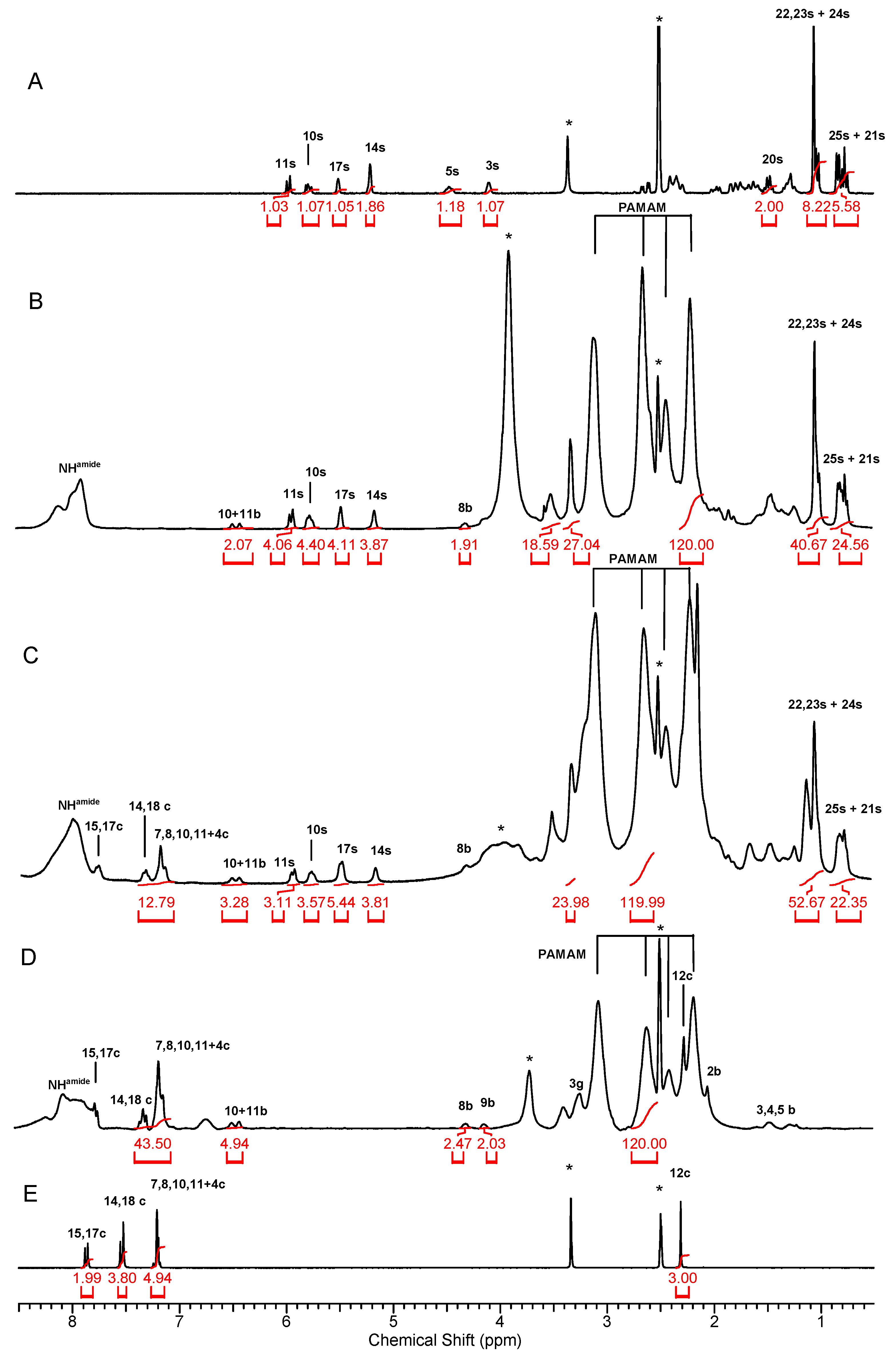
Simvastatin (S) was covalently attached via amide bond to both G32B and G32B2C to obtain equally substituted conjugates G32B4S and G32B2C4S, respectively. This conversion was elaborated from the beginning. Namely, we monitored a reaction between S and ethylenediamine (en) by the 1H NMR spectroscopy in deuterated methanol (Figure S1). We found that S lactone ring opening occurred within 4 h of refluxing 20 mM S and 23 mM en in methanol with formation of amide linkage between S and amine group of en. The addition of carbonyl diimidazole, which is a known carboxylate group-activating agent [25], sped up the reaction twice. Therefore, we applied this reagent to test the S addition into PAMAM G3 dendrimer (Scheme 1) in methanol. We obtained highly substituted G312S derivative and characterized it by the 1H NMR and 2-D 1H-1H COSY spectroscopy in DMSO-d6 (Figure S1). The most dramatic changes in the 1H NMR spectra were observed for 3s and 5s protons. These resonances shifted upfield from 4.20 ppm and 4.05 ppm in S (for spectral assignment, see Figure S3 and previously performed assignments [29]) into 3.65 ppm and 4.17 ppm in G312S, respectively. All the 1H resonances assigned to lactone ring in S (except 5s) shifted upfield in the G312S. Moreover, the 13C NMR resonances of lactone form of S shifted considerably in the 13C NMR spectrum of G312S. The 13C resonances were assigned on the basis of HSQC and HMBC experiments (Table S1).
Using the same reaction conditions, we added four equivalents of S into the G32B and G32B2C to obtain G32B4S and G32B2C4S, which were not isolated from methanol. Further, we blocked the remaining amine groups in G32B6C, G32B2C4S, and G32B4S by reaction of the conjugates with excess of R-glycidol in comparable conditions in methanol. Surprisingly, the remaining free amine groups of the starting conjugates were only partially substituted, as was observed before in the case of G3-fluorescein and G3-rhodamine conjugates [21]. On the basis of the integral intensity of the 3g resonance centered at around 3.32 ppm related to internal reference PAMAM resonance of [120H] intensity, we found that glycidylated conjugates were: G32B6C8gl, G32B2C4S12gl, and G32B4S14gl, as determined by the 1H NMR spectra (Figure 1B–D, respectively, Table S1).
The resonances in the 1H NMR spectrum of S (Figure 1A) in DMSO-d6 were assigned on the basis of COSY experiment (Figure S3). Amide conjugation of S into G32B4S14gl resulted in a considerable upfield shift of 5s and 3s protons, which are hidden under high-intensity residual HDO resonance (Figure 1B and Figure S4), whereas 10s, 11s, 14s, and 17s remained almost unchanged in the 1H NMR spectrum of G32B4S14gl related to S. However, the 3s and 5s were undoubtedly identified in the spectrum of G312S (Figure S2). The integral intensities of 10s, 11s, 14s, and 17s resonances corresponded to four S substituents in the G32B4S14gl conjugate in relation to [120H] integral intensity of PAMAM G3 resonance centered at 2.20 ppm (Figure 1B).
Analogously, the positions and intensities of the S resonances in ternary conjugate G32B2C4S12gl enabled the determination of the stoichiometry of the conjugate, containing average 4S substituents. In this 1H NMR spectral quantitation, another 1H internal reference, PAMAM resonance, was used, the one centered at 2.60, because the resonance at 2.20 ppm was overlapped with celecoxib-derived methyl 12c resonance (Figure 1C). The 1H NMR spectrum of G32B2C4S12gl conjugate showed very broad residual HDO resonance, which precluded a detailed spectrum analysis within 3.5–4.5 ppm region.
Celecoxib (C) resonances in the 1H NMR spectrum in DMSO-d6 (Figure 1E) were identified on the basis of C resonances integral intensity and COSY spectrum (not shown). All the aromatic proton resonances, except 15c and 17c of C in the 1H NMR spectrum of both G32B6C8gl and G32B2C4S12gl, were grouped within 7.3–7.4 ppm. The integration of these resonances originated from seven protons of C enabled to determine the number of C substituents equal to six and two in G32B6C8gl and G32B2C4S12gl, respectively (Figure 1D,C, respectively).
In conclusion, we obtained the binary conjugates G32B4S14gl and G32B6C8gl, as well as the ternary G32B2C4S12gl with remaining amine groups mono- or di-substituted with R-glycidol or unsubstituted at around 12, 16, and 12, respectively (Scheme 1). Glycidylation of conjugates was performed in order to increase a solubility of the conjugates in water but also to eradicate free amine groups, which are responsible for a systemic toxicity of PAMAM dendrimers, which increases considerably with PAMAM generation and amount of amine groups [30].
Although non-glycidylated analogues are well soluble in DMSO, they precipitate upon dilution with water. Surprisingly, the glycidylated conjugates obtained eventually were found to be better soluble in water and kept their molecular size at the molecular level, as determined by DLS method for 0.1 mM solution (Table 1).
We chose R-glycidol to derivatize the binary and ternary conjugates because we noticed more effective internalization of G3 and G2 PAMAM dendrimers covered with R-glycidol in comparison with those covered with S-glycidol into keratinocyte (HaCaT) and especially squamous carcinoma (SCC-15) cells in vitro [21].
The molecular size of conjugates, d(N), depends on primary amine groups available for protonation, which is achieved already at pH 5. The molecular size of the carrier, G32B14gl, was 3.3 nm (Table 1, Figure S5). The binary G32B6C8gl and G32B4S14gl sizes were 4.0 and 6.5, respectively, while a diameter of ternary G32B2C4S12gl molecule was 6.9 nm at pH 5 measured as d(N) at 0.05 mM concentration. In all cases, the volume-averaged diameter, d(V), was slightly larger than d(N), which is a regular feature of the conjugates. However, the ternary G32B2C4S12gl showed substantial association at pH 7, with d(N) 97 nm and d(V) around 150 nm as measured for 0.1 mM solution. The obtained results were important for further biological tests; the solutions used there were 10 times and more diluted. Thus, we assumed that all the conjugates were dispersed into the monomolecular level and the concentrations of all the conjugates were actual. In the case of G312S, the molecules remained associated both in pH 7 and pH 5 with particles sized at least 25 nm (Figure S5, Table 1).
Consistently, the zeta potential values for all conjugates, except G312S, fell into 8–38 mV, which are the numbers suggesting that high affinity of the conjugate molecules into negatively charged cell membranes is achievable (Figure S6, Table 1).

3.2. Conjugates Cytotoxicity

Dendrimer G32B14gl revealed rather low cytotoxicity for both cancer lines as estimated with neutral red (NR) assay: IC50 in order of 200 µM and 70 µM in SCC-15 and U-118 MG, respectively, and much higher cytotoxicity in BJ cells with IC50 fourfold lower in both assays (Table 2, Figure S7). This may indicate that under applied conditions, the G32B14gl penetrated the cell membrane without significant damage, whereas it entered the mitochondria and disrupted their transmembrane potential since the XTT assay indicated significantly lower IC50 values (Table 2, Figure S7).
The cytotoxicity of investigated repurposed drugs alone evaluated in all investigated cell lines revealed that their activities estimated with XTT assay as being in the order of 100 µM for both drugs and were higher than data published by the others for different cancer cell lines (Table 2). Gehrke et al. estimated the IC50 for C and S in the head and neck squamous carcinoma cell lines PE/CA-PJ 41 and HLaC78 using the MTT assay as 40 µM and 50 µM, respectively [19]. Comparable with those results are our data obtained with NR assay for C in both investigated cancer lines (IC50 = 48 µM for SCC-15) and (IC50 = 60 µM for U-118 MG) and with S (IC50 = 57 µM) in SCC-15 cells. Interestingly, the higher cytotoxicity of S (IC50 = 26 µM) was observed in glioma U-118MG cells. This may be explained by the observation including the breast (MCF-7 and SKBr-3), prostate (LNCaP and PC-3), colon (Caco-2 and HCT-116), skin (SCC-M7 and SCC-P9), and the lung cancer cell lines (Calu-3 and Calu6) that S inhibited the cell growth more efficiently in less differentiated tumors that spread faster, as in case of glioblastoma. The observed cytotoxicity for investigated cells was measured after treatment with 1 to 20 µM S for a prolonged period of 72 h [32]. The combination of treatment with both drugs by Gehrke et al. decreased tumor cell viability significantly more as compared to C and S alone, wherein a small additive effect and no synergistic effect were observed [19]. Our data revealed a higher additive effect of both drugs with XTT assay, but almost no effect with NR assay (Table 2). This may be explained by the data of Fujiwara et al., who reported that statins promote cell death by increasing the activation of caspases-3/-9, inducing pro-apoptotic protein (Bim) expression, arresting the cell cycle at the G1 phase, and decreasing the mitochondrial membrane potential (Δψm) [33]. Results obtained with either drug alone or both provided two interesting observations. The first is the higher resistance of fibroblasts to C and S as estimated by both assays as compared to cancer lines. The second is the highest cytotoxicity of S alone and in the combination with C in U-118 MG cells (IC50 = 26 µM and 38 µM, respectively; Figure 2).
Menter et al., 2017 [32] observed a time-dependent two-phased response in tumor cells to simvastatin. The first phase within the 6 h to 24 h involved changes in cell morphology, whereas the second phase between 24 and 72 h involved the loss of plasma membrane integrity as an effect of the cholesterol depletion. This is connected with disruption of the lipid rafts and expression of the caveolin-1 responsible for cellular transport and signaling, leading to the induction of apoptosis [32]. Thus, as was mentioned earlier, the NR assay showed higher sensitivity of U-118 MG to S for disintegration of the cellular membrane than SCC-15 and BJ cells.
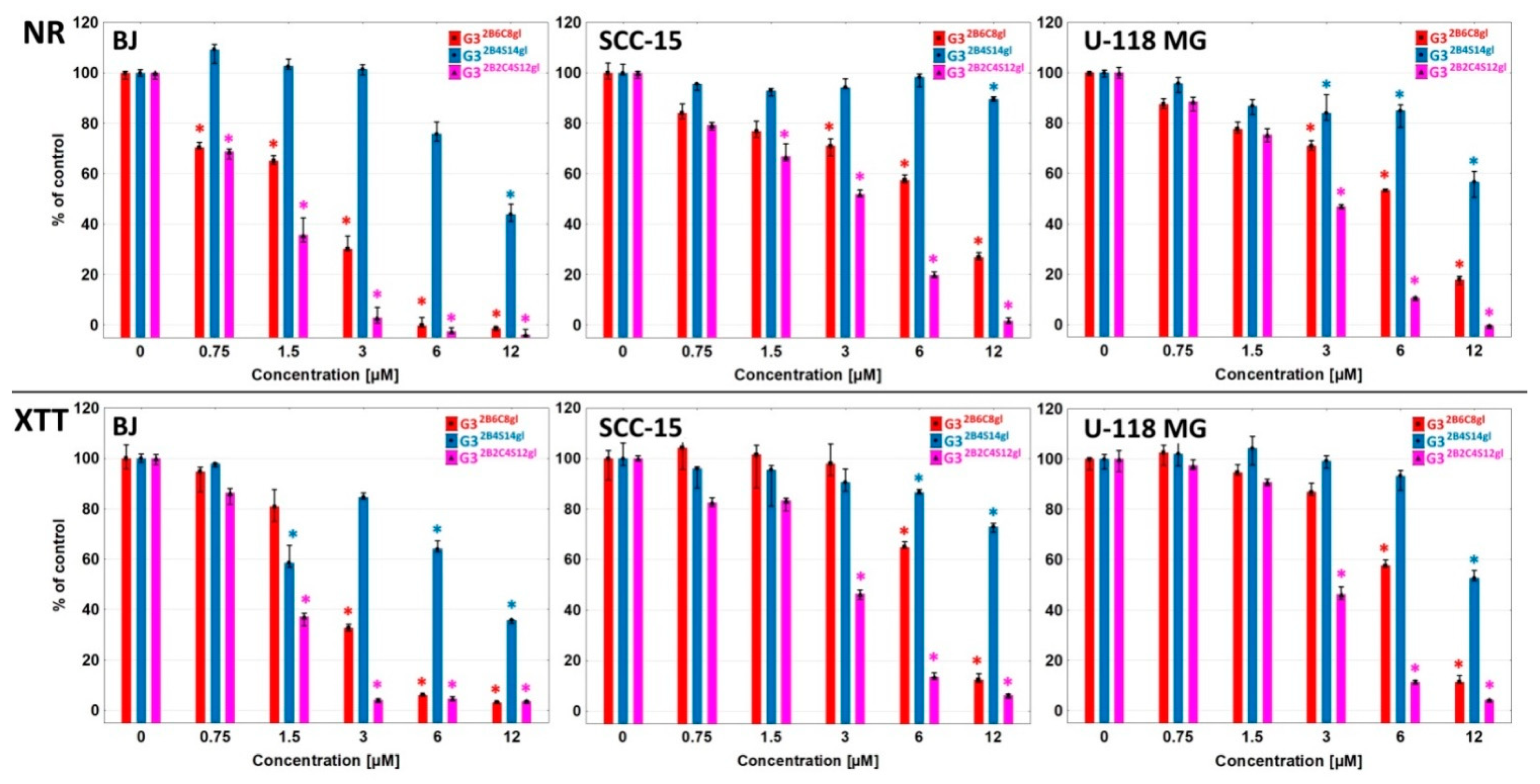
Conjugation of the G32B14gl with either drug alone or both dramatically increased their cytotoxicity as compared with drugs alone with much lower differences depending on used assays and investigated cancer cell lines (Figure 3). Estimated IC50 were in the order of 6–12 µM with lower values for G32B6C8gl in BJ cells (about 2 µM). This result was 10- to 50-fold lower as compared to both drugs alone (Table 2). A comparison of the IC50 values of the conjugates and the equivalent of the drugs (calculated concentration of introduced drugs) showed that the toxicity of the conjugates was not as impressively higher than that of the drugs themselves, but still the conjugates showed about 2-8-fold higher activity than the drug equivalents (Table 2). Conjugate with both drugs G32B2C4S12gl further increased the cytotoxicity of constructs as compared with conjugates of a single drug by 2–8-fold, with IC50 in the order of 1 µM for BJ and 3 µM for both cancer cell lines. This amounted to a 35–50-fold increase of cytotoxicity as compared to both drugs alone (Table 2). The negative finding is the lack of selectivity of obtained conjugate that is more toxic (threefold) for normal fibroblasts than for both cancer lines. However, this may have meaning for cancer-associated fibroblasts (CAF) constituting the tumor extracellular matrix (TEM) now recognized as a substantial factor for cancer progression and considered as a potential therapeutic and diagnostic target [34]. The interaction between tumor cells and the TEM plays a crucial role in tumor initiation, progression, metastasis, and response to therapy. The S has been shown to induce metabolic reprogramming in TEM and to target the immune microenvironment through cytokines or chemokines [35].

4. Conclusions

The presented results documented the very high effectiveness of obtained biotinylated and R-glycidylated PAMAM dendrimer conjugated with both C and S (G32B2C4S12g) as dramatically decreasing the squamous carcinoma SCC-15 and glioblastoma U-118 MG cell viability at very low concentrations with IC50 equal to about 3 µM. This makes it a prospective targeted drug carrier in malignant cancer, including glioblastoma therapy with decreased side effects of both drugs observed at concentrations applied in the clinics.

Supplementary Materials

The following supporting information can be downloaded at https://www.mdpi.com/article/10.3390/cancers14030714/s1, Figure S1: Progress of reaction between S and en monitored by the 1H NMR spectroscopy. Figure S2: COSY spectrum of G312S in DMSO-d6. Figure S3: COSY spectrum of simvastatin in DMSO-d6. Figure S4: COSY spectrum of G32B4S12gl in DMSO-d6. Figure S5: Size of conjugates by DLS. Figure S6: Zeta potential of conjugates. Figure S7: Cytotoxicity of G32B14gl carrier for BJ, SCC-15, and U-118 MG cells. Table S1: The 1H and 13C NMR chemical shifts of simvastatin (S); celecoxib (C); and G312S, G32B4S14gl, G32B2C4S12gl, and G32B6C8gl conjugates in DMSO-d6.

Author Contributions

Conceptualization, E.W.-R., S.W. and Ł.U.; methodology, E.W.-R., S.W. and Ł.U.; validation, S.W. and Ł.U.; formal analysis, Ł.U., S.W. and J.M.; investigation, K.W., S.W., J.M. and Ł.U.; resources, S.W. and Ł.U.; data curation, S.W., J.M. and Ł.U.; writing—original draft preparation, S.W., E.W-R. and Ł.U.; writing—review and editing, S.W., E.W-R. and Ł.U.; visualization, S.W., Ł.U. and J.M.; supervision, E.W-R.; project administration, S.W. and E.W-R.; funding acquisition, S.W. and Ł.U. All authors have read and agreed to the published version of the manuscript.

Funding

The biological part of this research was supported by financial resources of the Faculty of Chemistry, Rzeszów University of Technology, grant number PB25.CN.21.001.01.

Institutional Review Board Statement

Not applicable.

Informed Consent Statement

Not applicable.

Data Availability Statement

Data supporting reported results can be provided by authors upon request.

Acknowledgments

We gratefully acknowledge Małgorzata Walczak from Rzeszow University of Technology for kindly enabling us to measure size and zeta potential on Zetasizer Nano instrument.

Conflicts of Interest

The authors declare no conflict of interest.

References

  1. Abedi-Gaballu, F.; Dehghan, G.; Ghaffari, M.; Yekta, R.; Abbaspour-Ravasjani, S.; Baradaran, B.; Ezzati Nazhad Dolatabadi, J.; Hamblin, M.R. PAMAM Dendrimers as Efficient Drug and Gene Delivery Nanosystems for Cancer Therapy. Appl. Mater. Today 2018, 12, 177–190. [Google Scholar] [CrossRef]
  2. Tołoczko-Iwaniuk, N.; Dziemiańczyk-Pakieła, D.; Nowaszewska, B.K.; Celińska-Janowicz, K.; Miltyk, W. Celecoxib in Cancer Therapy and Prevention-Review. Curr. Drug Targets 2019, 20, 302–315. [Google Scholar] [CrossRef]
  3. Uram, Ł.; Markowicz, J.; Misiorek, M.; Filipowicz-Rachwał, A.; Wołowiec, S.; Wałajtys-Rode, E. Celecoxib Substituted Biotinylated Poly(Amidoamine) G3 Dendrimer as Potential Treatment for Temozolomide Resistant Glioma Therapy and Anti-Nematode Agent. Eur. J. Pharm. Sci. 2020, 152, 105439. [Google Scholar] [CrossRef]
  4. Uram, Ł.; Filipowicz, A.; Misiorek, M.; Pieńkowska, N.; Markowicz, J.; Wałajtys-Rode, E.; Wołowiec, S. Biotinylated PAMAM G3 Dendrimer Conjugated with Celecoxib and/or Fmoc-l-Leucine and Its Cytotoxicity for Normal and Cancer Human Cell Lines. Eur. J. Pharm. Sci. 2018, 124, 1–9. [Google Scholar] [CrossRef]
  5. Hemmer, R.; Hall, A.; Spaulding, R.; Rossow, B.; Hester, M.; Caroway, M.; Haskamp, A.; Wall, S.; Bullen, H.A.; Morris, C.; et al. Analysis of Biotinylated Generation 4 Poly(Amidoamine) (PAMAM) Dendrimer Distribution in the Rat Brain and Toxicity in a Cellular Model of the Blood-Brain Barrier. Molecules 2013, 18, 11537–11552. [Google Scholar] [CrossRef] [Green Version]
  6. Fana, M.; Gallien, J.; Srinageshwar, B.; Dunbar, G.L.; Rossignol, J. PAMAM Dendrimer Nanomolecules Utilized as Drug Delivery Systems for Potential Treatment of Glioblastoma: A Systematic Review. Int. J. Nanomed. 2020, 15, 2789–2808. [Google Scholar] [CrossRef] [Green Version]
  7. Hanurry, E.Y.; Mekonnen, T.W.; Andrgie, A.T.; Darge, H.F.; Birhan, Y.S.; Hsu, W.-H.; Chou, H.-Y.; Cheng, C.-C.; Lai, J.-Y.; Tsai, H.-C. Biotin-Decorated PAMAM G4.5 Dendrimer Nanoparticles to Enhance the Delivery, Anti-Proliferative, and Apoptotic Effects of Chemotherapeutic Drug in Cancer Cells. Pharmaceutics 2020, 12, 443. [Google Scholar] [CrossRef]
  8. Tilija Pun, N.; Jeong, C.-H. Statin as a Potential Chemotherapeutic Agent: Current Updates as a Monotherapy, Combination Therapy, and Treatment for Anti-Cancer Drug Resistance. Pharmaceuticals 2021, 14, 470. [Google Scholar] [CrossRef]
  9. Sirtori, C.R. The Pharmacology of Statins. Pharm. Res. 2014, 88, 3–11. [Google Scholar] [CrossRef]
  10. Guerra, B.; Recio, C.; Aranda-Tavío, H.; Guerra-Rodríguez, M.; García-Castellano, J.M.; Fernández-Pérez, L. The Mevalonate Pathway, a Metabolic Target in Cancer Therapy. Front. Oncol. 2021, 11, 626971. [Google Scholar] [CrossRef]
  11. Duarte, J.A.; de Barros, A.L.B.; Leite, E.A. The Potential Use of Simvastatin for Cancer Treatment: A Review. Biomed. Pharmacother. 2021, 141, 111858. [Google Scholar] [CrossRef]
  12. Göbel, A.; Zinna, V.M.; Dell’Endice, S.; Jaschke, N.; Kuhlmann, J.D.; Wimberger, P.; Rachner, T.D. Anti-Tumor Effects of Mevalonate Pathway Inhibition in Ovarian Cancer. BMC Cancer 2020, 20, 703. [Google Scholar] [CrossRef]
  13. Nath, S.D.; Linh, N.T.B.; Sadiasa, A.; Lee, B.T. Encapsulation of Simvastatin in PLGA Microspheres Loaded into Hydrogel Loaded BCP Porous Spongy Scaffold as a Controlled Drug Delivery System for Bone Tissue Regeneration. J. Biomater. Appl. 2014, 28, 1151–1163. [Google Scholar] [CrossRef]
  14. Kulhari, H.; Pooja, D.; Prajapati, S.K.; Chauhan, A.S. Performance Evaluation of PAMAM Dendrimer Based Simvastatin Formulations. Int. J. Pharm. 2011, 405, 203–209. [Google Scholar] [CrossRef]
  15. Kulhari, H.; Kulhari, D.P.; Prajapati, S.K.; Chauhan, A.S. Pharmacokinetic and Pharmacodynamic Studies of Poly(Amidoamine) Dendrimer Based Simvastatin Oral Formulations for the Treatment of Hypercholesterolemia. Mol. Pharm. 2013, 10, 2528–2533. [Google Scholar] [CrossRef]
  16. Sharma, R.; Liaw, K.; Sharma, A.; Jimenez, A.; Chang, M.; Salazar, S.; Amlani, I.; Kannan, S.; Kannan, R.M. Glycosylation of PAMAM Dendrimers Significantly Improves Tumor Macrophage Targeting and Specificity in Glioblastoma. J. Control. Release 2021, 337, 179–192. [Google Scholar] [CrossRef]
  17. Yan, S.; Ren, J.; Jian, Y.; Wang, W.; Yun, W.; Yin, J. Injectable Maltodextrin-Based Micelle/Hydrogel Composites for Simvastatin-Controlled Release. Biomacromolecules 2018, 19, 4554–4564. [Google Scholar] [CrossRef]
  18. Shojaei, S.; Koleini, N.; Samiei, E.; Aghaei, M.; Cole, L.K.; Alizadeh, J.; Islam, M.I.; Vosoughi, A.-R.; Albokashy, M.; Butterfield, Y.; et al. Simvastatin Increases Temozolomide-Induced Cell Death by Targeting the Fusion of Autophagosomes and Lysosomes. FEBS J. 2020, 287, 1005–1034. [Google Scholar] [CrossRef]
  19. Gehrke, T.; Scherzad, A.; Hackenberg, S.; Ickrath, P.; Schendzielorz, P.; Hagen, R.; Kleinsasser, N. Additive Antitumor Effects of Celecoxib and Simvastatin on Head and Neck Squamous Cell Carcinoma in Vitro. Int. J. Oncol. 2017, 51, 931–938. [Google Scholar] [CrossRef] [Green Version]
  20. Uram, Ł.; Szuster, M.; Filipowicz, A.; Zaręba, M.; Wałajtys-Rode, E.; Wołowiec, S. Cellular Uptake of Glucoheptoamidated Poly(Amidoamine) PAMAM G3 Dendrimer with Amide-Conjugated Biotin, a Potential Carrier of Anticancer Drugs. Bioorg. Med. Chem. 2017, 25, 706–713. [Google Scholar] [CrossRef]
  21. Malinga-Drozd, M.; Uram, Ł.; Wróbel, K.; Wołowiec, S. Chiral Recognition of Homochiral Poly (Amidoamine) Dendrimers Substituted with R- and S-Glycidol by Keratinocyte (HaCaT) and Squamous Carcinoma (SCC-15) Cells In Vitro. Polymer 2021, 13, 1049. [Google Scholar] [CrossRef] [PubMed]
  22. Tomalia, D.A.; Baker, H.; Dewald, J.; Hall, M.; Kallos, G.; Martin, S.; Roeck, J.; Ryder, J.; Smith, P. A New Class of Polymers: Starburst-Dendritic Macromolecules. Polym. J. 1985, 17, 117–132. [Google Scholar] [CrossRef] [Green Version]
  23. Uram, Ł.; Szuster, M.; Gargasz, K.; Filipowicz, A.; Wałajtys-Rode, E.; Wołowiec, S. In Vitro Cytotoxicity of the Ternary PAMAM G3-Pyridoxal-Biotin Bioconjugate. Int. J. Nanomed. 2013, 8, 4707–4720. [Google Scholar] [CrossRef] [Green Version]
  24. Kaczorowska, A.; Malinga-Drozd, M.; Kałas, W.; Kopaczyńska, M.; Wołowiec, S.; Borowska, K. Biotin-Containing Third Generation Glucoheptoamidated Polyamidoamine Dendrimer for 5-Aminolevulinic Acid Delivery System. Int. J. Mol. Sci. 2021, 22, 1982. [Google Scholar] [CrossRef]
  25. Lee, Y.; Kim, J.; Kim, H.; Kang, S.; Yoon, J.-H.; Kim, D.-D.; Kim, Y.M.; Jung, Y. N-Succinylaspart-1-Yl Celecoxib Is a Potential Colon-Specific Prodrug of Celecoxib with Improved Therapeutic Properties. J. Pharm. Sci. 2012, 101, 1831–1842. [Google Scholar] [CrossRef]
  26. Nizioł, J.; Uram, Ł.; Szuster, M.; Sekuła, J.; Ruman, T. Biological Activity of N(4)-Boronated Derivatives of 2′-Deoxycytidine, Potential Agents for Boron-Neutron Capture Therapy. Bioorganic Med. Chem. 2015, 23, 6297–6304. [Google Scholar] [CrossRef]
  27. Mukaiyama, T. New Synthetic Reactions Based on the Onium Salts of Aza-Arenes [New Synthetic Methods (29)]. Angew. Chem. Int. Ed. Engl. 1979, 18, 707–721. [Google Scholar] [CrossRef]
  28. Uram, Ł.; Misiorek, M.; Pichla, M.; Filipowicz-Rachwał, A.; Markowicz, J.; Wołowiec, S.; Wałajtys-Rode, E. The Effect of Biotinylated PAMAM G3 Dendrimers Conjugated with COX-2 Inhibitor (Celecoxib) and PPARγ Agonist (Fmoc-L-Leucine) on Human Normal Fibroblasts, Immortalized Keratinocytes and Glioma Cells in Vitro. Molecules 2019, 24, 3801. [Google Scholar] [CrossRef] [Green Version]
  29. Brus, J.; Jegorov, A. Through-Bonds and Through-Space Solid-State NMR Correlations at Natural Isotopic Abundance:  Signal Assignment and Structural Study of Simvastatin. J. Phys. Chem. A 2004, 108, 3955–3964. [Google Scholar] [CrossRef]
  30. Mukherjee, S.P.; Davoren, M.; Byrne, H.J. In Vitro Mammalian Cytotoxicological Study of PAMAM Dendrimers-towards Quantitative Structure Activity Relationships. Toxicol. In Vitro 2010, 24, 169–177. [Google Scholar] [CrossRef] [Green Version]
  31. AAT Bioquest, Inc. Quest GraphTM ANOVA Calculator. Available online: Https://www.aatbio.com/tools/anova-analysis-of-variance-one-two-way-calculator (accessed on 3 December 2021).
  32. Menter, D.G.; Ramsauer, V.P.; Harirforoosh, S.; Chakraborty, K.; Yang, P.; Hsi, L.; Newman, R.A.; Krishnan, K. Differential Effects of Pravastatin and Simvastatin on the Growth of Tumor Cells from Different Organ Sites. PLoS ONE 2011, 6, e28813. [Google Scholar] [CrossRef] [Green Version]
  33. Fujiwara, D.; Tsubaki, M.; Takeda, T.; Tomonari, Y.; Koumoto, Y.; Sakaguchi, K.; Nishida, S. Statins Induce Apoptosis through Inhibition of Ras Signaling Pathways and Enhancement of Bim and P27 Expression in Human Hematopoietic Tumor Cells. Tumour Biol. 2017, 39, 1010428317734947. [Google Scholar] [CrossRef] [PubMed] [Green Version]
  34. Belhabib, I.; Zaghdoudi, S.; Lac, C.; Bousquet, C.; Jean, C. Extracellular Matrices and Cancer-Associated Fibroblasts: Targets for Cancer Diagnosis and Therapy? Cancers 2021, 13, 3466. [Google Scholar] [CrossRef]
  35. Jiang, W.; Hu, J.-W.; He, X.-R.; Jin, W.-L.; He, X.-Y. Statins: A Repurposed Drug to Fight Cancer. J. Exp. Clin. Cancer Res. 2021, 40, 241. [Google Scholar] [CrossRef]
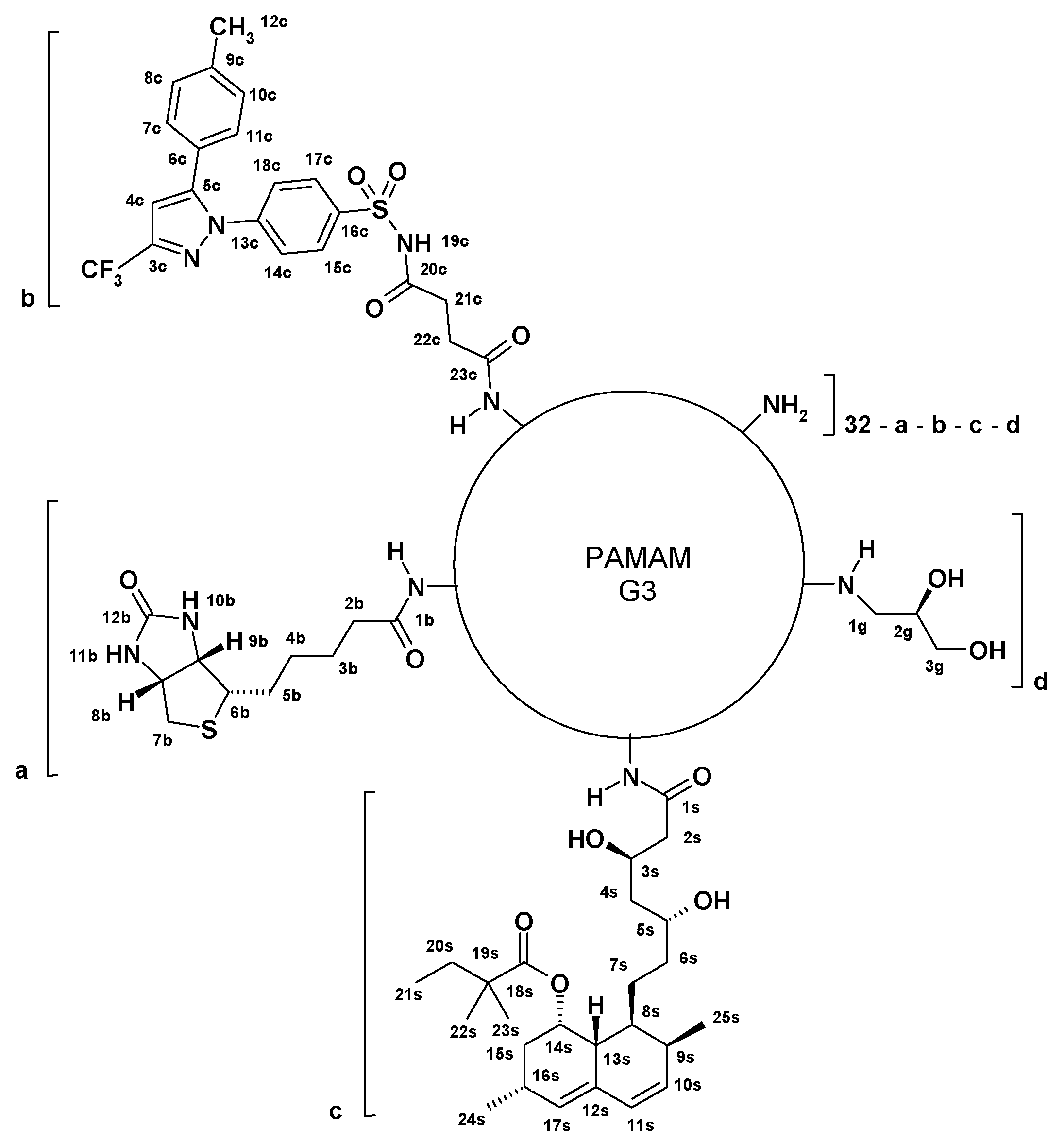
Scheme 1. Schematic formula of PAMAM G3 (G3) conjugates with biotin (B), celecoxib (C), simvastatin (S), and R-glycidol (gl). The following conjugates were tested: G32B6C8gl (a = 2; b = 6, c = 0, d = 8), G32B4S14gl (a = 2; b = 0, c = 4, d = 14), G32B2C4S12gl (a = 2; b = 2, c = 4, d = 12), and G32B14gl (a = 2; b = 0, c = 0, d = 14).
Cancers 14 00714 sch001
Figure 1. The 1H NMR spectra of simvastatin (A), G32B4S14gl (B), G32B2C4S12gl (C), G32B6C8gl (D), and celecoxib (E) in DMSO-d6. Residual HD2CS(O)CD3 and HDO peaks are labeled with asterisks *. The PAMAM CH2 resonances centered at 2.63 ppm or 2.20 were used as internal integration reference (the integral intensity of both resonances corresponds to [120H]).
Cancers 14 00714 g001
Scheme 2. Functionalization of celecoxib and simvastatin: (i) succinic anhydride in acetonitrile and triethylamine, (ii) 2-chloro-N-methylpyridine iodide in the presence of 4-(dimethylamino)pyridine in DMSO, (iii) carbonyl diimidazole in methanol.
Cancers 14 00714 sch002
Figure 2. Effect of single drugs celecoxib, simvastatin, and 0.5:1 mixture of celecoxib and simvastatin on normal BJ and cancer SCC-15 and U-118 MG cell viability, estimated with NR and XTT assays after 24 h incubation. Cell viability is presented as medians of a percent against non-treated control (control expressed as 100%). The whiskers are lower (25%) and upper (75%) quartile ranges. * p ≤ 0.05; Kruskal–Wallis test (against non-treated control).
Cancers 14 00714 g002
Figure 3. Cytotoxicity of single conjugates G32B6C8gl, G32B4S14gl, and combined conjugate G32B2C4S12gl for normal (BJ) and cancer (SCC-15) and (U-118 MG) cells after 24 h treatment, estimated with neutral red (NR) or XTT assay. Cell viability is presented as medians of a percent against non-treated control (control expressed as 100%). The whiskers are lower (25%) and upper (75%) quartile ranges. * p ≤ 0.05; Kruskal–Wallis test (against non-treated control).
Cancers 14 00714 g003
Table 1. Size of conjugates averaged by volume (d(V)) and by number of molecules (d(N)) and zeta potential values ± standard deviation determined by DLS analysis.
CompoundSize (nm)Zeta Potential (mV)
pH 7pH 5pH 7pH 5
d(V)d(N)d(V)d(N)
G32B14gl2.1 ± 0.491.8 ± 0.425.0 ± 0.963.3 ± 0.859.6 ± 0.897.8 ± 2.51
G32B6C8gl7.8 ± 3.565.7 ± 2.355.8 ± 1.594.0 ± 1.4618.6 ± 3.7034.6 ± 2.52
G32B4S14gl10.3 ± 1.678.6 ± 1.449.5 ± 1.976.5 ± 1.9237.7 ± 3.8236.5 ± 4.11
G32B2Cel4S12gl155 ± 5.7786.7 ± 6.838.5 ± 2.196.9 ± 2.1430.3 ± 0.7332.5 ± 3.14
G312S85.8 ± 1.8357.2 ± 2.9644.2 ± 1.9925.3 ± 7.0742.4 ± 1.7732.9 ± 2.84
Table 2. Cytotoxicity of investigated repurposed drugs alone and their conjugates with G32B14gl estimated for the normal BJ and cancer SCC-15 and U-118 MG cells, expressed as half maximal inhibitory concentration (IC50) in micromolar concentration (medians). Data were estimated with NR and XTT assay after 24 h incubation and calculated with AAT Bioquest IC50 calculator [31]. a—concentration of introduced C, b—concentration of introduced S.
AssayCell LineConjugate/Carrier Alone/Drug Alone (IC50 µM)
G32B6C8glG32B4S14glG32B2C4S12glG32B14glCSC + S
0.5:1 Ratio
NRBJ1.909.991.1053.6088.6890.1463.40
11.40 a39.96 b2.20 a
4.40 b
SCC-156.65>12.006.69>200.0047.4856.5456.45
39.9 a>48.00 b13.38 a
26.76 b
U-118 MG5.8616.902.73>200.0065.5726.1937.65
35.16 a67.60 b5.46 a
10.92 b
XTTBJ2.408.751.2823.45100.13100.8666.00
14.40 a35.00 b2.56 a
5.12 b
SCC-156.98>12.002.9864.5897.1384.3860.14
41.88 a>48.00 b5.96 a
11.92 b
U-118 MG6.4912.272.9162.6593.74112.3370.67
38.94 a49.08 b5.82 a
11.64 b
Publisher’s Note: MDPI stays neutral with regard to jurisdictional claims in published maps and institutional affiliations.