Review

MicroRNAs’ Crucial Role in Salivary Gland Cancers’ Onset and Prognosis

1
Department of Precision Medicine, University of Campania “Luigi Vanvitelli”, Via L. De Crecchio, 7, 80138 Naples, Italy
2
Laboratory of Precision and Molecular Oncology, Biogem Scarl, Institute of Genetic Research, Contrada Camporeale, 83031 Ariano Irpino, Italy
3
Department of Pharmacy, School of Medicine and Surgery, University of Naples “Federico II”, Via D. Montesano 49, 80131 Naples, Italy
4
College of Medical, Veterinary and Life Sciences, School of Infection and Immunity, University of Glasgow, Glasgow G12 8TA, UK
5
Department of Medicine, Surgery and Dentistry, University of Salerno, Via S. Allende, 84081 Baronissi, Italy
6
Otolaryngology Department, San Giovanni-Addolorata Hospital, Via dell’Amba Aradam, 8, 00184 Rome, Italy
7
Department of Advanced Medical and Surgical Sciences “DAMSS”, University of Campania “Luigi Vanvitelli”, Via S.M. di Costantinopoli 104, 80138 Naples, Italy
8
Ear Nose Throt Departement AORN Cardarelli, 80100 Napoli, Italy
*
Authors to whom correspondence should be addressed.
Cancers 2022, 14(21), 5304; https://doi.org/10.3390/cancers14215304
Submission received: 21 September 2022 / Revised: 22 October 2022 / Accepted: 24 October 2022 / Published: 28 October 2022

Abstract

:

Simple Summary

Salivary gland cancers are incredibly heterogeneous, both in the physical onset and in the aggressiveness. Setting up a novel diagnostic and prognostic detection method based on the noninvasive microRNAs’ profiling might represent a goal for the clinical management of those particular malignancies, saving precious time for the patients.

Abstract

Salivary gland cancer (SGC) is an uncommon and heterogeneous disease that accounts for around 8.5% of all head and neck cancers. MicroRNAs (miRNAs) consist of a class of highly conserved, short, single-stranded segments (18–25 nucleotides) of noncoding RNA that represent key gene-transcription regulators in physiological and pathological human conditions. However, their role in SGC development and progression is not completely clear. This review aims to compile and summarize the recent findings on the topic, focusing on the prognostic and diagnostic value of the major modulated and validated microRNAs in SGC. Their differential expression could possibly aid the clinician in delivering an early diagnosis, therapeutic strategy and precision medicine.

1. Introduction

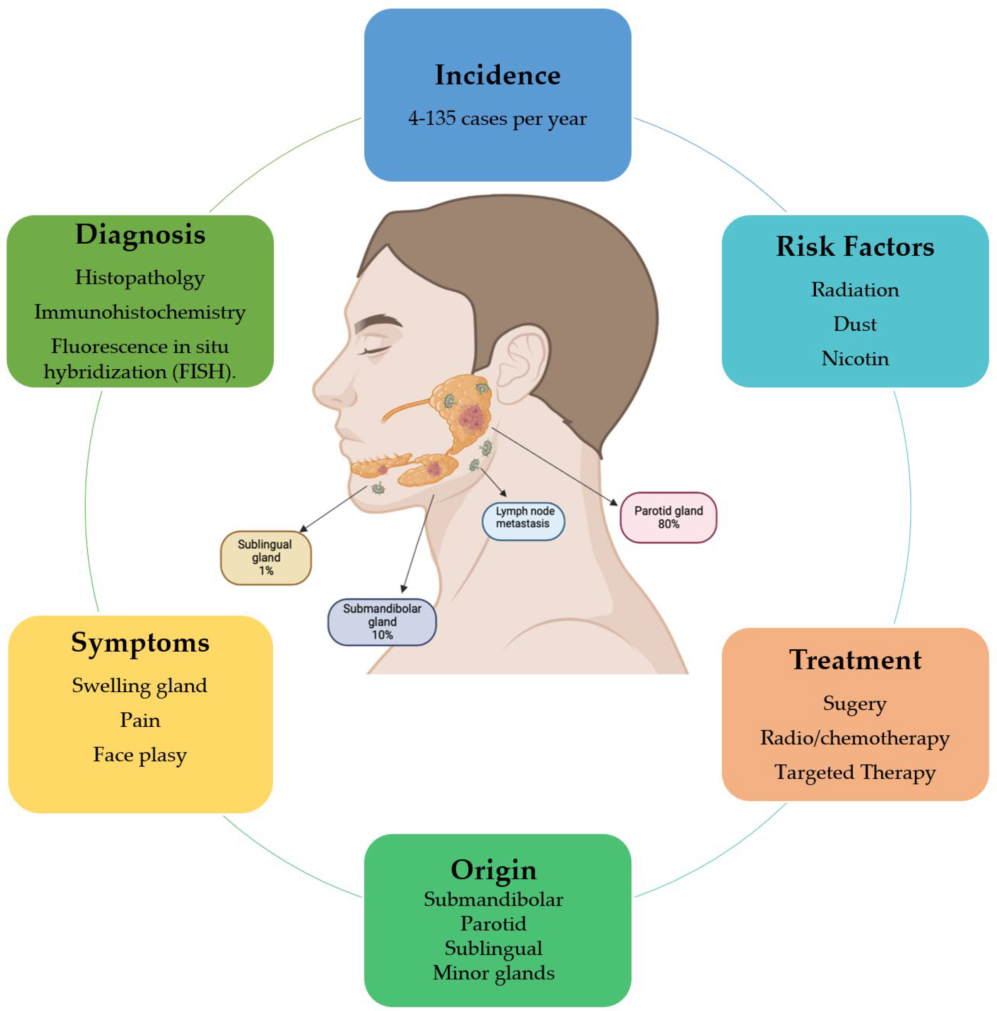
1.1. Characteristics of Salivary Gland Tumors

The salivary glands are oral cavity structures divided into three major glands: the parotid, the submandibular and the sublingual, followed by over 600 minor glands [1]. The function of the major glands is the secretion of saliva and protein-rich fluid that coats teeth and oral mucosa, facilitating mastication, speech, and the preservation of oral hygiene [2]. The salivary glands can be affected by different diseases, such as infections, obstructions and benign and malignant neoplasms, which compromise their normal functions [3].
Salivary gland cancer (SGC) is a rare and heterogeneous disease with an annual incidence of 1.4/100,000 in most countries (Figure 1), accounting for about 8.5% of all head and neck cancer. Men are more affected than women, and the incidence increases significantly in individuals over the age of 65 [4]. The significant risk factors are long-term radiation exposure, dust and nicotine (Figure 1). Benign tumors are characterized by slow growth, normal facial nerve function and no metastasis. In contrast, gland swelling, pain, inflammation and facial palsy are frequent symptoms of malignant salivary gland cancer (Figure 1) [5].
Almost 80% of all salivary gland tumors arise in the parotid gland, but approximately 75% are benign. Tumors occurring in the submandibular and sublingual sites represent 10% and 1%, respectively, of all SGC and are for the most part malignant [6]. Finally, cancers of the minor glands are instead located in the oral cavity, oropharynx, nasopharynx, larynx and upper airway and are usually malignant (Figure 1) [7]. According to the World Health Organization Classification of Salivary Gland Malignancies, there are more than 20 malignant histopathologic cancers and numerous benign types (Table 1) [8].
The most common malignancy is mucoepidermoid carcinoma, followed by adenoid cystic and acinic cell carcinoma. Mucoepidermoid carcinoma (MEC) usually originates in the parotid gland and is composed of mucin-secreting and epidermoid cells. Low-grade MEC does not present symptoms, with a slow-growing mass and no metastasis. The high-grade MEC tumor grows rapidly and causes pain and neuropathies [9]. Adenoid cystic carcinoma (ACC), composed of ductal and myoepithelial cells, commonly arises in the minor salivary glands. It is characterized by perineural invasion and cervical lymph node metastasis [10]. Acinic cell carcinoma is usually located in the parotid gland and has a higher incidence in women than men. It shows serous acinar cell differentiation and has a high rate of recurrence and metastasis [11]; moreover, it has recently been linked to the recurrent translocation of NR4A2- or NR4A3-activating genes [12]. The different histological features of these subtypes and the different degrees of malignancy can result in a problematic prognosis for the tumor [9]. The diagnostic scheme of salivary gland cancer starts with conventional histopathology and is then confirmed by immunohistochemistry and fluorescence in situ hybridization (FISH). According to the WHO in 2021, the first line of treatment is surgery; however, it is not always easy due to infiltration of facial and trigeminal nerve branches and cases of metastasis. Radiotherapy and chemotherapy play a secondary and not fully evidence-based role, except for those tumors for which treatment is available based on hormone receptor expression or targetable mutations. Identification of new targets for personalized therapy is increasing, even if it is still not routinely available (Figure 1) [13]. Genetic abnormalities and mutations are, in fact, recurrent in SGC. The most frequent in the MEC subtype is the fusion of CTRC1 and MAML2 genes, which leads to the upregulation of EGFR. The ACC type is instead characterized by MYB, MYBL1 and NFIB rearrangements and NOTCH gene mutations [14].
Other typical alterations in SGC are in human epidermal growth factor receptor 2 (HER2), PIK3CA and BRAF mutation. Specific therapy against these molecular features is effective in other cancers and is leading to promising future treatment strategies for salivary gland cancers [15].

1.2. Features of MicroRNAs

MicroRNAs (miRNAs) are small, single-stranded, endogenous RNA molecules that play an essential role in regulating gene expression and RNA silencing. The biosynthesis process of miRNAs is very complex (Figure 2); their maturation begins in the cell nucleus, where they are transcribed from specific DNA sequences into primary miRNAs (Pri-miRNAs). The RNase type III enzyme DROSHA processes Pri-miRNAs into precursor miRNAs (Pre-miRNAs), transcripts about 60–110 nucleotides in length with a shorter stem–loop structure [16]. RAS-related nuclear protein guanosine-5’-triphosphatase (RAN GTPase) and exportin-5 proteins carry Pre-miRNAs from the nucleus to the cytoplasm [17], and the miRNAs’ maturation process continues in the cytoplasm, where Pre-miRNA is cleaved by the RNase type III enzyme Dicer to generate molecules of duplex RNA of about 22 nucleotides. These shorter miRNA duplexes are loaded into the RNA-induced silencing complex (RISC), a multiprotein complex that includes proteins such as argonaute RISC catalytic component 2 (Ago2). Ago2 possesses catalytic properties and cleaves one strand of the RNA duplex [18]. The strand linked to the RISC is an active strand called the “guide strand”, which represents a mature miRNA. However, some studies have shown how a miRNA’s biosynthesis and maturation process can occur following noncanonical pathways, characterized by a different involvement of proteins such as Drosha, Dicer and Ago [19].
RISC, with the loaded miRNA (miRISC), acts by inhibiting protein synthesis and interfering with the stability of the target mRNA. The binding specificity is complementary-nucleotides-based, and usually the interaction of the miRNA with the target mRNA occurs through pairing a 2–8-nucleotide-long region of the miRNA itself, called the seed sequence, with sequences called miRNA response elements (MREs) localized on the target mRNA. The miRNAs’ binding sites are usually located in the 3′ untranslated region (3′UTR) of the target mRNA to induce its translational repression and decay. However, several studies have shown that miRNAs can also interact with sequences located in noncanonical binding sites such as 5’ untranslated region (5’UTR) [19,20], coding sequences [21] and gene promoter regions [22]. Gene silencing induced by miRNAs is mainly carried out by blocking protein synthesis, inhibiting elongation and ribosomal drop-off and destabilizing the mRNA following deadenylation and decapping processes that lead to its degradation [23]. In biological processes such as cellular proliferation, apoptosis, inflammation, cellular motility, secretome and cell differentiation, miRNAs have a central role. Since miRNAs can control and modulate gene expression, it is easy to understand how an alteration of their function can also be associated with human diseases and tumorigenesis. Many studies have shown that miRNAs can act as tumor suppressor genes and oncogenes according to the target mRNA type and the cellular and histological context. Many miRNAs have been identified to have different expression profiles in blood malignancies and solid tumors such as breast and lung cancers, glioblastoma, melanoma and ovarian cancer [24]. Patrick et al. discovered different serum levels of miR-141 in prostate cancer patients compared to healthy controls. This study represented a starting point for future investigations aimed at defining and validating the role of miRNAs as circulating cancer biomarkers [25]. Many studies also confirmed that altered miRNAs expression patterns can reflect the tumor stage, invasiveness and metastasis [26]. Therefore, today, efforts are being made to develop new therapeutic strategies aimed at exploiting miRNAs as potential diagnostic and prognostic biomarkers in response to pharmacological therapies, and in our case, precisely related to salivary gland cancer.

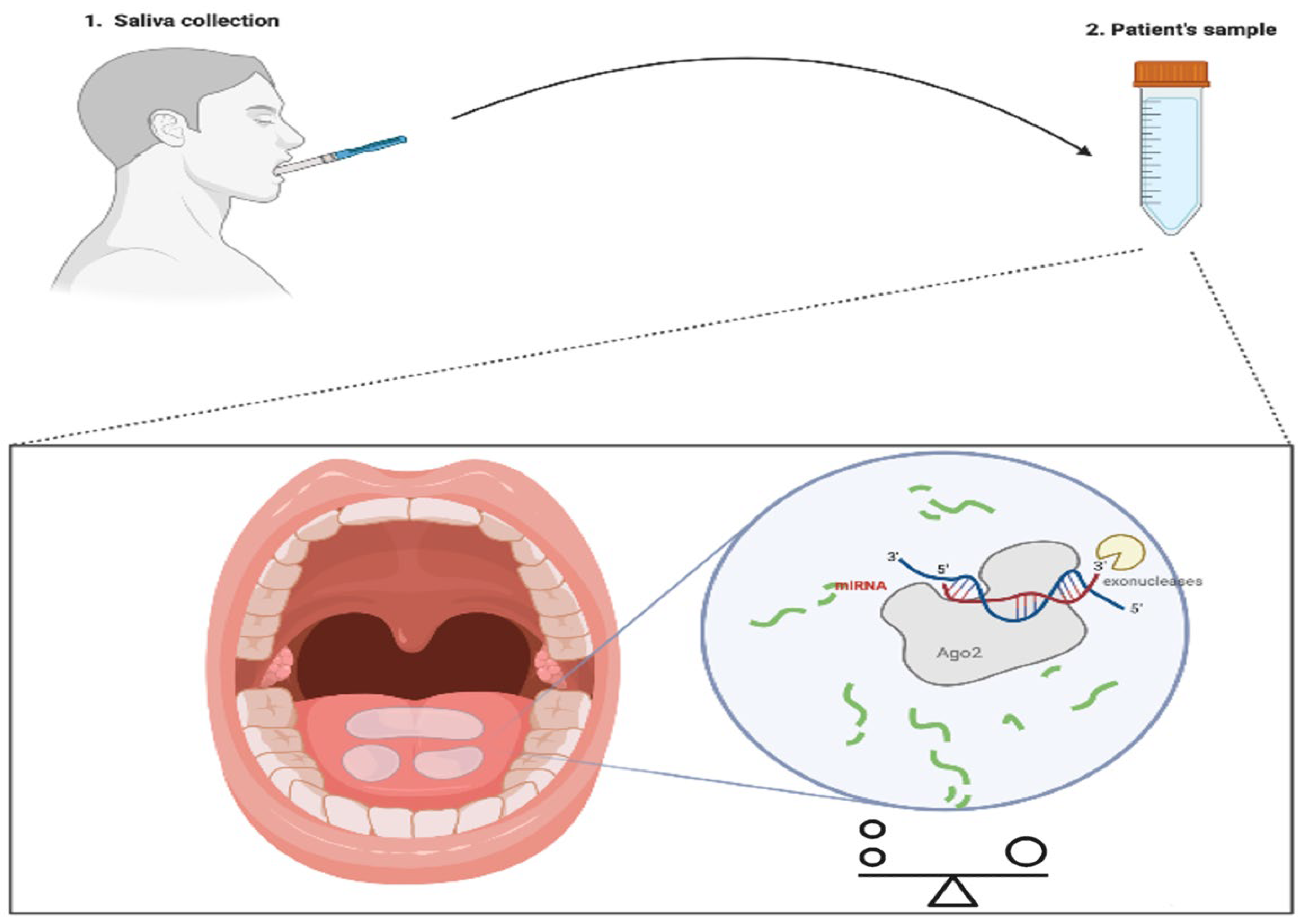
2. Liquid Biopsy: Kill Two Birds with One Stone

Liquid biopsy plays a crucial role in the noninvasive detection of cancer. The importance of liquid biopsy lies in the early stages for determining prognoses, therapeutic interventions, survival rates and the recurrence of diseases [27]. Recently, specific biomarkers of tumors in body fluids such as blood, urine or cerebrospinal fluid have been used. Current studies have highlighted saliva’s usefulness in identifying biomarkers, representing an excellent opportunity to improve the diagnosis and monitoring of general health and diseases [28]. The salivary secretome represents an attractive source of miRNAs with high value as cancer diagnostic biomarkers, especially in SGTs. MicroRNAs in body fluids have emerged as potential tumor biomarkers in clinical practice due to their stability and resistance to degradation [19,29,30]. The protection of miRNAs against degradation is supported by the formation of ribonucleoprotein complexes and incorporation into extracellular vesicles. MicroRNAs are usually secreted into the body fluids in membrane-bound vesicles known as exosomes [31]. Aberrant circulating miRNA profiles make it possible to define and understand the clinical–pathological characteristics at the base of the tumor in a noninvasive way [32]. Furthermore, highlighting miRNA expression can provide promising information for early diagnosis, prognosis and therapy [33]. In recent years, “whole mouth fluid” (WMF) containing the saliva secreted by the three major salivary glands (parotid, submandibular and sublingual), for the simplicity of collection, noninvasiveness and economic advantages, has been found to be a source of potential new tumor markers for diagnosis and prognosis (Figure 3) [34]. Screening salivary miRNAs has emerged as a valid diagnostic method for human cancers, especially those of the salivary glands and oral mucosa [35]. The capacity of miRNAs to simultaneously target many effectors of pathways involved in cell proliferation, differentiation and survival is one of the advantages of miRNA-based cancer treatment [36]. Many diagnostic miRNAs have recently been discovered through qRT-PCR, microarray hybridization and sequencing techniques [37]. Several reference genes are used to normalize miRNA expressions in saliva to reduce any technical variations or misinterpretations of the data. In detail, miRNA sequences used for saliva normalization are: miR-16 and miR-191, but also two snoRNAs (SNORD68 and SNORD96A) and an snRNA (U6). In particular, miR-191 was used as a reference gene for miRNA normalization in patients with oral cancer [35]. In one of the first studies, in which the Oral squamous cell carcinoma (OSCC) patient saliva supernatant was analyzed, differentially expressed miRNAs were detected; these are more varied than in total saliva, probably because the latter contains miRNA from the oral desquamation of epithelial cells [38]. Several miRNAs have been identified as metastatic and nonmetastatic biomarkers (miR-181, miR-296, miR-31 and miR-130b) in OSCC [39]. Liu et al. reported miR-31 as a potential tool for diagnosing precancerous lesions, as it is more abundant in the saliva of OSCC patients [40]. Several miRNAs (miR-372, miR-134, miR-146a) were associated with metastasis [41]. The overexpression of miR-146 mediated OSCC cell migration and invasion and increased metastasis by downregulating the expression of IRAK1, TRAF6 and NUMB [42]. Salivary miR-30c-5p regulates OSCC growth, metastasis and radioresistance, representing a promising approach to discriminating OSCC patients from healthy individuals. A recent genome-wide study of salivary miRNAs identified high expression levels of miR-106b-5p and miR-193b-3p in OSCC patients. Clearly, liquid biopsy represents a cost-effective method for diagnosing and identifying cancer types from biofluids, especially saliva, rather than conventional biofluids or the biopsy of tissue samples.
Nevertheless, further research is needed to define a practical diagnostic test for guiding miRNA signatures from saliva. Despite this, recent advances have been sufficient to obtain grants for the development of the “Test NanoSensor Fluido Orale” device (OFNASET), designed “for the detection of specific salivary biomarkers”. It is a prototype of point-of-care sensor nanotechnology capable of multiplex detection of analytes in saliva for oral cancer detection through the combination of mRNA and electrochemical detection proteins [43]. Continuous field research is needed to be able to translate salivary diagnostics into a clinical routine. This may be made possible by introducing a gold-standard normalization method and through the identification of specific salivary miRNAs that can be used to personalize the management of cancer patients.

3. MicroRNAs’ Role in Salivary Gland Cancers

To date, very few studies with an adequate number of patients and low bias have been carried out on miRNAs’ role in SGTs (MEC and ACC in particular). The main focus of this paper is to deepen the understanding of the following: how miRNAs, taken individually or collectively, could possibly be involved in the onset and progression of this form of cancer; what specific diagnostic or prognostic value miRNAs might offer; and how this value can be exploited in clinical practice. Taking into account the variability of cancer onset site and the difficulty of correct diagnosis, in a previous study, miR-17-92 cluster expression was demonstrated to be deregulated in MEC and ACC, representing a potential target for precision medicine [44]. Chen et al. also investigated the miRNA expression profile in adenoid cystic carcinoma, with miR-4487, miR-4430, miR486-3p, miR-5191, miR-3131 and miR-211-3p showing different expression patterns in cancer cells during metastatic development [45]. Further researchers examined miRNAs’ profile in saliva samples, which are easily and widely obtainable. Still, contradictory findings emerged [46]. Field researchers demonstrated dysregulation of miRNAs in SGTs, as well as differences in the expression patterns of miRNAs across healthy, benign and cancerous tissue, thus sparking interest in them as potentially novel biomarkers of various types and stages of cancer development [46,47]. Matse et al. discovered four miRNAs (miR-15b, 140, 132 and 223), the expression of which in saliva samples might have deep diagnostic value, possibly allowing for discrimination between benign and malignant SGTs. At the same time, another independent study found that these molecular markers could be exploited for differential diagnosis in both tissue and plasma/serum. MiR-119a (in serum), together with miR-30 (in plasma), were found to be significantly upregulated in patients with malignant SGTs compared to benign tumors. Moreover, an analysis of malignant and benign tissue samples was carried out, showing miR-199a-5p upregulation in malignant SGTs. In those samples, miR-21, 31, 146b and 345 were found to be upregulated as well [46]. In order to better understand the molecular characteristics of malignant SGTs, another independent study, carried out by Santos et al., investigated miRNAs’ expression patterns in both histotypes. Only miR-9 was shown to be considerably more abundant in benign tumors, whereas miR-195 was found to be significantly less abundant in malignant tumors [48]. Zhang et al. showed that miR-21, required for the embryological development of the submandibular gland, is implicated in SGTs’ progression [49]. Other publications identified miR-21 to be closely linked to cancer initiation, having as its specific target a wide panel of cancer-related genes (PTEN, TGF, p53) in different cancer subsets, including head and neck cancers. Chen et al. also showed some microRNAs to be downregulated in SGTs compared to healthy controls (miR-100, miR-99a and miR-125b); in particular, miR-125b downregulation was linked to tumorigenesis and an aggressive phenotype in squamous head and neck cancer and OCCs, together with miR-100, a member of the miR-99 family [47,48]. Deregulation of members of the miR-99 family has been linked to aggressive cancer behavior [50]. Furthermore, the researchers showed that miR-135a-5p, miR-195-5p, miR-199a-5p, miR-222-3p and miR-320edemonstrated different expressions between malignant and benign tumors, with some of those already associated with different expressions between benign and malignant thyroid cancers. miR-195 and miR-9 have also been linked to tumor cell interactions with the microenvironment. Santos et al. recently demonstrated a substantial inverse relationship between miR-9 expression and microvessel density in SGTs [48]. Two different study groups found a link between miR-195 overexpression, angiogenesis suppression and salivary gland tumors [48,51]. miR-195 was shown to be considerably upregulated in malignant lesions, suggesting that it may play a role in tumor etiology. The miR-17-92 cluster was also linked to the aggressiveness of ACC, and as a result, dysregulation of these miRNAs was proposed as likely involved in SGTs’ pathogenesis, which has implications for the prognosis of these cancers. In Matse et al.’s study [46], miR-374a-5p, miR-222-3p, miR-15b-5p, let-7g-5p and miR-140-5p demonstrated different expressions in saliva samples of patients with malignant tumors and patients with benign tumors. More specifically, miR-374 and miR-140 were up- and downregulated in two studies [46,52]. Furthermore, some microRNAs have shown differential expression patterns specifically tied to MEC grading. This is the case for miR-582-5p, miR-3125 and miR-4324. In particular, miRNA-582-5p downregulation in salivary gland cancers has previously been identified [52], and its activation reduced invasion and migration in salivary adenoid cystic carcinoma (ACC) [53]. miR-4324 expression was shown to be significantly lower in high-grade MEC compared to low/intermediate-grade malignancies, and it was also related to shorter overall survival [54]. miR-22 and miR-205 deficits impair cell survival, migration and invasion in MEC cell lines by increasing ZEB2 and ESR1 mRNA expression. Their overexpression is linked to a worse overall survival rate in MEC patients [55]. All the above-described microRNAs, and many more, together with their functions and peculiarities in SGTs, are summarized schematically in the table below to help readers quickly find the relevant information and assemble a personalized signature to validate in their particular subset, in line with the existing literature (Table 2).

4. Conclusions

MicroRNAs have recently emerged as potential cancer-related regulators, with widespread attention on the topic being a rather recent phenomenon. Nevertheless, their dysregulation is known to be associated with a number of human diseases, and it appears clear that microRNAs are expressed and regulated during the cancerogenesis and metastasis formation processes. Salivary gland cancer (SGC) is a rare and heterogeneous disease representing about 8.5% of all head and neck cancer. Due to its characteristics, the diagnosis is problematic, and the prognosis is often poor. This is why clinicians and scientists are cooperating in order to discover novel strategies and tools to enhance the patients’ chances of survival and quality of life. In this review, we briefly sought to clarify the role of small noncoding RNAs in the SGT etiopathogenesis and their potential role in this particular class of cancers. We presented how microRNAs act through different mechanisms, causing molecular alterations and modulating post-transcriptional programs.
We believe the insights and data we provided, based on our analysis of high-quality literature on the matter, will come in handy for clinicians assessing the microRNA profiles of their patients, since nowadays, these profiles are becoming easier to obtain. Based on this differential profile and the phenotype observed, the clinician will be able to take appropriate action with their patients. The most notable microRNAs, in our opinion, are miR-9, 21 and 132 for their diagnostic potential, and miR-100, 125b, 132, 140, 17, 20a, 21, 223-3p, 374, 375 and 99a for their prognostic value. Those microRNAs have been validated by several independent studies with different methods and could be important to check in the first place, even before a full miRNA-omic analysis. Please note that these methods are not intended to substitute for official guidelines, but rather to provide a different approach based on the significance of these novel, widespread biomarkers. Increased knowledge and understanding of these molecules are giving us new hope, and a better grasp of these mechanisms will aid us in our research efforts and clinical patient management in the coming years. They can, today and in the future, be exploited in precision medicine and specific targeted therapy, since restoring the altered microRNA at the physiological level might represent a powerful tool to avoid or delay cancer onset or even prevent its worsening, in tandem with standard therapy. This strategy, coupled with noninvasive and increasingly viable liquid biopsy methods, could prove invaluable in our fight against cancer.

Author Contributions

Conceptualization, M.B. and F.R.; Writing—Original Draft, M.B., P.G., F.M., M.G.F., A.M.C. and M.S.; Writing—Review and Editing, M.B., P.G., F.M., M.G.F., P.D.L., A.C., M.F., M.A., G.M., R.A., N.A., F.O., A.M.C., M.C., M.S. and F.R.; Supervision, M.B., P.D.L., A.C., G.M., A.M.C., M.S., M.C. and F.R.; Funding Acquisition, P.D.L., A.C., M.C. and F.R. All authors have read and agreed to the published version of the manuscript and meet all four ICMJE criteria.

Funding

This research received no external funding.

Institutional Review Board Statement

Not applicable.

Informed Consent Statement

Not applicable.

Data Availability Statement

Not applicable.

Conflicts of Interest

The authors declare no conflict of interest.

References

  1. Pedersen, A.M.L.; Sørensen, C.E.; Proctor, G.B.; Carpenter, G.; Ekström, J. Salivary secretion in health and disease. J. Oral Rehabilit. 2018, 45, 730–746. [Google Scholar] [CrossRef] [PubMed]
  2. Porcheri, C.; Mitsiadis, T.A. Physiology, Pathology and Regeneration of Salivary Glands. Cells 2019, 8, 976. [Google Scholar] [CrossRef] [PubMed] [Green Version]
  3. Ogle, O.E. Salivary Gland Diseases. Dent. Clin. N. Am. 2020, 64, 87–104. [Google Scholar] [CrossRef] [PubMed]
  4. Gatta, G.; Guzzo, M.; Locati, L.D.; McGurk, M.; Prott, F.J. Major and minor salivary gland tumours. Crit. Rev. Oncol. 2020, 152, 102959. [Google Scholar] [CrossRef]
  5. Jałocha-Kaczka, A.; Kolary-Siekierska, K.; Miłoński, J.; Olszewski, J. Clinic and own experience in the treatment of large salivary glands’ tumors. Otolaryngol. Pol. 2020, 74, 1–5. [Google Scholar] [CrossRef]
  6. Goyal, G.; Mehdi, S.A.; Ganti, A.K. Salivary Gland Cancers: Biology and Systemic Therapy. Oncology 2015, 29, 773–780. [Google Scholar]
  7. Lan, L.-F.; Gao, C.-K.; Ma, C.-W. Prediction of Minor Salivary Gland Carcinoma: A Novel Nomogram and Risk Classification System for Overall Survival and Cancer-Specific Survival. Otolaryngol. Neck Surg. 2020, 164, 359–368. [Google Scholar] [CrossRef]
  8. El-Naggar, A.K. Editor’s perspective on the 4th edition of the WHO head and neck tumor classification. J. Egypt. Natl. Cancer Inst. 2017, 29, 65–66. [Google Scholar] [CrossRef]
  9. Son, E.; Panwar, A.; Mosher, C.H.; Lydiatt, D. Cancers of the Major Salivary Gland. J. Oncol. Pract. 2018, 14, 99–108. [Google Scholar] [CrossRef]
  10. Kessler, A.T.; Bhatt, A. Review of the Major and Minor Salivary Glands, Part 2: Neoplasms and Tumor-like Lesions. J. Clin. Imaging Sci. 2018, 8, 48. [Google Scholar] [CrossRef]
  11. Al-Zaher, N.; Obeid, A.; Al-Salam, S.; Al-Kayyali, B.S. Acinic cell carcinoma of the salivary glands: A literature review. Hematol. Stem Cell Ther. 2009, 2, 259–264. [Google Scholar] [CrossRef]
  12. Lee, D.Y.; Brayer, K.J.; Mitani, Y.; Burns, E.A.; Rao, P.H.; Bell, D.; Williams, M.D.; Ferrarotto, R.; Pytynia, K.B.; El-Naggar, A.K.; et al. Oncogenic Orphan Nuclear Receptor NR4A3 Interacts and Cooperates with MYB in Acinic Cell Carcinoma. Cancers 2020, 12, 2433. [Google Scholar] [CrossRef]
  13. Keller, G.; Steinmann, D.; Quaas, A.; Grünwald, V.; Janssen, S.; Hussein, K. New concepts of personalized therapy in salivary gland carcinomas. Oral Oncol. 2017, 68, 103–113. [Google Scholar] [CrossRef]
  14. Lassche, G.; van Boxtel, W.; Ligtenberg, M.J.; Grunsven, A.C.V.E.-V.; van Herpen, C.M. Advances and challenges in precision medicine in salivary gland cancer. Cancer Treat. Rev. 2019, 80, 101906. [Google Scholar] [CrossRef]
  15. Kurzrock, R.; Bowles, D.; Kang, H.; Meric-Bernstam, F.; Hainsworth, J.; Spigel, D.; Bose, R.; Burris, H.; Sweeney, C.; Beattie, M.; et al. Targeted therapy for advanced salivary gland carcinoma based on molecular profiling: Results from MyPathway, a phase IIa multiple basket study. Ann. Oncol. 2020, 31, 412–421. [Google Scholar] [CrossRef] [Green Version]
  16. Han, J.; Lee, Y.; Yeom, K.-H.; Kim, Y.-K.; Jin, H.; Kim, V.N. The Drosha-DGCR8 complex in primary microRNA processing. Genes Dev. 2004, 18, 3016–3027. [Google Scholar] [CrossRef] [Green Version]
  17. Okada, C.; Yamashita, E.; Lee, S.J.; Shibata, S.; Katahira, J.; Nakagawa, A.; Yoneda, Y.; Tsukihara, T. A High-Resolution Structure of the Pre-microRNA Nuclear Export Machinery. Science 2009, 326, 1275–1279. [Google Scholar] [CrossRef]
  18. Liu, J.; Carmell, M.A.; Rivas, F.V.; Marsden, C.G.; Thomson, J.M.; Song, J.-J.; Hammond, S.M.; Joshua-Tor, L.; Hannon, G.J. Argonaute2 Is the Catalytic Engine of Mammalian RNAi. Science 2004, 305, 1437–1441. [Google Scholar] [CrossRef] [Green Version]
  19. O’Brien, J.; Hayder, H.; Zayed, Y.; Peng, C. Overview of MicroRNA Biogenesis, Mechanisms of Actions, and Circulation. Front. Endocrinol. 2018, 9, 402. [Google Scholar] [CrossRef] [Green Version]
  20. Gu, W.; Xu, Y.; Xie, X.; Wang, T.; Ko, J.-H.; Zhou, T. The role of RNA structure at 5′ untranslated region in microRNA-mediated gene regulation. RNA 2014, 20, 1369–1375. [Google Scholar] [CrossRef] [Green Version]
  21. Forman, J.J.; Coller, H.A. The code within the code: microRNAs target coding regions. Cell Cycle 2010, 9, 1533–1541. [Google Scholar] [CrossRef] [PubMed]
  22. Xu, W.; Lucas, A.S.; Wang, Z.; Liu, Y. Identifying microRNA targets in different gene regions. BMC Bioinform. 2014, 15 (Suppl. 7), S4. [Google Scholar] [CrossRef] [PubMed] [Green Version]
  23. Jonas, S.; Izaurralde, E. Towards a molecular understanding of microRNA-mediated gene silencing. Nat. Rev. Genet. 2015, 16, 421–433. [Google Scholar] [CrossRef] [PubMed]
  24. Munker, R.; Calin, G.A. MicroRNA profiling in cancer. Clin. Sci. 2011, 121, 141–158. [Google Scholar] [CrossRef] [PubMed]
  25. Summerer, I.; Unger, K.; Braselmann, H.; Schuettrumpf, L.; Maihöfer, C.; Baumeister, P.; Kirchner, T.R.; Niyazi, M.; Sage, E.H.; Specht, H.M.; et al. Circulating microRNAs as prognostic therapy biomarkers in head and neck cancer patients. Br. J. Cancer 2015, 113, 76–82. [Google Scholar] [CrossRef]
  26. Kawasaki, H.; Takeuchi, T.; Ricciardiello, F.; Lombardi, A.; Biganzoli, E.; Fornili, M.; De Bortoli, D.; Mesolella, M.; Cossu, A.M.; Scrima, M.; et al. Definition of miRNA Signatures of Nodal Metastasis in LCa: miR-449a Targets Notch Genes and Suppresses Cell Migration and Invasion. Mol. Ther. Nucleic Acids 2020, 20, 711–724. [Google Scholar] [CrossRef]
  27. Falco, M.; Palma, G.; Rea, D.; De Biase, D.; Scala, S.; D’Aiuto, M.; Facchini, G.; Perdonà, S.; Barbieri, A.; Arra, C. Tumour biomarkers: Homeostasis as a novel prognostic indicator. Open Biol. 2016, 6. [Google Scholar] [CrossRef] [Green Version]
  28. Malathi, N.; Mythili, S.; Vasanthi, H.R. Salivary Diagnostics: A Brief Review. ISRN Dent. 2014, 2014, 1–8. [Google Scholar] [CrossRef]
  29. Peng, Y.; Croce, C.M. The role of MicroRNAs in human cancer. Signal Transduct. Target. Ther. 2016, 1, 15004. [Google Scholar] [CrossRef] [Green Version]
  30. Liang, Y.; Ridzon, D.; Wong, L.; Chen, C. Characterization of microRNA expression profiles in normal human tissues. BMC Genom. 2007, 8, 166. [Google Scholar] [CrossRef] [Green Version]
  31. Schwarzenbach, H.; Nishida, N.; Calin, G.; Pantel, K. Clinical relevance of circulating cell-free microRNAs in cancer. Nat. Rev. Clin. Oncol. 2014, 11, 145–156. [Google Scholar] [CrossRef]
  32. Calin, G.A.; Croce, C.M. MicroRNA Signatures in Human Cancers. Nat. Rev. Cancer 2006, 6, 857–866. [Google Scholar] [CrossRef]
  33. Zhu, C.; Ren, C.; Han, J.; Ding, Y.; Du, J.; Dai, N.; Dai, J.; Ma, H.; Hu, Z.; Shen, H.; et al. A five-microRNA panel in plasma was identified as potential biomarker for early detection of gastric cancer. Br. J. Cancer 2014, 110, 2291–2299. [Google Scholar] [CrossRef]
  34. Humphrey, S.P.; Williamson, R.T. A review of saliva: Normal composition, flow, and function. J. Prosthet. Dent. 2001, 85, 162–169. [Google Scholar] [CrossRef]
  35. Rapado-González, Ó.; Majem, B.; Muinelo-Romay, L.; Álvarez-Castro, A.; Santamaría, A.; Gil-Moreno, A.; López, R.L.; Suárez-Cunqueiro, M.M. Human salivary microRNAs in Cancer. J. Cancer 2018, 9, 638–649. [CrossRef] [Green Version]
  36. Wang, S.; Claret, F.-X.; Wu, W. MicroRNAs as Therapeutic Targets in Nasopharyngeal Carcinoma. Front. Oncol. 2019, 9, 756. [Google Scholar] [CrossRef] [Green Version]
  37. Donati, S.; Ciuffi, S.; Brandi, M.L. Human Circulating miRNAs Real-time qRT-PCR-based Analysis: An Overview of Endogenous Reference Genes Used for Data Normalization. Int. J. Mol. Sci. 2019, 20, 4353. [Google Scholar] [CrossRef] [Green Version]
  38. Park, N.J.; Zhou, H.; Elashoff, D.; Henson, B.S.; Kastratovic, D.A.; Abemayor, E.; Wong, D.T. Salivary microRNA: Discovery, Characterization, and Clinical Utility for Oral Cancer Detection. Clin. Cancer Res. 2009, 15, 5473–5477. [Google Scholar] [CrossRef] [Green Version]
  39. Severino, P.; Oliveira, L.; Andreghetto, F.M.; Torres, N.; Curioni, O.A.; Cury, P.M.; Toporcov, T.N.; Paschoal, A.R.; Durham, A.M. Small RNAs in metastatic and non-metastatic oral squamous cell carcinoma. BMC Med. Genom. 2015, 8, 31. [Google Scholar] [CrossRef] [Green Version]
  40. Liu, C.-J.; Lin, S.-C.; Yang, C.-C.; Cheng, H.-W.; Chang, K.-W. Exploiting salivary miR-31 as a clinical biomarker of oral squamous cell carcinoma. Head Neck 2011, 34, 219–224. [Google Scholar] [CrossRef]
  41. Wang, L.; Chen, Y.; Yan, Y.; Guo, X.; Fang, Y.; Su, Y.; Pathak, J.L.; Ge, L. miR-146a Overexpression in Oral Squamous Cell Carcinoma Potentiates Cancer Cell Migration and Invasion Possibly via Targeting HTT. Front. Oncol. 2020, 10, 585976. [Google Scholar] [CrossRef]
  42. Hung, P.-S.; Liu, C.-J.; Chou, C.-S.; Kao, S.-Y.; Yang, C.-C.; Chang, K.-W.; Chiu, T.-H.; Lin, S.-C. miR-146a Enhances the Oncogenicity of Oral Carcinoma by Concomitant Targeting of the IRAK1, TRAF6 and NUMB Genes. PLoS ONE 2013, 8, e79926. [Google Scholar] [CrossRef] [PubMed]
  43. Gau, V.; Wong, D. Oral Fluid Nanosensor Test (OFNASET) with Advanced Electrochemical-Based Molecular Analysis Platform. Ann. N. Y. Acad. Sci. 2007, 1098, 401–410. [Google Scholar] [CrossRef] [PubMed]
  44. Mitani, Y.; Roberts, D.B.; Fatani, H.; Weber, R.S.; Kies, M.S.; Lippman, S.M.; El-Naggar, A.K. MicroRNA Profiling of Salivary Adenoid Cystic Carcinoma: Association of miR-17-92 Upregulation with Poor Outcome. PLoS ONE 2013, 8, e66778. [Google Scholar] [CrossRef] [PubMed]
  45. Chen, W.; Zhao, X.; Dong, Z.; Cao, G.; Zhang, S. Identification of microRNA profiles in salivary adenoid cystic carcinoma cells during metastatic progression. Oncol. Lett. 2014, 7, 2029–2034. [Google Scholar] [CrossRef] [Green Version]
  46. Matse, J.H.; Yoshizawa, J.; Wang, X.; Elashoff, D.; Bolscher, J.G.; Veerman, E.C.; Bloemena, E.; Wong, D.T. Discovery and Prevalidation of Salivary Extracellular microRNA Biomarkers Panel for the Noninvasive Detection of Benign and Malignant Parotid Gland Tumors. Clin. Cancer Res. 2013, 19, 3032–3038. [Google Scholar] [CrossRef] [Green Version]
  47. Cinpolat, O.; Unal, Z.N.; Ismi, O.; Gorur, A.; Unal, M. Comparison of microRNA profiles between benign and malignant salivary gland tumors in tissue, blood and saliva samples: A prospective, case-control study. Braz. J. Otorhinolaryngol. 2017, 83, 276–284. [Google Scholar] [CrossRef] [Green Version]
  48. Santos, P.R.B.; Coutinho-Camillo, C.M.; Soares, F.A.; Freitas, V.; Vilas-Bôas, D.S.; Xavier, F.; Rocha, C.A.G.; de Araújo, I.B.; dos Santos, J.N. MicroRNAs expression pattern related to mast cell activation and angiogenesis in paraffin-embedded salivary gland tumors. Pathol. Res. Pract. 2017, 213, 1470–1476. [Google Scholar] [CrossRef]
  49. Zhang, X.; Cairns, M.; Rose, B.; O’Brien, C.; Shannon, K.; Clark, J.; Gamble, J.; Tran, N. Alterations in miRNA processing and expression in pleomorphic adenomas of the salivary gland. Int. J. Cancer 2009, 124, 2855–2863. [Google Scholar] [CrossRef]
  50. Sun, J.-Y.; Huang, Y.; Li, J.-P.; Zhang, X.; Wang, L.; Meng, Y.-L.; Yan, B.; Bian, Y.-Q.; Zhao, J.; Wang, W.-Z.; et al. MicroRNA-320a suppresses human colon cancer cell proliferation by directly targeting β-catenin. Biochem. Biophys. Res. Commun. 2012, 420, 787–792. [Google Scholar] [CrossRef]
  51. Li, B.H.; Zhou, J.S.; Ye, F.; Cheng, X.D.; Zhou, C.Y.; Lu, W.G.; Xie, X. Reduced miR-100 expression in cervical cancer and precursors and its carcinogenic effect through targeting PLK1 protein. Eur. J. Cancer 2011, 47, 2166–2174. [Google Scholar] [CrossRef]
  52. Denaro, M.; Navari, E.; Ugolini, C.; Seccia, V.; Donati, V.; Casani, A.P.; Basolo, F. A microRNA signature for the differential diagnosis of salivary gland tumors. PLoS ONE 2019, 14, e0210968. [Google Scholar] [CrossRef] [Green Version]
  53. Wang, W.-W.; Chen, B.; Lei, C.-B.; Liu, G.-X.; Wang, Y.-G.; Yi, C.; Wang, Y.-Y.; Zhang, S.-Y. miR-582-5p inhibits invasion and migration of salivary adenoid cystic carcinoma cells by targeting FOXC1. Jpn. J. Clin. Oncol. 2017, 47, 690–698. [Google Scholar] [CrossRef] [Green Version]
  54. Naakka, E.; Barros-Filho, M.C.; Adnan-Awad, S.; Al-Samadi, A.; Marchi, F.A.; Kuasne, H.; Korelin, K.; Suleymanova, I.; Brown, A.L.; Scapulatempo-Neto, C.; et al. miR-22 and miR-205 Drive Tumor Aggressiveness of Mucoepidermoid Carcinomas of Salivary Glands. Front. Oncol. 2022, 11, 786150. [Google Scholar] [CrossRef]
  55. Boštjančič, E.; Hauptman, N.; Grošelj, A.; Glavač, D.; Volavšek, M. Expression, Mutation, and Amplification Status of EGFR and Its Correlation with Five miRNAs in Salivary Gland Tumours. BioMed Res. Int. 2017, 2017, 9150402. [Google Scholar] [CrossRef] [Green Version]
  56. Matse, J.H.; Yoshizawa, J.; Wang, X.; Elashoff, D.; Bolscher, J.G.M.; Veerman, E.C.I.; Leemans, C.R.; Pegtel, M.D.; Wong, D.T.W.; Bloemena, E. Human Salivary Micro-RNA in Patients with Parotid Salivary Gland Neoplasms. PLoS ONE 2015, 10, e0142264. [Google Scholar] [CrossRef] [Green Version]
  57. Andreasen, S.; Tan, Q.; Agander, T.K.; Hansen, T.V.O.; Steiner, P.; Bjørndal, K.; Høgdall, E.; Larsen, S.R.; Erentaite, D.; Olsen, C.H.; et al. MicroRNA dysregulation in adenoid cystic carcinoma of the salivary gland in relation to prognosis and gene fusion status: A cohort study. Virchows Arch. 2018, 473, 329–340. [Google Scholar] [CrossRef]
Figure 1. Schematic representation of salivary gland cancer features and prevalence in the major glands.
Cancers 14 05304 g001
Figure 2. MicroRNA: biogenesis and biological functions.
Cancers 14 05304 g002
Figure 3. Liquid biopsy scheme.
Cancers 14 05304 g003
Table 1. WHO classification of malignant and benign salivary gland cancers.
Malignant TypesBenign Types
Mucoepideroid carcinoma Pleomorphic adenoma
Adenoid cystic carcinoma Myoepithelioma
Acinic cell carcinoma Basal cell adenoma
Polymorphous adenocarcinoma Whartin tumor
Clear cell carcinoma Oncocytoma
Basal cell adenocarcinoma Lymphadenoma
Intraductal carcinoma Cystadenoma
Adenocarcinoma Sialadenoma papilliferum
Salivary duct carcinoma Ductal papillomas
Myoepithelial carcinoma Sebaceous adenoma
Epithelial–myoepithelial carcinoma Canalicular adenoma and other ductal adenomas
Carcinoma ex pleomorphic adenoma
Secretory carcinoma
Sebaceous adenocarcinoma
Carcinosarcoma
Poorly differentiated carcinoma
Undifferentiated carcinoma
Large-cell neuroendocrine carcinoma
Small-cell neuroendocrine carcinoma
Lymphoepithelial carcinoma
Squamous cell carcinoma
Oncocytic carcinoma
Sialoblastoma
Table 2. MicroRNA modulation and function in SGTs.
MicroRNASGC SubtypeFunctionLevels 1References
hsa-let-7aSGTsPrognostic+[44,55]
hsa-let7gSGTsDiagnostic[46]
hsa-let7g-5pSGTsPrognostic[52,56]
hsa-miR-100SGTsPrognostic[45,52]
hsa-miR-103a-3pPGTPrognostic+[56]
hsa-miR-106ACCPrognostic+[44]
hsa-miR-106aSGTsPrognostic+[52]
hsa-miR-106bSGTsPrognostic+[52]
hsa-miR-1180ACCPrognostic+[57]
hsa-miR-1233PGTDiagnostic+[56]
hsa-miR-125a-5pACCPrognostic+[30]
hsa-miR-125bSGTsPrognostic[45,52]
hsa-miR-1267PGTDiagnostic+[56]
hsa-mir-132AP ACC MECPrognostic+[48]
hsa-miR-132PGTDiagnostic[46,48]
hsa-miR-133bSGTsPrognostic+[54]
hsa-miR-135a-5pSGTsDiagnostic[52]
hsa-miR-140SGTsPrognostic[46,52,55]
hsa-miR-140-5pPGTDiagnostic[46]
hsa-miR-140-5pSGTsPrognostic[46,52]
hsa-miR-146bSGTsDiagnostic+[47]
hsa-miR-150ACCPrognostic+[44]
hsa-miR-152ACCPrognostic+[57]
hsa-miR-15bPGTDiagnostic[46]
hsa-mir-16AP ACC MECPrognostic+[48]
hsa-miR-17AP ACC MECPrognostic+[44,48]
hsa-miR-17SGTsDiagnostic+[48]
hsa-mir-181a-2ACCPrognostic+[57]
hsa-miR-1825PGTDiagnostic+[56]
hsa-mir-195AP ACC MECPrognostic[48]
hsa-miR-195SGTsDiagnostic[52]
hsa-miR-195-5pSGTsDiagnostic[52]
hsa-miR-199aSGTsDiagnostic+[47]
hsa-miR-199a-5pSGTsDiagnostic+[52]
hsa-miR-205MECPrognostic+[54]
hsa-miR-20aACCPrognostic+[44,52]
hsa-mir-21ACCPrognostic+[57]
hsa-miR-21SGTsDiagnostic+[47,52]
hsa-miR-211PGTDiagnostic+[56]
hsa-miR-211-3pACCPrognostic[45]
hsa-miR-22MECPrognostic+[54]
hsa-mir-221AP ACC MECPrognostic[48]
hsa-miR-221AP ACC MECDiagnostic[48]
hsa-miR-222PGTDiagnostic[46]
hsa-miR-222-3pSGTsDiagnostic+[52]
hsa-miR-222-3pSGTsPrognostic+[46,52]
hsa-miR-30eSGTsDiagnostic+[47]
hsa-miR-31SGTsDiagnostic+[47]
hsa-miR-3125MECPrognostic[54]
hsa-miR-3131ACCPrognostic[45]
hsa-miR-320aACCPrognostic+[50]
hsa-miR-320eSGTsDiagnostic+[52]
hsa-miR-345SGTsDiagnostic+[47]
hsa-miR-374SGTsDiagnostic+[56]
hsa-miR-374SGTsPrognostic+[46,52]
hsa-miR-374cACCPrognostic+[57]
hsa-miR-375ACCPrognostic[44,45,52]
hsa-miR-4430ACCPrognostic+[45]
hsa-miR-4487ACCPrognostic+[45]
hsa-miR-455-3pACCPrognostic+[44]
hsa-miR-4676ACCPrognostic+[57]
hsa-miR-4717-5pACCPrognostic+[57]
hsa-miR-486-3pACCPrognostic+[45]
hsa-miR-5191ACCPrognostic[45]
hsa-miR-577PGTDiagnostic+[46]
hsa-miR-582-5pMECPrognostic[54]
hsa-mir-6865ACCPrognostic+[57]
hsa-miR-9AP ACC MECPrognostic[48]
hsa-miR-9AP ACC MECDiagnostic[48,52]
hsa-miR-93SGTsPrognostic+[52]
hsa-miR-99aSGTsPrognostic[45,52]
hsa-miR-99bSGTsPrognostic+[55]
1 Expression levels compared to healthy controls and/or benign tumors if the Diagnostic value is assigned. If the Prognostic value is assigned, the level increases (+) or decreases (−) with the cancer progression.
Publisher’s Note: MDPI stays neutral with regard to jurisdictional claims in published maps and institutional affiliations.

