Review

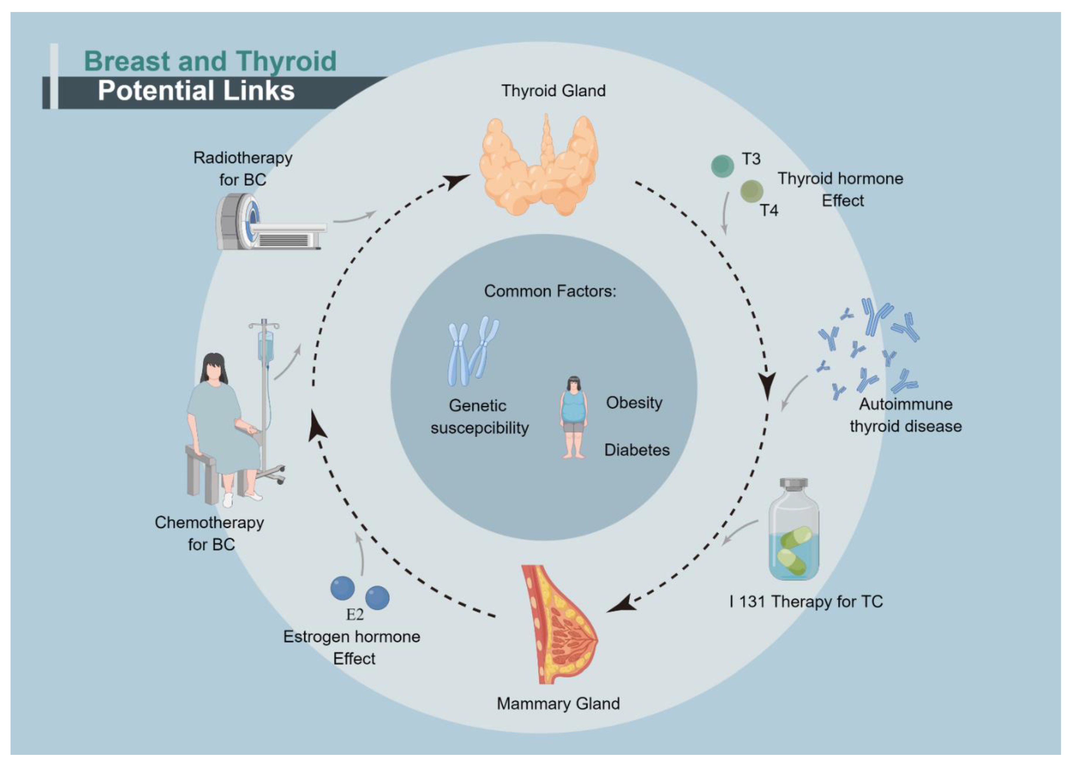
Links between Breast and Thyroid Cancer: Hormones, Genetic Susceptibility and Medical Interventions

Department of Breast and Thyroid Surgery, Renmin Hospital of Wuhan University, Wuhan 430060, China
*
Authors to whom correspondence should be addressed.
These authors contributed equally to this work.
Cancers 2022, 14(20), 5117; https://doi.org/10.3390/cancers14205117
Submission received: 14 September 2022 / Revised: 7 October 2022 / Accepted: 12 October 2022 / Published: 19 October 2022

Abstract

:

Simple Summary

Breast and thyroid cancer are commonly diagnosed in women. Physicians have recognized and evaluated the phenomenon of two cancers occurring synchronously or asynchronously. The potential mechanisms are complex and various. Hormone, autoimmune attack and genetic predisposition are significant and shared factors influencing two cancers. Medical interventions for the first cancer and other life-related factors are reviewed and discussed, as well. This article aims to expound the relationship between breast and thyroid cancer, and suggests that physicians should monitor for the second cancer appropriately whenever one cancer occurs.

Abstract

Breast and thyroid glands are two common sites of female malignancies. Since the late 19th century, physicians have found that the cancers in either thyroid or mammary gland might increase the risk of second primary cancers in the other site. From then on, many observational clinical studies have confirmed the hypothesis and more than one theory has been developed to explain the phenomenon. Since the two glands both have secretory functions and are regulated by the hypothalamic–pituitary axis, they may share some common oncogenic molecular pathways. However, other risks factors, including medical interventions and hormones, are also observed to play a role. This article aims to provide a comprehensive review of the associations between the two cancers. The putative mechanisms, such as hormone alteration, autoimmune attack, genetic predisposition and other life-related factors are reviewed and discussed. Medical interventions, such as chemotherapy and radiotherapy, can also increase the risk of second primary cancers. This review will provide novel insights into the research designs, clinical managements and treatments of thyroid and breast cancer patients.

1. Introduction

Breast cancer (BC) and thyroid cancer (TC) are among the most commonly diagnosed cancers in women, ranking 1st place and 7th place, respectively [1]. Since the late 19th century, the association of the two cancers has been recognized and evaluated by many researchers [2,3]. The earliest study in this field using epidemiological methods was published in 1984 [4]. Ron et al. collected data from 1618 women with primary TC and 39,194 women with primary BC. The standardized incidence ratios (SIR) were 1.68 and 1.89 for TC following BC and BC following TC, respectively. Although the elevated risks have been challenged by some other retrospective cohort studies [5,6,7,8], the discrepancy might be explained by different population screening programs, more accurate screening techniques and selection bias. More recently, data from multi-national large-cohort studies have demonstrated increasing risks with significant differences for TC in patients with primary BC, and vice versa [9,10,11]. A meta-analysis [12] further demonstrated that the odd ratio of secondary TC after BC was 1.55 (95%CI: 1.44–1.67). The odd ratio was somewhat lower for secondary BC after TC (1.18, 95%CI: 1.09–1.26). These studies and their associated standardized incidence ratios are summarized in Supplement Tables S1 and S2. And the level of evidence of these studies cited above is listed in Table S3. Interestingly, the incidence of TC has been revealed to increase in patients with other sex-hormone-related diseases, such as uterine fibroids and benign breast disease [13,14]. Additionally, the elevated risk of BC was also observed in those with autoimmune thyroid diseases [3,15]. Taken together, we can conclude with evidence that TC and BC can mutually increase the risk of each other.
The major question is “what is the mechanism”. Multiple explanations have been proposed by researchers. First, the two glands have secretory functions and are both regulated by the hypothalamic–pituitary axis, implying that they could be influenced by the same hormones (e.g., thyroxine and estrogen) [3,16,17,18]. Autoimmune attack to the thyroid gland, which increases the risk of TC, may also play a role in the oncogenesis of BC. Second, TC and BC share some alteration of geneses of in common, such as PTEN [19], KLLN [20], SDHx [21], PARP4 [22], MANCR [23,24] and VEGF [25]. Thus, genetic susceptibility is believed to cause co-occurrence of BC and TC [26,27,28]. Third, some medical interventions can increase the risk for secondary malignancies. Patients with advanced TC usually receive radioiodine therapy, according to the guideline. External beam radiation and chemotherapy are common therapies for progressed BC. These treatments are assumed to produce intracellular reactive oxygen species, to damage cells and to cause secondary cancers [29,30,31]. Finally, some other factors, including surveillance bias, obesity and diabetes mellitus, may also play roles in the co-occurrence of TC and BC.
The aim of this article is to give a comprehensive review of the association between TC and BC and putative mechanisms (Figure 1).

2. Hormones and Their Receptors

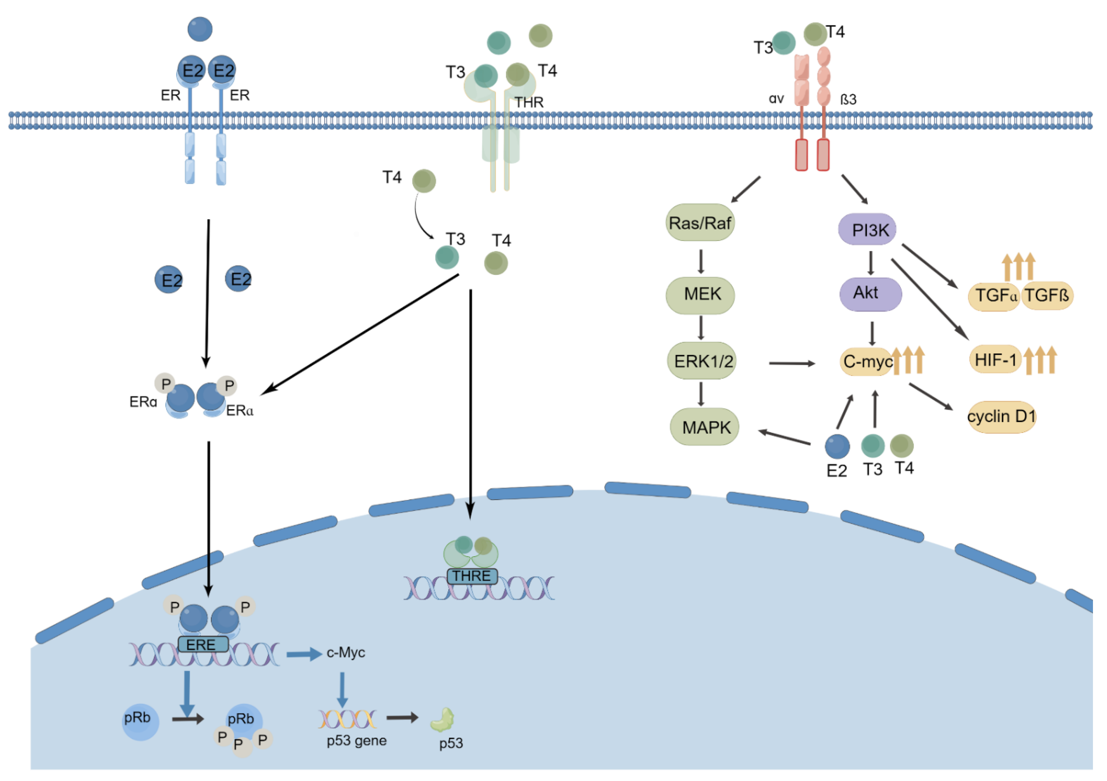
2.1. Thyroid Hormones, Thyroid Hormone Receptor and BC

Thyroid hormones (TH) exert critical effects on skeletal growth, basal metabolism, nervous system development and cell proliferation and differentiation. The mammary glands are target tissues for THs, and their effects are complex. Postsurgical hypothyroidism is a common complication in thyroid cancer patients. Previous studies have suggested that TH dysfunctions, such as hyperthyroidism and hypothyroidism, can affect the risk of glandular epithelium-derived carcinomas [32].
In 1976, Kapdi and Wolfe found for the first time that there was a higher risk of breast cancer in those who received thyroid supplements due to hypothyroidism [33]. From then on, many studies have been performed over the last several decades, and the association between TH and BC remains inconclusive. Data from preoperative observational studies showed that TH and thyrotropin (TSH) are significantly associated with the risks of overall cancer, especially breast and ovarian cancer [15,34,35,36]. Søgaard and colleagues studied a large population-based cohort in Denmark, which recruited 61,873 women diagnosed with hypothyroidism and 80,343 women diagnosed with hyperthyroidism [3]. Standardized incidence ratios (SIRs) of BC increased in women with hyperthyroidism (SIR: 1.11, 95%CI: 1.07–1.16) and slightly decreased in women with hypothyroidism (SIR: 0.94, 95%CI: 0.88–1.00). Moreover, during the follow-up of more than 5 years, the SIRs in women with hyperthyroidism were further elevated (SIR: 1.13, 95%CI: 1.08–1.19), while the SIRs showed no significant difference in women with hypothyroidism (SIR: 0.96, 95%CI: 0.88–1.04). Notably, there are some limitations in this study, such as short administration time and confounding lifestyle factors. Kim et al. examined serum TH concentration in 62,546 healthy Korean females over 40 years of age and 834 incident BCs were observed [36]. Compared to the normal free thyronine (FT4) group, the high FT4 group showed an increasing hazard ratio (HR) for BC (1.98, 95%CI: 1.02–3.83). The association was revealed in postmenopausal women as well. Patients were divided into three groups based on TSH values. Individuals with higher TSH were revealed to have lower risk of BC than those with lower TSH (HR: 0.68, 95%CI: 0.55–0.84). Another prospective study in Sweden showed that FT4 was correlated positively with BC, yet free triiodothyronine (T3) showed no association. The results were consistent with Kim’s study [15,37].
Nevertheless, contrasting results were also presented. By following 2775 women, the study by Kuijpens et al. [16] found that hypothyroidism (OR = 3.8, 95%CI: 1.3–10.9) and the use of thyroid medication (OR = 3.2, 95%CI: 1.0–10.7) were associated with the incidence of BC. Interestingly, among patients without thyroxine supplements, those with FT4 levels in the lowest tenth percentile (OR = 2.3, 95%CI: 1.2–4.6) and TSH in the lowest tenth percentile (OR = 2.9, 95%CI: 1.5–5.7) were both at a high risk of BC. The reason remained unclear. Another case-control study supported the hypothesis that hyperthyroidism and high TH within normal ranges could increase the risk of BC, while hypothyroidism is a protective factor [38]. Some other case-control studies demonstrated there was no significant relationship between BC and TH [39,40]. Recently, a meta-analysis [32] demonstrated that hyperthyroidism was associated with higher risk of BC (pooled risk ratio: 1.20, 95%CI: 1.04–1.38). But hypothyroidism did not increase the risk of breast cancer. Another meta-analysis [41] published in 2021 was in accordance with Søgaard’s study [41]. The study revealed that breast cancer occurred more commonly in hyperthyroidism (OR = 1.12, 95%CI: 1.08–1.16) and less frequently in hypothyroidism (OR = 0.95, 95%CI: 0.91–1.00). The level of evidence of these studies cited above is listed in Table S4. The available evidences have some limitations: the design of retrospective observational studies, residual covariate and detection bias. For instance, once diagnosed with thyroid dysfunction, patients are more prone to have medical visits, therefore, incident cancers are more likely to be detected.
The potential mechanisms between thyroid dysfunction and BC were studied by a relatively small number of studies [18,26,42,43,44,45]. The most proposed mechanism is that TH can initiate nongenomic actions via activating nuclear thyroid hormone receptor (THR) [46]. It can also cross-talk with other receptors, such as estrogen receptors (ER), progesterone receptors (PR), and human epidermal growth factor receptor 2 (HER-2) [42]. Firstly, Moretto FC demonstrated that T3 could induce the high expression of hypoxia inducing factor 1 (HIF-1) and transform growth factor alpha (TGFα) in the MCF7 breast cancer cell line by activating the PI3K pathway [47]. The process increases the aggressiveness and malignancy of BC. Secondly, TH can promote cell growth, which is similar to estrogen (E2). In Hall’s study [48], both T3 and E2 could promote cell proliferation in a dose-dependent manner in the MCF-7 and T47-D cell lines. Although the effect of TH was less strong, the activation of ERE-mediated gene expression in MCF-7 cells by T3 was proven. In BC cells with positive expression of ER, T3 treatment also increased the P53 level and induced Rb hyperphosphorylation, while an ER antagonist blocked these effects [49]. Additionally, T4 could also induce the serine phosphorylation of ERα, which then resulted in DNA binding and transcriptional activation. In addition, a significant crosstalk between the two hormones was proved. T3 enhances aerobic glycolysis (Warburg effect) of E2 in triple negative breast cancer cells, which was on behalf of transformed cells [50]. Finally, the fast signal pathway of avβ3, which mediates the balance between apoptosis inhibitors and promotors, was influenced by T3 [51]. Programmed death ligand 1 (PD-L1) gene expression was impacted by T4 via activating ERK1/2 [52] (Figure 2).
There are two subtypes of THR in human bodies: THRα and THRβ. They show adverse effects in the prognosis of BC patients. Heublein et al. found that THR was associated with BC patients with BRCA1 gene mutations [53]. THRβ showed a significant positive correlation with their five-year and overall survival rate, while THRα showed an adverse effect. In addition, according to a retrospective statistical analysis, higher expression of THRα1 showed a significantly worse disease free survival (DFS), while THRα2 expression may predict a better outcome of BC [43,45] Recently, Wahdan-Alaswad et al. [18] reported that TH treatment significantly and independently reduced disease-free and breast cancer-specific overall survival in steroid receptor positive BC patients in a long-term observational study. In their in vivo and in vitro experiments, TH treatment altered the nuclear colocalization of ER and THR. To conclude, THR and postsurgical thyroid dysfunction are important factors in the co-occurrence of BC and TC.

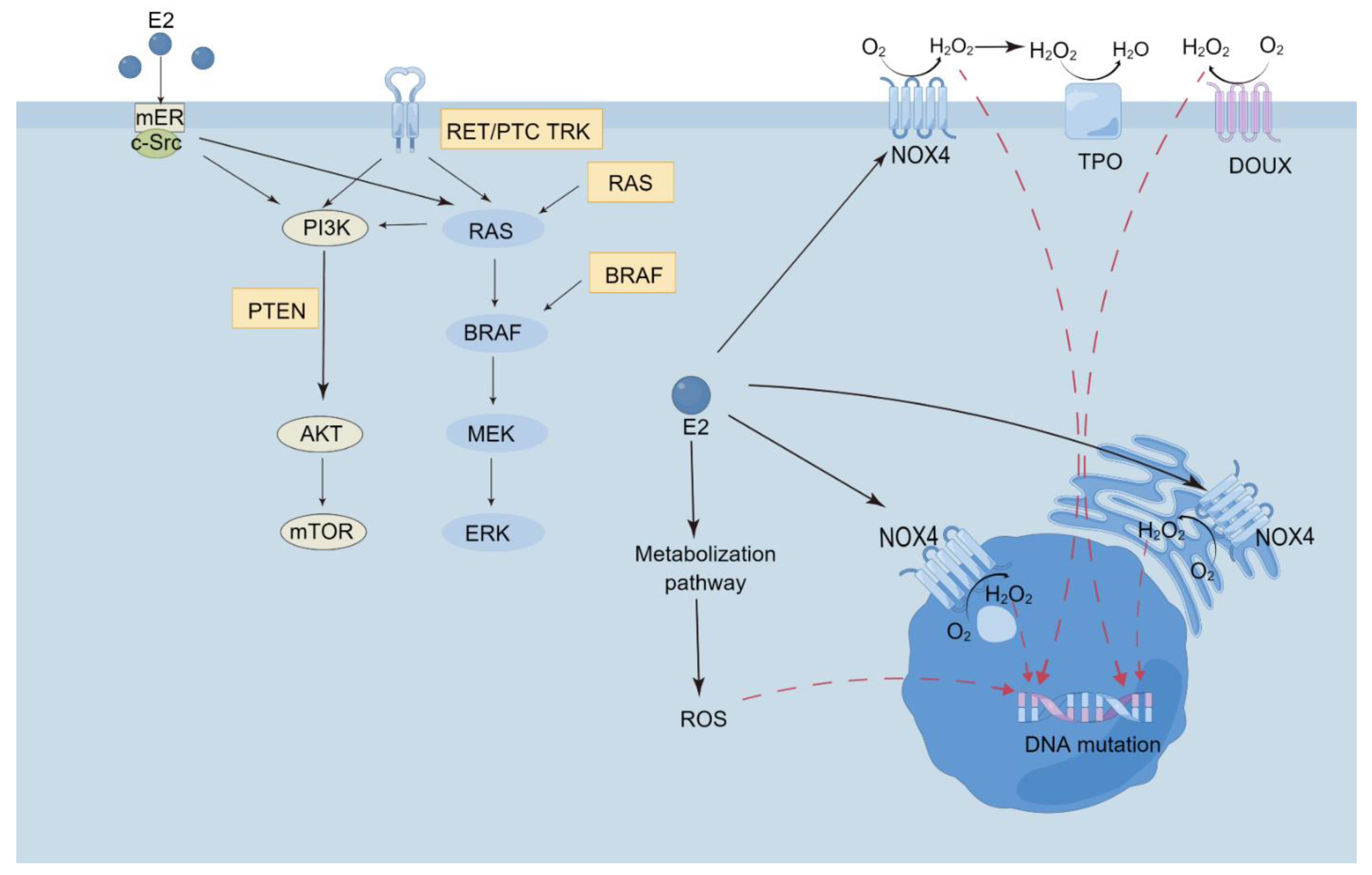
2.2. Estrogen, Progestin, and Its Receptors: ER/PR/HER-2 and TC

The most common cancer in women is BC, which is closely related to estrogen and progesterone. The prevalence of benign and malignant thyroid tumors occurs four or five times more in females than in males [54,55,56]. Notably, the difference is less obvious before puberty and after menopause [57]. Although the intrinsic causes of the gender discrepancy in thyroid disease have not been sufficiently elucidated, sex hormones are suspected to play a role.
Estrogen, a steroid hormone, exerts a pivotal impact on the regulation of body growth and the development of the immune system and reproductive organs. Patients with uterine fibroids, which are closely related to estrogen, had a significantly increased risk of thyroid cancer (HR = 1.64, 95%CI: 1.26–2.13), irrespective of whether or not they took myomectomy [14]. Persistence and recurrence of TC was significantly higher in patients who were diagnosed during pregnancy or within the second year after delivery, in comparison with the control group (p = 0.023) [58]. However, in a recent nationwide cohort study performed by Kim et al. in Korea [59], the finding did not support the theory that lack of estrogen is a protective factor. The risk of TC was higher after patients experience hysterectomy and oophorectomy, and there was no significant correlation between oral contraceptives and TC [60]. The level of evidence of these studies cited above is listed in Table S5.
These observative results were contradictory. Ex vivo studies provide more information. First, in the classical genomic pathway, the action of estrogen is mediated via two types of estrogen receptors, ERα and ERβ, which enhance DNA synthesis and proliferation. In another fast non-genomic pathway, a membrane-associated estrogen receptor (mER) is involved [61]. In the genomic pathway, estrogen enters the cell and transforms into estrogen-ER complex, which promotes the expression of target genes by binding an estrogen-responsive element (ERE) [62]. In the non-genomic pathway, E2 exerts function by mER, which activate MAPK and the PI3K signaling pathway. E2 also synergistically promotes the activation of the tyrosine kinase pathway in cells with RET/PTC fusion and BRAF mutation [61]. Second, thyroid cancer cells are replicated from mutated thyroid stem cells or progenitor cells, rather than primary thyroid cells [63]. In Xu’s study, the level of Erα in thyroid stem and progenitor cells was eight times higher than normal thyroid cells [64]. Third, Estrogen stimulates ROS production by NOX4, and ROS can reach the nucleus and contribute to thyroid carcinogenesis [65]. Mutual effect between thyroid redox homeostasis and estrogen in the development of thyroid carcinogenesis is another potential pathway [65] (Figure 3).
The expression of ER in thyroid epithelium cells is debatable. Some researchers failed to find ERs with immunohistochemical staining in normal thyroid tissue and cancer tissue [66,67]. Nevertheless, others detected ER on benign thyroid tissue [68]. With advanced techniques, ERα and PR expression was found by Vannucchi in 66.5% and 75.8% of patients, respectively, in 182 patients with papillary thyroid cancer [69]. It was the first time to find the occurrence of the ‘receptor conversion’ phenomenon in TC [69]. Overexpression of ERα in cancer tissue and lack expression of ERβ in surrounding tissue were reported in 2011 [70]. Low or lack of ER can be viewed as a hallmark of thyroid carcinomas. Heikkila found that ERβ expression was significantly lower in follicular thyroid cancer than in follicular adenomas [71]. Similarly, undifferentiated thyroid stem and progenitor cells expressed ERβ in a low level compared with differentiated human thyrocytes [64]. Therefore, ER expression in a low level may indicate dedifferentiation in thyroid cancer [72,73]. Tafani et al. found that HIF-1 and kB (NFkB), which mediate the immune and inflammation process, helped to transform thyroid cells to the malignant phenotype [74]. Importantly, HIF1a also regulated the ERα expression [75]. Tafani et al. proposed a hypothesis that ERα linked the two transcription factors in the progression of thyroid cancer [74].
HER-2 is an essential marker in the molecular target and prognosis of breast cancer. HER-2 expression in TC was also debatable, varying between 0% and 83.8% [76,77,78,79,80]. In 2001, Kim et al. found that HER-2 overexpression in thyroid cancer (83.8%) was more frequent than in benign tumors (16.7%), and was related to the reduction of PTEN protein [76]. Ruggeri et al. analyzed the HER-2 expression in differentiated thyroid cancer (DTC). The HER-2 overexpression was found in 44% (20/45) of the patients with follicular thyroid cancer (FTC) and 18% (8/45) with papillary thyroid cancer (PTC). After a median nine-year follow-up, six patients developed metastatic disease and five of them had HER-2-positive tumors [77]. The results from Tang et al. was consistent with Ruggeri’s study [78]. The positive HER-2 expression (45.71%) in PTC was significantly associated with lymph node metastatic (p < 0.05). Nevertheless, contradictory results are reported, as well. In Apostol’s study [79], the HER-2 expression was proposed to be a low risk factor. HER-2/neu positivity was found in twenty-five (20.8%) cases with twenty cases of high-risk and five cases of low-risk. Additionally, Mdah et al. demonstrated that the positive rate of HER-2 was just 6.9% in PTC [80]. The possible reasons for the HER-2 expression discrepancy might be varied size of sample, the general characteristic differences and the scoring methods. Large and independent researches with a unified estimated method of HER-2 are needed to provide more information.

3. Autoimmune Thyroid Disease and BC

Autoimmune thyroid diseases (ATD) include Graves’ disease and Hashimoto’s thyroiditis. The two specific biomarkers for Hashimoto thyroiditis, thyroid peroxidase antibody (TPOAb) and thyroglobulin antibody (TgAb), present in 90% patients [81]. Thyroid-stimulating hormone antibody (TSHRAb), which can bind with TSHR and increase the synthesis and release of TH, is a specific biomarker for Graves’ disease. Broadly speaking, the proposed mechanisms between ATD and cancer can be explained in two ways. On one hand, disordered immune system fails to eliminate cancerogenic cells. On the other hand, damaged immune system is prone to attack normal cells and abnormal cells indistinctively [82].The high rate of BC in ATD women was observed in 1975 [83]. Researchers recognized 18 BCs in 1810 cases with Hashimoto’s thyroiditis which was far more than the expected number (3.19 cases). The relationship between ATD and BC has been researched since then.
TPO is a member in the family of mammalian peroxidases. The family includes lactoperoxidase, myeloperoxidase, and so on. TPO express weakly in breast tumor and peritumoral tissue [84,85,86]. In 1996, Giani [87] evaluated 102 BC patients and 100 age-matched control healthy women. All patients experienced iodine deficiency. Hashimoto’s thyroiditis was found in 13.7% of BC patients and in only 2% of the controls (p < 0.005). And the detection rate of TPOAb was higher in BC patients than in the controls (23.5% vs. 8%, p < 0.005). They postulated that iodine deficiency might be an important factor in the oncogenic process. Subsequently, other studies recruiting BC patients showed similar results, demonstrating that TPOAb were detected more frequently in BC patients than in the controls [88,89,90,91,92]. Giustarini [90] detected TPOAb in 36 BC patients, 25 with breast benign disease (BBD) and 100 healthy women. The prevalence of TPOAb in BC patients (12/36, 33.33%) was significantly higher than in BBD patients (5/25, 20%) (p < 0.01) and in the controls (8/100, 8%) (p < 0.01). Since the study was retrospective, the diseases sequence remained unclear. A prospective study was conducted by Kuijpens in 2005 [16]. An unselected cohort of 2775 women around menopause was screened for TPOAb in 1994. There was an independent relationship between BC and the detection of TPOAb (OR = 3.3, 95%CI: 1.3–8.5). Contrary to expectation, the presence of TPOAb showed no relationship with BC after 9 years follow-up (OR = 1.1, 95%: 0.4–2.7). These results were in line with Hedley’s study [93], which revealed no correlation between ATD and BC. TPOAb may be the first sign of thyroiditis induced by irradiation or chemotherapy for BC. Another explanation is that the presence of TPOAb is the immune response of the tumor itself. Third, patients’ fear and stress due to BC may influence the immune system [16]. Recently, a meta-analysis showed that TPOAb presence was significantly associated with an increased risk of BC [94] (OR = 2.86, 95%CI: 2.17–3.77, p < 0.001). In summary, a correlation exists between TPOAb and BC. The discrepancy between studies might be explained by the small sample size, the different racial make-up of the study cohort, the diagnosis criteria, and lifestyle differences. A large prospective cohort study is needed to ascertain the association between TPOAb and BC.
TgAb is another indicator for ATD. Similar results have been presented. Some cross-sectional studies confirmed the link between TgAb and BC [90,91]. Giustarini [90] showed that the rates of TgAb in the BC patients (33.33%) was higher than of the BBD patients (16%, p < 0.01) and in the controls (12%, p < 0.01). However, contrary results were also demonstrated. The detection of TgAb was higher in BC patients, without significant difference (16.6% vs. 12%) [87]. The possible reason might be the small sample size. A prospective study is lacking. A meta-analysis in 2020 [94] demonstrated that TgAb was associated with BC (OR = 2.71, 95%CI: 1.81–4.05, p < 0.001). Thus, ATD shows a connection with BC. Further and prospective studies are also required.
Few studies have evaluated the relationship between TSHRAb and BC. In 2013, Szychta [95] found that Graves’ disease and BC was statistically correlated. This study detected TSHRAb in 9 BC patients, 47 BBD patients and 1630 women hospitalized for several non-oncological diseases. The mean values of serum TSHRAb were statistically higher in BC patients than in the controls (p = 0.0006). In univariate regression analysis, breast cancer had a predictive value for TSHRAb (OR = 1.10, 95%CI: 1.01–1.20, p = 0.0222). In addition, TSHR expression is common in BC (34/44 cases, 77%) [96]. No unanimous conclusion has been drawn concerning TSHRAb in BC patients. The shortcoming of Szychta’s study is that the sample was too small to trust. Large and prospective research is required to verify the association of TSHRAb and BC. The level of evidence of these studies cited above is listed in Table S6.
Moreover, sclerosing lymphocytic lobulitis (SLL), a rare benign disease of the breast, was viewed as an autoimmune disease of the breast and strongly associated with other autoimmune disorders [97]. SLL of breast and thyroid microsomal antibodies was first reported by Soler and Khardori in 1984 [98]. Dubenko, M. reported a case described the association of SLL of the breast with Graves’ disease [97]. Park also published a case-report about SLL of breast in patient with Hashimoto’s thyroiditis [99]. Interestingly, compared to healthy people, lobulitis was encountered in 21 of 41 (51%) patients at hereditary high risk of BC [100]. Recently, Hieken found moderate or severe lobulitis was more common in BC (73%) than benign disease (13%) (p = 0.003) [101]. Published case-reports about SSL of the breast and ATD are few. But the viewpoint that SLL of the breast was strongly linked autoimmune disorders was common in many authors. More studies concentrating on SLL and ATD are needed to illuminate the issue.

4. Iodine, Sodium Iodide Symporter and BC

Thyroid and breast both need iodide to produce iodoproteins, which participate in the biosynthesis of TH and breast milk as a source of neonatal nutrition [102].
There is also an interesting observation that the breast cancer incidence was relatively low in Japanese women. This is likely due to their seafood-rich diet [103]. Funahashi’s study revealed that Lugol’s iodine or iodine-rich Wakame seaweed is a protective factor in rats with BC [104].
Sodium iodide symporter (NIS), a membrane-bound glycoprotein, is located in the basolateral cell membrane. Its function is to transport and accumulate iodide ion (I−) into cells. NIS mediates the active uptake of I− in the thyroid, which is the crucial step in thyroid hormone biosynthesis. Other than the thyroid, NIS can mediate I− uptake in other tissues, such as salivary glands, gastric mucosa, and lactating glands. More than 80% BC samples and 23% peri-tumor breast samples are observed to express NIS [105]. Benign breast diseases, such as fibroadenoma, show a higher expression of NIS proteins and accumulation of iodide [106,107]. Thus, it is difficult to regard NIS as a specific indicator of BC [17,105]. Relatively higher expression of NIS does not indicate higher iodide uptake due to the dislocation in cytoplasm rather than cell membrane. Another explanation of this phenomenon is that the NIS may be overestimated due to the defective method of detecting NIS protein with polyclonal NISAbs [17,108].

5. Oncogenic Effects of the Therapies for Primary Cancer

5.1. Radioactive Iodine Therapy and Breast Cancer

Since the 1940s, radioactive iodine (RAI) has been used in the treatment of hyperthyroidism. Postsurgical RAI therapy is also used in patients with cervical lymph node metastasis, distant metastasis and extrathyroidal extension. RAI can be transported into thyroid epithelium cells via NIS to perform the tumoricidal effect. NIS is also found to be expressed in the breast, salivary lacrimal gland, ovaries and gastric mucosa [109]. Furthermore, BC cells have been proven to have functional expression of NIS [110]. Concerns have been raised with respect to radioactive iodine therapy, given the possible increased incidences and mortality of second primary malignancy, including breast cancer.
Some studies shed light on evaluating cancer risk after RAI, and their results are conflicting [111,112,113]. The large long-term follow-up analysis by Kitahara [114] included 18,805 patients. The study revealed that for every 1000 patients after RAI therapy, an estimated 18 to 30 deaths due to solid cancer would occur (4 to 6 were BC). A larger part of these deaths would occur more than 20 years after RAI treatment. By developing a biokinetic model [115], the high-quality, individualized organ- and tissue-specific dose estimation were obtained. The association between RAI treatment and the mortality of BC was revealed (RR = 1.12, 95%CI: 1.003–1.32, p = 0.04) at the dose of 100-mGy. However, the study did not take covariates into considerations, such as smoking, obesity and alcohol use and concomitant diseases. In 2017, Silva-Vieira [116] evaluated second primary cancer (SPC) incidence in 2031 patients with/without DTC receiving RAI treatment, with a median follow-up period of 8.8 years. A total of 130 SPC were diagnosed and the most common cancer was BC (31%). Compared to control groups, a statistically significant high risk of SPC in RAI treatment was found (RR = 1.84, 95%CI: 1.02–3.31, p = 0.026). Notably, an increasing incidence of SPC by year was revealed. The 10-year cumulative incidence rates of SPC in groups who received 0, <100, 100–199, 200–299, and >300 mCi were 4.4%, 3.9%, 7.5%, 11.8%, and 10.9%, respectively. Compared to no RAI group, the relative risk of SPC in 200–299 and >300 mCi group was 2.43 (95%CI: 1.17–5.01) and 2.29 (95%CI: 1.03–5.08), respectively. The incidence of breast cancer in patients with thyroid cancer receiving RAI treatment increased compared to the controls. Yet there was no significant elevation compared different cumulative dose of RAI [29]. The author postulated that increasing sodium-iodide symporter (NIS) expression before RAI is responsible for carcinogenesis, rather than radiation exposure. Chen proposed that immortal time bias might bring about false results [117].
Different opinions on postsurgical RAI in thyroid cancer patients still exist [113,118]. Recently, a meta-analysis showed no statistically significant elevations in the risk of SPC (OR = 1.02, 0.95–1.09) [119]. However, in a dose-response analysis based on two original studies, RAI was significantly associated with BC (p = 0.03) [119]. This study suggested that only high cumulative doses led to increased risk of SPC. Thus, the dose of RAI is needed to take into careful consideration in clinical practice. And more researches exploring rational and safe dose of RAI is needed. Because of the lengthy latent period of some solid cancers, a long-term follow-up is required. As the most common of SPC, BC may have particular mechanisms related to RAI.

5.2. Chemotherapy and Thyroid Cancer

Chemotherapy is a conventional and crucial treatment for breast cancer. The application and dose of chemotherapeutic drugs is under strict management, due to their potent adverse effect on normal organs and cells [120]. Therefore, thyroid function seems to be vulnerable to chemotherapeutic drugs. Normal thyroid function may be impacted due to cell death. Another theory suggested that hypothalamus–pituitary–thyroid axis down-regulation was an adaptation to adverse physical conditions in ill patients [121]. A low TH level of protects the body against tissue damage by down-regulating cellular metabolism [122].
A number of studies found that thyroid dysfunction occurs in patients who receive systemic chemotherapy. However, no unanimous conclusion was reached. Kailajärvi [123] performed a study testing FT3, FT4 and TSH in 15 women receiving cyclophosphamide, methotrexate and 5-fluorouracil chemotherapy. The results revealed that T3 and T4 declined temporarily but the TSH level had no alteration. Kumar [124] designed a prospective observational study to evaluate TH change in 198 BC patients. Patients received systemic chemotherapy agents of cytoxan, adriamycin and 5-fluorouracil. A significant reduction in mean serum T3 uptake (p < 0.05) was observed. Other studies show that T3 and T4 declined but TSH increased [122,125]. In 2015, de Groot [122] obtained serum samples at baseline, before the 2nd chemotherapy cycle, and at end of neoadjuvant treatment with docetaxel, doxorubicin and cyclophosphamide. FT4 levels decreased (p = 0.0001) and TSH levels increased significantly (p = 0.019). It is reasonable that lower FT4 level was regarded as a protective response [122]. A systematic review showed that T3 and T4 levels decreased after chemotherapy but TSH remained unchanged [126]. Hence, a routine screening for TH needs to be taken into consideration in BC patients after chemotherapy. Although there are studies focusing on thyroid dysfunction, few researches have evaluated the incidence of second primary cancer and different chemotherapy regimens. The potential reasons might be that chemotherapeutic agents aims to kill malignant cell and microenvironment of systematic treatment is not appropriate for cancer growth and invasion.

5.3. External Beam Radiation, Mammography and Thyroid Cancer

Postoperative radiotherapy for breast cancer can decrease the risks of local recurrence and death. This adjuvant treatment is used in patients receiving breast conserving surgery (BCS) and mastectomy with axillary lymph node metastasis. With a mean 5-year relative survival rate of over 80%, the majority of women were observed to have one or more treatment-induced SPCs. The balance between the expected benefit and the risk of SPC needed attention. Thyroid cancer and radiation have close relation. Data from Chernobyl showed that the risk of thyroid cancer in neighborhood has increased since the accident [127,128]. Besides, patients with Hodgkin’s lymphoma who received radiotherapy above the diaphragm have a a higher risk of second thyroid cancer [129,130].
Radiotherapy for breast cancer inevitably exposes the adjacent normal organs to unwanted radiation, with a gradual dose-fall outside the field-edge [131]. A national population-based study including 46,176 patients treated for early BC was conducted by Grantzau’s team in 2013 [132]. They classified SPC into two groups: radiotherapy-associated sites and non-radiotherapy-associated sites. The former involves esophagus, lung, heart/mediastinum, pleura, bones, and connective tissue. The other cancers, thyroid, stomach, liver and so on, are included in another group. The SPC of the first group showed an increased risk (HR = 1.34, 95%CI: 1.11–1.61) and the HR was even higher when the follow-up period was extended to 15 years. There was no increased risk for the second group (HR = 1.04, 95%CI: 0.94–1.1). The thyroid gland was exposed to considerable doses (>26 Gy) in breast radiation treatment in a retrospective study [133]. In 2017, Burt [134] used the SEER database to obtain data from 374,993 BC patients. A total of 154,697 patients received external beam radiation. The rate of SPC was significantly greater than the endemic rate in BC patients without radiation therapy (SIR = 1.2, 95%CI: 1.19–1.22). However, the rates of second primary thyroid cancer in radiation group showed no statistical significance (SIR = 1.09, 95%CI: 0.97–1.22). A systematic review and meta-analysis including 522,739 patients revealed that pooled incidence of second TC at ≥15 years after irradiation for BC were 3.15 [30]. The risk of second thyroid cancer for non-radiated patients increased without significant difference [30]. In summary, there was no evidence to support the theory that patients receiving radiation are at higher risk for TC.
The incidence of hypothyroidism increases in BC patients after postoperative radiotherapy [135,136,137]. A study aimed to assess the association of different radiation targets with hypothyroidism [135]. By comparing groups of regional node irradiation (supraclavicular lymph node) and breast only, the cox model revealed an adjusted hazard ratio of 2.25 (95%CI, 1.49–3.38). The risk was more prominent in patients aged under 60 years. Thus, a routine examination of the thyroid is needed, especially 10–15 years after the initial treatment of breast cancer radiation. Physicians need to balance the expected benefits and long-term side effects after radiation in patients with early-onset BC.
Concern over thyroid exposure during mammography has been expressed. Thyroid shields are supposed to protect during mammography. Sechopoulos [138] demonstrated that thyroid exposure had a maximum average effective dose of 0.13 μSv from digital, and 0.17 μSv from film-screen mammography. The cumulative lifetime risk of TC is approximately 56 per billion (or 1 in 17.8 million) if individuals received serial annual screening mammography examinations between the ages of 40 and 80 years. This effect to the thyroid is negligible. In a large population-based study [139], there was also no evidence supporting the higher risk of TC in the group who received mammography (HR = 1.2, 95%CI: 0.81–1.77). Therefore, mammography is unlikely to be a possible factor increasing the risk of TC. The level of evidence of studies cited about Oncogenic effects of the therapies for primary cancer is listed in Table S7.

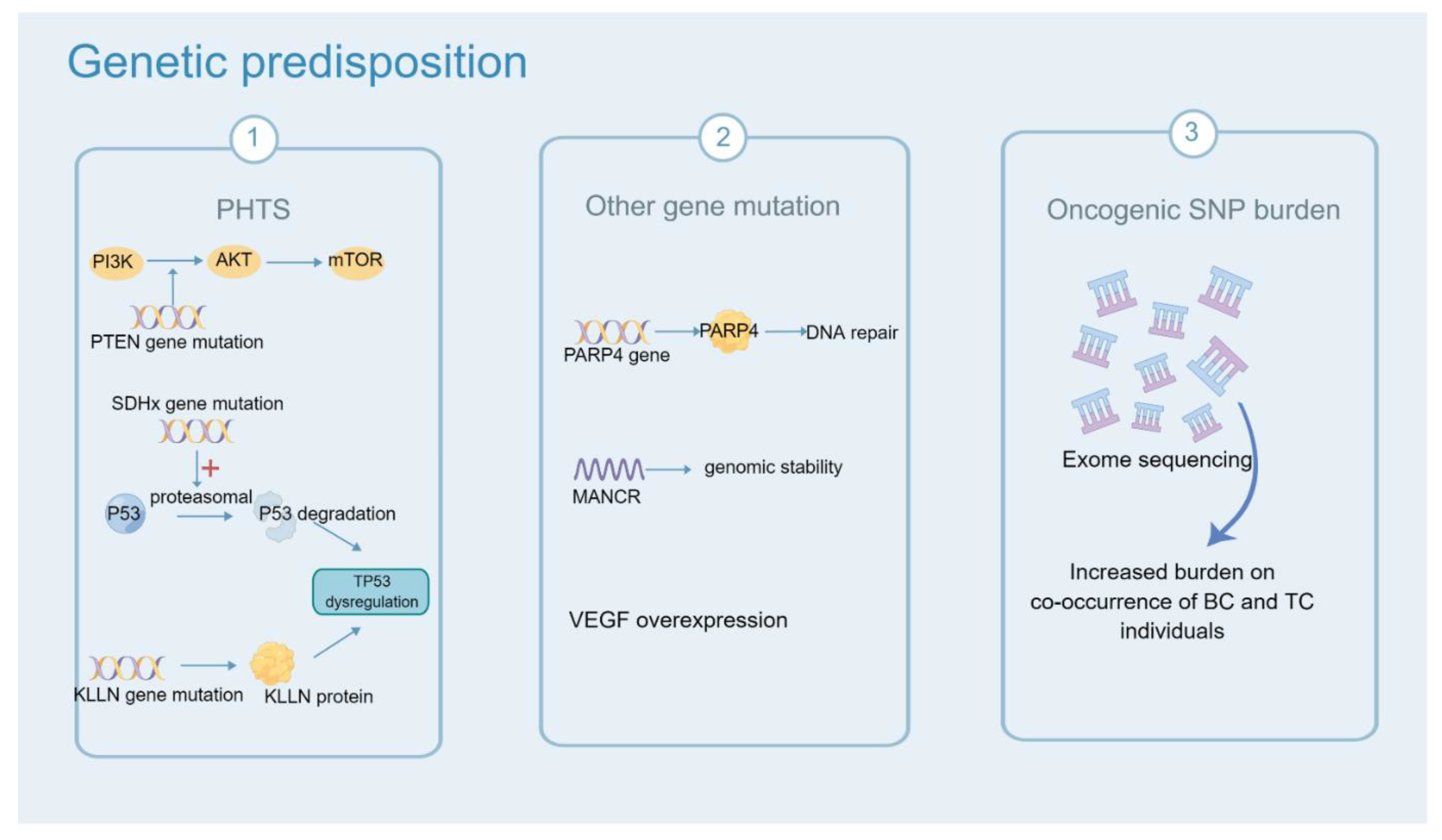
6. Genetic Predisposition

Not all second primary cancers are ascribed to molecular immunization environment, hormones or oncogenic effects of the therapies for the first cancer. Some sporadic cases may be attributed to genetic susceptibility and lifestyle factors. A large retrospective study among twins in Nordic countries revealed that hereditability accounted for approximately 33% of cancer risk [140]. A familial link between thyroid and breast cancer was found by a study based on the Swedish Family-Cancer Database [141]. The incidence of thyroid cancer increased in individuals who have ≥ two first-degree relatives with BC (RR = 1.90, 95%CI: 1.38–2.63). The associations between ovarian and prostate cancers are known because of the BRCA1/2 mutation and hormonal effects. But the genetic association with BC and TC was novel with the current statistical support [141].
PTEN hamartoma tumor syndrome (PHTS, comprising Cowden, Bannayan-Riley-Ruvalcaba, and Proteus-like syndromes). PHTS are featured as an increased risk for several solid malignances, including breast cancer, thyroid cancer, colorectal cancer, endometrial cancer and melanoma [142]. The standardized incidence of BC and TC ranged from 6 to 9 [143]. PHTS is due to germline mutations of tumor suppressor gene: phosphatase and tensin homolog (PTEN). It inhibits the catalytic activity of the enzyme PI3K and then upregulates the PI3K-AKT pathway, which facilitates the survival, proliferation and migration of tumor cells [19,144]. In a 7-year multicenter prospective study [143], 2024 patients with invasive cancer histories were included and 5.6% of them were detected to have PTEN mutations. Compared to the general population, the risk of second BC and TC significantly increased (SIR = 8.92, 95%CI: 5.85–13.07; SIR = 5.83; 95%CI: 3.01–10.18). Additionally, PHTS can be induced by mutations of KLLN and the succinate dehydrogenase complex (SDHx). SDHx participates in the composition of mitochondrial complex II, which transports electrons in Kreb’s cycle. SDHx mutations can dysregulate TP53, which induce apoptosis by upregulating the proteasomal degradation of p53 [21]. In Ni’s research [21], germline variations of SDHx occurred in 8% (49/608) of individuals with no PTEN mutation and 6% (26/444) of individuals with Cowden syndrome. It was not found in the control group of 700 individuals. Of note, the group with only SDHx mutation showed the highest TC incidence. KLLN, a transcription factor, shares a bidirectional promoter with PTEN and encodes KLLN protein [20].Patients with a KLLN mutation had a 3-fold higher incidence of BC (p < 0.0001). Ngeow et al. showed that the SIR of TC was 45 for KLLN mutation in individuals with PHTS (95%CI: 26–73, p < 0.001) [145]. New variants and mutations will continue to be identified.
In addition, PARP4 is another shared factor in patients with both BC and TC [22]. PARP4 encodes poly-ADP-ribose polymerases (PARPs) and is an important component of DNA repair. In 2016, Ikeda [22] found that the variants in PARP4 gene were detected at high frequency (OR = 5.2, p = 0.00001) in genomic analysis among 14 patients with co-occurrence of BC and TC. Epigenetic changes in the genome are worth taking into account. Long non-coding RNAs (lncRNA) named MANCR (mitotically-associated long noncoding RNA), which play a role in cell proliferation, viability, and genomic stability, are important regulators in the genomic stability of aggressive breast cancer [24]. Many long noncoding RNA(lncRNAs) are differentially expressed in TC tissues when compared with normal tissues [23]. Angiogenesis in patients with both two primary cancers has been illustrated. Wei [25] found that the vascular endothelial growth factor (VEGF) was overexpressed in thyroid and breast carcinomas in 14 patients with BC compared with the benign breast disease group (p < 0.01). The incidence of microvascular angiogenesis in TC increased in patients with a history of BC. Thus, contrast-enhanced ultrasound is a promising tool to examine the thyroid gland if the patient has a history of BC. However, the sample of this study was small. Large and multicentric research is needed to verify this result.
Some cases were associated with monogenic disorders of autosomal inheritance, but the majority cases of co-occurrence of BC and TC was considered to be polygenic. A recent retrospective case-control study is in line with hypothesis of genetic predisposition [27]. Bakos [27] compared the genetic profile of 15 cases with breast cancer and 19 cases with co-occurrence of the two cancers by using whole exome sequencing. The level of evidence of studies cited about genetic predisposition is listed in Table S8. Increased oncogenic single nucleotide polymorphism burden was associated with co-occurrence of BC and TC. This research further confirmed that the genetic predisposition plays a significant role in tumorigenic progression (Figure 4).

7. Other Factors

The International Agency for Cancer Research has demonstrated the correlation between being overweight or obese and 13 types of cancer [146], including postmenopausal BC and TC. The obesity groups whose BMI ≥ 35kg/m2 were at the highest risk of invasive BC (HR = 1.58, 95%CI = 1.40–1.79) [147]. Obesity is also related to advanced BC, including larger tumor size, positive lymph nodes, regional or distant stage, and deaths [147]. Ewertz [148] found that being obese or overweight may represent a modifiable risk factor in BC occurrence and progressive. Engeland [149] reported that the rates of TC increased moderately with increasing BMI and height in males and females (RR = 1.29, 95%CI = 1.13–1.46). Kitahara [150] further demonstrated central adiposity influenced high incidence of TC. Some other types of studies supported the association between the risk of TC and obesity [151,152,153]. In 2013, Kim [154] reported a strong relationship between BMI and aggressive features, including larger tumor size (OR = 1.31, p < 0.001), advanced TNM stage (OR, 1.30, p = 0.003) and extra-thyroidal invasion (OR 1.23, p = 0.006). The great prognosis of TC after surgical therapy may be a possible reason that the harm of obesity is too weak to change it. Being obese or overweight are linked with the incidence and pathological features of TC.
It is well-known that excessive adiposity is able to progressively cause a series of co-morbidities. Park [155] found triple-negative BC was in a higher risk in the T2D group (HR = 1.40; 95%CI = 0.90–2.16). Interestingly, longer duration metformin use for T2D showed a protective role in ER-positive BC (HR = 0.62; 95%CI = 0.38–1.01; p = 0.09). Moreover, a study involving 8482 patients in 2021 [156], demonstrated that the prognosis of BC was better in patients who persisted in maintain a diabetes risk reduction diet. This diet decreased the risk of BC-specific mortality (HR = 0.80; 95%CI = 0.65–0.97; p = 0.02) and risk of all-cause mortality (HR = 0.66; 95%CI = 0.58–0.76; p < 0.0001). Meanwhile, the connection between diabetes mellitus and TC was observed. In a study that applied Mendelian randomization, the casual link between diabetes mellitus and TC was confirmed [157]. Compared to individuals in the lowest quartile for genetic liability of T2D, higher odds of TC were found in the highest quartile (OR, 1.45; CI, 1.11–1.90). Diabetes mellitus may be a conceivable factor connecting BC and TC. And the level of evidence of these studies cited above is listed in Table S9.

8. Conclusions

The association between BC and TC has been evaluated. Patients who have either cancer history are at an increased risk of the other second primary cancer compared to the general population. “What is the mechanism?”, this problem has been illustrated and explored partially. The shared common features may be the etiologies and possible causative factors of BC and TC. For example, the hormone effects of TH and E2, autoimmune attack, genetic predisposition and other life-related factors. However, some results remain inconsistent. Well-designed and large cohort studies are needed to prove the causative factors linking BC and TC. Further investigation into gene mutation and disordered gene expression underlying BC and TC development is promising. Complicated, different, and cross-talk signal pathways exploration is needed as well. On one hand, RAI therapy should be taken into consideration by clinicians when balancing the benefits and risks. On the other hand, systematic chemotherapy and partial external beam radiation can both affect the thyroid gland. Systematic chemotherapy and immunity therapy lack convincing evidence to support their relation with TC. Large cohort studies are needed to evaluate the oncogenic effect of external beam radiation on certain regions. Common tumorigenic pathways to BC and TC and shared risk factors can be screened. The studies on co-occurrence of BC and TC can reveal the biological behavior of two cancers and provide novel treatment strategies, which might guide clinical practice in the future.

Supplementary Materials

The following supporting information can be downloaded at: https://www.mdpi.com/article/10.3390/cancers14205117/s1, Table S1: Standardized incidence ratios of thyroid cancer after breast cancer; Table S2: Standardized incidence ratios of breast cancer after thyroid cancer; Table S3: Level of evidence about Thyroid cancer and Breast cancer; Table S4: Level of evidence about Thyroid Hormone and Breast cancer; Table S5: Level of evidence about Estrogen and Thyroid cancer; Table S6: Level of evidence about Autoimmune thyroid disease and Breast cancer; Table S7: Level of evidence about Oncogenic effects of the therapies for primary cancer; Table S8: Level of evidence about Genetic predisposition; Table S9: Level of evidence about other factors and Breast cancer. And the level of evidence was classified by standard of OCEBM (Centre for Evidence-Based Medicine, University of Oxford, OCEBM). References [157,158,159,160,161,162,163,164,165,166,167,168,169,170,171,172,173,174,175,176,177,178,179,180,181,182,183,184,185,186,187,188,189,190,191,192,193] are cited in the supplementary materials.

Author Contributions

S.S. and C.C. conceived the idea. M.L., H.L. and B.Z. contributed to the writing of manuscript. M.L. and H.L. contribute to the proof reading, editing and figure preparation for the manuscript. All authors have read and agreed to the published version of the manuscript.

Funding

This research was supported by the grants from Beijing Xisike Clinical Oncology Research Foundation (Y-SY201901-0189), the Fundamental Research Funds for the Central Universities (2042019kf0229), the Science and Technology Major Project of Hubei Province (Next-Generation AI Technologies) (2019AEA170), and the National Natural Science Foundation of China (grant no. 82103671).

Conflicts of Interest

The authors declare that there are no conflict of interest. Figures were created by Figdraw (www.figdraw.com).

References

  1. Runowicz, C.D.; Leach, C.R.; Henry, N.L.; Henry, K.S.; Mackey, H.T.; Cowens-Alvarado, R.L.; Cannady, R.S.; Pratt-Chapman, M.L.; Edge, S.B.; Jacobs, L.A.; et al. American Cancer Society/American Society of Clinical Oncology Breast Cancer Survivorship Care Guideline. J. Clin. Oncol. 2016, 34, 611–635. [Google Scholar] [CrossRef] [Green Version]
  2. Beatson, G.T. On the Treatment of Inoperable Cases of Carcinoma of the Mamma: Suggestions for a New Method of Treatment, with Illustrative Cases. Trans. Med. Chir. Soc. Edinb. 1896, 15, 153–179. [Google Scholar]
  3. Søgaard, M.; Farkas, D.K.; Ehrenstein, V.; Jørgensen, J.O.L.; Dekkers, O.M.; Sørensen, H.T. Hypothyroidism and hyperthyroidism and breast cancer risk: A nationwide cohort study. Eur. J. Endocrinol. 2016, 174, 409–414. [Google Scholar] [CrossRef] [PubMed] [Green Version]
  4. Ron, E.; Curtis, R.; Hoffman, D.A.; Flannery, J.T. Multiple primary breast and thyroid cancer. Br. J. Cancer 1984, 49, 87–92. [Google Scholar] [CrossRef] [Green Version]
  5. Osterlind, A.; Olsen, J.H.; Lynge, E.; Ewertz, M. Second cancer following cutaneous melanoma and cancers of the brain, thyroid, connective tissue, bone, and eye in Denmark, 1943–1980. Natl. Cancer Inst. Monogr. 1985, 68, 361–388. [Google Scholar]
  6. Edhemović, I.; Volk, N.; Auersperg, M. Second primary cancers following thyroid cancer in Slovenia. A population-based cohort study. Eur. J. Cancer 1998, 34, 1813–1814. [Google Scholar]
  7. Evans, H.S.; Lewis, C.M.; Robinson, D.; Bell, C.M.; Møller, H.; Hodgson, S.V. Incidence of multiple primary cancers in a cohort of women diagnosed with breast cancer in southeast England. Br. J. Cancer 2001, 84, 435–440. [Google Scholar] [CrossRef] [Green Version]
  8. Soerjomataram, I.; Louwman, W.J.; de Vries, E.; Lemmens, V.E.P.P.; Klokman, W.J.; Coebergh, J.W.W. Primary malignancy after primary female breast cancer in the South of the Netherlands, 1972–2001. Breast Cancer Res. Treat. 2005, 93, 91–95. [Google Scholar] [CrossRef]
  9. Mellemkjær, L.; Christensen, J.; Frederiksen, K.; Pukkala, E.; Weiderpass, E.; Bray, F.; Friis, S.; Andersson, M.; Olsen, J.H. Risk of primary non-breast cancer after female breast cancer by age at diagnosis. Cancer Epidemiol. Biomark. Prev. 2011, 20, 1784–1792. [Google Scholar] [CrossRef] [Green Version]
  10. Mellemkjaer, L.; Friis, S.; Olsen, J.H.; Scélo, G.; Hemminki, K.; Tracey, E.; Andersen, A.; Brewster, D.H.; Pukkala, E.; McBride, M.L.; et al. Risk of second cancer among women with breast cancer. Int. J. Cancer 2006, 118, 2285–2292. [Google Scholar] [CrossRef]
  11. Kim, C.; Bi, X.; Pan, D.; Chen, Y.; Carling, T.; Ma, S.; Udelsman, R.; Zhang, Y. The risk of second cancers after diagnosis of primary thyroid cancer is elevated in thyroid microcarcinomas. Thyroid 2013, 23, 575–582. [Google Scholar] [CrossRef] [PubMed] [Green Version]
  12. Nielsen, S.M.; White, M.G.; Hong, S.; Aschebrook-Kilfoy, B.; Kaplan, E.L.; Angelos, P.; Kulkarni, S.A.; Olopade, O.I.; Grogan, R.H. The Breast-Thyroid Cancer Link: A Systematic Review and Meta-analysis. Cancer Epidemiol. Biomark. Prev. 2016, 25, 231–238. [Google Scholar] [CrossRef] [PubMed] [Green Version]
  13. Luo, J.; Hendryx, M.; Nassir, R.; Cheng, T.-Y.D.; Lane, D.; Margolis, K.L. Benign breast disease and risk of thyroid cancer. Cancer Causes Control 2017, 28, 913–920. [Google Scholar] [CrossRef] [PubMed]
  14. Sun, L.-M.; Chung, L.-M.; Lin, C.-L.; Kao, C.-H. Uterine Fibroids Increase the Risk of Thyroid Cancer. Int. J. Environ. Res. Public Health 2020, 17, 3821. [Google Scholar] [CrossRef]
  15. Tosovic, A.; Becker, C.; Bondeson, A.-G.; Bondeson, L.; Ericsson, U.-B.; Malm, J.; Manjer, J. Prospectively measured thyroid hormones and thyroid peroxidase antibodies in relation to breast cancer risk. Int. J. Cancer 2012, 131, 2126–2133. [Google Scholar] [CrossRef]
  16. Kuijpens, J.L.P.; Nyklíctek, I.; Louwman, M.W.J.; Weetman, T.A.P.; Pop, V.J.M.; Coebergh, J.-W.W. Hypothyroidism might be related to breast cancer in post-menopausal women. Thyroid 2005, 15, 1253–1259. [Google Scholar] [CrossRef] [Green Version]
  17. Muller, I.; Barrett-Lee, P.J. The antigenic link between thyroid autoimmunity and breast cancer. Semin. Cancer Biol. 2020, 64, 122–134. [Google Scholar] [CrossRef]
  18. Wahdan-Alaswad, R.S.; Edgerton, S.M.; Salem, H.; Kim, H.M.; Tan, A.C.; Finlay-Schultz, J.; Wellberg, E.A.; Sartorius, C.A.; Jacobsen, B.M.; Haugen, B.R.; et al. Exogenous Thyroid Hormone Is Associated with Shortened Survival and Upregulation of High-Risk Gene Expression Profiles in Steroid Receptor-Positive Breast Cancers. Clin. Cancer Res. 2021, 27, 585–597. [Google Scholar] [CrossRef]
  19. Ngeow, J.; Sesock, K.; Eng, C. Clinical Implications for Germline PTEN Spectrum Disorders. Endocrinol. Metab. Clin. N. Am. 2017, 46, 503–517. [Google Scholar] [CrossRef]
  20. Wang, Y.; He, X.; Yu, Q.; Eng, C. Androgen receptor-induced tumor suppressor, KLLN, inhibits breast cancer growth and transcriptionally activates p53/p73-mediated apoptosis in breast carcinomas. Hum. Mol. Genet. 2013, 22, 2263–2272. [Google Scholar] [CrossRef] [Green Version]
  21. Ni, Y.; He, X.; Chen, J.; Moline, J.; Mester, J.; Orloff, M.S.; Ringel, M.D.; Eng, C. Germline SDHx variants modify breast and thyroid cancer risks in Cowden and Cowden-like syndrome via FAD/NAD-dependant destabilization of p53. Hum. Mol. Genet. 2012, 21, 300–310. [Google Scholar] [CrossRef] [PubMed] [Green Version]
  22. Ikeda, Y.; Kiyotani, K.; Yew, P.Y.; Kato, T.; Tamura, K.; Yap, K.L.; Nielsen, S.M.; Mester, J.L.; Eng, C.; Nakamura, Y.; et al. Germline PARP4 mutations in patients with primary thyroid and breast cancers. Endocr. Relat. Cancer 2016, 23, 171–179. [Google Scholar] [CrossRef] [PubMed] [Green Version]
  23. Lu, W.; Xu, Y.; Xu, J.; Wang, Z.; Ye, G. Identification of differential expressed lncRNAs in human thyroid cancer by a genome-wide analyses. Cancer Med. 2018, 7, 3935–3944. [Google Scholar] [CrossRef] [PubMed]
  24. Tracy, K.M.; Tye, C.E.; Ghule, P.N.; Malaby, H.L.H.; Stumpff, J.; Stein, J.L.; Stein, G.S.; Lian, J.B. Mitotically-Associated lncRNA (MANCR) Affects Genomic Stability and Cell Division in Aggressive Breast Cancer. Mol. Cancer Res. 2018, 16, 587–598. [Google Scholar] [CrossRef] [Green Version]
  25. Wei, X.; Li, Y.; Zhang, S.; Ming, G. Evaluation of thyroid cancer in Chinese females with breast cancer by vascular endothelial growth factor (VEGF), microvessel density, and contrast-enhanced ultrasound (CEUS). Tumour Biol. 2014, 35, 6521–6529. [Google Scholar] [CrossRef]
  26. Yuan, S.; Kar, S.; Vithayathil, M.; Carter, P.; Mason, A.M.; Burgess, S.; Larsson, S.C. Causal associations of thyroid function and dysfunction with overall, breast and thyroid cancer: A two-sample Mendelian randomization study. Int. J. Cancer 2020, 147, 1895–1903. [Google Scholar] [CrossRef] [Green Version]
  27. Bakos, B.; Kiss, A.; Árvai, K.; Szili, B.; Deák-Kocsis, B.; Tobiás, B.; Putz, Z.; Ármós, R.; Balla, B.; Kósa, J.; et al. Co-occurrence of thyroid and breast cancer is associated with an increased oncogenic SNP burden. BMC Cancer 2021, 21, 706. [Google Scholar] [CrossRef]
  28. Marcheselli, R.; Marcheselli, L.; Cortesi, L.; Bari, A.; Cirilli, C.; Pozzi, S.; Ferri, P.; Napolitano, M.; Federico, M.; Sacchi, S. Risk of Second Primary Malignancy in Breast Cancer Survivors: A Nested Population-Based Case-Control Study. J. Breast Cancer 2015, 18, 378–385. [Google Scholar] [CrossRef] [Green Version]
  29. Lin, C.-Y.; Lin, C.-L.; Huang, W.-S.; Kao, C.-H. Risk of Breast Cancer in Patients with Thyroid Cancer Receiving or Not Receiving 131I Treatment: A Nationwide Population-Based Cohort Study. J. Nucl. Med. 2016, 57, 685–690. [Google Scholar] [CrossRef] [Green Version]
  30. Grantzau, T.; Overgaard, J. Risk of second non-breast cancer among patients treated with and without postoperative radiotherapy for primary breast cancer: A systematic review and meta-analysis of population-based studies including 522,739 patients. Radiother. Oncol. 2016, 121, 402–413. [Google Scholar] [CrossRef]
  31. Planck, T.; Hedberg, F.; Calissendorff, J.; Nilsson, A. Liothyronine Use in Hypothyroidism and its Effects on Cancer and Mortality. Thyroid 2021, 31, 732–739. [Google Scholar] [CrossRef] [PubMed]
  32. Tran, T.-V.-T.; Kitahara, C.M.; de Vathaire, F.; Boutron-Ruault, M.-C.; Journy, N. Thyroid dysfunction and cancer incidence: A systematic review and meta-analysis. Endocr. Relat. Cancer 2020, 27, 245–259. [Google Scholar] [CrossRef] [PubMed]
  33. Kapdi, C.C.; Wolfe, J.N. Breast cancer. Relationship to thyroid supplements for hypothyroidism. JAMA 1976, 236, 1124–1127. [Google Scholar] [CrossRef] [PubMed]
  34. Hellevik, A.I.; Asvold, B.O.; Bjøro, T.; Romundstad, P.R.; Nilsen, T.I.L.; Vatten, L.J. Thyroid function and cancer risk: A prospective population study. Cancer Epidemiol. Biomark. Prev. 2009, 18, 570–574. [Google Scholar] [CrossRef] [PubMed] [Green Version]
  35. Journy, N.M.Y.; Bernier, M.-O.; Doody, M.M.; Alexander, B.H.; Linet, M.S.; Kitahara, C.M. Hyperthyroidism, Hypothyroidism, and Cause-Specific Mortality in a Large Cohort of Women. Thyroid 2017, 27, 1001–1010. [Google Scholar] [CrossRef] [PubMed]
  36. Kim, E.Y.; Chang, Y.; Lee, K.H.; Yun, J.-S.; Park, Y.L.; Park, C.H.; Ahn, J.; Shin, H.; Ryu, S. Serum concentration of thyroid hormones in abnormal and euthyroid ranges and breast cancer risk: A cohort study. Int. J. Cancer 2019, 145, 3257–3266. [Google Scholar] [CrossRef]
  37. Brandt, J.; Borgquist, S.; Almquist, M.; Manjer, J. Thyroid function and survival following breast cancer. Br. J. Surg. 2016, 103, 1649–1657. [Google Scholar] [CrossRef]
  38. Ortega-Olvera, C.; Ulloa-Aguirre, A.; Ángeles-Llerenas, A.; Mainero-Ratchelous, F.E.; González-Acevedo, C.E.; Hernández-Blanco, M.d.L.; Ziv, E.; Avilés-Santa, L.; Pérez-Rodríguez, E.; Torres-Mejía, G. Thyroid hormones and breast cancer association according to menopausal status and body mass index. Breast Cancer Res. 2018, 20, 94. [Google Scholar] [CrossRef] [Green Version]
  39. Bach, L.; Kostev, K.; Schiffmann, L.; Kalder, M. Association between thyroid gland diseases and breast cancer: A case-control study. Breast Cancer Res. Treat. 2020, 182, 207–213. [Google Scholar] [CrossRef]
  40. Freitas, P.A.V.C.J.; Vissoci, G.M.; Pinto, R.M.; Lajolo, P.P.; Jorge, P.T. Study Of the Prevalence Of Autoimmune Thyroid Disease In Women With Breast Cancer. Endocr. Pract. 2016, 22, 16–21. [Google Scholar] [CrossRef]
  41. Chen, S.; Wu, F.; Hai, R.; You, Q.; Xie, L.; Shu, L.; Zhou, X. Thyroid disease is associated with an increased risk of breast cancer: A systematic review and meta-analysis. Gland Surg. 2021, 10, 336–346. [Google Scholar] [CrossRef] [PubMed]
  42. Zyla, L.E.; Cano, R.; Gómez, S.; Escudero, A.; Rey, L.; Santiano, F.E.; Bruna, F.A.; Creydt, V.P.; Carón, R.W.; Fontana, C.L. Effects of thyroxine on apoptosis and proliferation of mammary tumors. Mol. Cell. Endocrinol. 2021, 538, 111454. [Google Scholar] [CrossRef] [PubMed]
  43. Zehni, A.Z.; Batz, F.; Vattai, A.; Kaltofen, T.; Schrader, S.; Jacob, S.-N.; Mumm, J.-N.; Heidegger, H.H.; Ditsch, N.; Mahner, S.; et al. The Prognostic Impact of Retinoid X Receptor and Thyroid Hormone Receptor alpha in Unifocal vs. Multifocal/Multicentric Breast Cancer. Int. J. Mol. Sci. 2021, 22, 957. [Google Scholar] [CrossRef]
  44. Jerzak, K.J.; Cockburn, J.G.; Dhesy-Thind, S.K.; Pond, G.R.; Pritchard, K.I.; Nofech-Mozes, S.; Sun, P.; Narod, S.A.; Bane, A. Thyroid hormone receptor beta-1 expression in early breast cancer: A validation study. Breast Cancer Res. Treat. 2018, 171, 709–717. [Google Scholar] [CrossRef]
  45. Jerzak, K.J.; Cockburn, J.; Pond, G.R.; Pritchard, K.I.; Narod, S.A.; Dhesy-Thind, S.K.; Bane, A. Thyroid hormone receptor α in breast cancer: Prognostic and therapeutic implications. Breast Cancer Res. Treat. 2015, 149, 293–301. [Google Scholar] [CrossRef] [PubMed]
  46. Davis, P.J.; Goglia, F.; Leonard, J.L. Nongenomic actions of thyroid hormone. Nat. Rev. Endocrinol. 2016, 12, 111–121. [Google Scholar] [CrossRef] [PubMed]
  47. Moretto, F.C.F.; De Sibio, M.T.; Luvizon, A.C.; Olimpio, R.M.C.; de Oliveira, M.; Alves, C.A.B.; Conde, S.J.; Nogueira, C.R. Triiodothyronine (T3) induces HIF1A and TGFA expression in MCF7 cells by activating PI3K. Life Sci. 2016, 154, 52–57. [Google Scholar] [CrossRef] [PubMed]
  48. Hall, L.C.; Salazar, E.P.; Kane, S.R.; Liu, N. Effects of thyroid hormones on human breast cancer cell proliferation. J. Steroid Biochem. Mol. Biol. 2008, 109, 57–66. [Google Scholar] [CrossRef]
  49. Dinda, S.; Sanchez, A.; Moudgil, V. Estrogen-like effects of thyroid hormone on the regulation of tumor suppressor proteins, p53 and retinoblastoma, in breast cancer cells. Oncogene 2002, 21, 761–768. [Google Scholar] [CrossRef] [Green Version]
  50. Suhane, S.; Ramanujan, V.K. Thyroid hormone differentially modulates Warburg phenotype in breast cancer cells. Biochem. Biophys. Res. Commun. 2011, 414, 73–78. [Google Scholar] [CrossRef] [Green Version]
  51. Flamini, M.I.; Uzair, I.D.; Pennacchio, G.E.; Neira, F.J.; Mondaca, J.M.; Cuello-Carrión, F.D.; Jahn, G.A.; Simoncini, T.; Sanchez, A.M. Thyroid Hormone Controls Breast Cancer Cell Movement via Integrin αV/β3/SRC/FAK/PI3-Kinases. Horm. Cancer 2017, 8, 16–27. [Google Scholar] [CrossRef] [PubMed]
  52. Lin, H.-Y.; Chin, Y.-T.; Nana, A.W.; Shih, Y.-J.; Lai, H.-Y.; Tang, H.-Y.; Leinung, M.; Mousa, S.A.; Davis, P.J. Actions of l-thyroxine and Nano-diamino-tetrac (Nanotetrac) on PD-L1 in cancer cells. Steroids 2016, 114, 59–67. [Google Scholar] [CrossRef] [PubMed] [Green Version]
  53. Heublein, S.; Mayr, D.; Meindl, A.; Angele, M.; Gallwas, J.; Jeschke, U.; Ditsch, N. Thyroid Hormone Receptors Predict Prognosis in BRCA1 Associated Breast Cancer in Opposing Ways. PLoS ONE 2015, 10, e0127072. [Google Scholar] [CrossRef]
  54. Li, N.; Du, X.L.; Reitzel, L.R.; Xu, L.; Sturgis, E.M. Impact of enhanced detection on the increase in thyroid cancer incidence in the United States: Review of incidence trends by socioeconomic status within the surveillance, epidemiology, and end results registry, 1980–2008. Thyroid 2013, 23, 103–110. [Google Scholar] [CrossRef] [PubMed] [Green Version]
  55. Enewold, L.; Zhu, K.; Ron, E.; Marrogi, A.J.; Stojadinovic, A.; Peoples, G.E.; Devesa, S.S. Rising thyroid cancer incidence in the United States by demographic and tumor characteristics, 1980–2005. Cancer Epidemiol. Biomark. Prev. 2009, 18, 784–791. [Google Scholar] [CrossRef] [PubMed] [Green Version]
  56. Jemal, A.; Siegel, R.; Ward, E.; Hao, Y.; Xu, J.; Thun, M.J. Cancer statistics, 2009. CA Cancer J. Clin. 2009, 59, 225–249. [Google Scholar] [CrossRef] [PubMed]
  57. Colonna, M.; Borson-Chazot, F.; Delafosse, P.; Schvartz, C.; Guizard, A.-V. Progression of incidence and estimate of net survival from papillary thyroid cancers diagnosed between 2008 and 2016 in France. Ann. D’Endocrinologie 2020, 81, 530–538. [Google Scholar] [CrossRef]
  58. Messuti, I.; Corvisieri, S.; Bardesono, F.; Rapa, I.; Giorcelli, J.; Pellerito, R.; Volante, M.; Orlandi, F. Impact of pregnancy on prognosis of differentiated thyroid cancer: Clinical and molecular features. Eur. J. Endocrinol. 2014, 170, 659–666. [Google Scholar] [CrossRef] [Green Version]
  59. Kim, M.; Kim, B.H.; Lee, H.; Nam, H.; Park, S.; Jang, M.H.; Kim, J.M.; Kim, E.H.; Jeon, Y.K.; Kim, S.S.; et al. Thyroid cancer after hysterectomy and oophorectomy: A nationwide cohort study. Eur. J. Endocrinol. 2021, 184, 143–151. [Google Scholar] [CrossRef]
  60. Rosenblatt, K.A.; Gao, D.L.; Ray, R.M.; Nelson, Z.C.; Wernli, K.J.; Li, W.; Thomas, D.B. Oral contraceptives and the risk of all cancers combined and site-specific cancers in Shanghai. Cancer Causes Control 2009, 20, 27–34. [Google Scholar] [CrossRef] [Green Version]
  61. Derwahl, M.; Nicula, D. Estrogen and its role in thyroid cancer. Endocr. Relat. Cancer 2014, 21, T273–T283. [Google Scholar] [CrossRef]
  62. Nilsson, S.; Mäkelä, S.; Treuter, E.; Tujague, M.; Thomsen, J.; Andersson, G.; Enmark, E.; Pettersson, K.; Warner, M.; Gustafsson, J.A. Mechanisms of estrogen action. Physiol. Rev. 2001, 81, 1535–1565. [Google Scholar] [CrossRef]
  63. Derwahl, M. Linking stem cells to thyroid cancer. J. Clin. Endocrinol. Metab. 2011, 96, 610–613. [Google Scholar] [CrossRef]
  64. Xu, S.; Chen, G.; Peng, W.; Renko, K.; Derwahl, M. Oestrogen action on thyroid progenitor cells: Relevant for the pathogenesis of thyroid nodules? J. Endocrinol. 2013, 218, 125–133. [Google Scholar] [CrossRef] [Green Version]
  65. Faria, C.C.; Peixoto, M.S.; Carvalho, D.P.; Fortunato, R.S. The Emerging Role of Estrogens in Thyroid Redox Homeostasis and Carcinogenesis. Oxidative Med. Cell. Longev. 2019, 2019, 2514312. [Google Scholar] [CrossRef]
  66. Arain, S.A.; Shah, M.H.; Meo, S.A.; Jamal, Q. Estrogen receptors in human thyroid gland. An immunohistochemical study. Saudi Med. J. 2003, 24, 174–178. [Google Scholar]
  67. Jaklic, B.R.; Rushin, J.; Ghosh, B.C. Estrogen and progesterone receptors in thyroid lesions. Ann. Surg. Oncol. 1995, 2, 429–434. [Google Scholar] [CrossRef]
  68. Tavangar, S.M.; Monajemzadeh, M.; Larijani, B.; Haghpanah, V. Immunohistochemical study of oestrogen receptors in 351 human thyroid glands. Singap. Med. J. 2007, 48, 744–747. [Google Scholar]
  69. Vannucchi, G.; De Leo, S.; Perrino, M.; Rossi, S.; Tosi, D.; Cirello, V.; Colombo, C.; Bulfamante, G.; Vicentini, L.; Fugazzola, L. Impact of estrogen and progesterone receptor expression on the clinical and molecular features of papillary thyroid cancer. Eur. J. Endocrinol. 2015, 173, 29–36. [Google Scholar] [CrossRef] [Green Version]
  70. Di Vito, M.; De Santis, E.; Perrone, G.A.; Mari, E.; Giordano, M.C.; De Antoni, E.; Coppola, L.; Fadda, G.; Tafani, M.; Carpi, A.; et al. Overexpression of estrogen receptor-α in human papillary thyroid carcinomas studied by laser- capture microdissection and molecular biology. Cancer Sci. 2011, 102, 1921–1927. [Google Scholar] [CrossRef]
  71. Heikkilä, A.; Hagström, J.; Mäenpää, H.; Louhimo, J.; Siironen, P.; Heiskanen, I.; Haglund, C.; Arola, J. Loss of estrogen receptor Beta expression in follicular thyroid carcinoma predicts poor outcome. Thyroid 2013, 23, 456–465. [Google Scholar] [CrossRef]
  72. Thomas, C.; Gustafsson, J.-Å. The different roles of ER subtypes in cancer biology and therapy. Nat. Rev. Cancer 2011, 11, 597–608. [Google Scholar] [CrossRef]
  73. Božović, A.; Mandušić, V.; Todorović, L.; Krajnović, M. Estrogen Receptor Beta: The Promising Biomarker and Potential Target in Metastases. Int. J. Mol. Sci. 2021, 22, 1656. [Google Scholar] [CrossRef]
  74. Tafani, M.; Pucci, B.; Russo, A.; Schito, L.; Pellegrini, L.; Perrone, G.A.; Villanova, L.; Salvatori, L.; Ravenna, L.; Petrangeli, E.; et al. Modulators of HIF1α and NFkB in Cancer Treatment: Is it a Rational Approach for Controlling Malignant Progression? Front. Pharmacol. 2013, 4, 13. [Google Scholar] [CrossRef] [Green Version]
  75. Tafani, M.; Schito, L.; Pellegrini, L.; Villanova, L.; Marfe, G.; Anwar, T.; Rosa, R.; Indelicato, M.; Fini, M.; Pucci, B.; et al. Hypoxia-increased RAGE and P2X7R expression regulates tumor cell invasion through phosphorylation of Erk1/2 and Akt and nuclear translocation of NF-{kappa}B. Carcinogenesis 2011, 32, 1167–1175. [Google Scholar] [CrossRef]
  76. Kim, J.S.; Bae, J.S.; Kim, K.H.; Ahn, C.H.; Oh, S.J.; Jeon, H.M.; Lim, K.W.; Chun, C.S. Clinical Analysis of PTEN, p53 and Her-2/neu Expressions in Thyroid Cancers. Cancer Res. Treat. 2001, 33, 433–437. [Google Scholar] [CrossRef]
  77. Ruggeri, R.M.; Campennì, A.; Giuffrè, G.; Giovanella, L.; Siracusa, M.; Simone, A.; Branca, G.; Scarfì, R.; Trimarchi, F.; Ieni, A.; et al. HER2 Analysis in Sporadic Thyroid Cancer of Follicular Cell Origin. Int. J. Mol. Sci. 2016, 17, 2040. [Google Scholar] [CrossRef] [Green Version]
  78. Tang, T.; Zhang, D.-L. Study on extracellular matrix metalloproteinase inducer and human epidermal growth factor receptor-2 protein expression in papillary thyroid carcinoma using a quantum dot-based immunofluorescence technique. Exp. Ther. Med. 2015, 9, 1331–1335. [Google Scholar] [CrossRef] [Green Version]
  79. Apostol, D.C.; Caruntu, I.-D.; Lozneanu, L.; Andriescu, E.C.; Giusca, S.E. HER-2/neu expression in different histological subtypes of papillary thyroid carcinoma. Rom. J. Morphol. Embryol. 2017, 58, 439–444. [Google Scholar]
  80. Mdah, W.; Mzalbat, R.; Gilbey, P.; Stein, M.; Sharabi, A.; Zidan, J. Lack of HER-2 gene amplification and association with pathological and clinical characteristics of differentiated thyroid cancer. Mol. Clin. Oncol. 2014, 2, 1107–1110. [Google Scholar] [CrossRef] [Green Version]
  81. Medici, M.; Porcu, E.; Pistis, G.; Teumer, A.; Brown, S.J.; Jensen, R.A.; Rawal, R.; Roef, G.L.; Plantinga, T.S.; Vermeulen, S.H.; et al. Identification of novel genetic Loci associated with thyroid peroxidase antibodies and clinical thyroid disease. PLoS Genet. 2014, 10, e1004123. [Google Scholar] [CrossRef]
  82. Abu-Shakra, M.; Buskila, D.; Ehrenfeld, M.; Conrad, K.; Shoenfeld, Y. Cancer and autoimmunity: Autoimmune and rheumatic features in patients with malignancies. Ann. Rheum. Dis. 2001, 60, 433–441. [Google Scholar] [CrossRef] [Green Version]
  83. Ito, K.; Maruchi, N. Breast cancer in patients with Hashimoto’s thyroiditis. Lancet 1975, 2, 1119–1121. [Google Scholar] [CrossRef]
  84. Muller, I.; Giani, C.; Zhang, L.; Grennan-Jones, F.A.; Fiore, E.; Belardi, V.; Rosellini, V.; Funel, N.; Campani, D.; Giustarini, E.; et al. Does thyroid peroxidase provide an antigenic link between thyroid autoimmunity and breast cancer? Int. J. Cancer 2014, 134, 1706–1714. [Google Scholar] [CrossRef]
  85. Godlewska, M.; Arczewska, K.D.; Rudzińska, M.; Łyczkowska, A.; Krasuska, W.; Hanusek, K.; Ruf, J.; Kiedrowski, M.; Czarnocka, B. Thyroid peroxidase (TPO) expressed in thyroid and breast tissues shows similar antigenic properties. PLoS ONE 2017, 12, e0179066. [Google Scholar] [CrossRef] [Green Version]
  86. Muller, I.; Zhang, L.; Giani, C.; Dayan, C.M.; Ludgate, M.E.; Grennan-Jones, F. The sodium iodide symporter is unlikely to be a thyroid/breast shared antigen. J. Endocrinol. Investig. 2016, 39, 323–331. [Google Scholar] [CrossRef]
  87. Giani, C.; Fierabracci, P.; Bonacci, R.; Gigliotti, A.; Campani, D.; De Negri, F.; Cecchetti, D.; Martino, E.; Pinchera, A. Relationship between breast cancer and thyroid disease: Relevance of autoimmune thyroid disorders in breast malignancy. J. Clin. Endocrinol. Metab. 1996, 81, 990–994. [Google Scholar]
  88. Smyth, P.P.; Shering, S.G.; Kilbane, M.T.; Murray, M.J.; McDermott, E.W.; Smith, D.F.; O’Higgins, N.J. Serum thyroid peroxidase autoantibodies, thyroid volume, and outcome in breast carcinoma. J. Clin. Endocrinol. Metab. 1998, 83, 2711–2716. [Google Scholar] [CrossRef]
  89. Turken, O.; NarIn, Y.; DemIrbas, S.; Onde, M.E.; Sayan, O.; KandemIr, E.G.; YaylacI, M.; Ozturk, A. Breast cancer in association with thyroid disorders. Breast Cancer Res. 2003, 5, R110–R113. [Google Scholar] [CrossRef]
  90. Giustarini, E.; Pinchera, A.; Fierabracci, P.; Roncella, M.; Fustaino, L.; Mammoli, C.; Giani, C. Thyroid autoimmunity in patients with malignant and benign breast diseases before surgery. Eur. J. Endocrinol. 2006, 154, 645–649. [Google Scholar] [CrossRef] [Green Version]
  91. Jiskra, J.; Barkmanova, J.; Limanova, Z.; Lánská, V.; Smutek, D.; Potlukova, E.; Antosova, M. Thyroid autoimmunity occurs more frequently in women with breast cancer compared to women with colorectal cancer and controls but it has no impact on relapse-free and overall survival. Oncol. Rep. 2007, 18, 1603–1611. [Google Scholar] [CrossRef] [Green Version]
  92. Graceffa, G.; Scerrino, G.; Militello, G.; Laise, I.; Randisi, B.; Melfa, G.; Orlando, G.; Mazzola, S.; Cipolla, C.; Cocorullo, G. Breast cancer in previously thyroidectomized patients: Which thyroid disorders are a risk factor? Future Sci. OA 2021, 7, FSO699. [Google Scholar] [CrossRef]
  93. Hedley, A.J.; Jones, S.J.; Spiegelhalter, D.J.; Clements, P.; Bewsher, P.D.; Simpson, J.G.; Weir, R.D. Breast cancer in thyroid disease: Fact or fallacy? Lancet 1981, 1, 131–133. [Google Scholar] [CrossRef]
  94. Pan, X.-F.; Ma, Y.-J.; Tang, Y.; Yu, M.-M.; Wang, H.; Fan, Y.-R. Breast cancer populations may have an increased prevalence of thyroglobulin antibody and thyroid peroxidase antibody: A systematic review and meta-analysis. Breast Cancer 2020, 27, 828–836. [Google Scholar] [CrossRef]
  95. Szychta, P.; Szychta, W.; Gesing, A.; Lewiński, A.; Karbownik-Lewińska, M. TSH receptor antibodies have predictive value for breast cancer-retrospective analysis. Thyroid Res. 2013, 6, 8. [Google Scholar] [CrossRef] [Green Version]
  96. Oh, H.J.; Chung, J.-K.; Kang, J.H.; Kang, W.J.; Noh, D.Y.; Park, I.A.; Jeong, J.M.; Lee, D.S.; Lee, M.C. The relationship between expression of the sodium/iodide symporter gene and the status of hormonal receptors in human breast cancer tissue. Cancer Res. Treat. 2005, 37, 247–250. [Google Scholar] [CrossRef] [Green Version]
  97. Dubenko, M.; Breining, D.; Surks, M.I. Sclerosing lymphocytic lobulitis of the breast in a patient with Graves’ disease. Thyroid 2003, 13, 309–311. [Google Scholar] [CrossRef]
  98. Soler, N.G.; Khardori, R. Fibrous disease of the breast, thyroiditis, and cheiroarthropathy in type I diabetes mellitus. Lancet 1984, 1, 193–195. [Google Scholar] [CrossRef]
  99. Park, S.H.; Choi, S.J.; Jung, H.K. Sclerosing lymphocytic lobulitis manifesting as suspicious microcalcifications with Hashimoto’s thyroiditis in a young woman. Breast J. 2013, 19, 539–541. [Google Scholar] [CrossRef]
  100. Hermsen, B.B.J.; von Mensdorff-Pouilly, S.; Fabry, H.F.J.; Winters, H.A.H.; Kenemans, P.; Verheijen, R.H.M.; van Diest, P.J. Lobulitis is a frequent finding in prophylactically removed breast tissue from women at hereditary high risk of breast cancer. J. Pathol. 2005, 206, 220–223. [Google Scholar] [CrossRef]
  101. Hieken, T.J.; Chen, J.; Chen, B.; Johnson, S.; Hoskin, T.L.; Degnim, A.C.; Walther-Antonio, M.R.; Chia, N. The breast tissue microbiome, stroma, immune cells and breast cancer. Neoplasia 2022, 27, 100786. [Google Scholar] [CrossRef]
  102. Smyth, P.P.A. The thyroid, iodine and breast cancer. Breast Cancer Res. 2003, 5, 235–238. [Google Scholar] [CrossRef] [Green Version]
  103. Pisani, P.; Parkin, D.M.; Bray, F.; Ferlay, J. Estimates of the worldwide mortality from 25 cancers in 1990. Int. J. Cancer 1999, 83, 18–29. [Google Scholar] [CrossRef]
  104. Funahashi, H.; Imai, T.; Tanaka, Y.; Tsukamura, K.; Hayakawa, Y.; Kikumori, T.; Mase, T.; Itoh, T.; Nishikawa, M.; Hayashi, H.; et al. Wakame seaweed suppresses the proliferation of 7,12-dimethylbenz(a)-anthracene-induced mammary tumors in rats. Jpn. J. Cancer Res. 1999, 90, 922–927. [Google Scholar] [CrossRef]
  105. Tazebay, U.H.; Wapnir, I.L.; Levy, O.; Dohan, O.; Zuckier, L.S.; Zhao, Q.H.; Deng, H.F.; Amenta, P.S.; Fineberg, S.; Pestell, R.G.; et al. The mammary gland iodide transporter is expressed during lactation and in breast cancer. Nat. Med. 2000, 6, 871–878. [Google Scholar] [CrossRef]
  106. Ryan, J.; Curran, C.E.; Hennessy, E.; Newell, J.; Morris, J.C.; Kerin, M.J.; Dwyer, R.M. The sodium iodide symporter (NIS) and potential regulators in normal, benign and malignant human breast tissue. PLoS ONE 2011, 6, e16023. [Google Scholar] [CrossRef] [Green Version]
  107. Berger, F.; Unterholzner, S.; Diebold, J.; Knesewitsch, P.; Hahn, K.; Spitzweg, C. Mammary radioiodine accumulation due to functional sodium iodide symporter expression in a benign fibroadenoma. Biochem. Biophys. Res. Commun. 2006, 349, 1258–1263. [Google Scholar] [CrossRef]
  108. Beyer, S.J.; Jimenez, R.E.; Shapiro, C.L.; Cho, J.Y.; Jhiang, S.M. Do cell surface trafficking impairments account for variable cell surface sodium iodide symporter levels in breast cancer? Breast Cancer Res. Treat. 2009, 115, 205–212. [Google Scholar] [CrossRef] [Green Version]
  109. Portulano, C.; Paroder-Belenitsky, M.; Carrasco, N. The Na+/I symporter (NIS): Mechanism and medical impact. Endocr. Rev. 2014, 35, 106–149. [Google Scholar] [CrossRef] [Green Version]
  110. Dadachova, E.; Carrasco, N. The Na/I symporter (NIS): Imaging and therapeutic applications. Semin. Nucl. Med. 2004, 34, 23–31. [Google Scholar] [CrossRef]
  111. Ron, E.; Doody, M.M.; Becker, D.V.; Brill, A.B.; Curtis, R.E.; Goldman, M.B.; Harris, B.S.; Hoffman, D.A.; McConahey, W.M.; Maxon, H.R.; et al. Cancer mortality following treatment for adult hyperthyroidism. Cooperative Thyrotoxicosis Therapy Follow-up Study Group. JAMA 1998, 280, 347–355. [Google Scholar] [CrossRef] [PubMed] [Green Version]
  112. Metso, S.; Auvinen, A.; Huhtala, H.; Salmi, J.; Oksala, H.; Jaatinen, P. Increased cancer incidence after radioiodine treatment for hyperthyroidism. Cancer 2007, 109, 1972–1979. [Google Scholar] [CrossRef] [PubMed]
  113. Ryödi, E.; Metso, S.; Jaatinen, P.; Huhtala, H.; Saaristo, R.; Välimäki, M.; Auvinen, A. Cancer Incidence and Mortality in Patients Treated Either With RAI or Thyroidectomy for Hyperthyroidism. J. Clin. Endocrinol. Metab. 2015, 100, 3710–3717. [Google Scholar] [CrossRef] [PubMed]
  114. Kitahara, C.M.; Berrington de Gonzalez, A.; Bouville, A.; Brill, A.B.; Doody, M.M.; Melo, D.R.; Simon, S.L.; Sosa, J.A.; Tulchinsky, M.; Villoing, D.; et al. Association of Radioactive Iodine Treatment With Cancer Mortality in Patients With Hyperthyroidism. JAMA Intern. Med. 2019, 179, 1034–1042. [Google Scholar] [CrossRef]
  115. Melo, D.R.; Brill, A.B.; Zanzonico, P.; Vicini, P.; Moroz, B.; Kwon, D.; Lamart, S.; Brenner, A.; Bouville, A.; Simon, S.L. Organ Dose Estimates for Hyperthyroid Patients Treated with (131)I: An Update of the Thyrotoxicosis Follow-Up Study. Radiat. Res. 2015, 184, 595–610. [Google Scholar] [CrossRef]
  116. Silva-Vieira, M.; Carrilho Vaz, S.; Esteves, S.; Ferreira, T.C.; Limbert, E.; Salgado, L.; Leite, V. Second Primary Cancer in Patients with Differentiated Thyroid Cancer: Does Radioiodine Play a Role? Thyroid 2017, 27, 1068–1076. [Google Scholar] [CrossRef]
  117. Chen, K.-T.; Hu, Y.-W. Risk of Breast Cancer in Patients with Thyroid Cancer Receiving 131I Treatment: Is There an Immortal Time Bias? J. Nucl. Med. 2016, 57, 1324. [Google Scholar] [CrossRef]
  118. Ko, K.-Y.; Kao, C.-H.; Lin, C.-L.; Huang, W.-S.; Yen, R.-F. (131)I treatment for thyroid cancer and the risk of developing salivary and lacrimal gland dysfunction and a second primary malignancy: A nationwide population-based cohort study. Eur. J. Nucl. Med. Mol. Imaging 2015, 42, 1172–1178. [Google Scholar] [CrossRef]
  119. Shim, S.R.; Kitahara, C.M.; Cha, E.S.; Kim, S.-J.; Bang, Y.J.; Lee, W.J. Cancer Risk After Radioactive Iodine Treatment for Hyperthyroidism: A Systematic Review and Meta-analysis. JAMA Netw. Open 2021, 4, e2125072. [Google Scholar] [CrossRef]
  120. Haghi-Aminjan, H.; Farhood, B.; Rahimifard, M.; Didari, T.; Baeeri, M.; Hassani, S.; Hosseini, R.; Abdollahi, M. The protective role of melatonin in chemotherapy-induced nephrotoxicity: A systematic review of non-clinical studies. Expert Opin. Drug Metab. Toxicol. 2018, 14, 937–950. [Google Scholar] [CrossRef]
  121. Warner, M.H.; Beckett, G.J. Mechanisms behind the non-thyroidal illness syndrome: An update. J. Endocrinol. 2010, 205, 1–13. [Google Scholar] [CrossRef] [PubMed] [Green Version]
  122. de Groot, S.; Janssen, L.G.M.; Charehbili, A.; Dijkgraaf, E.M.; Smit, V.T.H.B.M.; Kessels, L.W.; van Bochove, A.; van Laarhoven, H.W.M.; Meershoek-Klein Kranenbarg, E.; van Leeuwen-Stok, A.E.; et al. Thyroid function alters during neoadjuvant chemotherapy in breast cancer patients: Results from the NEOZOTAC trial (BOOG 2010-01). Breast Cancer Res. Treat. 2015, 149, 461–466. [Google Scholar] [CrossRef] [PubMed] [Green Version]
  123. Kailajärvi, M.; Ahokoski, O.; Virtanen, A.; Salminen, E.; Irjala, K. Alterations in laboratory test results during adjuvant breast cancer treatment. Clin. Chem. Lab. Med. 2000, 38, 443–451. [Google Scholar] [CrossRef] [PubMed]
  124. Kumar, N.; Allen, K.A.; Riccardi, D.; Bercu, B.B.; Cantor, A.; Minton, S.; Balducci, L.; Jacobsen, P.B. Fatigue, weight gain, lethargy and amenorrhea in breast cancer patients on chemotherapy: Is subclinical hypothyroidism the culprit? Breast Cancer Res. Treat. 2004, 83, 149–159. [Google Scholar] [CrossRef] [PubMed]
  125. Khan, M.A.; Bhurani, D.; Agarwal, N.B. Alteration of Thyroid Function in Indian HER 2-Negative Breast Cancer Patients Undergoing Chemotherapy. Asian Pac. J. Cancer Prev. 2015, 16, 7701–7705. [Google Scholar] [CrossRef] [Green Version]
  126. Mortezaee, K.; Ahmadi, A.; Haghi-Aminjan, H.; Khanlarkhani, N.; Salehi, E.; Shabani Nashtaei, M.; Farhood, B.; Najafi, M.; Sahebkar, A. Thyroid function following breast cancer chemotherapy: A systematic review. J. Cell Biochem. 2019, 120, 12101–12107. [Google Scholar] [CrossRef]
  127. Rahu, K.; Hakulinen, T.; Smailyte, G.; Stengrevics, A.; Auvinen, A.; Inskip, P.D.; Boice, J.D.; Rahu, M. Site-specific cancer risk in the Baltic cohort of Chernobyl cleanup workers, 1986–2007. Eur. J. Cancer 2013, 49, 2926–2933. [Google Scholar] [CrossRef]
  128. Ivanov, V.K.; Tsyb, A.F.; Petrov, A.V.; Maksioutov, M.A.; Shilyaeva, T.P.; Kochergina, E.V. Thyroid cancer incidence among liquidators of the Chernobyl accident. Absence of dependence of radiation risks on external radiation dose. Radiat. Environ. Biophys. 2002, 41, 195–198. [Google Scholar] [CrossRef]
  129. Schaapveld, M.; Aleman, B.M.P.; van Eggermond, A.M.; Janus, C.P.M.; Krol, A.D.G.; van der Maazen, R.W.M.; Roesink, J.; Raemaekers, J.M.M.; de Boer, J.P.; Zijlstra, J.M.; et al. Second Cancer Risk Up to 40 Years after Treatment for Hodgkin’s Lymphoma. N. Engl. J. Med. 2015, 373, 2499–2511. [Google Scholar] [CrossRef]
  130. Wijnen, M.; van den Heuvel-Eibrink, M.M.; Medici, M.; Peeters, R.P.; van der Lely, A.J.; Neggers, S.J.C.M.M. Risk factors for subsequent endocrine-related cancer in childhood cancer survivors. Endocr. Relat. Cancer 2016, 23, R299–R321. [Google Scholar] [CrossRef] [Green Version]
  131. Stovall, M.; Weathers, R.; Kasper, C.; Smith, S.A.; Travis, L.; Ron, E.; Kleinerman, R. Dose reconstruction for therapeutic and diagnostic radiation exposures: Use in epidemiological studies. Radiat. Res. 2006, 166, 141–157. [Google Scholar] [CrossRef] [PubMed]
  132. Grantzau, T.; Mellemkjær, L.; Overgaard, J. Second primary cancers after adjuvant radiotherapy in early breast cancer patients: A national population based study under the Danish Breast Cancer Cooperative Group (DBCG). Radiother. Oncol. 2013, 106, 42–49. [Google Scholar] [CrossRef] [PubMed] [Green Version]
  133. Akın, M.; Ergen, A.; Unal, A.; Bese, N. Irradiation doses on thyroid gland during the postoperative irradiation for breast cancer. J. Cancer Res. Ther. 2014, 10, 942–944. [Google Scholar] [CrossRef] [PubMed]
  134. Burt, L.M.; Ying, J.; Poppe, M.M.; Suneja, G.; Gaffney, D.K. Risk of secondary malignancies after radiation therapy for breast cancer: Comprehensive results. Breast 2017, 35, 122–129. [Google Scholar] [CrossRef] [PubMed]
  135. Choi, S.H.; Chang, J.S.; Byun, H.K.; Son, N.-H.; Hong, C.-S.; Hong, N.; Park Ms, Y.-I.; Kim, J.; Kim, J.S.; Kim, Y.B. Risk of Hypothyroidism in Women After Radiation Therapy for Breast Cancer. Int. J. Radiat. Oncol. Biol. Phys. 2021, 110, 462–472. [Google Scholar] [CrossRef]
  136. Darvish, L.; Ghorbani, M.; Teshnizi, S.H.; Roozbeh, N.; Seif, F.; Bayatiani, M.R.; Knaup, C.; Amraee, A. Evaluation of thyroid gland as an organ at risk after breast cancer radiotherapy: A systematic review and meta-analysis. Clin. Transl. Oncol. 2018, 20, 1430–1438. [Google Scholar] [CrossRef]
  137. Reinertsen, K.V.; Cvancarova, M.; Wist, E.; Bjøro, T.; Dahl, A.A.; Danielsen, T.; Fosså, S.D. Thyroid function in women after multimodal treatment for breast cancer stage II/III: Comparison with controls from a population sample. Int. J. Radiat. Oncol. Biol. Phys. 2009, 75, 764–770. [Google Scholar] [CrossRef]
  138. Sechopoulos, I.; Hendrick, R.E. Mammography and the Risk of Thyroid Cancer. Am. J. Roentgenol. 2012, 198, 705–707. [Google Scholar] [CrossRef]
  139. Yuan, M.-K.; Chang, S.-C.; Hsu, L.-C.; Pan, P.-J.; Huang, C.-C.; Leu, H.-B. Mammography and the Risk of Thyroid and Hematological Cancers: A Nationwide Population-based Study. Breast J. 2014, 20, 496–501. [Google Scholar] [CrossRef]
  140. Mucci, L.A.; Hjelmborg, J.B.; Harris, J.R.; Czene, K.; Havelick, D.J.; Scheike, T.; Graff, R.E.; Holst, K.; Möller, S.; Unger, R.H.; et al. Familial Risk and Heritability of Cancer Among Twins in Nordic Countries. JAMA 2016, 315, 68–76. [Google Scholar] [CrossRef] [Green Version]
  141. Zheng, G.; Yu, H.; Hemminki, A.; Försti, A.; Sundquist, K.; Hemminki, K. Familial associations of female breast cancer with other cancers. Int. J. Cancer 2017, 141, 2253–2259. [Google Scholar] [CrossRef] [PubMed] [Green Version]
  142. Online Mendelian Inheritance in Man, OMIM®. Johns Hopkins University, Baltimore, MD. MIM Number: {158350}: {01/10/2019}. Available online: https://omim.org/ (accessed on 13 September 2022).
  143. Ngeow, J.; Stanuch, K.; Mester, J.L.; Barnholtz-Sloan, J.S.; Eng, C. Second malignant neoplasms in patients with Cowden syndrome with underlying germline PTEN mutations. J. Clin. Oncol. 2014, 32, 1818–1824. [Google Scholar] [CrossRef] [PubMed] [Green Version]
  144. Liu, P.; Cheng, H.; Roberts, T.M.; Zhao, J.J. Targeting the phosphoinositide 3-kinase pathway in cancer. Nat. Rev. Drug Discov. 2009, 8, 627–644. [Google Scholar] [CrossRef] [Green Version]
  145. Bennett, K.L.; Mester, J.; Eng, C. Germline epigenetic regulation of KILLIN in Cowden and Cowden-like syndrome. JAMA 2010, 304, 2724–2731. [Google Scholar] [CrossRef] [PubMed]
  146. Lauby-Secretan, B.; Scoccianti, C.; Loomis, D.; Grosse, Y.; Bianchini, F.; Straif, K. Body Fatness and Cancer--Viewpoint of the IARC Working Group. N. Engl. J. Med. 2016, 375, 794–798. [Google Scholar] [CrossRef] [Green Version]
  147. Neuhouser, M.L.; Aragaki, A.K.; Prentice, R.L.; Manson, J.E.; Chlebowski, R.; Carty, C.L.; Ochs-Balcom, H.M.; Thomson, C.A.; Caan, B.J.; Tinker, L.F.; et al. Overweight, Obesity, and Postmenopausal Invasive Breast Cancer Risk: A Secondary Analysis of the Women’s Health Initiative Randomized Clinical Trials. JAMA Oncol. 2015, 1, 611–621. [Google Scholar] [CrossRef]
  148. Ewertz, M.; Jensen, M.-B.; Gunnarsdóttir, K.Á.; Højris, I.; Jakobsen, E.H.; Nielsen, D.; Stenbygaard, L.E.; Tange, U.B.; Cold, S. Effect of obesity on prognosis after early-stage breast cancer. J. Clin. Oncol. 2011, 29, 25–31. [Google Scholar] [CrossRef]
  149. Engeland, A.; Tretli, S.; Akslen, L.A.; Bjørge, T. Body size and thyroid cancer in two million Norwegian men and women. Br. J. Cancer 2006, 95, 366–370. [Google Scholar] [CrossRef] [Green Version]
  150. Kitahara, C.M.; Platz, E.A.; Park, Y.; Hollenbeck, A.R.; Schatzkin, A.; Berrington de González, A. Body fat distribution, weight change during adulthood, and thyroid cancer risk in the NIH-AARP Diet and Health Study. Int. J. Cancer 2012, 130, 1411–1419. [Google Scholar] [CrossRef] [Green Version]
  151. He, Q.; Sun, H.; Li, F.; Liang, N. Obesity and risk of differentiated thyroid cancer: A large-scale case-control study. Clin. Endocrinol. 2019, 91, 869–878. [Google Scholar] [CrossRef]
  152. Hwang, Y.; Lee, K.E.; Park, Y.J.; Kim, S.-J.; Kwon, H.; Park, D.J.; Cho, B.; Choi, H.-C.; Kang, D.; Park, S.K. Annual Average Changes in Adult Obesity as a Risk Factor for Papillary Thyroid Cancer: A Large-Scale Case-Control Study. Medicine 2016, 95, e2893. [Google Scholar] [CrossRef] [PubMed]
  153. Rinaldi, S.; Lise, M.; Clavel-Chapelon, F.; Boutron-Ruault, M.-C.; Guillas, G.; Overvad, K.; Tjønneland, A.; Halkjær, J.; Lukanova, A.; Kaaks, R.; et al. Body size and risk of differentiated thyroid carcinomas: Findings from the EPIC study. Int. J. Cancer 2012, 131, E1004–E1014. [Google Scholar] [CrossRef] [PubMed] [Green Version]
  154. Kim, H.J.; Kim, N.K.; Choi, J.H.; Sohn, S.Y.; Kim, S.W.; Jin, S.-M.; Jang, H.W.; Suh, S.; Min, Y.-K.; Chung, J.H.; et al. Associations between body mass index and clinico-pathological characteristics of papillary thyroid cancer. Clin. Endocrinol. 2013, 78, 134–140. [Google Scholar] [CrossRef] [PubMed]
  155. Park, Y.M.M.; Bookwalter, D.B.; O’Brien, K.M.; Jackson, C.L.; Weinberg, C.R.; Sandler, D.P. A prospective study of type 2 diabetes, metformin use, and risk of breast cancer. Ann. Oncol. 2021, 32, 351–359. [Google Scholar] [CrossRef] [PubMed]
  156. Wang, T.; Farvid, M.S.; Kang, J.H.; Holmes, M.D.; Rosner, B.A.; Tamimi, R.M.; Willett, W.C.; Eliassen, A.H. Diabetes Risk Reduction Diet and Survival after Breast Cancer Diagnosis. Cancer Res. 2021, 81, 4155–4162. [Google Scholar] [CrossRef]
  157. Fussey, J.M.; Beaumont, R.N.; Wood, A.R.; Vaidya, B.; Smith, J.; Tyrrell, J. Does Obesity Cause Thyroid Cancer? A Mendelian Randomization Study. J. Clin. Endocrinol. Metab. 2020, 105, e2398–e2407. [Google Scholar] [CrossRef]
  158. Langballe, R.; Olsen, J.H.; Andersson, M.; Mellemkjær, L. Risk for second primary non-breast cancer in pre- and postmenopausal women with breast cancer not treated with chemotherapy, radiotherapy or endocrine therapy. Eur. J. Cancer 2011, 47, 946–952. [Google Scholar] [CrossRef]
  159. Jin, Y.J.; Kwon, M.J.; Kim, J.H.; Kim, J.H.; Choi, H.G. Association between Thyroid Cancer and Breast Cancer: Two Longitudinal Follow-Up Studies Using a National Health Screening Cohort. J. Pers. Med. 2022, 12, 133. [Google Scholar] [CrossRef]
  160. Cieszyńska, M.; Kluźniak, W.; Wokołorczyk, D.; Cybulski, C.; Huzarski, T.; Gronwald, J.; Falco, M.; Dębniak, T.; Jakubowska, A.; Derkacz, R.; et al. Risk of Second Primary Thyroid Cancer in Women with Breast Cancer. Cancers 2022, 14, 957. [Google Scholar] [CrossRef]
  161. Bright, C.J.; Reulen, R.C.; Winter, D.L.; Stark, D.P.; McCabe, M.G.; Edgar, A.B.; Frobisher, C.; Hawkins, M.M. Risk of subsequent primary neoplasms in survivors of adolescent and young adult cancer (Teenage and Young Adult Cancer Survivor Study): A population-based, cohort study. Lancet. Oncol. 2019, 20, 531–545. [Google Scholar] [CrossRef] [Green Version]
  162. Huang, N.S.; Chen, X.X.; Wei, W.J.; Mo, M.; Chen, J.Y.; Ma, B.; Yang, S.W.; Xu, W.B.; Wu, J.; Ji, Q.H.; et al. Association between breast cancer and thyroid cancer: A study based on 13 978 patients with breast cancer. Cancer Med. 2018, 7, 6393–6400. [Google Scholar] [CrossRef] [PubMed]
  163. Corso, G.; Veronesi, P.; Santomauro, G.I.; Maisonneuve, P.; Morigi, C.; Peruzzotti, G.; Intra, M.; Sacchini, V.; Galimberti, V. Multiple primary non-breast tumors in breast cancer survivors. J. Cancer Res. Clin. Oncol. 2018, 144, 979–986. [Google Scholar] [CrossRef] [PubMed]
  164. Silverman, B.G.; Lipshitz, I.; Keinan-Boker, L. Second Primary Cancers After Primary Breast Cancer Diagnosis in Israeli Women, 1992 to 2006. J. Glob. Oncol. 2017, 3, 135–142. [Google Scholar] [CrossRef]
  165. Jung, H.K.; Park, S.; Kim, N.W.; Lee, J.E.; Kim, Z.; Han, S.W.; Hur, S.M.; Kim, S.Y.; Lim, C.W.; Lee, M.H.; et al. Development of second primary cancer in Korean breast cancer survivors. Ann. Surg. Treat Res. 2017, 93, 287–292. [Google Scholar] [CrossRef] [Green Version]
  166. Hung, M.H.; Liu, C.J.; Teng, C.J.; Hu, Y.W.; Yeh, C.M.; Chen, S.C.; Chien, S.H.; Hung, Y.P.; Shen, C.C.; Chen, T.J.; et al. Risk of Second Non-Breast Primary Cancer in Male and Female Breast Cancer Patients: A Population-Based Cohort Study. PLoS ONE 2016, 11, e0148597. [Google Scholar] [CrossRef] [PubMed]
  167. Molina-Montes, E.; Pollán, M.; Payer, T.; Molina, E.; Dávila-Arias, C.; Sánchez, M.-J. Risk of second primary cancer among women with breast cancer: A population-based study in Granada (Spain). Gynecol. Oncol. 2013, 130, 340–345. [Google Scholar] [CrossRef]
  168. Lee, K.-D.; Chen, S.-C.; Chan, C.H.; Lu, C.-H.; Chen, C.-C.; Lin, J.-T.; Chen, M.-F.; Huang, S.-H.; Yeh, C.-M.; Chen, M.-C. Increased risk for second primary malignancies in women with breast cancer diagnosed at young age: A population-based study in Taiwan. Cancer Epidemiol. Biomarkers Prev. 2008, 17, 2647–2655. [Google Scholar] [CrossRef]
  169. Kirova, Y.M.; De Rycke, Y.; Gambotti, L.; Pierga, J.Y.; Asselain, B.; Fourquet, A. Second malignancies after breast cancer: The impact of different treatment modalities. Br. J. Cancer 2008, 98, 870–874. [Google Scholar] [CrossRef] [Green Version]
  170. Raymond, J.S.; Hogue, C.J.R. Multiple primary tumours in women following breast cancer, 1973-2000. Br. J. Cancer 2006, 94, 1745–1750. [Google Scholar] [CrossRef]
  171. Sadetzki, S.; Calderon-Margalit, R.; Peretz, C.; Novikov, I.; Barchana, M.; Papa, M.Z. Second primary breast and thyroid cancers (Israel). Cancer Causes Control 2003, 14, 367–375. [Google Scholar] [CrossRef]
  172. Levi, F.; Te, V.C.; Randimbison, L.; La Vecchia, C. Cancer risk in women with previous breast cancer. Ann. Oncol. 2003, 14, 71–73. [Google Scholar] [CrossRef] [PubMed]
  173. Tanaka, H.; Tsukuma, H.; Koyama, H.; Kinoshita, Y.; Kinoshita, N.; Oshima, A. Second primary cancers following breast cancer in the Japanese female population. Jpn. J. Cancer Res. 2001, 92, 1–8. [Google Scholar] [CrossRef] [PubMed]
  174. Huang, J.; Walker, R.; Groome, P.G.; Shelley, W.; Mackillop, W.J. Risk of thyroid carcinoma in a female population after radiotherapy for breast carcinoma. Cancer 2001, 92, 1411–1418. [Google Scholar] [CrossRef]
  175. Rubino, C.; de Vathaire, F.; Diallo, I.; Shamsaldin, A.; Lê, M.G. Increased risk of second cancers following breast cancer: Role of the initial treatment. Breast Cancer Res. Treat 2000, 61, 183–195. [Google Scholar] [CrossRef]
  176. Volk, N.; Pompe-Kirn, V. Second primary cancers in breast cancer patients in Slovenia. Cancer Causes Control 1997, 8, 764–770. [Google Scholar] [CrossRef]
  177. Doherty, M.A.; Rodger, A.; Langlands, A.O.; Kerr, G.R. Multiple primary tumours in patients treated with radiotherapy for breast cancer. Radiother. Oncol. 1993, 26, 125–131. [Google Scholar] [CrossRef]
  178. Murakami, R.; Hiyama, T.; Hanai, A.; Fujimoto, I. Second primary cancers following female breast cancer in Osaka, Japan--a population-based cohort study. Jpn. J. Clin. Oncol. 1987, 17, 293–302. [Google Scholar]
  179. Teppo, L.; Pukkala, E.; Saxén, E. Multiple cancer--an epidemiologic exercise in Finland. J. Natl. Cancer Inst. 1985, 75, 207–217. [Google Scholar]
  180. Harvey, E.B.; Brinton, L.A. Second cancer following cancer of the breast in Connecticut, 1935-82. Natl. Cancer Inst. Monogr. 1985, 68, 99–112. [Google Scholar]
  181. Schenker, J.G.; Levinsky, R.; Ohel, G. Multiple primary malignant neoplasms in breast cancer patients in Israel. Cancer 1984, 54, 145–150. [Google Scholar] [CrossRef]
  182. Lu, C.-H.; Lee, K.-D.; Chen, P.-T.; Chen, C.-C.; Kuan, F.-C.; Huang, C.-E.; Chen, M.-F.; Chen, M.-C. Second primary malignancies following thyroid cancer: A population-based study in Taiwan. Eur. J. Endocrinol. 2013, 169, 577–585. [Google Scholar] [CrossRef] [PubMed] [Green Version]
  183. Consorti, F.; Di Tanna, G.; Milazzo, F.; Antonaci, A. Nulliparity enhances the risk of second primary malignancy of the breast in a cohort of women treated for thyroid cancer. World J. Surg. Oncol. 2011, 9, 88. [Google Scholar] [CrossRef] [PubMed]
  184. Verkooijen, R.B.; Smit, J.W.; Romijn, J.A.; Stokkel, M.P. The incidence of second primary tumors in thyroid cancer patients is increased, but not related to treatment of thyroid cancer. Eur. J. Endocrinol. 2006, 155, 801–806. [Google Scholar] [CrossRef] [PubMed] [Green Version]
  185. Berthe, E.; Henry-Amar, M.; Michels, J.-J.; Rame, J.-P.; Berthet, P.; Babin, E.; Icard, P.; Samama, G.; Galateau-Sallé, F.; Mahoudeau, J.; et al. Risk of second primary cancer following differentiated thyroid cancer. Eur. J. Nucl. Med. Mol. Imaging 2004, 31, 685–691. [Google Scholar] [CrossRef] [PubMed]
  186. Rubino, C.; de Vathaire, F.; Dottorini, M.E.; Hall, P.; Schvartz, C.; Couette, J.E.; Dondon, M.G.; Abbas, M.T.; Langlois, C.; Schlumberger, M. Second primary malignancies in thyroid cancer patients. Br. J. Cancer 2003, 89, 1638–1644. [Google Scholar] [CrossRef] [PubMed]
  187. Adjadj, E.; Rubino, C.; Shamsaldim, A.; Lê, M.G.; Schlumberger, M.; de Vathaire, F. The risk of multiple primary breast and thyroid carcinomas. Cancer 2003, 98, 1309–1317. [Google Scholar] [CrossRef] [PubMed]
  188. Hemminki, K.; Jiang, Y. Second primary neoplasms after 19281 endocrine gland tumours: Aetiological links? Eur. J. Cancer 2001, 37, 1886–1894. [Google Scholar] [CrossRef]
  189. Dottorini, M.E.; Lomuscio, G.; Mazzucchelli, L.; Vignati, A.; Colombo, L. Assessment of female fertility and carcinogenesis after iodine-131 therapy for differentiated thyroid carcinoma. J. Nucl. Med. 1995, 36, 21–27. [Google Scholar]
  190. Glanzmann, C. Subsequent malignancies in patients treated with 131-iodine for thyroid cancer. Strahlenther Onkol. 1992, 168, 337–343. [Google Scholar]
  191. Hall, P.; Holm, L.E.; Lundell, G.; Bjelkengren, G.; Larsson, L.G.; Lindberg, S.; Tennvall, J.; Wicklund, H.; Boice, J.D. Cancer risks in thyroid cancer patients. Br. J. Cancer 1991, 64, 159–163. [Google Scholar] [CrossRef] [Green Version]
  192. Johns, M.E.; Shikhani, A.H.; Kashima, H.K.; Matanoski, G.M. Multiple primary neoplasms in patients with salivary gland or thyroid gland tumors. Laryngoscope 1986, 96, 718–721. [Google Scholar] [CrossRef] [PubMed]
  193. Edmonds, C.J.; Smith, T. The long-term hazards of the treatment of thyroid cancer with radioiodine. Br. J. Radiol. 1986, 59, 45–51. [Google Scholar] [CrossRef] [PubMed]
Figure 1. Summary of the potential links of breast and thyroid cancer. Abbreviation: TC: Thyroid Cancer; T3: Triiodothyronine; T4: Thyroxine; BC: Breast Cancer; I 131Therapy: Radioactive iodine therapy.
Cancers 14 05117 g001
Figure 2. Thyroid hormone and estrogen-mediated signaling pathway.Estrogen and TH enhance nuclear localization of both THRɑ and ERɑ in breast cancer cells. Thyroid hormones facilitate the genomic effect of estrogen through ERa which bind estrogen response elements (ERE) [48]. Thyroid hormone and E2 regulate p53 and pRb(retinoblastoma protein) together (Left) [49]. TH can induce aberrant activation of MAP kinase and the PI3 kinase signaling pathways by binding ɑvß3 integrin as well. Metastasis and proliferation of BC is improved by increased C-myc, which is activated by the MAP kinase pathway [18]. TH could induce the high expression of hypoxia inducing factor 1 (HIF-1) and transform growth factor alpha (TGFα) in BC cell lines by activating the PI3K pathway (Right) [47].
Cancers 14 05117 g002
Figure 3. Estrogen non-genomic signaling pathway and estrogen-induced ROS generation in thyrocyte. The non-genomic signaling of E2 occurs via the membrane-bound receptor mER, which stimulates activation of the MAP kinase and the PI3 kinase signaling pathways (left). Due to the chromosomal rearrangement of the tyrosine kinase receptor TRKA, PET/PTC genes, BRAF gene, and RAS gene mutation, aberrant activation of the tyrosine kinase pathway occurs. Additionally, the PI3K AKT pathway may also be abnormally activated by mutational inactivation of PTEN. E2 stimulates these pathways [61]. In addition, E2 stimulates NOX4 to product ROS, as well as generates ROS through its own metabolization [65]. NOX4 located in the plasma membrane, endoplasmic reticulum, and nuclear membrane. ROS is able to reach nuclear, then promote some alterations which help thyroid carcinogenesis. DUOX: dual oxidase; E2: estrogen; NOX4: NAPDH oxidase 4; ROS: reactive oxygen species; TPO: thyroperoxidase.
Cancers 14 05117 g003
Figure 4. Summary of genetic susceptibility of BC and TC. In part 1, PTEN hamartoma tumor syndrome (PHTS, comprising Cowden, Bannayan-Riley-Ruvalcaba, and Proteus-like syndromes) is due to germline mutations of tumor suppressor gene: phosphatase and tensin homolog (PTEN). The gene mutation of PTEN upregulates the PI3K-AKT pathway. SDHx (the succinate dehydrogenase complex) mutations can dysregulate TP53 by upregulating p53 proteasomal. KLLN gene encodes the KLLN protein, which is a transcription factor. KLLN mutation influences TP53 dysregulation. In part 2, PARP4 gene encodes poly-ADP-ribose polymerases (PARPs) and is an important component of DNA repair. MANCR (mitotically-associated long noncoding RNA) is an important regulator of the genomic stability of aggressive breast cancer. The vascular endothelial growth factor (VEGF) was overexpressed in TC and BC. In part 3, increased oncogenic single nucleotide polymorphism (SNP) burden in co-occurrence of BC and TC.
Cancers 14 05117 g004
Publisher’s Note: MDPI stays neutral with regard to jurisdictional claims in published maps and institutional affiliations.

