Article

Metabolomics by NMR Combined with Machine Learning to Predict Neoadjuvant Chemotherapy Response for Breast Cancer

by
Marcella R. Cardoso
1,2,†,
Alex Ap. Rosini Silva
3,†,
Maria Cecília R. Talarico
1,†,
Pedro H. Godoy Sanches
3,
Maurício L. Sforça
4,
Silvana A. Rocco
4,
Luciana M. Rezende
5,
Melissa Quintero
6,
Tassia B. B. C. Costa
6,
Laís R. Viana
7,
Rafael R. Canevarolo
8,
Amanda C. Ferracini
9,
Susana Ramalho
1,
Junier Marrero Gutierrez
3,
Fernando Guimarães
10,
Ljubica Tasic
6,
Alessandra Tata
11,
Luís O. Sarian
1,
Leo L. Cheng
2,12,
Andreia M. Porcari
3,*,† and
Sophie F. M. Derchain
1,*,†
add Show full author list remove Hide full author list
1
Department of Obstetrics and Gynecology, Division of Gynecologic and Breast Oncology, School of Medical Sciences, University of Campinas (UNICAMP–Universidade Estadual de Campinas), Campinas 13083881, SP, Brazil
2
Department of Pathology, Massachusetts General Hospital, Harvard Medical School, Boston, MA 02129, USA
3
MS4Life Laboratory of Mass Spectrometry, Health Sciences Postgraduate Program, São Francisco University, Bragança Paulista 12916900, SP, Brazil
4
Brazilian Biosciences National Laboratory (LNBio), Brazilian Center for Research in Energy and Materials (CNPEM), Campinas 13083970, SP, Brazil
5
Department of Medical Genetics, School of Medical Sciences, University of Campinas (UNICAMP–Universidade Estadual de Campinas), Campinas 13083887, SP, Brazil
6
Laboratory of Chemical Biology, Department of Organic Chemistry, Institute of Chemistry, University of Campinas (UNICAMP–Universidade Estadual de Campinas), Campinas 13083862, SP, Brazil
7
Laboratory of Nutrition and Cancer, Department of Structural and Functional Biology, Biology Institute, University of Campinas (UNICAMP–Universidade Estadual de Campinas), Campinas 13083862, SP, Brazil
8
Department of Cancer Physiology, H. Lee Moffitt Cancer Center and Research Institute, Tampa, FL 33612, USA
9
Postgraduate Program in Medical Sciences, School of Medical Sciences, University of Campinas (UNICAMP–Universidade Estadual de Campinas), Campinas 13083970, SP, Brazil
10
Women’s Hospital Prof. Dr. José Aristodemo Pinotti, Centro de Atenção Integral à Saúde da Mulher (CAISM), University of Campinas (UNICAMP–Universidade Estadual de Campinas), Campinas 13083891, SP, Brazil
11
Laboratorio di Chimica Sperimentale, Istituto Zooprofilattico Sperimentale delle Venezie (IZSVe), Viale Fiume 78, 36100 Vicenza, Italy
12
Department of Radiology, Massachusetts General Hospital, Harvard Medical School, Boston, MA 02129, USA
*
Authors to whom correspondence should be addressed.
These authors contributed equally to this work.
Cancers 2022, 14(20), 5055; https://doi.org/10.3390/cancers14205055
Submission received: 23 August 2022 / Revised: 5 October 2022 / Accepted: 10 October 2022 / Published: 15 October 2022
(This article belongs to the Section Cancer Biomarkers)

Abstract

:

Simple Summary

Neoadjuvant chemotherapy (NACT) is offered to breast cancer (BC) patients to downstage the disease. However, some patients may not respond to NACT, being resistant. We used the serum metabolic profile by Nuclear Magnetic Resonance (NMR) combined with disease characteristics to differentiate between sensitive and resistant BC patients. We obtained accuracy above 80% for the response prediction and showcased how NMR can substantially enhance the prediction of response to NACT.

Abstract

Neoadjuvant chemotherapy (NACT) is offered to patients with operable or inoperable breast cancer (BC) to downstage the disease. Clinical responses to NACT may vary depending on a few known clinical and biological features, but the diversity of responses to NACT is not fully understood. In this study, 80 women had their metabolite profiles of pre-treatment sera analyzed for potential NACT response biomarker candidates in combination with immunohistochemical parameters using Nuclear Magnetic Resonance (NMR). Sixty-four percent of the patients were resistant to chemotherapy. NMR, hormonal receptors (HR), human epidermal growth factor receptor 2 (HER2), and the nuclear protein Ki67 were combined through machine learning (ML) to predict the response to NACT. Metabolites such as leucine, formate, valine, and proline, along with hormone receptor status, were discriminants of response to NACT. The glyoxylate and dicarboxylate metabolism was found to be involved in the resistance to NACT. We obtained an accuracy in excess of 80% for the prediction of response to NACT combining metabolomic and tumor profile data. Our results suggest that NMR data can substantially enhance the prediction of response to NACT when used in combination with already known response prediction factors.

1. Introduction

Breast cancer (BC) is the most common cancer worldwide [1] and the leading cause of cancer-related mortality among women in Brazil [2]. Although BC presents histological similarity, clinically it can be very heterogeneous, with several phenotypic and genotypic subtypes [3], which results in a challenge in treatment effectiveness. Molecular and immunohistochemical markers generally are used to classify BC subtypes. The classification is based on the expression of hormonal receptors (HR) such as estrogen and progesterone receptors (ER; PR), human epidermal growth factor receptor 2 (HER2), and the cell proliferation marker (nuclear protein Ki67). These markers are also related to therapeutic decisions and prognostics [4,5].
One of the treatments used prior to definitive surgical therapy is neoadjuvant chemotherapy (NACT), which has gained attention due to its ability to reduce tumor size and cancer burden, avoiding mutilating surgical procedures [6]. It also has the potential for increasing resectability and controlling the micrometastatic disease. NACT provides a viable alternative when there is poor radiotherapy access or there are unavoidable delays in delivering radiotherapeutic treatment. To evaluate whether the patient had a favorable response after completion of NACT, pathological complete response (pCR) is defined as the absence of invasive tumors in the breast and axilla. This response is associated with improved long-term survival rates or far lower risk of subsequent recurrence [7]. BC patients who achieved a pCR after the NACT showed higher rates of disease-free survival (DFS) and overall survival (OS) than women with residual disease in their surgical specimens obtained after NACT [8]. Unfortunately, about 30% of patients show a pCR to NACT.
Patients with HR-positive tumors have a lower probability of pCR after NACT than patients with HER2-positive and triple-negative tumors [9]. The well-known HR, HER2, as well as the Ki67 markers triad, explain a substantial portion of the NACT response variability. Nevertheless, a better understanding of the mechanisms that define the response to NACT is still needed to determine which patients will gain clinical benefits from NACT. Many patients undergoing NACT have the adverse effects of therapy without enjoying pCR, and oncologists are largely incapable of predicting unsuccessful outcomes. To further improve the knowledge of the molecular events leading to pCR after NACT, we designed and carried out the present metabolomic study to construct a biomolecular and metabolic portrait of the biochemical processes in fluids and tissues of BC patients who had undergone NACT [10,11,12].
Metabolites are final byproducts acquired from the interaction between intracellular pathways and their microenvironment [13]. Metabolomics, when faced with a stimulus such as a disease, measure a comprehensive set of metabolites, thus representing the bioactivity of a system [14]. Thus, in cancer, metabolomics detects oncological developments by evaluating measurable metabolic profiles and selecting metabolic pathways through global variations in metabolites [15,16]. Specifically for breast cancer, several articles have proposed that assessing a metabolite profile might allow for an understanding of the biochemical processes that occurred or were occurring at the time of diagnosis [17,18,19]. In addition, in the chemoresistance field, metabolomics allows for the individual characterization of the patient, enabling the personalization of treatment with strategies focused on maximizing the action of drugs. Thus, metabolomics can be developed as a sensitive prognostic tool for associated therapy for diseases such as breast cancer [20,21]. However, a few studies have evaluated metabolites associated with better response to NACT using metabolomics [22,23,24].
The use of bioinformatics allows the use of approaches for predictive modeling, using medical data, for cancer evaluation [25]. For example, machine learning is able to generate models for prediction by testing vastly through model and parameter space, as opposed to traditional statistics approaches, which have a limited set of hypotheses [26,27,28,29,30].
In our study, we analyzed the serum metabolites of patients with different molecular subtypes of BC who had undergone NACT using Nuclear Magnetic Resonance (NMR). We also created machine learning classifiers by correlating the observed metabolites with the expression of BC markers and creating models to predict the response to NACT. These models were able to predict the response to NACT using pre-treatment serum samples.

2. Materials and Methods

2.1. Subjects

This is a prospective cohort study on 80 women aged between 29 and 77 years with invasive breast carcinoma who underwent NACT followed by surgery. Participants were diagnosed and treated at the Women’s Hospital (Hospital da Mulher Prof. Dr. José Aristodemo Pinotti, Centro de Atenção Integral à Saúde da Mulher–CAISM) of the University of Campinas (UNICAMP) in Brazil between January 2017 and January 2019. Demographic and clinical data from patients’ records are summarized in Table 1. The biological samples were stored in the CAISM’s biobank (CONEP 56, Campinas, SP, Brazil). All study subjects signed informed consent forms before removing their biological samples and being included in the institutional biobank. The Research Ethics Committee approved the study of UNICAMP (CAAE, 69699717.0.0000.5404).

2.1.1. Clinical and Histopathologic Diagnosis of Breast Cancer

Tissue samples were collected with an ultrasound-guided percutaneous needle (core) biopsy either at biopsy (pre-treatment specimens) or during surgical resection after NACT (post-treatment specimens). The samples were formalin-fixed and paraffin-embedded. Hematoxylin-eosin-stained sections were reviewed to confirm the histologic diagnosis. NACT was prescribed according to the standard protocols, including doxorubicin and cyclophosphamide, followed by paclitaxel (with carboplatin in triple-negative cases). HER2-positive patients received trastuzumab. See Supplementary Tables S1 and S2 for the detailed information of the therapeutic regimen. After NACT, all women underwent surgical treatment (mastectomy or quadrantectomy with sentinel lymph node biopsy or axillary lymph node dissection). All histological diagnoses were determined according to the World Health Organization (WHO) criteria and following the grade as per the Nottingham classification (Supplementary Methods) [31].

2.1.2. Immunohistochemical Diagnosis

For BC subtype classification, a conventional manual immunohistochemical technique was used [31,32,33,34,35]. Anti-estrogen receptor (ER, clone 1D5, diluted at 1:1000 v/v), anti-progesterone receptor (PR, clone PR636, diluted at 1:800 v/v), anti-HER2 (Clone PN2A, diluted at 1:1100 v/v), and anti-Ki67 (clone MIB1, diluted at 1:500 v/v) were used as primary antibodies (all by Dako, Agilent, Santa Clara, CA, USA). Evaluation of ER and PR was performed accordingly, on the diagnostic (pre-treatment) specimens [32]. Cases were considered positive when ≥1% of tumor cells were positive. For Ki67, we scored the percentage of positive tumor cells in a minimum of 500 cells in randomly selected representative fields [33]. When the mean percentage of stained cells was higher than the median (40%), cases were assigned as Ki67 high. Human epidermal growth factor receptor 2 (HER2) staining was scored as 0+/1+ (negative), 2+ (equivocal), or 3+ (positive). Equivocal (2+) cases were further confirmed by in situ hybridization, according to the recommendations of the American Society of Clinical Oncology/College of American Pathologists (ASCO/CAP) [34]. Due to tumor heterogeneity, hormone-receptor-negative cases and/or HER2-negative had the immunohistochemistry repeated in the surgical specimen of women with a residual disease for subtype confirmation [35].

2.1.3. Response to NACT

The response to NACT was evaluated on surgical specimens removed after NACT according to the Residual Cancer Burden (RCB) guidelines [36,37]. For statistical analyses, all cases were clustered into two major groups based on the response to NACT: (a) cases with a pCR and RCB-I (minimum residual disease) as sensitive, and (b) cases with residual breast carcinoma RCB-II (moderate residual disease) and RCB-III (extensive residual disease) as resistant [38].

2.2. Untargeted Nuclear Magnetic Resonance (NMR) Metabolomic Analysis of Serum Samples

Aliquots of peripheral blood were collected in dry tubes (Vacuette® tube 2.5 mL CAT Serum Separator Clot Activator 13 × 75 red cap-white ring, non-ridged, North Carolina, USA) and obtained from women before they initiated the NACT. Immediately after blood collection, the tubes were mixed by inverting several times and incubated for ≥30 min at 4 °C. The tubes were centrifuged for 10 min at 1000× g. The samples were stored at −80 °C until analysis.
Serum samples were thawed at room temperature before the spectroscopy analysis. Then, 400 μL of serum was slowly mixed with 200 μL of D2O (99.9% deuterium oxide with 0.03% of TSP) and transferred to 5 mm NMR tubes. The proton NMR spectra (1H-NMR) were acquired at 298 K using a Varian Inova® of 599.887 MHz NMR spectrometer (Agilent Technologies® Inc., Santa Clara, CA, USA) equipped with a triple resonance cryoprobe.
Regular one-dimensional 1H-NMR spectra were obtained using CPMG (Carr–Purcell–Meiboom–Gill) pulse sequence with ns = 128 with 8 K data points at a spectral sweep width of 16 ppm; total relaxation delay of 4 s; water presaturation applied during 2 s delay; T2 filtering was obtained with an echo time of 300 μs repeated 166 times, resulting in a total duration of effective echo time of 50 ms. The NMR acquisitions were performed at the Brazilian Biosciences National Laboratory (Brazilian Center for Research in Energy and Materials, CNPEM, Campinas, SP, Brazil).

2.2.1. NMR Analysis

The obtained spectra were processed using the MestreNova software (MestrelabResearch S.L.). The chemical shifts of the spectra were referenced using the doublet corresponding to the lactate’s methyl group (–CH3) at 1.324 ppm (3H, d, 3JHH = 7.0 Hz). The extent of information was reduced by using a binning of 0.005 ppm. The samples were normalized by the sum. The processed spectra were converted into data matrices to prepare them for identification and analysis. In addition, regions of the spectrum corresponding to substances with a potential for interference in the analyses, such as water (4.40–5.23 ppm), were removed. The NMR analyses were performed at the Biological Chemistry Laboratory from UNICAMP. Chenomx NMR® Suite 8.1 software (Chenomx® Inc., Edmonton, AB, Canada) was used to quantify relative concentrations.

2.2.2. Statistical Analysis

All statistical calculations were performed using the R Foundation for Statistical Computing (v3.6.2), Vienna, Austria. The epitools package was used for the odds ratio (OR) calculation of clinical characteristics of the women according to their response to NACT. Confidence levels of 95% and p-values <0.05 were assumed as statistically significant [39]. In addition, we used standard methodology to calculate the sample size for the study. Assuming a 0.05 significance level, a Cohen coefficient of 0.8 (high), the power for our analysis sits at a comfortable 0.801, considering the proportion of NACT sensitive/resistant patients in the cohort of 80 women with complete clinical data available for analyses. The caret package was used for recursive feature elimination (RFE) and logistic regression (LR) modeling [40]. RFE was applied for continuous elimination of features (i.e., metabolites or clinical markers) with a low contribution to the model [41,42,43].
Nine classification prediction models for the response to NACT were generated based on the alternative combinations of HR, Ki67, HER2 statuses, and the metabolite panel (Supplementary Table S3). We performed separate analyses for the entire (Supplementary Table S3) and triple-negative/HER2+ tumors (n = 45, Supplementary Table S4).
Models were built based on a training set comprising 75% of the data. To evaluate the performance of the models, we performed Leave-One-Out Cross-Validation (LOOCV). The area under the curve (AUC) of the receiver operator characteristics (ROC) curve of the selected features was also used to evaluate their prediction power. A validation set comprising 25% of data was also used for performance evaluation in terms of sensitivity, specificity, and accuracy [44].
For metabolomics analysis, normalization by sum and pareto scaling data were performed using the web platform MetaboAnalyst™ 5.0 [45]. Using this platform, we further interrogated the KEGG Library for quantitative Metabolite Pool enrichment, and the pathways with a p-value < 0.05 were considered significant.

3. Results

3.1. Subjects and Clinical Features

Eighty patients included in this study and undergoing NACT presented invasive ductal carcinoma (100%), most of which were histological grade 3 (51%), ER-positive (71%), and PR-positive (65%). Patients with tumors of histological grade 3 had a higher probability of pCR/RCB-I (OR = 5.57 (1.439–21.470); p = 0.0161) than their counterparts whose tumors were grade 1/2. In contrast, women with HR-positive tumors were less likely to enjoy pCR/RCB-I (OR = 0.18 (0.04–0.670); p = 0.005) than the women with non-luminal tumors. Table 2 shows the distribution of women according to the disease characteristics and NACT response.

3.2. NMR-Based Metabolomic Analysis

Untargeted NMR analysis identified and quantified the relative abundances of more than 27 compounds, including 14 amino acids (arginine, asparagine, glutamate, histidine, leucine, lysine, phenylalanine, proline, serine, threonine, tyrosine, and valine) and other metabolites (Table S5). Overall, no significant differences in the number of metabolites identified by 1H-NMR in the spectra of resistant and sensitive patients were observed. Representative spectra obtained from serum patients in the resistant and sensitive groups are shown in Figure 1. Univariate analysis shows that leucine and formate were found to be significantly altered when comparing resistant and sensitive women (p-value < 0.05) (Figure 2 and Table S5); however, their predictive power evaluated using a logistic regression model showed to be unsatisfactory (AUC = 0.67) (data not shown).

3.3. Classification Models for Predicting Response to NACT

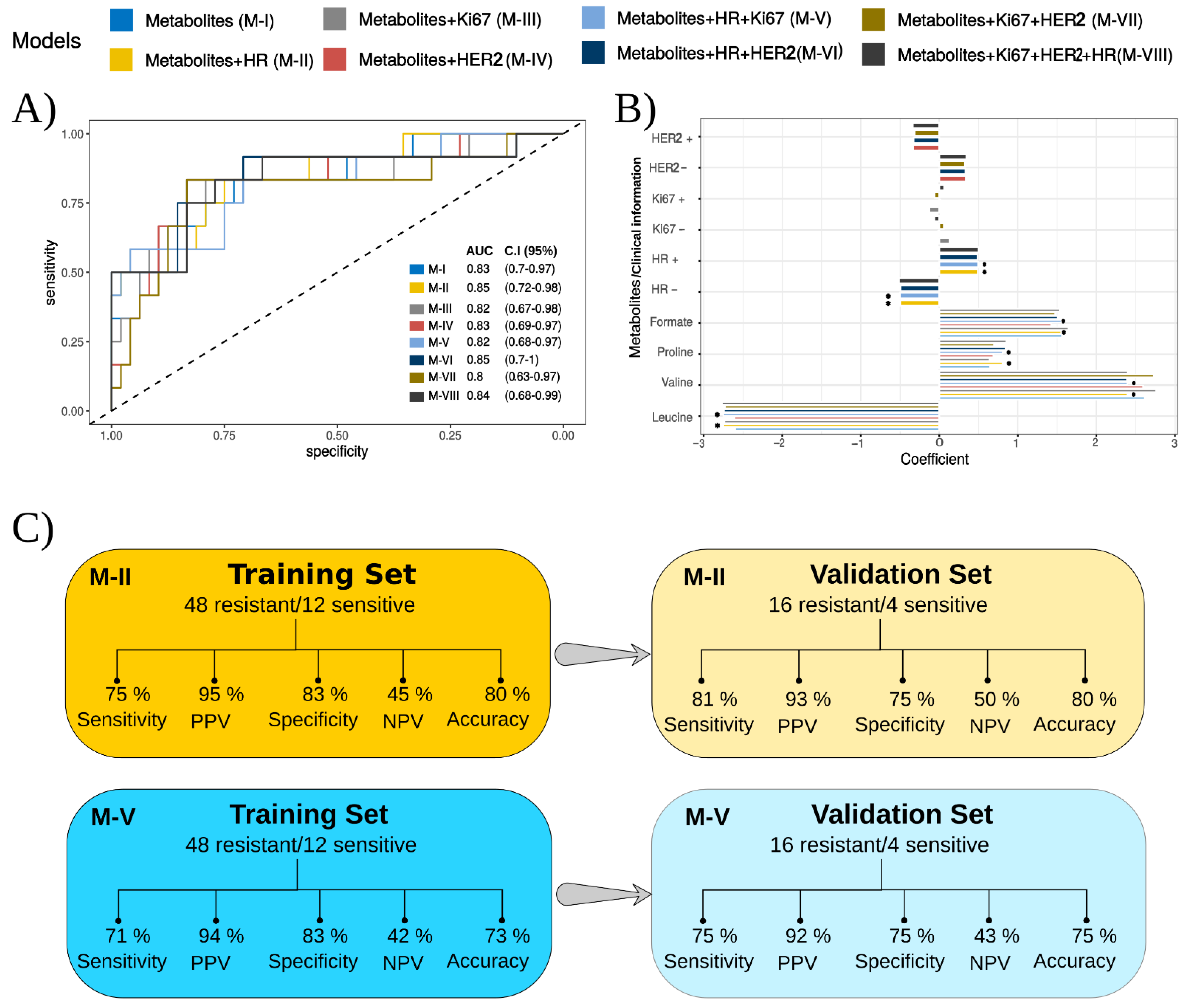
Logistic regression (LR) models have supported the assessment of biomarkers in cancer [46,47,48,49] and can also be coupled with RFE [50], as we present in Figure 3. When compared to the univariate approach, we observed an increase in the predictive power for the model using only the abundance of the metabolites (AUC = 0.83 – Model I, Figure 3A). Using the LR-RFE, we found formate, proline, valine, and leucine as potential markers of NACT response (Figure 3B). By combining the abundances of these metabolites with the information on HR status (Model II), HR and HER2 (Model VI), and Ki67, HER, and HR (Model VIII), we obtained AUC values higher than those obtained for the model that solely used metabolites.
The contributions of the metabolites were found to be more significant for the models than the contribution of the molecular markers, as presented by the observed coefficient values (Figure 3B). For the total contribution of the HR, Ki67, HER2, and serum metabolites as predictors of the response to NACT in the different models obtained, see Table S6.
The coefficient values display the patient’s chance of being resistant to NACT. A positive value means a positive correlation with NACT resistance, whereas a negative value means a positive correlation with NACT sensitivity. For example, formate, proline, valine, HR+, and HER2− display positive coefficient values, thus being directly related to the NACT resistance. It means that once any of these predictors show an increase, the patient’s chance of being resistant to NACT increases. Conversely, leucine, HR−, and HER2+ have negative coefficient values and are inversely related to NACT resistance or directly associated with NACT sensitivity. It means that an increase in leucine concentration, keeping the remaining predictors constant, increases the patient’s chance of being sensitive to the treatment, in accordance with what was previously pointed out by the univariate analysis of the metabolite set. Ki67, both positive and negative, was found to have a low influence on the models that contain it.
When exploring the performances of the models based on their predictive power and agreement with the pathologic results, equivalent specificity rates were found for the eight models when considering the training set (Supplementary Table S3). For the validation sets, the specificity reached 75% only for the models II and V (Figure 3C), which were considered the ones with the highest efficiency in the classification. The sensitivity of the models, for both training and validation sets, was higher than 70% for all the models (Supplementary Table S3). The results suggest that the interconnection of clinical variables (HR/Ki67/HER2) and serum metabolites can improve the prediction of the response to NACT. When the analyses were performed in a restricted cohort of patients with triple-negative and HER2+ tumors, similar performance estimators were obtained (Supplementary Table S4).
To gain insights into metabolome changes associated with resistance and sensitivity to NACT, the pathway enrichment analysis was carried out with the metabolites found as discriminants for models I–VIII. Glyoxylate and dicarboxylate metabolism was the most enrolled pathway for acquiring resistance (Supplementary Figure S1, Supplementary Materials).

4. Discussion

The potential of the combination of HR, Ki67, and HER2 statuses and serum metabolites in predicting the response to NACT was evaluated in this study. The untargeted NMR analysis identified and quantified the relative abundances of 28 compounds, including 14 amino acids, carnitine, and other metabolites. Amino acids are an indispensable source of nutrients for all types of cells. Both essential and non-essential amino acids have an important role in providing build blocks for cell growth and proliferation. The critical role of these amino acids is even more important for tumor cells due to their rapid growth and proliferation, supporting protein synthesis [51]. It is also known that amino acids cannot cross the cell membrane without the assistance of specific transporters due to their hydrophilic component [52]. Therefore, since tumor cells require a high demand of amino acids to satisfy their rapid growth and proliferation, amino acid transporters’ expression is higher than normal cells [53]. Furthermore, the amino acid transporters are expressed in different levels in types of cancers and exhibit different properties in substrate selectivity [54].
When studying the serum abundance of this set of metabolites, we noticed the significant differences between the concentrations of leucine and formate. Leucine, which has been extensively studied for its role in breast cancer, showed a lower concentration in the serum of resistant women [55,56,57]. Saito et al., 2019, proposed that resistance is based on the increased expression of transporters that incorporate leucine to fuel the accelerated proliferation of cells resistant to therapy, which supports our observation of a lower concentration of this amino acid in resistant patients to NACT [58]. The higher concentration of leucine observed in the serum from sensitive in comparison to resistant patients could reflect less leucine uptake from sensitive tumors. One possible explanation could be a lower expression of L-type amino acids transporter 1 (LAT1) in sensitive tumors. Leucine is an essential amino acid [59] that activates the mammalian target of rapamycin (mTOR) which regulates cell growth and cell cycle progression [60], stimulating insulin signaling [61] or oxidized for energy purposes by tumors [62]. Therefore, within a lower leucine uptake, the mTOR pathway will be down-regulated, growth stimuli and energy source will be decreased, and consequently, less cell growth and proliferation could lead to the sensitive phenotype. Contrarily, Yang and collaborators (2018) performed a metabolomics study evaluating the plasma profile associated with response to neoadjuvant chemotherapy for colorectal cancer patients and found that leucine concentration was decreased in responsive patients [61]. Compared to Yang et al. (2018), this discrepancy in the present study could be explained by the heterogeneity among different cancers, such as cell type and histologic classification. A study by Saito and colleagues (2019) demonstrated that leucine imported by LAT1 was involved in tamoxifen resistance in ER-positive breast cancer [58].
In addition to leucine, another amino acid, arginine, had different serum profiles when comparing sensitive and resistant patients. Arginine is a semi-essential amino acid that could be derived primarily from diet and synthesis in the kidney [63]. Arginine is crucial in many biologic processes, including the immune system. Furthermore, arginine is essential to cellular growth and may become limited in cases of rapid proliferation such as malignancy [64]. Jayant and Anant (2017) evaluated the serum levels of arginine in breast cancer patients and found lower levels in cancer patients, independent of stage, compared to healthy controls [65]. In the present study, we found higher levels of arginine in resistant patients in comparison to sensitive ones. This result could reflect that the required arginine may differ between sensitive and resistant tumors.
In addition to alterations in the serum amino acid profile, other metabolites such as formate have their profile changed among sensitive and resistant breast cancer patients. Formate is produced by many metabolic pathways such as serine catabolism, through the mitochondrial pathway. Sterol synthesis and tryptophan catabolism are also involved in this process. Formate may have different directions depending on cell type and environmental conditions. In proliferating cells, such as cancer, formate contributes to the one-carbon (1C) demand for the synthesis of nucleotides [66]. The high demand for nucleotide, RNA, and DNA is essential for rapid tumor growth [67]. For this reason, the circulating formate levels are reduced in some cancer patients, such as breast and lung cancer, relative to healthy controls [68]. Formate was increased in resistant women and can be considered a potential predictive biomarker, in agreement with several publications on cancer [22,68,69]. Jiang et al. (2018) performed a pharmacometabolomics study in metastatic breast cancer patients evaluating the response to gemcitabine–carboplatin chemotherapy. The authors found that significantly lower baseline levels of serum formate in patients with the resistant disease may reflect the higher demand from them for alternate/additional nutritional sources to fuel the accelerated proliferation of breast cancer cells biologically more aggressive or resistant to therapy [70]. Contrarily, our results revealed significantly higher serum formate in resistant patients. This controversial result could be explained by the fact that the oxidative nature of cancers and the chemotherapy treatment for our patients may differ from those included in the study performed by Jiang.
Subsequently, we used the recursive feature elimination (RFE) method to assist in the selection of putative biomarkers. RFE continually removes the resources with low contribution scores based on the iterative method and then classifies each resource in each cycle to exclude the resources with low scores [43]. Studies have proposed that RFE allows the extraction of potential biomarker subsets among different cancer types [71,72,73]. Specifically, for breast cancer, RFE was applied to classify the complete pathological response and distinguish triple-negative breast cancer from other subtypes of breast cancer based on the selection of miRNA biomarkers [74,75]. Logistic regression (LR) models, following the RFE approach, are also widely used in classification problems applied to breast cancer [76,77,78,79].
In our study, RFE + LR was applied as a supervised learning machine by combining the abundance of metabolites with the clinical information on HR, Ki67, and HER2 status. The resulting classification models presented sensitivity and specificity values greater than 70% and 80%, respectively, for the training set. Remarkable performance was achieved for Model II, with 75% sensitivity and 83% specificity for the training set and 81% sensitivity and 75% specificity for the validation set. This indicates that combining the HR status with the metabolite panel (leucine/valine/formate/proline) can generate good classifiers of the response to NACT.
A few studies reported similar results for predicting the response to NACT in BC patients based on metabolite panels [22,23,24]. Lin et al. (2019) assessed the metabolic biomarker signature of serum samples of BC patients by liquid chromatography–mass spectrometry (LC-MS). The authors applied partial least squares discriminant analysis (PLS-DA) to build the statistical model of classification, which identified nine informative metabolites and achieved performances with a specificity of 100% and sensitivity of 81.2% [23]. Some authors recently examined serum metabolite levels during chemotherapy treatment. Debik et al. (2019) observed unfavorable changes in lipid levels during NACT. By using NMR, they observed no metabolic difference in serum samples from survivors and non-survivors, although a PLS-DA model based on their analysis of tissue achieved 72% accuracy in predicting a 5-year survival rate. Among other metabolites, lactate, glycine, choline, and alanine were reported as the critical variables useful for the model set-up [22]. Vignoli et al. (2020) also studied metabolic profiles capable of codifying a complete response to NACT within the ER-positive group. After investigating the plasma by NMR, they built a classifier with low performance. Among other findings, branched-chain amino acids, such as valine and isoleucine, were reported as the key differentiators of their groups of study [24]. Unlike the previous studies mentioned above, applying the RFE + LR method allowed us to obtain models with successful results in predicting the response to NACT that links the selected biomarkers with the clinical information of patients.
Glyoxylate and dicarboxylate metabolism was the pathway most impacted by the acquisition of resistance (Supplementary Figure S1). This pathway has been associated with breast cancer cell metastasis by gas chromatography–mass spectrometry (GC-MS) and direct infusion mass spectrometry [80]. In addition, other pathways represented by our panel of metabolites are associated with breast cancer, such as the metabolism of branched-chain amino acids (BCAAs, i.e., valine, leucine, and isoleucine), the Aminoacyl-tRNA, arginine and proline, Pantothenate, and CoA [50,81]. Together, this set of relationships highlights the metabolic alterations involved in the cellular reprogramming that provoked the BC resistance to chemotherapy.
The current analysis is based on a strong dataset of comprehensive clinical and metabolomic data, derived from a well-maintained biobank, coupled with clinical facilities designed to collect world-class clinical data. We acknowledge, on the other hand, that a larger cohort of patients with non-luminal tumors would have enhanced the possibility of subset analyses aimed at refining the performance estimations on less common tumor types, such as triple-negative and HER2+ tumors. Although untreatable at this point, we are conducting further recruitment of patients with these less common tumor types.

5. Conclusions

In conclusion, NMR of serum offered rapid access to metabolic alterations in BC associated with resistance and sensitivity to NACT. The reported proof-of-principle study attested the power of these techniques in accelerating the definition of BC prognosis with excellent accuracy. Screening methods for predicting the response and effects of chemotherapies are much desired and would be of paramount importance for cancer patients. Despite BC being one of the most treatable cancers, a massive number of women do not benefit from NACT pre-treatment because of resistance to chemotherapy. Capitalizing on the close relationship between cancer formation and changes in serum, the analysis of the metabolites by NMR allowed insights into the metabolic alterations associated with marker status and resistance to NACT. When coupled by LR + RFE to the clinical marker status, a simple metabolomic panel can be applied successfully as an add-in prognosis and clinical forecasting for the response to NACT of BC patients. Although rigorous multisite validation of this untargeted approach with a larger patient pool is needed, this is the first milestone for developing an efficient strategy for the early discovery of NACT-resistant BC patients.

Supplementary Materials

The following supporting information can be downloaded at: https://www.mdpi.com/article/10.3390/cancers14205055/s1, Table S1: Standard NACT regimens and response evaluation parameters according to the molecular subtype of breast cancer. Table S2: Women’s distribution into the therapeutic regimen. Table S3: Performance for the training and validation sets based on the models built using logistic regression and recursive feature elimination considering the different combinations of metabolites, hormone receptor (HR), ki67, and HER2 statuses. Table S4: Performance for the models built using logistic regression and recursive feature elimination considering only triple-negative or HER2+ patients. Table S5: Average relative abundances of the serum metabolites detected by 1H-NMR according to response to NACT. Table S6: Contribution of the HR, Ki67, HER2, and serum metabolites as predictors of the response to NACT in different models obtained with RFE + LR. Figure S1: Pathway enrichment analysis for the metabolites found as discriminatory, assuming as relevant those pathways with a p-value < 0.05.

Author Contributions

M.R.C., R.R.C., L.O.S., A.M.P., L.R.V., F.G., M.L.S., S.A.R. and S.F.M.D. conceived and designed the study. M.R.C., A.A.R.S., M.C.R.T., S.F.M.D., A.C.F., S.R., F.G., L.M.R. and P.H.G.S. collected clinical samples and data. M.R.C., M.L.S., S.A.R., L.R.V., T.B.B.C.C. and M.C.R.T. performed NMR analysis. M.L.S., L.T., L.L.C., L.R.V., L.L.C. M.Q., T.B.B.C.C. and S.A.R. defined parameters for NMR analysis. M.L.S., S.A.R., M.Q., T.B.B.C.C., L.R.V., L.L.C., J.M.G., R.R.C., A.M.P., A.T. and L.T. interpreted the results for NMR. M.R.C., L.M.R., S.R., F.G., S.F.M.D. and A.C.F. assisted in patient recruitment and follow up. M.R.C., M.C.R.T., L.O.S., A.M.P., L.T., S.R., A.C.F., A.A.R.S., A.T. and S.F.M.D. interpreted clinical results. J.M.G., M.R.C., L.M.R., A.M.P. and A.A.R.S. checked the underlying data. J.M.G., L.O.S., M.Q. and T.B.B.C.C., P.H.G.S. performed the statistical analysis. M.R.C., S.F.M.D., A.A.R.S., M.C.R.T., A.M.P., A.T., P.H.G.S., J.M.G. and L.O.S. drafted the manuscript. M.Q., T.B.B.C.C., S.R., L.L.C., P.H.G.S., L.T., F.G., L.M.R., A.C.F., A.T., and R.R.C. revised the manuscript. All authors have read and agreed to the published version of the manuscript.

Funding

This research was funded by the Fundação de Amparo à Pesquisa do Estado de São Paulo (FAPESP), grant numbers 2019/04314-6 and 2018/24069-3, by the Conselho Nacional de Desenvolvimento Científico e Tecnológico (CNPq), grant number 303742/2018-6, and by Coordenação de Aperfeiçoamento de Pessoal de Nível Superior (CAPES), Finance Code 001.

Institutional Review Board Statement

The study was conducted according to the guidelines of the Declaration of Helsinki and approved by the Ethics Committee of UNICAMP (Research Ethics Committee approval CAAE# 69699717.0.0000.5404, 6 August 2017).

Informed Consent Statement

Informed consent was obtained from all subjects involved in the study before removing their biological samples and included in the institutional biobank (CAISM´s biobank—CONEP 56, Brazil). To publish this paper, written informed consent was obtained from the patients.

Data Availability Statement

The study protocol and the datasets generated during and/or analyzed during the current study, including the identified participant data, are available upon publication from the corresponding author upon reasonable request.

Acknowledgments

We are grateful to Geisilene R. Paiva and her team from the Experimental Pathology Laboratory of CAISM, for their unrestricted support. We are grateful to every women who consented to be part of this study.

Conflicts of Interest

The authors declare no conflict of interest.

References

  1. Sung, H.; Ferlay, J.; Siegel, R.L.; Laversanne, M.; Soerjomataram, I.; Jemal, A.; Bray, F. Global Cancer Statistics 2020: GLOBOCAN Estimates of Incidence and Mortality Worldwide for 36 Cancers in 185 Countries. CA Cancer J. Clin. 2021, 71, 209–249. [Google Scholar] [CrossRef] [PubMed]
  2. INCA/Estimativa de Câncer No Brasil. Available online: https://www.gov.br/inca/pt-br/assuntos/cancer/numeros/ (accessed on 26 July 2022).
  3. Esserman, L.J.; Berry, D.A.; DeMichele, A.; Carey, L.; Davis, S.E.; Buxton, M.; Hudis, C.; Gray, J.W.; Perou, C.; Yau, C.; et al. Pathologic Complete Response Predicts Recurrence-Free Survival More Effectively by Cancer Subset: Results from the I-SPY 1 TRIAL--CALGB 150007/150012, ACRIN 6657. J. Clin. Oncol. Off. J. Am. Soc. Clin. Oncol. 2012, 30, 3242–3249. [Google Scholar] [CrossRef] [PubMed] [Green Version]
  4. Cheang, M.C.U.; Chia, S.K.; Voduc, D.; Gao, D.; Leung, S.; Snider, J.; Watson, M.; Davies, S.; Bernard, P.S.; Parker, J.S.; et al. Ki67 Index, HER2 Status, and Prognosis of Patients with Luminal B Breast Cancer. J. Natl. Cancer Inst. 2009, 101, 736–750. [Google Scholar] [CrossRef] [PubMed] [Green Version]
  5. Goldhirsch, A.; Wood, W.C.; Coates, A.S.; Gelber, R.D.; Thürlimann, B.; Senn, H.-J. Strategies for Subtypes—Dealing with the Diversity of Breast Cancer: Highlights of the St. Gallen International Expert Consensus on the Primary Therapy of Early Breast Cancer 2011. Ann. Oncol. Off. J. Eur. Soc. Med. Oncol. 2011, 22, 1736–1747. [Google Scholar] [CrossRef]
  6. Yardley, D.A. Drug Resistance and the Role of Combination Chemotherapy in Improving Patient Outcomes. Int. J. Breast Cancer 2013, 2013, 137414. [Google Scholar] [CrossRef] [PubMed] [Green Version]
  7. von Minckwitz, G.; Untch, M.; Blohmer, J.-U.; Costa, S.D.; Eidtmann, H.; Fasching, P.A.; Gerber, B.; Eiermann, W.; Hilfrich, J.; Huober, J.; et al. Definition and Impact of Pathologic Complete Response on Prognosis after Neoadjuvant Chemotherapy in Various Intrinsic Breast Cancer Subtypes. J. Clin. Oncol. Off. J. Am. Soc. Clin. Oncol. 2012, 30, 1796–1804. [Google Scholar] [CrossRef] [Green Version]
  8. Haque, W.; Verma, V.; Hatch, S.; Klimberg, V.S.; Butler, E.B.; Teh, B.S. Response Rates and Pathologic Complete Response by Breast Cancer Molecular Subtype Following Neoadjuvant Chemotherapy. Breast Cancer Res. Treat. 2018, 170, 559–567. [Google Scholar] [CrossRef] [PubMed]
  9. Masoud, V.; Pagès, G. Targeted Therapies in Breast Cancer: New Challenges to Fight against Resistance. World J. Clin. Oncol. 2017, 8, 120–134. [Google Scholar] [CrossRef] [Green Version]
  10. Bilkey, J.; Tata, A.; McKee, T.D.; Porcari, A.M.; Bluemke, E.; Woolman, M.; Ventura, M.; Eberlin, M.N.; Zarrine-Afsar, A. Variations in the Abundance of Lipid Biomarker Ions in Mass Spectrometry Images Correlate to Tissue Density. Anal. Chem. 2016, 88, 12099–12107. [Google Scholar] [CrossRef]
  11. Cardoso, M.R.; Santos, J.C.; Ribeiro, M.L.; Talarico, M.C.R.; Viana, L.R.; Derchain, S.F.M. A Metabolomic Approach to Predict Breast Cancer Behavior and Chemotherapy Response. Int. J. Mol. Sci. 2018, 19, 617. [Google Scholar] [CrossRef] [PubMed]
  12. Silva, A.A.R.; Cardoso, M.R.; Rezende, L.M.; Lin, J.Q.; Guimaraes, F.; Silva, G.R.P.; Murgu, M.; Priolli, D.G.; Eberlin, M.N.; Tata, A.; et al. Multiplatform Investigation of Plasma and Tissue Lipid Signatures of Breast Cancer Using Mass Spectrometry Tools. Int. J. Mol. Sci. 2020, 21, 3611. [Google Scholar] [CrossRef]
  13. Pavlova, N.N.; Thompson, C.B. The Emerging Hallmarks of Cancer Metabolism. Cell Metab. 2016, 23, 27–47. [Google Scholar] [CrossRef] [PubMed] [Green Version]
  14. Cheng, L.L.; Pohl, U. Chapter 13-The Role of NMR-Based Metabolomics in Cancer. In The Handbook of Metabonomics and Metabolomics; Lindon, J.C., Nicholson, J.K., Holmes, E., Eds.; Elsevier Science B.V.: Amsterdam, The Netherland, 2007; pp. 345–374. ISBN 978-0-444-52841-4. [Google Scholar]
  15. Vandergrift, L.A.; Decelle, E.A.; Kurth, J.; Wu, S.; Fuss, T.L.; Defeo, E.M.; Halpern, E.F.; Taupitz, M.; McDougal, W.S.; Olumi, A.F.; et al. Metabolomic Prediction of Human Prostate Cancer Aggressiveness: Magnetic Resonance Spectroscopy of Histologically Benign Tissue. Sci. Rep. 2018, 8, 1–12. [Google Scholar] [CrossRef] [Green Version]
  16. Schult, T.A.; Lauer, M.J.; Berker, Y.; Cardoso, M.R.; Vandergrift, L.A.; Habbel, P.; Nowak, J.; Taupitz, M.; Aryee, M.; Mino-Kenudson, M.A.; et al. Screening Human Lung Cancer with Predictive Models of Serum Magnetic Resonance Spectroscopy Metabolomics. Proc. Natl. Acad. Sci. USA 2021, 118, e2110633118. [Google Scholar] [CrossRef] [PubMed]
  17. Kerbel, R.S.; Kobayashi, H.; Graham, C.H. Intrinsic or Acquired Drug Resistance and Metastasis: Are They Linked Phenotypes? J. Cell Biochem. 1994, 56, 37–47. [Google Scholar] [CrossRef] [PubMed]
  18. Dettmer, K.; Aronov, P.A.; Hammock, B.D. Mass Spectrometry-Based Metabolomics. Mass Spectrom. Rev. 2007, 26, 51–78. [Google Scholar] [CrossRef] [PubMed]
  19. Patel, S.; Ahmed, S. Emerging Field of Metabolomics: Big Promise for Cancer Biomarker Identification and Drug Discovery. J. Pharm. Biomed. Anal. 2015, 107, 63–74. [Google Scholar] [CrossRef] [PubMed]
  20. Shajahan-Haq, A.N.; Cheema, M.S.; Clarke, R. Application of Metabolomics in Drug Resistant Breast Cancer Research. Metabolites 2015, 5, 100–118. [Google Scholar] [CrossRef] [PubMed] [Green Version]
  21. Denkert, C.; Bucher, E.; Hilvo, M.; Salek, R.; Orešič, M.; Griffin, J.; Brockmöller, S.; Klauschen, F.; Loibl, S.; Barupal, D.K.; et al. Metabolomics of Human Breast Cancer: New Approaches for Tumor Typing and Biomarker Discovery. Genome Med. 2012, 4, 37. [Google Scholar] [CrossRef] [Green Version]
  22. Debik, J.; Euceda, L.R.; Lundgren, S.; Gythfeldt, H.V.D.L.; Garred, Ø.; Borgen, E.; Engebraaten, O.; Bathen, T.F.; Giskeødegård, G.F. Assessing Treatment Response and Prognosis by Serum and Tissue Metabolomics in Breast Cancer Patients. J. Proteome Res. 2019, 18, 3649–3660. [Google Scholar] [CrossRef]
  23. Lin, X.; Xu, R.; Mao, S.; Zhang, Y.; Dai, Y.; Guo, Q.; Song, X.; Zhang, Q.; Li, L.; Chen, Q. Metabolic Biomarker Signature for Predicting the Effect of Neoadjuvant Chemotherapy of Breast Cancer. Ann. Transl. Med. 2019, 7, 670. [Google Scholar] [CrossRef] [PubMed]
  24. Vignoli, A.; Muraro, E.; Miolo, G.; Tenori, L.; Turano, P.; Gregorio, D.E.; Steffan, A.; Luchinat, C.; Corona, G. Effect of Estrogen Receptor Status on Circulatory Immune and Metabolomics Profiles of HER2-Positive Breast Cancer Patients Enrolled for Neoadjuvant Targeted Chemotherapy. Cancers 2020, 12, 314. [Google Scholar] [CrossRef] [PubMed] [Green Version]
  25. Shortliffe, E.H.; Blois, M.S. The Computer Meets Medicine and Biology: Emergence of a Discipline. In Biomedical Informatics; Springer: Berlin, Germany, 2006; pp. 3–45. [Google Scholar]
  26. Nattkemper, T.W.; Arnrich, B.; Lichte, O.; Timm, W.; Degenhard, A.; Pointon, L.; Hayes, C.; Leach, M.O. Evaluation of Radiological Features for Breast Tumour Classification in Clinical Screening with Machine Learning Methods. Artif. Intell. Med. 2005, 34, 129–139. [Google Scholar] [CrossRef]
  27. Wei, L.; Yang, Y.; Nishikawa, R.M.; Jiang, Y. A Study on Several Machine-Learning Methods for Classification of Malignant and Benign Clustered Microcalcifications. IEEE Trans. Med. Imaging 2005, 24, 371–380. [Google Scholar] [CrossRef] [PubMed]
  28. Delen, D.; Walker, G.; Kadam, A. Predicting Breast Cancer Survivability: A Comparison of Three Data Mining Methods. Artif. Intell. Med. 2005, 34, 113–127. [Google Scholar] [CrossRef] [PubMed]
  29. Antropova, N.; Huynh, B.Q.; Giger, M.L. A Deep Feature Fusion Methodology for Breast Cancer Diagnosis Demonstrated on Three Imaging Modality Datasets. Med. Phys. 2017, 44, 5162–5171. [Google Scholar] [CrossRef] [PubMed] [Green Version]
  30. Antropova, N.; Abe, H.; Giger, M.L. Use of Clinical MRI Maximum Intensity Projections for Improved Breast Lesion Classification with Deep Convolutional Neural Networks. J. Med. Imaging 2018, 5, 14503. [Google Scholar] [CrossRef] [PubMed]
  31. Lakhani, S.; Ellis, I.; Schnitt, S.; Tan, P.; van de Vijver, M. Breast Tumours, WHO Classification of Tumours, 5th ed.; WHO/IARC, Ed.; Publisher: Lyon, France, 2019; Volume 2, ISBN 9789283245001. [Google Scholar]
  32. Allison, K.H.; Hammond, M.E.H.; Dowsett, M.; McKernin, S.E.; Carey, L.A.; Fitzgibbons, P.L.; Hayes, D.F.; Lakhani, S.R.; Chavez-MacGregor, M.; Perlmutter, J.; et al. Estrogen and Progesterone Receptor Testing in Breast Cancer: American Society of Clinical Oncology/College of American Pathologists Guideline Update. Arch. Pathol. Lab. Med. 2020, 144, 545–563. [Google Scholar] [CrossRef] [PubMed] [Green Version]
  33. Dowsett, M.; Nielsen, T.O.; A’Hern, R.; Bartlett, J.; Coombes, R.C.; Cuzick, J.; Ellis, M.; Henry, N.L.; Hugh, J.C.; Lively, T.; et al. Assessment of Ki67 in Breast Cancer: Recommendations from the International Ki67 in Breast Cancer Working Group. J. Natl. Cancer Inst. 2011, 103, 1656–1664. [Google Scholar] [CrossRef] [Green Version]
  34. Wolff, A.C.; Hammond, M.E.H.; Allison, K.H.; Harvey, B.E.; McShane, L.M.; Dowsett, M. HER2 Testing in Breast Cancer: American Society of Clinical Oncology/College of American Pathologists Clinical Practice Guideline Focused Update Summary. J. Oncol. Pract. 2018, 14, 437–441. [Google Scholar] [CrossRef] [PubMed]
  35. Robertson, S.; Rönnlund, C.; de Boniface, J.; Hartman, J. Re-Testing of Predictive Biomarkers on Surgical Breast Cancer Specimens Is Clinically Relevant. Breast Cancer Res. Treat. 2019, 174, 795–805. [Google Scholar] [CrossRef] [Green Version]
  36. Bossuyt, V.; Provenzano, E.; Symmans, W.F.; Boughey, J.C.; Coles, C.; Curigliano, G.; Dixon, J.M.; Esserman, L.J.; Fastner, G.; Kuehn, T.; et al. Recommendations for Standardized Pathological Characterization of Residual Disease for Neoadjuvant Clinical Trials of Breast Cancer by the BIG-NABCG Collaboration. Ann. Oncol. Off. J. Eur. Soc. Med. Oncol. 2015, 26, 1280–1291. [Google Scholar] [CrossRef] [PubMed]
  37. Symmans, W.F.; Peintinger, F.; Hatzis, C.; Rajan, R.; Kuerer, H.; Valero, V.; Assad, L.; Poniecka, A.; Hennessy, B.; Green, M.; et al. Measurement of Residual Breast Cancer Burden to Predict Survival after Neoadjuvant Chemotherapy. J. Clin. Oncol. Off. J. Am. Soc. Clin. Oncol. 2007, 25, 4414–4422. [Google Scholar] [CrossRef]
  38. Provenzano, E.; Bossuyt, V.; Viale, G.; Cameron, D.; Badve, S.; Denkert, C.; MacGrogan, G.; Penault-Llorca, F.; Boughey, J.; Curigliano, G.; et al. Standardization of Pathologic Evaluation and Reporting of Postneoadjuvant Specimens in Clinical Trials of Breast Cancer: Recommendations from an International Working Group. Mod. Pathol. Off. J. U.S. Can. Acad. Pathol. Inc. 2015, 28, 1185–1201. [Google Scholar] [CrossRef] [PubMed] [Green Version]
  39. Brown, L.D.; Cai, T.T.; DasGupta, A. Interval Estimation for a Binomial Proportion. Stat. Sci. 2001, 16, 101–133. [Google Scholar] [CrossRef]
  40. Kuhn, M.; Wing, J.; Weston, S.; Williams, A.; Keefer, C.; Engelhardt, A.; Cooper, T.; Mayer, Z.; Kenkel, B.; Team, R.C.; et al. Classification and Regression Training. R Package Version 6.0–88. Available online: https://cran.r-project.org/package=caret (accessed on 20 November 2021).
  41. Guan, W.; Zhou, M.; Hampton, C.Y.; Benigno, B.B.; Walker, L.D.; Gray, A.; McDonald, J.F.; Fernández, F.M. Ovarian Cancer Detection from Metabolomic Liquid Chromatography/Mass Spectrometry Data by Support Vector Machines. BMC Bioinform. 2009, 10, 259. [Google Scholar] [CrossRef] [PubMed] [Green Version]
  42. Guyon, I.; Weston, J.; Barnhill, S.; Vapnik, V. Gene Selection for Cancer Classification Using Support Vector Machines. Mach. Learn. 2002, 46, 389–422. [Google Scholar] [CrossRef]
  43. Sheng, J.; Shao, M.; Zhang, Q.; Zhou, R.; Wang, L.; Xin, Y. Alzheimer’s Disease, Mild Cognitive Impairment, and Normal Aging Distinguished by Multi-Modal Parcellation and Machine Learning. Sci. Rep. 2020, 10, 5475. [Google Scholar] [CrossRef] [Green Version]
  44. Murata, T.; Yanagisawa, T.; Kurihara, T.; Kaneko, M.; Ota, S.; Enomoto, A.; Tomita, M.; Sugimoto, M.; Sunamura, M.; Hayashida, T.; et al. Salivary Metabolomics with Alternative Decision Tree-Based Machine Learning Methods for Breast Cancer Discrimination. Breast Cancer Res. Treat. 2019, 177, 591–601. [Google Scholar] [CrossRef] [PubMed]
  45. Pang, Z.; Chong, J.; Zhou, G.; Morais, D.A.D.L.; Chang, L.; Barrette, M.; Gauthier, C.; Jacques, P.-É.; Li, S.; Xia, J. MetaboAnalyst 5.0: Narrowing the Gap between Raw Spectra and Functional Insights. Nucleic Acids Res. 2021, 49, W388–W396. [Google Scholar] [CrossRef]
  46. Eberlin, L.S.; Tibshirani, R.J.; Zhang, J.; Longacre, T.A.; Berry, G.J.; Bingham, D.B.; Norton, J.A.; Zare, R.N.; Poultsides, G.A. Molecular Assessment of Surgical-Resection Margins of Gastric Cancer by Mass-Spectrometric Imaging. Proc. Natl. Acad. Sci. USA 2014, 111, 2436–2441. [Google Scholar] [CrossRef] [PubMed] [Green Version]
  47. Hosseinnataj, A.; RezaBaneshi, M.; Bahrampour, A. Mortality Risk Factors in Patients with Gastric Cancer Using Bayesian and Ordinary Lasso Logistic Models: A Study in the Southeast of Iran. Gastroenterol. Hepatol. Bed Bench 2020, 13, 31–36. [Google Scholar] [PubMed]
  48. Kim, S.M.; Kim, Y.; Jeong, K.; Jeong, H.; Kim, J. Logistic LASSO Regression for the Diagnosis of Breast Cancer Using Clinical Demographic Data and the BI-RADS Lexicon for Ultrasonography. Ultrasonography 2018, 37, 36–42. [Google Scholar] [CrossRef] [Green Version]
  49. Sans, M.; Gharpure, K.; Tibshirani, R.; Zhang, J.; Liang, L.; Liu, J.; Young, J.H.; Dood, R.L.; Sood, A.K.; Eberlin, L.S. Metabolic Markers and Statistical Prediction of Serous Ovarian Cancer Aggressiveness by Ambient Ionization Mass Spectrometry Imaging. Cancer Res. 2017, 77, 2903–2913. [Google Scholar] [CrossRef] [Green Version]
  50. Yang, L.; Wang, Y.; Cai, H.; Wang, S.; Shen, Y.; Ke, C. Application of Metabolomics in the Diagnosis of Breast Cancer: A Systematic Review. J. Cancer 2020, 11, 2540–2551. [Google Scholar] [CrossRef]
  51. Ganapathy, V.; Thangaraju, M.; Prasad, P.D. Nutrient Transporters in Cancer: Relevance to Warburg Hypothesis and Beyond. Pharmacol. Ther. 2009, 121, 29–40. [Google Scholar] [CrossRef] [PubMed]
  52. Bhutia, Y.D.; Babu, E.; Ramachandran, S.; Ganapathy, V. Amino Acid Transporters in Cancer and Their Relevance to “Glutamine Addiction”: Novel Targets for the Design of a New Class of Anticancer Drugs. Cancer Res. 2015, 75, 1782–1788. [Google Scholar] [CrossRef] [PubMed] [Green Version]
  53. Cha, Y.J.; Kim, E.-S.; Koo, J.S. Amino Acid Transporters and Glutamine Metabolism in Breast Cancer. Int. J. Mol. Sci. 2018, 19, 907. [Google Scholar] [CrossRef] [PubMed] [Green Version]
  54. Häfliger, P.; Graff, J.; Rubin, M.; Stooss, A.; Dettmer, M.S.; Altmann, K.-H.; Gertsch, J.; Charles, R.-P. The LAT1 Inhibitor JPH203 Reduces Growth of Thyroid Carcinoma in a Fully Immunocompetent Mouse Model. J. Exp. Clin. Cancer Res. 2018, 37, 234. [Google Scholar] [CrossRef] [PubMed]
  55. Bae, H.; Kim, B.; Lee, H.; Lee, S.; Kang, H.-S.; Kim, S.J. Epigenetically Regulated Fibronectin Leucine Rich Transmembrane Protein 2 (FLRT2) Shows Tumor Suppressor Activity in Breast Cancer Cells. Sci. Rep. 2017, 7, 272. [Google Scholar] [CrossRef]
  56. Shennan, D.B.; Thomson, J.; Gow, I.F.; Travers, M.T.; Barber, M.C. L-Leucine Transport in Human Breast Cancer Cells (MCF-7 and MDA-MB-231): Kinetics, Regulation by Estrogen and Molecular Identity of the Transporter. Biochim. Biophys. Acta 2004, 1664, 206–216. [Google Scholar] [CrossRef] [Green Version]
  57. Speers, C.; Zhao, S.G.; Kothari, V.; Santola, A.; Liu, M.; Wilder-Romans, K.; Evans, J.; Batra, N.; Bartelink, H.; Hayes, D.F.; et al. Maternal Embryonic Leucine Zipper Kinase (MELK) as a Novel Mediator and Biomarker of Radioresistance in Human Breast Cancer. Clin. Cancer Res. Off. J. Am. Assoc. Cancer Res. 2016, 22, 5864–5875. [Google Scholar] [CrossRef] [Green Version]
  58. Saito, Y.; Li, L.; Coyaud, E.; Luna, A.; Sander, C.; Raught, B.; Asara, J.M.; Brown, M.; Muthuswamy, S.K. LLGL2 Rescues Nutrient Stress by Promoting Leucine Uptake in ER (+) Breast Cancer. Nature 2019, 569, 275–279. [Google Scholar] [CrossRef] [PubMed]
  59. Zhang, L.; Sui, C.; Yang, W.; Luo, Q. Amino Acid Transporters: Emerging Roles in Drug Delivery for Tumor-Targeting Therapy. Asian J. Pharm. Sci. 2020, 15, 192–206. [Google Scholar] [CrossRef] [PubMed]
  60. Boutouja, F.; Stiehm, C.M.; Platta, H.W. MTOR: A Cellular Regulator Interface in Health and Disease. Cells 2019, 8, 18. [Google Scholar] [CrossRef] [PubMed] [Green Version]
  61. Yang, K.; Zhang, F.; Han, P.; Wang, Z.-Z.; Deng, K.; Zhang, Y.-Y.; Zhao, W.-W.; Song, W.; Cai, Y.-Q.; Li, K.; et al. Metabolomics Approach for Predicting Response to Neoadjuvant Chemotherapy for Colorectal Cancer. Metabolomics 2018, 14, 110. [Google Scholar] [CrossRef] [PubMed]
  62. Ananieva, E.A.; Wilkinson, A.C. Branched-Chain Amino Acid Metabolism in Cancer. Curr. Opin. Clin. Nutr. Metab. Care 2018, 21, 64–70. [Google Scholar] [CrossRef] [PubMed] [Green Version]
  63. Lind, D.S. Arginine and Cancer. J. Nutr. 2004, 134, 2837S–2841S. [Google Scholar] [CrossRef] [PubMed] [Green Version]
  64. Albaugh, V.L.; Pinzon-Guzman, C.; Barbul, A. Arginine-Dual Roles as an Onconutrient and Immunonutrient. J. Surg. Oncol. 2017, 115, 273–280. [Google Scholar] [CrossRef] [PubMed]
  65. Jayant, S.; Anant, A. The Relation of Serum Arginine Levels with Serum Arginase and Nitric Oxide Synthase Activity in Patients with Breast Cancer. J. Clin. Diagn. Res. 2017, 11, 11–14. [Google Scholar] [CrossRef]
  66. Pietzke, M.; Meiser, J.; Vazquez, A. Formate Metabolism in Health and Disease. Mol. Metab. 2020, 33, 23–37. [Google Scholar] [CrossRef] [PubMed]
  67. Ducker, G.S.; Rabinowitz, J.D. One-Carbon Metabolism in Health and Disease. Cell Metab. 2017, 25, 27–42. [Google Scholar] [CrossRef] [PubMed] [Green Version]
  68. Pietzke, M.; Arroyo, S.F.; Sumpton, D.; Mackay, G.M.; Martin-Castillo, B.; Camps, J.; Joven, J.; Menendez, J.A.; Vazquez, A. Stratification of Cancer and Diabetes Based on Circulating Levels of Formate and Glucose. Cancer Metab. 2019, 7, 3. [Google Scholar] [CrossRef] [PubMed] [Green Version]
  69. Delbrouck, C.; Pozdeev, V.I.; Oudin, A.; Grzyb, K.; Neises, L.; Kiweler, N.; Skupin, A.; Letellier, E.; Niclou, S.P.; Meiser, J. FSMP-09. Formate Promotes Cancer Cell Invasion and Metastasis Via Calcium Signaling. Neurocol. Adv. 2021, 3, i18. [Google Scholar] [CrossRef]
  70. Jiang, L.; Lee, S.C.; Ng, T.C. Pharmacometabonomics Analysis Reveals Serum Formate and Acetate Potentially Associated with Varying Response to Gemcitabine-Carboplatin Chemotherapy in Metastatic Breast Cancer Patients. J. Proteome. Res. 2018, 17, 1248–1257. [Google Scholar] [CrossRef]
  71. Chen, Z.; Dai, Y.; Huang, X.; Chen, K.; Gao, Y.; Li, N.; Wang, D.; Chen, A.; Yang, Q.; Hong, Y.; et al. Combined Metabolomic Analysis of Plasma and Tissue Reveals a Prognostic Risk Score System and Metabolic Dysregulation in Esophageal Squamous Cell Carcinoma. Front. Oncol. 2020, 10, 1545. [Google Scholar] [CrossRef]
  72. Ge, C.; Luo, L.; Zhang, J.; Meng, X.; Chen, Y. FRL: An Integrative Feature Selection Algorithm Based on the Fisher Score, Recursive Feature Elimination, and Logistic Regression to Identify Potential Genomic Biomarkers. Biomed. Res. Int. 2021, 2021, 4312850. [Google Scholar] [CrossRef]
  73. Han, Y.; Huang, L.; Zhou, F. A Dynamic Recursive Feature Elimination Framework (DRFE) to Further Refine a Set of OMIC Biomarkers. Bioinformatics 2021, 37, 2183–2189. [Google Scholar] [CrossRef]
  74. Sutton, E.J.; Onishi, N.; Fehr, D.A.; Dashevsky, B.Z.; Sadinski, M.; Pinker, K.; Martinez, D.F.; Brogi, E.; Braunstein, L.; Razavi, P.; et al. A Machine Learning Model That Classifies Breast Cancer Pathologic Complete Response on MRI Post-Neoadjuvant Chemotherapy. Breast Cancer Res. 2020, 22, 57. [Google Scholar] [CrossRef]
  75. Lopez-Rincon, A.; Mendoza-Maldonado, L.; Martinez-Archundia, M.; Schönhuth, A.; Kraneveld, A.D.; Garssen, J.; Tonda, A. Machine Learning-Based Ensemble Recursive Feature Selection of Circulating MiRNAs for Cancer Tumor Classification. Cancers 2020, 12, 1785. [Google Scholar] [CrossRef]
  76. Liu, L. Research on Logistic Regression Algorithm of Breast Cancer Diagnose Data by Machine Learning. In Proceedings of the 2018 International Conference on Robots & Intelligent System (ICRIS), Amsterdam, The Netherlands, 21–23 February 2018; pp. 157–160. [Google Scholar]
  77. Pfob, A.; Sidey-Gibbons, C.; Lee, H.-B.; Tasoulis, M.K.; Koelbel, V.; Golatta, M.; Rauch, G.M.; Smith, B.D.; Valero, V.; Han, W.; et al. Identification of Breast Cancer Patients with Pathologic Complete Response in the Breast after Neoadjuvant Systemic Treatment by an Intelligent Vacuum-Assisted Biopsy. Eur. J. Cancer 2021, 143, 134–146. [Google Scholar] [CrossRef] [PubMed]
  78. Tahmassebi, A.; Wengert, G.J.; Helbich, T.H.; Bago-Horvath, Z.; Alaei, S.; Bartsch, R.; Dubsky, P.; Baltzer, P.; Clauser, P.; Kapetas, P.; et al. Impact of Machine Learning With Multiparametric Magnetic Resonance Imaging of the Breast for Early Prediction of Response to Neoadjuvant Chemotherapy and Survival Outcomes in Breast Cancer Patients. Investig. Radiol. 2019, 54, 110–117. [Google Scholar] [CrossRef] [PubMed]
  79. Zhang, J.; Xiao, L.; Pu, S.; Liu, Y.; He, J.; Wang, K. Can We Reliably Identify the Pathological Outcomes of Neoadjuvant Chemotherapy in Patients with Breast Cancer? Development and Validation of a Logistic Regression Nomogram Based on Preoperative Factors. Ann. Surg. Oncol. 2021, 28, 2632–2645. [Google Scholar] [CrossRef] [PubMed]
  80. Kim, H.Y.; Lee, K.M.; Kim, S.H.; Kwon, Y.J.; Chun, Y.J.; Choi, H.K. Comparative Metabolic and Lipidomic Profiling of Human Breast Cancer Cells with Different Metastatic Potentials. Oncotarget 2016, 7, 67111–67128. [Google Scholar] [CrossRef] [PubMed] [Green Version]
  81. Peng, H.; Wang, Y.; Luo, W. Multifaceted Role of Branched-Chain Amino Acid Metabolism in Cancer. Oncogene 2020, 39, 6747–6756. [Google Scholar] [CrossRef]
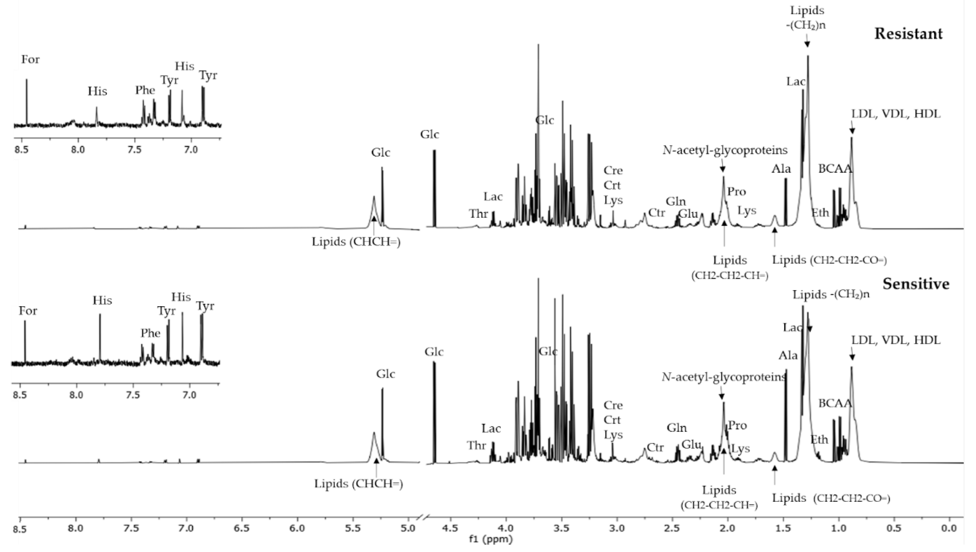
Figure 1. Representative 1H-NMR spectra from serum obtained from women before they initiated the NACT. Resistant and sensitive refer to response to neoadjuvant chemotherapy.
Cancers 14 05055 g001
Figure 2. Volcano plot showing the metabolite variation by NACT outcomes (log2(resistant/sensitive)) in function of their statistical significance (log2(p-value)). Non-significant metabolites are plotted below the horizontal dashed line, and the metabolites above this line presented significant variation (p-value < 0.05). In addition, we show a boxplot for metabolites with significant variation (blue represents resistant patients and pink the sensitive ones); p-values were calculated using the Mann–Whitney–Wilcoxon test or t-test as a function of whether the data came from a normal distribution, proved with the Shapiro test.
Cancers 14 05055 g002
Figure 3. Classification models obtained with the combination of metabolites and clinical information. (A) The plot of the ROC curves for each model, containing the area under the curve (AUC). (B) The coefficients of the predictors and (C) performance of models II and V for training (cross-validation) and validation sets.
Cancers 14 05055 g003
Table 1. Key patient information as related to response to neoadjuvant chemotherapy.
Characteristicn (%)SensitiveResistantORp-Value
n = 16 (%)n = 64 (%)(95% CI)
Age≥5046 (57.5)8 (50.0)38 (59.4)ref
<5034 (42.5)8 (50.0)26 (40.6)0.68 (0.23–2.05)0.499
RaceCaucasian69 (86.3)15 (93.8)54 (84.4)ref
Non-Caucasian11 (13.8)1 (6.2)10 (15.6)2.78 (0.33–23.45)0.293
Age of menarche>1242 (52.5)9 (56.2)33 (51.6)ref
≤1238 (47.5)7 (43.8)31 (48.4)1.21 (0.4–3.64)0.737
MenopauseNo36 (45.0)9 (56.2)27 (42.2)ref
Yes44 (55.0)7 (43.8)37 (57.8)1.76 (0.58–5.32)0.313
Hormone therapyNo68 (85.0)15 (93.8)53 (82.8)ref
Yes12 (15.0)1 (6.2)11 (17.2)3.11 (0.37–26.09)0.233
Pregnancy (previous or current)Yes73 (91.3)15 (93.8)58 (90.6)ref
No7 (8.7)1 (6.2)6 (9.4)1.55 (0.17–13.89)0.681
Lactation *Yes63 (78.8)13 (81.2)50 (78.1)ref
No17 (21.2)3 (18.8)14 (21.9)1.21 (0.3–4.86)0.782
SmokingYes17 (21.2)4 (25.0)13 (20.3)ref
No63 (78.8)12 (75.0)51 (79.7)1.31 (0.36–4.73)0.686
Chronic alcoholismNo79 (98.8)16 (100.0)63 (98.4)ref
Yes1 (1.2)0 (0.0)1 (1.6)NC0.503
BMINormal weight24 (30.0)6 (37.5)18 (28.1)ref
Overweight21 (26.3)2 (12.5)19 (29.7)3.17 (0.56–17.77)0.19
Obese35 (43.7)8 (50.0)27 (42.2)1.13 (0.33–3.79)0.849
DiabetesNo71 (88.7)15 (93.8)56 (87.5)ref
Yes9 (11.3)1 (6.2)8 (12.5)2.14 (0.25–18.5)0.452
Family history of breast or ovarian cancerNo59 (73.8)9 (56.2)50 (78.1)ref
Yes21 (26.2)7 (43.8)14 (21.9)0.36 (0.11–1.14)0.087
Sensitive (pCR and RCB I); resistant (RCB II and RCB III); NC, non-calculable; BMI, body mass index; * women who had no pregnancies. The following demographic and clinical data were obtained: age at diagnosis; self-declared ethnicity; age of menarche; menopausal status (premenopausal and postmenopausal); hormone therapy history; number of pregnancies; lactation regardless of the number of pregnancies or duration; smoking; chronic alcoholism; body mass index before the beginning of neoadjuvant chemotherapy (NACT) (normal weight: <25 kg/m2; overweight: 25 < 30; obese: ≥30); diabetes mellitus; family history of breast and/or ovarian cancer.
Table 2. Main breast cancer features as related to response to neoadjuvant chemotherapy.
Characteristicn (%)SensitiveResistantORp-Value
n = 16 (%)n = 64 (%)(95% CI)
Histological grade1/238 (48.75)3 (18.8)36 (56.2)ref
341 (51.25)13 (81.2)28 (43.8)0.18 (0.05–0.69)0.006
Ki67Low34 (42.5)5 (31.2)29 (45.3)ref
High46 (57.5)11 (68.8)35 (54.7)0.55 (0.17–1.76)0.303
HER2Negative46 (57.5)6 (37.5)40 (62.5)ref
Positive *34 (42.5)10 (62.5)24 (37.5)0.36 (0.12–1.12)0.072
Tumor size **T1/T253 (66.25)12 (75.0)41 (64.1)ref
T3/T427 (33.75)4 (25.0)23 (35.9)1.68 (0.49–5.82)0.399
Regional lymph nodeN033 (41.25)6 (37.5)27 (42.2)ref
N1 or higher47 (58.75)10 (62.5)37 (57.8)0.82 (0.27–2.54)0.732
MetastasisM074 (92.5)16 (100.0)58 (90.6)ref
M16 (7.5)0 (0.0)6 (9.4)NC0.094
Hormonal ReceptorNegative21 (26.25)9 (56.2)12 (18.8)ref
Positive59 (73.75)7 (43.8)52 (81.2)5.57 (1.73–17.96)0.004
* Five patients had negative HER2 at core biopsy and positive HER2 in the surgical specimen; therefore, these patients were not treated with trastuzumab prior to surgery. ** One patient had an occult breast tumor with axillary disease. Legend: sensitive (pCR and RCB I); resistant (RCB II and RCB III); NC, non-calculable; Ki67, Ki67 protein (low ≤ 30 and high > 30); HER2, human epidermal growth factor receptor (scored as 0+/1+ (negative), 2+ (equivocal), or 3+ (positive), equivocal cases were further confirmed by in situ hybridization); T1, tumor ≤ 20 mm; T2, tumor > 20 mm and ≤ 50 mm; T3, tumor > 50 mm; T4, tumor of any size with direct extension to the chest wall and/or skin; Regional lymph node negative, no regional lymph node metastasis; positive, included N1 (mobile ipsilateral lymph node metastases, axillary levels I, II), N2 (clinically fixed or entangled lymph node metastases I, II, or ipsilateral internal mammary lymph node metastasis) or N3 (axillary level III ipsilateral lymph node metastasis with or without axillary involvement at levels I and II, or metastasis in internal mammary lymph node with level I and II axillary involvement, or supraclavicular lymph node metastasis with or without axillary or internal mammary involvement); M0, without clinical or radiographic evidence of distant metastasis; M1, distant metastasis determined clinically and radiographically and/or histologically. Hormonal receptor, positive if ER and/or PR positive; negative if both RE and RP negative.
Publisher’s Note: MDPI stays neutral with regard to jurisdictional claims in published maps and institutional affiliations.