
Evaluating Pancreatic and Biliary Neoplasms with Small Biopsy-Based Next Generation Sequencing (NGS): Doing More with Less

 ,
,  , and
, and 
Abstract
Simple Summary
Abstract
1. Introduction
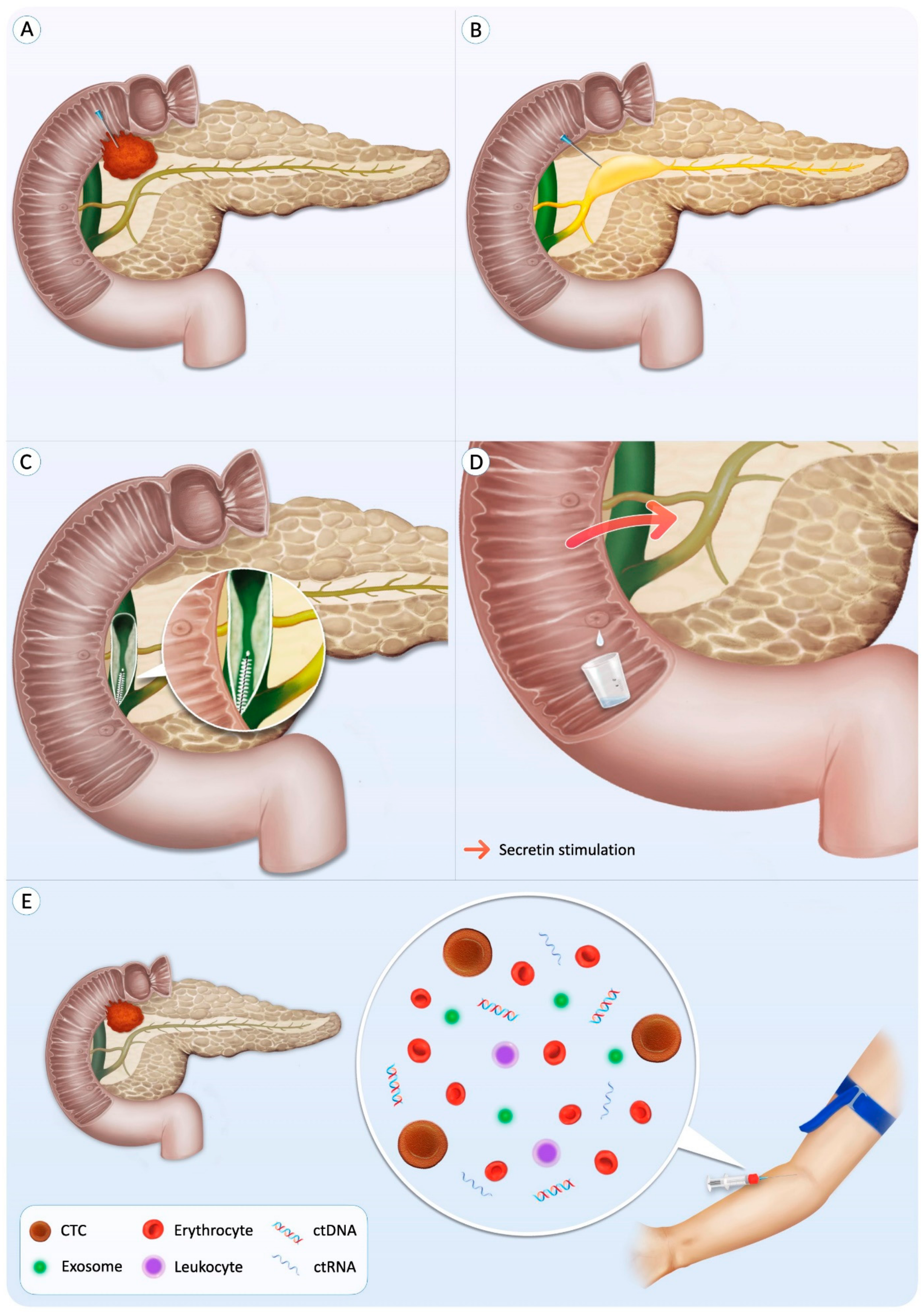
2. The Role of NGS Performed on Pancreatic Small Biopsies
2.1. Most Common Mutations Detected in PDACs
2.2. Preoperative Evaluation of Pancreatic Cysts
2.3. Evaluation of High-Risk Patients under Surveillance with Pancreatic Juice-Based NGS
2.4. Identification of Potentially Actionable Mutations in PDAC Patients
2.5. Evaluation of Neoplasms Other Than PDAC and Its Precursors
2.6. NGS Performed on FNA vs. Tissue Biopsy Samples
3. The Role of NGS Performed on Biliary Small Biopsies
4. The Role of NGS Performed on Blood-Based Liquid Biopsies
4.1. Monitoring Disease Course and Response to Therapy in PDAC Patients
4.2. Assessing Prognosis of PDAC Patients
4.3. Identifying Potentially Actionable Mutations in PDAC Patients
4.4. NGS Performed on Blood-Based Liquid Biopsy vs. Tissue Biopsy Samples
5. Discussion
6. Conclusions
Author Contributions
Funding
Conflicts of Interest
References
- Ilic, M.; Ilic, I. Epidemiology of Pancreatic Cancer. World J. Gastroenterol. 2016, 22, 9694–9705. [Google Scholar] [CrossRef]
- Brosens, L.A.A.; Hackeng, W.M.; Offerhaus, G.J.; Hruban, R.H.; Wood, L.D. Pancreatic Adenocarcinoma Pathology: Changing “Landscape”. J. Gastrointest. Oncol. 2015, 6, 358–374. [Google Scholar]
- Sung, H.; Ferlay, J.; Siegel, R.L.; Laversanne, M.; Soerjomataram, I.; Jemal, A.; Bray, F. Global Cancer Statistics 2020: GLOBOCAN Estimates of Incidence and Mortality Worldwide for 36 Cancers in 185 Countries. CA Cancer J. Clin. 2021, 71, 209–249. [Google Scholar] [CrossRef]
- Kandel, P.; Wallace, M.B. Advanced EUS Guided Tissue Acquisition Methods for Pancreatic Cancer. Cancers 2018, 10, 54. [Google Scholar] [CrossRef]
- Kleeff, J.; Korc, M.; Apte, M.; La Vecchia, C.; Johnson, C.D.; Biankin, A.V.; Neale, R.E.; Tempero, M.; Tuveson, D.A.; Hruban, R.H.; et al. Pancreatic Cancer. Nat. Rev. Dis. Primers 2016, 2, 16022. [Google Scholar] [CrossRef]
- Taghizadeh, H.; Müllauer, L.; Mader, R.M.; Schindl, M.; Prager, G.W. Applied Precision Medicine in Metastatic Pancreatic Ductal Adenocarcinoma. Ther. Adv. Med. Oncol. 2020, 12, 1758835920938611. [Google Scholar] [CrossRef]
- Conroy, T.; Desseigne, F.; Ychou, M.; Bouché, O.; Guimbaud, R.; Bécouarn, Y.; Adenis, A.; Raoul, J.-L.; Gourgou-Bourgade, S.; de la Fouchardière, C.; et al. FOLFIRINOX versus Gemcitabine for Metastatic Pancreatic Cancer. N. Engl. J. Med. 2011, 364, 1817–1825. [Google Scholar] [CrossRef] [PubMed]
- Pittman, M.E.; Rao, R.; Hruban, R.H. Classification, Morphology, Molecular Pathogenesis, and Outcome of Premalignant Lesions of the Pancreas. Arch. Pathol. Lab. Med. 2017, 141, 1606–1614. [Google Scholar] [CrossRef] [PubMed]
- Hoda, R.S.; Arpin, R.N., 3rd; Rosenbaum, M.W.; Pitman, M.B. Risk of Malignancy Associated with Diagnostic Categories of the Proposed World Health Organization International System for Reporting Pancreaticobiliary Cytopathology. Cancer Cytopathol. 2021. [Google Scholar] [CrossRef]
- Gonzalez, R.S.; Raza, A.; Propst, R.; Adeyi, O.; Bateman, J.; Sopha, S.C.; Shaw, J.; Auerbach, A. Recent Advances in Digestive Tract Tumors: Updates from the 5th Edition of the World Health Organization “Blue Book”. Arch. Pathol. Lab. Med. 2021, 145, 607–626. [Google Scholar] [CrossRef] [PubMed]
- Frampas, E.; Morla, O.; Regenet, N.; Eugène, T.; Dupas, B.; Meurette, G. A Solid Pancreatic Mass: Tumour or Inflammation? Diagn. Interv. Imaging 2013, 94, 741–755. [Google Scholar] [CrossRef]
- Hoda, R.S.; Pitman, M.B. Pancreatic Cytology. Surg. Pathol. Clin. 2018, 11, 563–588. [Google Scholar] [CrossRef]
- Bollen, T.L.; Wessels, F.J. Radiological Workup of Cystic Neoplasms of the Pancreas. Visc. Med. 2018, 34, 182–190. [Google Scholar] [CrossRef] [PubMed]
- Sydney, G.I.; Ioakim, K.J.; Michaelides, C.; Sepsa, A.; Sopaki-Valalaki, A.; Tsiotos, G.G.; Theocharis, S.; Salla, C.; Nikas, I. EUS-FNA Diagnosis of Pancreatic Serous Cystadenoma with the Aid of Cell Blocks and α-Inhibin Immunochemistry: A Case Series. Diagn. Cytopathol. 2019, 48, 239–243. [Google Scholar] [CrossRef]
- Kromrey, M.-L.; Bülow, R.; Hübner, J.; Paperlein, C.; Lerch, M.M.; Ittermann, T.; Völzke, H.; Mayerle, J.; Kühn, J.-P. Prospective Study on the Incidence, Prevalence and 5-Year Pancreatic-Related Mortality of Pancreatic Cysts in a Population-Based Study. Gut 2018, 67, 138–145. [Google Scholar] [CrossRef] [PubMed]
- Banales, J.M.; Marin, J.J.G.; Lamarca, A.; Rodrigues, P.M.; Khan, S.A.; Roberts, L.R.; Cardinale, V.; Carpino, G.; Andersen, J.B.; Braconi, C.; et al. Cholangiocarcinoma 2020: The Next Horizon in Mechanisms and Management. Nat. Rev. Gastroenterol. Hepatol. 2020, 17, 557–588. [Google Scholar] [CrossRef]
- Hong, S.-M.; Park, J.Y.; Hruban, R.H.; Goggins, M. Molecular Signatures of Pancreatic Cancer. Arch. Pathol. Lab. Med. 2011, 135, 716–727. [Google Scholar] [CrossRef] [PubMed]
- Hruban, R.H.; Goggins, M.; Parsons, J.; Kern, S.E. Progression Model for Pancreatic Cancer. Clin. Cancer Res. 2000, 6, 2969–2972. [Google Scholar]
- Maitra, A.; Adsay, N.V.; Argani, P.; Iacobuzio-Donahue, C.; De Marzo, A.; Cameron, J.L.; Yeo, C.J.; Hruban, R.H. Multicomponent Analysis of the Pancreatic Adenocarcinoma Progression Model Using a Pancreatic Intraepithelial Neoplasia Tissue Microarray. Mod. Pathol. 2003, 16, 902–912. [Google Scholar] [CrossRef]
- Iacobuzio-Donahue, C.A. Genetic Evolution of Pancreatic Cancer: Lessons Learnt from the Pancreatic Cancer Genome Sequencing Project. Gut 2012, 61, 1085–1094. [Google Scholar] [CrossRef]
- Löhr, M.; Klöppel, G.; Maisonneuve, P.; Lowenfels, A.B.; Lüttges, J. Frequency of K-Ras Mutations in Pancreatic Intraductal Neoplasias Associated with Pancreatic Ductal Adenocarcinoma and Chronic Pancreatitis: A Meta-Analysis. Neoplasia 2005, 7, 17–23. [Google Scholar] [CrossRef] [PubMed]
- Sekita-Hatakeyama, Y.; Fujii, T.; Nishikawa, T.; Mitoro, A.; Sawai, M.; Itami, H.; Morita, K.; Uchiyama, T.; Takeda, M.; Sho, M.; et al. Evaluation and Diagnostic Value of Next-Generation Sequencing Analysis of Residual Liquid-Based Cytology Specimens of Pancreatic Masses. Cancer Cytopathol. 2021. [Google Scholar] [CrossRef]
- Suenaga, M.; Yu, J.; Shindo, K.; Tamura, K.; Almario, J.A.; Zaykoski, C.; Witmer, P.D.; Fesharakizadeh, S.; Borges, M.; Lennon, A.-M.; et al. Pancreatic Juice Mutation Concentrations Can Help Predict the Grade of Dysplasia in Patients Undergoing Pancreatic Surveillance. Clin. Cancer Res. 2018, 24, 2963–2974. [Google Scholar] [CrossRef] [PubMed]
- Takano, S.; Fukasawa, M.; Kadokura, M.; Shindo, H.; Takahashi, E.; Hirose, S.; Maekawa, S.; Mochizuki, K.; Kawaida, H.; Itakura, J.; et al. Next-Generation Sequencing Revealed TP53 Mutations to Be Malignant Marker for Intraductal Papillary Mucinous Neoplasms That Could Be Detected Using Pancreatic Juice. Pancreas 2017, 46, 1281–1287. [Google Scholar] [CrossRef]
- Rosenbaum, M.W.; Jones, M.; Dudley, J.C.; Le, L.P.; Iafrate, A.J.; Pitman, M.B. Next-Generation Sequencing Adds Value to the Preoperative Diagnosis of Pancreatic Cysts. Cancer Cytopathol. 2017, 125, 41–47. [Google Scholar] [CrossRef]
- Yu, J.; Sadakari, Y.; Shindo, K.; Suenaga, M.; Brant, A.; Almario, J.A.N.; Borges, M.; Barkley, T.; Fesharakizadeh, S.; Ford, M.; et al. Digital next-Generation Sequencing Identifies Low-Abundance Mutations in Pancreatic Juice Samples Collected from the Duodenum of Patients with Pancreatic Cancer and Intraductal Papillary Mucinous Neoplasms. Gut 2017, 66, 1677–1687. [Google Scholar] [CrossRef]
- Paziewska, A.; Polkowski, M.; Goryca, K.; Karczmarski, J.; Wiechowska-Kozlowska, A.; Dabrowska, M.; Mikula, M.; Ostrowski, J. Mutational Mosaics of Cell-Free DNA from Pancreatic Cyst Fluids. Dig. Dis. Sci. 2020, 65, 2294–2301. [Google Scholar] [CrossRef] [PubMed]
- Nikiforova, M.N.; Khalid, A.; Fasanella, K.E.; McGrath, K.M.; Brand, R.E.; Chennat, J.S.; Slivka, A.; Zeh, H.J.; Zureikat, A.H.; Krasinskas, A.M.; et al. Integration of KRAS Testing in the Diagnosis of Pancreatic Cystic Lesions: A Clinical Experience of 618 Pancreatic Cysts. Mod. Pathol. 2013, 26, 1478–1487. [Google Scholar] [CrossRef]
- Takano, S.; Fukasawa, M.; Maekawa, S.; Kadokura, M.; Miura, M.; Shindo, H.; Takahashi, E.; Sato, T.; Enomoto, N. Deep Sequencing of Cancer-Related Genes Revealed GNAS Mutations to Be Associated with Intraductal Papillary Mucinous Neoplasms and Its Main Pancreatic Duct Dilation. PLoS ONE 2014, 9, e98718. [Google Scholar] [CrossRef]
- Layfield, L.J.; Ehya, H.; Filie, A.C.; Hruban, R.H.; Jhala, N.; Joseph, L.; Vielh, P.; Pitman, M.B. Utilization of Ancillary Studies in the Cytologic Diagnosis of Biliary and Pancreatic Lesions: The Papanicolaou Society of Cytopathology Guidelines. Cytojournal 2014, 11, 28–39. [Google Scholar] [CrossRef]
- Yohe, S.; Thyagarajan, B. Review of Clinical Next-Generation Sequencing. Arch. Pathol. Lab. Med. 2017, 141, 1544–1557. [Google Scholar] [CrossRef] [PubMed]
- de Biase, D.; Visani, M.; Acquaviva, G.; Fornelli, A.; Masetti, M.; Fabbri, C.; Pession, A.; Tallini, G. The Role of Next-Generation Sequencing in the Cytologic Diagnosis of Pancreatic Lesions. Arch. Pathol. Lab. Med. 2018, 142, 458–464. [Google Scholar] [CrossRef]
- Horak, P.; Fröhling, S.; Glimm, H. Integrating Next-Generation Sequencing into Clinical Oncology: Strategies, Promises and Pitfalls. ESMO Open 2016, 1, e000094. [Google Scholar] [CrossRef] [PubMed]
- Shatsky, R.; Parker, B.A.; Bui, N.Q.; Helsten, T.; Schwab, R.B.; Boles, S.G.; Kurzrock, R. Next-Generation Sequencing of Tissue and Circulating Tumor DNA: The UC San Diego Moores Center for Personalized Cancer Therapy Experience with Breast Malignancies. Mol. Cancer Ther. 2019, 18, 1001–1011. [Google Scholar] [CrossRef] [PubMed]
- Esagian, S.M.; Grigoriadou, G.Ι.; Nikas, I.P.; Boikou, V.; Sadow, P.M.; Won, J.-K.; Economopoulos, K.P. Comparison of Liquid-Based to Tissue-Based Biopsy Analysis by Targeted next Generation Sequencing in Advanced Non-Small Cell Lung Cancer: A Comprehensive Systematic Review. J. Cancer Res. Clin. Oncol. 2020, 146, 2051–2066. [Google Scholar] [CrossRef]
- Roy-Chowdhuri, S.; Roy, S.; Pantanowitz, L. Next-Generation Sequencing in Cytopathology; Karger: Basel, Switzerland, 2020; ISBN 9783318065756. [Google Scholar]
- Satyal, U.; Srivastava, A.; Abbosh, P.H. Urine Biopsy-Liquid Gold for Molecular Detection and Surveillance of Bladder Cancer. Front. Oncol. 2019, 9, 1266. [Google Scholar] [CrossRef] [PubMed]
- Grigoriadou, G.Ι.; Esagian, S.M.; Ryu, H.S.; Nikas, I.P. Molecular Profiling of Malignant Pleural Effusions with Next Generation Sequencing (NGS): Evidence That Supports Its Role in Cancer Management. J. Pers. Med. 2020, 10, 206. [Google Scholar] [CrossRef]
- Tsamis, K.I.; Sakkas, H.; Giannakis, A.; Ryu, H.S.; Gartzonika, C.; Nikas, I.P. Evaluating Infectious, Neoplastic, Immunological, and Degenerative Diseases of the Central Nervous System with Cerebrospinal Fluid-Based Next-Generation Sequencing. Mol. Diagn. Ther. 2021, 25, 207–229. [Google Scholar] [CrossRef]
- Sweeney, J.; Soong, L.; Goyal, A. Endoscopic Ultrasound-Guided Tissue Acquisition of Solid Mass Lesions of the Pancreas: A Retrospective Comparison Study of Fine-Needle Aspiration and Fine-Needle Biopsy. Diagn. Cytopathol. 2020, 48, 322–329. [Google Scholar] [CrossRef]
- Maruta, A.; Iwashita, T.; Yoshida, K.; Uemura, S.; Yasuda, I.; Shimizu, M. Evaluation of Preoperative Diagnostic Methods for Resectable Pancreatic Cancer: A Diagnostic Capability and Impact on the Prognosis of Endoscopic Ultrasound-Guided Fine Needle Aspiration. BMC Gastroenterol. 2021, 21, 382. [Google Scholar] [CrossRef] [PubMed]
- Pitman, M.B.; Centeno, B.A.; Ali, S.Z.; Genevay, M.; Stelow, E.; Mino-Kenudson, M.; Fernandez-del Castillo, C.; Max Schmidt, C.; Brugge, W.; Layfield, L.; et al. Standardized Terminology and Nomenclature for Pancreatobiliary Cytology: The Papanicolaou Society of Cytopathology Guidelines. Diagn. Cytopathol. 2014, 42, 338–350. [Google Scholar] [CrossRef]
- Heredia-Soto, V.; Rodríguez-Salas, N.; Feliu, J. Liquid Biopsy in Pancreatic Cancer: Are We Ready to Apply It in the Clinical Practice? Cancers 2021, 13, 1986. [Google Scholar] [CrossRef]
- Bowlus, C.L.; Olson, K.A.; Gershwin, M.E. Evaluation of Indeterminate Biliary Strictures. Nat. Rev. Gastroenterol. Hepatol. 2016, 13, 28–37. [Google Scholar] [CrossRef] [PubMed]
- Qian, Y.; Gong, Y.; Fan, Z.; Luo, G.; Huang, Q.; Deng, S.; Cheng, H.; Jin, K.; Ni, Q.; Yu, X.; et al. Molecular Alterations and Targeted Therapy in Pancreatic Ductal Adenocarcinoma. J. Hematol. Oncol. 2020, 13, 130. [Google Scholar] [CrossRef] [PubMed]
- Valero, V., 3rd; Saunders, T.J.; He, J.; Weiss, M.J.; Cameron, J.L.; Dholakia, A.; Wild, A.T.; Shin, E.J.; Khashab, M.A.; O’Broin-Lennon, A.M.; et al. Reliable Detection of Somatic Mutations in Fine Needle Aspirates of Pancreatic Cancer With Next-Generation Sequencing: Implications for Surgical Management. Ann. Surg. 2016, 263, 153–161. [Google Scholar] [CrossRef]
- Vidula, N.; Rich, T.A.; Sartor, O.; Yen, J.; Hardin, A.; Nance, T.; Lilly, M.B.; Nezami, M.A.; Patel, S.P.; Carneiro, B.A.; et al. Routine Plasma-Based Genotyping to Comprehensively Detect Germline, Somatic, and Reversion BRCA Mutations among Patients with Advanced Solid Tumors. Clin. Cancer Res. 2020, 26, 2546–2555. [Google Scholar] [CrossRef] [PubMed]
- Ren, R.; Krishna, S.G.; Chen, W.; Frankel, W.L.; Shen, R.; Zhao, W.; Avenarius, M.R.; Garee, J.; Caruthers, S.; Jones, D. Activation of the RAS Pathway through Uncommon BRAF Mutations in Mucinous Pancreatic Cysts without KRAS Mutation. Mod. Pathol. 2021, 34, 438–444. [Google Scholar] [CrossRef] [PubMed]
- Haeberle, L.; Schramm, M.; Goering, W.; Frohn, L.; Driescher, C.; Hartwig, W.; Preissinger-Heinzel, H.-K.; Beyna, T.; Neuhaus, H.; Fuchs, K.; et al. Molecular Analysis of Cyst Fluids Improves the Diagnostic Accuracy of Pre-Operative Assessment of Pancreatic Cystic Lesions. Sci. Rep. 2021, 11, 2901. [Google Scholar] [CrossRef] [PubMed]
- Takano, S.; Fukasawa, M.; Shindo, H.; Takahashi, E.; Hirose, S.; Fukasawa, Y.; Kawakami, S.; Hayakawa, H.; Kuratomi, N.; Kadokura, M.; et al. Clinical Significance of Genetic Alterations in Endoscopically Obtained Pancreatic Cancer Specimens. Cancer Med. 2021, 10, 1264–1274. [Google Scholar] [CrossRef]
- Herranz Pérez, R.; de la Morena López, F.; Majano Rodríguez, P.L.; Molina Jiménez, F.; Vega Piris, L.; Santander Vaquero, C. Molecular Analysis of Pancreatic Cystic Neoplasm in Routine Clinical Practice. World J. Gastrointest. Endosc. 2021, 13, 56–71. [Google Scholar] [CrossRef]
- Schmitz, D.; Kazdal, D.; Allgäuer, M.; Trunk, M.; Vornhusen, S.; Nahm, A.-M.; Doll, M.; Weingärtner, S.; Endris, V.; Penzel, R.; et al. KRAS/GNAS-Testing by Highly Sensitive Deep Targeted next Generation Sequencing Improves the Endoscopic Ultrasound-Guided Workup of Suspected Mucinous Neoplasms of the Pancreas. Genes Chromosomes Cancer 2021, 60, 489–497. [Google Scholar] [CrossRef] [PubMed]
- Kuratomi, N.; Takano, S.; Fukasawa, M.; Maekawa, S.; Kadokura, M.; Shindo, H.; Takahashi, E.; Hirose, S.; Fukasawa, Y.; Kawakami, S.; et al. MiR-10a in Pancreatic Juice as a Biomarker for Invasive Intraductal Papillary Mucinous Neoplasm by miRNA Sequencing. Int. J. Mol. Sci. 2021, 22, 3221. [Google Scholar] [CrossRef] [PubMed]
- Habib, J.R.; Zhu, Y.; Yin, L.; Javed, A.A.; Ding, D.; Tenior, J.; Wright, M.; Ali, S.Z.; Burkhart, R.A.; Burns, W.; et al. Reliable Detection of Somatic Mutations for Pancreatic Cancer in Endoscopic Ultrasonography-Guided Fine Needle Aspirates with Next-Generation Sequencing: Implications from a Prospective Cohort Study. J. Gastrointest. Surg. 2021, 25, 3149–3159. [Google Scholar] [CrossRef] [PubMed]
- Dupain, C.; Masliah-Planchon, J.; Gu, C.; Girard, E.; Gestraud, P.; du Rusquec, P.; Borcoman, E.; Bello, D.; Ricci, F.; Hescot, S.; et al. Fine-Needle Aspiration as an Alternative to Core Needle Biopsy for Tumour Molecular Profiling in Precision Oncology: Prospective Comparative Study of next-Generation Sequencing in Cancer Patients Included in the SHIVA02 Trial. Mol. Oncol. 2020, 15, 104–115. [Google Scholar] [CrossRef]
- de Biase, D.; Acquaviva, G.; Visani, M.; Sanza, V.; Argento, C.M.; De Leo, A.; Maloberti, T.; Pession, A.; Tallini, G. Molecular Diagnostic of Solid Tumor Using a Next Generation Sequencing Custom-Designed Multi-Gene Panel. Diagnostics 2020, 10, 250. [Google Scholar] [CrossRef]
- Carrara, S.; Soldà, G.; Di Leo, M.; Rahal, D.; Peano, C.; Giunta, M.; Lamonaca, L.; Auriemma, F.; Anderloni, A.; Fugazza, A.; et al. Side-by-Side Comparison of next-Generation Sequencing, Cytology, and Histology in Diagnosing Locally Advanced Pancreatic Adenocarcinoma. Gastrointest. Endosc. 2020, 93, 597–604.e5. [Google Scholar] [CrossRef]
- Fulmer, C.G.; Park, K.; Dilcher, T.; Ho, M.; Mirabelli, S.; Alperstein, S.; Hissong, E.M.; Pittman, M.; Siddiqui, M.; Heymann, J.J.; et al. Next-Generation Sequencing of Residual Cytologic Fixative Preserved DNA from Pancreatic Lesions: A Pilot Study. Cancer Cytopathol. 2020, 128, 840–851. [Google Scholar] [CrossRef]
- Plougmann, J.I.; Klausen, P.; Toxvaerd, A.; Abedi, A.A.; Kovacevic, B.; Karstensen, J.G.; Poulsen, T.S.; Kalaitzakis, E.; Høgdall, E.; Vilmann, P. DNA Sequencing of Cytopathologically Inconclusive EUS-FNA from Solid Pancreatic Lesions Suspicious for Malignancy Confirms EUS Diagnosis. Endosc Ultrasound 2020, 9, 37–44. [Google Scholar] [CrossRef]
- Ishizawa, T.; Makino, N.; Matsuda, A.; Kakizaki, Y.; Kobayashi, T.; Ikeda, C.; Sugahara, S.; Tsunoda, M.; Ueno, Y. Usefulness of Rapid on-Site Evaluation Specimens from Endoscopic Ultrasound-Guided Fine-Needle Aspiration for Cancer Gene Panel Testing: A Retrospective Study. PLoS ONE 2020, 15, e0228565. [Google Scholar] [CrossRef]
- Laquière, A.E.; Lagarde, A.; Napoléon, B.; Bourdariat, R.; Atkinson, A.; Donatelli, G.; Pol, B.; Lecomte, L.; Curel, L.; Urena-Campos, R.; et al. Genomic Profile Concordance between Pancreatic Cyst Fluid and Neoplastic Tissue. World J. Gastroenterol. 2019, 25, 5530–5542. [Google Scholar] [CrossRef]
- Yamaguchi, T.; Akahane, T.; Harada, O.; Kato, Y.; Aimono, E.; Takei, H.; Tasaki, T.; Noguchi, H.; Nishihara, H.; Kamata, H.; et al. Next-Generation Sequencing in Residual Liquid-Based Cytology Specimens for Cancer Genome Analysis. Diagn. Cytopathol. 2020, 48, 965–971. [Google Scholar] [CrossRef]
- Sugimori, M.; Sugimori, K.; Tsuchiya, H.; Suzuki, Y.; Tsuyuki, S.; Kaneta, Y.; Hirotani, A.; Sanga, K.; Tozuka, Y.; Komiyama, S.; et al. Quantitative Monitoring of Circulating Tumor DNA in Patients with Advanced Pancreatic Cancer Undergoing Chemotherapy. Cancer Sci. 2020, 111, 266–278. [Google Scholar] [CrossRef]
- Park, J.K.; Lee, J.H.; Noh, D.H.; Park, J.K.; Lee, K.T.; Lee, J.K.; Lee, K.H.; Jang, K.-T.; Cho, J. Factors of Endoscopic Ultrasound-Guided Tissue Acquisition for Successful Next-Generation Sequencing in Pancreatic Ductal Adenocarcinoma. Gut Liver 2019, 14, 387–394. [Google Scholar] [CrossRef]
- Volckmar, A.-L.; Endris, V.; Gaida, M.M.; Leichsenring, J.; Stögbauer, F.; Allgäuer, M.; von Winterfeld, M.; Penzel, R.; Kirchner, M.; Brandt, R.; et al. Next Generation Sequencing of the Cellular and Liquid Fraction of Pancreatic Cyst Fluid Supports Discrimination of IPMN from Pseudocysts and Reveals Cases with Multiple Mutated Driver Clones: First Findings from the Prospective ZYSTEUS Biomarker Study. Genes Chromosom. Cancer 2019, 58, 3–11. [Google Scholar] [CrossRef]
- Vestrup Rift, C.; Melchior, L.C.; Kovacevic, B.; Toxvaerd, A.; Klausen, P.; Karstensen, J.G.; Kalaitzakis, E.; Storkholm, J.; Palnaes Hansen, C.; Vilmann, P.; et al. Next-Generation Sequencing of Endoscopic Ultrasound Guided Microbiopsies from Pancreatic Cystic Neoplasms. Histopathology 2019, 75, 767–771. [Google Scholar] [CrossRef] [PubMed]
- Takano, S.; Fukasawa, M.; Kadokura, M.; Shindo, H.; Takahashi, E.; Hirose, S.; Fukasawa, Y.; Kawakami, S.; Hayakawa, H.; Maekawa, S.; et al. Mutational Patterns in Pancreatic Juice of Intraductal Papillary Mucinous Neoplasms and Concomitant Pancreatic Cancer. Pancreas 2019, 48, 1032–1040. [Google Scholar] [CrossRef]
- Sakhdari, A.; Moghaddam, P.A.; Ok, C.Y.; Walter, O.; Tomaszewicz, K.; Caporelli, M.-L.; Meng, X.; LaFemina, J.; Whalen, G.; Belkin, E.; et al. Somatic Molecular Analysis Augments Cytologic Evaluation of Pancreatic Cyst Fluids as a Diagnostic Tool. Oncotarget 2019, 10, 4026–4037. [Google Scholar] [CrossRef] [PubMed]
- Choi, M.H.; Mejlænder-Andersen, E.; Manueldas, S.; El Jellas, K.; Steine, S.J.; Tjensvoll, K.; Sætran, H.A.; Knappskog, S.; Hoem, D.; Nordgård, O.; et al. Mutation Analysis by Deep Sequencing of Pancreatic Juice from Patients with Pancreatic Ductal Adenocarcinoma. BMC Cancer 2019, 19, 11. [Google Scholar] [CrossRef] [PubMed]
- Elhanafi, S.; Mahmud, N.; Vergara, N.; Kochman, M.L.; Das, K.K.; Ginsberg, G.G.; Rajala, M.; Chandrasekhara, V. Comparison of Endoscopic Ultrasound Tissue Acquisition Methods for Genomic Analysis of Pancreatic Cancer. J. Gastroenterol. Hepatol. 2019, 34, 907–913. [Google Scholar] [CrossRef]
- Larson, B.K.; Tuli, R.; Jamil, L.H.; Lo, S.K.; Deng, N.; Hendifar, A.E. Utility of Endoscopic Ultrasound-Guided Biopsy for Next-Generation Sequencing of Pancreatic Exocrine Malignancies. Pancreas 2018, 47, 990–995. [Google Scholar] [CrossRef]
- Sibinga Mulder, B.G.; Mieog, J.S.D.; Farina Sarasqueta, A.; Handgraaf, H.J.; Vasen, H.F.A.; Swijnenburg, R.-J.; Luelmo, S.A.C.; Feshtali, S.; Inderson, A.; Vahrmeijer, A.L.; et al. Diagnostic Value of Targeted next-Generation Sequencing in Patients with Suspected Pancreatic or Periampullary Cancer. J. Clin. Pathol. 2018, 71, 246–252. [Google Scholar] [CrossRef]
- Sibinga Mulder, B.G.; Mieog, J.S.D.; Handgraaf, H.J.M.; Farina Sarasqueta, A.; Vasen, H.F.A.; Potjer, T.P.; Swijnenburg, R.-J.; Luelmo, S.A.C.; Feshtali, S.; Inderson, A.; et al. Targeted Next-Generation Sequencing of FNA-Derived DNA in Pancreatic Cancer. J. Clin. Pathol. 2017, 70, 174–178. [Google Scholar] [CrossRef]
- Gleeson, F.C.; Voss, J.S.; Kipp, B.R.; Kerr, S.E.; Van Arnam, J.S.; Mills, J.R.; Marcou, C.A.; Schneider, A.R.; Tu, Z.J.; Henry, M.R.; et al. Assessment of Pancreatic Neuroendocrine Tumor Cytologic Genotype Diversity to Guide Personalized Medicine Using a Custom Gastroenteropancreatic Next-Generation Sequencing Panel. Oncotarget 2017, 8, 93464–93475. [Google Scholar] [CrossRef]
- Gleeson, F.C.; Kerr, S.E.; Kipp, B.R.; Voss, J.S.; Minot, D.M.; Tu, Z.J.; Henry, M.R.; Graham, R.P.; Vasmatzis, G.; Cheville, J.C.; et al. Targeted next Generation Sequencing of Endoscopic Ultrasound Acquired Cytology from Ampullary and Pancreatic Adenocarcinoma Has the Potential to Aid Patient Stratification for Optimal Therapy Selection. Oncotarget 2016, 7, 54526–54536. [Google Scholar] [CrossRef]
- Jones, M.; Zheng, Z.; Wang, J.; Dudley, J.; Albanese, E.; Kadayifci, A.; Dias-Santagata, D.; Le, L.; Brugge, W.R.; Fernandez-del Castillo, C.; et al. Impact of next-Generation Sequencing on the Clinical Diagnosis of Pancreatic Cysts. Gastrointest. Endosc. 2016, 83, 140–148. [Google Scholar] [CrossRef]
- Kameta, E.; Sugimori, K.; Kaneko, T.; Ishii, T.; Miwa, H.; Sato, T.; Ishii, Y.; Sue, S.; Sasaki, T.; Yamashita, Y.; et al. Diagnosis of Pancreatic Lesions Collected by Endoscopic Ultrasound-Guided Fine-Needle Aspiration Using next-Generation Sequencing. Oncol. Lett. 2016, 12, 3875–3881. [Google Scholar] [CrossRef]
- Dudley, J.C.; Zheng, Z.; McDonald, T.; Le, L.P.; Dias-Santagata, D.; Borger, D.; Batten, J.; Vernovsky, K.; Sweeney, B.; Arpin, R.N.; et al. Next-Generation Sequencing and Fluorescence in Situ Hybridization Have Comparable Performance Characteristics in the Analysis of Pancreaticobiliary Brushings for Malignancy. J. Mol. Diagn. 2016, 18, 124–130. [Google Scholar] [CrossRef] [PubMed]
- Springer, S.; Wang, Y.; Dal Molin, M.; Masica, D.L.; Jiao, Y.; Kinde, I.; Blackford, A.; Raman, S.P.; Wolfgang, C.L.; Tomita, T.; et al. A Combination of Molecular Markers and Clinical Features Improve the Classification of Pancreatic Cysts. Gastroenterology 2015, 149, 1501–1510. [Google Scholar] [CrossRef] [PubMed]
- Wang, J.; Paris, P.L.; Chen, J.; Ngo, V.; Yao, H.; Frazier, M.L.; Killary, A.M.; Liu, C.-G.; Liang, H.; Mathy, C.; et al. Next Generation Sequencing of Pancreatic Cyst Fluid microRNAs from Low Grade-Benign and High Grade-Invasive Lesions. Cancer Lett. 2015, 356, 404–409. [Google Scholar] [CrossRef] [PubMed]
- Kubota, Y.; Kawakami, H.; Natsuizaka, M.; Kawakubo, K.; Marukawa, K.; Kudo, T.; Abe, Y.; Kubo, K.; Kuwatani, M.; Hatanaka, Y.; et al. CTNNB1 Mutational Analysis of Solid-Pseudopapillary Neoplasms of the Pancreas Using Endoscopic Ultrasound-Guided Fine-Needle Aspiration and next-Generation Deep Sequencing. J. Gastroenterol. 2015, 50, 203–210. [Google Scholar] [CrossRef] [PubMed]
- Di Marco, M.; Astolfi, A.; Grassi, E.; Vecchiarelli, S.; Macchini, M.; Indio, V.; Casadei, R.; Ricci, C.; D’Ambra, M.; Taffurelli, G.; et al. Characterization of Pancreatic Ductal Adenocarcinoma Using Whole Transcriptome Sequencing and Copy Number Analysis by Single-Nucleotide Polymorphism Array. Mol. Med. Rep. 2015, 12, 7479–7484. [Google Scholar] [CrossRef] [PubMed][Green Version]
- de Biase, D.; Visani, M.; Baccarini, P.; Polifemo, A.M.; Maimone, A.; Fornelli, A.; Giuliani, A.; Zanini, N.; Fabbri, C.; Pession, A.; et al. Next Generation Sequencing Improves the Accuracy of KRAS Mutation Analysis in Endoscopic Ultrasound Fine Needle Aspiration Pancreatic Lesions. PLoS ONE 2014, 9, e87651. [Google Scholar]
- Amato, E.; Molin, M.D.; Mafficini, A.; Yu, J.; Malleo, G.; Rusev, B.; Fassan, M.; Antonello, D.; Sadakari, Y.; Castelli, P.; et al. Targeted next-Generation Sequencing of Cancer Genes Dissects the Molecular Profiles of Intraductal Papillary Neoplasms of the Pancreas. J. Pathol. 2014, 233, 217–227. [Google Scholar] [CrossRef]
- Young, G.; Wang, K.; He, J.; Otto, G.; Hawryluk, M.; Zwirco, Z.; Brennan, T.; Nahas, M.; Donahue, A.; Yelensky, R.; et al. Clinical next-Generation Sequencing Successfully Applied to Fine-Needle Aspirations of Pulmonary and Pancreatic Neoplasms. Cancer Cytopathol. 2013, 121, 688–694. [Google Scholar] [CrossRef]
- Stark, A.; Donahue, T.R.; Reber, H.A.; Hines, O.J. Pancreatic Cyst Disease: A Review. JAMA 2016, 315, 1882–1893. [Google Scholar] [CrossRef]
- Sakhdari, A.; Moghaddam, P.A.; Pejchal, M.; Cosar, E.F.; Hutchinson, L. Sequential Molecular and Cytologic Analyses Provides a Complementary Approach to the Diagnosis of Pancreatic Cystic Lesions: A Decade of Clinical Practice. J. Am. Soc. Cytopathol 2020, 9, 38–44. [Google Scholar] [CrossRef]
- Arechederra, M.; Rullán, M.; Amat, I.; Oyon, D.; Zabalza, L.; Elizalde, M.; Latasa, M.U.; Mercado, M.R.; Ruiz-Clavijo, D.; Saldaña, C.; et al. Next-Generation Sequencing of Bile Cell-Free DNA for the Early Detection of Patients with Malignant Biliary Strictures. Gut 2021. [Google Scholar] [CrossRef]
- Driescher, C.; Fuchs, K.; Haeberle, L.; Goering, W.; Frohn, L.; Opitz, F.V.; Haeussinger, D.; Knoefel, W.T.; Keitel, V.; Esposito, I. Bile-Based Cell-Free DNA Analysis Is a Reliable Diagnostic Tool in Pancreatobiliary Cancer. Cancers 2021, 13, 39. [Google Scholar] [CrossRef]
- Rosenbaum, M.W.; Arpin, R.; Limbocker, J.; Casey, B.; Le, L.; Dudley, J.; Iafrate, A.J.; Pitman, M.B. Cytomorphologic Characteristics of next-Generation Sequencing-Positive Bile Duct Brushing Specimens. J. Am. Soc. Cytopathol 2020, 9, 520–527. [Google Scholar] [CrossRef] [PubMed]
- Harbhajanka, A.; Michael, C.W.; Janaki, N.; Gokozan, H.N.; Wasman, J.; Bomeisl, P.; Yoest, J.; Sadri, N. Tiny but Mighty: Use of next Generation Sequencing on Discarded Cytocentrifuged Bile Duct Brushing Specimens to Increase Sensitivity of Cytological Diagnosis. Mod. Pathol. 2020, 33, 2019–2025. [Google Scholar] [CrossRef] [PubMed]
- Singhi, A.D.; Nikiforova, M.N.; Chennat, J.; Papachristou, G.I.; Khalid, A.; Rabinovitz, M.; Das, R.; Sarkaria, S.; Ayasso, M.S.; Wald, A.I.; et al. Integrating next-Generation Sequencing to Endoscopic Retrograde Cholangiopancreatography (ERCP)-Obtained Biliary Specimens Improves the Detection and Management of Patients with Malignant Bile Duct Strictures. Gut 2020, 69, 52–61. [Google Scholar] [CrossRef]
- Affolter, K.E.; Hellwig, S.; Nix, D.A.; Bronner, M.P.; Thomas, A.; Fuertes, C.L.; Hamil, C.L.; Garrido-Laguna, I.; Scaife, C.L.; Mulvihill, S.J.; et al. Detection of Circulating Tumor DNA without a Tumor-Informed Search Using next-Generation Sequencing Is a Prognostic Biomarker in Pancreatic Ductal Adenocarcinoma. Neoplasia 2021, 23, 859–869. [Google Scholar] [CrossRef]
- van der Sijde, F.; Azmani, Z.; Besselink, M.G.; Bonsing, B.A.; de Groot, J.W.B.; Groot Koerkamp, B.; Haberkorn, B.C.M.; Homs, M.Y.V.; van IJcken, W.F.J.; Janssen, Q.P.; et al. Circulating TP53 Mutations Are Associated with Early Tumor Progression and Poor Survival in Pancreatic Cancer Patients Treated with FOLFIRINOX. Ther. Adv. Med. Oncol. 2021, 13, 17588359211033704. [Google Scholar] [CrossRef] [PubMed]
- Botrus, G.; Kosirorek, H.; Sonbol, M.B.; Kusne, Y.; Uson Junior, P.L.S.; Borad, M.J.; Ahn, D.H.; Kasi, P.M.; Drusbosky, L.M.; Dada, H.; et al. Circulating Tumor DNA-Based Testing and Actionable Findings in Patients with Advanced and Metastatic Pancreatic Adenocarcinoma. Oncologist 2021, 26, 569–578. [Google Scholar] [CrossRef]
- Yu, J.; Gemenetzis, G.; Kinny-Köster, B.; Habib, J.R.; Groot, V.P.; Teinor, J.; Yin, L.; Pu, N.; Hasanain, A.; van Oosten, F.; et al. Pancreatic Circulating Tumor Cell Detection by Targeted Single-Cell next-Generation Sequencing. Cancer Lett. 2020, 493, 245–253. [Google Scholar] [CrossRef] [PubMed]
- Yin, L.; Pu, N.; Thompson, E.D.; Miao, Y.; Wolfgang, C.L.; Yu, J. Improved Assessment of Response Status in Patients with Pancreatic Cancer Treated with Neoadjuvant Therapy Using Somatic Mutations and Liquid Biopsy Analysis. Clin. Cancer Res. 2020, 27, 740–748. [Google Scholar] [CrossRef] [PubMed]
- Guo, S.; Shi, X.; Shen, J.; Gao, S.; Wang, H.; Shen, S.; Pan, Y.; Li, B.; Xu, X.; Shao, Z.; et al. Preoperative Detection of KRAS G12D Mutation in ctDNA Is a Powerful Predictor for Early Recurrence of Resectable PDAC Patients. Br. J. Cancer 2020, 122, 857–867. [Google Scholar] [CrossRef]
- Metzenmacher, M.; Váraljai, R.; Hegedüs, B.; Cima, I.; Forster, J.; Schramm, A.; Scheffler, B.; Horn, P.A.; Klein, C.A.; Szarvas, T.; et al. Plasma Next Generation Sequencing and Droplet Digital-qPCR-Based Quantification of Circulating Cell-Free RNA for Noninvasive Early Detection of Cancer. Cancers 2020, 12, 353. [Google Scholar] [CrossRef]
- Wei, T.; Zhang, J.; Li, J.; Chen, Q.; Zhi, X.; Tao, W.; Ma, J.; Yang, J.; Lou, Y.; Ma, T.; et al. Genome-Wide Profiling of Circulating Tumor DNA Depicts Landscape of Copy Number Alterations in Pancreatic Cancer with Liver Metastasis. Mol. Oncol. 2020, 14, 1966–1977. [Google Scholar] [CrossRef]
- Bachet, J.-B.; Blons, H.F.; Hammel, P.; El Hariry, I.; Portales, F.; Mineur, L.; Metges, J.-P.; Mulot, C.; Bourreau, C.; Cain, J.; et al. Circulating Tumor DNA Is Prognostic and Potentially Predictive of Eryaspase Efficacy in Second-Line in Patients with Advanced Pancreatic Adenocarcinoma. Clin. Cancer Res. 2020, 26, 5208–5216. [Google Scholar] [CrossRef]
- Uesato, Y.; Sasahira, N.; Ozaka, M.; Sasaki, T.; Takatsuki, M.; Zembutsu, H. Evaluation of Circulating Tumor DNA as a Biomarker in Pancreatic Cancer with Liver Metastasis. PLoS ONE 2020, 15, e0235623. [Google Scholar] [CrossRef] [PubMed]
- Li, H.; Di, Y.; Li, J.; Jiang, Y.; He, H.; Yao, L.; Gu, J.; Lu, J.; Song, J.; Chen, S.; et al. Blood-Based Genomic Profiling of Circulating Tumor DNA from Patients with Advanced Pancreatic Cancer and Its Value to Guide Clinical Treatment. J. Cancer 2020, 11, 4316–4323. [Google Scholar] [CrossRef]
- Zakka, K.; Nagy, R.; Drusbosky, L.; Akce, M.; Wu, C.; Alese, O.B.; El-Rayes, B.F.; Kasi, P.M.; Mody, K.; Starr, J.; et al. Blood-Based next-Generation Sequencing Analysis of Neuroendocrine Neoplasms. Oncotarget 2020, 11, 1749–1757. [Google Scholar] [CrossRef]
- Yang, Z.; LaRiviere, M.J.; Ko, J.; Till, J.E.; Christensen, T.; Yee, S.S.; Black, T.A.; Tien, K.; Lin, A.; Shen, H.; et al. A Multianalyte Panel Consisting of Extracellular Vesicle miRNAs and mRNAs, cfDNA, and CA19-9 Shows Utility for Diagnosis and Staging of Pancreatic Ductal Adenocarcinoma. Clin. Cancer Res. 2020, 26, 3248–3258. [Google Scholar] [CrossRef]
- Macgregor-Das, A.; Yu, J.; Tamura, K.; Abe, T.; Suenaga, M.; Shindo, K.; Borges, M.; Koi, C.; Kohi, S.; Sadakari, Y.; et al. Detection of Circulating Tumor DNA in Patients with Pancreatic Cancer Using Digital Next-Generation Sequencing. J. Mol. Diagn. 2020, 22, 748–756. [Google Scholar] [CrossRef]
- Kumar, S.R.; Kimchi, E.T.; Manjunath, Y.; Gajagowni, S.; Stuckel, A.J. RNA Cargos in Extracellular Vesicles Derived from Blood Serum in Pancreas Associated Conditions. Sci. Rep. 2020, 10, 2800. [Google Scholar] [CrossRef]
- Strijker, M.; Soer, E.C.; de Pastena, M.; Creemers, A.; Balduzzi, A.; Beagan, J.J.; Busch, O.R.; van Delden, O.M.; Halfwerk, H.; van Hooft, J.E.; et al. Circulating Tumor DNA Quantity Is Related to Tumor Volume and Both Predict Survival in Metastatic Pancreatic Ductal Adenocarcinoma. Int. J. Cancer 2020, 146, 1445–1456. [Google Scholar] [CrossRef]
- Mohan, S.; Ayub, M.; Rothwell, D.G.; Gulati, S.; Kilerci, B.; Hollebecque, A.; Sun Leong, H.; Smith, N.K.; Sahoo, S.; Descamps, T.; et al. Analysis of Circulating Cell-Free DNA Identifies KRAS Copy Number Gain and Mutation as a Novel Prognostic Marker in Pancreatic Cancer. Sci. Rep. 2019, 9, 11610. [Google Scholar] [CrossRef]
- Liu, X.; Liu, L.; Ji, Y.; Li, C.; Wei, T.; Yang, X.; Zhang, Y.; Cai, X.; Gao, Y.; Xu, W.; et al. Enrichment of Short Mutant Cell-Free DNA Fragments Enhanced Detection of Pancreatic Cancer. EBioMedicine 2019, 41, 345–356. [Google Scholar] [CrossRef]
- Li, Q.; Geng, S.; Yuan, H.; Li, Y.; Zhang, S.; Pu, L.; Ge, J.; Niu, X.; Li, Y.; Jiang, H. Circular RNA Expression Profiles in Extracellular Vesicles from the Plasma of Patients with Pancreatic Ductal Adenocarcinoma. FEBS Open Bio 2019, 9, 2052–2062. [Google Scholar] [CrossRef]
- Patel, H.; Okamura, R.; Fanta, P.; Patel, C.; Lanman, R.B.; Raymond, V.M.; Kato, S.; Kurzrock, R. Clinical Correlates of Blood-Derived Circulating Tumor DNA in Pancreatic Cancer. J. Hematol. Oncol. 2019, 12, 130. [Google Scholar] [CrossRef]
- Wei, T.; Zhang, Q.; Li, X.; Su, W.; Li, G.; Ma, T.; Gao, S.; Lou, J.; Que, R.; Zheng, L.; et al. Monitoring Tumor Burden in Response to FOLFIRINOX Chemotherapy Via Profiling Circulating Cell-Free DNA in Pancreatic Cancer. Mol. Cancer Ther. 2019, 18, 196–203. [Google Scholar] [CrossRef]
- Perets, R.; Greenberg, O.; Shentzer, T.; Semenisty, V.; Epelbaum, R.; Bick, T.; Sarji, S.; Ben-Izhak, O.; Sabo, E.; Hershkovitz, D. Mutant KRAS Circulating Tumor DNA Is an Accurate Tool for Pancreatic Cancer Monitoring. Oncologist 2018, 23, 566–572. [Google Scholar] [CrossRef]
- Riviere, P.; Fanta, P.T.; Ikeda, S.; Baumgartner, J.; Heestand, G.M.; Kurzrock, R. The Mutational Landscape of Gastrointestinal Malignancies as Reflected by Circulating Tumor DNA. Mol. Cancer Ther. 2018, 17, 297–305. [Google Scholar] [CrossRef]
- Park, G.; Park, J.K.; Son, D.-S.; Shin, S.-H.; Kim, Y.J.; Jeon, H.-J.; Lee, J.; Park, W.-Y.; Lee, K.H.; Park, D. Utility of Targeted Deep Sequencing for Detecting Circulating Tumor DNA in Pancreatic Cancer Patients. Sci. Rep. 2018, 8, 11631. [Google Scholar] [CrossRef]
- Berger, A.W.; Schwerdel, D.; Ettrich, T.J.; Hann, A.; Schmidt, S.A.; Kleger, A.; Marienfeld, R.; Seufferlein, T. Targeted Deep Sequencing of Circulating Tumor DNA in Metastatic Pancreatic Cancer. Oncotarget 2018, 9, 2076–2085. [Google Scholar] [CrossRef] [PubMed]
- Pishvaian, M.J.; Joseph Bender, R.; Matrisian, L.M.; Rahib, L.; Hendifar, A.; Hoos, W.A.; Mikhail, S.; Chung, V.; Picozzi, V.; Heartwell, C.; et al. A Pilot Study Evaluating Concordance between Blood-Based and Patient-Matched Tumor Molecular Testing within Pancreatic Cancer Patients Participating in the Know Your Tumor (KYT) Initiative. Oncotarget 2017, 8, 83446–83456. [Google Scholar] [CrossRef]
- Vietsch, E.E.; Graham, G.T.; McCutcheon, J.N.; Javaid, A.; Giaccone, G.; Marshall, J.L.; Wellstein, A. Circulating Cell-Free DNA Mutation Patterns in Early and Late Stage Colon and Pancreatic Cancer. Cancer Genet. 2017, 218–219, 39–50. [Google Scholar] [CrossRef]
- Pietrasz, D.; Pécuchet, N.; Garlan, F.; Didelot, A.; Dubreuil, O.; Doat, S.; Imbert-Bismut, F.; Karoui, M.; Vaillant, J.-C.; Taly, V.; et al. Plasma Circulating Tumor DNA in Pancreatic Cancer Patients Is a Prognostic Marker. Clin. Cancer Res. 2017, 23, 116–123. [Google Scholar] [CrossRef]
- Adamo, P.; Cowley, C.M.; Neal, C.P.; Mistry, V.; Page, K.; Dennison, A.R.; Isherwood, J.; Hastings, R.; Luo, J.; Moore, D.A.; et al. Profiling Tumour Heterogeneity through Circulating Tumour DNA in Patients with Pancreatic Cancer. Oncotarget 2017, 8, 87221–87233. [Google Scholar] [CrossRef]
- Chen, I.; Raymond, V.M.; Geis, J.A.; Collisson, E.A.; Jensen, B.V.; Hermann, K.L.; Erlander, M.G.; Tempero, M.; Johansen, J.S. Ultrasensitive Plasma ctDNA KRAS Assay for Detection, Prognosis, and Assessment of Therapeutic Response in Patients with Unresectable Pancreatic Ductal Adenocarcinoma. Oncotarget 2017, 8, 97769–97786. [Google Scholar] [CrossRef] [PubMed]
- Takai, E.; Totoki, Y.; Nakamura, H.; Kato, M.; Shibata, T.; Yachida, S. Clinical Utility of Circulating Tumor DNA for Molecular Assessment and Precision Medicine in Pancreatic Cancer. Adv. Exp. Med. Biol. 2016, 924, 13–17. [Google Scholar]
- Le Calvez-Kelm, F.; Foll, M.; Wozniak, M.B.; Delhomme, T.M.; Durand, G.; Chopard, P.; Pertesi, M.; Fabianova, E.; Adamcakova, Z.; Holcatova, I.; et al. KRAS Mutations in Blood Circulating Cell-Free DNA: A Pancreatic Cancer Case-Control. Oncotarget 2016, 7, 78827–78840. [Google Scholar] [CrossRef]
- San Lucas, F.A.; Allenson, K.; Bernard, V.; Castillo, J.; Kim, D.U.; Ellis, K.; Ehli, E.A.; Davies, G.E.; Petersen, J.L.; Li, D.; et al. Minimally Invasive Genomic and Transcriptomic Profiling of Visceral Cancers by next-Generation Sequencing of Circulating Exosomes. Ann. Oncol. 2016, 27, 635–641. [Google Scholar] [CrossRef] [PubMed]
- Ko, A.H.; Bekaii-Saab, T.; Van Ziffle, J.; Mirzoeva, O.M.; Joseph, N.M.; Talasaz, A.; Kuhn, P.; Tempero, M.A.; Collisson, E.A.; Kelley, R.K.; et al. A Multicenter, Open-Label Phase II Clinical Trial of Combined MEK plus EGFR Inhibition for Chemotherapy-Refractory Advanced Pancreatic Adenocarcinoma. Clin. Cancer Res. 2016, 22, 61–68. [Google Scholar] [CrossRef]
- Zill, O.A.; Greene, C.; Sebisanovic, D.; Siew, L.M.; Leng, J.; Vu, M.; Hendifar, A.E.; Wang, Z.; Atreya, C.E.; Kelley, R.K.; et al. Cell-Free DNA Next-Generation Sequencing in Pancreatobiliary Carcinomas. Cancer Discov. 2015, 5, 1040–1048. [Google Scholar] [CrossRef]
- Hosoda, W.; Chianchiano, P.; Griffin, J.F.; Pittman, M.E.; Brosens, L.A.; Noë, M.; Yu, J.; Shindo, K.; Suenaga, M.; Rezaee, N.; et al. Genetic Analyses of Isolated High-Grade Pancreatic Intraepithelial Neoplasia (HG-PanIN) Reveal Paucity of Alterations in TP53 and SMAD4. J. Pathol. 2017, 242, 16–23. [Google Scholar] [CrossRef]
- Tanaka, M.; Fernández-Del Castillo, C.; Kamisawa, T.; Jang, J.Y.; Levy, P.; Ohtsuka, T.; Salvia, R.; Shimizu, Y.; Tada, M.; Wolfgang, C.L. Revisions of International Consensus Fukuoka Guidelines for the Management of IPMN of the Pancreas. Pancreatology 2017, 17, 738–753. [Google Scholar] [CrossRef]
- Vege, S.S.; Ziring, B.; Jain, R.; Moayyedi, P. Clinical Guidelines Committee; American Gastroenterology Association American Gastroenterological Association Institute Guideline on the Diagnosis and Management of Asymptomatic Neoplastic Pancreatic Cysts. Gastroenterology 2015, 148, 819–822. [Google Scholar] [CrossRef]
- Salla, C.; Karvouni, E.; Nikas, I.; Ikonomakis, A.; Konstantinou, P.; Karoumpalis, I.; Sepsa, A.; Papaparaskeva, K.; Tsopanomichalou, M.; Georgiadou, D.; et al. Imaging and Cytopathological Criteria Indicating Malignancy in Mucin-Producing Pancreatic Neoplasms: A Series of 68 Histopathologically Confirmed Cases. Pancreas 2018, 47, 1283–1289. [Google Scholar] [CrossRef] [PubMed]
- Pitman, M.B.; Centeno, B.A.; Daglilar, E.S.; Brugge, W.R.; Mino-Kenudson, M. Cytological Criteria of High-Grade Epithelial Atypia in the Cyst Fluid of Pancreatic Intraductal Papillary Mucinous Neoplasms. Cancer Cytopathol. 2014, 122, 40–47. [Google Scholar] [CrossRef]
- Wu, J.; Wang, Y.; Li, Z.; Miao, H. Accuracy of Fukuoka and American Gastroenterological Association Guidelines for Predicting Advanced Neoplasia in Pancreatic Cyst Neoplasm: A Meta-Analysis. Ann. Surg. Oncol. 2019, 26, 4522–4536. [Google Scholar] [CrossRef]
- Poruk, K.E.; Firpo, M.A.; Adler, D.G.; Mulvihill, S.J. Screening for Pancreatic Cancer: Why, How, and Who? Ann. Surg. 2013, 257, 17–26. [Google Scholar] [CrossRef]
- Luchini, C.; Veronese, N.; Nottegar, A.; Cappelletti, V.; Daidone, M.G.; Smith, L.; Parris, C.; Brosens, L.A.A.; Caruso, M.G.; Cheng, L.; et al. Liquid Biopsy as Surrogate for Tissue for Molecular Profiling in Pancreatic Cancer: A Meta-Analysis Towards Precision Medicine. Cancers 2019, 11, 1152. [Google Scholar] [CrossRef]
- Wang, Y.; Lakoma, A.; Zogopoulos, G. Building towards Precision Oncology for Pancreatic Cancer: Real-World Challenges and Opportunities. Genes 2020, 11, 1098. [Google Scholar] [CrossRef]
- Colomer, R.; Mondejar, R.; Romero-Laorden, N.; Alfranca, A.; Sanchez-Madrid, F.; Quintela-Fandino, M. When Should We Order a next Generation Sequencing Test in a Patient with Cancer? EClinicalMedicine 2020, 25, 100487. [Google Scholar] [CrossRef]
- Mafficini, A.; Scarpa, A. Genomic Landscape of Pancreatic Neuroendocrine Tumours: The International Cancer Genome Consortium. J. Endocrinol. 2018, 236, R161–R167. [Google Scholar] [CrossRef]
- Cowey, C.L.; Rathmell, W.K. VHL Gene Mutations in Renal Cell Carcinoma: Role as a Biomarker of Disease Outcome and Drug Efficacy. Curr. Oncol. Rep. 2009, 11, 94–101. [Google Scholar] [CrossRef]
- Ioakim, K.J.; Sydney, G.I.; Michaelides, C.; Sepsa, A.; Psarras, K.; Tsiotos, G.G.; Salla, C.; Nikas, I.P. Evaluation of Metastases to the Pancreas with Fine Needle Aspiration: A Case Series from a Single Centre with Review of the Literature. Cytopathology 2020, 31, 96–105. [Google Scholar] [CrossRef]
- Imaoka, H.; Sasaki, M.; Hashimoto, Y.; Watanabe, K.; Ikeda, M. New Era of Endoscopic Ultrasound-Guided Tissue Acquisition: Next-Generation Sequencing by Endoscopic Ultrasound-Guided Sampling for Pancreatic Cancer. J. Clin. Med. Res. 2019, 8, 1173. [Google Scholar] [CrossRef]
- Roy-Chowdhuri, S.; Mehrotra, M.; Bolivar, A.M.; Kanagal-Shamanna, R.; Barkoh, B.A.; Hannigan, B.; Zalles, S.; Ye, W.; Duose, D.; Broaddus, R.; et al. Salvaging the Supernatant: Next Generation Cytopathology for Solid Tumor Mutation Profiling. Mod. Pathol. 2018, 31, 1036–1045. [Google Scholar] [CrossRef]
- Chen, H.; Luthra, R.; Goswami, R.S.; Singh, R.R.; Roy-Chowdhuri, S. Analysis of Pre-Analytic Factors Affecting the Success of Clinical Next-Generation Sequencing of Solid Organ Malignancies. Cancers 2015, 7, 1699–1715. [Google Scholar] [CrossRef] [PubMed]
- Roy-Chowdhuri, S.; Pisapia, P.; Salto-Tellez, M.; Savic, S.; Nacchio, M.; de Biase, D.; Tallini, G.; Troncone, G.; Schmitt, F. Invited Review-next-Generation Sequencing: A Modern Tool in Cytopathology. Virchows Arch. 2019, 475, 3–11. [Google Scholar] [CrossRef] [PubMed]


| First Author, Year | Small Biopsy Type Clinical Setting | NGS Strategy | Main Findings |
|---|---|---|---|
| Ren, 2021 [48] | EUS-FNA Pancreatic mucinous cystic lesions | 48 gene panel | KRAS and/or GNAS mutations were detected in 59/68 cases tested; NGS was more sensitive to detect a neoplastic mucinous cyst than cytologic examination or elevated CEA cystic fluid levels, whereas their combination showed a sensitivity of 94.1% and a specificity of 100%; in 6/10 mucinous cysts without a KRAS mutation, a combination of BRAF and GNAS mutations were detected |
| Haeberle, 2021 [49] | EUS-FNA Pancreatic mucinous cystic lesions | 50 gene panel | NGS enhanced the diagnostic accuracy of EUS-FNA cytology to detect neoplastic mucinous cysts |
| Takano, 2021 [50] | EUS-FNA/FNB PDACs | 50 gene panel | Mutations in KRAS, TP53, SMAD4, and PTEN genes were the most common ones detected; 22.4% of the cases exhibited potentially targetable alterations |
| Perez, 2021 [51] | EUS-FNA Pancreatic cystic lesions | 39 gene panel | KRAS and/or GNAS mutations were 83.3% sensitive and 60% specific to detect a neoplastic mucinous cyst |
| Schmitz, 2021 [52] | EUS-FNA Pancreatic mucinous cystic lesions | 14 gene panel | KRAS or GNAS mutations were found in 43/47 patients tested; NGS exhibited higher sensitivity to detect a neoplastic mucinous cyst than cytology or elevated CEA levels |
| Kuratomi, 2021 [53] | Pancreatic juice IPMNs with and without invasion | miRNA sequencing | The miR-10a-5p was upregulated at a significant level in invasive, compared with noninvasive IPMNs |
| Sekita-Hatakeyama, 2021 [22] | FNA Pancreatic and periampullary lesions suspicious for malignancy | 6 gene panel | Mutations in KRAS, TP53, CDKN2A, and SMAD4 genes were the most common ones detected; 18/33 PDACs were identified as carrying at least HGD (KRAS and CDKN2A/PIK3CA/TP53/SMAD4 mutations) with NGS performed on residual LBC specimens, whereas 10/11 benign cases showed no mutations |
| Habib, 2021 [54] | FNA; plasma cfDNA Lesions suspicious for PDAC | 9 gene panel | FNA-based NGS identified 16/16 of the KRAS mutations found in their paired histological specimens, in contrast to 6/8 identified by the plasma-based molecular analysis; mutations in the KRAS and TP53 genes were the most common ones detected |
| Dupain, 2020 [55] | CT or EUS-FNA and EUS-FNB Pancreatic cancer metastases | 87 gene panel | Among the metastatic tumors (e.g., from pancreas, breast, and colon) prospectively tested, FNA-based was highly concordant with the CNB-based NGS; potentially actionable alterations were also identified |
| De Biase, 2020 [56] | FNAs and direct fluid samples Solid and cystic pancreatic lesions | 22 gene panel | KRAS p.G12V and p.G12D were the most common mutations detected in the 42 pancreatic lesions tested |
| Carrara, 2020 [57] | EUS-FNA and EUS-FNB PDACs | 161 gene panel | In this clinical trial, NGS was successful in almost all samples tested and exhibited higher diagnostic yield (94%) than histology (91%) or cytology (88%); at least two mutations were found in the majority of PDAC cases, whereas KRAS mutations were the most common ones detected |
| Fulmer, 2020 [58] | EUS-FNA Solid and cystic pancreatic lesions | 143 gene panel | DNA of high quality was retrieved from most samples; NGS revealed clinically significant mutations in 10/14 mucinous cysts (e.g., KRAS, GNAS, TP53 mutations) and 13/15 PDACs (KRAS mutations in 10 and TP53 in 9 samples), whereas it did not exhibit any mutation in the 4 PanNETs tested |
| Plougmann, 2020 [59] | EUS-FNA Solid pancreatic lesions | 19 gene panel | Mutations in KRAS and TP53 were only detected in the malignant and indeterminate cases; NGS could aid in the stratification of imaging and cytology indeterminate cases |
| Ishisawa, 2020 [60] | EUS-FNA Pancreatic cancers | 409 gene panel | In addition to improving the diagnostic accuracy of EUS-FNA, ROSE facilitated the acquisition of material for subsequent NGS testing, sparing patients from additional invasive procedures; mutations in KRAS, TP53, SMAD4, and CDKN2A genes were the most common ones detected |
| Laquiere, 2020 [61] | EUS-FNA Pancreatic cystic lesions | 526 gene panel | Cystic fluid-based NGS was concordant with its paired post-surgical NGS testing in 15/17 matched samples, whereas it also identified additional molecular alterations; mutations in KRAS and GNAS genes were the most common ones detected |
| Paziewska, 2020 [27] | EUS-FNA Pancreatic cystic lesions | 409 gene panel | Mutations were mostly found in the TP53, KRAS, PI3CA, and GNAS genes; except for IPMNs, MCNs, and malignant cysts, 13% of SCAs and 14% of pseudocysts also exhibited KRAS mutations |
| Yamaguchi, 2020 [62] | Pancreatic juice PDACs | 28 gene panel | SMAD4, CDKN2A, and TP53 mutations were identified by performing NGS on residual LBC specimens |
| Sugimori, 2020 [63] | EUS-FNA PDACs | 50 gene panel | NGS was performed in two PDACs and was concordant to digital PCR concerning the absence of KRAS G12/13 mutations; NGS additionally detected KRAS Q61K and TP53 mutations in one of the cases tested |
| Park JK, 2019 [64] | EUS-FNA and FNB PDACs | 83 gene panel | Larger gauge needles were more likely to result in successful NGS results (OR = 2.19; 95% CI: 1.08 to 4.47; p = 0.031) |
| Volckmar, 2019 [65] | EUS-FNA Pancreatic cystic lesions | 14 gene panel | Mutations were found in all tested IPMNs (n = 12), most often in the KRAS and GNAS genes, whereas none of the tested pseudocysts (n = 3) showed any KRAS/GNAS mutations; cellular fraction exhibited superior results than the liquid fraction molecular analysis |
| Vestrup Rift, 2019 [66] | EUS-FNB Pancreatic cystic lesions | 50 gene panel | Mutations in KRAS and GNAS genes were the most common ones detected in IPMNs (11/19 and 13/19 cases, respectively), whereas the three SCAs tested did not show any mutations |
| Takano, 2019 [67] | Pancreatic juice IPMNs with and without invasive component | 2 panels, targeting 50 and 6 genes | TP53 or multiple KRAS mutations were associated with invasive IPMN |
| Sakhdari, 2019 [68] | EUS-FNA Pancreatic cystic lesions | 50 gene panel | NGS was more sensitive than cytology, whereas their combination improved the diagnostic sensitivity; KRAS and GNAS mutations were the ones most often detected, whereas SMAD4 and VHL mutations were found in PDACs and SCAs, respectively |
| Choi, 2019 [69] | Pancreatic juice PDACs | 15 gene panel | Most pancreatic juice samples revealed KRAS mutations, even when these were not found in the resected primary tissue molecular analysis; six juice samples (29%) also revealed TP53 mutations, whereas the cases with a concurrent KRAS and TP53 mutational profile were concordant between the paired tissue and pancreatic juice molecular analysis |
| Elhanafi, 2019 [70] | EUS-FNA and FNB PDACs | 47 gene panel | FNB was more likely to result in adequate material for subsequent NGS testing than FNA (OR = 4.95; 95% CI: 1.11–22.05; p = 0.04), especially in PDACs ≤ 3 cm or PDACs located in the head or neck of the pancreas; KRAS, TP53, and SMAD4 mutations were the most frequent mutations found, whereas actionable alterations (e.g., in BRAF, MET, ERBB2, ARID1A, and BRCA1 genes) were identified in several PDACs |
| Larson, 2018 [71] | EUS-FNA and FNB, forceps biopsies, percutaneous CNBs PDACs (also one ACC and one AAC) | 324 gene panel | Adequacy for subsequent NGS analysis was significantly associated with larger-gauge needles and sampling of the metastatic lesions |
| Sibinga Mulder, 2018 [72] | EUS-FNA and brushings Pancreatic or periampullary lesions | 50 gene panel | KRAS, TP53, SMAD4, and CDKN2A mutations were the ones most often detected; NGS exhibited high diagnostic accuracy and facilitated preoperative risk stratification, leading to management change in 10% of the patients |
| Suenaga, 2018 [23] | Pancreatic juice PDACs and precursors; non-neoplastic controls | 12 gene panel | Patients with HGD or cancer showed higher number and concentration of mutations other than KRAS/GNAS (also higher overall mutation concentration) in their pancreatic juice; mutations in TP53 and/or SMAD4 or a high SMAD4/TP53 mutation score were associated with HGD or cancer, whereas they were not detected in the controls; NGS could facilitate the stratification of high-risk patients under pancreatic surveillance, by identifying patients harboring HGD or cancer |
| Takano, 2017 [24] | Pancreatic juice IPMNs | 2 panels, targeting 50 and 6 genes | Mutations in the KRAS and GNAS genes were the most common ones detected, whereas TP53 mutations were associated with malignant IPMNs, both in the pancreatic juice and tumor resection specimens tested |
| Rosenbaum, 2017 [25] | EUS-FNA Pancreatic cystic lesions | 39 gene panel | Mutations in the KRAS and GNAS genes supported the diagnosis of an IPMN over a non-mucinous cyst; additional non-KRAS/GNAS aberrations (SMAD4, TP53, CDKN2A, or NOTCH1 mutations) indicated the presence of IPMN with HGD or invasion; NGS improved the overall diagnostic accuracy when added to cytology for both the detection of mucinous vs. non-mucinous cysts and the presence of at least HGD (high-risk cysts) |
| Sibinga Mulder, 2017 [73] | EUS-FNA PDAC | 50 gene panel | Mutations in KRAS, TP53, and CDKN2A were detected in both the EUS-FNA and matched tumor resection specimen tested (SMAD4 mutation was found only in the former); NGS modified the management plan of this patient |
| Yu, 2017 [26] | Pancreatic juice Pancreatic solid and cystic lesions, also non-neoplastic controls | 9 gene panel | PDAC patients showed higher mutation concentrations than IPMNs or controls; mutations in the TP53 and SMAD4 genes were found most often in PDACs, whereas they were also detected in 15/57 and 1/57 of the IPMNs tested, respectively, albeit in none of the controls; KRAS mutations were also found in 10/24 of the controls; two high-risk patients under surveillance showed TP53 or SMAD4 mutations in the pancreatic juice-based molecular analysis, more than a year before their cancer diagnosis |
| Gleeson, 2017 [74] | EUS-FNA PanNETs (primary and liver metastases) | 15 gene panel | Alterations in the MEN1, DAXX, ATRX, and TSC2 genes were the most common ones detected in primary PanNETs; TSC2, KRAS, and TP53 alterations were associated with poor prognosis; potentially actionable alterations in members of the mTOR pathway (PTEN, TSC2, and PIK3CA) were identified in 10% of the primary and 12.5% metastatic PanNETs tested |
| Gleeson, 2016 [75] | EUS-FNA PDACs, IPMNs with invasion, AACs | 160 gene panel | Mutations in the KRAS, TP53, SMAD4, and GNAS genes were the most common ones detected; SMAD4 mutations were detected in nine patients, yet in none of the four AAC patients tested; FNA-based NGS was highly concordant with the matched tumor resection-based NGS analysis |
| Jones, 2016 [76] | EUS-FNA Pancreatic cystic lesions | 39 gene panel | Mutations in the KRAS, GNAS, and CDKN2A genes were the most common ones detected; KRAS and GNAS mutations supported the diagnosis of IPMN, even when the CEA levels were low; additional non-KRAS/GNAS aberrations (SMAD4, TP53, or CDKN2A) indicated the presence of IPMN with HGD or cancer; VHL mutations supported the diagnosis of SCA |
| Valero, 2016 [46] | EUS-FNA Unresectable PDACs | 409 gene panel | NGS revealed at least one mutation in 17/19 PDAC patients tested; mutations in KRAS, TP53, SMAD4, and ARID1A genes were the most common ones detected; actionable mutations (e.g., in the ATM or mTOR genes) were also detected in a few cases |
| Kameta, 2016 [77] | EUS-FNA Solid and cystic pancreatic lesions | 50 gene panel | KRAS mutations were found in 26/27 PDAC albeit none of the non-PDAC cases; KRAS, TP53, CDKN2A, and SMAD4 mutations were the most common ones detected |
| Dudley, 2016 [78] | Main pancreatic and bile duct brushings Pancreatobiliary duct strictures | 39 gene panel | Mutations in the KRAS, TP53, SMAD4, and CDKN2A genes were the most common ones detected; a KRAS mutation was also found in a non-neoplastic case (cholecystitis); NGS was more sensitive, specific, and accurate than FISH, whereas it improved the overall sensitivity and diagnostic accuracy when combined with cytology |
| Springer, 2015 [79] | EUS-FNA or direct collection from the resected tissue specimens Pancreatic cystic lesions | 11 gene panel | KRAS and GNAS mutations were the most common ones found in IPMNs (78% and 58% of the cases, respectively); KRAS mutations were the most common ones found in MCNs (6/12 cases tested); CTNNB1 mutations were found in SPNs, whereas VHL mutations were found in SCAs |
| Wang, 2015 [80] | EUS-FNA Pancreatic cystic lesions | Non-coding RNA sequencing | miRNA expression profiling was used to distinguish low-grade from high-grade/malignant pancreatic cystic lesions; the latter showed enrichment of 13 and depletion of two miRNAs |
| Kubota, 2015 [81] | EUS-FNA Pancreatic solid and cystic lesions | WES (CTNNB1 gene) | A CTNNB1 mutation in exon 3 was found in all seven SPNs tested 1/11 NETs but none of the PDACs, ACC, or non-neoplastic cases tested displayed a CTNNB1 mutation |
| Di Marco, 2015 [82] | EUS-FNB PDACs | WTS | KRAS, TP53, SMAD4, and CDKNA mutations were the most common ones found in PDACs; ARID1A alterations were found in 6/16 of the PDACs tested, whereas PTEN inactivation was identified only in advanced PDACs |
| De Biase, 2014 [83] | EUS-FNA Pancreatic solid and cystic lesions | KRAS (exons 2 and 3) | KRAS mutations were found in most of the PDACs and IPMNs, but in none of the PanNET cases tested; NGS exhibited superior sensitivity than PCR or Sanger sequencing, whereas it maintained a high specificity; sensitivity was higher when cytology slide scraping of selected areas (rather than fresh aliquots) was used for NGS analysis |
| Amato, 2014 [84] | Direct cystic fluid collection from surgical specimens IPMNs | 50 gene panel | GNAS, KRAS, and TP53 mutations were the most common ones found in PDACs |
| Takano, 2014 [29] | Pancreatic juice Pancreatic solid and cystic lesions | 46 gene panel | GNAS mutations were found in 41.5% of the IPMNs tested; all PDAC cases with GNAS mutations had concurrent IPMN; GNAS mutations were associated with main duct IPMNs exhibiting dilatation ≥6 mm |
| Young, 2013 [85] | FNA PDACs (also one PanNET) | Exons of 287 and introns of 19 genes | Mutations in KRAS, TP53, CDKN2A/B, SMAD4, and PTEN were the most common ones found; FNA-based NGS was 100% concordant with its matched tissue-based NGS analysis for the aberrations discovered |
| First Author, Year | Small Biopsy Type Clinical Setting | NGS Strategy | Main Findings |
|---|---|---|---|
| Arechederra, 2021 [88] | Bile Bile duct strictures | 52 and 161 gene panels | NGS was more sensitive to detect malignancies compared with the initial pathologic evaluation (performed either with FNA or FNB);mutations in the KRAS, TP53, ERBB3, and GNAS genes were the most common ones detected |
| Driescher, 2020 [89] | Bile; plasma cfDNA Biliary obstruction (in PDAC and CCA patients) | 50 gene panel | Bile-based NGS identified 96.2 % of the molecular alterations found in the paired histological specimens, in contrast to 31.6% identified by the plasma-based molecular analysis |
| Rosenbaum, 2020 [90] | Bile duct brushings (LBC samples) Bile duct strictures | 39 gene panel | NGS exhibited higher sensitivity than cytology to diagnose HGD or cancer, whereas the presence of late mutations (TP53, SMAD4, CDKN2A) was 100% specific; KRAS/GNAS mutations were found in both benign and malignant strictures; selected cytomorphologic characteristics (anisonucleosis, nucleomegaly, coarse chromatin, and stripped nuclei) were associated with late rather than early (e.g., KRAS) mutations |
| Harbhajanka, 2020 [91] | Bile duct brushings Bile duct strictures | 52 and 69 gene panels | NGS improved the overall diagnostic accuracy when combined with cytology; mutations were found in 93% of the malignant cases tested, most often in the KRAS and TP53 genes |
| Singhi, 2020 [92] | Bile duct brushings; forceps biopsies Bile duct strictures | 28 gene panel | NGS exhibited a sensitivity of 73% and a specificity of 100% to detect malignancy, performing better than CA19-9 serum levels or the pathologic evaluation (conducted in biliary brushings, biopsies, or both); NGS also improved the overall diagnostic accuracy, when combined with the pathologic evaluation, both in the brushing and biopsy specimens; lastly, it revealed potentially actionable alterations (e.g., ERBB2 amplification) in 8% of the patients tested |
| Dudley, 2016 [78] | Bile and main pancreatic duct brushings Biliary and pancreatic duct strictures | 39 gene panel | Mutations in the KRAS, TP53, SMAD4, and CDKN2A genes were the most common ones detected; a KRAS mutation was also found in a non-neoplastic case (cholecystitis); NGS was more sensitive, specific, and accurate than FISH, whereas it improved the overall sensitivity and diagnostic accuracy when added to cytology |
| First Author, Year | Liquid Biopsy Yype Clinical Setting | NGS Strategy | Blood Collection Time Point | Main Findings |
|---|---|---|---|---|
| Affolter, 2021 [93] | Plasma cfDNA PDAC patients | 118 gene panel | Before and after surgery | High ctDNA levels before surgery were significantly associated with poor survival |
| van der Sijde, 2021 [94] | Plasma cfDNA PDAC patients under chemotherapy | 57 gene panel | Before and after the first chemotherapy cycle | TP53 mutations and the TP53 Pro72Arg germline variant were independent predictors of PDAC progression; this combination of genetic lesions was linked with poor OS |
| Botrus, 2021 [95] | Plasma cfDNA Patients with locally advanced or metastatic PDAC | 54, 68, 70, 73, and 74 gene panels | Before and during treatment, also at disease progression | Mutations in TP53 and KRAS genes were the most common ones detected; almost half of the patients (48%) exhibited potentially targetable alterations, such as KRAS (G12C) and EGFR |
| Yu, 2020 [96] | DNA from CTCs Patients with stage IA, IIB, and IV PDAC, also one healthy control | scNGS; 3 gene panel | NA | Mutations (KRAS, 6/12 patients; TP53, 5/12 patients; and SMAD4, 3/12 patients) were found only in the patients with metastatic PDAC |
| Yin, 2020 [97] | Plasma cfDNA and CTCs PDAC patients with pCR after NAT | 6 gene panel | At the time of surgery and during follow-up | ctDNA was detected in 7/16, whereas CTCs were found in 5/5 patients with pCR after NAT tested, suggesting recurrence and worse survival |
| Guo, 2020 [98] | Plasma cfDNA Patients with resectable PDAC | 50 gene panel | Before surgery | NGS was highly concordant with digital PCR;KRAS mutations (especially the KRAS G12D) were associated with poor prognosis (shorter OS and RFS) and early distant metastasis |
| Metzenmacher, 2020 [99] | Plasma cfRNA Patients with stage III PDACs and healthy controls | Total RNA sequencing | Before treatment initiation | PDAC patients exhibited higher cfRNA quantity and POU6F2-AS expression than the controls |
| Vidula, 2020 [47] | Plasma cfDNA Patients with advanced PDAC | 73 gene panel | NA | NGS detected germline, somatic, and reversion BRCA1/2 mutations, tailoring patients for treatment with PARPi therapy; NGS also identified mechanisms of PAPRi resistance (BRCA1/2 reversion mutations) |
| Wei, 2020 [100] | Plasma cfDNA Patients with locally advanced or metastatic PDAC | WGS | Before or following therapy; serial sampling (monitoring) for 14 patients | Higher tumor fraction was correlated with liver metastasis, shorter OS, and higher serum CA19-9 levels; CNAs were detected in almost half of the patients, especially in the ones with liver metastases, and were linked with favorable chemotherapy response; in the serial samples, tumor fraction estimated the tumor burden and response to treatment for most patients |
| Bachet, 2020 [101] | Plasma cfDNA Patients with advanced PDAC | 22 gene panel | At first day of the first, second and third cycle of therapy | In this randomized phase 2b trial, presence of ctDNA at baseline was associated with shorter OS and PFS, also with response to eryaspase (patients who responded to therapy exhibited negative or low ctDNA levels); the ctDNA quantity alterations detected in the consecutive plasma samples were associated with ORR, OS, and PFS |
| Uesato, 2020 [102] | Plasma cfDNA Patients with metastatic PDAC | 14 gene panel | Before or during therapy | Mutations in TP53 and KRAS were the most common ones found; ctDNA presence was associated with shorter OS and PFS, metastasis, tumor burden, and higher serum CA19-9 levels |
| Li, 2020 [103] | Plasma cfDNA PDAC patients | 150 gene panel | NA | ctDNA was identified in almost 70% of the patients; mutations in KRAS, TP53 and CDKN2A were the most common ones found, whereas actionable alterations (e.g., in NTRK, BRCA1/2) were also identified; two patients were successfully treated with ICI or PARPi based on detected MLH1 and BRCA1 mutations, respectively |
| Zakka, 2020 [104] | Plasma cfDNA Patients with PanNET | 73 gene panel | NA | Mutations in the TP53, KRAS, and APC genes were the most common ones found; potentially actionable alterations (e.g., in BRCA1 EGFR, MET, BRAF, PIK3CA, and ERBB2) were also identified |
| Yang, 2020 [105] | Plasma EV-derived RNA (NGS or qPCR); plasma cfDNA (digital or qPCR) Patients with PDAC, a non-PDAC pancreatic lesion, and healthy controls | miRNA sequencing | Before therapy (baseline) | Multi-analyte liquid biopsy (EV-derived mRNA/miRNA, cfDNA concentration, KRAS MAF, and CA19-9 levels) exhibited superior diagnostic accuracy to detect and stage PDACs than CA19-9 and imaging, respectively; this approach also spotted metastases missed by imaging at baseline, which were later discovered during surgery or follow-up imaging, exhibiting the potential to identify suitable surgical candidates |
| Macgregor-Das, 2020 [106] | Plasma cfDNA PDAC patients and healthy controls | Digital NGS: KRAS (codons 12, 13) and GNAS (codon 201) | Before surgery for resectable PDACs | Mutations in KRAS codon 12 were the most common ones detected; KRAS ctDNA combined with CA19-9 levels showed a diagnostic sensitivity of 66.7%; enzymatic pretreatment before digital NGS decreased the background errors of the assay, thus potential false positive results |
| Kumar, 2020 [107] | Exosomal RNA Patients with Stage III and IV PDACs, IPMNs, and healthy controls | Exosomal RNA analysis | NA | Diverse RNA types (mRNAs, miRNAs, lincRNAs, tRNAs, piRNAs) were identified in exosomes; exosome RNA profiling could potentially differentiate among PDACs, its precursors (e.g., IPMN), and non-neoplastic conditions |
| Strijker, 2020 [108] | Plasma cfDNA Patients with metastatic PDACs | Panel including KRAS, GNAS, TP53, SMAD4, CDKN2A, PIK3CA, BRAF, and NRAS | Before therapy (baseline) mostly; during follow-up in 10 patients (1–6 samples per patient) | KRAS and TP53 mutations were the most common ones found; ctDNA was most often found in patients with large tumors and liver metastases, yet in no case with lymph node metastasis only; ctDNA quantity was associated with tumor 3D volume (as measured by imaging), whereas both of them predicted OS |
| Mohan, 2019 [109] | Plasma cfDNA Patients with locally advanced or metastatic PDAC | WGS and targeted (641 gene panel) | Before therapy | ctDNA was detected more commonly in the metastatic than the locally advanced PDAC cases (87% vs. 62.5%); presence of KRAS copy number gains and mutations were associated with poor prognosis |
| Liu, 2019 [110] | Plasma cfDNA Patients with pancreatic cancer or IPMN | 62 gene panel | NA | Mutations were found in 88% of the patients tested (most common in the TP53, KRAS, CDKN2A, and SMAD4 genes), whereas potentially actionable mutations were also identified (e.g., BRAF, ERBB2); the use of single-strand library preparation enriched the short cfDNA fragments harboring mutations, improving the diagnostic NGS performance concerning early stage pancreatic cancers; short fragment enrichment enhanced the diagnostic capacity of plasma NGS and results were concordant to tissue NGS analysis and the publicly available tissue-based sequencing data |
| Li, 2019 [111] | Plasma EV-derived RNA PDAC patients and healthy controls | WTS | NA | circRNA profiling from EVs differed between PDACs and healthy controls |
| Patel, 2019 [112] | CfDNA Patients with resectable or advanced PDAC | 54–73 gene panel | During the advanced setting, before or after surgery | TP53 and KRAS mutations were the most common ones found, whereas potentially actionable mutations were also identified in most advanced PDACs; advanced PDACs also showed higher number of aberrations and ctDNA amount (% ctDNA) than the resectable ones; concordance between plasma and tissue NGS was 61% and 52% for TP53 and KRAS mutations, respectively; increased total % ctDNA was associated with shorter OS |
| Wei, 2019 [113] | Plasma cfDNA Patients with stage III or IV PDAC | 560 gene panel | Before (baseline) and during therapy; serial sampling (monitoring) in 17 patients | ctDNA was detected in most patients; compared with stage III, stage IV PDACs showed higher ctDNA quantity; patients with multiple metastatic foci also had higher ctDNA quantity than the ones with fewer foci, reflecting increased tumor burden; in the serial samples, ctDNA quantity was reduced in 11/12 patients who responded to chemotherapy, whereas it was increased in five patients that showed resistance to therapy and progression |
| Peters, 2018 [114] | Plasma cfDNA Patients with metastatic PDAC | KRAS (exon 2) | At each session, before therapy starts (in total, 1–8 samples per patient) | KRAS mutations were identified in five patients; detection of KRAS mutations in the plasma was associated with serum CA19-9 levels and shorter survival |
| Riviere, 2018 [115] | Plasma cfDNA Patients with unresectable PDAC or PanNET | 68 gene panel | NA | In a cohort composed of gastrointestinal cancers (e.g., colorectal, liver, pancreas), at least one aberration was detected in most patients (most common ones: TP53, KRAS, and PIK3CA), whereas several were potentially actionable; high concordance between liquid and tissue biopsy for four aberrations (KRAS, MYC, and EGFR amplifications; KRAS G12V mutation) was detected |
| Park, 2018 [116] | Plasma cfDNA PDAC patients | 83 gene panel | Before and during therapy | ctDNA was found at most baseline cases (15/17 samples);ctDNA levels were successful to monitor tumor burden, response to therapy or disease progression; the lowest ctDNA levels were found in complete/partial disease response |
| Berger, 2018 [117] | Plasma cfDNA Patients with metastatic PDAC | 7 gene panel | Before therapy (baseline), during the 1st, 2nd, and 3rd line of therapy, and during progression | KRAS and TP53 mutations were the most common ones detected at baseline and during treatment, whereas the mutational landscape was often altered from baseline to the 1st, 2nd, and 3rd lines of treatment; ctDNA quantity dropped from the baseline levels during therapy, whereas it surged during progression; in treatment-naive patients, decrease in ctDNA quantity during therapy was associated with longer PFS |
| Pishvaian, 2017 [118] | cfDNA and DNA from CTCs Patients with locally advanced and metastatic PDAC | 68 gene panel (cfDNA); 50 gene panel (CTCs) | Within 6 weeks from tumor biopsy for most patients | Blood-based liquid biopsy exhibited low concordance compared with the tissue-based molecular analysis, as KRAS mutations were detected in 29% of the liquid, albeit 87% of tissue biopsies; the presence of ctDNA was associated with shorter OS |
| Vietsch, 2017 [119] | Plasma cfDNA Patients with resectable PDAC | 56 gene panel | Before surgery and at disease progression | Although not detecting all mutations found in the tissue-based NGS, liquid biopsy identified a much higher number of alterations not detected in its paired biopsies, reflecting more efficiently the intratumoral heterogeneity; cfDNA collected during progression revealed additional mutations not identified at the pre-operative cfDNA samples |
| Pietrasz, 2017 [120] | Plasma cfDNA Patients with resectable, locally advanced, or metastatic PDAC | 22 gene panel | Before the first cycle of chemotherapy (after surgery for the resectable patients); serial sampling for 8 patients | KRAS, TP53, and SMAD4 mutations were the most common ones detected; the presence of ctDNA was associated with tumor grade and stage (higher detection rates in high-grade and metastatic PDACs); ctDNA presence and quantity was associated with shorter OS in advanced PDACs, whereas its absence conferred longer OS and DFS in resected PDACs |
| Adamo, 2017 [121] | Plasma cfDNA Patients with PDAC or CP, and healthy controls | 50 gene panel | Before therapy | PDACs exhibited higher cfDNA yields than CPs and controls; KRAS mutations were the most common ones detected and were associated with poor prognosis; when both plasma and tissue biopsy were available, plasma NGS failed to detect any mutations detected in their paired tissue biopsies |
| Chen, 2017 [122] | Plasma cfDNA Patients with stage III or IV PDAC | KRAS (exon 2) | Before (baseline) and during chemotherapy, also with each CT | ctDNA was found in 93.7% of the patients at baseline, even in cases where CA19-9 was undetectable; the combination of ctDNA and CA19-9 increased sensitivity; ctDNA quantity was higher in stage IV than III PDACs, whereas higher ctDNA amount was associated with disease progression and shorter TTP and OS at baseline, being a more significant prognostic marker than serum CA19-9; ctDNA quantity changes at the longitudinal plasma samples predicted response to therapy in most patients |
| Takai, 2016 [123] | Plasma cfDNA PDAC patients | 60 gene panel | Before therapy | At least one mutation was found in all patients; potentially actionable alterations were detected in 14/48 patients (e.g., in ALK, ATM, EGFR, and PIK3CA) |
| Le Calvez-Kelm, 2016 [124] | Plasma cfDNA Patients with PDAC or CP and healthy controls | KRAS (exons 2 and 3) | NA | Sensitivity was low, as mutations were detected only in 21.1% of the cases; KRAS mutations were more often detected in advanced PDACs, whereas they were also found (at low MAFs though) in a small portion of CPs and healthy controls |
| San Lucas, 2016 [125] | Exosomal DNA and RNA Patients with PDAC or ampullary carcinoma | WGS WES WTS | Before therapy or during progression | Genomic and transcriptomic profiling was comprehensively performed using exosomal DNA and RNA; potentially actionable alterations (e.g., ERBB2 amplification, NOTCH1 and BRCA2 mutation) were also identified |
| Ko, 2016 [126] | Plasma cfDNA Patients with locally advanced or metastatic PDAC | 54 gene panel | Before (baseline) and during therapy | In this phase II clinical trial, ctDNA was detected in most patients, whereas mutations in KRAS, TP53, ATM, and CDKN2A were the most common ones found at baseline; when paired plasma and tissue biopsy were available in the same patient KRAS mutation detection was 100% concordant between them; most mutations detected at baseline were also found at the follow-up samples, whereas relative ctDNA quantity was linked with the serum CA19-9 levels and tumor burden |
| Zill, 2015 [127] | Plasma cfDNA Patients with advanced PDAC or biliary carcinoma | 54 gene panel | Baseline; serial sampling for 8 patients (monitoring) | Plasma NGS exhibited high sensitivity, specificity, and diagnostic accuracy, whereas it even detected additional alterations from its paired tissue-based NGS; KRAS and TP53 mutations were the most common ones found, whereas actionable alterations (e.g., BRAF or EGFR mutations) were also identified; in the serial samples, changes in ctDNA quantity correlated with the tumor marker (e.g., CA19-9) altered levels, reflecting disease progression or therapy response |
Publisher’s Note: MDPI stays neutral with regard to jurisdictional claims in published maps and institutional affiliations. |
© 2022 by the authors. Licensee MDPI, Basel, Switzerland. This article is an open access article distributed under the terms and conditions of the Creative Commons Attribution (CC BY) license (https://creativecommons.org/licenses/by/4.0/).
Share and Cite
Nikas, I.P.; Mountzios, G.; Sydney, G.I.; Ioakim, K.J.; Won, J.-K.; Papageorgis, P. Evaluating Pancreatic and Biliary Neoplasms with Small Biopsy-Based Next Generation Sequencing (NGS): Doing More with Less. Cancers 2022, 14, 397. https://doi.org/10.3390/cancers14020397
Nikas IP, Mountzios G, Sydney GI, Ioakim KJ, Won J-K, Papageorgis P. Evaluating Pancreatic and Biliary Neoplasms with Small Biopsy-Based Next Generation Sequencing (NGS): Doing More with Less. Cancers. 2022; 14(2):397. https://doi.org/10.3390/cancers14020397
Chicago/Turabian StyleNikas, Ilias P., Giannis Mountzios, Guy I. Sydney, Kalliopi J. Ioakim, Jae-Kyung Won, and Panagiotis Papageorgis. 2022. "Evaluating Pancreatic and Biliary Neoplasms with Small Biopsy-Based Next Generation Sequencing (NGS): Doing More with Less" Cancers 14, no. 2: 397. https://doi.org/10.3390/cancers14020397
APA StyleNikas, I. P., Mountzios, G., Sydney, G. I., Ioakim, K. J., Won, J.-K., & Papageorgis, P. (2022). Evaluating Pancreatic and Biliary Neoplasms with Small Biopsy-Based Next Generation Sequencing (NGS): Doing More with Less. Cancers, 14(2), 397. https://doi.org/10.3390/cancers14020397