Management of Optic Pathway Glioma: A Systematic Review and Meta-Analysis
Abstract
Simple Summary
Abstract
1. Introduction
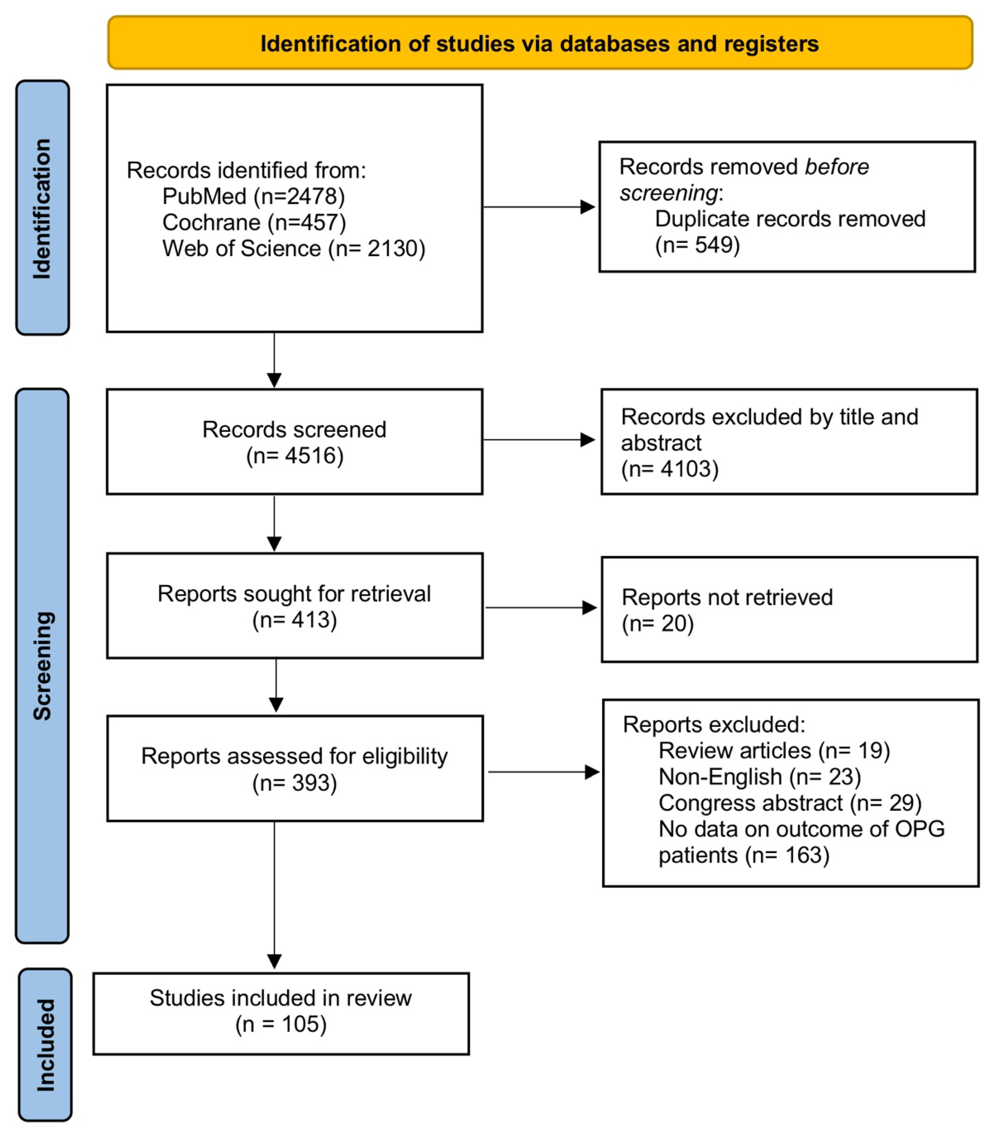
2. Materials and Methods
2.1. Search Strategy
2.2. Eligibility Criteria
2.3. Data Extraction
2.4. Risk of Bias
2.5. Statistical Analysis
3. Results
3.1. Neurofibromatosis Type 1
3.2. Tumor Location
3.3. Pathology of the Tumor
3.4. Symptoms
3.5. Complications
3.6. Mortality
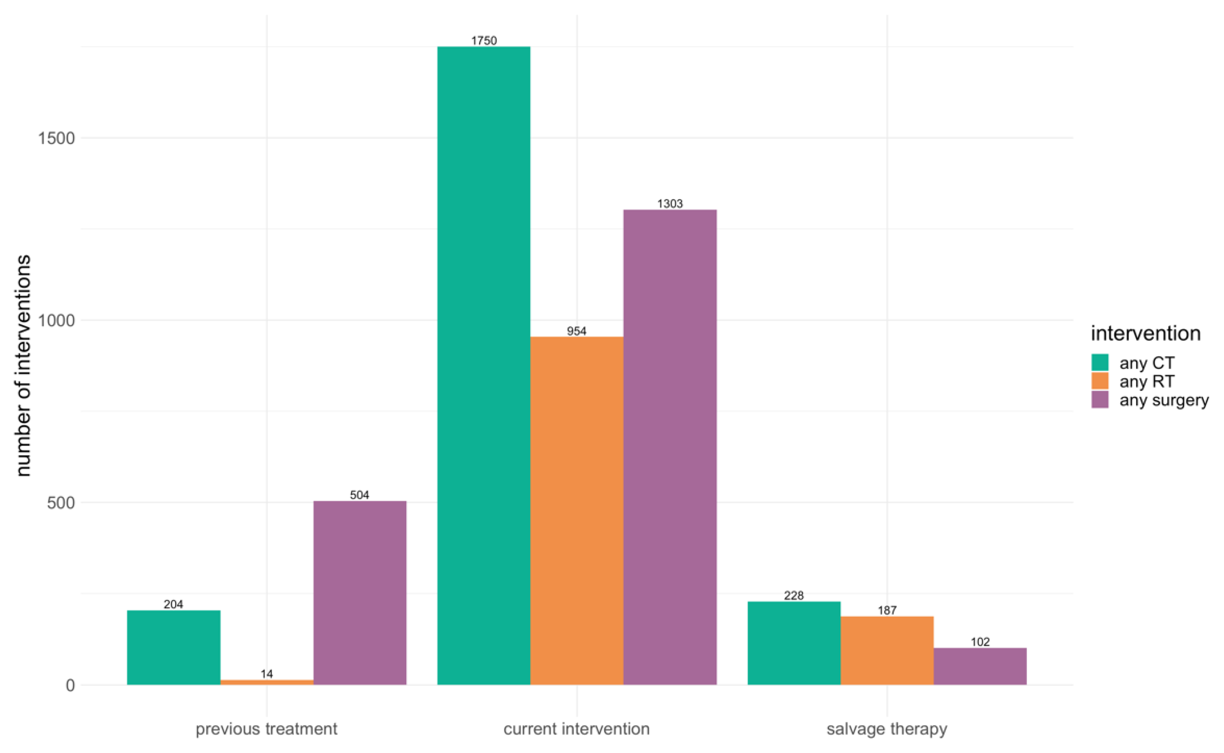
3.7. Treatment Strategies
3.8. Clinical Outcome
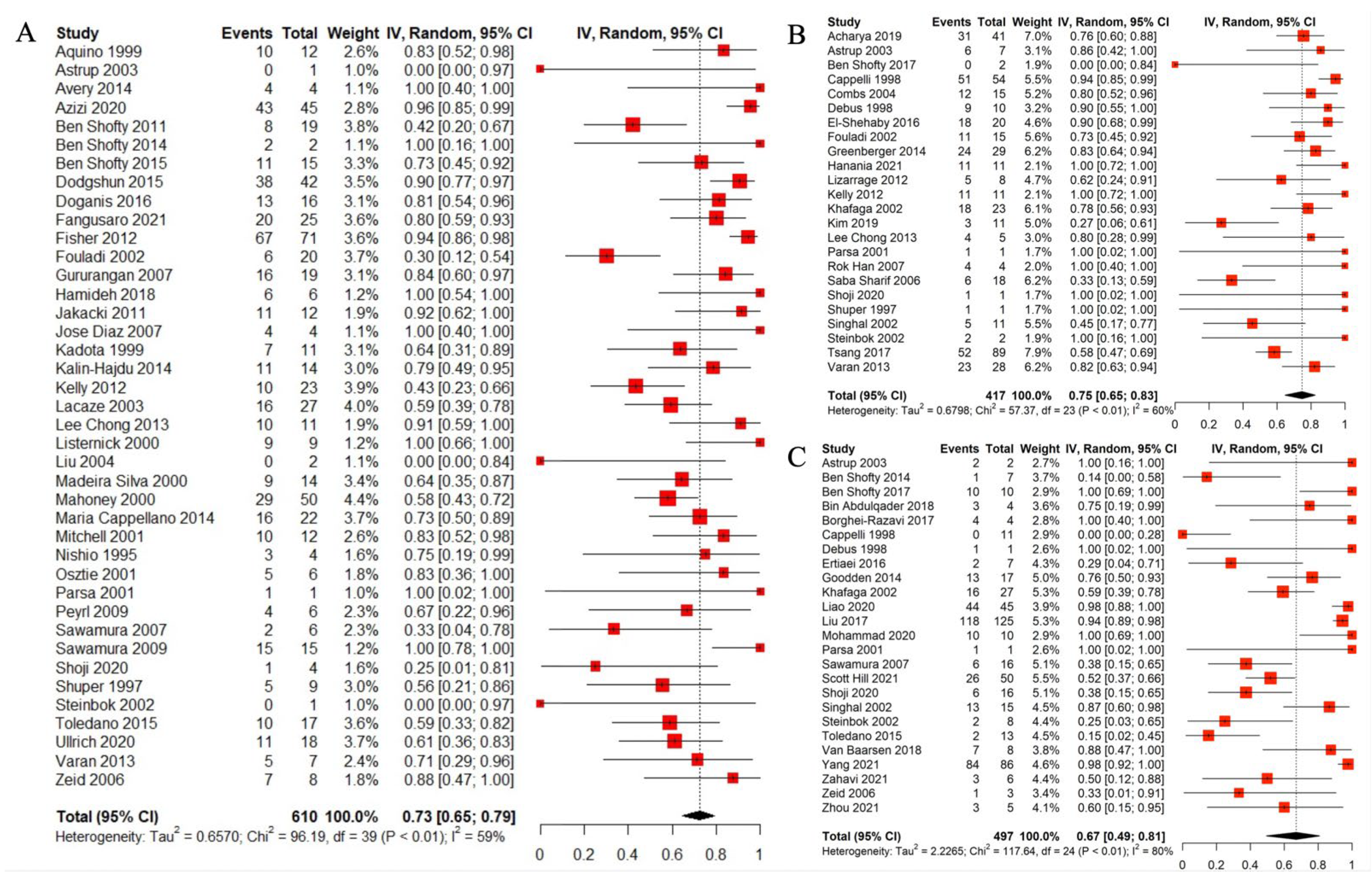
3.9. Radiological Outcome
4. Discussion
4.1. Observation
4.2. Radiotherapy
4.3. Chemotherapy
4.4. Surgery
4.5. Limitations and Future Research
5. Conclusions
Author Contributions
Funding
Institutional Review Board Statement
Informed Consent Statement
Data Availability Statement
Conflicts of Interest
References
- Binning, M.J.; Liu, J.K.; Kestle, J.R.; Brockmeyer, D.L.; Walker, M.L. Optic pathway gliomas: A review. Neurosurg. Focus 2007, 23, E2. [Google Scholar] [CrossRef] [PubMed]
- Fried, I.; Tabori, U.; Tihan, T.; Reginald, A.; Bouffet, E. Optic pathway gliomas: A review. CNS Oncol. 2013, 2, 143–159. [Google Scholar] [CrossRef] [PubMed]
- Peckham-Gregory, E.C.; Montenegro, R.E.; Stevenson, D.A.; Viskochil, D.H.; Scheurer, M.E.; Lupo, P.J.; Schiffman, J.D. Evaluation of racial disparities in pediatric optic pathway glioma incidence: Results from the Surveillance, Epidemiology, and End Results Program, 2000–2014. Cancer Epidemiol. 2018, 54, 90–94. [Google Scholar] [CrossRef] [PubMed]
- Robert-Boire, V.; Rosca, L.; Samson, Y.; Ospina, L.H.; Perreault, S. Clinical presentation and outcome of patients with optic pathway glioma. Pediatr. Neurol. 2017, 75, 55–60. [Google Scholar] [CrossRef]
- Campen, C.J.; Gutmann, D.H. Optic pathway gliomas in neurofibromatosis type 1. J. Child Neurol. 2018, 33, 73–81. [Google Scholar] [CrossRef]
- Cassina, M.; Frizziero, L.; Opocher, E.; Parrozzani, R.; Sorrentino, U.; Viscardi, E.; Miglionico, G.; Midena, E.; Clementi, M.; Trevisson, E. Optic pathway glioma in type 1 neurofibromatosis: Review of its pathogenesis, diagnostic assessment, and treatment recommendations. Cancers 2019, 11, 1790. [Google Scholar] [CrossRef]
- Thomas, R.P.; Gibbs, I.C.; Xu, L.W.; Recht, L. Treatment options for optic pathway gliomas. Curr. Treat. Options Neurol. 2015, 17, 2. [Google Scholar] [CrossRef]
- ZHANG, T.; SUN, H.; JI, Y.; YU, J.; WEI, A.; GE, M. Spontaneous regression of optic pathway glioma in children: Report of three cases and review of literature. Cancer Res. Clin. 2020, 6, 182–185. [Google Scholar]
- Page, M.J.; McKenzie, J.E.; Bossuyt, P.M.; Boutron, I.; Hoffmann, T.C.; Mulrow, C.D.; Shamseer, L.; Tetzlaff, J.M.; Akl, E.A.; Brennan, S.E. The PRISMA 2020 statement: An updated guideline for reporting systematic reviews. Int. J. Surg. 2021, 88, 105906. [Google Scholar] [CrossRef]
- NHLBI. Study Quality Assessment Tools. 2021. Available online: https://www.nhlbi.nih.gov/health-topics/study-quality-assessment-tools (accessed on 14 December 2021).
- Hozo, S.P.; Djulbegovic, B.; Hozo, I. Estimating the mean and variance from the median, range, and the size of a sample. BMC Med. Res. Methodol. 2005, 5, 13. [Google Scholar] [CrossRef]
- Schwarzer, G. meta: An R package for meta-analysis. R News 2007, 7, 40–45. [Google Scholar]
- Rücker, G.; Schwarzer, G.; Carpenter, J.R.; Binder, H.; Schumacher, M. Treatment-effect estimates adjusted for small-study effects via a limit meta-analysis. Biostatistics 2011, 12, 122–142. [Google Scholar] [CrossRef]
- Yang, P.; Liu, H.-C.; Qiu, E.; Wang, W.; Zhang, J.-L.; Jiang, L.-B.; Kang, J. Comparison of two surgical methods for the treatment of optic pathway gliomas in the intraorbital segment: An analysis of long-term clinical follow-up, which evaluates the surgical outcomes. Transl. Pediatr. 2021, 10, 1586–1597. [Google Scholar] [CrossRef]
- Fangusaro, J.; Onar-Thomas, A.; Poussaint, T.Y.; Wu, S.; Ligon, A.H.; Lindeman, N.; Campagne, O.; Banerjee, A.; Gururangan, S.; Kilburn, L.; et al. A Phase 2 Trial of Selumetinib in Children with Recurrent Optic Pathway and Hypothalamic Low-Grade Glioma without NF1: A Pediatric Brain Tumor Consortium Study. Neuro-Oncol. 2021, 23, 1777–1788. [Google Scholar] [CrossRef]
- Liao, C.; Zhang, H.; Liu, Z.; Han, Z.; Li, C.; Gong, J.; Liu, W.; Ma, Z.; Tian, Y. The Visual Acuity Outcome and Relevant Factors Affecting Visual Improvement in Pediatric Sporadic Chiasmatic-Hypothalamic Glioma Patients Who Received Surgery. Front. Neurol. 2020, 11, 766. [Google Scholar] [CrossRef]
- Heidary, G.; Fisher, M.J.; Liu, G.T.; Ferner, R.E.; Gutmann, D.H.; Listernick, R.H.; Kapur, K.; Loguidice, M.; Ardern-Holmes, S.L.; Avery, R.A.; et al. Visual field outcomes in children treated for neurofibromatosis type 1–associated optic pathway gliomas: A multicenter retrospective study. J. AAPOS 2020, 24, 349.e341–349.e345. [Google Scholar] [CrossRef]
- Quesada, S.; Coca, K.; Hoehn, M.; Qaddoumi, I.; Merchant, T.E.; Acharya, S. Visual Outcomes After Radiation Therapy for Optic Pathway Glioma. Int. J. Radiat. Oncol. Biol. Phys. 2019, 105, E631. [Google Scholar] [CrossRef]
- Kim, H.; Kim, T.; Jung, J.; Kim, J. The regression patterns of pediatric optic pathway glioma after Proton Beam Therapy. Int. J. Radiat. Oncol. Biol. Phys. 2019, 105, 912. [Google Scholar] [CrossRef]
- Acharya, S.; Quesada, S.; Coca, K.; Richardson, C.; Hoehn, M.E.; Chiang, J.; Qaddoumi, I.; Boop, F.A.; Gajjar, A.; Merchant, T.E. Long-term visual acuity outcomes after radiation therapy for sporadic optic pathway glioma. J. Neuro-Oncol. 2019, 144, 603–610. [Google Scholar] [CrossRef]
- Halliday, G.; Zapotocky, M.; Reginald, A.; Tabori, U.; Bouffet, E.; Dupuis, L.; Fried, I.; Bartels, U. Theophylline in vision-impaired children with optic pathway glioma. Pediatr. Blood Cancer 2018, 65, S434–S435. [Google Scholar]
- Falzon, K.; Drimtzias, E.; Picton, S.; Simmons, I. Visual outcomes after chemotherapy for optic pathway glioma in children with and without neurofibromatosis type 1: Results of the international society of paediatric oncology (siop) low-grade glioma 2004 trial UK cohort. Br. J. Ophthalmol. 2018, 102, 1367–1371. [Google Scholar] [CrossRef]
- Bin Abdulqader, S.; Al-Ajlan, Z.; Albakr, A.; Issawi, W.; Al-Bar, M.; Recinos, P.F.; Alsaleh, S.; Ajlan, A. Endoscopic transnasal resection of optic pathway pilocytic astrocytoma. Child’s Nerv. Syst. 2019, 35, 73–81. [Google Scholar] [CrossRef]
- Borghei-Razavi, H.; Shibao, S.; Schick, U. Prechiasmatic transection of the optic nerve in optic nerve glioma: Technical description and surgical outcome. Neurosurg. Rev. 2017, 40, 135–141. [Google Scholar] [CrossRef]
- Ertiaei, A.; Hanaei, S.; Habibi, Z.; Moradi, E.; Nejat, F. Optic pathway gliomas: Clinical manifestation, treatment, and follow-up. Pediatr. Neurosurg. 2016, 51, 223–228. [Google Scholar] [CrossRef]
- El-Shehaby, A.M.; Reda, W.A.; Abdel Karim, K.M.; Emad Eldin, R.M.; Nabeel, A.M. Single-session Gamma Knife radiosurgery for optic pathway/hypothalamic gliomas. J. Neurosurg. 2016, 125, 50–57. [Google Scholar] [CrossRef]
- Doganis, D.; Pourtsidis, A.; Tsakiris, K.; Baka, M.; Kouri, A.; Bouhoutsou, D.; Varvoutsi, M.; Servitzoglou, M.; Dana, H.; Kosmidis, H. Optic pathway glioma in children: 10 years of experience in a single institution. Pediatr. Hematol. Oncol. 2016, 33, 102–108. [Google Scholar] [CrossRef]
- Dodgshun, A.J.; Elder, J.E.; Hansford, J.R.; Sullivan, M.J. Long-term visual outcome after chemotherapy for optic pathway glioma in children: Site and age are strongly predictive. Cancer 2015, 121, 4190–4196. [Google Scholar] [CrossRef]
- Kalin-Hajdu, E.; Décarie, J.-C.; Marzouki, M.; Carret, A.-S.; Ospina, L.H. Visual acuity of children treated with chemotherapy for optic pathway gliomas. Pediatr. Blood Cancer 2014, 61, 223–227. [Google Scholar] [CrossRef]
- Chong, A.L.; Pole, J.D.; Scheinemann, K.; Hukin, J.; Tabori, U.; Huang, A.; Bouffet, E.; Bartels, U. Optic pathway gliomas in adolescence-time to challenge treatment choices? Neuro-Oncol. 2013, 15, 391–400. [Google Scholar] [CrossRef]
- Kelly, J.P.; Leary, S.; Khanna, P.; Weiss, A.H. Longitudinal measures of visual function, tumor volume, and prediction of visual outcomes after treatment of optic pathway gliomas. Ophthalmology 2012, 119, 1231–1237. [Google Scholar] [CrossRef]
- Fisher, M.J.; Loguidice, M.; Gutmann, D.H.; Listernick, R.; Ferner, R.E.; Ullrich, N.J.; Packer, R.J.; Tabori, U.; Hoffman, R.O.; Ardern-Holmes, S.L.; et al. Visual outcomes in children with neurofibromatosis type 1-associated optic pathway glioma following chemotherapy: A multicenter retrospective analysis. Neuro-Oncol. 2012, 14, 790–797. [Google Scholar] [CrossRef] [PubMed]
- Shofty, B.; Ben-Sira, L.; Freedman, S.; Yalon, M.; Dvir, R.; Weintraub, M.; Toledano, H.; Constantini, S.; Kesler, A. Visual outcome following chemotherapy for progressive optic pathway gliomas. Pediatr. Blood Cancer 2011, 57, 481–485. [Google Scholar] [CrossRef] [PubMed]
- Sawamura, Y.; Kamoshima, Y.; Kato, T.; Tajima, T.; Tsubaki, J. Chemotherapy with cisplatin and vincristine for optic pathway/ hypothalamic astrocytoma in young children. Jpn. J. Clin. Oncol. 2009, 39, 277–283. [Google Scholar] [CrossRef] [PubMed]
- Peyrl, A.; Azizi, A.; Czech, T.; Gruber-Olipitz, M.; Jones, N.; Haberler, C.; Prayer, D.; Autzinger, E.; Slavc, I. Tumor stabilization under treatment with imatinib in progressive hypothalamic-chiasmatic glioma. Pediatr. Blood Cancer 2009, 52, 476–480. [Google Scholar] [CrossRef]
- Via, P.D.; Opocher, E.; Pinello, M.L.; Calderone, M.; Viscardi, E.; Clementi, M.; Battistella, P.A.; Laverda, A.M.; Dalt, L.D.; Perilongo, G. Visual outcome of a cohort of children with neurofibromatosis type 1 and optic pathway glioma followed by a pediatric neuro-oncology program. Neuro-Oncol. 2007, 9, 430–437. [Google Scholar] [CrossRef]
- Silva, M.M.; Goldman, S.; Keating, G.; Marymont, M.A.; Kalapurakal, J.; Tomita, T. Optic pathway hypothalamic gliomas in children under three years of age: The role of chemotherapy. Pediatr. Neurosurg. 2000, 33, 151–158. [Google Scholar] [CrossRef]
- Listernick, R.; Charrow, J.; Tomita, T.; Goldman, S. Carboplatin therapy for optic pathway tumors in children with neurofibromatosis type-1. J. Neuro-Oncol. 1999, 45, 185–190. [Google Scholar] [CrossRef]
- Parsa, C.F.; Hoyt, C.S.; Lesser, R.L.; Weinstein, J.M.; Strother, C.M.; Muci-Mendoza, R.; Ramella, M.; Manor, R.S.; Fletcher, W.A.; Repka, M.X.; et al. Spontaneous regression of optic gliomas: Thirteen cases documented by serial neuroimaging. Arch. Ophthalmol. 2001, 119, 516–529. [Google Scholar] [CrossRef]
- Osztie, E.; Várallyay, P.; Doolittle, N.D.; Lacy, C.; Jones, G.; Nickolson, H.S.; Neuwelt, E.A. Combined intraarterial carboplatin, intraarterial etoposide phosphate, and IV cytoxan chemotherapy for progressive optic-hypothalamic gliomas in young children. Am. J. Neuroradiol. 2001, 22, 818–823. [Google Scholar]
- Mitchell, A.E.; Elder, J.E.; Mackey, D.A.; Waters, K.D.; Ashley, D.M. Visual improvement despite radiologically stable disease after treatment with carboplatin in children with progressive low-grade optic/thalamic gliomas. Am. J. Pediatr. Hematol./Oncol. 2001, 23, 572–577. [Google Scholar] [CrossRef]
- Aquino, V.M.; Fort, D.W.; Kamen, B.A. Carboplatin for the treatment of children with newly diagnosed optic chiasm gliomas: A phase II study. J. Neuro-Oncol. 1999, 41, 255–259. [Google Scholar] [CrossRef]
- Cappelli, C.; Grill, J.; Raquin, M.; Pierre-Kahn, A.; Lellouch-Tubiana, A.; Terrier-Lacombe, M.J.; Habrand, J.L.; Couanet, D.; Brauner, R.; Rodriguez, D.; et al. Long-term follow up of 69 patients treated for optic pathway tumours before the chemotherapy era. Arch. Dis. Child. 1998, 79, 334–338. [Google Scholar] [CrossRef]
- Nishio, S.; Morioka, T.; Takeshita, I.; Shono, T.; Inamura, T.; Fujiwara, S.; Fukui, M. Chemotherapy for progressive pilocytic astrocytomas in the chiasmo-hypothalamic regions. Clin. Neurol. Neurosurg. 1995, 97, 300–306. [Google Scholar] [CrossRef]
- Ullrich, N.J.; Prabhu, S.P.; Packer, R.J.; Goldman, S.; Robison, N.J.; Allen, J.C.; Viskochil, D.H.; Gutmann, D.H.; Perentesis, J.P.; Korf, B.R.; et al. Visual outcomes following everolimus targeted therapy for neurofibromatosis type 1-associated optic pathway gliomas in children. Pediatr. Blood Cancer 2021, 68, e28833. [Google Scholar] [CrossRef]
- Shoji, T.; Kanamori, M.; Saito, R.; Watanabe, Y.; Watanabe, M.; Fujimura, M.; Ogawa, Y.; Sonoda, Y.; Kumabe, T.; Kure, S.; et al. Frequent clinical and radiological progression of optic pathway/hypothalamic pilocytic astrocytoma in adolescents and young adults. Neurol. Med.-Chir. 2020, 60, 277–285. [Google Scholar] [CrossRef]
- Hidalgo, E.T.; Kvint, S.; Orillac, C.; North, E.; Dastagirzada, Y.; Chang, J.C.; Addae, G.; Jennings, T.S.; Snuderl, M.; Wisoff, J.H. Long-term clinical and visual outcomes after surgical resection of pediatric pilocytic/pilomyxoid optic pathway gliomas. J. Neurosurg. Pediatr. 2019, 24, 166–173. [Google Scholar] [CrossRef]
- Awdeh, R.M.; Kiehna, E.N.; Drewry, R.D.; Kerr, N.C.; Haik, B.G.; Wu, S.; Xiong, X.; Merchant, T.E. Visual Outcomes in Pediatric Optic Pathway Glioma after Conformal Radiation Therapy. Int. J. Radiat. Oncol. Biol. Phys. 2012, 84, 46–51. [Google Scholar] [CrossRef]
- Zeid, J.L.; Charrow, J.; Sandu, M.; Goldman, S.; Listernick, R. Orbital optic nerve gliomas in children with neurofibromatosis type 1. J. AAPOS 2006, 10, 534–539. [Google Scholar] [CrossRef]
- Han, S.R.; Yoon, S.W.; Yee, G.T.; Choi, C.Y.; Sohn, M.J.; Lee, D.J.; Whang, C.J. Novalis radiosurgery of optic gliomas in children: Preliminary report. Pediatr. Neurosurg. 2007, 43, 251–257. [Google Scholar] [CrossRef]
- Hanania, A.N.; Paulino, A.C.; Ludmir, E.B.; Shah, V.S.; Su, J.M.; McGovern, S.L.; Baxter, P.A.; McAleer, M.F.; Grosshans, D.R.; Okcu, M.F.; et al. Early radiotherapy preserves vision in sporadic optic pathway glioma. Cancer 2021, 127, 2358–2367. [Google Scholar] [CrossRef]
- Azizi, A.A.; Walker, D.A.; Liu, J.-F.; Sehested, A.; Jaspan, T.; Pemp, B.; Simmons, I.; Ferner, R.; Grill, J.; Hargrave, D.; et al. NF1 optic pathway glioma: Analyzing risk factors for visual outcome and indications to treat. Neuro-Oncol. 2021, 23, 100–111. [Google Scholar] [CrossRef]
- Dodgshun, A.J.; Maixner, W.J.; Heath, J.A.; Sullivan, M.J.; Hansford, J.R. Single agent carboplatin for pediatric low-grade glioma: A retrospective analysis shows equivalent efficacy to multiagent chemotherapy. Int. J. Cancer 2016, 138, 481–488. [Google Scholar] [CrossRef]
- Kadota, R.P.; Kun, L.E.; Langston, J.W.; Burger, P.C.; Cohen, M.E.; Mahoney, D.H.; Walter, A.W.; Rodman, J.H.; Parent, A.; Buckley, E.; et al. Cyclophosphamide for the treatment of progressive low-grade astrocytoma: A Pediatric Oncology Group Phase II study. J. Pediatr. Hematol./Oncol. 1999, 21, 198–202. [Google Scholar] [CrossRef]
- Varan, A.; Batu, A.; Cila, A.; Soylemezoǧlu, F.; Balc, S.; Akalan, N.; Zorlu, F.; Akyüz, C.; Kutluk, T.; Büyükpamukçu, M. Optic glioma in children: A retrospective analysis of 101 cases. Am. J. Clin. Oncol. Cancer Clin. Trials 2013, 36, 287–292. [Google Scholar] [CrossRef] [PubMed]
- Tsang, D.S.; Murphy, E.S.; Merchant, T.E. Radiation Therapy for Optic Pathway and Hypothalamic Low-Grade Gliomas in Children. Int. J. Radiat. Oncol. Biol. Phys. 2017, 99, 642–651. [Google Scholar] [CrossRef] [PubMed]
- Hill, C.S.; Khan, M.; Phipps, K.; Green, K.; Hargrave, D.; Aquilina, K. Neurosurgical experience of managing optic pathway gliomas. Child’s Nerv. Syst. 2021, 37, 1917–1929. [Google Scholar] [CrossRef] [PubMed]
- Zahavi, A.; Luckman, J.; Ben-David, G.S.; Toledano, H.; Michowiz, S.; Vardizer, Y.; Goldenberg-Cohen, N. Proptosis due to intraorbital space-occupying lesions in children. Graefe’s Arch. Clin. Exp. Ophthalmol. 2020, 258, 2541–2550. [Google Scholar] [CrossRef]
- Liu, Y.; Hao, X.; Liu, W.; Li, C.; Gong, J.; Ma, Z.; Tian, Y. Analysis of Survival Prognosis for Children with Symptomatic Optic Pathway Gliomas Who Received Surgery. World Neurosurg. 2018, 109, e1–e15. [Google Scholar] [CrossRef]
- Hamideh, D.; Hoehn, M.E.; Harreld, J.H.; Klimo, P.D.; Gajjar, A.; Qaddoumi, I. Isolated Optic Nerve Glioma in Children With and Without Neurofibromatosis: Retrospective Characterization and Analysis of Outcomes. J. Child Neurol. 2018, 33, 375–382. [Google Scholar] [CrossRef]
- Shofty, B.; Ben-Sira, L.; Kesler, A.; Jallo, G.; Groves, M.L.; Iyer, R.R.; Lassaletta, L.; Tabori, U.; Bouffet, E.; Thomale, U.-W.; et al. Isolated optic nerve gliomas: A multicenter historical cohort study. J. Neurosurg. Pediatr. 2017, 20, 549–555. [Google Scholar] [CrossRef]
- Falsini, B.; Chiaretti, A.; Rizzo, D.; Piccardi, M.; Ruggiero, A.; Manni, L.; Soligo, M.; Dickmann, A.; Federici, M.; Salerni, A.; et al. Nerve growth factor improves visual loss in childhood optic gliomas: A randomized, double-blind, phase II clinical trial. Brain 2016, 139, 404–414. [Google Scholar] [CrossRef]
- Parness-Yossifon, R.; Listernick, R.; Charrow, J.; Barto, H.; Zeid, J.L. Strabismus in patients with neurofibromatosis type 1-associated optic pathway glioma. J. Am. Assoc. Pediatr. Ophthalmol. Strabismus 2015, 19, 422–425. [Google Scholar] [CrossRef]
- Millward, C.P.; Da Rosa, S.P.; Avula, S.; Ellenbogen, J.R.; Spiteri, M.; Lewis, E.; Didi, M.; Mallucci, C. The role of early intra-operative MRI in partial resection of optic pathway/hypothalamic gliomas in children. Child’s Nerv. Syst. 2015, 31, 2055–2062. [Google Scholar] [CrossRef]
- Shofty, B.; Constantini, S.; Bokstein, F.; Ram, Z.; Ben-Sira, L.; Freedman, S.; Vainer, G.; Kesler, A. Optic pathway gliomas in adults. Neurosurgery 2014, 74, 273–280. [Google Scholar] [CrossRef]
- Mandiwanza, T.; Kaliaperumal, C.; Khalil, A.; Sattar, M.; Crimmins, D.; Caird, J. Suprasellar pilocytic astrocytoma: One national centre’s experience. Child’s Nerv. Syst. 2014, 30, 1243–1248. [Google Scholar] [CrossRef]
- Greenberger, B.A.; Pulsifer, M.B.; Ebb, D.H.; Macdonald, S.M.; Jones, R.M.; Butler, W.E.; Huang, M.S.; Marcus, K.J.; Oberg, J.A.; Tarbell, N.J.; et al. Clinical outcomes and late endocrine, neurocognitive, and visual profiles of proton radiation for pediatric low-grade gliomas. Int. J. Radiat. Oncol. Biol. Phys. 2014, 89, 1060–1068. [Google Scholar] [CrossRef]
- Goodden, J.; Pizer, B.; Pettorini, B.; Williams, D.; Blair, J.; Didi, M.; Thorp, N.; Mallucci, C. The role of surgery in optic pathway/hypothalamic gliomas in children: Clinical article. J. Neurosurg. Pediatr. 2014, 13, 1–12. [Google Scholar] [CrossRef]
- Magli, A.; Forte, R.; Cinalli, G.; Esposito, F.; Parisi, S.; Capasso, M.; Papparella, A. Functional changes after treatment of optic pathway paediatric low-grade gliomas. Eye 2013, 27, 1288–1292. [Google Scholar] [CrossRef]
- Shriver, E.M.; Ragheb, J.; Tse, D.T. Combined transcranial-orbital approach for resection of optic nerve gliomas: A clinical and anatomical study. Ophthalmic Plast. Reconstr. Surg. 2012, 28, 184–191. [Google Scholar]
- Gras-Combe, G.; Moritz-Gasser, S.; Herbet, G.; Duffau, H. Intraoperative subcortical electrical mapping of optic radiations in awake surgery for glioma involving visual pathways: Clinical article. J. Neurosurg. 2012, 117, 466–473. [Google Scholar] [CrossRef]
- Jakacki, R.I.; Bouffet, E.; Adamson, P.C.; Pollack, I.F.; Ingle, A.M.; Voss, S.D.; Blaney, S.M. A phase 1 study of vinblastine in combination with carboplatin for children with low-grade gliomas: A Children’s Oncology Group phase 1 consortium study. Neuro-Oncol. 2011, 13, 910–915. [Google Scholar] [CrossRef]
- Massimino, M.; Spreafico, F.; Riva, D.; Biassoni, V.; Poggi, G.; Solero, C.; Gandola, L.; Genitori, L.; Modena, P.; Simonetti, F.; et al. A lower-dose, lower-toxicity cisplatin-etoposide regimen for childhood progressive low-grade glioma. J. Neuro-Oncol. 2010, 100, 65–71. [Google Scholar] [CrossRef]
- Nicolin, G.; Parkin, P.; Mabbott, D.; Hargrave, D.; Bartels, U.; Tabori, U.; Rutka, J.; Buncic, J.R.; Bouffet, E. Natural history and outcome of optic pathway gliomas in children. Pediatr. Blood Cancer 2009, 53, 1231–1237. [Google Scholar] [CrossRef]
- Sawamura, Y.; Kamada, K.; Kamoshima, Y.; Yamaguchi, S.; Tajima, T.; Tsubaki, J.; Fujimaki, T. Role of surgery for optic pathway/hypothalamic astrocytomas in children. Neuro-Oncol. 2008, 10, 725–733. [Google Scholar] [CrossRef]
- Diaz, R.J.; Laughlin, S.; Nicolin, G.; Buncic, J.R.; Bouffet, E.; Bartels, U. Assessment of chemotherapeutic response in children with proptosis due to optic nerve glioma. Child’s Nerv. Syst. 2008, 24, 707–712. [Google Scholar] [CrossRef]
- Sharif, S.; Ferner, R.; Birch, J.M.; Gillespie, J.E.; Gattamaneni, H.R.; Baser, M.E.; Evans, D.G.R. Second primary tumors in neurofibromatosis 1 patients treated for optic glioma: Substantial risks after radiotherapy. J. Clin. Oncol. 2006, 24, 2570–2575. [Google Scholar] [CrossRef]
- Kaufman, L.M.; Doroftei, O. Optic glioma warranting treatment in children. Eye 2006, 20, 1149–1164. [Google Scholar] [CrossRef]
- Combs, S.E.; Schulz-Ertner, D.; Moschos, D.; Thilmann, C.; Huber, P.E.; Debus, J. Fractionated stereotactic radiotherapy of optic pathway gliomas: Tolerance and long-term outcome. Int. J. Radiat. Oncol. Biol. Phys. 2005, 62, 814–819. [Google Scholar] [CrossRef] [PubMed]
- Lacaze, E.; Kieffer, V.; Streri, A.; Lorenzi, C.; Gentaz, E.; Habrand, J.-L.; Dellatolas, G.; Kalifa, C.; Grill, J. Neuropsychological outcome in children with optic pathway tumours when first-line treatment is chemotherapy. Br. J. Cancer 2003, 89, 2038–2044. [Google Scholar] [CrossRef] [PubMed]
- Khafaga, Y.; Hassounah, M.; Kandil, A.; Kanaan, I.; Allam, A.; El Husseiny, G.; Kofide, A.; Belal, A.; Al Shabanah, M.; Schultz, H.; et al. Optic gliomas: A retrospective analysis of 50 cases. Int. J. Radiat. Oncol. Biol. Phys. 2003, 56, 807–812. [Google Scholar] [CrossRef]
- Fouladi, M.; Wallace, D.; Langston, J.W.; Mulhern, R.; Rose, S.R.; Gajjar, A.; Sanford, R.A.; Merchant, T.E.; Jenkins, J.J.; Kun, L.E.; et al. Survival and functional outcome of children with Hypothalamic/Chiasmatic tumors. Cancer 2003, 97, 1084–1092. [Google Scholar] [CrossRef]
- Astrup, J. Natural history and clinical management of optic pathway glioma. Br. J. Neurosurg. 2003, 17, 327–335. [Google Scholar] [CrossRef]
- Steinbok, P.; Hentschel, S.; Almqvist, P.; Cochrane, D.D.; Poskitt, K. Management of optic chiasmatic/hypothalamic astrocytomas in children. Can. J. Neurol. Sci. 2002, 29, 132–138. [Google Scholar] [CrossRef]
- Singhal, S.; Birch, J.M.; Kerr, B.; Lashford, L.; Evans, D.G.R. Neurofibromatosis type 1 and sporadic optic gliomas. Arch. Dis. Child. 2002, 87, 65–70. [Google Scholar] [CrossRef]
- Mahoney, D.H.J.; Cohen, M.E.; Friedman, H.S.; Kepner, J.L.; Gemer, L.; Langston, J.W.; James, H.E.; Duffner, P.K.; Kun, L.E. Carboplatin is effective therapy for young children with progressive optic pathway tumors: A Pediatric Oncology Group phase II study. Neuro-Oncol. 2000, 2, 213–220. [Google Scholar] [CrossRef]
- Debus, J.; Kocagöncü, K.O.; Ḧoss, A.; Wenz, F.; Wannenmacher, M. Fractionated stereotactic radiotherapy (FSRT) for optic glioma. Int. J. Radiat. Oncol. Biol. Phys. 1999, 44, 243–248. [Google Scholar] [CrossRef]
- Zhou, Z.-Y.; Wang, X.-S.; Gong, Y.; La Ali Musyafar, O.; Yu, J.-J.; Huo, G.; Mou, J.-M.; Yang, G. Treatment with endoscopic transnasal resection of hypothalamic pilocytic astrocytomas: A single-center experience. BMC Surg. 2021, 21, 103. [Google Scholar] [CrossRef]
- Lassaletta, A.; Scheinemann, K.; Zelcer, S.M.; Hukin, J.; Wilson, B.A.; Jabado, N.; Carret, A.S.; Lafay-Cousin, L.; Larouche, V.; Hawkins, C.E.; et al. Phase II weekly vinblastine for chemotherapy-naïve children with progressive low-grade glioma: A Canadian pediatric brain tumor consortium study. J. Clin. Oncol. 2016, 34, 3537–3543. [Google Scholar] [CrossRef]
- Toledano, H.; Muhsinoglu, O.; Luckman, J.; Goldenberg-Cohen, N.; Michowiz, S. Acquired nystagmus as the initial presenting sign of chiasmal glioma in young children. Eur. J. Paediatr. Neurol. 2015, 19, 694–700. [Google Scholar] [CrossRef]
- Liu, G.T.; Brodsky, M.C.; Phillips, P.C.; Belasco, J.; Janss, A.; Golden, J.C.; Bilaniuk, L.L.; Burson, G.T.; Duhaime, A.-C.; Sutton, L.N. Optic radiation involvement in optic pathway gliomas in neurofibromatosis. Am. J. Ophthalmol. 2004, 137, 407–414. [Google Scholar] [CrossRef]
- Gururangan, S.; Fisher, M.J.; Allen, J.C.; Herndon II, J.E.; Quinn, J.A.; Reardon, D.A.; Vredenburgh, J.J.; Desjardins, A.; Phillips, P.C.; Watral, M.A.; et al. Temozolomide in children with progressive low-grade glioma. Neuro-Oncol. 2007, 9, 161–168. [Google Scholar] [CrossRef] [PubMed]
- Kinori, M.; Armarnik, S.; Listernick, R.; Charrow, J.; Zeid, J.L. Neurofibromatosis Type 1-Associated Optic Pathway Glioma in Children: A Follow-Up of 10 Years or More. American journal of ophthalmology 2021, 221, 91–96. [Google Scholar] [CrossRef] [PubMed]
- Rakotonjanahary, J.; Gravier, N.; Lambron, J.; De Carli, E.; Toulgoat, F.; Delion, M.; Pellier, I.; Rialland, X. Long-term visual acuity in patients with optic pathway glioma treated during childhood with up-front BB-SFOP chemotherapy-Analysis of a French pediatric historical cohort. PLoS ONE 2019, 14, e0212107. [Google Scholar] [CrossRef] [PubMed]
- Campagna, M.; Opocher, E.; Viscardi, E.; Calderone, M.; Severino, S.M.; Cermakova, I.; Perilongo, G. Optic pathway glioma: Long-term visual outcome in children without neurofibromatosis type-1. Pediatr. Blood Cancer 2010, 55, 1083–1088. [Google Scholar] [CrossRef]
- Suárez, J.C.; Viano, J.C.; Zunino, S.; Herrera, E.J.; Gomez, J.; Tramunt, B.; Marengo, I.; Hiramatzu, E.; Miras, M.; Pena, M.; et al. Management of child optic pathway gliomas: New therapeutical option. Child’s Nerv. Syst. 2006, 22, 679–684. [Google Scholar] [CrossRef]
- Rakotonjanahary, J.; De Carli, E.; Delion, M.; Kalifa, C.; Grill, J.; Doz, F.; Leblond, P.; Bertozzi, A.-I.; Rialland, X.; Alapetite, C.; et al. Mortality in children with optic pathway glioma treated with up-front BB-SFOP chemotherapy. PLoS ONE 2015, 10, e0127676. [Google Scholar] [CrossRef]
- Estrada, M.; Kelly, J.P.; Wright, J.; Phillips, J.O.; Weiss, A. Visual Function, Brain Imaging, and Physiological Factors in Children With Asymmetric Nystagmus due to Chiasmal Gliomas. Pediatric Neurology 2019, 97, 30–37. [Google Scholar] [CrossRef]
- Cambiaso, P.; Galassi, S.; Palmiero, M.; Mastronuzzi, A.; Del Bufalo, F.; Capolino, R.; Cacchione, A.; Buonuomo, P.S.; Gonfiantini, M.V.; Bartuli, A.; et al. Growth hormone excess in children with neurofibromatosis type-1 and optic glioma. Am. J. Med. Genet. Part A 2017, 173, 2353–2358. [Google Scholar] [CrossRef]
- Wan, M.J.; Ullrich, N.J.; Manley, P.E.; Kieran, M.W.; Goumnerova, L.C.; Heidary, G. Long-term visual outcomes of optic pathway gliomas in pediatric patients without neurofibromatosis type 1. J. Neuro-Oncol. 2016, 129, 173–178. [Google Scholar] [CrossRef]
- Gan, H.-W.; Phipps, K.; Aquilina, K.; Gaze, M.N.; Hayward, R.; Spoudeas, H.A. Neuroendocrine morbidity after pediatric optic gliomas: A longitudinal analysis of 166 children over 30 years. J. Clin. Endocrinol. Metab. 2015, 100, 3787–3799. [Google Scholar] [CrossRef]
- Mishra, M.V.; Andrews, D.W.; Glass, J.; Evans, J.J.; Dicker, A.P.; Shen, X.; Lawrence, Y.R. Characterization and outcomes of optic nerve gliomas: A population-based analysis. J. Neuro-Oncol. 2012, 107, 591–597. [Google Scholar] [CrossRef]
- Hupp, M.; Falkenstein, F.; Bison, B.; Mirow, C.; Krauss, J.; Gnekow, A.; Solymosi, L.; Warmuth-Metz, M. Infarction following chiasmatic low grade glioma resection. Child’s Nerv. Syst. 2012, 28, 391–398. [Google Scholar] [CrossRef]
- Marcus, K.J.; Goumnerova, L.; Billett, A.L.; Lavally, B.; Scott, R.M.; Bishop, K.; Xu, R.; Young Poussaint, T.; Kieran, M.; Kooy, H.; et al. Stereotactic radiotherapy for localized low-grade gliomas in children: Final results of a prospective trial. Int. J. Radiat. Oncol. Biol. Phys. 2005, 61, 374–379. [Google Scholar] [CrossRef]
- Thiagalingam, S.; Flaherty, M.; Billson, F.; North, K. Neurofibromatosis type 1 and optic pathway gliomas: Follow-up of 54 patients. Ophthalmology 2004, 111, 568–577. [Google Scholar] [CrossRef]
- Bowers, D.C.; Krause, T.P.; Aronson, L.J.; Barzi, A.; Burger, P.C.; Carson, B.S.; Weingart, J.D.; Wharam, M.D.; Melhem, E.R.; Cohen, K.J. Second surgery for recurrent pilocytic astrocytoma in children. Pediatr. Neurosurg. 2001, 34, 229–234. [Google Scholar] [CrossRef]
- Parsons, M.W.; Whipple, N.S.; Poppe, M.M.; Mendez, J.S.; Cannon, D.M.; Burt, L.M. The use and efficacy of chemotherapy and radiotherapy in children and adults with pilocytic astrocytoma. J. Neuro-Oncol. 2021, 151, 93–101. [Google Scholar] [CrossRef]
- Sani, I.; Albanese, A. Endocrine Long-Term Follow-Up of Children with Neurofibromatosis Type 1 and Optic Pathway Glioma. Horm. Res. Paediatr. 2017, 87, 179–188. [Google Scholar] [CrossRef]
- El Beltagy, M.A.; Reda, M.; Enayet, A.; Zaghloul, M.S.; Awad, M.; Zekri, W.; Taha, H.; El-Khateeb, N. Treatment and Outcome in 65 Children with Optic Pathway Gliomas. World Neurosurg. 2016, 89, 525–534. [Google Scholar] [CrossRef]
- Mohammad, A.E.-N.A. En-Bloc Resection Versus Resection After Evacuation and Suction of the Content for Orbital Optic Nerve Glioma Causing Visual Loss and Disfiguring Proptosis. Ophthalmic Plast. Reconstr. Surg. 2020, 36, 399–402. [Google Scholar] [CrossRef]
- Shofty, B.; Mauda-Havakuk, M.; Weizman, L.; Constantini, S.; Ben-Bashat, D.; Dvir, R.; Pratt, L.-T.; Joskowicz, L.; Kesler, A.; Yalon, M.; et al. The effect of chemotherapy on optic pathway gliomas and their sub-components: A volumetric MR analysis study. Pediatr. Blood Cancer 2015, 62, 1353–1359. [Google Scholar] [CrossRef]
- Cappellano, A.M.; Petrilli, A.S.; da Silva, N.S.; Silva, F.A.; Paiva, P.M.; Cavalheiro, S.; Bouffet, E. Single agent vinorelbine in pediatric patients with progressive optic pathway glioma. J. Neuro-Oncol. 2015, 121, 405–412. [Google Scholar] [CrossRef]
- Shuper, A.; Horev, G.; Kornreich, L.; Michowiz, S.; Weitz, R.; Zaizov, R.; Cohen, I. Visual pathway glioma: An erratic tumour with therapeutic dilemmas. Arch. Dis. Child. 1997, 76, 259–263. [Google Scholar] [CrossRef]
- Han, S.; Yang, Z.; Wang, L.; Yang, Y.; Qi, X.; Yan, C.; Yu, C. Postoperative hydrocephalus is a high-risk lethal factor for patients with low-grade optic pathway glioma. Br. J. Neurosurg. 2021, 35, 1–7. [Google Scholar] [CrossRef]
- van Baarsen, K.; Roth, J.; Serova, N.; Packer, R.J.; Shofty, B.; Thomale, U.-W.; Cinalli, G.; Toledano, H.; Michowiz, S.; Constantini, S. Optic pathway–hypothalamic glioma hemorrhage: A series of 9 patients and review of the literature. J. Neurosurg. 2018, 129, 1407–1415. [Google Scholar] [CrossRef]
- Lizarraga, K.J.; Gorgulho, A.; Lee, S.P.; Rauscher, G.; Selch, M.T.; Desalles, A.A.F. Stereotactic radiation therapy for progressive residual pilocytic astrocytomas. J. Neuro-Oncol. 2012, 109, 129–135. [Google Scholar] [CrossRef]
- Kandels, D.; Pietsch, T.; Bison, B.; Warmuth-Metz, M.; Thomale, U.-W.; Kortmann, R.-D.; Timmermann, B.; Hernáiz Driever, P.; Witt, O.; Schmidt, R.; et al. Loss of efficacy of subsequent nonsurgical therapy after primary treatment failure in pediatric low-grade glioma patients—Report from the German SIOP-LGG 2004 cohort. Int. J. Cancer 2020, 147, 3471–3489. [Google Scholar] [CrossRef]
- Avery, R.A.; Hwang, E.I.; Jakacki, R.I.; Packer, R.J. Marked recovery of vision in children with optic pathway gliomas treated with bevacizumab. JAMA Ophthalmol. 2014, 132, 111–114. [Google Scholar] [CrossRef]
- Dodge, H.W.; Love, J.G.; Craig, W.M.; Dockerty, M.B.; Kearns, T.P.; Holman, C.B.; Hayles, A.B. Gliomas of the optic nerves. AMA Arch. Neurol. Psychiatry 1958, 79, 607–621. [Google Scholar] [CrossRef] [PubMed]
- Louis, D.N.; Perry, A.; Wesseling, P.; Brat, D.J.; Cree, I.A.; Figarella-Branger, D.; Hawkins, C.; Ng, H.K.; Pfister, S.M.; Reifenberger, G.; et al. The 2021 WHO Classification of Tumors of the Central Nervous System: A summary. Neuro Oncol. 2021, 23, 1231–1251. [Google Scholar] [CrossRef] [PubMed]
- Stokland, T.; Liu, J.-F.; Ironside, J.W.; Ellison, D.W.; Taylor, R.; Robinson, K.J.; Picton, S.V.; Walker, D.A. A multivariate analysis of factors determining tumor progression in childhood low-grade glioma: A population-based cohort study (CCLG CNS9702). Neuro-Oncol. 2010, 12, 1257–1268. [Google Scholar] [CrossRef] [PubMed]
- Gnekow, A.K.; Falkenstein, F.; von Hornstein, S.; Zwiener, I.; Berkefeld, S.; Bison, B.; Warmuth-Metz, M.; Driever, P.H.; Soerensen, N.; Kortmann, R.-D. Long-term follow-up of the multicenter, multidisciplinary treatment study HIT-LGG-1996 for low-grade glioma in children and adolescents of the German Speaking Society of Pediatric Oncology and Hematology. Neuro-Oncol. 2012, 14, 1265–1284. [Google Scholar] [CrossRef]
- Gnekow, A.; Kortmann, R.-D.; Pietsch, T.; Emser, A. Low grade chiasmatic-hypothalamic glioma-carboplatin and vincristin chemotherapy effectively defers radiotherapy within a comprehensive treatment strategy. Klin. Pädiatrie 2004, 216, 331–342. [Google Scholar] [CrossRef]
- Laithier, V.; Grill, J.; Le Deley, M.-C.; Ruchoux, M.-M.; Couanet, D.; Doz, F.; Pichon, F.; Rubie, H.; Frappaz, D.; Vannier, J.-P. Progression-free survival in children with optic pathway tumors: Dependence on age and the quality of the response to chemotherapy-results of the first French prospective study for the French Society of Pediatric Oncology. J. Clin. Oncol. 2003, 21, 4572–4578. [Google Scholar]
- Shuper, A.; Kornreich, L.; Michowitz, S.; Schwartz, M.; Yaniv, I.; Cohen, I.J. Visual pathway tumors and hydrocephalus. Pediatr. Hematol. Oncol. 2000, 17, 463–468. [Google Scholar] [CrossRef]
- Opocher, E.; Kremer, L.C.; Da Dalt, L.; van de Wetering, M.D.; Viscardi, E.; Caron, H.N.; Perilongo, G. Prognostic factors for progression of childhood optic pathway glioma: A systematic review. Eur. J. Cancer 2006, 42, 1807–1816. [Google Scholar] [CrossRef]
- Parsa, C.F. Why optic gliomas should be called hamartomas. Ophthalmic Plast. Reconstr. Surg. 2010, 26, 497. [Google Scholar] [CrossRef]
- Tow, S.L.; Chandela, S.; Miller, N.R.; Avellino, A.M. Long-term outcome in children with gliomas of the anterior visual pathway. Pediatr. Neurol. 2003, 28, 262–270. [Google Scholar] [CrossRef]
- Noureldine, M.H.A.; Rasras, S.; Safari, H.; Sabahi, M.; Jallo, G.I.; Arjipour, M. Spontaneous regression of multiple intracranial capillary hemangiomas in a newborn—Long-term follow-up and literature review. Child’s Nerv. Syst. 2021, 37, 3225–3234. [Google Scholar] [CrossRef]
- Piccirilli, M.; Lenzi, J.; Delfinis, C.; Trasimeni, G.; Salvati, M.; Raco, A. Spontaneous regression of optic pathways gliomas in three patients with neurofibromatosis type I and critical review of the literature. Child’s Nerv. Syst. 2006, 22, 1332–1337. [Google Scholar] [CrossRef]
- Listernick, R.; Ferner, R.E.; Liu, G.T.; Gutmann, D.H. Optic pathway gliomas in neurofibromatosis-1: Controversies and recommendations. Ann. Neurol. Off. J. Am. Neurol. Assoc. Child Neurol. Soc. 2007, 61, 189–198. [Google Scholar] [CrossRef]
- Janss, A.J.; Grundy, R.; Cnaan, A.; Packer, R.J.; Zackai, E.H.; Sutton, L.N.; Molloy, P.T.; Phillips, P.C.; Lange, B.J.; Savino, P.J. Optic pathway and hypothalamic/chiasmatic gliomas in children younger than age 5 years with a 6-year follow-up. Cancer 1995, 75, 1051–1059. [Google Scholar] [CrossRef]
- Hoffman, H.J.; Humphreys, R.P.; Drake, J.M.; Rutka, J.T.; Becker, L.E.; Jenkin, D.; Greenberg, M. Optic Pathway/Hypothaiamic Gliomas: A Dilemma in Management. Pediatr. Neurosurg. 1993, 19, 186–195. [Google Scholar] [CrossRef] [PubMed]
- Merchant, T.E.; Kun, L.E.; Wu, S.; Xiong, X.; Sanford, R.A.; Boop, F.A. Phase II trial of conformal radiation therapy for pediatric low-grade glioma. J. Clin. Oncol. 2009, 27, 3598. [Google Scholar] [CrossRef]
- Fisher, B.J.; Bauman, G.S.; Leighton, C.E.; Stitt, L.; Cairncross, J.G.; Macdonald, D.R. Low-grade gliomas in children: Tumor volume response to radiation. J. Neurosurg. 1998, 88, 969–974. [Google Scholar] [CrossRef]
- Jenkin, D.; Angyalfi, S.; Becker, L.; Berry, M.; Buncic, R.; Chan, H.; Doherty, M.; Drake, J.; Greenberg, M.; Hendrick, B. Optic glioma in children: Surveillance, resection, or irradiation? Int. J. Radiat. Oncol. * Biol. * Phys. 1993, 25, 215–225. [Google Scholar] [CrossRef]
- Kovalic, J.J.; Grigsby, P.W.; Shepard, M.J.; Fineberg, B.B.; Thomas, P.R. Radiation therapy for gliomas of the optic nerve and chiasm. Int. J. Radiat. Oncol. * Biol. * Phys. 1990, 18, 927–932. [Google Scholar] [CrossRef]
- Brenner, D.J.; Carlson, D.J. Radiobiological principles underlying stereotactic radiation therapy. In Principles and Practice of Stereotactic Radiosurgery; Springer: Berlin/Heidelburg, Germany, 2015; pp. 57–71. [Google Scholar]
- Merchant, T.E.; Conklin, H.M.; Wu, S.; Lustig, R.H.; Xiong, X. Late effects of conformal radiation therapy for pediatric patients with low-grade glioma: Prospective evaluation of cognitive, endocrine, and hearing deficits. J. Clin. Oncol. 2009, 27, 3691. [Google Scholar] [CrossRef] [PubMed]
- Ullrich, N.; Robertson, R.; Kinnamon, D.; Scott, R.; Kieran, M.; Turner, C.; Chi, S.; Goumnerova, L.; Proctor, M.; Tarbell, N. Moyamoya following cranial irradiation for primary brain tumors in children. Neurology 2007, 68, 932–938. [Google Scholar] [CrossRef] [PubMed]
- Bowers, D.C.; Mulne, A.F.; Reisch, J.S.; Elterman, R.D.; Munoz, L.; Booth, T.; Shapiro, K.; Doxey, D.L. Nonperioperative strokes in children with central nervous system tumors. Cancer 2002, 94, 1094–1101. [Google Scholar] [CrossRef]
- Bowers, D.C.; Liu, Y.; Leisenring, W.; McNeil, E.; Stovall, M.; Gurney, J.G.; Robison, L.L.; Packer, R.J.; Oeffinger, K.C. Late-occurring stroke among long-term survivors of childhood leukemia and brain tumors: A report from the Childhood Cancer Survivor Study. J. Clin. Oncol. 2006, 24, 5277–5282. [Google Scholar] [CrossRef]
- Inskip, P.D.; Curtis, R.E. New malignancies following childhood cancer in the United States, 1973–2002. Int. J. Cancer 2007, 121, 2233–2240. [Google Scholar] [CrossRef]
- Merchant, T.E.; Hua, C.H.; Shukla, H.; Ying, X.; Nill, S.; Oelfke, U. Proton versus photon radiotherapy for common pediatric brain tumors: Comparison of models of dose characteristics and their relationship to cognitive function. Pediatr. Blood Cancer 2008, 51, 110–117. [Google Scholar] [CrossRef]
- Rosenstock, J.G.; Evans, A.E.; Schut, L. Response to vincristine of recurrent brain tumors in children. J. Neurosurg. 1976, 45, 135–140. [Google Scholar] [CrossRef]
- Lefkowitz, I.B.; Packer, R.J.; Sutton, L.N.; Siegel, K.R.; Bruce, D.A.; Evans, A.E.; Schut, L. Results of the treatment of children with recurrent gliomas with lomustine and vincristine. Cancer 1988, 61, 896–902. [Google Scholar] [CrossRef]
- Packer, R.J.; Sutton, L.N.; Bilaniuk, L.T.; Radcliffe, J.; Rosenstock, J.G.; Siegel, K.R.; Bunin, G.R.; Savino, P.J.; Bruce, D.A.; Schut, L. Treatment of chiasmatic/hypothalamic gliomas of childhood with chemotherapy: An update. Ann. Neurol. Off. J. Am. Neurol. Assoc. Child Neurol. Soc. 1988, 23, 79–85. [Google Scholar] [CrossRef]
- Packer, R.J.; Ater, J.; Allen, J.; Phillips, P.; Geyer, R.; Nicholson, H.S.; Jakacki, R.; Kurczynski, E.; Needle, M.; Finlay, J. Carboplatin and vincristine chemotherapy for children with newly diagnosed progressive low-grade gliomas. J. Neurosurg. 1997, 86, 747–754. [Google Scholar] [CrossRef]
- Ater, J.L.; Zhou, T.; Holmes, E.; Mazewski, C.M.; Booth, T.N.; Freyer, D.R.; Lazarus, K.H.; Packer, R.J.; Prados, M.; Sposto, R. Randomized study of two chemotherapy regimens for treatment of low-grade glioma in young children: A report from the Children’s Oncology Group. J. Clin. Oncol. 2012, 30, 2641. [Google Scholar] [CrossRef]
- Massimino, M.; Spreafico, F.; Cefalo, G.; Riccardi, R.; Tesoro-Tess, J.D.; Gandola, L.; Riva, D.; Ruggiero, A.; Valentini, L.; Mazza, E. High response rate to cisplatin/etoposide regimen in childhood low-grade glioma. J. Clin. Oncol. 2002, 20, 4209–4216. [Google Scholar] [CrossRef]
- Reddy, A.T.; Packer, R.J. Chemotherapy for low-grade gliomas. Child’s Nerv. Syst. 1999, 15, 506–513. [Google Scholar] [CrossRef]
- Moreno, L.; Bautista, F.; Ashley, S.; Duncan, C.; Zacharoulis, S. Does chemotherapy affect the visual outcome in children with optic pathway glioma? A systematic review of the evidence. Eur. J. Cancer 2010, 46, 2253–2259. [Google Scholar] [CrossRef]
- Lafay-Cousin, L.; Sung, L.; Carret, A.S.; Hukin, J.; Wilson, B.; Johnston, D.L.; Zelcer, S.; Silva, M.; Odame, I.; Mpofu, C. Carboplatin hypersensitivity reaction in pediatric patients with low-grade glioma: A Canadian Pediatric Brain Tumor Consortium experience. Cancer 2008, 112, 892–899. [Google Scholar] [CrossRef] [PubMed]
- Avery, R.A.; Fisher, M.J.; Liu, G.T. Optic pathway gliomas. J. Neuro-Ophthalmol. 2011, 31, 269–278. [Google Scholar] [CrossRef] [PubMed]
- Le Deley, M.-C.; Leblanc, T.; Shamsaldin, A.; Raquin, M.-A.; Lacour, B.; Sommelet, D.; Chompret, A.; Cayuela, J.-M.; Bayle, C.; Bernheim, A. Risk of secondary leukemia after a solid tumor in childhood according to the dose of epipodophyllotoxins and anthracyclines: A case-control study by the Societe Francaise d’Oncologie Pediatrique. J. Clin. Oncol. 2003, 21, 1074–1081. [Google Scholar] [CrossRef] [PubMed]
- Cappellano, A.; Bouffet, E.; Silva, F.; Paiva, P.; Alves, M.d.S.; Cavalheiro, S.; Silva, N. Vinorelbine in progressive unresectable low-grade glioma in children. J. Clin. Oncol. 2011, 29, 9524. [Google Scholar] [CrossRef]
- Bouffet, E.; Jakacki, R.; Goldman, S.; Hargrave, D.; Hawkins, C.; Shroff, M.; Hukin, J.; Bartels, U.; Foreman, N.; Kellie, S. Phase II study of weekly vinblastine in recurrent or refractory pediatric low-grade glioma. J. Clin. Oncol. 2012, 30, 1358–1363. [Google Scholar] [CrossRef]
- Packer, R.J.; Jakacki, R.; Horn, M.; Rood, B.; Vezina, G.; MacDonald, T.; Fisher, M.J.; Cohen, B. Objective response of multiply recurrent low-grade gliomas to bevacizumab and irinotecan. Pediatr. Blood Cancer 2009, 52, 791–795. [Google Scholar] [CrossRef]
- Scheinemann, K.; Bartels, U.; Tsangaris, E.; Hawkins, C.; Huang, A.; Dirks, P.; Fried, I.; Bouffet, E.; Tabori, U. Feasibility and efficacy of repeated chemotherapy for progressive pediatric low-grade gliomas. Pediatr. Blood Cancer 2011, 57, 84–88. [Google Scholar] [CrossRef]
- Sabahi, M.; Bordes, S.J.; Najera, E.; Mohammadi, A.M.; Barnett, G.H.; Adada, B.; Borghei-Razavi, H. Laser interstitial thermal therapy for posterior fossa lesions: A systematic review and analysis of multi-institutional outcomes. Cancers 2022, 14, 456. [Google Scholar] [CrossRef]
- Yousefi, M.; Sabahi, M.; Malcolm, M.; Adada, M.; Borghei-Razavi, M. Laser Interstitial Thermal Therapy for cavernous malformations; A systematic review. Front. Surg. 2022, 9, 676. [Google Scholar] [CrossRef]
- Cross, K.A.; Salehi, A.; Abdelbaki, M.S.; Gutmann, D.H.; Limbrick, D.D. MRI-guided laser interstitial thermal therapy for deep-seated gliomas in children with neurofibromatosis type 1: Report of two cases. Child’s Nerv. Syst. 2022, 38, 1–5. [Google Scholar] [CrossRef]
- Shlobin, N.A.; Montgomery, E.Y.; Mohammad, L.M.; Kandula, V.; Beestrum, M.; DeCuypere, M.; Lam, S.K. Visual Outcomes After Treatment for Sporadic Optic Pathway Gliomas in Pediatric Patients: A Systematic Review. World Neurosurg. 2022, 164, 436–449. [Google Scholar] [CrossRef]
- de Blank, P.M.; Fisher, M.J.; Lu, L.; Leisenring, W.M.; Ness, K.K.; Sklar, C.A.; Stovall, M.; Vukadinovich, C.; Robison, L.L.; Armstrong, G.T. Impact of vision loss among survivors of childhood central nervous system astroglial tumors. Cancer 2016, 122, 730–739. [Google Scholar] [CrossRef]
- Walker, D.A.; Liu, J.; Kieran, M.; Jabado, N.; Picton, S.; Packer, R.; St. Rose, C.; CPN Paris 2011 Conference Consensus Group. A multi-disciplinary consensus statement concerning surgical approaches to low-grade, high-grade astrocytomas and diffuse intrinsic pontine gliomas in childhood (CPN Paris 2011) using the Delphi method. Neuro-Oncol. 2013, 15, 462–468. [Google Scholar] [CrossRef]
- Taylor, T.; Jaspan, T.; Milano, G.; Gregson, R.; Parker, T.; Ritzmann, T.; Benson, C.; Walker, D. Radiological classification of optic pathway gliomas: Experience of a modified functional classification system. Br. J. Radiol. 2008, 81, 761–766. [Google Scholar] [CrossRef]


| Symptoms | Number of Patients |
|---|---|
| Decreased visual acuity | 1656 |
| Raised ICP | 485 |
| Endocrine disorder | 378 |
| Proptosis | 250 |
| Optic nerve disorder | 232 |
| Visual field defect | 206 |
| Neurological disorder | 200 |
| Headache | 185 |
| Nystagmus | 176 |
| Diencephalic syndrome | 165 |
| Strabismus | 116 |
| Other | 87 |
| Developmental delay | 67 |
| Other ophthalmologic disorder | 49 |
| Seizure | 39 |
| Cranial nerve involvement | 11 |
| Complications | Number of Patients |
|---|---|
| Neurological disorder | 57 |
| Seizure | 15 |
| Endocrine disorder | 134 |
| Electrolyte disturbance | 94 |
| Toxicity | 334 |
| Infection | 37 |
| Vasculopathy/hemorrhagic phenomenon | 70 |
| Ophthalmologic disorder | 264 |
| Hydrocephalus | 48 |
| Secondary tumor formation | 44 |
| Other | 37 |
| Treatments Performed before the Study Design | Treatment | Number of Interventions |
|---|---|---|
| Surgery | 339 | |
| Chemotherapy | 201 | |
| Radiotherapy | 13 | |
| Surgery + chemotherapy | 2 | |
| Surgery + chemotherapy + radiotherapy | 1 | |
| No specific report | 90 | |
| Treatments reported as the outcome of the studies | Treatment | Number of interventions |
| Chemotherapy | 1338 | |
| Surgery | 853 | |
| Radiotherapy | 604 | |
| Conservation | 563 | |
| Surgery + Chemotherapy | 214 | |
| Surgery + Radiotherapy | 152 | |
| Radiotherapy + Chemotherapy | 114 | |
| Surgery + Chemotherapy + Radiotherapy | 84 | |
| Salvage therapies | Treatment | Number of interventions |
| Chemotherapy | 217 | |
| Radiotherapy | 169 | |
| Surgery | 91 | |
| Other | 37 | |
| Radiotherapy + Chemotherapy | 11 | |
| Surgery + Radiotherapy | 1 |
| Outcome | Analysis | Estimate Point | 95% Confidence Interval | Tests of Heterogenicity | Number of Studies-Cases | Egger’s Test, p-Value | |
|---|---|---|---|---|---|---|---|
| I2 | Q Value | ||||||
| p Value | |||||||
| Chemotherapy Visual Outcome | Pooled rate | 0.7453 | 0.66; 0.80 | 51.6% | 82.6 | 41–664 | 0.0042 |
| 0.001 | |||||||
| outliers removed | 0.7647 | 0.70; 0.81 | 27.8% | 51.2 | 38 | ||
| 0.059 | |||||||
| Trim and fill method * | 0.6596 | 0.55; 0.74 | 59.6% | 128.8 | 53 | ||
| 0.0001 | |||||||
| Rucker’s limit meta-analysis * | 0.6337 | 0.50; 0.74 | 51.6% | 82.62 | |||
| 0.0001 | |||||||
| Radiotherapy Visual Outcome | Pooled rate | 0.8110 | 0.74; 0.86 | 0% | 16 | 18–257 | 0.3532 |
| 0.49 | |||||||
| Surgical intervention Visual Outcome | Pooled rate | 0.7532 | 0.58; 0.86 | 63.8% | 60 | 23–450 | 0.6281 |
| 0.0001 | |||||||
| outliers removed | 0.7535 | 0.65; 0.82 | 28.5% | 26 | 20 | ||
| 0.11 | |||||||
| Outcome | Analysis | Estimate Point | 95% Confidence Interval | Tests of Heterogenicity | Number of Studies-Cases | Egger’s Test, p-Value | |
|---|---|---|---|---|---|---|---|
| I2 | Q Value | ||||||
| p Value | |||||||
| Chemotherapy Radiological Outcome | Pooled rate | 0.7256 | 0.64; 0.79 | 59.5 | 96.19 | 40–610 | 0.0704 |
| 0.0001 | |||||||
| outliers removed | 0.7238 | 0.65; 0.78 | 27.5% | 46.93 | 35 | ||
| 0.069 | |||||||
| Radiotherapy Radiological Outcome | Pooled rate | 0.7470 | 0.64; 0.82 | 59.9% | 57.3 | 24–417 | 0.1076 |
| 0.0001 | |||||||
| outliers removed | 0.7570 | 0.68; 0.81 | 31.3% | 29.1 | 21 | ||
| 0.08 | |||||||
| Surgical intervention Radiological Outcome | Pooled rate | 0.6704 | 0.49; 0.80 | 79.6% | 117.6 | 25–497 | 0.8007 |
| 0.0001 | |||||||
| outliers removed | 0.5927 | 0.45; 0.71 | 44.9% | 34.4 | 20 | ||
| 0.01 | |||||||
Publisher’s Note: MDPI stays neutral with regard to jurisdictional claims in published maps and institutional affiliations. |
© 2022 by the authors. Licensee MDPI, Basel, Switzerland. This article is an open access article distributed under the terms and conditions of the Creative Commons Attribution (CC BY) license (https://creativecommons.org/licenses/by/4.0/).
Share and Cite
Yousefi, O.; Azami, P.; Sabahi, M.; Dabecco, R.; Adada, B.; Borghei-Razavi, H. Management of Optic Pathway Glioma: A Systematic Review and Meta-Analysis. Cancers 2022, 14, 4781. https://doi.org/10.3390/cancers14194781
Yousefi O, Azami P, Sabahi M, Dabecco R, Adada B, Borghei-Razavi H. Management of Optic Pathway Glioma: A Systematic Review and Meta-Analysis. Cancers. 2022; 14(19):4781. https://doi.org/10.3390/cancers14194781
Chicago/Turabian StyleYousefi, Omid, Pouria Azami, Mohammadmahdi Sabahi, Rocco Dabecco, Badih Adada, and Hamid Borghei-Razavi. 2022. "Management of Optic Pathway Glioma: A Systematic Review and Meta-Analysis" Cancers 14, no. 19: 4781. https://doi.org/10.3390/cancers14194781
APA StyleYousefi, O., Azami, P., Sabahi, M., Dabecco, R., Adada, B., & Borghei-Razavi, H. (2022). Management of Optic Pathway Glioma: A Systematic Review and Meta-Analysis. Cancers, 14(19), 4781. https://doi.org/10.3390/cancers14194781


