Article

Prevalence of ARID1A Mutations in Cell-Free Circulating Tumor DNA in a Cohort of 71,301 Patients and Association with Driver Co-Alterations

1
WIN Consortium for Personalized Cancer Therapy, BP 90059, 94801 Villejuif, France
2
Genomic Sciences and Precision Medicine Center, Medical College of Wisconsin, Milwaukee, WI 53226, USA
3
Division of Hematology and Oncology, Department of Medicine, University of Pennsylvania Perelman School of Medicine, Philadelphia, PA 19104, USA
4
Guardant Health, Palo Alto, CA 94304, USA
5
Division of Hematology and Oncology, University of California Davis Comprehensive Cancer Center, Sacramento, CA 95817, USA
6
Norris Comprehensive Cancer Center, Keck School of Medicine, University of Southern California, Los Angeles, CA 90033, USA
*
Author to whom correspondence should be addressed.
Cancers 2022, 14(17), 4281; https://doi.org/10.3390/cancers14174281
Submission received: 15 August 2022 / Revised: 25 August 2022 / Accepted: 29 August 2022 / Published: 1 September 2022

Abstract

:

Simple Summary

ARID1A abnormalities disturb gene coding processes and correlate with immunotherapy responsiveness. We report the first blood sample-based genomic sequencing of ARID1A in DNA shed from tumors into the circulation (known as cell-free DNA (cfDNA) from liquid biopsy). Altogether, of 62,851 cancer patients with ≥1 cfDNA alteration in the blood, 3137 (5%) had ≥1 deleterious ARID1A alteration (a frequency similar to the ~6% generally reported in tissue sequencing), suggesting this non-invasive test’s value in detecting ARID1A. ARID1A alterations were most frequent in endometrial (21.3% of patients) and bladder cancer (12.9% of patients). As compared to blood samples without ARID1A aberrations, those with a functional (deleterious) ARID1A abnormality had more DNA alterations/sample (median, 6 versus 4; p < 0.0001) and more frequent co-alterations in one or more genes in key pathways promoting cancer development/progression, which may inform therapeutic strategies.

Abstract

ARID1A abnormalities disturb transcriptional processes regulated by chromatin remodeling and correlate with immunotherapy responsiveness. We report the first blood-based cell-free DNA (cfDNA) next-generation sequencing (NGS) ARID1A analysis. From November 2016 through August 2019, 71,301 patients with advanced solid tumors underwent clinical blood-derived cfDNA testing. Of these patients, 62,851 (88%) had ≥1 cfDNA alteration, and 3137 (of the 62,851) (5%) had ≥1 deleterious ARID1A alteration (a frequency similar to the ~6% generally reported in tissue NGS), suggesting this non-invasive test’s value in interrogating ARID1A. ARID1A cfDNA alterations were most frequent in endometrial cancer, 21.3% of patients; bladder cancer, 12.9%; gastric cancer, 11%; cholangiocarcinoma, 10.9%; and hepatocellular carcinoma, 10.6%. Blood samples with a functional ARID1A abnormality had more alterations/sample (median, 6 versus 4; p < 0.0001) and more frequent co-alterations in ≥1 gene in key oncogenic pathways: signal transduction, RAS/RAF/MAPK, PI3K/Akt/mTor, and the cell cycle. Taken together, our data suggest that liquid (blood) biopsies identify ARID1A alterations at a frequency similar to that found in primary tumor material. Furthermore, co-alterations in key pathways, some of which are pharmacologically tractable, occurred more frequently in samples with functional (deleterious) ARID1A alterations than in those without such aberrations, which may inform therapeutic strategies.

Graphical Abstract

1. Introduction

The AT-rich interaction domain 1A (ARID1A) protein is the DNA-binding subunit in SWItch/Sucrose Non-Fermentable (SWI/SNF) chromatin remodeling complexes [1]. Alterations in genes encoding subunits of SWI/SNF complexes are of significant importance in cancer, being discerned in ~20% of human malignancies [2], with ARID1A loss-of-function alterations found in ~6% of solid tumor tissue samples [3].
The SWI/SNF chromatin remodeling complex controls transcription by way of an enzyme-assisted process that enables interaction with DNA by remodeling nucleosome conformation. This transcriptional regulation, combined with a high frequency of loss-of-function mutations in human cancers, points to a fundamental role of involved genes as tumor suppressors [1,4]. These observations have inspired dialog regarding how best to impact SWI/SNF chromatin remodeling aberrations such as ARID1A mutations in oncology.
Recent data indicate that ARID1A impacts anti-tumor immunity and may influence responses to multiple therapies. ARID1A alterations interact with the mismatch repair protein MSH2 and, therefore, attenuate mismatch repair [5]. Furthermore, ARID1A alterations predict better outcomes after immune checkpoint blockade in pan-cancer analysis, independent of microsatellite instability or tumor mutational burden [6]. In the Phase 3 MYSTIC study, ARID1A alterations predicted better outcomes of anti-PD-(L)1 monotherapy [7]. ARID1A has also been associated with impaired DNA damage repair, and, in colorectal cancer, ARID1A was enriched in patients who developed resistance to an anti-EGFR monoclonal antibody, suggesting it may play a role in resistance in this setting [8,9].
Most studies of ARID1A gene alterations have been conducted using tissue-based sequencing. Liquid biopsies that interrogate cell-free circulating DNA (cfDNA) via next-generation sequencing (NGS) are a non-invasive alternative to tissue biopsies. Liquid biopsy is currently used clinically for therapeutic guidance, especially when tissue testing is challenging [10,11,12,13,14]. Herein, we explore, for the first time, the blood-derived cfDNA landscape of ARID1A alterations, a gene gaining increasing attention for therapeutic targeting in cancer, in a pan-cancer cohort.

2. Materials and Methods

2.1. Patient Material

We queried a deidentified database containing results from consecutive patients who underwent clinical testing with a CLIA-accredited, College of American Pathologists-approved, New York State Department of Health-approved cfDNA assay (Guardant360®, Guardant Health, Redwood City, CA, USA). Patients with any solid tumor cancer type (as reported by the ordering physician on the test order form) tested between November 2016 and August 2019 were included in this analysis. All patients had advanced disease (stage IIIB or higher), as reported by the ordering physician. This research was conducted in accordance with ethics standards and institutional review board approval, which waived the need for informed consent to analyze deidentified data (Advarra IRB Pro00034566/CR00218935).

2.2. Liquid Biopsy cfDNA NGS Assay

Guardant360 is a well-validated targeted NGS cfDNA assay, with blood collection, cfDNA extraction, and sequencing procedures previously described [15,16]. Briefly, extracted cfDNA was subjected to paired-end NGS on an Illumina NextSeq500 and/or HiSeq 2500 (Illumina, Inc., San Diego, CA, USA, average read depth 15,000×) following the generation of sequencing libraries using non-random oligonucleotide adapters and hybrid capture enrichment (IDT, Inc. and Aligent Technologies, Inc.). Sequencing reads were mapped to the hg19/GRCh37 human reference sequence and were evaluated for single nucleotide variants (SNVs) in 73–74 clinically relevant cancer genes (the assay evolved from a 73-gene to a 74-gene panel over the course of the study period), as well as small insertions/deletions (indels), copy number amplifications (CNAs), and gene rearrangements/fusions in a subset of genes. The reportable ranges for SNVs, indels, fusions, and CNAs are ≥0.04%, ≥0.02%, ≥0.04%, and ≥2.12 copies, respectively, with >99.9999% per-position analytic specificity [16]. Microsatellite instability-high (MSI-H) status was available for a subset of patients upon validation of its inclusion in the assay [17].
As loss-of-function ARID1A alterations are considered pathogenic, only nonsense, frameshift, and canonical splice site alterations in ARID1A were classified as functional (implying a (deleterious) impact on function). Samples without a functional (deleterious) ARID1A variant were designated as wild-type ARID1A. Only samples with ≥1 cfDNA alteration reported were included in frequency calculations. The median number of alterations per sample was calculated using the total number of reported alterations per sample (including variants of uncertain significance and synonymous variants). Clonality was calculated using the following ratio: ARID1A loss-of-function alteration/maximum somatic allele fraction detected in the sample, with alterations having a ratio ≥ 0.5 defined as clonal.

2.3. Statistical Analysis

The frequencies of alterations in specific pathways by patient were compared using Fisher’s exact test, with variants of uncertain significance and synonymous variants excluded for the genes included in each pathway (see Table 1). Patients with MSI-H were excluded from this pathway analysis, and thus, only patients who were tested once MSI-H testing was added to the cfDNA assay were included in this analysis. SNVs, indels, splice site variants, amplifications, and fusions were assessed in the pathway analysis. A Mann–Whitney U test was performed to compare the median number of alterations per sample for samples with and without at least one functional ARID1A alteration. Sub-analyses were performed in the cohort of patients with NSCLC, as identified by the ordering physician on the test order form. The associations between functional ARID1A and several biomarkers in NSCLC were assessed, with differences in mutation frequencies compared using Fisher’s exact test. All statistical analyses were performed using GraphPad Prism version 9.1.0 for macOS (GraphPad Software, San Diego, CA, USA, www.graphpad.com).

3. Results

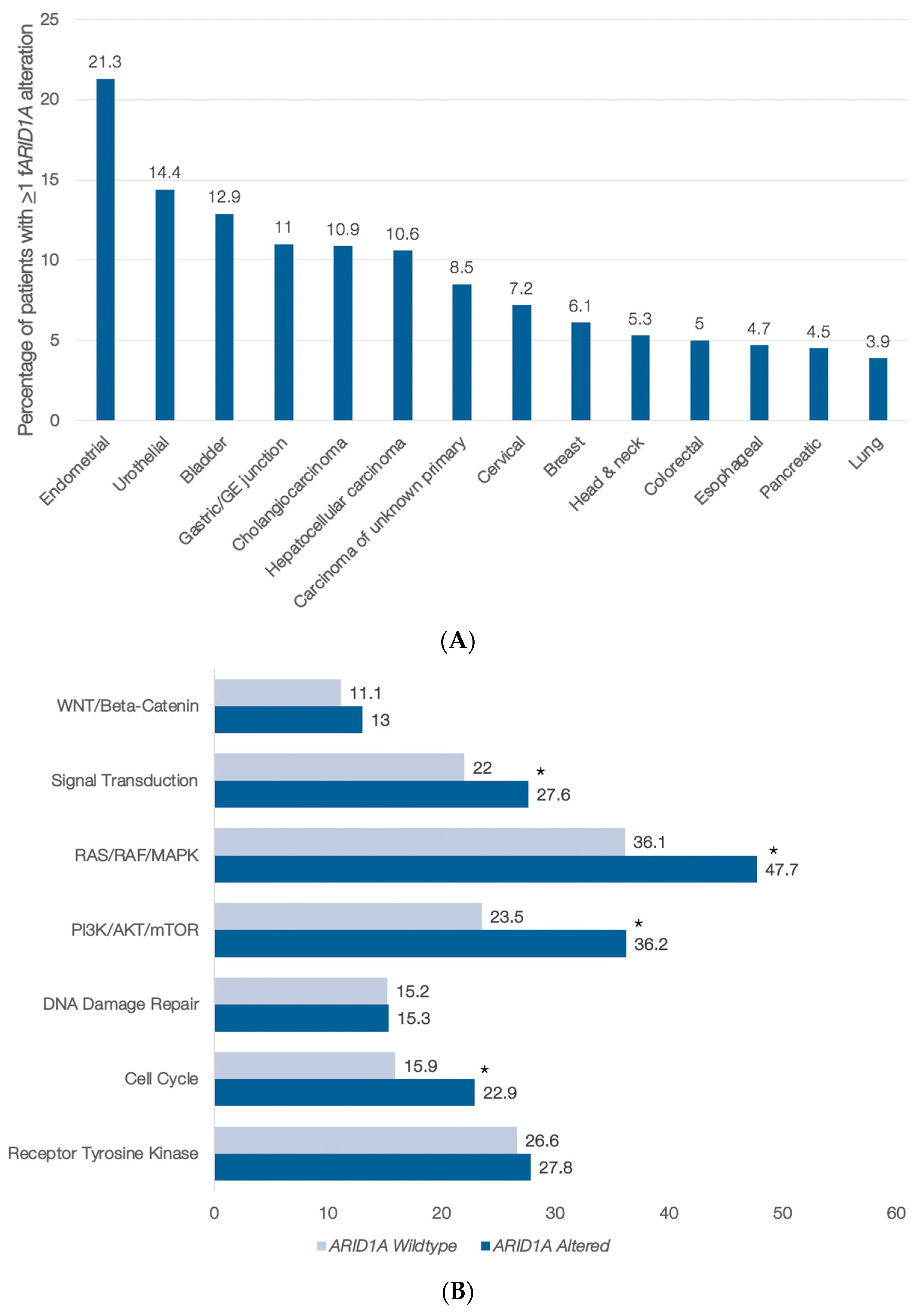
3.1. ARID1A Abnormalities in Blood-Derived cfDNA

From November 2016 through August 2019, 71,301 patients with advanced solid tumors underwent clinical cfDNA testing; 62,851 (88%) had ≥1 cfDNA alteration; 3137 (5%) had ≥1 deleterious ARID1A alteration; and 50% of ARID1A alterations detected were clonal. The frequency of ARID1A alterations by cancer type is as follows (Figure 1A): endometrial cancer, 21.3% of patients; bladder cancer, 12.9%; gastric cancer, 11% (the latter includes gastric/GE junction for cfDNA); cholangiocarcinoma, 10.9%; hepatocellular carcinoma, 10.6%; carcinoma of unknown primary, 8.5%; cervical cancer, 7.2%; breast cancer, 6.1%; head and neck cancer, 5.3%; colorectal cancer, 5%; pancreatic cancer, 4.5%; and lung cancer, 3.9%. (The following were the total numbers of patients analyzed: endometrial (N = 517); urothelial (N = 146); bladder (N = 653); gastric/GE junction (N = 1764); cholangiocarcinoma (N = 1537); hepatocellular carcinoma (N = 481); carcinoma of unknown primary (N = 1589); cervical (N = 167); breast (N = 6880); head and neck (N = 582); colorectal (N = 5742); esophageal (N = 379); pancreatic (N = 3095); and lung (N = 29,378).)

3.2. cfDNA Genomic Alterations Co-Occurring with ARID1A Anomalies in the Pan-Cancer Setting

The number of alterations was examined as a rough surrogate of mutational burden (since many of these samples predated the availability of blood tumor mutational burden). Samples with a functional ARID1A alteration had a greater number of overall alterations per sample compared to those without a functional ARID1A alteration (median 6 versus 4; p < 0.0001), including variants of unknown significance (VUS) and synonymous variants (ARID1A was also included in this count) (data not shown). Tumors with versus without functional ARID1A alterations also more frequently harbored alterations in at least one gene in many important pathways, including signal transduction, RAS/RAF/MAPK, and the cell cycle (Figure 1B); anomalies in WNT/Beta-catenin or DNA damage repair genes were not more common in ARID1A-altered samples.

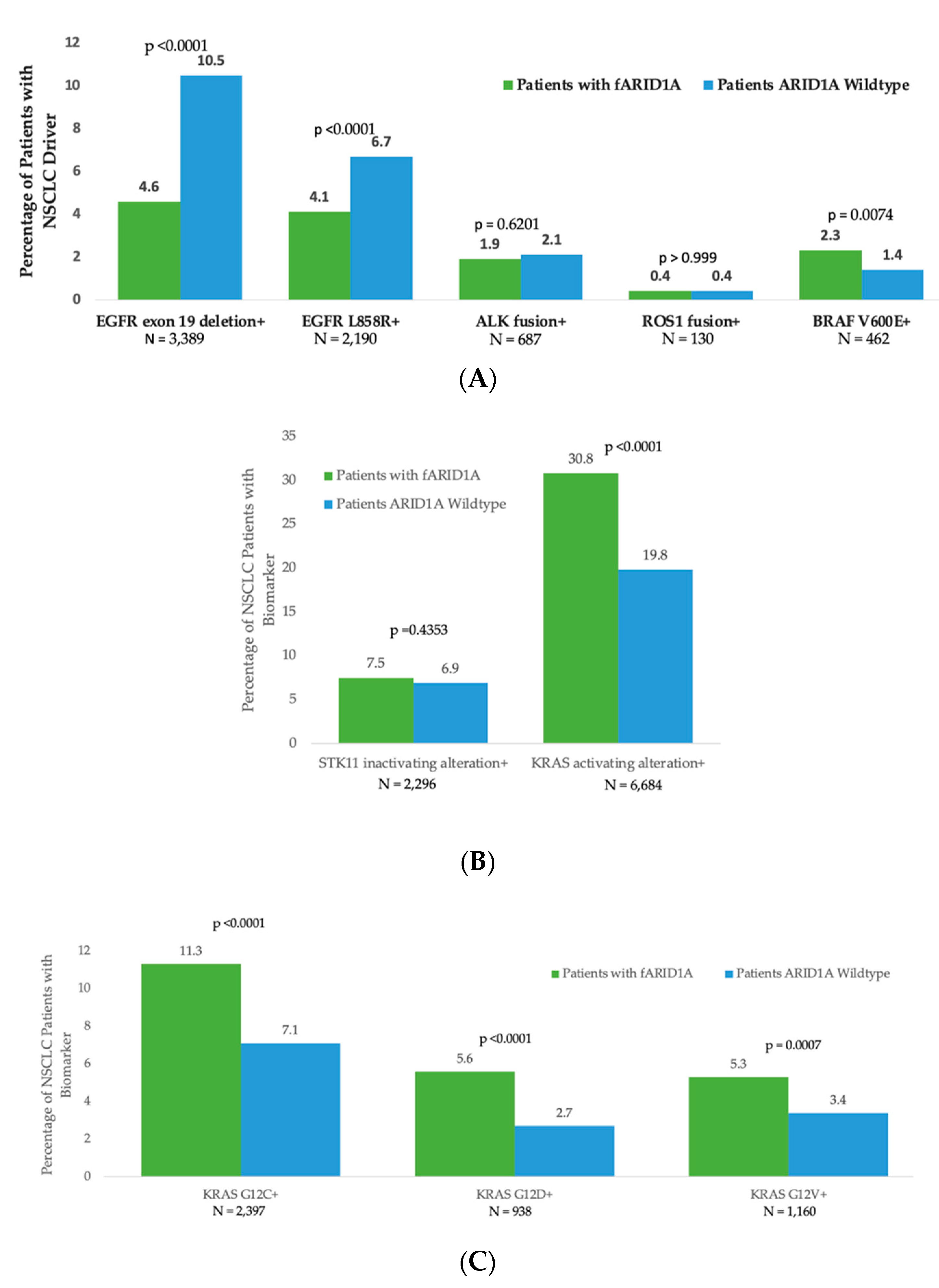
3.3. Segregation of ARID1A cfDNA Alterations with Other Oncogenic Drivers in Non-Small Cell Lung Cancer (NSCLC)

We examined NSCLC because of the large number of targetable alterations in this tumor type [18]. In the specific subset analyses of NSCLC, certain driver alterations segregated by ARID1A alteration status. We specifically focused on targetable alterations and alterations implicated in resistance or sensitivity to immunotherapy [19,20]. For instance, oncogenic EGFR driver alterations occurred more frequently in samples without ARID1A versus those with functional ARID1A alterations, while activating KRAS mutations (e.g., KRAS G12C) were seen more frequently in samples bearing functional ARID1A alterations versus those without (Figure 2).

4. Discussion

ARID1A has a critical role in controlling gene expression that promotes carcinogenesis. Indeed, ARID1A participates in immune responsiveness to neoplastic transformation, control of PI3K/AKT/mTOR signaling, EZH2 methyltransferase activity, steroid receptor modulation, DNA damage, and modulation of KRAS and p53 signaling [12]. Many agents may theoretically be of benefit in ARID1A-altered malignancies: immune checkpoint blockade and inhibitors of EZH2, mTOR, histone deacetylases, PARP, and/or ATR. ARID1A alterations may induce resistance to platinum chemotherapy and estrogen receptor degraders/modulators [12,21,22]. Hence, ARID1A alterations are important to the malignant process and have therapeutic implications.
In our dataset of over 60,000 patients with advanced solid cancers, 5% had cfDNA ARID1A alterations in their blood-based liquid biopsy. These data are consistent with reported tissue sequencing, which shows ARID1A gene anomalies in approximately 6% of human malignancies [3,11,12]. Furthermore, in comparing previously published data on the frequency of ARID1A alterations detected via tissue testing and the current results for blood cfDNA, the frequency of ARID1A alterations by cancer type is as follows (tissue vs. cfDNA) [3,11,12] (Figure 1A, cfDNA shown): endometrial cancer, ~37% vs. 21.3%; bladder cancer, ~24% vs. 12.9%; gastric cancer, ~29% vs. 11% (the latter includes gastric/GE junction for cfDNA); cholangiocarcinoma, ~14% vs. 10.9%; hepatocellular carcinoma, ~9% vs. 10.6%; carcinoma of unknown primary, ~15% vs. 8.5%; cervical cancer, ~6% vs. 7.2%; breast cancer, ~5% vs. 6.1%; head and neck cancer, ~4.5% vs. 5.3%; colorectal cancer, ~7% vs. 5%.; pancreatic cancer, ~7% vs. 4.5%; and lung cancer, ~8% vs. 3.9%. Although ARID1A alterations occur in ~30–50% of ovarian clear cell carcinomas, ovarian clear cell cancers are a rare subset of ovarian cancer, and only one patient was designated with this pathologic entity in the dataset; hence, the frequency of ARID1A mutations in this histology was not evaluable [13]. It should also be noted that certain types of endometrial cancers have higher rates of ARID1A alterations: ~40% in uterine endometrioid carcinomas and 20–36% in uterine carcinosarcomas, but they are less frequent in endometrial serous carcinoma [14]. Our cohort did not have available details on specific histologic subtypes of endometrial cancer. Since the tissue frequencies of ARID1A alterations are derived from the literature, it is not clear why there are some differences in ARID1A alteration frequencies between tissue DNA and blood cfDNA (even if the overall frequency is similar). While certain tumor types did have higher rates of ARID1A alterations in the tissue than we found in the blood, others had lower frequencies: breast, ~5% vs. 6.1%; head and neck, ~4.5% vs. 5.3% (tissue versus blood). It is also plausible that tissue has been analyzed for a wider array of rare cancers that have fewer ARID1A alterations and that that diluted overall tissue numbers. Furthermore, certain tumor types may shed less tumor DNA into the circulation.
Blood samples with a functional (deleterious) ARID1A alteration had a significantly greater number of overall alterations compared to those without a functional ARID1A alteration (median 6 versus 4; p < 0.0001). Blood biopsies with versus without functional ARID1A alterations also more frequently harbored alterations in ≥1 gene in many important cancer-related pathways, such as signal transduction, RAS/RAF/MAPK, PI3K/Akt/mTor, and the cell cycle (Figure 1B). Importantly, some of these pathways, such as the RAS/RAF/MAPK and PI3K/Akt/mTor pathways, may be targetable by existing drugs approved or in clinical trials. The tissue gene co-alterations occurring with ARID1A alterations have not been extensively studied, but co-alterations in PI3K/Akt/mTor pathway genes have been noted [12]. Co-occurring pathway abnormalities may be important when planning therapy.
We did not include tumor mutational burden in the analysis because many of the samples predated the inclusion of tumor mutational burden as a blood-based analysis. In addition, we have previously shown, in multivariate analysis, that ARID1A alterations predicted longer PFS after checkpoint blockade, and this result was independent of microsatellite instability or mutational burden [6].
A subgroup analysis of NSCLC was also performed because of the many crucial molecular abnormalities identified in this malignancy. Specific driver alterations segregated by ARID1A status. As an example, oncogenic EGFR driver alterations, which are associated with low efficacy of immune checkpoint therapy, occurred more frequently in samples without versus those with functional (deleterious) ARID1A abnormalities, while activating KRAS mutations (e.g., KRAS G12C, previously considered undruggable but now therapeutically actionable with the FDA-approved sotorasib) were seen more often in blood samples harboring functional ARID1A (deleterious) alterations versus those without these aberrations. These findings may be notable given recent data suggesting that KRAS mutations may be a positive predictor of immune checkpoint response in NSCLC [19,20]. Additional research is needed to determine whether similar patterns of ARID1A co-alterations are seen in tumor tissue.

5. Conclusions

The ARID1A protein binds specific motifs of DNA as part of an SWI/SNF complex and contributes to the remarkable specificity of these chromatin remodeling complexes. These complexes exploit ATP to alter nucleosome architecture in order to hide or expose regions of DNA as part of the process mediating transcriptional regulation [12]. Through SWI/SNF complexes’ participation in the regulation of gene expression, ARID1A impacts many important cellular pathways and processes in healthy cells, and altered ARID1A plays a crucial role in the generation and propagation of the cancer state [12]. Hence, we studied ARID1A alterations in a large dataset of patients with cancer.
Taken together, our data from a validated cfDNA assay [16,17] suggest that liquid biopsies performed using this assay detect ARID1A alterations at frequencies similar to those found in tissue. Furthermore, at a patient level, certain co-alterations occur more frequently in samples with functional (deleterious) ARID1A alterations than those without such aberrations, most prominently activation via mutation of the MAPK pathway and PI3k/Akt/mTor signals. Recent studies and ongoing research suggest ARID1A may be targetable via immune checkpoint inhibitors and/or several other agents, such as PARP, EZH2, HDAC, and ATR inhibitors [12,21,22]. The impact of co-activated pathways on these strategies needs to be evaluated [23,24].

Author Contributions

Conceptualization, R.K., J.R., C.A., H.-J.L., D.G., C.W. and L.K.; methodology, R.K., J.R., C.A., H.-J.L., D.G. and C.W.; formal analysis, C.W.; data curation, C.W.; writing—original draft preparation, R.K. and C.W.; writing—review and editing, R.K., J.R., C.A., H.-J.L., D.G., C.W. and L.K. All authors have read and agreed to the published version of the manuscript.

Funding

This research was funded by Guardant Health and National Cancer Institute grants P30 CA023100 (RK).

Institutional Review Board Statement

The study was conducted in accordance with the Declaration of Helsinki and approved by Advarra IRB Pro00034566/CR00218935.

Informed Consent Statement

Patient consent was waived due to the retrospective nature of the research using a de-identified dataset of patients tested as part of usual clinical care.

Data Availability Statement

The dataset generated and/or analyzed for the current study are not publicly available, as they are derived from commercial testing. This data may be made available under a fully executed data use agreement.

Conflicts of Interest

The authors declare the following competing interests: R.K. receives research funding from Genentech, Merck Serono, Pfizer, Boehringer Ingelheim, TopAlliance, Takeda, Incyte, Debiopharm, Medimmune, Sequenom, Foundation Medicine, Konica Minolta, Grifols, Omniseq, and Guardant, as well as consultant and/or speaker fees and/or serves on the advisory board for X-Biotech, Neomed, Pfizer, Actuate Therapeutics, Roche, Turning Point Therapeutics, TD2/Volastra, and Bicara Therapeutics, Inc., has an equity interest in IDbyDNA and CureMatch Inc, serves on the Board of CureMatch and CureMetrix, and is a co-founder of CureMatch. J.R. receives research funding from AstraZeneca, Novartis, Merck, Spectrum, Revolution Medicines, and GlaxoSmithKline, as well as serves as a consultant/advisory board member for Turning Point Therapeutics, EMD Serano, Daiichi Sankyo, Boehringer Ingelheim, Novartis, Janssen, Beigene, and BluPrint. C.A. has served as a consultant for AZ, Celgene, Daichii Sankyo, BluPrint, Merck, and Roche. Research support to the institution was received from Macrogenics, Novartis, Merck, and Medimmune. H.L. receives consulting fees/advisory board fees from Bayer, BMS, Roche, Merck, Merck Serono, Bicara, G1 Therapeutics, Jazz Therapeutics, Oncocyte, and Fulgent. D.G. receives institutional research grants from Amgen, AstraZeneca, Genentech, and Merck, as well as consultant and/or advisory board fees from AstraZeneca (institutional), Roche-Genentech (institutional), Guardant Health (institutional), IO Biotech (institutional), Oncocyte (institutional), Lilly, Merck, and Novartis. He is the Chief Medical Officer (unpaid) for the International Society of Liquid Biopsy. L.K. is an employee and stockholder of Guardant Health. C.W. is an employee and stockholder of Guardant Health.

References

  1. Mathur, R. ARID1A loss in cancer: Towards a mechanistic understanding. Pharmacol. Ther. 2018, 190, 15–23. [Google Scholar] [CrossRef] [PubMed]
  2. Helming, K.C.; Wang, X.; Roberts, C.W.M. Vulnerabilities of mutant SWI/SNF complexes in cancer. Cancer Cell 2014, 26, 309–317. [Google Scholar] [CrossRef] [PubMed]
  3. Jiang, T.; Chen, X.; Su, C.; Ren, S.; Zhou, C. Pan-cancer analysis of ARID1A Alterations as Biomarkers for Immunotherapy Outcomes. J. Cancer 2020, 11, 776–780. [Google Scholar] [CrossRef]
  4. Pulice, J.L.; Kadoch, C. Composition and Function of Mammalian SWI/SNF Chromatin Remodeling Complexes in Human Disease. Cold Spring Harb. Symp. Quant. Biol. 2017, 81, 53–60. [Google Scholar] [CrossRef] [PubMed]
  5. Shen, J.; Ju, Z.; Zhao, W.; Wang, L.; Peng, Y.; Ge, Z.; Nagel, Z.D.; Zou, J.; Wang, C.; Kapoor, P.; et al. ARID1A deficiency promotes mutability and potentiates therapeutic antitumor immunity unleashed by immune checkpoint blockade. Nat. Med. 2018, 24, 556–562. [Google Scholar] [CrossRef]
  6. Okamura, R.; Kato, S.; Lee, S.; Jimenez, R.E.; Sicklick, J.K.; Kurzrock, R. ARID1A alterations function as a biomarker for longer progression-free survival after anti-PD-1/PD-L1 immunotherapy. J. Immunother. Cancer 2020, 8, e000438. [Google Scholar] [CrossRef]
  7. Rizvi, N.; Cho, B.C.; Reinmuth, N.; Lee, K.H.; Luft, A.; Ahn, M.; Papadimitrakopoulou, V.; Heymach, J.; Scheuring, U.; Higgs, B.; et al. OA04.07 Mutations Associated with Sensitivity or Resistance to Immunotherapy in mNSCLC: Analysis from the MYSTIC Trial. J. Thorac. Oncol. 2019, 14, S217. [Google Scholar] [CrossRef]
  8. Johnson, R.M.; Qu, X.; Thomas, J.; Kschonsak, Y.; Huw, L.-Y.; Ou, F.-S.; Sokol, E.; Ihuegbu, N.; Zill, O.; Kabbarah, O.; et al. ARID1A mutations induce an EGFR-like gene expression signature and confer intrinsic and acquired resistance to cetuximab treatment in first line metastatic CRC. Cancer Res. 2020, 80 (Suppl. S16), LB-063. [Google Scholar] [CrossRef]
  9. Shen, J.; Peng, Y.; Wei, L.; Zhang, W.; Yang, L.; Lan, L.; Kapoor, P.; Ju, Z.; Mo, Q.; Shih, I.-M.; et al. ARID1A Deficiency Impairs the DNA Damage Checkpoint and Sensitizes Cells to PARP Inhibitors. Cancer Discov. 2015, 5, 752–767. [Google Scholar] [CrossRef]
  10. Adashek, J.J.; Janku, F.; Kurzrock, R. Signed in Blood: Circulating Tumor DNA in Cancer Diagnosis, Treatment and Screening. Cancers 2021, 13, 3600. [Google Scholar] [CrossRef]
  11. Wu, J.N.; Roberts, C.W. ARID1A mutations in cancer: Another epigenetic tumor suppressor? Cancer Discov. 2013, 3, 35–43. [Google Scholar] [CrossRef] [PubMed]
  12. Mullen, J.; Kato, S.M.; Sicklick, J.K.; Kurzrock, R. Targeting ARID1A Mutations in Cancer. Cancer Treat. Rev. 2021, 100, 102287. [Google Scholar] [CrossRef] [PubMed]
  13. Iida, Y.; Okamoto, A.; Hollis, R.L.; Gourley, C.; Herrington, C.S. Clear cell carcinoma of the ovary: A clinical and molecular perspective. Int. J. Gynecol. Cancer 2021, 31, 605–616. [Google Scholar] [CrossRef] [PubMed]
  14. Toumpeki, C.; Liberis, A.; Tsirkas, I.; Tsirka, T.; Kalagasidou, S.; Inagamova, L.; Anthoulaki, X.; Tsatsaris, G.; Kontomanolis, E.N. The Role of ARID1A in Endometrial Cancer and the Molecular Pathways Associated with Pathogenesis and Cancer Progression. In Vivo 2019, 33, 659–667. [Google Scholar] [CrossRef]
  15. Zill, O.A.; Banks, K.C.; Fairclough, S.R.; Mortimer, S.A.; Vowles, J.V.; Mokhtari, R.; Gandara, D.R.; Mack, P.C.; Odegaard, J.I.; Nagy, R.J.; et al. The Landscape of Actionable Genomic Alterations in Cell-Free Circulating Tumor DNA from 21,807 Advanced Cancer Patients. Clin. Cancer Res. 2018, 24, 3528–3538. [Google Scholar] [CrossRef]
  16. Odegaard, J.I.; Vincent, J.J.; Mortimer, S.; Vowles, J.V.; Ulrich, B.C.; Banks, K.C.; Fairclough, S.R.; Zill, O.A.; Sikora, M.; Mokhtari, R.; et al. Validation of a Plasma-Based Comprehensive Cancer Genotyping Assay Utilizing Orthogonal Tissue- and Plasma-Based Methodologies. Clin. Cancer Res. 2018, 24, 3539–3549. [Google Scholar] [CrossRef]
  17. Willis, J.; Lefterova, M.I.; Artyomenko, A.; Kasi, P.M.; Nakamura, Y.; Mody, K.; Catenacci, D.V.T.; Fakih, M.; Barbacioru, C.; Zhao, J.; et al. Validation of Microsatellite Instability Detection Using a Comprehensive Plasma-Based Genotyping Panel. Clin. Cancer Res. 2019, 25, 7035–7045. [Google Scholar] [CrossRef]
  18. Majeed, U.; Manochakian, R.; Zhao, Y.; Lou, Y. Targeted therapy in advanced non-small cell lung cancer: Current advances and future trends. J. Hematol. Oncol. 2021, 14, 108–115. [Google Scholar] [CrossRef]
  19. Fountzilas, E.; Kurzrock, R.; Hiep Vo, H.; Tsimberidou, A.M. Wedding of Molecular Alterations and Immune Checkpoint Blockade: Genomics as a Matchmaker. J. Natl. Cancer Inst. 2021, 113, 1634–1647. [Google Scholar] [CrossRef]
  20. Torralvo, J.; Friedlaender, A.; Achard, V.; Addeo, A. The Activity of Immune Checkpoint Inhibition in KRAS Mutated Non-small Cell Lung Cancer: A Single Centre Experience. Cancer Genom. Proteom. 2019, 16, 577–582. [Google Scholar] [CrossRef] [Green Version]
  21. Bitler, B.G.; Aird, K.M.; Garipov, A.; Li, H.; Amatangelo, M.; Kossenkov, A.V.; Schultz, D.C.; Liu, Q.; Shih, I.-M.; Conejo-Garcia, J.; et al. Synthetic lethality by targeting EZH2 methyltransferase activity in ARID1A-mutated cancers. Nat. Med. 2015, 21, 231–238. [Google Scholar] [CrossRef] [PubMed]
  22. Fukumoto, T.; Park, P.H.; Wu, S.; Fatkhutdinov, N.; Karakashev, S.; Nacarelli, T.; Kossenkov, A.V.; Speicher, D.W.; Jean, S.; Zhang, L.; et al. Repurposing Pan-HDAC Inhibitors for ARID1A-Mutated Ovarian Cancer. Cell Rep. 2018, 22, 3393–3400. [Google Scholar] [CrossRef] [PubMed]
  23. Kato, S.; Kim, K.H.; Lim, H.J.; Boichard, A.; Nikanjam, M.; Weihe, E.; Kuo, D.J.; Eskander, R.N.; Goodman, A.; Galanina, N.; et al. Real-world data from a molecular tumor board demonstrates improved outcomes with a precision N-of-One strategy. Nat. Commun. 2020, 11, 4965. [Google Scholar] [CrossRef] [PubMed]
  24. Sicklick, J.K.; Kato, S.; Okamura, R.; Schwaederle, M.; Hahn, M.E.; Williams, C.B.; De, P.; Krie, A.; Piccioni, D.E.; Miller, V.A.; et al. Molecular profiling of cancer patients enables personalized combination therapy: The I-PREDICT study. Nat. Med. 2019, 25, 744–750. [Google Scholar] [CrossRef] [PubMed]
Figure 1. (A,B) Frequency of functional (deleterious) ARID1A (fARID1A) by cancer type and examination of co-occurring alterations by pathway. (A) Percentage of patients with functional ARID1A (fARID1A) cfDNA alterations by cancer type. Of 62,851 patients with ≥1 cfDNA alteration detected, 3137 (5%) had ≥1 deleterious (functional) ARID1A alteration (fARID1A alteration). Nonsense, frameshift, and canonical splice site alterations were considered functional variants. (B) The frequency of alterations in certain pathways was examined on a per-patient basis (i.e., counting each patient once even if they had multiple alterations in the same pathway). The denominator equates to the total number of patients with (n = 1088) or without (n = 23,935) a functional ARID1A alteration and at least one cfDNA alteration detected. In this pan-cancer cohort, numerous pathways were more frequently altered (statistically significant differences, p ≤ 0.05, marked with *, Fisher’s exact test) in patients with functional ARID1A genomic alterations. The genes included in each pathway are provided in Table 1. The numbers beside the bars represent percentages. For instance, the “13” beside “WNT/Beta-catenin” means that 13% of ARID1A-altered samples had ≥1 WNT/Beta-catenin pathway alteration. (Note: MSI-H samples were excluded from this analysis.) Abbreviations: MSI-H = microsatellite instability-high.
Cancers 14 04281 g001
Figure 2. Frequency of select NSCLC biomarkers in patients with functional ARID1A alterations versus those without. The denominator equates to total number of NSCLC patients with (n = 1298) or without (n = 31,788) a functional ARID1A alteration, respectively. The number provided under each bar graph is the total number of patients in the cohort with the specified alteration. Wild-type ARID1A refers to samples without a functional ARID1A alteration. (A) NSCLC drivers in patients with a functional ARID1A alteration versus those without. For instance, 10.5% of patients without a functional ARID1A alteration had an EGFR exon 19 deletion, while only 4.6% of patients with a functional ARID1A alteration had such a deletion (p < 0.0001). (B) STK11 and KRAS alterations (both implicated in prognosis and/or immunotherapy response) in patients without a functional ARID1A alteration versus those with such an alteration. (C). Specific KRAS alterations in patients with functional ARID1A alterations versus those without.
Cancers 14 04281 g002
Table 1. Genes used in pathway analysis (see also Figure 1B).
PathwayGenes Included
Receptor Tyrosine KinaseEGFR, FGFR1, FGFR2, KIT, MET, MPL, and PDGFRA
Cell CycleCCNE1, CDH1, CDK4, CDK6, CDKN2A, FBXW7, and RB1
DNA DamageATM, BRCA1, BRCA2, CCND1, and MLH1
PI3K/AKT/mTORAKT1, MTOR, PIK3CA, PTEN, STK11, and TSC1
RAS/RAF/MAPKARAF, BRAF, ERBB2, GNA11, HRAS, KRAS, MAP2K1, MAP2K2, MAPK1, MAPK3, NF1, NRAS, RAF1, and RIT1
Signal TransductionALK, AR, DDR, ESR1, GATA3, GNAS, GNAQ, MYC, NOTCH1,
NTRK1, NTRK3, PTPN11, RET, RHOA, ROS1, and SMAD4
WNT / β -CateninAPC and CTNNB1
Publisher’s Note: MDPI stays neutral with regard to jurisdictional claims in published maps and institutional affiliations.

