Simple Summary
In the present study we evaluated the frequency and the clinical relevance of ESR1 mutations in high-grade serous ovarian cancer (HGSOC). Drop-off droplet digital PCR (ddPCR) was first used to screen for ESR1 mutations in primary tumors (formalin-fixed paraffin-embedded, FFPEs) from HGSOC patients and plasma cell-free DNA (cfDNA) samples from advanced and metastatic ovarian cancer patients. We further used the recently developed ESR1-NAPA assay to detect individual ESR1 mutations in drop-off ddPCR-positive samples. We report for the first time the presence of ESR1 mutations in 15% of FFPEs and in 13.8% of plasma cfDNA samples from advanced and metastatic ovarian cancer patients.
Abstract
ESR1 mutations have been recently associated with resistance to endocrine therapy in metastatic breast cancer and their detection has led to the development and current evaluation of novel, highly promising therapeutic strategies. In ovarian cancer there have been just a few reports on the presence of ESR1 mutations. The aim of our study was to evaluate the frequency and the clinical relevance of ESR1 mutations in high-grade serous ovarian cancer (HGSOC). Drop-off droplet digital PCR (ddPCR) was first used to screen for ESR1 mutations in 60 primary tumors (FFPEs) from HGSOC patients and in 80 plasma cell-free DNA (cfDNA) samples from advanced and metastatic ovarian cancer patients. We further used our recently developed ESR1-NAPA assay to identify individual ESR1 mutations in drop-off ddPCR-positive samples. We report for the first time the presence of ESR1 mutations in 15% of FFPEs and in 13.8% of plasma cfDNA samples from advanced and metastatic ovarian cancer patients. To define the clinical significance of this finding, our results should be further validated in a large and well-defined cohort of ovarian cancer patients.
1. Introduction
Ovarian cancer remains the cancer with the worst survival rates in women, as in most cases it is diagnosed at an advanced stage [1]. It is the second most frequent malignancy, following breast cancer, in women over the age of 40, especially in developed countries [2]. For primary disease, the customary treatment is debulking surgery accompanied by first-line platinum and paclitaxel-based chemotherapy [1]. The majority of patients respond to primary treatment but more than half of them will acquire chemo-resistance and consequently recurrent disease [1]. Epithelial ovarian cancer is the most frequent type, with histological and molecular heterogeneity [1,2]. Serous tumors are classified into high-grade serous carcinomas (HGSCs) and low-grade serous carcinomas (LGSCs) [2]. The former is a highly aggressive disease that is often diagnosed at an advanced FIGO (International Federation of Gynecology and Obstetrics) stage [1]. Overall survival (OS) remains low, even though there have been slight improvements in therapy [1]. There are a few available targeted therapies for ovarian cancer, such us the anti-angiogenetic antibody bevacizumab [3] and the PARP (poly (ADP-ribose) polymerase) inhibitors olaparib and rucaparib, which are FDA (Food and Drug Administration)-approved for platinum-sensitive recurrent BRCA-mutated ovarian cancer patients [4,5].
The most commonly reported gene mutations, highly associated with epithelial ovarian cancer, are for TP53, BRCA1/2, PIK3CA and KRAS genes [6]. The frequency of the above-mentioned mutations varies among the subtypes of epithelial ovarian cancer. Mutations in TP53 are present in more than 96% of ovarian cancer cases [6,7]. BRCA1/2 mutations are associated with the majority of hereditary ovarian cancer or Lynch syndrome and the mutation rate of BRCA1/2 increases in recurrent HGSOC [6,7]. PIK3CA mutations have been also detected at high frequencies in ovarian clear cell carcinoma (OCCC) and endometrioid ovarian cancer related to endometriosis [6]. NOTCH3 mutations have been detected in 66% of HGSOC cases and NOTCH3 inactivation could be a potential therapeutic approach [7]. Low-grade serous ovarian carcinomas (LGSOCs) are associated with BRAF (especially V600E) and KRAS mutations [6,7,8]. LGSOCs present with a lower frequency of somatic TP53 and BRCA1/2 mutations and are not associated with germline BRCA1/2 mutations [8]. RAD51C and RAD51D have been demonstrated to be inherited ovarian cancer predisposition genes with mutation carriers showing HGSOC [8]. In addition, the deleterious germline mutations BRP1 (BRCA1-interacting protein 1) are mainly associated with the high-grade serous epithelial subtype [8,9].
Liquid biopsy, now widely recognized as an important tool for the follow-up of cancer patients, is mainly based on the analysis of circulating tumor cells (CTCs) and circulating tumor DNA (ctDNA), which provide a source of diagnostic or/and prognostic markers. The clinical significance of CTCs and ctDNA in ovarian cancer has been investigated in many studies to date [1,10,11,12,13,14,15,16]. We have recently reported that ESR1 is methylated in HGSOC patients, and that there is a statistically significant concordance between ESR1 methylation in primary tumors and paired ctDNA [17]. Recently, many studies have investigated the mutation profile of ovarian cancer patients in plasma-cfDNA in many genes, such as TP53, PIK3CA, KRAS, BRAC1, BRAC2 and EGFR [18,19,20,21,22,23].
ESR1 mutations have emerged as a key mechanism of resistance to endocrine therapy in patients with ER-positive metastatic breast cancer [24] and their detection is now considered to be highly promising as a prognostic and predictive biomarker in this type of cancer [24,25]. To date, only a few studies have investigated the presence of ESR1 mutations in endometrial and cervical cancer [26,27,28,29,30]. According to the cBioPortal cancer genomics database, ESR1 mutations have been detected in 4–6% of uterine corpus endometrial carcinoma samples [31]. As for ovarian cancer, the cBioPortal cancer genomics database includes one ovarian serous cystadenocarcinoma study in which ESR1 mutations were detected in 0.8% of samples [31]. In 2018, Stover et al., using targeted next-generation sequencing (NGS), detected a Y537S ESR1 mutation in one patient with low-grade serous ovarian cancer (LGSOC); this particular patient developed a single site of progressive disease in an abdominal wall nodule and maintained stable low-volume peritoneal disease during endocrine therapy for almost five years, but later presented progressive disease after a durable response to hormonal therapy [32].
The aim of our study was to evaluate the frequency and the clinical relevance of ESR1 mutations in HGSOC (HGSOC). We applied our recently developed highly sensitive and specific ESR1-NAPA assay for the detection of ESR1 hotspot mutations (Y537S, Y537C, L536R, Y537N and D538G) [33] in combination with drop-off ddPCR [34] to investigate ESR1 mutational status in primary tumors and plasma cfDNA in HGSOC patients.
2. Materials and Methods
2.1. Clinical Samples
The study material consisted of (a) primary formalin-fixed paraffin-embedded tumor tissues (FFPEs) from patients with HGSOC prior to any systemic treatment (n = 60) and, as a corresponding non-cancerous control, a group of 10 normal fallopian tube FFPEs that were obtained from women at the productive age; and (b) plasma-cfDNA samples from 80 patients with advanced (n = 20) and metastatic ovarian cancer (n = 60), and as a corresponding control, plasma-cfDNA samples from female healthy donors (HD, n = 11). All patients received at least six cycles of carboplatinum AUC 5 and paclitaxel at 175 mg/m2. Patients provided written informed consent to participate in the study, which was approved by the Local Essen Research Ethics Committee (16-6916-BO; 17-7859-BO), and the General University Hospital of Alexandroupolis’ ethics committee (date: 25 June 2020). The available clinicopathological features are shown in Table 1 and Table 2.

Table 1.
Clinicopathological characteristics of the advanced HGSOC patients.

Table 2.
Clinicopathological characteristics of the advanced and metastatic ovarian cancer patients.
2.2. DNA Isolation
FFPEs: FFPEs containing >60% tumor cells were used for genomic DNA (gDNA) extraction. gDNA was isolated from FFPEs with using the QIAamp® DNA FFPE Tissue Kit 50 (Qiagen®, Hilden, Germany), according to the manufacturer’s instructions. The DNA concentration was determined using a Nanodrop ND-1000 spectrophotometer (Nanodrop Technologies, Wilmington, NC, USA).
Plasma: 10 mL of peripheral blood in EDTA were used within 2–4 h to isolate plasma via centrifugation at 530× g for 10 min. Following a second centrifugation at 2000× g for 10 min, plasma was transferred into 2 mL tubes and stored at −70 °C until use. cfDNA was further isolated using the QIAamp Circulating Nucleic Acid Kit (Qiagen, Hilden, Germany), as previously described [35].
In all samples, cfDNA quality was checked prior to PCR using a previously described protocol [36]. Serial dilutions of a wild-type sample with a known DNA concentration (Human Reference DNA Female, Agilent Technologies, Santa Clara, CA, USA), prepared via serial 10-fold dilution in concentrations ranging from 200 ng/μL down to 0.5 ng/μL, were used to generate a standard curve for the quantification of the gDNA concentration in all cfDNA samples using a LightCycler z480 (Roche).
2.3. Drop-Off ddPCR for ESR1 Mutations (Y537S, Y537C, Y537N, L536R, D538G)
All cfDNA samples and controls were screened for ESR1 mutations in exon 8, including the Y537S, Y537C, Y537N, D538G and L536R mutations, using drop-off ddPCR in a QX200 Droplet Digital PCR System (Bio-Rad Laboratories, Hercules, CA, USA), as previously described [34].
2.4. ESR1-NAPA Assay
All samples that were found to be positive for ESR1 mutations via the ESR1 drop-off ddPCR and all controls were further analyzed to define each individual ESR1 mutation using our previously developed and validated ultrasensitive ESR1-NAPA assay for Y537S, Y537C, Y537N and D538G mutations [33]. Synthetic oligonucleotide sequences for each individual ESR1 mutation were used as positive controls. In this study, we additionally designed, analytically validated and added the L536R mutation into our ESR1-NAPA assay. The experimental conditions for the ESR1-L536R mutation assay were optimized in detail regarding the annealing temperature, time and concentration of primers, buffer, MgCl2 (magnesium chloride solution), dNTPs (deoxyribonucleotide triphosphates) and BSA (bovine serum albumin solution) (data not shown).
2.5. Statistical Analysis
SPSS version 28.0 (IBM® SPSS® Statistics, Endicott, NK, USA) was used for statistical analysis. Pearson’s χ2 and Cohen’s kappa coefficient tests were used to estimate the concordance between ESR1 mutations in primary tumors and paired cfDNA. The correlation between ESR1 mutations and the clinicopathological characteristics of the patients (Table 1) were estimated using Pearson’s χ2 and Fischer’s exact test (p-values < 0.05 were considered statistically significant). Kaplan–Meier analysis was used for overall survival (OS) and progression-free survival (PFS) curves.
3. Results
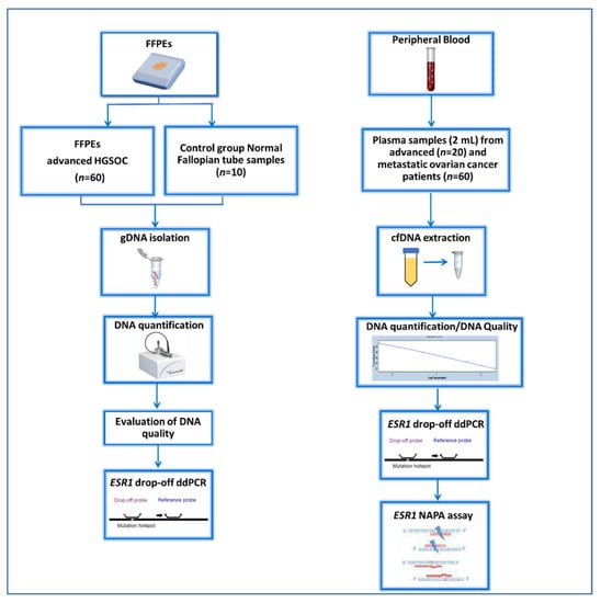
A schematic flowchart of our study is given in Figure 1.

Figure 1.
Schematic flowchart of the study.
3.1. Detection of ESR1 Mutations in FFPEs
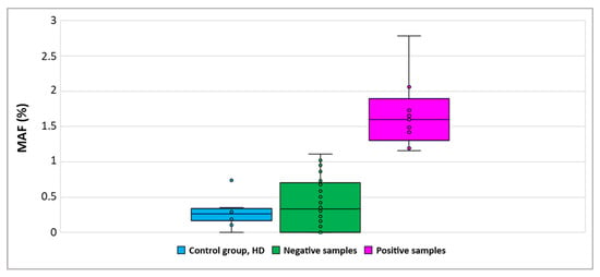
To ensure the specificity of the drop-off ddPCR assay we first evaluated the mutant allelic frequency (MAF) in 10 non-cancerous fallopian tube samples. A cut-off value was calculated by adding the 2SD (standard deviation) to the mean of the MAF values of these control samples. The MAF% was estimated using the program developed by Attali et al. specifically for this type of ddPCR assay [37]. Based on the defined cut-off (1.15), we detected the presence of ESR1 mutations in 9/60 (15%) of FFPE samples tested (Figure 2).

Figure 2.
Detection of ESR1 mutations in primary tumor samples using drop-off ddPCR. (MAF: mutant allele frequency).
In this patient group the median PFS was 41 months, and the median OS was 47 months. There was no significant correlation between OS, PFS, and ESR1 mutations in FFPEs when our results were evaluated via Kaplan–Meier analysis (data not shown). Furthermore, no significant correlation between ESR1 mutations and the patients’ clinicopathological characteristics was observed.
3.2. Detection of ESR1 Mutations in Plasma-cfDNA
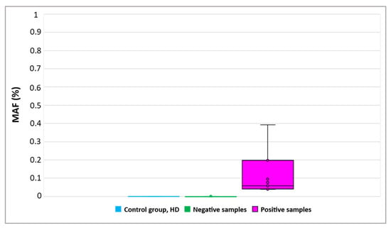
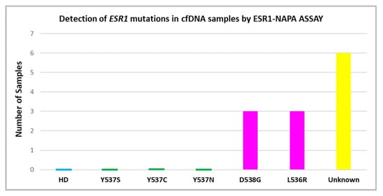
Using drop-off ddPCR, ESR1 mutations were detected in 11/80 (13.8%) plasma-cfDNA samples (Figure 3), more specifically, in eight plasma-cfDNA samples from patients with metastatic ovarian cancer (8/60, 13.3%) and in three plasma-cfDNA samples from patients with advanced ovarian cancer (3/20, 15%). All these ESR1-mutation-positive samples were further analyzed to define ESR1 mutations using the ESR1-NAPA assay (Figure 4). The D538G mutation was detected in three plasma-cfDNA samples from patients with metastatic ovarian cancer and L536R was detected in two plasma-cfDNA samples from patients with metastatic ovarian cancer and in one plasma-cfDNA sample from one patient with advanced ovarian cancer. It should be mentioned that in one patient with metastatic ovarian cancer, both D538G and L536R were detected in plasma-cfDNA.

Figure 3.
Detection of ESR1 mutations in plasma-cfDNA samples using drop-off ddPCR. (MAF: mutant allele frequency).

Figure 4.
Detection of ESR1 mutations in plasma-cfDNA samples using the NAPA-ESR1 assay.
The median PFS was 38 months and the median OS was 38 months in the group of n = 20 patients with advanced ovarian cancer, and the median PFS was 19 months and the median OS was 31 months in the group of n = 60 patients with metastatic ovarian cancer. Kaplan–Meier analysis was performed to estimate the correlation between OS and PFS with the detection of ESR1 mutations in both groups. No significant correlations were observed among OS, PFS and ESR1 mutations for both groups (data not shown). Furthermore, no significant correlations between ESR1 mutations and the patients’ clinicopathological characteristics were observed.
4. Discussion
We report, for the first time, the detection of ESR1 mutations in primary tumors (FFPEs) in plasma cfDNA samples from patients with advanced and metastatic ovarian cancer patients using highly sensitive and specific methodologies based on drop-off ddPCR for screening and the ESR1-NAPA assay for the definition of Y537S, Y537C, Y537N, L536R and D538G ESR1 mutations.
To date, the detection of ESR1 mutations has been reported in cervical squamous cell carcinoma [26] and in a patient with endometrial cancer treated with an aromatase inhibitor [27]. It has also been reported that the presence of ESR1 mutations is associated with worse outcomes in endometrial cancer [28]. Apart from endometrial cancer studies, there are very few studies that show the existence of ESR1 mutations in ovarian cancer. More specifically, in 2017, McIntyre et al. detected ESR1 Y537S mutation in one patient with low-grade serous ovarian carcinoma, when analyzing 26 primary tumor samples using NGS [38]. In 2018, Stover et al., using targeted NGS, detected a Y537S ESR1 mutation in one patient with LGSOC; this particular patient developed a single site of progressive disease in an abdominal wall nodule and maintained stable low-volume peritoneal disease during endocrine therapy for almost five years, but later presented progressive disease after a durable response to hormonal therapy [32]. In 2019, Gaillard et al. reported that ESR1 mutations were detected in 4.4% (24/548) of uterine endometrioid carcinomas vs. 0.2% (1/446) of uterine serous carcinomas and 3.5% (5/144) of ovarian endometrioid carcinomas compared to 0.3% (12/3502) of ovarian serous carcinomas, whereas in an ovarian serous carcinoma both ESR1 Y537S and D538G mutations were detected [39]. Since then, there have been no reports on the detection of ESR1 mutations in ovarian cancer.
In the present study, we report that, using drop-off ddPCR, ESR1 mutations were detected in 15% of primary tumor tissues and in 13.8% of plasma-cfDNA samples tested. More specifically, eight plasma-cfDNA samples from patients with metastatic cancer and three plasma-cfDNA samples from patients with advanced ovarian cancer were found to be positive for ESR1 mutations. All plasma-cfDNA samples found to be positive via ESR1 drop-off ddPCR were further analyzed using the ESR1-NAPA assay in order to define the specific mutation. In patients with metastatic ovarian cancer, the D538G mutation was detected in three plasma-cfDNA samples and L536R was detected in two plasma-cfDNA samples, whereas both D538G and L536R were detected in one patient. In patients with advanced ovarian cancer, L536R was detected only in one plasma-cfDNA sample. Drop-off ddPCR screens for ESR1 mutations were clustered in exon 8. Hence, any mutation in this region could be detected in addition to Y537S, Y537C, Y537N, L536R and D538G. In this region, additional mutations were present, such as L536H, which has been detected in endometrial cancer [29,39].
These findings could be of clinical importance if we consider that in metastatic breast cancer the detection of ESR1 mutations has led to the development of novel highly promising therapeutic strategies. In patients with metastatic breast cancer that are positive for ESR1 mutations, selective estrogen receptor modulators (SERMs) and selective estrogen receptor covalent antagonists (SERCAs) are now being evaluated as promising drugs. Lasofoxifene is currently in Phase 2 trials for patients with ESR1 mutations and for patients after progression on endocrine therapy and CDK4/6 inhibition [40]. The FDA has granted a fast-track designation to lasofoxifene for use as a treatment of female patients with estrogen receptor (ER)-positive, HER2-negative metastatic breast cancer who harbor ESR1 mutations. Bazedoxifene, a SERM/SERD hybrid, which has been approved for use in postmenopausal hot flashes and osteoporosis, is now in a Phase 2 trial for patients after progression on endocrine therapy (NCT02448771) [40]. Fanning et al. reported that bazedoxifene possessed improved inhibitory potency against the Y537S and D538G mutants compared to tamoxifen and had additional inhibitory activity in combination with the CDK4/6 inhibitor palbociclib [41]. In parallel, the efficacy of H3B-6545, a drug optimized from the SERCA class, against ESR1 mutations was demonstrated in patients with metastatic breast cancer previously treated with endocrine therapy and CDK4/6i [42]. H3B-6545 is now in a Phase 2 trial for patients after progression on endocrine therapy and CDK4/6i (NCT03250676) [40]. The combined analysis of SoFEA and EFFECT showed that patients with ESR1 mutations detected in plasma-cfDNA samples [43] had shorter PFS and OS when treated with exemestane therapy, compared with fulvestrant. In the PALOMA-3 trial, patients on fulvestrant and a placebo tended to have poorer PFS in the presence of mutations compared to the absence of mutations [44]. O’Leary et al. reported that ESR1 Y537S mutation promotes resistance to fulvestrant and that acquired mutations from fulvestrant are a major driver of resistance to fulvestrant and palbociclib combination therapy [45]. In the phase 3 PADA-1 trial presented at the 2021 San Antonio Breast Cancer Symposium, it was observed that when switching from an aromatase inhibitor plus palbociclib to fulvestrant and palbociclib upon early identification of the ESR1 mutation in plasma—before disease progression—the median PFS was doubled. This trial has also shown that ESR1 mutations are rarely detected in the plasma-cfDNA of ER + HER2− metastatic breast cancer patients with no overt resistance to aromatase inhibitors and that the detection of ESR1 mutations was associated with a significantly shorter PFS, suggesting that the presence of the ESR1 mutation at baseline could accelerate the outset of resistance to AI-palbociclib [46,47]. Novel therapies could include possible strategies to overcome the endocrine resistance induced by ESR1 mutations.
5. Conclusions
To our knowledge, this is the first time that the presence of ESR1 mutations has been reported in primary tumors and plasma-cfDNA from HGSOC patients. The clinical significance of this finding should be examined prospectively in a large group of ovarian cancer patients.
Author Contributions
Conceptualization, E.L.; methodology, D.S. and A.M.; validation, D.S, A.M., L.G. and E.L.; formal analysis, D.S., A.M. and L.G.; investigation: D.S.; resources, E.L., S.K.-B., S.K. and N.X.; data curation, D.S., A.M., I.B., P.B. and E.L.; writing—original draft preparation, D.S. and E.L.; writing—review and editing, A.M. and E.L.; visualization, D.S; supervision, E.L.; project administration, E.L.; funding acquisition, E.L. All authors have read and agreed to the published version of the manuscript.
Funding
This study was financially supported by the European Union and Greek National funds through the Operational Program Competitiveness, Entrepreneurship and Innovation, under the call RESEARCH—CREATE—INNOVATE (project code: T1RCI-02935).
Institutional Review Board Statement
The study was conducted according to the guidelines of the Declaration of Helsinki and approved was approved by the Local Essen Research Ethics Committee (16-6916-BO; 17-7859-BO) and General University Hospital of Alexandroupolis Ethics Committee (Date: 25 June 2020) for plasma samples.
Informed Consent Statement
Informed consent was obtained from all subjects involved in the study.
Data Availability Statement
The data presented in this study are available on request from the corresponding author. The data are not publicly available due to ethical restrictions.
Acknowledgments
We would like to thank all patients who participated in this study and all healthy volunteers. We would also like to thank Kitty Pavlakis (Pathology Department, Metropolitan—IASO Hospital, Athens) for kindly providing fallopian tube samples used as controls.
Conflicts of Interest
The authors declare no conflict of interest.
References
- Giannopoulou, L.; Lianidou, E.S. Liquid Biopsy in Ovarian Cancer. In Advances in Clinical Chemistry; Academic Press Inc.: Cambridge, MA, USA, 2020; Volume 97, pp. 13–71. [Google Scholar]
- Stewart, C.; Ralyea, C.; Lockwood, S. Ovarian Cancer: An Integrated Review. Semin. Oncol. Nurs. 2019, 35, 151–156. [Google Scholar] [CrossRef]
- Burger, R.A.; Brady, M.F.; Bookman, M.A.; Fleming, G.F.; Monk, B.J.; Huang, H.; Mannel, R.S.; Homesley, H.D.; Fowler, J.; Greer, B.E.; et al. Incorporation of Bevacizumab in the Primary Treatment of Ovarian Cancer. N. Engl. J. Med. 2011, 365, 2473–2483. [Google Scholar] [CrossRef] [PubMed]
- Montemorano, L.; Michelle, D.S.; Bixel, L.K. Role of Olaparib as Maintenance Treatment for Ovarian Cancer: The Evidence to Date. OncoTargets Ther. 2019, 12, 11497–11506. [Google Scholar] [CrossRef] [PubMed]
- Shirley, M. Rucaparib: A Review in Ovarian Cancer. Target. Oncol. 2019, 14, 237–246. [Google Scholar] [CrossRef] [PubMed]
- Guo, T.; Dong, X.; Xie, S.; Zhang, L.; Zeng, P.; Zhang, L. Cellular Mechanism of Gene Mutations and Potential Therapeutic Targets in Ovarian Cancer. Cancer Manag. Res. 2021, 13, 3081–3100. [Google Scholar] [CrossRef]
- Elsherif, S.B.; Faria, S.C.; Lall, C.; Iyer, R.; Bhosale, P.R. Ovarian Cancer Genetics and Implications for Imaging and Therapy. J. Comput. Assist. Tomogr. 2019, 43, 835–845. [Google Scholar] [CrossRef]
- Andrews, L.; Mutch, D.G. Hereditary Ovarian Cancer and Risk Reduction. Best Pract. Res. Clin. Obstet. Gynaecol. 2017, 41, 31–48. [Google Scholar] [CrossRef]
- Pietragalla, A.; Arcieri, M.; Marchetti, C.; Scambia, G.; Fagotti, A. Ovarian Cancer Predisposition beyond BRCA1 and BRCA2 Genes. Int. J. Gynecol. Cancer 2020, 30, 1803–1810. [Google Scholar] [CrossRef]
- Asante, D.B.; Calapre, L.; Ziman, M.; Meniawy, T.M.; Gray, E.S. Liquid Biopsy in Ovarian Cancer Using Circulating Tumor DNA and Cells: Ready for Prime Time? Cancer Lett. 2020, 468, 59–71. [Google Scholar] [CrossRef]
- Giannopoulou, L.; Kasimir-Bauer, S.; Lianidou, E.S. Liquid Biopsy in Ovarian Cancer: Recent Advances on Circulating Tumor Cells and Circulating Tumor DNA. Clin. Chem. Lab. Med. 2018, 56, 186–197. [Google Scholar] [CrossRef]
- Feeney, L.; Harley, I.J.; McCluggage, W.G.; Mullan, P.B.; Beirne, J.P. Liquid Biopsy in Ovarian Cancer: Catching the Silent Killer before It Strikes. World J. Clin. Oncol. 2020, 11, 868–889. [Google Scholar] [CrossRef]
- Chebouti, I.; Kasimir-Bauer, S.; Buderath, P.; Wimberger, P.; Hauch, S.; Kimmig, R.; Kuhlmann, J.D. EMT-like Circulating Tumor Cells in Ovarian Cancer Patients Are Enriched by Platinum-Based Chemotherapy. Oncotarget 2017, 8, 48820–48831. [Google Scholar] [CrossRef] [PubMed]
- Aktas, B.; Kasimir-Bauer, S.; Heubner, M.; Kimmig, R.; Wimberger, P. Molecular Profiling and Prognostic Relevance of Circulating Tumor Cells in the Blood of Ovarian Cancer Patients at Primary Diagnosis and after Platinum-Based Chemotherapy. Int. J. Gynecol. Cancer 2011, 21, 822–830. [Google Scholar] [CrossRef]
- Obermayr, E.; Bednarz-Knoll, N.; Orsetti, B.; Weier, H.U.; Lambrechts, S.; Castillo-Tong, D.C.; Reinthaller, A.; Braicu, E.I.; Mahner, S.; Sehouli, J.; et al. Circulating Tumor Cells: Potential Markers of Minimal Residual Disease in Ovarian Cancer?—A Study of the OVCAD Consortium. Oncotarget 2017, 8, 106415–106428. [Google Scholar] [CrossRef]
- Giannopoulou, L.; Chebouti, I.; Pavlakis, K.; Kasimir-Bauer, S.; Lianidou, E.S. RASSF1A Promoter Methylation in HGSOC: A Direct Comparison Study in Primary Tumors, Adjacent Morphologically Tumor Cell-Free Tissues and Paired Circulating Tumor DNA. Oncotarget 2017, 8, 21429–21443. [Google Scholar] [CrossRef]
- Giannopoulou, L.; Mastoraki, S.; Buderath, P.; Strati, A.; Pavlakis, K.; Kasimir-Bauer, S.; Lianidou, E.S. ESR1 Methylation in Primary Tumors and Paired Circulating Tumor DNA of Patients with HGSOC. Gynecol. Oncol. 2018, 150, 355–360. [Google Scholar] [CrossRef]
- Ogasawara, A.; Hihara, T.; Shintani, D.; Yabuno, A.; Ikeda, Y.; Tai, K.; Fujiwara, K.; Watanabe, K.; Hasegawa, K. Evaluation of Circulating Tumor DNA in Patients with Ovarian Cancer Harboring Somatic PIK3CA or KRAS Mutations. Cancer Res. Treat. 2020, 52, 1219–1228. [Google Scholar] [CrossRef] [PubMed]
- Noguchi, T.; Iwahashi, N.; Sakai, K.; Matsuda, K.; Matsukawa, H.; Toujima, S.; Nishio, K.; Ino, K. Comprehensive Gene Mutation Profiling of Circulating Tumor DNA in Ovarian Cancer: Its Pathological and Prognostic Impact. Cancers 2020, 12, 3382. [Google Scholar] [CrossRef]
- Quigley, D.; Alumkal, J.J.; Wyatt, A.W.; Kothari, V.; Foye, A.; Lloyd, P.; Aggarwal, R.; Kim, W.; Lu, E.; Schwartzman, J.; et al. Analysis of Circulating CfDNA Identifies Multiclonal Heterogeneity of BRCA2 Reversion Mutations Associated with Resistance to PARP Inhibitors. Cancer Discov. 2017, 7, 999–1005. [Google Scholar] [CrossRef]
- Kim, Y.M.; Lee, S.W.; Lee, Y.J.; Lee, H.Y.; Lee, J.E.; Choi, E.K. Prospective Study of the Efficacy and Utility of TP53 Mutations in Circulating Tumor DNA as a Non-Invasive Biomarker of Treatment Response Monitoring in Patients with High-Grade Serous Ovarian Carcinoma. J. Gynecol. Oncol. 2019, 30, e32. [Google Scholar] [CrossRef]
- Du, Z.H.; Bi, F.F.; Wang, L.; Yang, Q. Next-Generation Sequencing Unravels Extensive Genetic Alteration in Recurrent Ovarian Cancer and Unique Genetic Changes in Drug-Resistant Recurrent Ovarian Cancer. Mol. Genet. Genom. Med. 2018, 6, 638–647. [Google Scholar] [CrossRef] [PubMed]
- Weigelt, B.; Comino-Méndez, I.; De Bruijn, I.; Tian, L.; Meisel, J.L.; García-Murillas, I.; Fribbens, C.; Cutts, R.; Martelotto, L.G.; Ng, C.K.Y.; et al. Diverse BRCA1 and BRCA2 Reversion Mutations in Circulating CfDNA of Therapy-Resistant Breast or Ovarian Cancer. Clin. Cancer Res. 2017, 23, 6708–6720. [Google Scholar] [CrossRef]
- Carausu, M.; Bidard, F.C.; Callens, C.; Melaabi, S.; Jeannot, E.; Pierga, J.Y.; Cabel, L. ESR1 Mutations: A New Biomarker in Breast Cancer. Expert Rev. Mol. Diagn. 2019, 19, 599–611. [Google Scholar] [CrossRef] [PubMed]
- De Santo, I.; McCartney, A.; Malorni, L.; Migliaccio, I.; Di Leo, A. The Emerging Role of ESR1 Mutations in Luminal Breast Cancer as a Prognostic and Predictive Biomarker of Response to Endocrine Therapy. Cancers 2019, 11, 1894. [Google Scholar] [CrossRef] [PubMed]
- Yang, X.M.; Wu, Z.M.; Huang, H.; Chu, X.Y.; Lou, J.; Xu, L.X.; Chen, Y.T.; Wang, L.Q.; Huang, O.P. Estrogen Receptor 1 Mutations in 260 Cervical Cancer Samples from Chinese Patients. Oncol. Lett. 2019, 18, 2771–2776. [Google Scholar] [CrossRef] [PubMed]
- Morel, A.; Masliah-Planchon, J.; Bataillon, G.; Becette, V.; Morel, C.; Antonio, S.; Girard, E.; Bièche, I.; Le Tourneau, C.; Kamal, M. De Novo ESR1 Hotspot Mutation in a Patient With Endometrial Cancer Treated With an Aromatase Inhibitor. JCO Precis. Oncol. 2019, 3, 1–3. [Google Scholar] [CrossRef]
- Blanchard, Z.; Vahrenkamp, J.M.; Berrett, K.C.; Arnesen, S.; Gertz, J. Estrogen-Independent Molecular Actions of Mutant Estrogen Receptor 1 in Endometrial Cancer. Genome Res. 2019, 29, 1429–1441. [Google Scholar] [CrossRef]
- Backes, F.J.; Walker, C.J.; Goodfellow, P.J.; Hade, E.M.; Agarwal, G.; Mutch, D.; Cohn, D.E.; Suarez, A.A. Estrogen Receptor-Alpha as a Predictive Biomarker in Endometrioid Endometrial Cancer. Gynecol. Oncol. 2016, 141, 312–317. [Google Scholar] [CrossRef]
- Farolfi, A.; Altavilla, A.; Morandi, L.; Capelli, L.; Chiadini, E.; Prisinzano, G.; Gurioli, G.; Molari, M.; Calistri, D.; Foschini, M.P.; et al. Endometrioid Cancer Associated With Endometriosis: From the Seed and Soil Theory to Clinical Practice. Front. Oncol. 2022, 12, 859510. [Google Scholar] [CrossRef]
- CBioPortal for Cancer Genomics. Available online: https://www.cbioportal.org/ (accessed on 23 May 2022).
- Stover, E.H.; Feltmate, C.; Berkowitz, R.S.; Lindeman, N.I.; Matulonis, U.A.; Konstantinopoulos, P.A. Targeted Next-Generation Sequencing Reveals Clinically Actionable BRAF and ESR1 Mutations in Low-Grade Serous Ovarian Carcinoma. JCO Precis. Oncol. 2018, 2, 1–8. [Google Scholar] [CrossRef]
- Stergiopoulou, D.; Markou, A.; Tzanikou, E.; Ladas, I.; Mike Makrigiorgos, G.; Georgoulias, V.; Lianidou, E. ESR1 NAPA Assay: Development and Analytical Validation of a Highly Sensitive and Specific Blood-based Assay for the Detection of ESR1 Mutations in Liquid Biopsies. Cancers 2021, 13, 556. [Google Scholar] [CrossRef] [PubMed]
- Jeannot, E.; Darrigues, L.; Michel, M.; Stern, M.H.; Pierga, J.Y.; Rampanou, A.; Melaabi, S.; Benoist, C.; Bièche, I.; Vincent-Salomon, A.; et al. A Single Droplet Digital PCR for ESR1 Activating Mutations Detection in Plasma. Oncogene 2020, 39, 2987–2995. [Google Scholar] [CrossRef] [PubMed]
- Tzanikou, E.; Markou, A.; Politaki, E.; Koutsopoulos, A.; Psyrri, A.; Mavroudis, D.; Georgoulias, V.; Lianidou, E. PIK3CA Hotspot Mutations in Circulating Tumor Cells and Paired Circulating Tumor DNA in Breast Cancer: A Direct Comparison Study. Mol. Oncol. 2019, 13, 2515–2530. [Google Scholar] [CrossRef]
- Vorkas, P.A.; Poumpouridou, N.; Agelaki, S.; Kroupis, C.; Georgoulias, V.; Lianidou, E.S. PIK3CA Hotspot Mutation Scanning by a Novel and Highly Sensitive High-Resolution Small Amplicon Melting Analysis Method. J. Mol. Diagn. 2010, 12, 697–704. [Google Scholar] [CrossRef] [PubMed]
- Attali, D.; Bidshahri, R.; Haynes, C.; Bryan, J. Ddpcr: An R Package and Web Application for Analysis of Droplet Digital PCR Data. F1000Research 2016, 5, 1411. [Google Scholar] [CrossRef] [PubMed][Green Version]
- McIntyre, J.B.; Rambau, P.F.; Chan, A.; Yap, S.; Morris, D.; Nelson, G.S.; Köbel, M. Molecular Alterations in Indolent, Aggressive and Recurrent Ovarian Low-Grade Serous Carcinoma. Histopathology 2017, 70, 347–358. [Google Scholar] [CrossRef]
- Gaillard, S.L.; Andreano, K.J.; Gay, L.M.; Steiner, M.; Jorgensen, M.S.; Davidson, B.A.; Havrilesky, L.J.; Alvarez Secord, A.; Valea, F.A.; Colon-Otero, G.; et al. Constitutively Active ESR1 Mutations in Gynecologic Malignancies and Clinical Response to Estrogen-Receptor Directed Therapies. Gynecol. Oncol. 2019, 154, 199–206. [Google Scholar] [CrossRef]
- Brett, J.O.; Spring, L.M.; Bardia, A.; Wander, S.A. ESR1 Mutation as an Emerging Clinical Biomarker in Metastatic Hormone Receptor-Positive Breast Cancer. Breast Cancer Res. 2021, 23, 85. [Google Scholar] [CrossRef]
- Fanning, S.W.; Jeselsohn, R.; Dharmarajan, V.; Mayne, C.G.; Karimi, M.; Buchwalter, G.; Houtman, R.; Toy, W.; Fowler, C.E.; Han, R.; et al. The SERM/SERD Bazedoxifene Disrupts ESR1 Helix 12 to Overcome Acquired Hormone Resistance in Breast Cancer Cells. eLife 2018, 7, e37161. [Google Scholar] [CrossRef]
- Hamilton, E.P.; Dees, E.C.; Wang, J.S.-Z.; Kim, A.; Korpal, M.; Rimkunas, V.; Rioux, N.; Schindler, J.; Juric, D. Phase I Dose Escalation of H3B-6545, a First-in-Class Highly Selective ERα Covalent Antagonist (SERCA), in Women with ER-Positive, HER2-Negative Breast Cancer (HR+BC). J. Clin. Oncol. 2019, 37, 1059. [Google Scholar] [CrossRef]
- Turner, N.C.; Swift, C.; Kilburn, L.; Fribbens, C.; Beaney, M.; Garcia-Murillas, I.; Budzar, A.U.; Robertson, J.F.R.; Gradishar, W.; Piccart, M.; et al. ESR1 Mutations and Overall Survival on Fulvestrant versus Exemestane in Advanced Hormone Receptor-Positive Breast Cancer: A Combined Analysis of the Phase III SoFEA and EFECT Trials. Clin. Cancer Res. 2020, 26, 5172–5177. [Google Scholar] [CrossRef] [PubMed]
- Fribbens, C.; O’Leary, B.; Kilburn, L.; Hrebien, S.; Garcia-Murillas, I.; Beaney, M.; Cristofanilli, M.; Andre, F.; Loi, S.; Loibl, S.; et al. Plasma ESR1 Mutations and the Treatment of Estrogen Receptor-Positive Advanced Breast Cancer. J. Clin. Oncol. 2016, 34, 2961–2968. [Google Scholar] [CrossRef] [PubMed]
- O’Leary, B.; Hrebien, S.; Morden, J.P.; Beaney, M.; Fribbens, C.; Huang, X.; Liu, Y.; Bartlett, C.H.; Koehler, M.; Cristofanilli, M.; et al. Early Circulating Tumor DNA Dynamics and Clonal Selection with Palbociclib and Fulvestrant for Breast Cancer. Nat. Commun. 2018, 9, 896. [Google Scholar] [CrossRef]
- Bidard, F.C.; Callens, C.; Dalenc, F.; Pistilli, B.; Rouge, T.D.L.M.; Clatot, F.; D’hondt, V.; Teixeira, L.; Vegas, H.; Everhard, S.; et al. Prognostic Impact of ESR1 Mutations in ER+ HER2- MBC Patients Prior Treated with First Line AI and Palbociclib: An Exploratory Analysis of the PADA-1 Trial. J. Clin. Oncol. 2020, 38, 1010. [Google Scholar] [CrossRef]
- Berger, F.; Marce, M.; Delaloge, S.; Hardy-Bessard, A.C.; Bachelot, T.; Bièche, I.; Pradines, A.; de La Motte Rouge, T.; Canon, J.L.; André, F.; et al. Randomised, Open-Label, Multicentric Phase III Trial to Evaluate the Safety and Efficacy of Palbociclib in Combination with Endocrine Therapy, Guided by ESR1 Mutation Monitoring in Oestrogen Receptor-Positive, HER2-Negative Metastatic Breast Cancer Patients: Study Design of PADA-1. BMJ Open 2022, 12, e055821. [Google Scholar] [CrossRef]
Publisher’s Note: MDPI stays neutral with regard to jurisdictional claims in published maps and institutional affiliations. |
© 2022 by the authors. Licensee MDPI, Basel, Switzerland. This article is an open access article distributed under the terms and conditions of the Creative Commons Attribution (CC BY) license (https://creativecommons.org/licenses/by/4.0/).