Article

Comparison between Endoscopic Submucosal Dissection and Surgery in Patients with Early Gastric Cancer

1
Department of Gastroenterology, The First Affiliated Hospital of USTC, Division of Life Sciences and Medicine, University of Science and Technology of China, Hefei 230001, China
2
Graduate School, Bengbu Medical College, Bengbu 233000, China
3
Department of Gastroenterology, Affiliated Provincial Hospital, Anhui Medical University, Hefei 230001, China
*
Authors to whom correspondence should be addressed.
Cancers 2022, 14(15), 3603; https://doi.org/10.3390/cancers14153603
Submission received: 15 June 2022 / Revised: 19 July 2022 / Accepted: 22 July 2022 / Published: 24 July 2022

Abstract

:

Simple Summary

Conventional gastrectomy combined with regional lymph node dissection has been the standard treatment for early gastric cancer (EGC). This retrospective case–control study aimed to compare the clinical outcomes of endoscopic submucosal dissection (ESD) and surgical resection for EGC in China. After propensity score-matching, there were no significant differences between the two groups for OS, RFS, and DSS. Additionally, with similar R0 resection and recurrence rates, the ESD group showed less blood loss, fewer adverse events, lower hospital cost, and a shorter operative time and hospital duration than the surgery group. Therefore, ESD can be a first-line treatment of EGC in addition to surgery.

Abstract

Background: Endoscopic submucosal dissection (ESD) has become a preferred treatment option for early gastric cancer (EGC). This study aimed to compare the clinical outcomes of ESD and surgical resection for EGC. Methods: This was a retrospective case–control study. Patients with a diagnosis of EGC who underwent ESD or surgery in our hospital from 2011 to 2020 were enrolled. We compared the clinical characteristics and treatment outcomes of these two groups according to propensity score-matching. The primary outcome comparison was overall survival (OS). Secondary outcomes were disease-specific survival (DSS), recurrence-free survival (RFS), and treatment-related events. Results: In the matched cohort, the ESD group showed comparable OS, RFS, and DSS with the surgery group. Statistical differences were shown in blood loss and adverse events. Furthermore, the ESD group showed lower hospital cost, as well as a shorter operative time and hospital duration than the surgery group. The R0 resection and recurrence rates were similar between the two groups. In Cox regression analysis, age, tumor size, poor differentiation, and lymphovascular invasion were regarded as independent factors of OS. Conclusions: With sufficient safety and advantages, ESD can be a first-line treatment of EGC. Preoperative evaluation is vital to the appropriate treatment and prognosis.

1. Introduction

As one of the most common malignancies worldwide, gastric cancer ranks fifth in terms of prevalence and fourth in terms of mortality [1]. Although the total number of people with gastric cancer has decreased in recent years, the incidence rate is increasing among young people [1]. In China, there are about 478,508 new cases of gastric cancer each year, making it the third leading cause of cancer-related deaths [2]. Therefore, choosing the appropriate treatment remains critical to improving clinical outcomes.
Early gastric cancer (EGC) is defined as a gastric cancer confined to the mucosal and submucosal layers, regardless of lymph node metastasis. Conventional gastrectomy combined with regional lymph node dissection is the standard treatment for EGC. In recent years, endoscopic submucosal dissection (ESD) has become an alternative treatment option for EGC due to its minimal invasiveness [3,4,5]. Many studies have shown comparable survival outcomes between patients receiving ESD versus surgery [6,7,8]. However, there have been some concerns of ESD. The greatest limitation is that ESD can only resect primary lesions, not lymph node metastases; thus, patients treated with ESD are considered to have a higher incidence of metachronous cancers [9,10,11]. Additionally, with the development of minimally invasive surgical techniques, laparoscopic gastrectomy has also shown favorable oncological efficacy [12,13]. Given these issues, the Japanese Gastroenterological Endoscopy Society (JGES), National Comprehensive Cancer Network (NCCN), British Society of Gastroenterology (BSG), and European Society of Gastrointestinal Endoscopy (ESGE) have instituted indications for endoscopic resection. The indication for ESD includes (1) all differentiated intramucosal carcinomas without ulceration, (2) differentiated intramucosal cancers ≤ 3 cm in diameter with ulceration, (3) undifferentiated intramucosal cancers ≤ 2 cm in diameter without ulceration, and (4) superficial submucosal invasive cancer (sm1 < 500 μm) of differentiated type without ulceration and tumor diameter ≤ 3 cm [14,15,16,17]. According to these guidelines, when determining the indications for ESD in the EGC, it depends significantly on the depth of infiltration and the risk of lymph node metastasis. Therefore, cancers that meet the indicated criteria tend to have a very low risk of lymph node metastasis [18]. With adequate preoperative evaluation, these lesions are considered to be curable by endoscopy.
In recent years, with advanced endoscopic technology, the detection of EGC has been significantly increased, and a better definition for the role of ESD in EGC management is needed. Although many previous studies have demonstrated the clinical value of ESD, large-scale studies comparing ESD with surgery are still lacking in China. Therefore, we conducted this study to evaluate the short- and long-term results between ESD and surgery in the treatment of patients with EGC.

2. Materials and Methods
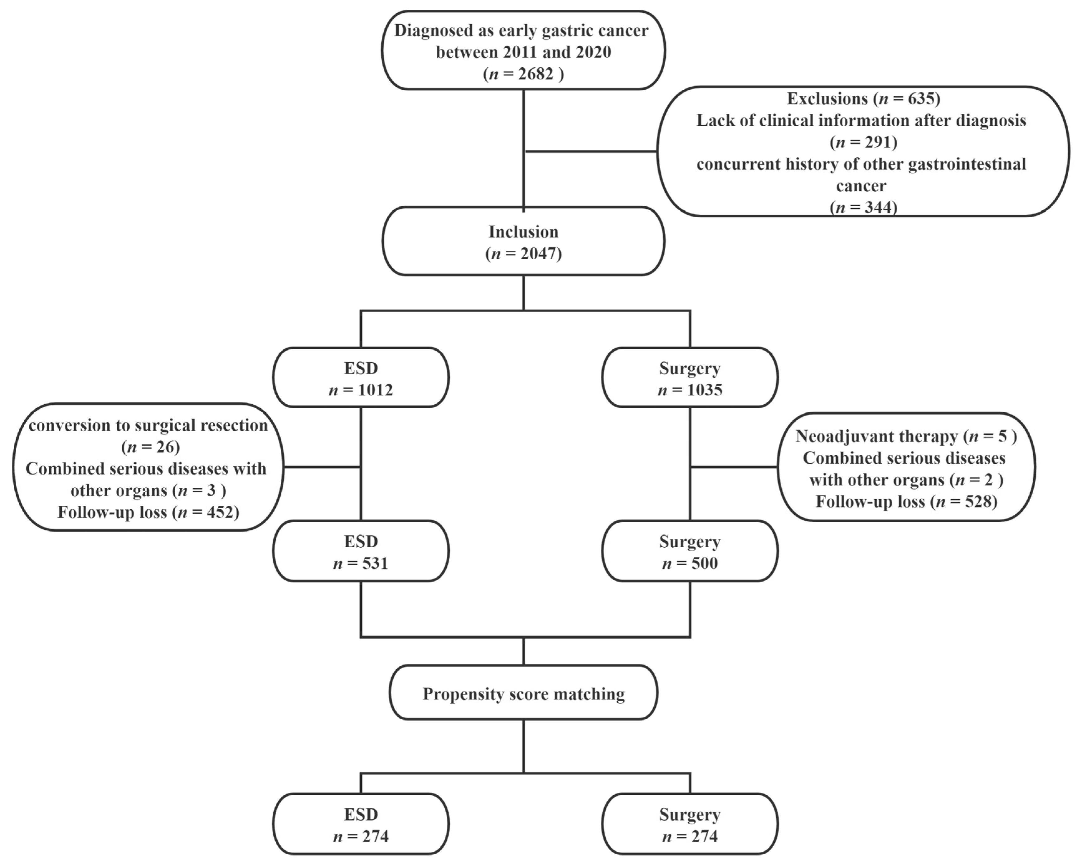
2.1. Patients

A total of 2682 patients diagnosed with EGC who underwent ESD or surgery at The First Affiliated Hospital of USTC between January 2010 and December 2020 were initially enrolled. Exclusion criteria were as follows: (1) patients who lacked clinical information after diagnosis; (2) patients with a concurrent history of other gastrointestinal cancer; (3) conversion to surgical resection immediately in the ESD group; (4) adjuvant chemotherapy (neoadjuvant chemotherapy or radiation therapy) after surgery; (5) patients combined with serious diseases of other organs; (6) patients lost to follow-up. Finally, 531 ESD cases and 500 surgery cases were included in the study. The study protocol was approved by the Medical Ethics Committee of the First Affiliated Hospital of USTC (approval number: 2022-RE-051)
To minimize potential selection bias, 1:1 propensity score-matching (PSM) was performed on the basis of sex, age, cigarette, alcohol, family history, tumor location, size, infiltration depth, and the grade of tumor differentiation. Finally, we analyzed 274 patients in each group. The study flowchart is shown in Figure 1.

2.2. Data Collection

The baseline clinicopathologic characteristics and treatment outcomes were collected from the electronic medical record system, including age, sex, cigarette, alcohol, family history of gastric cancer, tumor characteristics (such as location, size, morphology, ulceration, depth of infiltration, grade of differentiation, and lymphovascular invasion), estimated blood loss, operative time, hospital duration, cost, resection margin, adverse events, and recurrence rate.

2.3. Treatment Procedures and Follow-Up

All patients in the study underwent an intensive preoperative evaluation, including gastroduodenoscopy, endoscopic ultrasound, and computed tomography. Patients and their families were informed by a clinician about the indications and details of ESD. The choice of treatment was based on the patient’s physical condition and preference, after explaining the advantages and disadvantages of ESD and surgery. The typical ESD procedure at our institution involved staining, marking, submucosal injection, mucosal incision, and submucosal dissection of the lesion. After staining the lesion with indigo carmine dye, an electrocautery marking was made 5 mm outside the margin with a Dual Knife (KD-650L, Olympus Medical Systems, Tokyo, Japan). Saline mixed with indigo carmine and epinephrine was injected into the submucosa to elevate the lesion. A circumferential incision was made along the outside of marked area, and the lesion was dissected with an insulated-tipped knife (IT knife, KD-611L, Olympus Medical Systems, Tokyo, Japan) or a Dual Knife (KD-650L, Olympus Medical Systems, Tokyo, Japan). During the procedure, a hemoclip (hemostatic clip) was used to control the bleeding. All patients in the surgery group underwent open or laparoscopy-assisted gastrectomy with D1 or D1+ lymph node dissection. The extent of gastric resection was determined by the tumor location, and lymph node dissection was performed according to the guidelines of Japanese Gastric Cancer [14,19,20].
Follow-up was recommended for patients who underwent ESD or surgery. In the ESD group, endoscopy was scheduled at 3, 6, and 12 months after initial ESD and then annually for 5 years. In the surgery group, endoscopic evaluation and abdominal CT were scheduled every 6 months for the first year and annually thereafter until 5 years. If patients were lost to follow-up, survival and recurrence information was obtained via telephone.

2.4. Definitions and Outcomes

Macroscopic types of tumor were classified according to the Japanese classification system, type I (protruding), type IIa (superficial elevated), type IIb (flat), type IIc (superficial depressed), and type III (excavated) [21]. We classified types I and IIa as elevated types, and types IIb, IIc, and III as flat or depressed types.
R0 resection was defined as resection with negative margins, in which there was no horizontal or vertical residual tumor at the resection margins.
Recurrences included metachronous, local, and regional or distant metastatic recurrence, while synchronous recurrence was not considered as recurrence. Synchronous and metachronous recurrences were defined as cancer detected at a site different from the primary lesion location within and after 12 months of ESD/surgery treatment, respectively. Local recurrence was defined as cancer detected at the scar site of ESD or at the anastomotic site of the surgical group. Regional/distant metastases were defined as the detection of a recurrence in other organs or lymph nodes.
The primary outcome comparison was overall survival (OS). Secondary outcomes were disease-specific survival (DSS), recurrence-free survival (RFS), and treatment-related events (including estimated blood loss, operative time, hospital duration, hospital cost, resection margin, adverse events, and recurrence rate). OS was defined as the time from ESD/surgery treatment to death of any cause, DSS was defined as the time from ESD/surgery treatment to death of gastric cancer, and RFS was defined as the time from ESD/surgery treatment to first recurrence or death of any cause.

2.5. Statistical Analysis

Continuous variables were presented as means ± standard deviations (SDs) or medians with interquartile ranges (IQRs), and categorical variables were presented as numbers with percentages. For comparisons, continuous variables were analyzed using the Student’s t-test or Mann–Whitney U-test, and categorical variables were analyzed using the chi-squared test or Fisher’s exact test. The Kaplan–Meier method was used to estimate survival analyses, and outcomes were compared using the log-rank test. Using the Cox proportional hazards regression model to perform univariate and multivariate analyzes, those variables that were considered clinically relevant or with p < 0.1 in univariate analysis were subsequently entered into multivariate analyses.
PSM was performed to minimize potential selection bias at a 1:1 ratio, with a match tolerance of 0.2 standard deviations of the logit of the estimated propensity score [22]. All statistical analyses were performed using SPSS software (version 26.0, IBM Corp, Armonk NY, USA) and Prism software (version 9.0, Inc., San Diego, CA, USA). A p-value <0.05 was considered statistically significant.

3. Results

3.1. Patient Characteristics

The baseline and clinicopathological characteristics of the study population before and after PSM are shown in Table 1. Before PSM, there were statistically significant differences in the family history of gastric cancer (p = 0.013), tumor location (p = 0.005), tumor size (p < 0.001), ulceration (p < 0.001), tumor differentiation grade (p < 0.001), and lymphovascular invasion (p = 0.010). After PSM, all characteristics were well balanced between the ESD and surgery groups (all p > 0.05).

3.2. Clinical Outcomes and Treatment-Related Events

The clinical outcomes before and after PSM are reported in Table 2. Patients in the ESD group had less blood loss, a shorter operative time and hospital duration, and a lower hospital cost (all p < 0.001). There was no statistically significant difference in the R0 resection rate and the recurrence rate. Additionally, the ESD group had significantly fewer adverse events compared with the surgery group (p < 0.001). In the matched population, these variables (including estimated blood loss, operative time, hospital duration, hospital cost, and adverse events) still showed statistically significant differences between the ESD and surgery groups (p < 0.001).
Adverse events that occurred in the ESD and surgery groups are summarized in Table 3 and Table 4, respectively. In the ESD group, one patient required emergency surgery due to massive bleeding, and one patient was transferred to surgery due to postoperative perforation. In the surgery group, two patients were transferred to the intensive care unit (ICU) due to severe infection. In terms of complications, there was no related death in the ESD group, while there was one related death in the surgery group (these cases are marked with an asterisk in the tables).

3.3. Survival Analysis

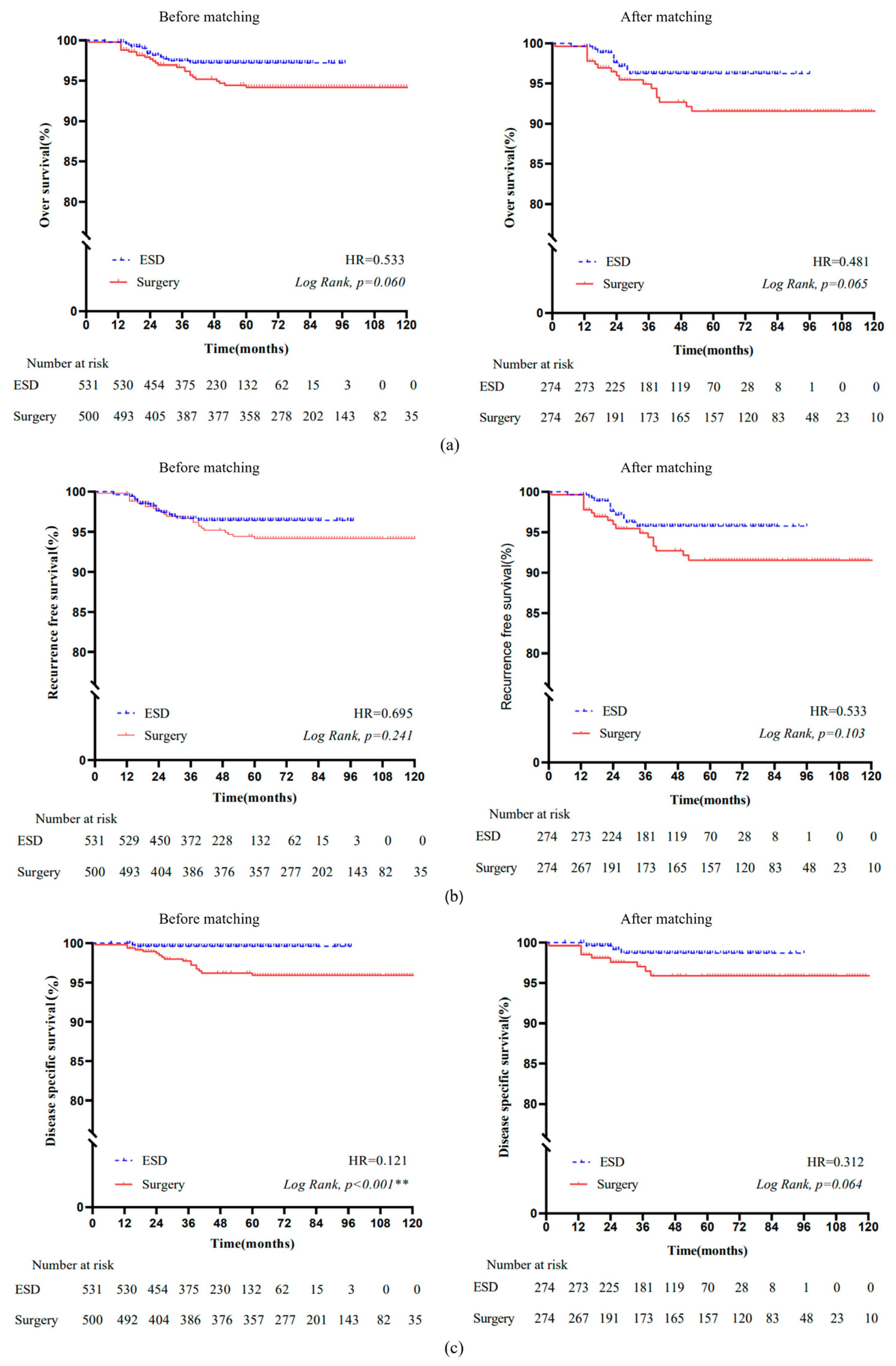
3.3.1. Overall Survival

The Kaplan–Meier OS curves before and after matching are presented in Figure 2a. No significant difference in OS was found between the ESD and surgery groups (hazard ratio (HR) = 0.533, p = 0.060). After matching, there was also no significant difference in OS between the two groups (HR = 0.481, p = 0.065). The 5 year OS was comparable between the ESD and surgery groups (96.1% vs. 91.4%, p = 0.081).
For the PSM cohort, we further used Cox regression analysis to identify the variables associated with OS (Table 5). In the univariate analysis, age (p < 0.001), tumor size (p < 0.001), submucosal infiltration (p = 0.025), poor differentiation (p < 0.001), ulceration (p = 0.025), and the rate of R0 resection (p < 0.001) were associated with OS. In the multivariate analysis, there were still associations of age (HR = 1.24, 95% CI: 1.15–1.29, p < 0.001), tumor size (HR = 1.37, 95% CI: 1.11–1.69, p = 0.003), and poor differentiation (HR = 3.21, 95% CI: 1.13–9.10, p = 0.022). Meanwhile, treatment method (HR = 0.10, 95% CI: 0.04–0.26, p < 0.001) showed an association with OS.

3.3.2. Recurrence-Free Survival

The Kaplan–Meier RFS curves before and after matching are presented in Figure 2b. There was no significant difference in RFS before (HR = 0.695, p = 0.241) and after (HR = 0.533, p = 0.103) matching between the ESD and surgery groups. The 5 year RFS in the ESD group of 95.8% was not significantly different from the 5 year RFS in the surgery group of 91.4% (p = 0.136).
The Cox regression model was used to analyze the factors contributing to RFS in the PSM cohort (Table 6). In the univariate analysis, factors associated with RFS included age (p < 0.001), tumor size (p < 0.001), poor differentiation (p = 0.001), and R1 resection (p = 0.001), while, in the multivariate analysis, age (HR = 1.24, 95% CI: 1.18–1.30, p < 0.001), tumor size (HR = 1.36, 95% CI: 1.07–1.73, p = 0.012), poor differentiation (HR = 3.41, 95% CI: 1.17–9.97, p = 0.025), and lymphovascular invasion (HR = 9.61, 95% CI: 3.21–28.79, p < 0.001) were the factors associated with RFS.

3.4. Disease-Specific Survival

The Kaplan–Meier DSS curves before and after matching are presented in Figure 2c. The DSS was significantly higher in the ESD group than in the surgery group (HR = 0.121, p < 0.001). However, it became comparable between the two groups in the matched cohort (HR = 0.312, p = 0.064). The 5 year DSS between the ESD and surgery groups was also not significantly different (98.6% vs. 95.7%, p = 0.062).
After matching, we also performed Cox regression analysis on the DSS (Table 7). The univariate analysis showed that age (p < 0.001), tumor size (p = 0.010), infiltration depth (p = 0.001), poor differentiation (p = 0.008), and ulceration (p = 0.027) were the factors for DSS, while multivariate analysis showed that treatment method (HR = 0.10, 95% CI: 0.02–0.46, p = 0.003), age (HR = 1.24, 95% CI: 1.15–1.33, p < 0.001), tumor size (HR = 1.38, 95% CI: 1.01–1.89, p = 0.041), and infiltration depth (HR = 5.75, 95% CI: 1.73–19.04, p = 0.004) were independent factors for DSS.

4. Discussion

In recent years, ESD has been developed as a treatment option for EGC that meets certain criteria [23]. Many studies have demonstrated the safety and efficacy of ESD in treating EGC. The outcomes of ESD treatment for gastric cancer included en bloc (92.4%), R0 (80.5%), curative resection rates (72.0%), complication (delayed bleeding: 5.9%, perforation: 4.2%), and recurrence rates (3 month: 2.3%; 12 month: 3.2%) [5]. A recent study reported favorable short- and long-term outcomes of endoscopic therapy in gastric cancer nationwide in Japan. In patients who met the absolute indications, the 5 year OS and DSS rates were 91.6% and 99.9%, respectively. In patients who belonged to expanded indications, the 5 year OS and DSS rates were still up to 90.3% and 99.7%, respectively [24]. Draganov et al. demonstrated the established outcomes of ESD for the treatment of gastrointestinal cancer. Regarding gastric cancer, the en bloc, R0, and curative resection rates were 98%, 82.2%, and 77.2%, respectively, and adverse events were reported in three cases of delayed bleeding (3%) and one case of perforation (1%) [25]. ESD is a safe and effective treatment option with favorable long-term results of early gastric cardiac cancer treated with ESD; en bloc, complete, and curative resection rates were 99.8%, 94.3%, and 80.5%, respectively. The 5 year OS rate was 89.6%, and the 5 year disease-specific survival rate reached 100% in patients with a curative resection [26].
However, there are still controversies regarding the oncological safety of ESD. For ESD specimens, R0 resection should be confirmed by pathological evaluation. A few patients who underwent ESD in our study needed additional surgery to get negative margins, but the rate was only 2.57% (26/1012). With its limitations, ESD can only remove the primary lesion, although the risk of lymph node metastasis in these EGCs is commonly low [27]. Compared with surgery, ESD has been reported to be associated with a higher incidence of metachronous tumors [28]. In addition, ESD requires a high level of professional skills, which limit its development in some areas. Due to factors such as epidemiological differences in gastric cancer, most reported results are from Asian regions, especially Korea and Japan. As one of the countries with a high prevalence of gastric cancer, China has contributed to nearly half of the gastric cancer-related deaths worldwide [29]. Although there have been some previous studies on comparing ESD with surgery in the treatment of EGC, reliable evidence from large-scale populations with long-term follow-up is still limited in China. We conducted this study to explore the role of ESD in patients with EGC in China.
Our results showed that blood loss in the ESD group was significantly less than that in the surgery group, while the operative time was significantly shorter in the ESD group. With the shorter duration of hospital stay, the cost of hospitalization was also significantly lower in the ESD group. These findings are consistent with previous studies [26,30]. Furthermore, a higher R0 resection rate of the ESD group was observed in the present study, which allowed ESD to show similar resection outcomes to surgery (99.4% vs. 100%, p = 0.092) [6,30]. Furthermore, the incidence of adverse events was significantly reduced in the ESD group compared with the surgery group [31]. From the results, we also observed that complications in the ESD group were milder than in the surgery group. Patients with complications in the ESD group could be successfully managed by conservative treatment or surgery, while some patients with severe complications in the surgery group needed to be transferred to the ICU, and even a case of treatment-related death was reported in the surgery group due to severe infection.
In the matched cohorts, patients in the ESD group showed comparable survival outcomes (including OS, RFS, and DSS) to that of the surgery group, along with additional advantages, such as shorter operative time and hospital duration, less blood loss, and lower treatment costs. Patients with gastric cancer who received ESD had a lower hospitalization cost, less traumatic experience, shorter recovery time, and better quality of life, while OS and DSS were not significantly different between the ESD and surgery groups [31]. Jeon et al. compared the long-term outcomes of ESD with surgery in the treatment of EGC. They concluded that the 5 year DSS rate of the ESD group was significantly better in the overall cohort, but similar to that of the surgery group in the matched cohort [32]. A study of young patients with gastric cancer further suggested that DSS was not constant, and that the prognostic factors that predict survival vary over time [33]. To verify the reliability of the results, we also compared the 5 year OS, RFS, and DSS between the ESD and surgery groups in the matched cohort, and we found that 5 year OS, RFS, and DSS were similar in the two populations. In this study, although only patients in the ESD group developed metachronous gastric cancer, we did not find a significant difference in the incidence of metachronous tumors between the two groups, and all of these cases were successfully treated with repeated ESD.
A multivariate analysis was used to identify the potential factors influencing OS, RFS, and DSS. For OS, age, tumor size, poor differentiation, and treatment method were independent factors. In a cohort matched study, Pyo et al. reported that the independent factors for OS included age, comorbidity index, performance index, sex, tumor morphology, and the depth of infiltration [34]. Regarding RFS, age, tumor size, poor differentiation, and lymphovascular invasion were influential factors. In terms of DSS, we found that age, tumor size, submucosal infiltration, and treatment method were negative prognostic factors. It is necessary to mention that age and tumor size were common risk factors. Tumor size has proven to be the independent risk factor for lymph node metastasis, significantly associated with the prognosis of patients with EGC [35,36,37]. Consequently, preoperative evaluation is crucial, and clinicians should comprehensively consider the relevant factors in choosing the appropriate treatment for patients with EGC.
For the treatment of EGC, ESD shows some advantages. First, ESD is more cost-effective, which can adequately save medical resources and reduce the burden of patients [3,38]. Second, ESD preserves the stomach integrity, avoiding potential functional complications relevant to surgery and, thus, improving the quality of life of patients [39]. Lastly, ESD provides comparable survival outcomes to surgery. Although ESD may have a higher incidence of metachronous lesions, successful treatment is available in most cases with repeated ESD, and a history of ESD does not negatively affect subsequent surgical treatment [40,41].
Our study had several limitations. Firstly, this was a single-center, retrospective study; hence, potential selection bias was unavoidable, although a PSM analysis was performed to reduce the clinicopathological differences between the two cohorts. Secondly, some patients were lost to follow-up during the study period. China is characterized by a large population and vast geographic area, and patients in the study were from all over the country, which increased the difficulty of follow-up. Due to the long timespan, some patients changed their phone numbers, and their follow-up information was not obtained. Fortunately, enough individuals were initially included in this study, and the sample was considered to be representative [42]. Therefore, we believe that the results were not significantly impacted. In the future, it is necessary to develop new strategies to improve the completeness and quality of follow-up information. Thirdly, the research time was 2 years later in the ESD group than in the surgery group; so as to minimize the effect of this limitation, we further compared the 5 year OS, RFS, and DSS. Lastly, the numbers for the matched cohort were limited; thus, we did not make subgroup comparisons based on the depth of infiltration, presence of ulceration, and lymphovascular invasion. Further multicenter and randomized studies are needed to present better evidence.

5. Conclusions

In conclusion, ESD provides comparable long-term OS, RFS, and DSS with surgery. Patients in the ESD group have significantly shorter hospital duration, lower hospital cost, and fewer adverse events. Thus, ESD is preferable to surgery for well-chosen localized gastric cancer when close follow-up is ensured. On the basis of the impact of tumor histological characteristics on survival outcomes, preoperative evaluation is essential for the selection of the appropriate treatment modality and patient prognosis.

Author Contributions

Conceptualization, M.Q., S.W., and K.Z.; methodology, M.Q., S.W., and K.Z.; software, M.Q.; validation, M.Q., S.W., and K.Z.; formal analysis, M.Q.; investigation, M.Q., S.W., and K.Z.; resources, M.Q., S.W., and K.Z.; data curation, M.Q., M.W., and Y.S.; writing—M.Q.; writing—review and editing, M.Q., S.W., and K.Z.; visualization, M.Q., M.W., Y.S., S.W., and K.Z.; supervision, K.Z.; project administration, K.Z. All authors have read and agreed to the published version of the manuscript.

Funding

This research received no external funding.

Institutional Review Board Statement

This study was reviewed and approved by the First Affiliated Hospital of USTC (approval number: 2022-RE-051, date of approval: 16 April 2022), All methods were carried out in accordance with relevant guidelines and regulations.

Informed Consent Statement

Since this is a retrospective study using clinical routine treatment or diagnostic medical records, and since no human immunodeficiency virus-positive cases were involved, the First Affiliated Hospital of USTC Medisch Ethische Institutional approved the waiver of participants’ consent.

Data Availability Statement

Deidentified individual participant data are available and can be provided on reasonable request to the corresponding author.

Conflicts of Interest

The authors declare no conflict of interest.

References

  1. Sung, H.; Ferlay, J.; Siegel, R.L.; Laversanne, M.; Soerjomataram, I.; Jemal, A.; Bray, F. Global Cancer Statistics 2020: GLOBOCAN Estimates of Incidence and Mortality Worldwide for 36 Cancers in 185 Countries. CA Cancer J. Clin. 2021, 71, 209–249. [Google Scholar] [CrossRef] [PubMed]
  2. Mei, Y.; Wang, S.; Feng, T.; Yan, M.; Yuan, F.; Zhu, Z.; Li, T.; Zhu, Z. Nomograms Involving HER2 for Predicting Lymph Node Metastasis in Early Gastric Cancer. Front Cell Dev. Biol. 2021, 9, 781824. [Google Scholar] [CrossRef] [PubMed]
  3. Draganov, P.V.; Wang, A.Y.; Othman, M.O.; Fukami, N. AGA Institute Clinical Practice Update: Endoscopic Submucosal Dissection in the United States. Clin. Gastroenterol. Hepatol. 2019, 17, 16–25. [Google Scholar] [CrossRef] [PubMed] [Green Version]
  4. Ono, H.; Yao, K.; Fujishiro, M.; Oda, I.; Uedo, N.; Nimura, S.; Yahagi, N.; Iishi, H.; Oka, M.; Ajioka, Y.; et al. Guidelines for endoscopic submucosal dissection and endoscopic mucosal resection for early gastric cancer (second edition). Dig. Endosc. 2021, 33, 4–20. [Google Scholar] [CrossRef]
  5. Fleischmann, C.; Probst, A.; Ebigbo, A.; Faiss, S.; Schumacher, B.; Allgaier, H.P.; Dumoulin, F.L.; Steinbrueck, I.; Anzinger, M.; Marienhagen, J.; et al. Endoscopic Submucosal Dissection in Europe: Results of 1000 Neoplastic Lesions From the German Endoscopic Submucosal Dissection Registry. Gastroenterology 2021, 161, 1168–1178. [Google Scholar] [CrossRef] [PubMed]
  6. Quero, G.; Fiorillo, C.; Longo, F.; Laterza, V.; Rosa, F.; Cina, C.; Menghi, R.; Tortorelli, A.P.; Barbaro, F.; Pecere, S.; et al. Propensity score-matched comparison of short- and long-term outcomes between surgery and endoscopic submucosal dissection (ESD) for intestinal type early gastric cancer (EGC) of the middle and lower third of the stomach: A European tertiary referral center experience. Surg. Endosc. 2021, 35, 2592–2600. [Google Scholar] [CrossRef]
  7. Pourmousavi, M.K.; Wang, R.; Kerdsirichairat, T.; Kamal, A.; Akshintala, V.S.; Hajiyeva, G.; Lopimpisuth, C.; Hanada, Y.; Kumbhari, V.; Singh, V.K.; et al. Comparable Cancer-Specific Mortality of Patients With Early Gastric Cancer Treated With Endoscopic Therapy vs Surgical Resection. Clin. Gastroenterol. Hepatol. 2020, 18, 2824–2832. [Google Scholar] [CrossRef]
  8. Li, H.; Feng, L.Q.; Bian, Y.Y.; Yang, L.L.; Liu, D.X.; Huo, Z.B.; Zeng, L. Comparison of endoscopic submucosal dissection with surgical gastrectomy for early gastric cancer: An updated meta-analysis. World J. Gastrointest. Oncol. 2019, 11, 161–171. [Google Scholar] [CrossRef]
  9. Choi, I.J.; Lee, J.H.; Kim, Y.I.; Kim, C.G.; Cho, S.J.; Lee, J.Y.; Ryu, K.W.; Nam, B.H.; Kook, M.C.; Kim, Y.W. Long-term outcome comparison of endoscopic resection and surgery in early gastric cancer meeting the absolute indication for endoscopic resection. Gastrointest. Endosc. 2015, 81, 333–341. [Google Scholar] [CrossRef]
  10. Kamarajah, S.K.; Markar, S.R.; Phillips, A.W.; Salti, G.I.; Dahdaleh, F.S. Local Endoscopic Resection is Inferior to Gastrectomy for Early Clinical Stage T1a and T1b Gastric Adenocarcinoma: A Propensity-Matched Study. Ann. Surg. Oncol. 2021, 28, 2992–2998. [Google Scholar] [CrossRef]
  11. Park, C.H.; Yang, D.H.; Kim, J.W.; Kim, J.H.; Kim, J.H.; Min, Y.W.; Lee, S.H.; Bae, J.H.; Chung, H.; Choi, K.D.; et al. Clinical practice guideline for endoscopic resection of early gastrointestinal cancer. Intest. Res. 2021, 19, 127–157. [Google Scholar] [CrossRef] [PubMed]
  12. van der Veen, A.; Brenkman, H.J.F.; Seesing, M.F.J.; Haverkamp, L.; Luyer, M.D.P.; Nieuwenhuijzen, G.A.P.; Stoot, J.; Tegels, J.J.W.; Wijnhoven, B.P.L.; Lagarde, S.M.; et al. Laparoscopic Versus Open Gastrectomy for Gastric Cancer (LOGICA): A Multicenter Randomized Clinical Trial. J. Clin. Oncol. 2021, 39, 978–989. [Google Scholar] [CrossRef] [PubMed]
  13. Hiramatsu, Y.; Kikuchi, H.; Takeuchi, H. Function-Preserving Gastrectomy for Early Gastric Cancer. Cancers 2021, 13, 2683–2692. [Google Scholar] [CrossRef] [PubMed]
  14. Japanese Gastric Cancer Association. Japanese gastric cancer treatment guidelines 2018 (5th edition). Gastric Cancer 2021, 24, 1–21. [Google Scholar] [CrossRef] [PubMed] [Green Version]
  15. Ajani, J.A.; D’Amico, T.A.; Bentrem, D.J.; Chao, J.; Cooke, D.; Corvera, C.; Das, P.; Enzinger, P.C.; Enzler, T.; Fanta, P.; et al. Gastric Cancer, Version 2.2022, NCCN Clinical Practice Guidelines in Oncology. J. Natl. Compr. Cancer Netw. 2022, 20, 167–192. [Google Scholar] [CrossRef]
  16. Banks, M.; Graham, D.; Jansen, M.; Gotoda, T.; Coda, S.; di Pietro, M.; Uedo, N.; Bhandari, P.; Pritchard, D.M.; Kuipers, E.J.; et al. British Society of Gastroenterology guidelines on the diagnosis and management of patients at risk of gastric adenocarcinoma. Gut 2019, 68, 1545–1575. [Google Scholar] [CrossRef] [Green Version]
  17. Pimentel-Nunes, P.; Dinis-Ribeiro, M.; Ponchon, T.; Repici, A.; Vieth, M.; De Ceglie, A.; Amato, A.; Berr, F.; Bhandari, P.; Bialek, A.; et al. Endoscopic submucosal dissection: European Society of Gastrointestinal Endoscopy (ESGE) Guideline. Endoscopy 2015, 47, 829–854. [Google Scholar] [CrossRef] [Green Version]
  18. Yim, K.; Jang, W.M.; Lee, S.H. Modified Tumor Budding as a Better Predictor of Lymph Node Metastasis in Early Gastric Cancer: Possible Real-World Applications. Cancers 2021, 13, 3405. [Google Scholar] [CrossRef]
  19. Japanese Gastric Cancer Association. Japanese gastric cancer treatment guidelines 2010 (ver. 3). Gastric Cancer 2011, 14, 113–123. [Google Scholar] [CrossRef] [Green Version]
  20. Japanese Gastric Cancer Association. Japanese gastric cancer treatment guidelines 2014 (ver. 4). Gastric Cancer 2017, 20, 1–19. [Google Scholar] [CrossRef] [Green Version]
  21. Japanese Gastric Cancer, A. Japanese Classification of Gastric Carcinoma—2nd English Edition. Gastric Cancer 1998, 1, 10–24. [Google Scholar] [CrossRef] [Green Version]
  22. Austin, P.C. The use of propensity score methods with survival or time-to-event outcomes: Reporting measures of effect similar to those used in randomized experiments. Stat. Med. 2014, 33, 1242–1258. [Google Scholar] [CrossRef] [PubMed] [Green Version]
  23. Shahidi, N.; Bourke, M.J. ESD, not EMR, should be the first-line therapy for early gastric neoplasia. Gut 2020, 69, 1–2. [Google Scholar] [CrossRef] [PubMed]
  24. Tanabe, S.; Hirabayashi, S.; Oda, I.; Ono, H.; Nashimoto, A.; Isobe, Y.; Miyashiro, I.; Tsujitani, S.; Seto, Y.; Fukagawa, T.; et al. Gastric cancer treated by endoscopic submucosal dissection or endoscopic mucosal resection in Japan from 2004 through 2006: JGCA nationwide registry conducted in 2013. Gastric Cancer 2017, 20, 834–842. [Google Scholar] [CrossRef] [Green Version]
  25. Draganov, P.V.; Aihara, H.; Karasik, M.S.; Ngamruengphong, S.; Aadam, A.A.; Othman, M.O.; Sharma, N.; Grimm, I.S.; Rostom, A.; Elmunzer, B.J.; et al. Endoscopic Submucosal Dissection in North America: A Large Prospective Multicenter Study. Gastroenterology 2021, 160, 2317–2327. [Google Scholar] [CrossRef]
  26. Cao, S.; Zou, T.; Sun, Q.; Liu, T.; Fan, T.; Yin, Q.; Fan, X.; Jiang, J.; Raymond, D.; Wang, Y.; et al. Safety and long-term outcomes of early gastric cardiac cancer treated with endoscopic submucosal dissection in 499 Chinese patients. Ther. Adv. Gastroenterol. 2020, 13, 1756284820966929. [Google Scholar] [CrossRef]
  27. Abdelfatah, M.M.; Barakat, M.; Lee, H.; Kim, J.J.; Uedo, N.; Grimm, I.; Othman, M.O. The incidence of lymph node metastasis in early gastric cancer according to the expanded criteria in comparison with the absolute criteria of the Japanese Gastric Cancer Association: Asystematic review of the literature and meta-analysis. Gastrointest. Endosc. 2018, 87, 338–347. [Google Scholar] [CrossRef]
  28. Nishizawa, T.; Yahagi, N. Long-Term Outcomes of Using Endoscopic Submucosal Dissection to Treat Early Gastric Cancer. Gut Liver 2018, 12, 119–124. [Google Scholar] [CrossRef] [Green Version]
  29. The global, regional, and national burden of stomach cancer in 195 countries, 1990-2017: A systematic analysis for the Global Burden of Disease study 2017. Lancet Gastroenterol. Hepatol. 2020, 5, 42–54. [CrossRef] [Green Version]
  30. Libânio, D.; Braga, V.; Ferraz, S.; Castro, R.; Lage, J.; Pita, I.; Ribeiro, C.; Abreu De Sousa, J.; Dinis-Ribeiro, M.; Pimentel-Nunes, P. Prospective comparative study of endoscopic submucosal dissection and gastrectomy for early neoplastic lesions including patients’ perspectives. Endoscopy 2019, 51, 30–39. [Google Scholar] [CrossRef] [Green Version]
  31. Liu, Q.; Ding, L.; Qiu, X.; Meng, F. Updated evaluation of endoscopic submucosal dissection versus surgery for early gastric cancer: A systematic review and meta-analysis. Int. J. Surg. 2020, 73, 28–41. [Google Scholar] [CrossRef] [PubMed]
  32. Jeon, H.K.; Kim, G.H.; Lee, B.E.; Park, D.Y.; Song, G.A.; Kim, D.H.; Jeon, T.Y. Long-term outcome of endoscopic submucosal dissection is comparable to that of surgery for early gastric cancer: A propensity-matched analysis. Gastric Cancer 2018, 21, 133–143. [Google Scholar] [CrossRef] [PubMed] [Green Version]
  33. Chen, Q.Y.; Zhong, Q.; Wang, W.; Chen, S.; Li, P.; Xie, J.W.; Wang, J.B.; Lin, J.X.; Lu, J.; Cao, L.L.; et al. Prognosis of Young Survivors of Gastric Cancer in China and the U.S.: Determining Long-Term Outcomes Based on Conditional Survival. Oncologist 2019, 24, e260–e274. [Google Scholar] [CrossRef] [PubMed] [Green Version]
  34. Pyo, J.H.; Lee, H.; Min, B.H.; Lee, J.H.; Choi, M.G.; Lee, J.H.; Sohn, T.S.; Bae, J.M.; Kim, K.M.; Ahn, J.H.; et al. Long-Term Outcome of Endoscopic Resection vs. Surgery for Early Gastric Cancer: A Non-inferiority-Matched Cohort Study. Am. J. Gastroenterol. 2016, 111, 240–249. [Google Scholar] [CrossRef] [PubMed]
  35. Wang, J.; Wang, L.; Li, S.; Bai, F.; Xie, H.; Shan, H.; Liu, Z.; Ma, T.; Tang, X.; Tang, H.; et al. Risk Factors of Lymph Node Metastasis and Its Prognostic Significance in Early Gastric Cancer: A Multicenter Study. Front Oncol. 2021, 11, 649035. [Google Scholar] [CrossRef] [PubMed]
  36. Hatta, W.; Gotoda, T.; Kanno, T.; Yuan, Y.; Koike, T.; Moayyedi, P.; Masamune, A. Prevalence and risk factors for lymph node metastasis after noncurative endoscopic resection for early gastric cancer: A systematic review and meta-analysis. J. Gastroenterol. 2020, 55, 742–753. [Google Scholar] [CrossRef]
  37. Kim, S.M.; Min, B.H.; Ahn, J.H.; Jung, S.H.; An, J.Y.; Choi, M.G.; Sohn, T.S.; Bae, J.M.; Kim, S.; Lee, H.; et al. Nomogram to predict lymph node metastasis in patients with early gastric cancer: A useful clinical tool to reduce gastrectomy after endoscopic resection. Endoscopy 2020, 52, 435–443. [Google Scholar] [CrossRef]
  38. Dahan, M.; Pauliat, E.; Liva-Yonnet, S.; Brischoux, S.; Legros, R.; Tailleur, A.; Carrier, P.; Charissoux, A.; Valgueblasse, V.; Loustaud-Ratti, V.; et al. What is the cost of endoscopic submucosal dissection (ESD)? A medico-economic study. United Eur. Gastroenterol. J. 2019, 7, 138–145. [Google Scholar] [CrossRef]
  39. Park, K.B.; Jeon, C.H.; Seo, H.S.; Jung, Y.J.; Song, K.Y.; Park, C.H.; Lee, H.H. Operative safety of curative gastrectomy after endoscopic submucosal dissection (ESD) for early gastric cancer—1:2 propensity score matching analysis: A retrospective single-center study (cohort study). Int. J. Surg. 2020, 80, 124–128. [Google Scholar] [CrossRef]
  40. Jung, D.H.; Youn, Y.H.; Kim, J.H.; Park, J.J.; Park, H. Secondary endoscopic submucosal dissection for locally recurrent or incompletely resected gastric neoplasms. World J. Gastroenterol. 2018, 24, 3776–3785. [Google Scholar] [CrossRef]
  41. Shimada, S.; Sawada, N.; Oae, S.; Seki, J.; Takano, Y.; Nakahara, K.; Takehara, Y.; Mukai, S.; Ishida, F.; Kudo, S.E. Impact of non-curative endoscopic submucosal dissection on short- and long-term outcome of subsequent laparoscopic gastrectomy for pT1 gastric cancer. Surg. Endosc. 2021, 36, 3985–3993. [Google Scholar] [CrossRef] [PubMed]
  42. Lie, H.C.; Rueegg, C.S.; Fosså, S.D.; Loge, J.H.; Ruud, E.; Kiserud, C.E. Limited evidence of non-response bias despite modest response rate in a nationwide survey of long-term cancer survivors-results from the NOR-CAYACS study. J. Cancer Surviv. 2019, 13, 353–363. [Google Scholar] [CrossRef] [PubMed]
Figure 1. Flowchart of the study population. ESD: endoscopic submucosal dissection.
Cancers 14 03603 g001
Figure 2. Kaplan–Meier survival curves of OS, RFS, and DSS in ESD and surgery groups. (a) OS before and after matching; (b) RFS before and after matching; (c) DSS before and after matching. Abbreviations: OS, overall survival; RFS, recurrence-free survival; DSS, disease-specific survival. **: p < 0.001.
Cancers 14 03603 g002
Table 1. Baseline and clinicopathologic characteristics of the study population before matching After matching.
ESD
N = 531
Surgery
N = 500
p-ValueESD
N = 274
Surgery
N = 274
p-Value
Age (mean ± SD; years)61.6 ± 10.761.4 ± 10.60.19661.1 ± 10.761.6 ± 10.70.448
Sex, n (%) 0.506 0.851
Male368 (69.3%)356 (71.2%) 192 (70.1%)194 (70.8%)
Female163 (30.7%)144 (28.8%) 82 (29.9%)80 (29.2%)
Lifestyle, n (%)
Cigarette104 (19.6%)108 (21.6%)0.42454 (19.7%)53 (19.3%)0.948
Alcohol
Positive family history, n (%)
89 (16.8%)
22 (4.1%)
89 (17.8%)
39 (7.8%)
0.659
0.013
46 (16.8%)
13 (4.7%)
44 (16.1%)
21 (6.6%)
0.818
0.355
Tumor location, n (%) 0.005 0.390
Cardia209 (39.4%)143 (28.6%) 102 (37.2%)94 (34.3%)
Fundus16 (3.0%)13 (2.6%) 9 (3.3%)8 (2.9%)
Body91 (17.1%)100 (20.0%) 49 (17.9%)54 (19.7%)
Antrum145 (27.3%)154 (30.8%) 87 (31.7%)77 (28.1%)
Incisura angularis or pylorus70 (13.2%)90 (18.0%) 27 (9.9%)41 (15.0%)
Size (mean ± SD; cm)2.6 ± 1.52.3 ± 1.1<0.001 **2.6 ± 1.52.4 ± 1.20.103
Tumor morphology, n (%) 0.932 0.913
Elevated86 (16.2%) 80 (16.0%) 52 (19.0%) 51 (18.6%)
Flat or depressed445 (83.8%)420 (84.0%) 222 (81.0%)223 (81.4%)
Tumor infiltration, n (%) 0.271 0.899
Mucosa450 (84.7%)411 (82.2%) 239 (87.2%)238 (86.9%)
Submucosa81 (15.3%)89 (17.8%) 35 (12.8%)36 (13.1%)
Ulceration, n (%)32 (6.0%)237 (47.4%)<0.001 **32 (11.7%)48 (17.5%)0.053
Tumor differentiation grade, n (%) <0.001 ** 0.062
Well-differentiated363 (68.3%)137 (15.3%) 122 (44.5%)114 (41.6%)
Moderately differentiated156 (29.4%)148 (15.3%) 140 (51.1%)134 (48.9%)
Poorly differentiated12 (2.3%)215 (15.3%) 12 (4.4%)26 (9.5%)
Lymphovascular invasion, n (%)16 (3.0%)32 (6.4%)0.01012 (4.4%)13 (4.7%)0.838
Abbreviations: ESD, endoscopic submucosal dissection; SD, standard deviation. **: p < 0.001.
Table 2. Clinical outcomes of the two study populations.
ESD
N = 531
Surgery
N = 500
p-ValueESD
N = 274
Surgery
N = 274
p-Value
Estimated blood loss (mL) <0.001 ** <0.001 **
≤50511 (96.2%)68 (13.6%) 265 (96.7%)50 (18.2%)
>5020 (3.8%)432 (86.4%) 9 (3.6%)224 (86.3%)
Operative time (min), median (IQR)73.5
(55.0–118.0)
195.5
(153.0–232.0)
<0.001 **94.0
(60.0–120.0)
197.0
(153.0–232.0)
<0.001 **
Hospital duration (day), median (IQR)9.0
(8.0–11.0)
17.0
(14.0–20.0)
<0.001 **10.3
(8.0–11.0)
19.1
(14.0–21.0)
<0.001 **
Hospital cost (USD), median (IQR)3121.1
(2693.1–3658.0)
6142.2
(5005.1–7211.7)
<0.001 **3304.2
(2660.9–3438.9)
6507.6
(5157.7–7357.6)
<0.001 **
Resection margin 0.092 0.082
R0 resection528 (99.4%)500 (100.0%) 271 (98.9%)274 (100.0%)
R1 resection3 (0.6%)0 (0.0%) 3 (1.1%)0 (0.0%)
Recurrence4 (0.8%)1 (0.2%)0.2011 (0.4%)0 (0.0%)0.317
Adverse events, n (%)7 (1.3%)26 (5.2%)<0.001 **1 (0.4%)16 (5.8%)<0.001 **
Abbreviations: IQR, interquartile range. **: p < 0.001.
Table 3. Adverse events after ESD treatment for early gastric cancer patients.
Adverse EventsTotal
N = 7
AgeSexLocationSize
(cm)
InfiltrationEstimated Blood
Loss (mL)
Operative Time
(min)
Hospital Duration
(Day)
Hospital Cost
(USD)
Gastropathy 2 (28.6%)59MaleCardia1.5Mucosa1055121203.9
53MaleCardia1.0Mucosa57093162.0
Bleeding4 (57.1%)84FemaleBody3.5Mucosa240143964.1
67MaleCardia1.5Mucosa1060123861.5
63 *MaleCardia4.0Mucosa1093348450.7
52MaleAntrum3.0Mucosa10120168708.5
Perforation1 (14.3%)54 *MaleCardia4.5Submucosa10140125934.4
*: Transferred to surgery.
Table 4. Adverse events after surgery treatment for early gastric cancer patients.
Adverse EventsTotal
N = 26
AgeSexLocationSize (cm)InfiltrationEstimated Blood
Loss (mL)
Operative Time
(min)
Hospital Duration
(day)
Hospital Cost
(USD)
Delayed
gastric emptying
8 (30.9%)55MaleAntrum2.0Mucosa2002143912,373.8
69FemaleBody3.0Submucosa20093379534.3
85MaleAntrum2.0Mucosa1001353716,754.6
64MaleIncisura angularis1.2Mucosa200150407747.8
66FemaleBody1.0Submucosa1002954011,897.8
54MaleIncisura angularis1.2Submucosa1002054210,712.3
68FemaleAntrum1.0Submucosa50125519484.9
63MaleBody3.0Submucosa100246145390.5
Anastomotic leakage3 (11.5%)63MaleBody5.0Submucosa100150548917.2
71FemaleCardia3.5Submucosa200317369378.0
60MaleAntrum1.5Mucosa100200418457.5
Anastomotic leakage
+ gastroparesis
1 (3.8%)70FemaleBody2.0Mucosa2001409913,457.5
Ileus4 (15.4%)61MaleAntrum3.0Submucosa1002654713,814.6
37MaleAntrum1.5Mucosa100180235488.9
78FemaleAntrum8.0Mucosa100215257170.1
67MaleIncisura angularis2.0Submucosa100205147371.2
Bleeding2 (7.7%)63MaleBody2.5Mucosa100154446542.9
79MaleCardia2.5Submucosa100180477887.4
Infection3 (11.5%)69MaleIncisura angularis1.0Submucosa100220426533.3
Severe infection ** 78MaleAntrum5.0Mucosa1001552821,170.6
Severe infection * 60MaleAntrum2.0Submucosa3002352521,101.8
Fever4 (15.4%)72FemaleCardia1.5Submucosa1001872911,168.6
59MaleCardia1.5Mucosa100190208965.7
67MaleCardia2.0Mucosa2001894611,228.6
70FemaleCardia2.5Submucosa200305299630.1
Other1 (3.8%)86MaleAntrum2.0Mucosa200162289204.0
**: Transferred to intensive care unit and then died; *: Transferred to intensive care unit.
Table 5. Univariate and multivariate regression analyses of overall survival for propensity score-matching patients.
VariablesUnivariate AnalysisMultivariate Analysis
Hazard Ratio (95% CI)p-ValueHazard Ratio (95% CI) p-Value
Treatment method (ESD vs. surgery)0.48 (0.22–1.07)0.0720.10 (0.04–0.26)<0.001 **
Age1.20 (1.15–1.25)<0.001 **1.24 (1.15–1.29)<0.001 **
Sex, male = 1 0.31 (0.09–1.03)0.055
Cigarette, no = 11.60 (0.55–4.64)0.384
Alcohol, no = 12.64 (0.63–11.15)0.286
Positive family history22.01 (0.02–26.86)0.394
Tumor location 0.593
Cardia1.00_
Fundus0.32 (0.07–1.44)_
Body1.04 (0.44–2.47)_
Antrum1.24 (0.44–3.53)_
Incisura angularis or pylorus0.02 (0.27–1.54)_
Tumor size1.49 (1.22–1.82)<0.001 **1.37 (1.11–1.69)0.003
Tumor morphology 0.505
Elevated1.00_
Flat or depressed1.44 (0.50–4.15)_
Submucosal infiltration2.47 (1.12–1.81)0.025
Tumor differentiation grade
Well-differentiated
1.00_
Moderately differentiated
1.68 (0.69–4.11)0.257
Poorly differentiated5.02 (1.88–13.41)0.0013.21 (1.13–9.10)0.022
Ulceration, no = 12.50 (0.18–0.89)0.025
Lymphovascular invasion, no = 11.97 (0.89–4.40)0.097
Resection margin
R0 resection1.00_
R1 resection 5.88 (0.06–1.47)0.001
**: p < 0.001.
Table 6. Univariate and multivariate regression analyses of recurrence-free survival for propensity score-matching patients.
VariablesUnivariate AnalysisMultivariate Analysis
Hazard Ratio (95% CI)p-ValueHazard Ratio (95% CI)p-Value
Treatment method (ESD vs. surgery)0.53 (0.25–1.16)0.111
Age1.18 (1.13–1.25)<0.001 **1.24 (1.18–1.30)<0.001 **
Sex, male = 10.30 (0.09–0.98)0.056
Cigarette, no = 11.27 (0.48–3.35)0.624
Alcohol, no = 12.75 (0.65–11.57)0.169
Positive family history22.02 (0.02–23.49)0.385
Tumor location 0.602
Cardia1.00_
Fundus0.43 (0.25–1.56)_
Body0.32 (0.07–1.44)_
Antrum0.92 (0.38–2.26)_
Incisura angularis or pylorus1.24 (0.44–3.53)_
Tumor size1.52 (1.24–1.85)<0.001 **1.36 (1.07–1.73)0.012
Tumor morphology 0.853
Elevated1.00_
Flat or depressed1.40 (0.42–2.99)_
Submucosal infiltration2.20 (1.96–5.04)0.061
Tumor differentiation grade
Well-differentiated1.00_
Moderately differentiated1.92 (0.76–4.88)0.170
Poorly differentiated5.72 (2.07–15.79)0.0013.41 (1.17–9.97)0.025
Ulceration, no = 12.17 (0.20–1.04)0.062
Lymphovascular invasion, no = 12.21 (0.96–5.09)0.0639.61 (3.21–28.79)<0.001 **
Resection margin
R0 resection1.00_
R1 resection5.88 (0.06–0.50)0.001
**: p < 0.001.
Table 7. Univariate and multivariate regression analyses of disease-specific survival for propensity score-matching patients.
VariablesUnivariate AnalysisMultivariate Analysis
Hazard Ratio (95% CI)p ValueHazard Ratio (95% CI)p-Value
Treatment method (ESD vs. surgery)0.31 (0.08–1.15)0.0800.10 (0.02–0.46)0.003
Age1.19 (1.11–1.27)<0.001 **1.24 (1.15–1.33)<0.001 **
Sex, male = 10.22 (0.03–1.73)0.151
Cigarette, no = 12.99 (0.39–23.24)0.293
Alcohol, no = 12.28 (0.57–3.41)0.431
Positive family history21.99 (0.10–9.54)0.572
Tumor location 0.951
Cardia1.00_
Fundus0.01 (0.02–0.54) _
Body0.37 (0.13–1.24) _
Antrum0.77 (0.22–2.62) _
Incisura angularis or pylorus0.43 (0.05–3.47)_
Tumor size1.48 (1.10–2.00)0.0101.38 (1.01–1.89)0.041
Tumor morphology 0.649
Elevated1.00_
Flat or depressed0.74 (0.20–2.73)_
Submucosal infiltration7.36 (2.33–23.21)0.0015.75 (1.73–19.04)0.004
Tumor differentiation grade
Well-differentiated1.00
Moderately differentiated7.82 (0.96–6.61)0.054
Poorly differentiated 19.40 (0.96–6.61)0.008
Ulceration, no = 10.27 (0.09–0.86)0.027
Lymphovascular invasion, no = 11.53 (0.41–5.67)0.144
Resection margin
R0 resection1.00_
R1 resection3.22 (0.04–2.44)0.269
**: p < 0.001.
Publisher’s Note: MDPI stays neutral with regard to jurisdictional claims in published maps and institutional affiliations.

