Article

Genetic Analysis Reveals the Prognostic Significance of the DNA Mismatch Repair Gene MSH2 in Advanced Prostate Cancer

1
Department of Urology, Kaohsiung Medical University Hospital, Kaohsiung 807, Taiwan
2
Graduate Institute of Clinical Medicine, College of Medicine, Kaohsiung Medical University, Kaohsiung 807, Taiwan
3
Department of Life Sciences and Institute of Genome Sciences, National Yang Ming Chiao Tung University, Taipei 112, Taiwan
4
Department of Urology, College of Medicine, National Taiwan University Hospital, National Taiwan University, Taipei 100, Taiwan
5
Division of Urology, Department of Surgery, Kaohsiung Veterans General Hospital, Kaohsiung 813, Taiwan
6
Department of Urology, School of Medicine, National Yang-Ming University, Taipei 112, Taiwan
7
Department of Pharmacy, Tajen University, Pingtung 907, Taiwan
8
Department of Urology, E-Da Hospital, Kaohsiung 824, Taiwan
9
School of Medicine for International Students, I-Shou University, Kaohsiung 840, Taiwan
10
Department of Urology, Kaohsiung Municipal Hsiao-Kang Hospital, Kaohsiung 812, Taiwan
11
Department of Pharmacy, China Medical University, Taichung 404, Taiwan
12
Department of Urology, Faculty of Medicine, College of Medicine, Kaohsiung Medical University, Kaohsiung 807, Taiwan
13
Ph.D. Program in Environmental and Occupational Medicine, College of Medicine, Kaohsiung Medical University, Kaohsiung 807, Taiwan
14
Sex Hormone Research Center, China Medical University Hospital, Taichung 404, Taiwan
15
Department of Nursing, Asia University, Taichung 413, Taiwan
*
Authors to whom correspondence should be addressed.
These authors contributed equally to this work.
Cancers 2022, 14(1), 223; https://doi.org/10.3390/cancers14010223
Submission received: 17 November 2021 / Revised: 22 December 2021 / Accepted: 23 December 2021 / Published: 4 January 2022
(This article belongs to the Special Issue Biomarkers in the Era of Precision Oncology)

Abstract

:

Simple Summary

Androgen deprivation therapy is the most effective and widely used treatment for advanced prostate cancer, but its efficacy is highly variable among patients. Therefore, the identification of potent prognostic biomarkers is needed to determine patients at risk. We demonstrated that MSH2 rs1400633 was notably associated with patient survival during androgen deprivation therapy even after adjustment for clinical predictors and false discovery rate correction. Furthermore, our meta-analyses demonstrated that the MSH2 gene is highly expressed in prostate cancer and correlates positively with poor prognosis for this disease.

Abstract

DNA damage repair is frequently dysregulated in advanced prostate cancer and has been linked to cancer susceptibility and survival outcomes. The aim of this study is to assess the influence of genetic variants in DNA damage repair pathways on the prognosis of prostate cancer. Specifically, 167 single nucleotide polymorphisms (SNPs) in 18 DNA damage repair pathway genes were assessed for association with cancer-specific survival (CSS), overall survival (OS), and progression-free survival (PFS) in a cohort of 630 patients with advanced prostate cancer receiving androgen deprivation therapy. Univariate analysis identified four SNPs associated with CSS, four with OS, and two with PFS. However, only MSH2 rs1400633 C > G showed a significant association upon multivariate analysis and multiple testing adjustments (hazard ratio = 0.75, 95% confidence interval = 0.63–0.90, p = 0.002). Furthermore, rs1400633 risk allele C increased MSH2 expression in the prostate and other tissues, which correlated with more aggressive prostate cancer characteristics. A meta-analysis of 31 gene expression datasets revealed significantly higher MSH2 expression in prostate cancer than in normal tissues (p < 0.001), and this high expression was associated with a poor prognosis of prostate cancer (p = 0.002). In summary, we identified MSH2 rs1400633 as an independent prognostic biomarker for prostate cancer survival, and the association of MSH2 with cancer progression lends relevance to our findings.

1. Introduction

Prostate cancer is one of the most common cancers in men. Therefore, preventing, diagnosing, and treating prostate cancer is an important public health issue. Currently, the known risk factors for prostate cancer are age, ethnicity, inherited genetic variants, and dietary factors. The risk of developing prostate cancer increases with age, particularly after age 50. It has been reported that age-adjusted prostate cancer incidence rates per 100,000 men vary substantially among ethnic groups, with 647 for African-Caribbeans, 213 for Europeans, and 199 for Asians [1]. These differences are partly explained by the findings that prostate cancer is highly heritable, with an estimated heritability of 58% in the Nordic Twin studies [2]. A variety of dietary factors have also been implicated with respect to prostate cancer risk. Considerable evidence has demonstrated that dairy intake is positively associated with the risk of prostate cancer [3], and cooked tomato/lycopene intake is inversely associated with the risk of advanced disease [4]. The incidence of prostate cancer in Taiwan has increased rapidly over the past decades, and it has become the fifth most common cancer with an age-adjusted incidence rate of 31.65 per 100,000 men in 2017. Following the introduction of prostate-specific antigen (PSA) screening, most prostate cancers are diagnosed at curative early stages, but approximately 10% are diagnosed at an already advanced stage [5]. Prostate cancer is a hormone-dependent cancer whereby androgen signalling plays a pivotal role in both normal prostate development and the pathogenesis of prostate cancer [6]. Therefore, androgen deprivation therapy (ADT) has become the main therapeutic approach against advanced prostate cancer. Although most prostate cancers initially respond to ADT, they gradually develop resistance and progress to castration-resistant prostate cancer, which eventually becomes lethal. Several clinical factors, such as PSA level, tumour stage, and Gleason score, can predict the outcomes of prostate cancer. However, the progression and survival during ADT still differ significantly among patients with similar disease characteristics.
The dysregulation of genes involved in DNA damage repair (DDR) pathways can lead to higher mutation rates and genomic instability, which are common drivers of tumorigenesis in prostate cancer [7]. A study used the comet assay to demonstrate that patients with prostate cancer exhibited higher DNA damage in peripheral blood lymphocytes [8]. It has also been reported that oxidative DNA damage in patients with prostate cancer, based on the measurement of urinary 8-hydroxy-2′-deoxyguanosine, is significantly higher than in healthy controls, while ADT is able to reduce the damage levels [9]. Eukaryotic cells have evolved five major DDR pathways aimed at recognising DNA damage, activating cell cycle checkpoints, initiating DNA repair, and maintaining genomic integrity. Base excision repair removes the small base adducts resulting from alkylating agents, ionizing radiation, and reactive oxygen species, whereas nucleotide excision repair removes DNA crosslinks and bulky lesions resulting from ultraviolet radiation and chemical mutagens [10]. The mismatch repair gene removes the base mismatches originating during DNA replication and recombination. DNA double-strand breaks are repaired by either homologous recombination, an error-free pathway that uses a homologous template to repair the DNA, or non-homologous end joining, an error-prone pathway that ligates the broken ends directly. DDR gene alterations have been found to be more common in advanced/metastatic than localized prostate tumours [11,12]. Among advanced prostate cancers, 60% have clinically actionable genetic alterations in non-androgen-related pathways, particularly in the DDR genes [13]. The most frequently mutated genes were homologous recombination genes, such as BRCA1, BRCA2, and ATM, and mismatch repair genes, such as MSH2 and MLH1 [12,13]. However, there are limited immunohistochemical analyses of DDR proteins that have been performed in prostate cancer clinical specimens, and the results are largely inconclusive. One cohort study on prostate cancer specimens from 80 patients demonstrated that the BRCA2 protein is significantly lost in carcinoma cells (77%) compared to normal and hyperplastic prostate tissues (20%) [14]. Conversely, a study with 510 cases found that the BRCA2 protein was overexpressed in 41.6% of prostate tumours and was correlated with poor biochemical recurrence-free survival [15]. Association studies have suggested that the genetic variants in DDR pathway genes may predispose individuals to prostate cancer onset [16,17], tumour aggressiveness [18,19], and treatment outcomes [20,21]. Variants in XRCC1, ERCC2, EXO1, and MSH6 have been shown to be associated with an altered risk of toxicity, disease recurrence, and survival after treatment for prostate cancer [22,23,24]. Identification of specific biomarkers that may predict the risk of aggressive disease would likely improve the clinical utility of genetic testing to offer patients with an effective and personalised therapeutic plan.
Genome-wide association studies have revealed more than 120 single nucleotide polymorphisms (SNPs) in multiple chromosomal regions, including 8q24, 17q12, and 17q24, associated with a hereditary predisposition to prostate cancer [25]. Potentially, functional SNPs of various genes involved in cancer progression could also determine the efficacy of ADT. Therefore, we comprehensively evaluated the associations of SNPs in major DDR pathway genes with prostate cancer survival in men treated with ADT. We also assessed the potential of the candidate variant/gene as a prognostic biomarker for prostate cancer.

2. Materials and Methods

2.1. Patients and Response Evaluation

A total of 630 patients who underwent ADT (orchiectomy or luteinizing hormone–releasing hormone with or without an antiandrogen) for advanced prostate cancer were recruited from the following three medical centres in Taiwan: National Taiwan University Hospital, Kaohsiung Medical University Hospital, and Kaohsiung Veterans General Hospital, as described previously [26,27]. Clinicopathological information was extracted from patient medical records. Patients were followed for survival outcomes from the initiation of ADT until 31 August 2020, the date of disease progression (progression-free survival, PFS), or the date of death due to cancer (cancer-specific survival, CSS) or any other disease (overall survival, OS) by linking the personal identification number with the National Death Registry via the Ministry of Health and Welfare, Taiwan. After signing an informed consent form, all participants underwent a structured face-to-face interview and provided blood samples for DNA extraction and genotyping.

2.2. SNP Selection and Genotyping

A literature review yielded a list of 18 commonly altered genes in DDR pathways [28], including ATM serine/threonine kinase (ATM), ATR serine/threonine kinase (ATR), BRCA1 DNA repair associated (BRCA1), BRCA2 DNA repair associated (BRCA2), BRCA1-interacting helicase 1 (BRIP1), checkpoint kinase 2 (CHEK2), abraxas 1 BRCA1-A complex subunit (ABRAXAS1), GEN1 Holliday junction 5′ flap endonuclease (GEN1), mutL homolog 1 (MLH1), MRE11 homolog double-strand break repair nuclease (MRE11), mutS homolog 2 (MSH2), mutS homolog 6 (MSH6), nibrin (NBN), partner and localizer of BRCA2 (PALB2), PMS1 homolog 2 mismatch repair system component (PMS2), RAD51 paralog C (RAD51C), RAD51 paralog D (RAD51D), and X-ray repair cross-complementing 2 (XRCC2). Haplotype tagging SNPs (htSNPs) within these genes and 10-kb flanking regions were selected by pairwise linkage disequilibrium with r2 > 0.8 and minor allele frequency >0.05 using the 1000 Genomes data for Han Chinese in Beijing, China, and Southern Han Chinese data [29] with tagger algorithm [30]. Genomic DNA was isolated from 3 mL of whole peripheral blood using the QIAamp DNA Blood Maxi Kit (Qiagen, Valencia, CA, USA) in accordance with manufacturer’s instructions, and genotyped using the Affymetrix Axiom genotyping arrays system (Thermo Fisher Scientific, Waltham, MA, USA) at the National Centre for Genome Medicine, Taiwan, as described previously [31]. Any SNP that failed in >10% of the samples, had minor allele frequency <0.05, and exhibited a deviation from Hardy–Weinberg equilibrium <0.001 was excluded. Finally, 167 htSNPs were kept for further exploration.

2.3. Bioinformatics Analysis

HaploReg v4.1 was used to predict the potential regulatory function of MSH2 rs1400633 [32]. Expression quantitative trait loci analysis was used to investigate the correlation between rs1400633 and MSH2 levels based on Genotypes-Tissue Expression (GTEx) data [33]. All publicly available prostate gene expression datasets in The Cancer Genome Atlas Prostate Adenocarcinoma (TCGA PRAD) [34], Oncomine [35], and Gene Expression database of Normal and Tumor tissues 2 [36] databases were used to compare MSH2 levels between tumour and normal tissues, tumour characteristics, and patient prognoses.

2.4. Statistical Analysis
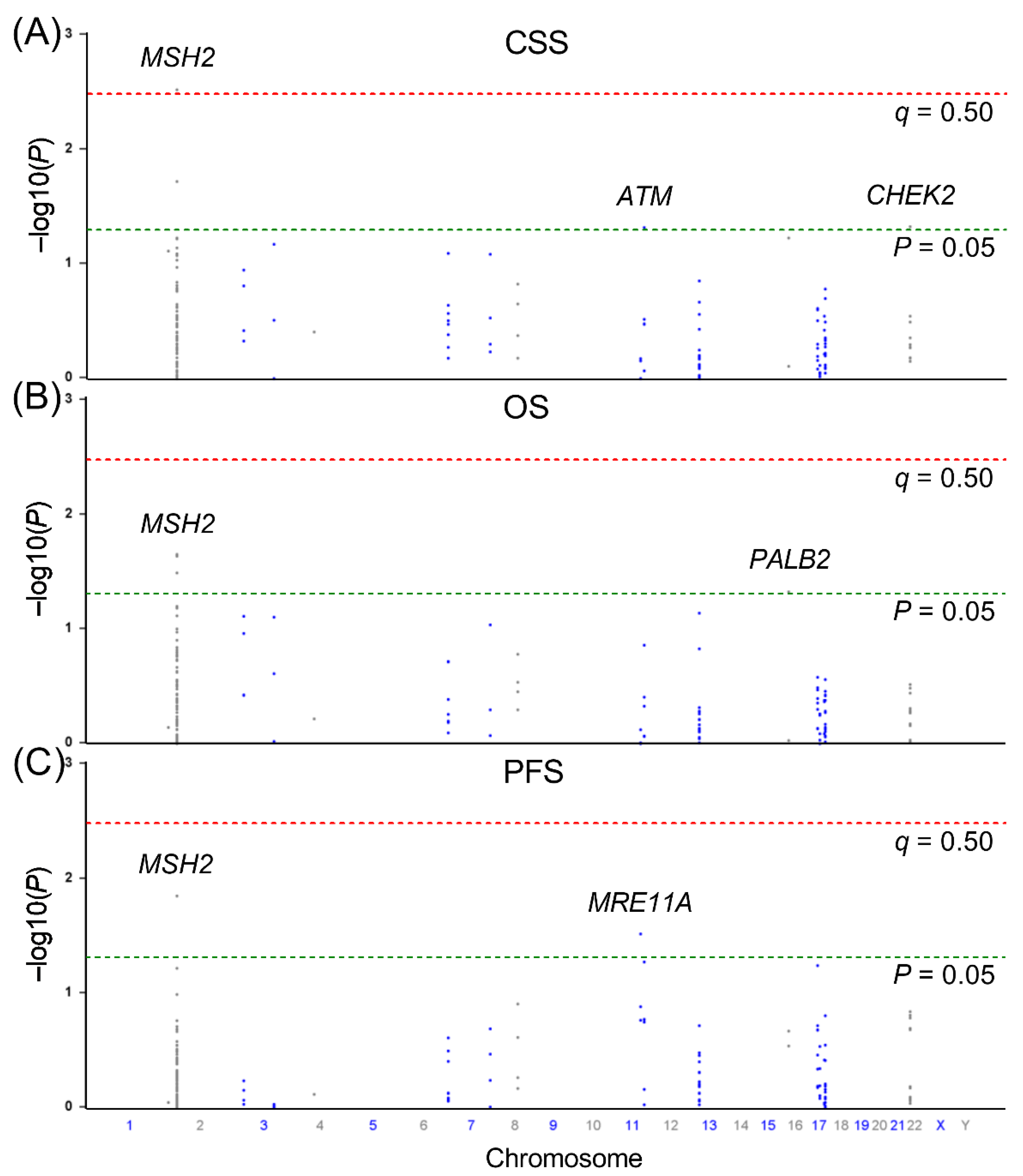
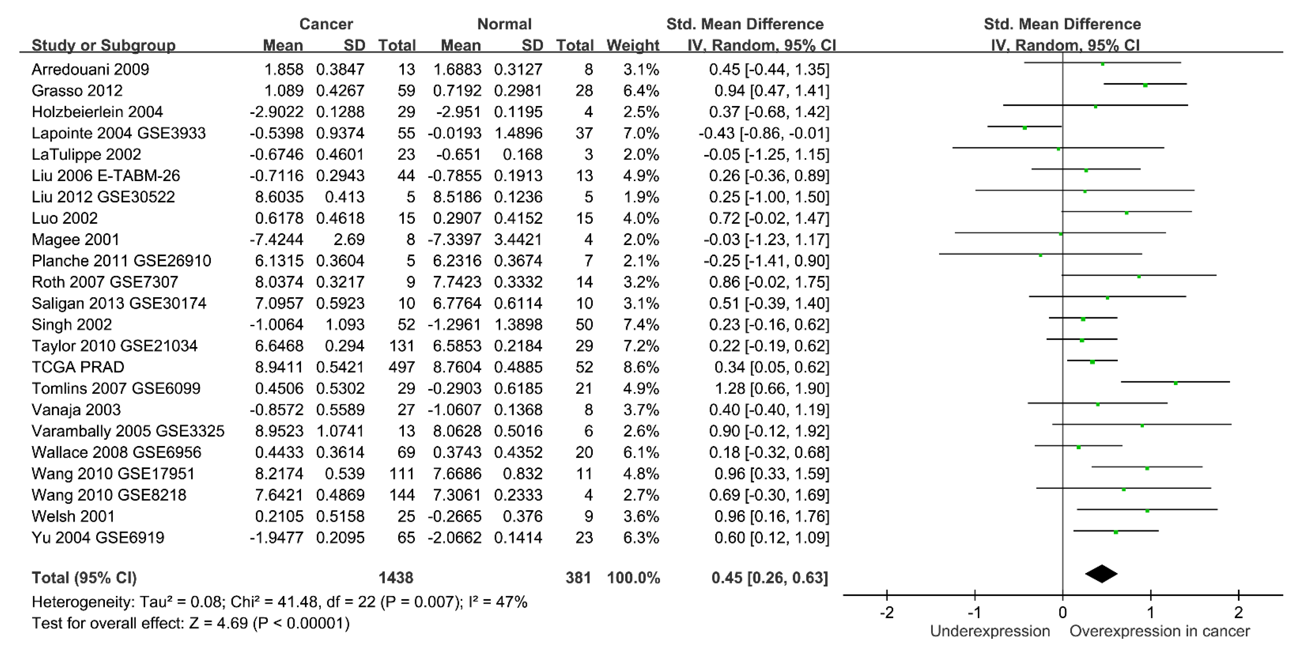
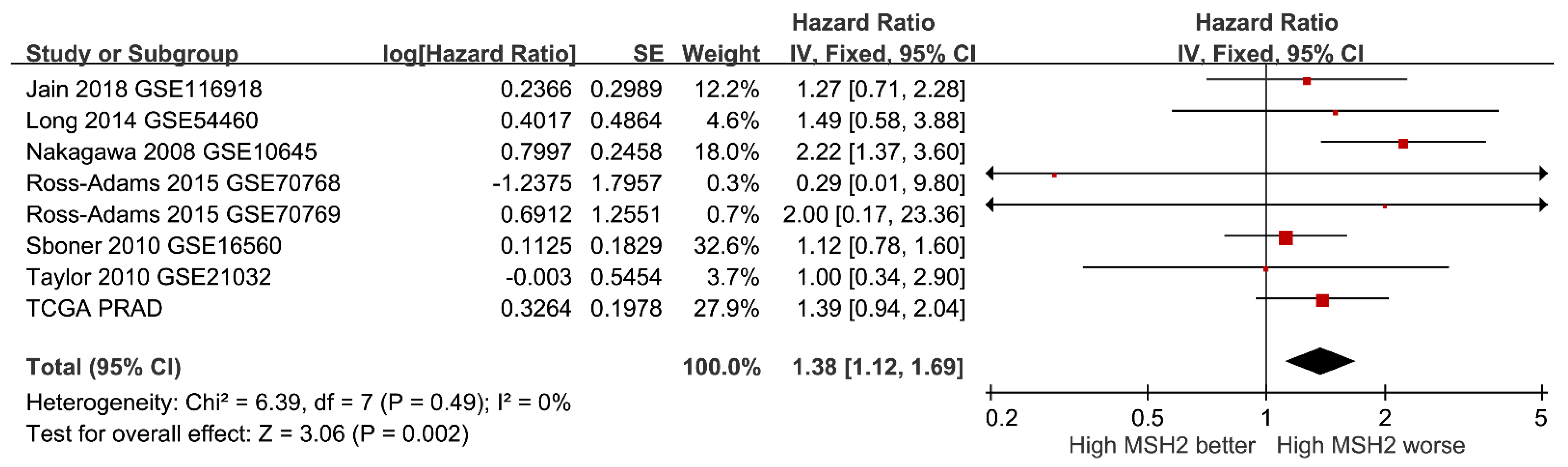
Univariate Cox regressions and log-rank tests were employed to compare differences in CSS, OS, and PFS among patients with diverse clinicopathological characteristics and SNP genotypes. Multivariate Cox regression was further performed to adjust the associations for age, stage, Gleason score at diagnosis, PSA at ADT initiation, PSA nadir, and time to PSA nadir. The false discovery rate (q Value) was used to adjust for multiple testing. The link between gene expression and tumour characteristics was assessed by Spearman correlation. Pooled standardised mean differences were used to assess the difference in gene expression between tumour and normal tissues, and the pooled hazard ratio (HR) was used to evaluate the relationship between gene expression and survival outcomes. If heterogeneity was absent from the included studies, a fixed-effect model was used to calculate pooled data; otherwise, the random-effect model was taken into account using RevMan 5.4.1 (Cochrane, London, UK). Other statistical analyses were conducted using Statistical Product and Service Solutions version 19.0.0 (IBM, Armonk, NY, USA). A two-sided p Value < 0.05 was considered significant.

3. Results

The present study included 630 patients who underwent ADT for prostate cancer, and whose characteristics are presented in Table 1. During a median follow-up of 165.8 months, 314 (49.8%) patients died from cancer, 414 (65.7%) died from another disease, and 518 (82.6%) exhibited disease progression. Age at diagnosis was only associated with OS and PFS. Five other clinicopathological covariates, including PSA, clinical stage, Gleason score at diagnosis, PSA nadir, and time to PSA nadir, were all significant predictors of CSS, OS, and PFS during ADT.
Univariate survival analysis on the association of 167 SNPs in the DDR pathway genes with CSS, OS, and PFS is summarised in Figure 1. Four SNPs (rs1400633 and rs4953519 in MSH2, rs4987981 in ATM, and rs9620817 in CHEK2) were associated with CSS, four SNPs (rs1400633, rs2303425, and rs13383462 in MSH2, and rs249954 in PALB2) were associated with OS, and two SNPs (rs11903353 in MSH2 and rs496797 in MRE11A) were associated with PFS (p < 0.05). After applying multiple testing correction (q < 0.50), only MSH2 rs1400633 was significantly associated with CSS. Each additional rs1400633 minor allele G was associated with a 22% decreased risk of cancer-specific mortality (HR = 0.78, 95% confidence interval [CI] = 0.65–0.92, p = 0.003, q = 0.492) (Table 2 and Figure 2A). This association remained significant after an adjustment for clinical predictors and coincided with a 25% decreased risk of cancer-specific mortality (adjusted HR = 0.75, 95% CI = 0.63–0.90, p = 0.002). Interestingly, MSH2 rs1400633 was also associated with OS in univariate and multivariate analyses (adjusted HR = 0.84, 95% CI = 0.72–0.98, p = 0.029) (Table 2 and Figure 2B).
The HaploReg analysis demonstrated that rs1400633 and several linked SNPs in linkage disequilibrium (r2 > 0.8) were located in functional promoters/enhancers with active histone marks and DNase hypersensitive sites, which made them more accessible to transcription factors in multiple tissues (Table 3). Additionally, these SNPs may alter the motif of some transcription regulators, which could affect MSH2 expression via alterations to chromatin remodelling during transcription. According to the GTEx database, rs1400633 risk allele C was associated with a significant increase in MSH2 mRNA expression in the brain nucleus accumbens (normalized effect size [NES] = 0.064, p = 0.035), and the same trend, positive NES, has also been observed in the prostate (NES = 0.10, p = 0.15) and several other types of tissues (Figure 3).
Finally, we assessed the clinical relevance of MSH2 expression in prostate cancer. A meta-analysis of 23 studies that included 1438 prostate cancer and 381 adjacent normal samples demonstrated elevated MSH2 expressions in prostate cancer (standardised mean difference = 0.45, 95% CI = 0.26–0.63, p < 0.001) (Figure 4). High MSH2 expression was significantly associated with a higher Gleason score and a more advanced tumour stage in the TCGA PRAD dataset (p < 0.001) (Figure 5). Furthermore, a meta-analysis of eight studies comprising 2071 patients suggested that a high expression of MSH2 was associated with a poor prognosis for prostate cancer (HR = 1.38, 95% CI = 1.12–1.69, p = 0.002) (Figure 6). These pooled results indicated that high MSH2 expression might play an important role in the development and progression of prostate cancer.

4. Discussion

In the present study, we explored the associations between 167 htSNPs of 18 DDR pathway genes and the efficacy of ADT. We report that MSH2 rs1400633 C > G was independently associated with both the CSS and OS of patients with advanced prostate cancer subjected to ADT. The bioinformatic analyses based on the GTEx database suggested that the risk allele C of rs1400633 was related to the increased MSH2 expression. Furthermore, a meta-analysis of 31 studies revealed that MSH2 expression was elevated in prostate cancer tissues and was significantly associated with worse patient prognosis.
The risk variant rs1400633 and its linked SNPs were found to be located in a regulatory region. Using a multivariate hidden Markov model to identify chromatin states from multiple histone modifications according to the HaploReg database, the region was classified as a functional promoter/enhancer in trophoblast, mesenchymal stem, placenta, lymphoblastoid, and mammary epithelial cells. Therefore, rs1400633 may affect gene expression by regulating the accessibility of DNA to the transcriptional machinery and transcription factors. Additionally, rs1400633 could disrupt the binding of some important transcription regulators, including the pancreas associated transcription factor 1a (PTF1A) and paired box 5 (PAX5). Paired box transcription factors play critical roles in early development, and the dysregulation of these genes is thought to contribute to neoplastic transformation, such as rhabdomyosarcoma and lymphoma [37]. Upregulation of PAX5 was found to induce the forkhead box P4 axis and promote tumorigenesis in prostate cancer [38]. Consistent with that, we found that the risk allele C of rs1400633 was associated with a significant increase in MSH2 mRNA expression in the brain nucleus accumbens and exhibited a similar trend in prostate tissues.
MSH2 is a core protein in the mammalian DNA mismatch repair pathway that is responsible for recognising and removing base pair mismatches introduced by replication errors. MSH2 forms two heterodimers with MSH3 or MSH6, and these protein complexes are responsible for the recognition of single nucleotide and small insertion/deletion mismatches [39]. Upon mismatch recognition, the complexes recruit the MLH1/PMS2 heterodimer to nick the 5′ of the mismatches via PMS2 endonuclease activity. Then, exonuclease 1 removes the mismatch, DNA polymerase δ 1 resynthesizes the strand, and DNA ligase 1 seals the nick, thereby completing the DNA mismatch repair [40,41]. Given the role of MSH2 in DNA repair, mutations in MSH2 are likely to affect this process, lead to genomic instability, and drive tumorigenesis. Conflicting data on the protein expression of MSH2 in prostate cancer exist; while some studies have suggested a link between loss of MSH2 expression and aggressive prostate cancer features [42,43], others have associated overexpression of MSH2 with prostate cancer progression and poor patient prognosis [44,45,46]. MSH2 deficiency has been described in 1.2–8.0% of prostate cancer patients and is more frequent in tumours with Gleason scores of 9–10 [42,43]. In contrast, several studies have demonstrated elevated MSH2 in prostate tumours, together with a higher Gleason score, as well as worse disease-free rates and overall survival [44,45,46], which is in line with our findings. Given that these studies have relied on protein expression in antibody-based approaches, the discrepancies might be associated with the methodology, sample size, and the specificity and sensitivity of the antibodies. Furthermore, the differentiation of functionally normal protein from abnormal protein remains difficult using immunohistochemical staining. Besides prostate cancer, MSH2 is often overexpressed in melanoma, gastric cancer, and oral squamous cell carcinoma, where it correlates with increased tumour aggressiveness and poor patient prognosis [47,48,49]. Increased expression of other DNA mismatch repair proteins, such as MSH6, MLH1, and PMS1, has also been observed in multiple cancers [50]. This phenomenon can be explained by the higher proliferation rate of cancer cells and their stronger need to correct mutations and DNA damage during DNA replication.
Two potential implications may translate our findings into clinical practice. First, genotyping patients prior to ADT could help identify patients at high risk of therapeutic failure, and these patients may benefit from a more aggressive treatment strategy, such as the addition of novel drugs to ADT. For example, the clinical trials supported the combination of docetaxel and ADT in the first-line treatment of metastatic castration-sensitive prostate cancer [51,52]. Second, this study provides evidence that the genetic variants in DDR pathways may influence the efficacy of ADT. Recently, poly (ADP-ribose) polymerase (PARP) inhibitors have been tried for the treatment of metastatic castration-resistant prostate cancer [53,54]. Because DDR-deficient cancers are unable to repair DNA double-strand breaks, they are sensitive to PARP inhibition, explaining the rationale for this approach. Moreover, androgen signalling has been found to regulate the expression and function of DDR genes in prostate cancer cells, which may justify the synergistic combination of ADT and PARP inhibitors [55,56]. Together, the above preclinical and clinical evidence offers a potential biological explanation for the observed association between MSH2 rs1400633 and prostate cancer progression; however, further experiments are required to identify the exact mechanism.
The major strength of this study is the availability of complete clinical information to adjust for confounding variables. Additional strength stems from our comprehensive evaluations of genetic variants in DDR pathways using the haplotype tagging approach. However, the results reported here are limited by the modest sample size and homogeneous Taiwanese study population, which reduces the ability to extrapolate our findings to other ethnic groups. Additional fine-mapping to narrow down the causal variants, followed by validation in independent and functional studies, is necessary.

5. Conclusions

The consistent risk associations across our genetic analyses and independent gene expression studies suggest that the genes involved in DDR, such as MSH2, could have a prognostic function in prostate cancer survival. For patients receiving ADT, MSH2 rs1400633 may serve as a novel prognostic biomarker or a clinical decision-making indicator. Future functional studies should elucidate the biological mechanisms defining how genetic variants can affect prostate cancer mortality.

Author Contributions

Conceptualization and methodology, H.-H.C., C.-H.L., S.-P.H. and B.-Y.B.; resources and funding acquisition, C.-Y.H., C.-C.Y., V.C.L., S.-P.H. and B.-Y.B.; formal analysis and investigation, H.-H.C., C.-H.L., S.-P.H. and B.-Y.B.; data curation and visualization, Y.-T.C., J.-H.G. and T.-L.L. All authors have read and agreed to the published version of the manuscript.

Funding

This work was supported by the Ministry of Science and Technology of Taiwan (grant nos: 108-2314-B-037-029, 108-2314-B-037-026-MY2, 108-2320-B-039-050-MY3, 109-2314-B-037-108-MY2, 109-2314-B-037-106-MY3, and 109-2320-B-037-007-MY3), the Kaohsiung Medical University Hospital (grant nos: KMUH105-5R42, KMUH108-8R53, KMUH108-8R55, KMUH109-9R63, and KMUH109-9R64), the Kaohsiung Medical University Research Center (grant no: KMU-TC108A04-4), and the China Medical University (grant nos: CMU109-SR-64, CMU109-MF-65, and CMU110-MF-59). The funders had no role in study design, data collection and analysis, decision to publish, or preparation of the manuscript.

Institutional Review Board Statement

The present study was conducted in accordance with the Declaration of Helsinki and was approved by the Institutional Review Board of Kaohsiung Medical University Hospital (protocol code: KMUHIRB-2013132; initial date of approval: 21 January 2014). The study was performed in compliance with the Good Clinical Practice guidelines.

Informed Consent Statement

Written informed consent was obtained from all subjects involved in the study.

Data Availability Statement

The data presented in this study are available on request from the corresponding author.

Acknowledgments

We thank Chao-Shih Chen for data analysis, and the National Center for Genome Medicine, Ministry of Science and Technology of Taiwan, for technical support. The results published here are based in part on data generated by the HaploReg, GTEx, and TCGA projects.

Conflicts of Interest

The authors declare no conflict of interest.

References

  1. Chinegwundoh, F.; Enver, M.; Lee, A.; Nargund, V.; Oliver, T.; Ben-Shlomo, Y. Risk and presenting features of prostate cancer amongst African-Caribbean, South Asian and European men in North-east London. BJU Int. 2006, 98, 1216–1220. [Google Scholar] [CrossRef] [PubMed]
  2. Mucci, L.A.; Hjelmborg, J.B.; Harris, J.R.; Czene, K.; Havelick, D.J.; Scheike, T.; Graff, R.E.; Holst, K.; Moller, S.; Unger, R.H.; et al. Familial Risk and Heritability of Cancer Among Twins in Nordic Countries. JAMA 2016, 315, 68–76. [Google Scholar] [CrossRef] [PubMed] [Green Version]
  3. Aune, D.; Navarro Rosenblatt, D.A.; Chan, D.S.; Vieira, A.R.; Vieira, R.; Greenwood, D.C.; Vatten, L.J.; Norat, T. Dairy products, calcium, and prostate cancer risk: A systematic review and meta-analysis of cohort studies. Am. J. Clin. Nutr. 2015, 101, 87–117. [Google Scholar] [CrossRef] [PubMed] [Green Version]
  4. Rowles, J.L., 3rd; Ranard, K.M.; Smith, J.W.; An, R.; Erdman, J.W., Jr. Increased dietary and circulating lycopene are associated with reduced prostate cancer risk: A systematic review and meta-analysis. Prostate Cancer Prostatic Dis. 2017, 20, 361–377. [Google Scholar] [CrossRef]
  5. Fletcher, S.A.; von Landenberg, N.; Cole, A.P.; Gild, P.; Choueiri, T.K.; Lipsitz, S.R.; Trinh, Q.D.; Kibel, A.S. Contemporary national trends in prostate cancer risk profile at diagnosis. Prostate Cancer Prostatic Dis. 2020, 23, 81–87. [Google Scholar] [CrossRef]
  6. Huggins, C.; Hodges, C.V. Studies on prostatic cancer. I. The effect of castration, of estrogen and androgen injection on serum phosphatases in metastatic carcinoma of the prostate. CA Cancer J. Clin. 1972, 22, 232–240. [Google Scholar] [CrossRef]
  7. Frank, S.; Nelson, P.; Vasioukhin, V. Recent advances in prostate cancer research: Large-scale genomic analyses reveal novel driver mutations and DNA repair defects. F1000Research 2018, 7, 1173. [Google Scholar] [CrossRef]
  8. Lockett, K.L.; Hall, M.C.; Clark, P.E.; Chuang, S.C.; Robinson, B.; Lin, H.Y.; Su, L.J.; Hu, J.J. DNA damage levels in prostate cancer cases and controls. Carcinogenesis 2006, 27, 1187–1193. [Google Scholar] [CrossRef] [Green Version]
  9. Miyake, H.; Hara, I.; Kamidono, S.; Eto, H. Oxidative DNA damage in patients with prostate cancer and its response to treatment. J. Urol. 2004, 171, 1533–1536. [Google Scholar] [CrossRef]
  10. Dronkert, M.L.; Kanaar, R. Repair of DNA interstrand cross-links. Mutat. Res. 2001, 486, 217–247. [Google Scholar] [CrossRef]
  11. Dall’Era, M.A.; McPherson, J.D.; Gao, A.C.; DeVere White, R.W.; Gregg, J.P.; Lara, P.N., Jr. Germline and somatic DNA repair gene alterations in prostate cancer. Cancer 2020, 126, 2980–2985. [Google Scholar] [CrossRef]
  12. Marshall, C.H.; Fu, W.; Wang, H.; Baras, A.S.; Lotan, T.L.; Antonarakis, E.S. Prevalence of DNA repair gene mutations in localized prostate cancer according to clinical and pathologic features: Association of Gleason score and tumor stage. Prostate Cancer Prostatic Dis. 2019, 22, 59–65. [Google Scholar] [CrossRef]
  13. Robinson, D.; Van Allen, E.M.; Wu, Y.M.; Schultz, N.; Lonigro, R.J.; Mosquera, J.M.; Montgomery, B.; Taplin, M.E.; Pritchard, C.C.; Attard, G.; et al. Integrative clinical genomics of advanced prostate cancer. Cell 2015, 161, 1215–1228. [Google Scholar] [CrossRef] [Green Version]
  14. Moro, L.; Arbini, A.A.; Yao, J.L.; di Sant’Agnese, P.A.; Marra, E.; Greco, M. Loss of BRCA2 promotes prostate cancer cell invasion through up-regulation of matrix metalloproteinase-9. Cancer Sci. 2008, 99, 553–563. [Google Scholar] [CrossRef]
  15. Kim, S.H.; Park, W.S.; Yun, S.I.; Joo, J.; Joung, J.Y.; Seo, H.K.; Chung, J.; Lee, G.K.; Lee, K.H. Overexpression of BRCA1 or BRCA2 in prostatectomy specimens is predictive of biochemical recurrence after radical prostatectomy. Histopathology 2016, 68, 673–679. [Google Scholar] [CrossRef] [PubMed]
  16. Leongamornlert, D.A.; Saunders, E.J.; Wakerell, S.; Whitmore, I.; Dadaev, T.; Cieza-Borrella, C.; Benafif, S.; Brook, M.N.; Donovan, J.L.; Hamdy, F.C.; et al. Germline DNA Repair Gene Mutations in Young-onset Prostate Cancer Cases in the UK: Evidence for a More Extensive Genetic Panel. Eur. Urol. 2019, 76, 329–337. [Google Scholar] [CrossRef] [Green Version]
  17. Na, R.; Zheng, S.L.; Han, M.; Yu, H.; Jiang, D.; Shah, S.; Ewing, C.M.; Zhang, L.; Novakovic, K.; Petkewicz, J.; et al. Germline Mutations in ATM and BRCA1/2 Distinguish Risk for Lethal and Indolent Prostate Cancer and are Associated with Early Age at Death. Eur. Urol. 2017, 71, 740–747. [Google Scholar] [CrossRef] [Green Version]
  18. Castro, E.; Goh, C.; Olmos, D.; Saunders, E.; Leongamornlert, D.; Tymrakiewicz, M.; Mahmud, N.; Dadaev, T.; Govindasami, K.; Guy, M.; et al. Germline BRCA mutations are associated with higher risk of nodal involvement, distant metastasis, and poor survival outcomes in prostate cancer. J. Clin. Oncol. 2013, 31, 1748–1757. [Google Scholar] [CrossRef] [Green Version]
  19. Leongamornlert, D.; Saunders, E.; Dadaev, T.; Tymrakiewicz, M.; Goh, C.; Jugurnauth-Little, S.; Kozarewa, I.; Fenwick, K.; Assiotis, I.; Barrowdale, D.; et al. Frequent germline deleterious mutations in DNA repair genes in familial prostate cancer cases are associated with advanced disease. Br. J. Cancer 2014, 110, 1663–1672. [Google Scholar] [CrossRef] [PubMed]
  20. Marshall, C.H.; Sokolova, A.O.; McNatty, A.L.; Cheng, H.H.; Eisenberger, M.A.; Bryce, A.H.; Schweizer, M.T.; Antonarakis, E.S. Differential Response to Olaparib Treatment Among Men with Metastatic Castration-resistant Prostate Cancer Harboring BRCA1 or BRCA2 Versus ATM Mutations. Eur. Urol. 2019, 76, 452–458. [Google Scholar] [CrossRef] [PubMed] [Green Version]
  21. Mateo, J.; Carreira, S.; Sandhu, S.; Miranda, S.; Mossop, H.; Perez-Lopez, R.; Nava Rodrigues, D.; Robinson, D.; Omlin, A.; Tunariu, N.; et al. DNA-Repair Defects and Olaparib in Metastatic Prostate Cancer. N. Engl. J. Med. 2015, 373, 1697–1708. [Google Scholar] [CrossRef] [PubMed]
  22. Damaraju, S.; Murray, D.; Dufour, J.; Carandang, D.; Myrehaug, S.; Fallone, G.; Field, C.; Greiner, R.; Hanson, J.; Cass, C.E.; et al. Association of DNA repair and steroid metabolism gene polymorphisms with clinical late toxicity in patients treated with conformal radiotherapy for prostate cancer. Clin. Cancer Res. 2006, 12, 2545–2554. [Google Scholar] [CrossRef] [PubMed] [Green Version]
  23. Gao, R.; Price, D.K.; Dahut, W.L.; Reed, E.; Figg, W.D. Genetic polymorphisms in XRCC1 associated with radiation therapy in prostate cancer. Cancer Biol. Ther. 2010, 10, 13–18. [Google Scholar] [CrossRef] [PubMed] [Green Version]
  24. Zanusso, C.; Bortolus, R.; Dreussi, E.; Polesel, J.; Montico, M.; Cecchin, E.; Gagno, S.; Rizzolio, F.; Arcicasa, M.; Novara, G.; et al. Impact of DNA repair gene polymorphisms on the risk of biochemical recurrence after radiotherapy and overall survival in prostate cancer. Oncotarget 2017, 8, 22863–22875. [Google Scholar] [CrossRef] [PubMed] [Green Version]
  25. Little, J.; Wilson, B.; Carter, R.; Walker, K.; Santaguida, P.; Tomiak, E.; Beyene, J.; Usman Ali, M.; Raina, P. Multigene panels in prostate cancer risk assessment: A systematic review. Genet. Med. 2016, 18, 535–544. [Google Scholar] [CrossRef] [Green Version]
  26. Bao, B.Y.; Pao, J.B.; Huang, C.N.; Pu, Y.S.; Chang, T.Y.; Lan, Y.H.; Lu, T.L.; Lee, H.Z.; Juang, S.H.; Chen, L.M.; et al. Polymorphisms inside microRNAs and microRNA target sites predict clinical outcomes in prostate cancer patients receiving androgen-deprivation therapy. Clin. Cancer Res. 2011, 17, 928–936. [Google Scholar] [CrossRef] [Green Version]
  27. Geng, J.H.; Lin, V.C.; Yu, C.C.; Huang, C.Y.; Yin, H.L.; Chang, T.Y.; Lu, T.L.; Huang, S.P.; Bao, B.Y. Inherited Variants in Wnt Pathway Genes Influence Outcomes of Prostate Cancer Patients Receiving Androgen Deprivation Therapy. Int. J. Mol. Sci. 2016, 17, 1970. [Google Scholar] [CrossRef] [Green Version]
  28. Gong, J.; Posadas, E.; Bhowmick, N.; Kim, H.; Daskivich, T.; Gupta, A.; Sandler, H.; Kamrava, M.; Zumsteg, Z.; Freedland, S.; et al. Integrating PARP Inhibitors Into Advanced Prostate Cancer Therapeutics. Oncology 2021, 35, 119–125. [Google Scholar] [CrossRef]
  29. Genomes Project, C.; Abecasis, G.R.; Auton, A.; Brooks, L.D.; De Pristo, M.A.; Durbin, R.M.; Handsaker, R.E.; Kang, H.M.; Marth, G.T.; McVean, G.A. An integrated map of genetic variation from 1092 human genomes. Nature 2012, 491, 56–65. [Google Scholar]
  30. de Bakker, P.I.; Yelensky, R.; Pe’er, I.; Gabriel, S.B.; Daly, M.J.; Altshuler, D. Efficiency and power in genetic association studies. Nat. Genet. 2005, 37, 1217–1223. [Google Scholar] [CrossRef]
  31. Bao, B.Y.; Pao, J.B.; Huang, C.N.; Pu, Y.S.; Chang, T.Y.; Lan, Y.H.; Lu, T.L.; Lee, H.Z.; Chen, L.M.; Ting, W.C.; et al. Significant associations of prostate cancer susceptibility variants with survival in patients treated with androgen-deprivation therapy. Int. J. Cancer 2012, 130, 876–884. [Google Scholar] [CrossRef] [PubMed]
  32. Ward, L.D.; Kellis, M. HaploReg v4: Systematic mining of putative causal variants, cell types, regulators and target genes for human complex traits and disease. Nucleic Acids Res. 2016, 44, D877–D881. [Google Scholar] [CrossRef] [PubMed]
  33. Consortium, G.T. The Genotype-Tissue Expression (GTEx) project. Nat. Genet. 2013, 45, 580–585. [Google Scholar]
  34. The Cancer Genome Atlas (TCGA) Research Network. Comprehensive genomic characterization defines human glioblastoma genes and core pathways. Nature 2008, 455, 1061–1068. [Google Scholar] [CrossRef]
  35. Rhodes, D.R.; Yu, J.; Shanker, K.; Deshpande, N.; Varambally, R.; Ghosh, D.; Barrette, T.; Pandey, A.; Chinnaiyan, A.M. ONCOMINE: A cancer microarray database and integrated data-mining platform. Neoplasia 2004, 6, 1–6. [Google Scholar] [CrossRef] [Green Version]
  36. Park, S.J.; Yoon, B.H.; Kim, S.K.; Kim, S.Y. GENT2: An updated gene expression database for normal and tumor tissues. BMC Med. Genom. 2019, 12, 101. [Google Scholar] [CrossRef]
  37. Barr, F.G. Chromosomal translocations involving paired box transcription factors in human cancer. Int. J. Biochem. Cell Biol. 1997, 29, 1449–1461. [Google Scholar] [CrossRef]
  38. Wu, X.; Xiao, Y.; Zhou, Y.; Zhou, Z.; Yan, W. LncRNA FOXP4-AS1 is activated by PAX5 and promotes the growth of prostate cancer by sequestering miR-3184-5p to upregulate FOXP4. Cell Death Dis. 2019, 10, 472. [Google Scholar] [CrossRef] [Green Version]
  39. Acharya, S.; Wilson, T.; Gradia, S.; Kane, M.F.; Guerrette, S.; Marsischky, G.T.; Kolodner, R.; Fishel, R. hMSH2 forms specific mispair-binding complexes with hMSH3 and hMSH6. Proc. Natl. Acad. Sci. USA 1996, 93, 13629–13634. [Google Scholar] [CrossRef] [Green Version]
  40. Liu, D.; Frederiksen, J.H.; Liberti, S.E.; Lutzen, A.; Keijzers, G.; Pena-Diaz, J.; Rasmussen, L.J. Human DNA polymerase delta double-mutant D316A;E318A interferes with DNA mismatch repair in vitro. Nucleic Acids Res. 2017, 45, 9427–9440. [Google Scholar] [CrossRef] [Green Version]
  41. Nielsen, F.C.; Jager, A.C.; Lutzen, A.; Bundgaard, J.R.; Rasmussen, L.J. Characterization of human exonuclease 1 in complex with mismatch repair proteins, subcellular localization and association with PCNA. Oncogene 2004, 23, 1457–1468. [Google Scholar] [CrossRef] [PubMed] [Green Version]
  42. Albero-Gonzalez, R.; Hernandez-Llodra, S.; Juanpere, N.; Lorenzo, M.; Lloret, A.; Segales, L.; Duran, X.; Fumado, L.; Cecchini, L.; Lloreta-Trull, J. Immunohistochemical expression of mismatch repair proteins (MSH2, MSH6, MLH1, and PMS2) in prostate cancer: Correlation with grade groups (WHO 2016) and ERG and PTEN status. Virchows Arch. 2019, 475, 223–231. [Google Scholar] [CrossRef] [PubMed]
  43. Guedes, L.B.; Antonarakis, E.S.; Schweizer, M.T.; Mirkheshti, N.; Almutairi, F.; Park, J.C.; Glavaris, S.; Hicks, J.; Eisenberger, M.A.; De Marzo, A.M.; et al. MSH2 Loss in Primary Prostate Cancer. Clin. Cancer Res. 2017, 23, 6863–6874. [Google Scholar] [CrossRef] [Green Version]
  44. Burger, M.; Denzinger, S.; Hammerschmied, C.G.; Tannapfel, A.; Obermann, E.C.; Wieland, W.F.; Hartmann, A.; Stoehr, R. Elevated microsatellite alterations at selected tetranucleotides (EMAST) and mismatch repair gene expression in prostate cancer. J. Mol. Med. 2006, 84, 833–841. [Google Scholar] [CrossRef]
  45. Prtilo, A.; Leach, F.S.; Markwalder, R.; Kappeler, A.; Burkhard, F.C.; Cecchini, M.G.; Studer, U.E.; Thalmann, G.N. Tissue microarray analysis of hMSH2 expression predicts outcome in men with prostate cancer. J. Urol. 2005, 174, 1814–1818. [Google Scholar] [CrossRef]
  46. Velasco, A.; Hewitt, S.M.; Albert, P.S.; Hossein, M.; Rosenberg, H.; Martinez, C.; Sagalowsky, A.I.; McConnell, J.D.; Marston, W.; Leach, F.S. Differential expression of the mismatch repair gene hMSH2 in malignant prostate tissue is associated with cancer recurrence. Cancer 2002, 94, 690–699. [Google Scholar] [CrossRef]
  47. Kauffmann, A.; Rosselli, F.; Lazar, V.; Winnepenninckx, V.; Mansuet-Lupo, A.; Dessen, P.; van den Oord, J.J.; Spatz, A.; Sarasin, A. High expression of DNA repair pathways is associated with metastasis in melanoma patients. Oncogene 2008, 27, 565–573. [Google Scholar] [CrossRef] [PubMed] [Green Version]
  48. Li, M.; Liu, L.; Wang, Z.; Wang, L.; Liu, Z.; Xu, G.; Lu, S. Overexpression of hMSH2 and hMLH1 protein in certain gastric cancers and their surrounding mucosae. Oncol. Rep. 2008, 19, 401–406. [Google Scholar] [CrossRef] [Green Version]
  49. Wagner, V.P.; Webber, L.P.; Salvadori, G.; Meurer, L.; Fonseca, F.P.; Castilho, R.M.; Squarize, C.H.; Vargas, P.A.; Martins, M.D. Overexpression of MutSalpha Complex Proteins Predicts Poor Prognosis in Oral Squamous Cell Carcinoma. Medicine 2016, 95, e3725. [Google Scholar] [CrossRef]
  50. Wilczak, W.; Rashed, S.; Hube-Magg, C.; Kluth, M.; Simon, R.; Buscheck, F.; Clauditz, T.S.; Grupp, K.; Minner, S.; Tsourlakis, M.C.; et al. Up-regulation of mismatch repair genes MSH6, PMS2 and MLH1 parallels development of genetic instability and is linked to tumor aggressiveness and early PSA recurrence in prostate cancer. Carcinogenesis 2017, 38, 19–27. [Google Scholar] [CrossRef]
  51. James, N.D.; Sydes, M.R.; Clarke, N.W.; Mason, M.D.; Dearnaley, D.P.; Spears, M.R.; Ritchie, A.W.; Parker, C.C.; Russell, J.M.; Attard, G.; et al. Addition of docetaxel, zoledronic acid, or both to first-line long-term hormone therapy in prostate cancer (STAMPEDE): Survival results from an adaptive, multiarm, multistage, platform randomised controlled trial. Lancet 2016, 387, 1163–1177. [Google Scholar] [CrossRef] [Green Version]
  52. Sweeney, C.J.; Chen, Y.H.; Carducci, M.; Liu, G.; Jarrard, D.F.; Eisenberger, M.; Wong, Y.N.; Hahn, N.; Kohli, M.; Cooney, M.M.; et al. Chemohormonal Therapy in Metastatic Hormone-Sensitive Prostate Cancer. N. Engl. J. Med. 2015, 373, 737–746. [Google Scholar] [CrossRef] [PubMed]
  53. Abida, W.; Campbell, D.; Patnaik, A.; Shapiro, J.D.; Sautois, B.; Vogelzang, N.J.; Voog, E.G.; Bryce, A.H.; McDermott, R.; Ricci, F.; et al. Non-BRCA DNA Damage Repair Gene Alterations and Response to the PARP Inhibitor Rucaparib in Metastatic Castration-Resistant Prostate Cancer: Analysis From the Phase II TRITON2 Study. Clin. Cancer Res. 2020, 26, 2487–2496. [Google Scholar] [CrossRef] [Green Version]
  54. de Bono, J.; Mateo, J.; Fizazi, K.; Saad, F.; Shore, N.; Sandhu, S.; Chi, K.N.; Sartor, O.; Agarwal, N.; Olmos, D.; et al. Olaparib for Metastatic Castration-Resistant Prostate Cancer. N. Engl. J. Med. 2020, 382, 2091–2102. [Google Scholar] [CrossRef] [PubMed]
  55. Goodwin, J.F.; Schiewer, M.J.; Dean, J.L.; Schrecengost, R.S.; de Leeuw, R.; Han, S.; Ma, T.; Den, R.B.; Dicker, A.P.; Feng, F.Y.; et al. A hormone-DNA repair circuit governs the response to genotoxic insult. Cancer Discov. 2013, 3, 1254–1271. [Google Scholar] [CrossRef] [Green Version]
  56. Thompson, T.C.; Li, L.; Broom, B.M. Combining enzalutamide with PARP inhibitors: Pharmaceutically induced BRCAness. Oncotarget 2017, 8, 93315–93316. [Google Scholar] [CrossRef]
Figure 1. Manhattan plots of the associations (−log10 (p) Values; Y-axis) between 167 single nucleotide polymorphisms (SNPs; X-axis by chromosome positions) in 18 DNA repair pathway genes and (A) cancer-specific survival, (B) overall survival, and (C) progression-free survival among patients with prostate cancer treated with androgen deprivation therapy. Labelled genes contain associated SNPs with p < 0.05. The red horizontal line represents the suggestive significance threshold of q = 0.50, and the green horizontal line represents the nominal significance threshold of p = 0.05.
Cancers 14 00223 g001
Figure 2. Kaplan-Meier curves of (A) cancer-specific survival and (B) overall survival after androgen deprivation therapy (ADT) for MSH2 rs1400633 genotypes. Values in brackets represent the number of patients.
Cancers 14 00223 g002
Figure 3. Correlation between rs1400633 genotypes and MSH2 expression in different organs. Values were calculated using prostate, brain nucleus accumbens, frontal cortex, amygdala, hippocampus, and tibial artery tissue data from the Genotype-Tissue Expression project. Values in brackets represent the number of samples. NES, normalized effect size.
Cancers 14 00223 g003
Figure 4. Meta-analysis of MSH2 expression between normal and prostate cancer tissues in 23 independent studies. MSH2 expression is significantly upregulated in prostate cancer. Abbreviations: TCGA PRAD, The Cancer Genome Atlas Prostate Adenocarcinoma; SD, standard deviation; IV, inverse variance; CI, confidence interval; Std, standardized; df, degrees of freedom.
Cancers 14 00223 g004
Figure 5. Correlation of MSH2 expression with (A) Gleason score and (B) tumour stage in The Cancer Genome Atlas Prostate Adenocarcinoma dataset. Rho, Spearman’s rank correlation coefficient. Values in brackets represent the number of samples.
Cancers 14 00223 g005
Figure 6. Meta-analysis of eight studies evaluating the prognostic role of MSH2 in prostate cancer. SE, standard error; IV, inverse variance; CI, confidence interval; TCGA PRAD, The Cancer Genome Atlas Prostate Adenocarcinoma.
Cancers 14 00223 g006
Table 1. Clinicopathological characteristics of the study population.
Characteristics CSS aOS aPFS a
HR (95% CI)pHR (95% CI)pHR (95% CI)p
Total, n (%)630314 (49.8) 414 (65.7) 518 (82.6)
Age at diagnosis, years
Median (IQR)73 (67–79)1.012 (0.999–1.026)0.0781.029 (1.016–1.041)<0.0010.980 (0.970–0.990)<0.001
PSA at ADT initiation, ng/mL
Median (IQR)34.5 (11.25–129)1.000 (1.000–1.000)<0.0011.000 (1.000–1.000)<0.0011.000 (1.000–1.000)0.001
PSA nadir, ng/mL
Median (IQR)0.14 (0.01–1.16)1.002 (1.001–1.002)<0.0011.002 (1.001–1.002)<0.0011.001 (1.000–1.001)0.019
Time to PSA nadir, months
Median (IQR)11 (5–20)0.972 (0.962–0.981)<0.0010.982 (0.975–0.990)<0.0010.956 (0.948–0.963)<0.001
Clinical stage at diagnosis
T1/T2, n (%)187 (29.9)Reference Reference Reference
T3/T4/N1, n (%)205 (32.8)1.080 (0.785–1.488)0.6361.065 (0.818–1.386)0.6420.860 (0.686–1.078)0.190
M1, n (%)233 (37.3)3.096 (2.334–4.106)<0.0012.508 (1.968–3.194)<0.0011.406 (1.136–1.739)0.002
Gleason score at diagnosis
2–6, n (%)188 (30.6)Reference Reference Reference
7, n (%)194 (31.6)1.063 (0.783–1.443)0.6941.064 (0.820–1.381)0.6401.162 (0.929–1.455)0.189
8–10, n (%)232 (37.8)2.144 (1.629–2.820)<0.0011.957 (1.542–2.484)<0.0011.483 (1.194–1.841)<0.001
Abbreviations: CSS, cancer-specific survival; OS, overall survival; PFS, progression-free survival; IQR, interquartile range; PSA, prostate-specific antigen. a With a median follow-up of 165.8 months. Subtotals do not sum to 630 due to missing data.
Table 2. Association of MSH2 rs1400633 with cancer-specific survival and overall survival in prostate cancer patients receiving androgen deprivation therapy.
GenotypeFrequencyCSSOS
HR (95% CI)pHR (95% CI) apaHR (95% CI)pHR (95% CI) apa
CC/CG/GG244/269/840.78 (0.65–0.92)0.0030.75 (0.63–0.90)0.0020.84 (0.73–0.98)0.0230.84 (0.72–0.98)0.029
Abbreviations: CSS, cancer-specific survival; OS, overall survival; HR, hazard ratio; CI, confidence interval. a Adjustment for age, stage, Gleason score at diagnosis, prostate-specific antigen at androgen deprivation therapy initiation, prostate-specific antigen nadir, and time to prostate-specific antigen nadir.
Table 3. Functional annotation of MSH2 rs1400633 and its linked variants.
VariantPositionLD (r2)Reference AlleleAlternate AlleleASN FrquencyPromoter Histone MarksEnhancer Histone MarksDNaseProteins BoundMotifs Changed
rs1882450476474640.82GC0.64 2 altered motifs
rs2091750476478220.8TG0.65 5 altered motifs
rs1400633476568631GC0.65 4 tissues5 tissues3 bound proteins2 altered motifs
rs2969775476583371GT0.65 2 tissues 7 altered motifs
rs2969774476616840.94TC0.65 2 tissues 2 altered motifs
rs2705766476631920.95TC0.662 tissues3 tissues1 tissue 2 altered motifs
rs2705767476648660.92GA0.65 2 tissues 1 bound protein5 altered motifs
rs2944783476658600.92GC0.65 8 altered motifs
rs2969772476673330.9AG0.65
Abbreviations: LD, linkage disequilibrium; ASN, Asia.
Publisher’s Note: MDPI stays neutral with regard to jurisdictional claims in published maps and institutional affiliations.

