
Effects of Red Blood Cell Transfusions on Distant Metastases of Oral Squamous Cell Carcinomas

 , ,
, ,  ,
,
Abstract
:Simple Summary
Abstract
1. Introduction
2. Materials and Methods
2.1. Study Group
2.2. Data Acquisition
2.3. Statistical Analysis
3. Results
3.1. Study Group Characteristics
3.2. Univariate Analysis
3.3. Multivariate Analysis
4. Discussion
5. Conclusions
Author Contributions
Funding
Institutional Review Board Statement
Informed Consent Statement
Data Availability Statement
Acknowledgments
Conflicts of Interest
References
- The Selection and Use of Essential Medicines: Report of the WHO Expert Committee on Selection and Use of Essential Medicines; WHO Technical Report Series; Report No.: 1021; WHO Expert Committee (Ed.) World Health Organization: Geneva, Italy, 2019; p. 681. Available online: https://apps.who.int/ (accessed on 11 August 2021).
- Perrotta, P.L.; Snyder, E.L. Non-infectious complications of transfusion therapy. Blood Rev. 2001, 15, 69–83. [Google Scholar] [CrossRef]
- Vamvakas, E.C.; Blajchman, M.A. Transfusion-related immunomodulation (TRIM): An update. Blood Rev. 2007, 21, 327–348. [Google Scholar] [CrossRef]
- Kleinman, S.H.; Busch, M.P. The risks of transfusion-transmitted infection: Direct estimation and mathematical modelling. Best Pract. Res. Clin. Haematol. 2000, 13, 631–649. [Google Scholar] [CrossRef]
- Opelz, G.; Sengar, D.P.; Mickey, M.R.; Terasaki, P.I. Effect of blood transfusions on subsequent kidney transplants. Transplant. Proc. 1973, 5, 253–259. [Google Scholar]
- Opelz, G.; Terasaki, P.I. Improvement of Kidney-Graft Survival with Increased Numbers of Blood Transfusions. N. Engl. J. Med. 1978, 299, 799–803. [Google Scholar] [CrossRef]
- Babcock, G.F.; Alexander, J.W. The effects of blood transfusion on cytokine production by TH1 and TH2 lymphocytes in the mouse. Transplantation 1996, 61, 465–468. [Google Scholar] [CrossRef]
- Blajchman, M.A.; Bardossy, L.; Carmen, R.; Sastry, A.; Singal, D.P. Allogeneic blood transfusion-induced enhancement of tumor growth: Two animal models showing amelioration by leukodepletion and passive transfer using spleen cells. Blood 1993, 81, 1880–1882. [Google Scholar] [CrossRef] [Green Version]
- Mincheff, M.S.; Meryman, H.T.; Kapoor, V.; Alsop, P.; Wötzel, M. Blood transfusion and immunomodulation: A possible mechanism. Vox Sang. 1993, 65, 18–24. [Google Scholar] [PubMed]
- Buelow, R.; Burlingham, W.J.; Clayberger, C. Immunomodulation by soluble HLA class I. Transplantation 1995, 59, 649–654. [Google Scholar] [CrossRef] [PubMed]
- Krensky, A.M.; Clayberger, C. Structure of HLA Molecules and Immunosuppressive Effects of HLA Derived Peptides. Int. Rev. Immunol. 1996, 13, 173–185. [Google Scholar] [CrossRef] [PubMed]
- Magee, C.C.; Sayegh, M.H. Peptide-mediated immunosuppression. Curr. Opin. Immunol. 1997, 9, 669–675. [Google Scholar] [CrossRef]
- Farrugia, A.; Vamvakas, E. Toward a patient-based paradigm for blood transfusion. J. Blood Med. 2014, 5, 5–13. [Google Scholar] [CrossRef] [PubMed] [Green Version]
- Paul-Ehrlich-Institut. Bekanntmachung über die Zulassung und Registrierung von Arzneimitteln Leukozytendepletion von Blutkomponenten zur Transfusion (Anhörung zur Beabsichtigten Anordnung der Leukozytendepletion) Abwehr von Arzneimittelrisiken Stufe 2 (vom 12. Mai 2000). 2000. Available online: https://www.pei.de/ (accessed on 3 March 2020).
- van de Watering, L.M.G.; Hermans, J.; Houbiers, J.G.A.; van den Broek, P.J.; Bouter, H.; Boer, F.; Harvey, M.S.; Huysmans, H.A.; Brand, A. Beneficial Effects of Leukocyte Depletion of Transfused Blood on Postoperative Complications in Patients Undergoing Cardiac Surgery. Circulation 1998, 97, 562–568. [Google Scholar] [CrossRef] [PubMed]
- Spahn, D.R. Strategies for transfusion therapy. Best Pract. Res. Clin. Anaesthesiol. 2004, 18, 661–673. [Google Scholar] [CrossRef] [PubMed]
- Blajchman, M.A. Allogeneic blood transfusions, immunomodulation, and postoperative bacterial infection: Do we have the answers yet? Transfusion 1997, 37, 121–125. [Google Scholar] [CrossRef]
- Chang, H.; Hall, G.A.; Geerts, W.H.; Greenwood, C.; McLeod, R.S.; Sher, G.D. Allogeneic Red Blood Cell Transfusion Is an Independent Risk Factor for the Development of Postoperative Bacterial Infection. Vox Sang. 2000, 78, 13–18. [Google Scholar] [CrossRef]
- Jensen, L.S.; Andersen, A.J.; Christiansen, P.M.; Hokland, P.; Juhl, C.O.; Madsen, G.; Mortensen, J.; Møller-Nielsen, C.; Hanberg-Sørensen, F.; Hokland, M. Postoperative infection and natural killer cell function following blood transfusion in patients undergoing elective colorectal surgery. BJS 1992, 79, 513–516. [Google Scholar] [CrossRef]
- Talukder, Y.; Stillwell, A.P.; Siu, S.K.; Ho, Y.-H. Comparing Survival and Recurrence in Curative Stage I to III Colorectal Cancer in Transfused and Nontransfused Patients. Int. Surg. 2014, 99, 8–16. [Google Scholar] [CrossRef]
- Luan, H.; Ye, F.; Wu, L.; Zhou, Y.; Jiang, J. Perioperative blood transfusion adversely affects prognosis after resection of lung cancer: A systematic review and a meta-analysis. BMC Surg. 2014, 14, 34. [Google Scholar] [CrossRef] [Green Version]
- Squires, M.H.; Kooby, D.A.; Poultsides, G.A.; Weber, S.M.; Bloomston, M.; Fields, R.C.; Pawlik, T.M.; Votanopoulos, K.I.; Schmidt, C.R.; Ejaz, A.; et al. Effect of Perioperative Transfusion on Recurrence and Survival after Gastric Cancer Resection: A 7-Institution Analysis of 765 Patients from the US Gastric Cancer Collaborative. J. Am. Coll. Surg. 2015, 221, 767–777. [Google Scholar] [CrossRef]
- Sarode, G.; Maniyar, N.; Sarode, S.C.; Jafer, M.; Patil, S.; Awan, K.H. Epidemiologic aspects of oral cancer. Disease-a-Month 2020, 66, 100988. [Google Scholar] [CrossRef]
- Bennardo, L.; Bennardo, F.; Giudice, A.; Passante, M.; Dastoli, S.; Morrone, P.; Provenzano, E.; Patruno, C.; Nisticò, S.P. Local Chemotherapy as an Adjuvant Treatment in Unresectable Squamous Cell Carcinoma: What Do We Know So Far? Curr. Oncol. 2021, 28, 2317–2325. [Google Scholar] [CrossRef] [PubMed]
- Pentangelo, G.; Nisticò, S.P.; Provenzano, E.; Cisale, G.Y.; Bennardo, L. Topical 5% Imiquimod Sequential to Surgery for HPV-Related Squamous Cell Carcinoma of the Lip. Medicina 2021, 57, 563. [Google Scholar] [CrossRef]
- Brandenburg, L.S.; Schwarz, S.J.; Weingart, J.V.; Metzger, M.C.; Fuessinger, M.A.; Ermer, M.A. Do Red Blood Cell Transfusions Influence Long-Term Outcomes in Patients Undergoing Primary Surgery for Oral Squamous Cell Carcinoma? J. Oral Maxillofac. Surg. 2021, 79, 1570–1579. [Google Scholar] [CrossRef] [PubMed]
- Szakmany, T.; Dodd, M.; Dempsey, G.A.; Lowe, D.; Brown, J.S.; Vaughan, E.D.; Rogers, S.N. The influence of allogenic blood transfusion in patients having free-flap primary surgery for oral and oropharyngeal squamous cell carcinoma. Br. J. Cancer 2006, 94, 647–653. [Google Scholar] [CrossRef] [Green Version]
- Fenner, M.; Vairaktaris, E.; Nkenke, E.; Weisbach, V.; Neukam, F.W.; Radespiel-Tröger, M. Prognostic impact of blood transfusion in patients undergoing primary surgery and free-flap reconstruction for oral squamous cell carcinoma. Cancer 2009, 115, 1481–1488. [Google Scholar] [CrossRef]
- Baumeister, P.; Canis, M.; Reiter, M. Preoperative anemia and perioperative blood transfusion in head and neck squamous cell carcinoma. PLoS ONE 2018, 13, e0205712. Available online: https://www.ncbi.nlm.nih.gov/pmc/articles/PMC6197687/ (accessed on 4 June 2020). [CrossRef] [PubMed]
- Danan, D.; Smolkin, M.E.; Varhegyi, N.E.; Bakos, S.R.; Jameson, M.J.; Shonka, D.C. Impact of blood transfusions on patients with head and neck cancer undergoing free tissue transfer. Laryngoscope 2015, 125, 86–91. [Google Scholar] [CrossRef]
- Taniguchi, Y.; Okura, M. Prognostic significance of perioperative blood transfusion in oral cavity squamous cell carcinoma. Head Neck. 2003, 25, 931–936. [Google Scholar] [CrossRef]
- Ram, H.; Sarkar, J.; Kumar, H.; Konwar, R.; Bhatt, M.L.B.; Mohammad, S. Oral Cancer: Risk Factors and Molecular Pathogenesis. J. Maxillofac. Oral Surg. 2011, 10, 132. [Google Scholar] [CrossRef] [Green Version]
- Wittekind, C. TNM Klassifikation Maligner Tumoren: Korrigierter Nachdruck 2020 Mit Allen Ergänzungen der UICC aus den Jahren 2017 bis 2019; John Wiley & Sons: Hoboken, NJ, USA, 2019; p. 237. [Google Scholar]
- Wolff, K.-D.; Follmann, M.; Nast, A. The Diagnosis and Treatment of Oral Cavity Cancer. Dtsch. Aerzteblatt Online 2012, 109, 829–835. Available online: https://www.aerzteblatt.de/10.3238/arztebl.2012.0829 (accessed on 27 August 2020). [CrossRef] [PubMed]
- Lim, J.-Y.; Lim, Y.C.; Kim, S.-H.; Kim, J.W.; Jeong, H.M.; Choi, E.C. Predictive factors of isolated distant metastasis after primary definitive surgery without systemic treatment for head and neck squamous cell carcinoma. Oral Oncol. 2010, 46, 504–508. [Google Scholar] [CrossRef] [PubMed]
- Garavello, W.; Ciardo, A.; Spreafico, R.; Gaini, R.M. Risk factors for distant metastases in head and neck squamous cell carcinoma. Arch. Otolaryngol.-Head Neck Surg. 2006, 132, 762–766. [Google Scholar] [CrossRef] [Green Version]
- Tomioka, H.; Yamagata, Y.; Oikawa, Y.; Ohsako, T.; Kugimoto, T.; Kuroshima, T.; Hirai, H.; Shimamoto, H.; Harada, H. Risk factors for distant metastasis in locoregionally controlled oral squamous cell carcinoma: A retrospective study. Sci. Rep. 2021, 11, 5213. [Google Scholar] [CrossRef] [PubMed]
- van der Kamp, M.F.; Muntinghe, F.O.W.; Iepsma, R.S.; Plaat, B.E.C.; van der Laan, B.F.A.M.; Algassab, A.; Steenbakkers, R.J.H.M.; Witjes, M.J.H.; van Dijk, B.A.C.; de Bock, G.H.; et al. Predictors for distant metastasis in head and neck cancer, with emphasis on age. Eur. Arch. Otorhinolaryngol. 2021, 278, 181–190. [Google Scholar] [CrossRef]
- Vach, W. Regression Models as a Tool in Medical Research; CRC Press: Boca Raton, FL, USA, 2012; p. 497. [Google Scholar]
- Liao, C.-T.; Wang, H.-M.; Chang, J.T.-C.; Ng, S.-H.; Hsueh, C.; Lee, L.-Y.; Lin, C.-H.; Chen, I.-H.; Huang, S.-F.; Yen, T.-C. Analysis of risk factors for distant metastases in squamous cell carcinoma of the oral cavity. Cancer 2007, 110, 1501–1508. [Google Scholar] [CrossRef]
- Kowalski, L.P.; Carvalho, A.L.; Martins Priante, A.V.; Magrin, J. Predictive factors for distant metastasis from oral and oropharyngeal squamous cell carcinoma. Oral Oncol. 2005, 41, 534–541. [Google Scholar] [CrossRef]
- Goubran, H.; Sheridan, D.; Radosevic, J.; Burnouf, T.; Seghatchian, J. Transfusion-related immunomodulation and cancer. Transfus. Apher. Sci. 2017, 56, 336–340. [Google Scholar] [CrossRef]
- Ma, X.; Liu, Y.; Han, Q.; Han, Y.; Wang, J.; Zhang, H. Transfusion-related immunomodulation in patients with cancer: Focus on the impact of extracellular vesicles from stored red blood cells (Review). Int. J. Oncol. 2021, 59, 108. [Google Scholar] [CrossRef]
- Martini, V.; Timme-Bronsert, S.; Fichtner-Feigl, S.; Hoeppner, J.; Kulemann, B. Circulating Tumor Cells in Pancreatic Cancer: Current Perspectives. Cancers 2019, 11, 1659. [Google Scholar] [CrossRef] [Green Version]
- Kulemann, B.; Pitman, M.B.; Liss, A.S.; Valsangkar, N.; Fernández-Del Castillo, C.; Lillemoe, K.D.; Hoeppner, J.; Mino-Kenudson, M.; Warshaw, A.L.; Thayer, S.P. Circulating tumor cells found in patients with localized and advanced pancreatic cancer. Pancreas 2015, 44, 547–550. [Google Scholar] [CrossRef] [PubMed] [Green Version]
- Gröbe, A.; Blessmann, M.; Hanken, H.; Friedrich, R.E.; Schön, G.; Wikner, J.; Effenberger, K.E.; Kluwe, L.; Heiland, M.; Pantel, K.; et al. Prognostic relevance of circulating tumor cells in blood and disseminated tumor cells in bone marrow of patients with squamous cell carcinoma of the oral cavity. Clin. Cancer Res. 2014, 20, 425–433. [Google Scholar] [CrossRef] [Green Version]
- Franken, A.; Behrens, B.; Reinhardt, F.; Yang, L.; Rivandi, M.; Marass, F.; Jaeger, B.; Krawczyk, N.; Cieslik, J.-P.; Honisch, E.; et al. Multiparametric Circulating Tumor Cell Analysis to Select Targeted Therapies for Breast Cancer Patients. Cancers 2021, 13, 6004. [Google Scholar] [CrossRef]
- Grisanti, S.; Almici, C.; Consoli, F.; Buglione, M.; Verardi, R.; Bolzoni-Villaret, A.; Bianchetti, A.; Ciccarese, C.; Mangoni, M.; Ferrari, L.; et al. Circulating tumor cells in patients with recurrent or metastatic head and neck carcinoma: Prognostic and predictive significance. PLoS ONE 2014, 9, e103918. [Google Scholar] [CrossRef] [PubMed]
- Harris, E.J.; Huang, J.; Carroll, E.; Lowe, A.C.; Chau, N.G.; Rabinowits, G.; Haddad, R.; Hanna, G.J.; Haddad, T.; Sanborn, M.; et al. Circulating tumor cell analysis in locally advanced and metastatic squamous cell carcinoma of the head and neck. Laryngoscope 2020, 5, 1063–1069. [Google Scholar] [CrossRef]
- Remy, K.E.; Hall, M.W.; Cholette, J.; Juffermans, N.P.; Nicol, K.; Doctor, A.; Blumberg, N.; Spinella, P.C.; Norris, P.J.; Dahmer, M.K.; et al. Pediatric Critical Care Blood Research Network (Blood Net). Mechanisms of red blood cell transfusion-related immunomodulation. Transfusion 2018, 58, 804–815. [Google Scholar] [CrossRef] [PubMed]
- de Visser, K.E.; Coussens, L.M. The inflammatory tumor microenvironment and its impact on cancer development. Contrib. Microbiol. 2006, 13, 118–137. [Google Scholar] [PubMed]
- An international association between Helicobacter pylori infection and gastric cancer. The EUROGAST Study Group. Lancet 1993, 341, 1359–1362.
- Donato, F.; Boffetta, P.; Puoti, M. A meta-analysis of epidemiological studies on the combined effect of hepatitis B and C virus infections in causing hepatocellular carcinoma. Int. J. Cancer 1998, 75, 347–354. [Google Scholar] [CrossRef]
- Eaden, J.A.; Abrams, K.R.; Mayberry, J.F. The risk of colorectal cancer in ulcerative colitis: A meta-analysis. Gut 2001, 48, 526–535. [Google Scholar] [CrossRef] [Green Version]
- García-Rodríguez, L.A.; Huerta-Alvarez, C. Reduced risk of colorectal cancer among long-term users of aspirin and nonaspirin nonsteroidal antiinflammatory drugs. Epidemiology 2001, 12, 88–93. [Google Scholar] [CrossRef] [PubMed]
- Goertzen, C.; Mahdi, H.; Laliberte, C.; Meirson, T.; Eymael, D.; Gil-Henn, H.; Magalhaes, M. Oral inflammation promotes oral squamous cell carcinoma invasion. Oncotarget 2018, 9, 29047–29063. [Google Scholar] [CrossRef] [PubMed]


| No. | Not Transfused | Transfused | p-Value | |
|---|---|---|---|---|
| Total | 588 | 436/74.15% | 152/28.85% | |
| Age | ||||
| <45 | 33 | 23/69.70% | 10/30.3% | 0.209 |
| 45–65 | 291 | 208/71.48% | 83/28.52% | |
| >65 | 264 | 205/77.65 | 59/22.35% | |
| Gender | ||||
| Female | 254 | 195/76.77% | 59/23.23% | 0.217 |
| Male | 334 | 241/72.16% | 93/27.84% | |
| Nicotine | ||||
| No | 303 | 240/79.21% | 63/20.79% | 0.005 |
| Yes | 285 | 196/68.77% | 89/31.23% | |
| Alcohol | ||||
| No | 379 | 297/78.36% | 82/21.64% | 0.002 |
| Yes | 209 | 139/66.51% | 70/33.49% | |
| pT | ||||
| 1 | 271 | 243/89.67% | 28/10.33% | <0.0001 |
| 2 | 182 | 135/74.18% | 47/25.82% | |
| 3 | 62 | 33/53.23% | 29/46.77 | |
| 4 | 73 | 25/34.25% | 48/65.75% | |
| pN | ||||
| 1 | 353 | 278/78.75% | 75/21.25% | <0.0001 |
| 2 | 79 | 49/62.03% | 30/37.97 | |
| 3 | 85 | 48/56.47% | 37/43.53% | |
| 4 | 11 | 4/36.36% | 7/63.64% | |
| Localization | ||||
| 1 | 267 | 182/68.16% | 85/31.84% | <0.0001 |
| 2 | 148 | 129/87.16% | 19/12.84% | |
| 3 | 19 | 10/52.63% | 9/47.37% | |
| 4 | 48 | 40/83.33% | 8/16.67% | |
| 5 | 65 | 54/83.08% | 11/16.92% | |
| 6 | 40 | 21/52.5% | 19/47.5% | |
| L | ||||
| No | 387 | 296/76.49% | 91/23.51% | <0.0001 |
| Yes | 98 | 55/56.12% | 43/43.88% | |
| V | ||||
| No | 478 | 346/72.38% | 132/27.62% | 0.955 |
| Yes | 7 | 5/71.43% | 2/28.57% | |
| G | ||||
| 1 | 74 | 59/79.73% | 15/20.27% | 0.187 |
| 2 | 496 | 284/71.72% | 112/28.28% | |
| 3 | 80 | 60/75% | 20/25% | |
| 4 | 3 | 1/33.33% | 2/66.67% | |
| UICC | ||||
| I | 234 | 209/89.32% | 25/10.68% | <0.0001 |
| II | 115 | 91/79.13% | 24/20.87% | |
| III | 94 | 65/69.15% | 29/30.85% | |
| IV | 145 | 71/48.97% | 74/51.03% | |
| Adjuvant Therapy | ||||
| no | 407 | 332 81.57% | 75/18.43% | <0.0001 |
| RTX | 134 | 84/62.69% | 50/37.31% | |
| RCTX | 47 | 20/42.55% | 27/57.45% | |
| Microvascular Transplant | ||||
| no | 374 | 343/91.71% | 31/8.29% | <0.0001 |
| yes | 214 | 93/43.46% | 121/56.51% |
| n (Total = 588) | M+ (Distant Metastasis) | OS (Overall Survival) | TFS (Tumor-Free Survival) | ||||
|---|---|---|---|---|---|---|---|
| Failures (n = 74) | p | Failures (n = 321) | p | Failures (n = 241) | p | ||
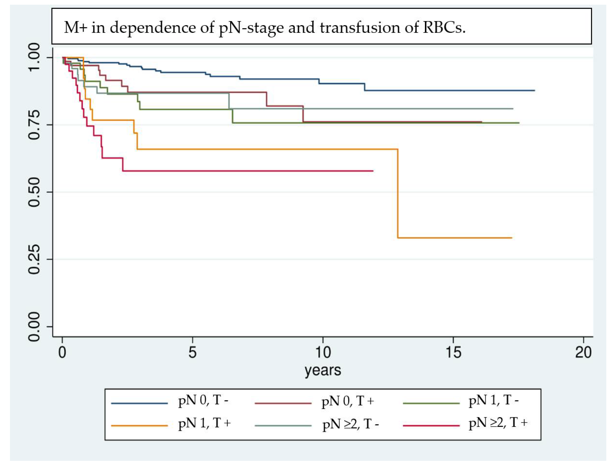
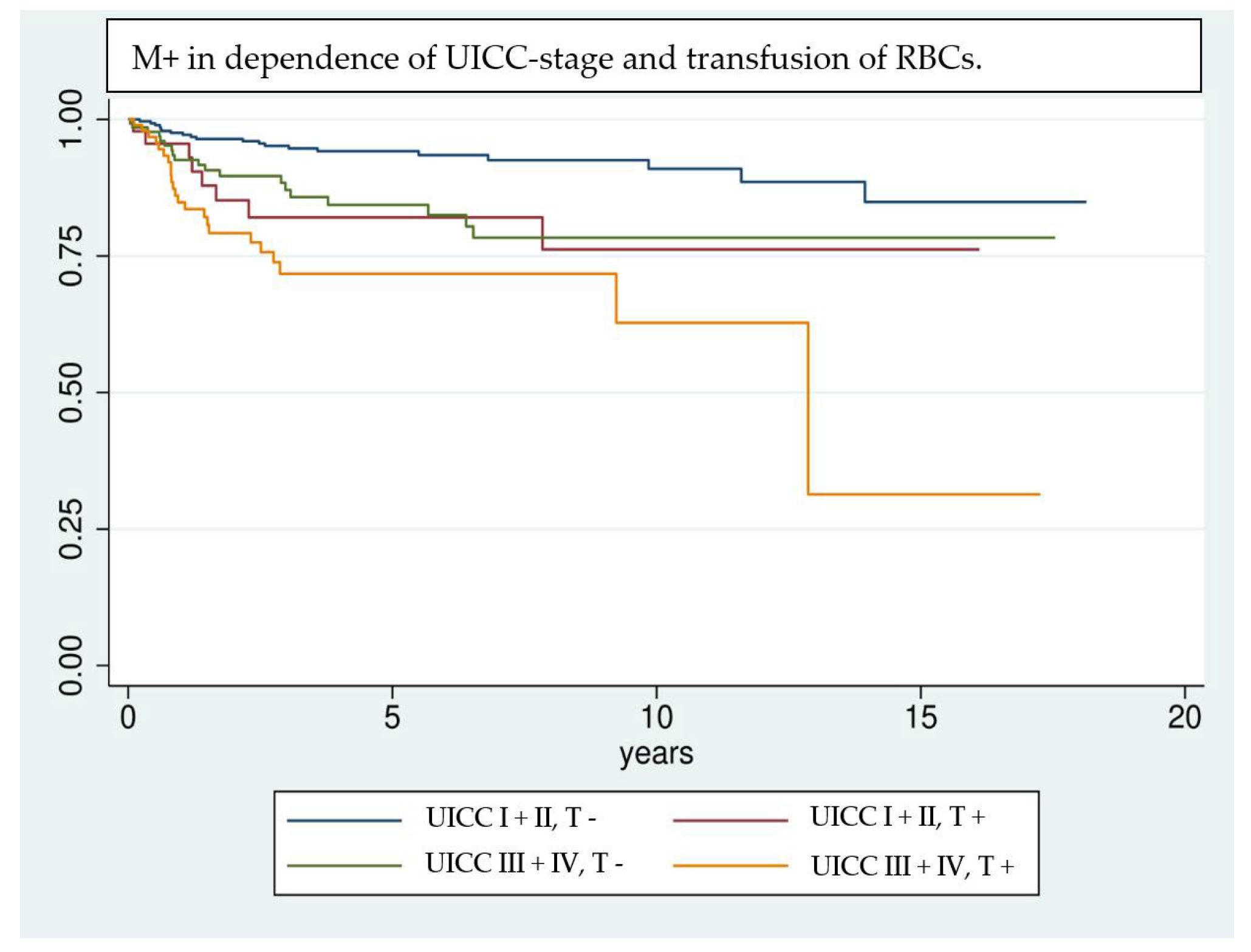
| Transfusion | <0.0001 | 0.0001 | <0.0001 | ||||
| 0 | 436 | 41/9.40% | 248/56.88% | 182/41.74% | |||
| 1–3 | 102 | 23/22.55% | 47/46.08% | 43/42.16% | |||
| >3 | 50 | 10/20% | 26/52% | 16/32% | |||
| Age | 0.3695 | <0.0001 | 0.3695 | ||||
| <45 | 33 | 2/6.06% | 23/69.7% | 18/54.55% | |||
| 45–65 | 291 | 42/14.43% | 173/59.45% | 128/43.99% | |||
| >65 | 264 | 30/11.36% | 125/47.35% | 95/35.98% | |||
| Gender | 0.9334 | 0.0449 | 0.9334 | ||||
| Male | 334 | 41/12.28% | 165/49.40% | 121/36.23% | |||
| Female | 254 | 33/12.99% | 156/61.42% | 120/47.24% | |||
| Nicotine | 0.4981 | 0.7823 | 0.4038 | ||||
| Yes | 285 | 39/13.68% | 151/52.98% | 108/37.89% | |||
| No | 303 | 35/11.55% | 170/56.11% | 133/43.89% | |||
| Alcohol | 0.7819 | 0.0533 | 0.7745 | ||||
| Yes | 209 | 24/11.48% | 101/48.33% | 71/33.97% | |||
| No | 379 | 50/13.19% | 220/58.05% | 170/44.85% | |||
| Cancer History | 0.3946 | 0.1917 | 0.6032 | ||||
| Yes | 46 | 6/13.04% | 26/56.52% | 21/45.65% | |||
| No | 542 | 68/12.55% | 295/54.43% | 220/40.59% | |||
| pT | 0.0281 | <0.0001 | <0.0001 | ||||
| 1 | 271 | 29/10.7% | 161/59.41% | 124/45.76% | |||
| 2 | 182 | 23/12.64% | 96/52.75% | 71/39.01% | |||
| 3 | 62 | 10/16.13% | 33/53.23% | 26/41.94% | |||
| 4 | 73 | 11/16.44% | 31/42.47% | 20/27.40% | |||
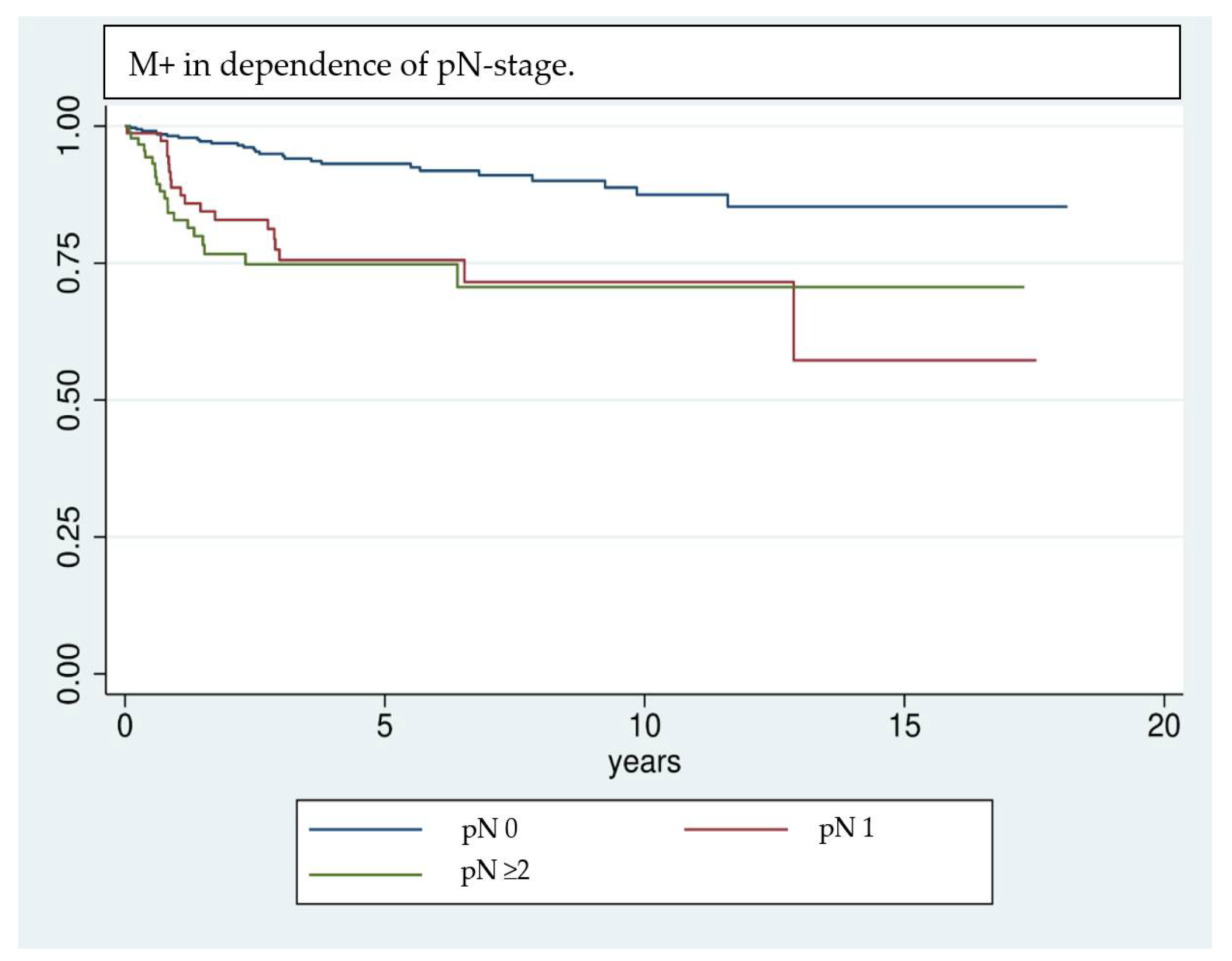
| pN | <0.0001 | <0.0001 | <0.0001 | ||||
| 0 | 353 | 27/7.65% | 224/63.46% | 167/47.31% | |||
| 1 | 79 | 19/24.05% | 36/45.57% | 27/34.18% | |||
| 2 | 85 | 19/22.35% | 32/37.65% | 23/27.06% | |||
| 3 | 11 | 2/18.18% | 7/63.64% | 6/54.55% | |||
| Localization | 0.3261 | 0.0001 | 0.0006 | ||||
| 1 | 267 | 35/13.11% | 135/50.56% | 97/36.33% | |||
| 2 | 148 | 15/10.14% | 100/67.57% | 82/55.41% | |||
| 3 | 19 | 3/15.79% | 3/15.79% | 2/10.53% | |||
| 4 | 48 | 7/14.58% | 26/54.17% | 18/37.50% | |||
| 5 | 65 | 6/9.23 % | 37/56.92% | 28/43.08% | |||
| 6 | 40 | 8/20% | 19/47.50% | 13/32.50% | |||
| L | <0.0001 | <0.0001 | <0.0001 | ||||
| Yes | 98 | 26/26.53% | 244/63.05% | 34/34.69% | |||
| No | 387 | 98/20.21% | 46/46.94% | 187/48.32% | |||
| V | 0.0456 | 0.0256 | 0.0799 | ||||
| Yes | 7 | 2/28.57% | 2/28.57% | 1/14.29% | |||
| No | 478 | 7/1.44% | 288/60.25% | 220/46.03% | |||
| Pn | 0.0093 | 0.0003 | 0.0019 | ||||
| 0 | 438 | 55/12.56% | 269/61.42% | 203/46.35% | |||
| 1 | 45 | 11/24.44% | 20/44.44% | 17/37.78% | |||
| G | 0.0062 | 0.0001 | 0.0032 | ||||
| 1 | 74 | 4/5.41% | 51/68.92% | 44/59.46% | |||
| 2 | 396 | 53/13.38% | 214/54.04% | 151/38.13% | |||
| 3 | 80 | 15/18.75% | 31/38.75% | 25/31.25% | |||
| 4 | 3 | 1/33.33% | 1/33.33% | 1/33.33% | |||
| UICC | 0.0002 | <0.0001 | <0.0001 | ||||
| I | 234 | 20/8.55% | 145/61.97% | 109/46.58% | |||
| II | 115 | 10/8.7% | 66/57.39% | 51/44.35% | |||
| III | 94 | 17/18.09% | 46/48.94% | 35/37.23% | |||
| IV | 145 | 27/18.62% | 64/44.14% | 46/31.72% | |||
| Adjuvant Therapy | <0.0001 | <0.0001 | <0.0001 | ||||
| No | 407 | 29/7.13% | 239/58.72% | 181/44.47% | |||
| RTx | 134 | 22/16.42% | 64/47.76% | 48/35.82% | |||
| RCTx | 47 | 23/48.94% | 18/38.30% | 12/25.53% | |||
| Microvascular Transplant | <0.0001 | <0.0001 | <0.0001 | ||||
| Yes | 214 | 40/18.69% | 94/43.93% | 72/33.64% | |||
| No | 374 | 34/9.09% | 227/60.70% | 169/45.19% | |||
| Preoperative Hemoglobin | 0.0256 | 0.0256 | 0.0002 | ||||
| ≥12 g/dL | 521 | 63/12.09% | 288/55.28% | 215/41.27% | |||
| <12 g/dL | 67 | 11/16.42% | 33/49.25% | 26/38.81% | |||
| M+ (Distant Metastasis) | OS (Overall Survival) | TFS (Tumor-Free Survival) | ||||||||||
|---|---|---|---|---|---|---|---|---|---|---|---|---|
| HR | SD | 95% CI | p | HR | SD | 95% CI | p | HR | SD | 95% CI | p | |
| Transfusion | ||||||||||||
| Yes | 2.42 | 0.78 | 1.28–4.56 | <0.01 | 1.11 | 0.2 | 0.78–1.57 | 0.566 | 1.16 | 0.22 | 0.8–1.67 | 0.437 |
| Age | ||||||||||||
| High | 1.01 | 0.01 | 0.99–1.04 | 0.334 | 1.03 | 0.01 | 1.02–1.04 | <0.01 | 1.02 | 0.01 | 1.01–1.03 | <0.01 |
| Gender | ||||||||||||
| Female | 0.73 | 0.2 | 0.42–1.26 | 0.26 | 1.41 | 0.21 | 1.06–1.88 | 0.02 | 1.07 | 0.16 | 0.80–1.44 | 0.633 |
| pT Stage | ||||||||||||
| pT2 | 1.04 | 0.34 | 0.55–1.97 | 0.894 | 1.21 | 0.2 | 0.87–1.68 | 0.251 | 1.4 | 0.24 | 1.01–1.96 | 0.045 |
| pT3 | 1.53 | 0.64 | 0.67–3.46 | 0.31 | 1.68 | 0.39 | 1.06–2.66 | 0.026 | 2.21 | 0.54 | 1.37–3.57 | <0.01 |
| pT4 | 0.97 | 0.41 | 0.42–2.24 | 0.952 | 1.51 | 0.34 | 0.97–2.34 | 0.065 | 1.97 | 0.44 | 1.27–3.1 | <0.01 |
| pN Stage | ||||||||||||
| pN1 | 2.99 | 0.97 | 1.59–5.63 | <0.01 | 1.36 | 0.25 | 0.95–1.95 | 0.094 | 1.24 | 0.23 | 0.85–1.81 | 0.266 |
| pN ≥ 2 | 3.37 | 1.07 | 1.81–6.29 | <0.01 | 2.07 | 0.35 | 1.49–2.88 | <0.01 | 1.97 | 0.35 | 1.39–2.79 | <0.01 |
| Microvascular Transplant | ||||||||||||
| yes | 1.44 | 0.47 | 0.76–2.73 | 0.262 | 1.59 | 0.25 | 1.17–2.17 | <0.01 | 1.28 | 0.21 | 0.93–1.77 | 0.131 |
| Preoperative Hemoglobin | ||||||||||||
| <12 g/dL | 1.21 | 0.11 | 1.00–1.45 | 0.5 | 0.93 | 0.04 | 0.87–1.02 | 0.122 | 0.93 | 0.44 | 0.85–1.02 | 0.149 |
Publisher’s Note: MDPI stays neutral with regard to jurisdictional claims in published maps and institutional affiliations. |
© 2021 by the authors. Licensee MDPI, Basel, Switzerland. This article is an open access article distributed under the terms and conditions of the Creative Commons Attribution (CC BY) license (https://creativecommons.org/licenses/by/4.0/).
Share and Cite
Brandenburg, L.S.; Metzger, M.C.; Poxleitner, P.; Voss, P.J.; Vach, K.; Hell, J.; Hasel, K.; Weingart, J.V.; Schwarz, S.J.; Ermer, M.A. Effects of Red Blood Cell Transfusions on Distant Metastases of Oral Squamous Cell Carcinomas. Cancers 2022, 14, 138. https://doi.org/10.3390/cancers14010138
Brandenburg LS, Metzger MC, Poxleitner P, Voss PJ, Vach K, Hell J, Hasel K, Weingart JV, Schwarz SJ, Ermer MA. Effects of Red Blood Cell Transfusions on Distant Metastases of Oral Squamous Cell Carcinomas. Cancers. 2022; 14(1):138. https://doi.org/10.3390/cancers14010138
Chicago/Turabian StyleBrandenburg, Leonard Simon, Marc Christian Metzger, Philipp Poxleitner, Pit Jacob Voss, Kirstin Vach, Johannes Hell, Konstantin Hasel, Julia Vera Weingart, Steffen Jochen Schwarz, and Michael Andreas Ermer. 2022. "Effects of Red Blood Cell Transfusions on Distant Metastases of Oral Squamous Cell Carcinomas" Cancers 14, no. 1: 138. https://doi.org/10.3390/cancers14010138
APA StyleBrandenburg, L. S., Metzger, M. C., Poxleitner, P., Voss, P. J., Vach, K., Hell, J., Hasel, K., Weingart, J. V., Schwarz, S. J., & Ermer, M. A. (2022). Effects of Red Blood Cell Transfusions on Distant Metastases of Oral Squamous Cell Carcinomas. Cancers, 14(1), 138. https://doi.org/10.3390/cancers14010138