Review

RGD-Binding Integrins Revisited: How Recently Discovered Functions and Novel Synthetic Ligands (Re-)Shape an Ever-Evolving Field

1
Department of Nuclear Medicine, University Hospital Klinikum Rechts der Isar and Central Institute for Translational Cancer Research (TranslaTUM), Technical University Munich, 81675 Munich, Germany
2
Department of Chemistry, Institute for Advanced Study, Technical University Munich, 85748 Garching, Germany
3
Clinical Research Unit, Department of Obstetrics and Gynecology, University Hospital Klinikum Rechts der Isar, Technical University Munich, 81675 Munich, Germany
*
Authors to whom correspondence should be addressed.
Cancers 2021, 13(7), 1711; https://doi.org/10.3390/cancers13071711
Submission received: 26 February 2021 / Revised: 22 March 2021 / Accepted: 29 March 2021 / Published: 4 April 2021
(This article belongs to the Special Issue Targeting Blood Vessel Components to Treat Cancer)

Abstract

:

Simple Summary

Integrins, a superfamily of cell adhesion receptors, were extensively investigated as therapeutic targets over the last decades, motivated by their multiple functions, e.g., in cancer (progression, metastasis, angiogenesis), sepsis, fibrosis, and viral infections. Although integrin-targeting clinical trials, especially in cancer, did not meet the high expectations yet, integrins remain highly interesting therapeutic targets. In this article, we analyze the state-of-the-art knowledge on the roles of a subfamily of integrins, which require binding of the tripeptide motif Arg-Gly-Asp (RGD) for cell adhesion and signal transduction, in cancer, in tumor-associated exosomes, in fibrosis and SARS-CoV-2 infection. Furthermore, we outline the latest achievements in the design and development of synthetic ligands, which are highly selective and affine to single integrin subtypes, i.e., αvβ3, αvβ5, α5β1, αvβ6, αvβ8, and αvβ1. Lastly, we present the substantial progress in the field of nuclear and optical molecular imaging of integrins, including first-in-human and clinical studies.

Abstract

Integrins have been extensively investigated as therapeutic targets over the last decades, which has been inspired by their multiple functions in cancer progression, metastasis, and angiogenesis as well as a continuously expanding number of other diseases, e.g., sepsis, fibrosis, and viral infections, possibly also Severe Acute Respiratory Syndrome Coronavirus (SARS-CoV-2). Although integrin-targeted (cancer) therapy trials did not meet the high expectations yet, integrins are still valid and promising targets due to their elevated expression and surface accessibility on diseased cells. Thus, for the future successful clinical translation of integrin-targeted compounds, revisited and innovative treatment strategies have to be explored based on accumulated knowledge of integrin biology. For this, refined approaches are demanded aiming at alternative and improved preclinical models, optimized selectivity and pharmacological properties of integrin ligands, as well as more sophisticated treatment protocols considering dose fine-tuning of compounds. Moreover, integrin ligands exert high accuracy in disease monitoring as diagnostic molecular imaging tools, enabling patient selection for individualized integrin-targeted therapy. The present review comprehensively analyzes the state-of-the-art knowledge on the roles of RGD-binding integrin subtypes in cancer and non-cancerous diseases and outlines the latest achievements in the design and development of synthetic ligands and their application in biomedical, translational, and molecular imaging approaches. Indeed, substantial progress has already been made, including advanced ligand designs, numerous elaborated pre-clinical and first-in-human studies, while the discovery of novel applications for integrin ligands remains to be explored.

1. Introduction

Cell adhesion to the surrounding extracellular matrix (ECM) and/or other cells constitutes the basis for the development, maintenance, and function of various cell and tissue types in multicellular organisms [1,2,3]. Since their discovery as cell-surface adhesion receptors 30 years ago, the integrin superfamily of cell adhesion and signaling receptors evolved as a key player in the link between the extracellular environment and the cytoskeleton [4,5,6]. Essential physiological processes such as embryogenesis, angiogenesis, homeostasis, hemostasis, and wound healing are mediated by integrins [1,5,7,8,9,10] as well as pathophysiological processes, such as tumor growth, metastasis, invasion, and tumor vascularization/angiogenesis. These facts and the occurrence of distinct integrin subtypes on tumor cell surfaces at elevated levels raised great hopes to classify them as valuable target structures in cancer treatment and diagnostic imaging [1,7,9,10,11].
Integrins are transmembrane heterodimers composed of one α- and one β-subunit constituting a protein family of 24 different members formed by the non-covalent combination of 18 α- and 8 β-subunits [5]. The elucidation of the crystal structure and the characterization of the extracellular domain of some family members in the beginning of the new century stimulated the generation of an enormous number of therapeutics targeting distinct integrins [12,13,14,15,16,17,18,19,20,21,22]. Docking experiments and crystal structure analyses allowed a tailor-made design of subtype-selective and highly affine integrin ligands, which is mainly based on the highly conserved recognition motif in ECM proteins, the tripeptide Arg-Gly-Asp (RGD) [4,12,13,14,15,16,17,18,19,20,21,22,23,24,25,26].
The knowledge of the biological roles of integrins and the discovery of new key functions of distinct integrin subtypes in pathophysiological processes, specifically in cancer, is continuously expanding and enables the development of new and improved integrin-targeting therapeutic strategies. To this end, significant improvements have been made in the field of synthetic integrin ligands that selectively address integrin subtypes with high affinity and specificity, which act as inhibitors and/or vehicles for drug delivery but also by agonistic properties of integrin ligands, which came recently in the focus of medicinal application [27,28,29].
In the present review, we summarize the latest discoveries in RGD-binding integrin (patho-) biology as well as progresses made in the design and application schemes of RGD-based integrin ligands for new and promising approaches in integrin-targeted therapy concepts.

2. Integrin/ECM Cell Adhesion and Signaling Receptors

Before we get into the role of integrins in cancer and the ligands to target them, we want to clearly lay out the basis for integrin-targeted therapy by describing the mechanisms of integrin ligand binding and the targetable vulnerabilities associated with it. Integrins are classified into four integrin subfamilies according to their binding specificity toward distinct ECM proteins [2,30]. These subfamilies are (i) the collagen-binding [5,31,32], (ii) the laminin-binding [5,31], (iii) the RGD-binding integrins [5,10,19], and (iv) the leukocyte-specific integrins that bind to intercellular adhesion molecules (ICAMs), polysaccharides, and plasma proteins, respectively [33,34,35] (Figure 1).
Most of the eight RGD binding integrins are expressed at elevated levels in various cancer types where they contribute to crucial pathophysiological functions in cancer as well as in other diseases, such as neurological disorders [36], sepsis [37], fibrosis [38], cardiovascular diseases [39], and viral infections [40]. Since this review focuses exclusively on RGD binding integrins, with respect to other integrin family members, we refer to excellent other reviews [32,41,42]. Various ECM proteins, e.g., fibronectin [43], vitronectin [44], von Willebrand factor [45], fibrinogen [46], osteopontin [47], or thrombospondin [48] encompass the RGD-motif, which is recognized by the integrin subtypes αvβ3, αvβ5, αvβ6, αvβ1, αvβ8, α5β1, αIIbβ3, and α8β1, respectively [31,34,49].
Each subunit consists of three domains: a long extracellular domain (700–1100 amino acids), a short transmembrane domain (20–30 amino acids), and a cytoplasmic region (20–75 amino acids) [2,48,50,51]. The integrin ligand-binding domain is constituted by the extracellular head domains of both integrin subunits [2,7,48,50,52]. One crucial structural element for integrin ligand binding is located within the head group of the β-subunit of all integrins, the so-called Metal Ion-Dependent Adhesion Site (MIDAS) [53,54,55,56]. In case of ligand binding, MIDAS is occupied by a divalent metal ion (Mn2+, Mg2+, Ca2+) to which the carboxy group of the integrin ligand coordinates [53,54,55,56,57,58,59]. Hence, the acidic residue represents a distinctive element of all integrin ligands [53,54,55,56,57,58,59].
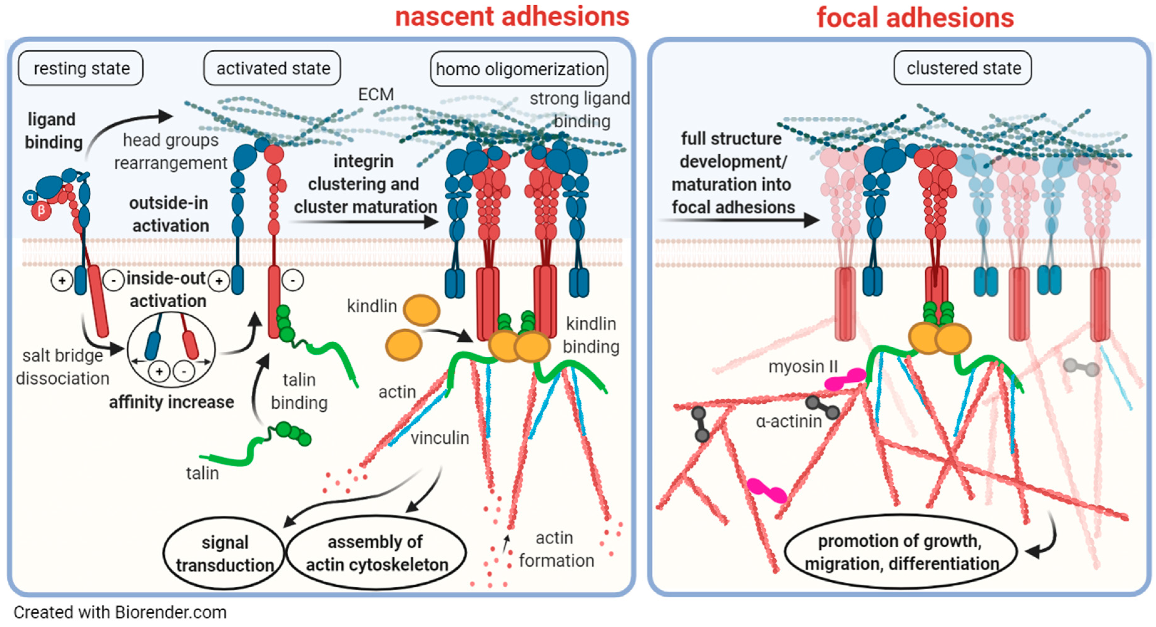
In addition to mediating firm cellular adhesion to the underlying ECM, integrins bear the capacity to transmit bidirectional signals across cell membranes (Figure 2) [60,61,62,63]. This enables internal signals to control cell adhesion to the ECM (inside-out signaling) as well as signals from the cellular microenvironment to regulate cellular processes (outside-in signaling), such as wound healing, cell differentiation, migration, and proliferation [64]. This requires conformational changes in the two integrin subunits [60,61,62,63]. In the “switchblade” model [65,66,67], three integrin conformational states have been proposed, bent, extended, and extended with an open headpiece [68,69,70,71]. Already in the bent conformation, integrins are capable of binding ECM ligands with low affinity [72]. In this resting state, the transmembrane and cytoplasmic domains of the α- and the β-subunit harbor a closed conformation. Here, the helices interact in a manner, similar to that observed for the strongly homodimerizing erythrocyte protein glycophorin A (GpA), which harbors the dimerization motif GXXXG in its transmembrane domains [24,73,74,75,76]. In fact, by sequence alignments, indeed, a GXXXG-like motif was shown to be highly conserved among most integrin subunits [73,77]. Then, priming and ligand binding to integrins instigate large-scale conformational rearrangements in which the integrin extracellular domains erect [65,66,67]. During inside-out activation, an intracellular force stimulates cytoplasmic proteins, such as talin or kindlin, to attach to the cytoplasmic domain of the β-subunit and to destabilize a salt bridge connecting the α- and the β-subunit (Figure 2) [20,71,78,79,80,81]. The resulting separation of the transmembrane and cytoplasmic domains to an open conformation leads to a high affinity ligand-binding headpiece and now exerts integrin signaling competence [64,66,82,83,84].
The knowledge of integrin transmembrane domain conformations is indispensable for elucidating the mechanisms involved. In a cell model harboring an engineered αvβ3 transmembrane domain variant, in which the complete integrin transmembrane domains were exchanged by that of GpA in the context of an otherwise unaltered αvβ3, it was shown that this resulted in a non-signaling receptor with low affinity due to firmly associated transmembrane domains [76]. This leads to an integrin conformation with pure antagonistic function, since for signal transduction, the unclasping of the transmembrane domains is required [85]. In contrast, when the GXXXG-motif was mutated to GXXXI, which is known to abrogate dimerization, an αvβ3 variant was generated that displayed dissociated transmembrane domains, exhibiting high-affinity and constitutively active signaling competence [76]. This allows the re-association of the separated subunits to form homooligomers (dimers in the α-subunit and trimers of the β-subunits) with new RGD binding pockets [85,86], which can further bind to other proteins to form nascent adhesions (Figure 2) [20,62,69,70,71,81,87,88,89]. Subsequently, upon multiprotein clustering, focal adhesions are formed, which are the sites where integrin signaling takes place [62,68,69,70,71,88,90]. This triggers a cascade of phosphorylation events of various downstream signaling molecules, among those, most importantly the focal adhesion kinase (FAK), the mitogen-activated protein kinase (MAPK), and the extracellular signal regulated kinase (ERK) [71,75,76,81,85,91]. Obviously, multivalent binding of clustered integrins and other proteins in focal adhesions results in enhanced integrin receptor avidity and thus strong cell/ECM contacts [92]. In order to interfere with this strong cell attachment to the ECM, high amounts of ligands are required for their antagonistic action. Thus, the behavior in the activation of integrins may explain why low concentrations of the αvβ3-targeting ligand Cilengitide cause agonism, whereas antagonism needs high doses of ligands to disrupt the strong multivalent binding events within focal adhesions (see Section 4.2).

3. The Role of RGD Binding Integrins in Cancer and Tumor Neovascularization

3.1. RGD Binding Integrin Receptors

Integrin αvβ3. One of the earliest studied and meanwhile best characterized integrins is αvβ3. The integrin αvβ3, originally named vitronectin receptor, binds besides vitronectin also to other ECM proteins, such as fibronectin, osteopontin, von Willebrand factor, or laminin. Other integrins, such as α5β1, bind selectively to one ECM protein, e.g., fibronectin [10,93]. During tumor angiogenesis, αvβ3 is highly upregulated on angiogenic endothelial cells but not on the quiescent endothelium and mediates the interaction of activated endothelial cells with the ECM [10,93,94,95,96,97]. On tumor cells, αvβ3 is highly overexpressed in the course of tumor progression and triggers the formation of lamelli- and filopodia, regulating cancer cell migration and invasion, cell/ECM stiffness, and cell contractility [10]. Additionally, through the interaction with various ECM proteins, αvβ3 associates with Vascular Endothelial Growth Factor (VEGF) receptors and Fibroblast Growth Factor-2 (FGF-2), promoting cancer cell invasiveness [93,98]. Especially in breast cancer, αvβ3 is detected at the invasive front as well as in distant metastases [99]. In addition to breast cancer, αvβ3 is upregulated in gastric cancer, mainly in stroma and endothelial cells [10,100]. Furthermore, αvβ3 was found to be highly expressed in glioma [101], lung cancer brain metastases [102], prostate cancer [103,104], pancreatic cancer [105], oral squamous cell carcinoma [106], ovarian [10], and non-small cell lung cancer correlating with tumor progression [107,108,109]. As it is overexpressed on tumor cells, as well as the vasculature, αvβ3 can be used for the targeted therapy of both [93,110].
Integrin αvβ5. Integrin αvβ5 has been found to be highly upregulated in gastric tumor and stromal cells as well as in the endothelial cells of newly formed vessels during angiogenesis [100]. Furthermore, αvβ5 is overexpressed in lung cancer [102], non-small cell lung cancer [108,111], and prostate cancer [104]. In contrast to αvβ3, it promotes tumor invasion and metastasis formation rather than impacting on primary tumor growth [97,112,113]. Similar to αvβ3, αvβ5 promotes distinct pathways of angiogenesis [97]. While αvβ3 is necessary for basic FGF- or Tumor Necrosis Factor-α (TNF-α)-mediated angiogenesis, angiogenesis mediated via VEGF or Transforming Growth Factor-α (TGF-α) requires the presence of αvβ5 [97,114]. Furthermore, downstream signaling of αvβ5 leads to FAK activation as well as of Raf, leading to endothelial cell protection against apoptosis induced by ECM detachment, cellular stress, and/or DNA damaging agents [97,115]. For tumor invasion and metastasis promoted by αvβ5, in contrast to αvβ3, cytokine or growth factor stimulation is necessary [97].
Integrin α5β1. Another important angiogenesis marker on endothelial cells is α5β1, where it correlates with tumor malignancy, invasiveness, and thus metastasis formation [116,117,118]. During invasion processes, α5β1 facilitates the increased generation of adhesion forces, focal adhesion assembly, stress fiber formation, and contractile forces, respectively [118]. Thereby, the cell overcomes easier the steric hindrance of the ECM, leading to a higher invasiveness [118,119]. Integrin α5β1 is also expressed on tumor cells, such as oral squamous cell carcinoma [106,120] and ovarian cancer cells [117]. Furthermore, α5β1 was capable of triggering pro-survival signaling on epidermoid carcinoma cells, thereby regulating cell proliferation through both a constant activation of the Akt kinase, an apoptosis suppressor, and the Epidermal Growth Factor Receptor (EGFR) [120].
Integrin αvβ6. Integrin αvβ6 is exclusively expressed in epithelial cells and overexpressed in various cancer entities [10], such as oral squamous cell carcinoma [121], breast [122], colon [123], liver [124], ovarian [125], and pancreatic cancer [126,127] at the invasive front. Here, it is involved in the activation of Transforming Growth Factor-β (TGF-β1). Integrin αvβ6 activates TGF-β1 through binding to the RGD motif contained within the Latency Associated Peptide (LAP) linked to inactive TGF-β1 [128]. This mechanism works in a protease-independent fashion implicating tractional forces conferred by the mechanotransducing properties of this integrin exerted by the actin cytoskeleton. As long as LAP is non-covalently linked to inactive TGF-β1, the binding of the latter to its receptors is abrogated and prevents signaling [129]. In order to enable receptor binding, TGF-β1 has to be activated and released from this complex. Upon force application to the LAP–TGF-β1 complex, the so-called Large Latent Complex (LLC), conformational structural alterations have to occur. As a consequence, αvβ6 is a driver in epithelial mesenchymal transition (EMT) in cancer and correlates with poor patient survival as well as chemo- and radioresistance [128,130,131].
Integrin αvβ8. More recently, another member of the αv-integrin family, αvβ8, has been detected in head and neck cancer, non-small cell lung cancer, as well as prostate cancer [104,108,132]. It has received increasing attention in current research, since it also interacts with the RGD sequence of LAP, thereby promoting the activation of TGF-β. However, in contrast to the above described mechanism of αvβ6-mediated TGF-β1 activation, αvβ8 activates TGF-β1 in a protease-dependent way, since it harbors a shorter cytoplasmic tail, which is unable to bind to actin. Therefore, αvβ8-dependent activation of latent TGF-β1 from the LLC has been proposed to require proteolytic cleavage of LAP by co-expressed MMP-14 (or MT1-MMP) rather than by force such as in the case of αvβ6. Subsequently, TGF-β1 is released from the LLC, allowing its binding to TGF-β receptors [132,133,134]. Via its capacity to activate TGF-β1, αvβ8 is also implicated in angiogenesis, which is not the case for αvβ3 and αvβ5 [133].
Integrin α8β1. Integrin α8β1 is mainly expressed in smooth muscle cells [135,136] and plays a crucial role in kidney morphogenesis [137]. Its role in cancer is so far unknown. However, in murine mammary tumors, α8 was found to be co-expressed with its ligand tenascin-W, which is implicated in cell proliferation and regenerative processes [138]. Thus, in the presence of tumor stromal tenascin-W, a facilitated invasion of α8β1-expressing cancer cells into the surrounding tissue might be conceivable [138]. So far, no selective, synthetic ligands targeting α8β1 have been reported.
Integrin αIIbβ3. Integrin αIIbβ3 occurs exclusively on thrombocytes and serves as a fibrinogen receptor to bridge platelets together. As a consequence, αIIbβ3 represents a valuable target structure in anti-thrombosis therapy [139]. Even it does not play an active role in cancer, for the design of integrin ligands for anti-cancer therapy, it is of utmost importance to assure that these ligands do no cross-react with αIIbβ3 in order to avoid severe hemorrhagic complications in patients receiving systemically administered ligands [140].
Integrin αvβ1. Integrin αvβ1 has received limited attention as a disease-relevant target in the past, which was possibly a consequence of the absence of specific and selective inhibitory peptides or antibodies. It is highly expressed on activated fibroblasts and directly binds to the latency-associated peptide of TGFβ1 and mediates TGFβ1 activation [141,142]. Thus, αvβ1 has been suggested as a promising target in fibrosis treatment, but its exact role in TGFβ1 activation, compared to other integrins, remains to be dissected. Integrin αvβ1 has also been described a receptor for viral entry, for example as an adenovirus co-receptor.

3.2. Integrin Functions in Angiogenesis and Tumor Vessel Phenotype

3.2.1. The Role of Integrins in Angiogenesis

One important cancer hallmark, in which integrins are majorly involved during tumor growth and progression, is the induction of tumor neovascularization [143,144,145]. Distinct integrin subtypes are majorly implicated in these angiogenic processes [146]. Integrin αvβ3 was the first member of this superfamily found to be overexpressed on angiogenic endothelial cells in concert with numerous pro- and anti-angiogenic factors, most importantly VEGF and its receptors [147]. Since VEGF positively affects the conformational activation state of αvβ3 and its signaling capabilities, a feed-forward loop of VEGF release by tumor cells and increased cell adhesion and migration is constituted. In turn, αvβ3 activation hampers VEGFR2 degradation, boosting continuous signal transduction even more [148]. Thus, αvβ3 was soon recognized as an important marker for angiogenesis.
In pre-clinical studies, the use of αvβ3 antagonists, such as antibodies, RGD-based cyclic peptides, or peptidomimetics proved to efficiently suppress tumor angiogenesis and disease progression. This strongly motivated their exploration in clinical cancer trials as new anti-cancer strategies [93,149]. In this regard, the first RGD-based peptide was Cilengitide (EMD 121974) [62,150], which is directed to the binding epitope for ECM protein ligands constituted by the headgroups of the two subunits of αvβ3 and αvβ5, respectively. The clinical trial experience with Cilengitide is further discussed in Section 4.2 and is the subject of a number of excellent and detailed reviews [95,151,152,153,154]. In brief, although Cilengitide exhibited a promising performance in in vitro cell studies and in pre-clinical animal models, it missed the high expectations of its therapeutic benefit in human therapy trials for patients afflicted with different cancer types [62,155,156]. In the course of the exploration of its clinical efficacy, it became obvious that treatment with low-dose Cilengitide led to unexpected pro-angiogenic effects. This observation inspired a series of interesting studies to unravel the underlying mechanisms. In fact, it was found that low concentrations of Cilengitide promoted VEGF-mediated angiogenesis, which is an effect whose mechanisms motivated further investigations [157]. In mouse endothelial cells treated with low-dose Cilengitide, an increased VEGFR2 protein expression was noticed, with no impact on VEGFR2 mRNA, indicating a post-transcriptional regulation. The same was observed in tumor blood vessels in vivo. Moreover, the exposure of cells to low-dose Cilengitide affected the cellular trafficking of VEGFR2 and αvβ3, since it was documented that the recycling of both receptors back to the cell membrane was strongly promoted, without impacting on receptor internalization. As a consequence of this enhanced recycling, a diminished VEGFR2 degradation was noticed that was concomitant with αvβ3 recruitment to focal adhesions [148,157]. The Cilengitide-triggered increase of VEGFR2 cell surface expression was traced to the level of downstream mediators leading to VEGFR2 phosphorylation as well as integrin and Src activation [157,158]. Even more, low-dose Cilengitide induced αvβ3 affinity and signaling, leading to augmented tumor cell proliferation and enhanced angiogenesis via increased VEGFR-triggered signaling and enforced endothelial cell migration [157]. In addition, Cilengitide has a short plasma half-life of 3–5 h, limiting its time for accumulation in the tumor. Hence, even though high doses were systemically applied, tumors were potentially exposed to the low-dose, pro-angiogenic effects of Cilengitide, explaining its inability to efficiently treat tumors [159]. Taken together, these Cilengitide effects are thought to explain its failure as an angiogenesis inhibitor [160]. In addition to the pro-angiogenic effects of low-dose Cilengitide in cancer, it was debated if it may also exert vascular-promoting effects in heart failure models [29]. Indeed, the treatment of mice who underwent abdominal aortic constriction surgery with low-dose Cilengitide stimulated coronary angiogenesis and affected cardiomyocyte hypertrophy, resulting in a mitigated course of the heart disease. Analyses at the molecular level revealed that low-dose Cilengitide affected the transcriptome of cardiac endothelial cells, involving pro-angiogenic signaling pathways as well as genes implicated in cell growth and DNA repair, also enhancing angiogenesis.

3.2.2. The Therapeutic Benefit of Vascular Normalization Therapy in Cancer

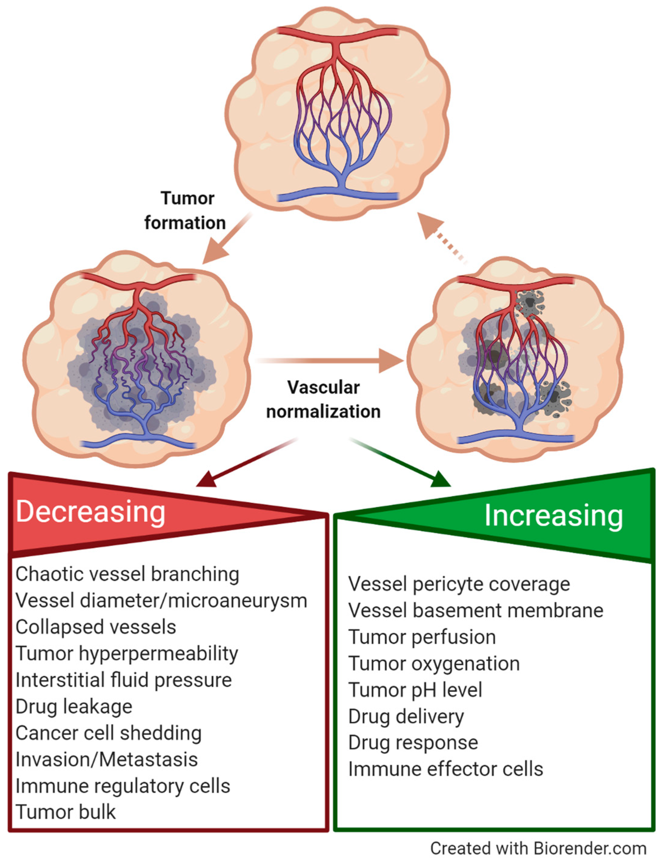
Tumor vessels are markedly different from their normal quiescent counterparts since they display rather immature features, such as chaotic branching, collapses, and microaneurysm. Moreover, tumor vessels show incomplete endothelial cell lining, a defective basement membrane, and a lack of other cellular vessel components, such as pericytes and smooth muscle cells, which stabilize and cover normal vessel walls [94,161,162,163,164,165,166,167]. Due to those facts, no regular blood flow is established in tumor vessels, reinforcing a hypoxic environment within the tumors, fibrosis, and extravasation of tumor cells into the blood or lymphatic system, resulting in the enhanced permeability and retention (EPR) effect. This leads to the entry of macromolecules into the tumor interstitial space, missing drug delivery and therapy response, as well as infiltration by immune regulatory cells [168]. Thus, new approaches are urgently needed to address these challenges by developing a vascular promotion therapy aiming at vascular normalization. Vessel maturation is achieved by the upregulation of adhesion molecules on endothelial cells as well as by the recruitment of vascular smooth muscle cells and pericytes that interact with activated endothelial cell and contribute to vessel branching and coverage. This leads to increased tumor perfusion, oxygen, and pH levels, the recruitment of immune effector cells, as well as improved drug delivery and thus therapy response (Figure 3). Thus, tumor-associated angiogenesis is the net result of a complex and fine-tuned interplay between cancer cells, stimulated vascular and perivascular cells, recruited myeloid and mesenchymal stem cells, as well as activated fibroblasts and immune cells within the tumor microenvironment [169,170]. These events involve integrin crosstalk with the VEGFR2 and the Platelet-Derived Growth Factor Receptor (PDGFR) [171,172].
Once it was discovered that low-dose treatment with Cilengitide exerted pro-angiogenic effects in tumors, an alternative use of low-dose Cilengitide was inspired in order to increase blood flow and, consequently, increased delivery and intracellular uptake of chemotherapeutic drugs [157]. These observations changed the paradigm in cancer therapy from anti-angiogenic strategies to vascular promotion/normalization therapy [173,174]. In mouse and human cancer models, the administration of Cilengitide in combination with Verapamil, a calcium channel blocker, which increased vessel dilation and blood flow in tumors by relaxing blood vessel muscles, enhanced gemcitabine delivery to the tumor bed and thus the efficacy of this drug to provoke tumor regression and metastasis suppression [27]. Moreover, vascular promotion therapy allowed the administration of significantly reduced doses of gemcitabine, thereby lowering side effects and enabling longer treatment windows [173,175,176,177,178].

3.2.3. Integrins in Tumor Cell Extravasation

Leaky tumor vessels do not only exert a negative impact on drug delivery within the tumor site but also favor the escape of shed or detached tumor cells into the bloodstream and lymphatic system as a crucial step for tumor cell dissemination and metastasis. This extravasation occurs as a function of the permeability and missing integrity of the tumor vascular endothelium. Vessel wall permeability is under the control of inflammatory mediators as well as growth factors and proteases, which are sequestered by tumor cells and other cells within the tumor microenvironment. Integrins, which are expressed on endothelial cells, take over important functions since they mediate their adhesion to the basement membrane, which represents an important prerequisite for tumor vessel integrity [179]. Integrin α5 affects tumor cell extravasation by interacting with neuropilin 2 (NRP-2). NRPs are multifunctional transmembrane non-kinase receptors that are expressed on cancer cells [180]. They function predominantly as co-receptors and thus rely on other cell surface receptors to transduce transmembrane signals. The dysregulation of NRPs has been implicated in many pathological conditions, including tumor cell survival, migration, and invasion involving them in the regulation of angiogenesis [181,182,183,184]. Thus, the overexpression of NRPs provokes hypervascularization due to leaky vessel formation [185,186]. Integrins also affect angiogenesis and tumor cell extravasation by their association with NRPs, which are found in integrin-containing multiprotein complexes of adhesomes and are capable of modulating the PI3K/Akt/PTEN signaling axis by activating integrins. Hereby, inside-out signaling of β1, β3, and β5 integrins, respectively, is activated, leading to enhanced binding to ECM proteins [187,188,189,190]. In addition, α5β1, α6β1, and α9β1 expressed on tumor cells are capable of binding to endothelial cell-expressed NRP-2, thereby facilitating cancer cell binding to the endothelium, followed by promoted tumor cell extravasation in distinct cancer types [187,191,192,193,194,195,196]. These findings underline the importance of NRP in concert with integrins and the VEGFR for the proper development of blood vessels and their stabilization. Thus, NRPs also represent potential new therapeutic targets due to their multifaceted roles and the fact that they are highly expressed on tumor cells and tumor endothelial cells [180].

3.3. The Cellular Fate of Tumor Cell-Expressed Integrins

3.3.1. Cellular Internalization of Integrins

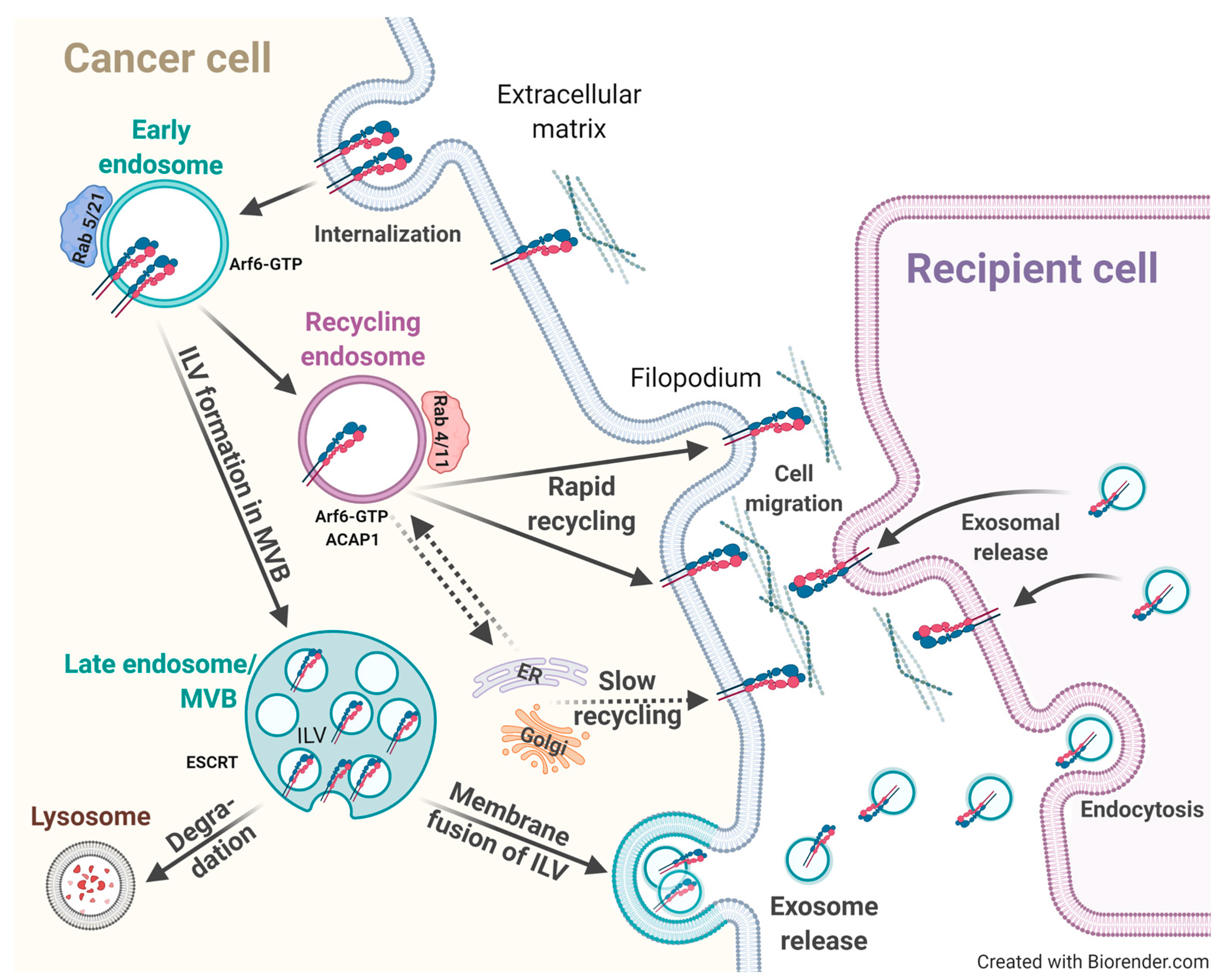
Integrins undergo constant endo-/exocytosis processes, which are crucial for directed cell migration and the dynamic formation and release of adhesive cell–ECM contacts in order to move a cell’s body forward [197,198]. Thus, the balance between endocytosis and recycling of cell-surface integrin receptors is decisive for proper cell migration. It is well known that integrins are constitutively internalized through clathrin- or caveolin-mediated pathways, implicating various additional players. Alternative routes include macropinocytosis from circular dorsal ruffles upon growth factor receptor-mediated signaling [199] and endocytosis via clathrin-independent carriers. The kind of pathway that will be pursued for trafficking either via caveolae, clathrin, neither, or both, strongly depends on the respective cell type and integrin subspecies [200], the latter depending on sequence motifs contained within the highly variable C-terminal tail of β-integrins [201]. As such, in fibroblasts and fibrosarcoma cells, α5β1 internalization occurred in a clathrin- and microtubule-dependent fashion [202,203], whereas in myofibroblasts, α5β1 is constitutively taken up in a caveolin-mediated manner [204] (Figure 4).

3.3.2. Intracellular Endosomal Integrin Trafficking and Sorting

Intracellular endosomal sorting decides over the fate of integrins, which may either be prone to degradation or recycling back to the cell surface [205]. Even when integrins are frequently shuttled to the degradative route initially, in many cases, they are eventually recycled back to the plasma membrane. As such, it has been found that α5β1, destined to be sorted to late endosomes or lysosomes, could still escape from this degradation route to be recycled back to the cell surface [206]. The blockade of this recycling route enhanced the degradation of internalized integrins [207,208,209,210]. In case integrin ligands are conjugated to drugs, e.g., for their α5β1-mediated intracellular delivery, a portion of the endocytosed integrin was recycled, even from lysosomes, while the drug was selectively degraded.
Integrin recycling occurs by spatially and temporally different pathways [211,212,213]. In principal, two different recycling routes have been discovered, the long-loop and the rapid (short) recycling track. The long-loop pathway has been described for the endocytosis of α5β1, α6β4, and αvβ3, respectively. It starts with integrin trafficking to the Perinuclear Recycling Compartment (PNRC), which is enriched by recycling endosomes carrying members of the Rab family of small GTPases, Rab11 and Rab25, as well as Arf6 [214,215,216,217,218,219]. Recycling of α5β1 is under the control of the PKB/Akt kinase pathway via distinct downstream target structures, such as the Arf6-specific GTPase Activating Protein (GAP) ACAP1, which is crucial for clathrin coat assembly [220,221]. However, certain differences regarding involved players in the recycling process have been documented. Long-loop recycling of α5β1 in fibroblasts did not depend on growth factors [214] and also β1 recycling—brought to terms by Rab11 and Arf6—was not reinforced in the presence of EGF or serum [215]. In contrast, the rapid (short) integrin recycling route from Rab4-positive endosomes was under the control of growth factors, such as PDGF, which directed αvβ3 to this route [214]. In addition, in cervical cancer cells, β1 and β3 integrins were recycled via the rapid (short) loop requiring EGF stimulation [222] (Figure 4).
Even different integrin subtypes exhibit similar preference toward distinct ECM ligands, the biological effects arising thereof turned out to be integrin subtype- and cell-type specific. For endocytosed α5β1 and αvβ3, differential routes for recycling and crosstalk have been reported. Even more, they were interdependent, since αvβ3 activation had a negative impact on α5β1 recycling [223]. Whereas αvβ3 recycling enforced the generation of lamellipodia and increased directional cell movement in a Rac-dependent manner, α5β1 recycling to invasive cellular protrusions accelerated random cell migration and invasion via the Rho–ROCK–cofilin pathway by altered actin cytoskeleton [148,224,225,226,227]. Thus, distinct functions of integrin subtypes that have to be temporally acquired by a (tumor) cell are tightly controlled by tipping the balance between integrin degradation and recycling [205]. The net effect of continued recycling from endosomes after internalization is an increased cell surface integrin receptor density and an altered membrane distribution, e.g., in newly formed focal adhesions at the leading migratory cell front. By binding to the ECM, directed and efficient cell motility was promoted, which was not the case upon constant integrin breakdown [211,214]. Moreover, there is also a need for molecules assisting in cell migration, which are also transported via endosomes. As such, Membrane Type 1 Matrix Metalloprotease (MT1-MMP) was trafficked to the cell invasive front, contributing to ECM remodeling for enhanced cell motility and chemotaxis [228].
Most interestingly, intracellular sorting of integrins is under the control of their activation state. In several cancer entities, both, active and inactive β1 receptors are internalized via clathrin- and dynamin-dependent routes to Rab4a-, Rab5-, or Rab21-positive early endosomes [229]. However, from there, different trafficking routes are being pursued: active β1 engaged by an ECM ligand was shuttled to late endosomes and lysosomes, either for degradation [230] or for ligand detachment, followed by the slow recycling of the ligand-free integrin back to the cell membrane via the Rab11-dependent pathway. In contrast, inactive unligated β1 was rapidly recycled back to Arf6-positive cell membrane protrusions in an actin- and Rab4-dependent fashion [229]. Ligation of α5β1 by fibronectin instigated the ubiquitination of the α5 cytoplasmic domain, ultimately resulting in the sorting of the α5β1/fibronectin complex to lysosomes by implicating the Endosomal-Sorting Complex Required for Transport (ESCRT) [231]. Still, the process of coordinated α5β1-provoked internalization of fibronectin as well as the secretion of de novo synthesized fibronectin is not fully resolved yet. Secreted fibronectin assembles into a fibrillar network via integrins, which triggers mechanical and chemical cues, which is also crucial for endothelial cell tubulogenesis [232], in vivo angiogenesis [233,234], and the establishment of endothelial cell apico-basal polarity [235]. After endocytosis into early endosomes, active α5β1 engaged by protease-cleaved fibronectin fragments [204,236] was detected in post-Golgi carriers following its trafficking back to the endothelial cell basolateral surface by carrying de novo synthesized fibronectin dimers. The latter kept α5β1 in an activated state within post-Golgi carriers where talin and kindlins are missing. On one hand, these processes lead to the disposal of endocytosed and old fibronectin, and on the other, they lead to the deposition of newly synthesized fibronectin into the ECM to assure the dynamic renewal of polarized fibronectin fibrils. These findings emphasize a role of integrin recycling via the endosomal pathway for the control of ECM protein accumulation and remodeling, enabling tumor cell invasion into adjacent tissues [237].
Moreover, in the fateful decisions between the breakdown and recycling of integrins, endocytic adaptor proteins are involved. As such, sorting nexin 17 prevented lysosomal β1 degradation by interacting in early endosomes with the NPxY-motif contained within its cytoplasmic domain [208,238]. In addition, Rab proteins are critical for integrin recycling and trafficking processes in cancer cells [219,239,240]: (i) Rab21 recognizes a conserved motif found in the α-tails of all integrins and controls integrin endocytosis [241,242]; (ii) Rab25 is expressed in specific epithelial and some cancer tissues [243] and drives α5β1 recycling [244]; and (iii) Rab4 and Rab5 contained within early endosomes are instrumental for integrin sorting to other cellular compartments. Integrins, which are destined for decay, are sorted to the late endosome and to lysosomes, involving the substitution of Rab5 by Rab7 implementing the Homotypic Fusion and Protein Sorting (HOPS) complex. Then, Rab7 takes over its functions, leading to the maturation of early into late endosomes [245,246].
Even the Rab11 recycling machinery is instrumental for almost all integrins during their transfer from the cytoplasm to the cell membrane [247]; still, each integrin subtype harbors distinct trafficking characteristics [248,249]. This is dependent on Rab11 Family Interacting Proteins (Rab11-FIPs). For α6β1 trafficking and recycling, Rab11-FIP5 is required. Consequently, its deletion led to intracellular α6β1 accumulation and thus to its decreased cell surface expression on prostate cancer cells, which is followed by their downregulated α6β1-dependent migratory and metastatic properties [250,251]. In ovarian cancer cells, Rab11-FIP5 expression correlated with poor patient outcomes [252]. Other examples for integrin subtype-specific Rab11-FIP regulators are given for α5β1 and αvβ3, whose trafficking is mediated by Rab11-FIP1 [148]. These findings underline that Rab11-FIPs are decisive for specific recycling events of distinct integrin subtypes depending on the respective α-integrin subunit [253]. Most importantly, the effects of recycling-associated factors on the net cancer cell surface integrin distribution also decide over the possible use of respective integrin-targeted ligands for therapy and/or diagnosis, which requires a sufficiently high integrin density and accessibility.
The density of cell surface integrins also determines the successful and firm adhesion of tumor cells to the ECM at the metastatic site [254,255]. In this context, Rab11b-mediated endosomal recycling is important to allow tumor cell seeding at metastatic brain sites [254]. In search of differentially and temporally regulated genes during the formation of brain metastases, Rab11b was identified to be localized to the Endosomal Recycling Center (ERC). In breast cancer cells metastasizing to the brain, increased Rab11b levels were detected, permitting among others the recycling and adapted localization of β1. In vivo, a lack of Rab11b drastically decreased the development of brain metastases [254].
Several other regulators of integrin trafficking had been identified, disclosing Rab13 and Rab10 as interaction partners of the Golgi-localized Gamma Ear-Containing Arf-Binding Protein 2 (GGA2). Arf-binding proteins associate with clathrin-coated vesicles and localize to the trans-Golgi network and endosomes [256,257,258,259]. GGA2 interacts with activated β1 and affects its recycling. Both Rab13 silencing and GGA2 depletion led to intracellular β1 accumulation and thus to decreased β1 activity in focal adhesions and thus impaired cell migration [260]. In addition, several other multiprotein complexes have been discovered, such as the Retriever- [261] and the CORVET-complex, which determine specific integrin subtype trafficking routes [262]. CORVET is composed of the two subunits Vps3 and Vps8. For fusion events between early endosomes, these directly interact with each other, localize to Rab4-positive recycling vesicles, and co-localize on Rab11-positive recycling endosomes. If both are lacking, the delivery of endocytosed integrins to recycling endosomes and thus their subsequent return to the cell membrane is postponed. This leads to disturbed cell adhesion, focal adhesion formation, and cell migration (Figure 4) [262].
The proportion between distinct cell surface-expressed integrin subtypes strongly depends on the balance between their recycling and degradation. This exerts tremendous consequences for integrin signaling and the biological events arising thereof, not only under physiological but also pathophysiological settings. In fact, dysregulated integrin recycling was shown to affect tumor cell growth, invasion, and metastasis, but also tumor cell survival, e.g., by their escape of apoptosis [255]. The better understanding of intracellular integrin trafficking, integrin recycling, or degradation mechanisms will majorly inspire the development of inhibitors for novel and revisited integrin-targeted cancer therapeutic strategies.

3.3.3. Exosomal Integrins in Cancer

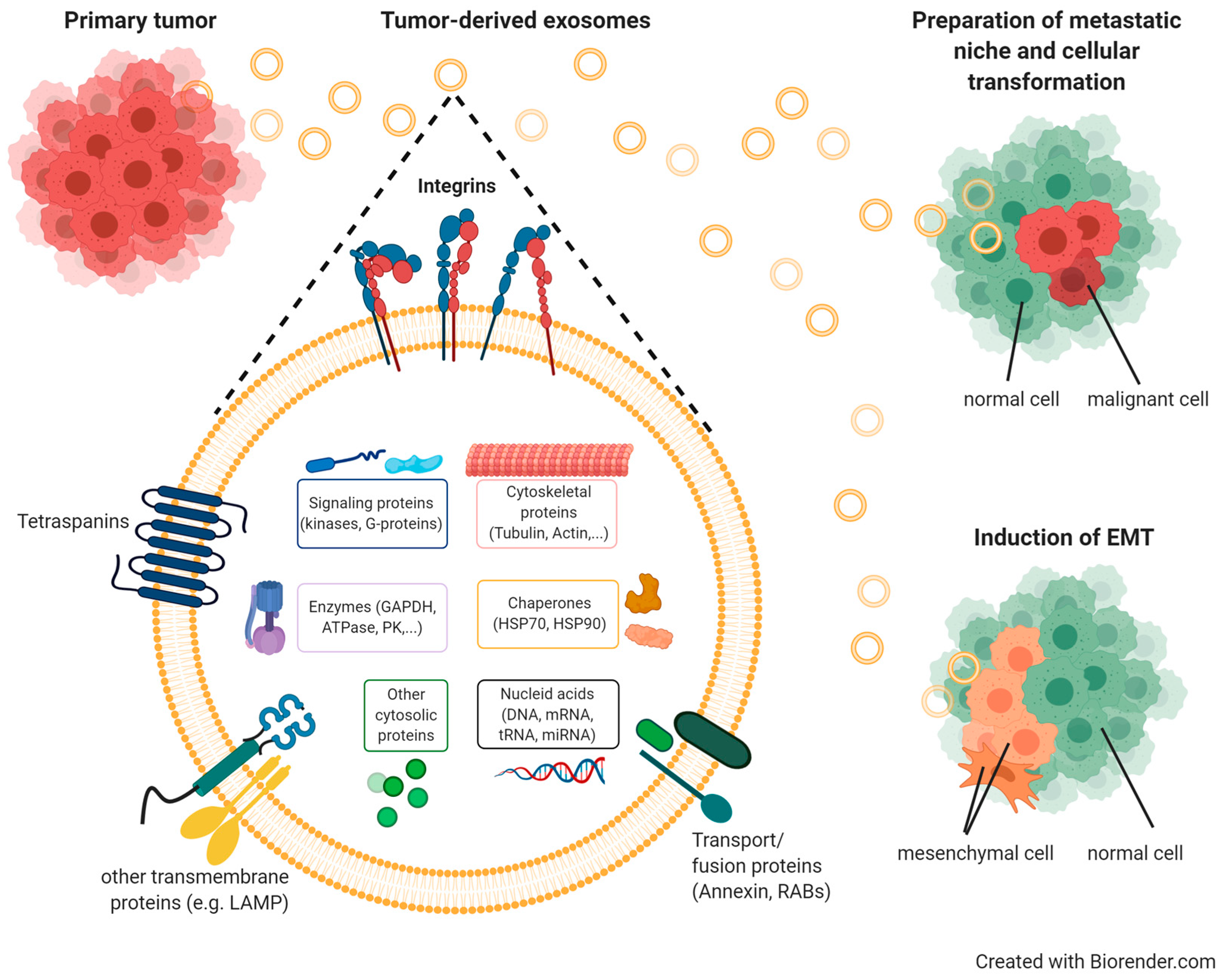
For maintaining tissue homeostasis, cell/cell communication is of high importance. The discovery of extracellular vesicles has revolutionized the perception of these events. Exosomes are the smallest among those vesicles, ranging in size between 30 and 150 nm. They have been detected in a number of body fluids, including blood, urine, and amniotic fluid [263,264]. Their generation starts on early endosomes, followed by their maturation in multivesicular bodies (MVB) via the invagination of the endosomal membrane resulting in intraluminal vesicles (ILVs). Constitutive exocytosis into the extracellular microenvironment occurs in most cell types upon the fusion of ILVs with the cell membrane [263,264,265]. Exosomes are recognized as potent vehicles in intercellular crosstalk via the delivery of their cargo composed of proteins, lipids, metabolites, DNAs, RNAs, and microRNAs to recipient cells [266,267,268,269] (Figure 5).
In addition, during cancer progression and metastasis, enhanced exosome release by tumor cells governs crucial functions by allowing communication with other cells, including endothelial, mesothelial, and stromal cells, as well as fibroblasts and infiltrating immune cells, by transmitting pro-tumorigenic factors to distant sites in specifically tumor cell-targeted organs [270]. This cargo of tumor exosomes includes cell surface-anchored proteases, transmembrane receptors, growth and angiogenic factors, intercellular signaling messengers, as well as ECM molecules and integrins [271,272,273], which all contribute to the preparation and organization of a pre-metastatic niche [274,275,276] (Figure 5). To this end, tumor exosomes also take over distinct functions related to vascular leakiness, stromal cell education at organotropic sites, and bone-marrow-derived cell education and recruitment [266,276,277]. Tumor exosomes harboring e.g., αvβ5, α6β4, or α6β1, are capable of anchoring to distinct ECM proteins, such as fibronectin, vitronectin, laminin, or collagen [278]. Thus, in lung-tropic metastatic exosomes, α6β4 and α6β1 were found, and in liver-tropic exosomes, αvβ5 was found. Thus, strategies targeting tumor exosomal integrins may effectively block organ-specific metastasis [276].
Furthermore, integrins associated with extracellular vesicles may be utilized for tracing tumor cells within an organism, as has been documented for α6, αv, and β1, respectively [279]. These findings underline that circulating tumor exosomes harvested as liquid biopsy may be utilized to predict the tendency of certain tumor cells to metastasize to specific organs and thus predict local tumor cell seeding [280].
In addition, αvβ3 is shuttled via exosomes to recipient non-tumorigenic as well as tumorigenic cells, where it enhances their adhesive and migratory/metastatic properties. As such, the formation of cell membrane protrusions, such as filopodia, in the recipient cell is a consequence of exosomal delivery, endocytosis, and consecutive αvβ3 recycling to the recipient’s cell surface in a functionally active form [281]. Similarly, also, αvβ6 is exosomally delivered to prostate cancer cells, where it localized on the cell membrane provoking increased cell migratory activity on the αvβ6-specific substrate LAP of TGF-β1. Hereby, metastasis is promoted in a paracrine manner [282]. Thus, tumor-derived exosomes are important contributors to the horizontal transfer of distinct integrin subtypes to specific recipient cells thereby affecting their cellular phenotype and behavior.

3.3.4. Exosomes as Therapeutic and/or Diagnostic Tools

The discovery of extracellular vesicles and exosomes and their important functions in the organism has inspired a series of concepts for their implementation as therapeutic and/or diagnostic vehicles. These include their application as circulating biomarkers to be harvested from body fluids as liquid biopsy as well as for the delivery of exosomally packaged drugs to tumor sites. Exosomes are uniquely suited for these purposes, since they are endowed with high delivery efficiency, excellent in vivo stability, and only minor cytotoxicity when compared to other currently used synthetic drug carriers [283]. Moreover, exosomes express transmembrane and membrane-anchored proteins that promote endocytosis, thereby enhancing the delivery of their internal cargo [284]. In fact, several exosomal vehicle variants have already been explored regarding their suitability for targeted drug delivery to cancer cells or cells related to other diseases: (i) native exosomes, (ii) native modified exosomes harboring ligands or receptors and carrying therapeutic agents as cargo, (iii) exosomes derived from genetically engineered cells, or (iv) exosome mimetics [285]. Via the encapsulation of drugs and/or the addition of specific ligands to the exosomal surface by chemical and physical procedures, a broad variety of specific and target-selective vehicles have been generated [286,287]. The ligands explored so far include monoclonal antibodies, proteins, peptides, small molecules, carbohydrates, and aptamers, which were designed to target different receptors overexpressed on the recipient cell membrane. Among those are human epidermal growth factor receptor 2 (HER2), VEGFR, e-selectine, nucleolin, transferrin, folate receptor, EGF receptor, and αvβ3 [288].
Regarding integrin targeting, several effective delivery platforms have been established. Prominent examples are doxorubicin-loaded exosomes, which have e.g., been decorated with RGD-based peptides on their surface and were shown to enable efficient and specific drug delivery into αv-expressing breast cancer models after intravenous injection [289]. In another example, doxorubicin-loaded exosomes were engineered to express the exosomal membrane protein Lamp2b fused to the αv-specific iRGD peptide and also showed highly efficient targeting of breast cancer models in vitro and in vivo [289]. Other types of exosomes have been used to target ischemic brain lesions in mouse models. These exosomes were surface modified with cyclo[RGDyK] and loaded with curcumin, and they were able to cross the blood–brain barrier to reduce inflammation and apoptosis [290]. Lastly, cyclo[RGDyK] decorated exosomes loaded with miR-210, a robust target of hypoxia-inducible factors (HIF), were also able to increase survival in mice after brain ischemia [291].
Whereas the role of distinct exosomal integrins has meanwhile been intensively explored, their function in recipient cells is not yet fully clear. In a recent study, a function of β3-integrin in the endocytosis of vesicles was described, emphasizing its role in exosomal intercellular communication critical for cancer metastasis. Based on earlier findings on the role of β3 in tumor stress resistance and metastasis in triple-negative breast cancer [292], its contribution to exosome biogenesis and endocytosis has been investigated [293]. Hereby, it was shown that clonal tumor cell growth stimulated by extracellular vesicles depended on the presence of β3 on the recipient tumor cell surface at the metastatic site, which permitted cell entry of the extracellular vesicles via β3-mediated internalization. By exploring the underlying mechanism, it was found that syndecans were involved in β3-mediated extracellular vesicle capture. Syndecans, heparan sulfate-decorated cell-surface proteoglycans, which recognize heparin-binding motifs, had been shown before to associate with β3 [294]. By applying heparin, which served as a glycosaminoglycan mimetic of heparan sulfate, extracellular vesicle uptake was blocked, indicating that heparan sulfate proteoglycans served as the bridge between exosomes and β3. The process was accompanied by the interaction of β3 with FAK leading to its activation and consequently, to clathrin-mediated and thus dynamin-driven endocytosis [293].

4. Design and Development of RGD-Based Integrin Ligands for Biomedical Applications and Translation

4.1. From Natural ECM Proteins to Synthethic RGD-Based Integrin Ligands: The Generation of Highly Selective and Affine Integrin Ligands

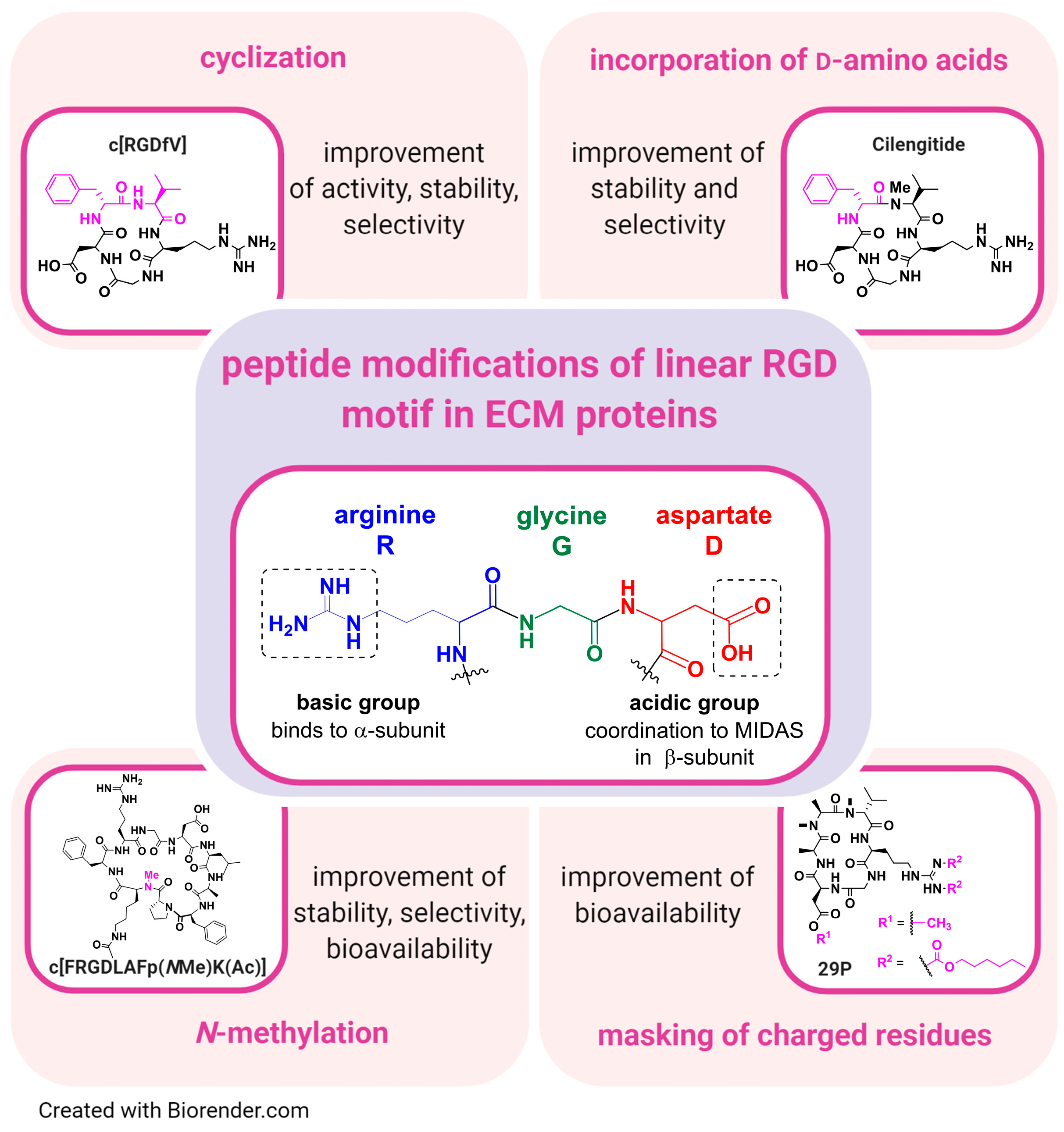
Historically, linear and cyclic peptidic integrin ligands had been designed, mimicking the RGD binding sequence of natural ECM proteins [295]. Derived from RGD-based cyclic peptides, antibodies, functionalized peptides, aptamers (short nucleic acid sequences), and non-peptidic RGD analogues had been developed as new classes of integrin binding compounds/antagonists [14,24,57,150,296,297,298,299]. Hereby, peptidomimetics, peptides, and small molecule ligands either imitate or block the biological effects of a natural ECM ligand [14,24,57,150,296,297,298,299,300]. Nonetheless, the overall goal in the design strategy was to generate highly selective ligands, which bind with high affinity to a respective integrin subtype (Figure 6). Given the number of integrin subtypes and their numerous and distinct roles in physiological and disease processes, a controlled therapeutic approach is usually aimed at highly selective ligands, while non-selective ligands could lead to strong side effects. For example, unwanted αIIbβ3 activity in cancer therapy can lead to severe hemorrhage [110,139]. Through cyclization (e.g., in case of cyclo[RGDfV]) as well as the incorporation of d-amino acids (e.g., Cilengitide), highly active and metabolically stable peptidic ligands were generated (Figure 6) [295,301,302,303]. In addition, the different modifications shown in Figure 6 may be combined, as e.g., for cyclo[RGDfV] or 29P, which harbor d-amino acids in their structures. In contrast, linear peptides seem to be rather metabolically instable and are often cleaved in vivo within a few minutes. Moreover, disulfide bridged peptides have lower in vivo stabilities than so-called homodetic cyclic peptides (cyclized head-to-tail). Furthermore, N-methylation of external and solvent-exposed amide bonds in cyclic peptides often resulted in a significant enhancement of selectivity among the group αvβ3–α5β1–αIIbβ3 [304,305,306,307]. On top of that, N-methylation confers better pharmacokinetic properties, such as enhancement of blood half-life in vivo (e.g., cyclo[FRGDLAFp(NMe)K(Ac)] (Ac = acetyl) (Figure 6) [300,305,308]. All synthetic integrin ligands share an essential acidic group for metal-coordination in the MIDAS region as a common feature, such as a carboxylate moiety, e.g., aspartate, as found in natural ECM ligands (Figure 6). Moreover, potent isosteric replacements, such as hydroxamic acids for αvβ3 and α5β1 [57], had been synthesized as well. Alongside the acidic group, which is necessary for binding to the β-subunit, a basic group is needed for optimal α-subunit interaction (Figure 6). For example, the direct introduction of an Arg (pKa ≈ 13.8) [309] or its isosteric replacement with less basic groups, such as 2-aminopyridine (pKa ≈ 7), 1,8-tetrahydronaphthyridine (pKa ≈ 7), or 2-aminoimidazole (pKa ≈ 8.5) [110,310,311,312]. However, it should be mentioned that e.g., Asp residues of different integrin α-subunits bind the guanidinium group of the Arg in the integrin ligand in a different way, e.g., in the αv integrins side-on but in the α5 integrins end-on (and side-on) [313]. Furthermore, through the isosteric replacement of the RGD mojety, peptidomimetic integrin ligands were generated, harboring higher metabolic stability and activity in comparison to linear, peptidic RGD-containing integrin ligands. For peptidic ligands in general, the basic and acidic moieties are arranged on a rigid peptide skeleton in order to achieve the most suitable integrin receptor interaction [314,315,316]. In addition, in case of peptidomimetic integrin ligands, a rational design, e.g., by docking studies, is applied for the generation of selective ligands. Furthermore, in case of αvβ3 ligands, an aromatic residue close to the acidic group can bind in a hydrophobic cleft, leading to an enhanced affinity of the integrin ligand [13,317]. The different affinities of many integrin ligands have been recently reviewed [318].
In general, many parameters have to be considered toward ligand translation into clinical use (e.g., affinity, selectivity, and pharmacokinetics). Whenever metabolic stability is required, cyclic peptides or peptidomimetics should be preferred.

4.2. Design of Synthetic RGD Binding Integrin Ligands

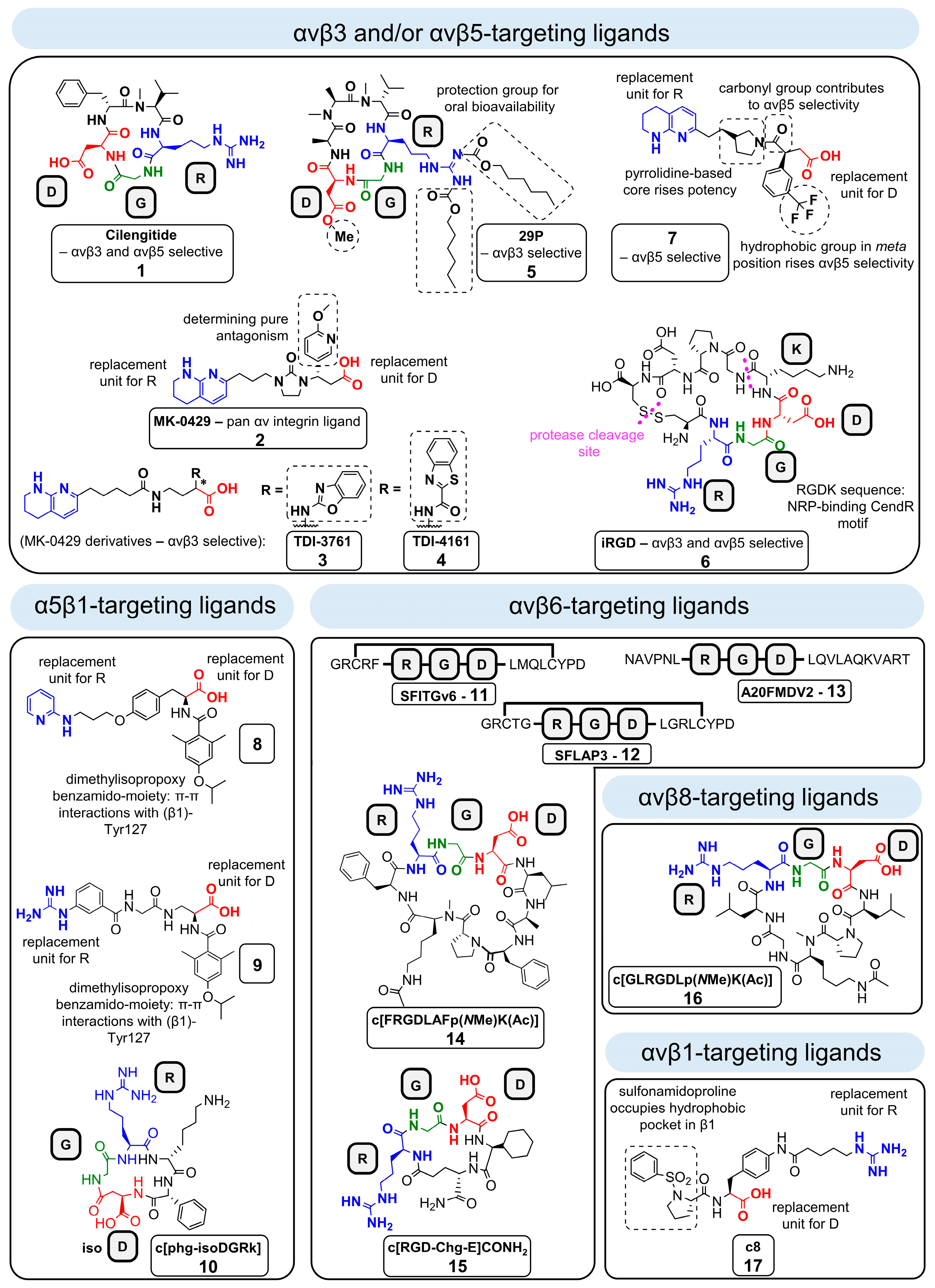
Integrin ligands targeting αvβ3. Due to the important role of αvβ3 in angiogenesis and tumor progression, many different ligands targeting this integrin have been designed over the past decades [19]. Nonetheless, only a few αvβ3 small molecule antagonists have entered clinical trials so far [10,110,319,320,321,322]. One of the major drawbacks of the most previously generated αvβ3 antagonists is their complex concentration-dependent receptor pharmacology. In fact, that has been uncovered after the first clinical trials with Cilengitide (cyclo[RGDf(NMe)V], ligand 1, Figure 7), which is a cyclic pentapeptide designed for the treatment of glioblastoma, whose performance did not lead to the expected anti-angiogenic effects. Cilengitide represents a ligand for both αvβ3 and αvβ5 (IC50 αvβ3 = 0.61 nM, IC50 αvβ5 = 8.4 nM) [318]. Since this compound was extensively described in recently published reviews, we briefly describe some important facts regarding the clinical evaluation of Cilengitide in this review [26,323]. Based on early phase I and II clinical studies in glioblastoma patients, there was initially great hope for both Cilengitide high-dose monotherapy [324] and combination treatments [325,326]. In the end, large multicenter randomized phase II (CORE) and phase III (CENTRIC) clinical cancer trials using Cilengitide in combination with standard treatment for patients with newly diagnosed glioblastoma or by intensified doses during radiotherapy, respectively, failed to reach the primary endpoints of those studies [154,160]. This disappointing clinical efficacy of Cilengitide instigated the search for explanations for these failures, along with the exploration of alternative applications for its clinical use. Unexpectedly, the administration of low-dose Cilengitide in vitro and in animal models revealed that opposite to its intended purpose, it promoted tumor angiogenesis instead of inhibiting it [157].
Accumulating evidence indicates that at low concentrations, Cilengitide actually operates as an agonist, where it binds to the resting state of αvβ3, which harbors a bent conformation [66,157,327]. This induces conformational changes resulting in erected extracellular domains presenting a high-affinity conformation of the headpiece. Subsequently, upon further conformational changes, the two integrin legs are separated and integrin signaling is triggered (see Section 2). In contrast, at high concentrations, Cilengitide acts as an antagonist, which effectively inhibits integrin signaling. This dose-dependent functional switch was confirmed in vitro, showing that Cilengitide treatment with concentrations below the IC50 value showed agonistic signaling, while antagonistic effects occurred at concentrations above the IC50 [157,328]. How this applies to organisms in vivo, i.e., in which a systemic dose results in a “low” (agonistic) or a “high” (antagonistic) concentration at the target site (i.e., the primary tumor and/or metastasis), has not been systematically studied. In this respect, especially due to the rapid pharmacokinetics, i.e., the 4 h blood half-life of Cilengitide in humans [62,329], even high systemic doses likely result in low tumor doses several hours after drug application. For example, in the CENTRIC study (Merck), patients received 2 g Cilengitide intravenously twice weekly, which will be reduced to only 1 µg after 3.5 days, and to an even lower dose finally at the tumor site. Hence, the agonistic low dose regimen cannot be avoided in such a dose set-up [154,160]. In contrast, in case of Cilengitide, it is also published that low-dose agonistic action leads to vascular promotion effects and stimulation of VEGF-mediated angiogenesis (see Section 3.2.2) [27,157]. Recently, interesting applications of the vascular stabilization effect in immune therapy, cancer treatment, and sepsis have emerged (see Section 6.1) [27,37,328,330].
In addition to Cilengitide [62,331], this dose-dependent functional switch has also been observed for MK-0429 (ligand 2, Figure 7) [319,320], which is an orally available αvβ3 integrin ligand that has been applied for the treatment of osteoporosis and melanoma [332,333]. The case of MK-0429 is curious, as it has initially been described to target only αvβ3, but later low nanomolar affinities with IC50 values of 1.6, 2.8, 0.1, 0.7, 0.5, and 12.2 nM for αvβ1, αvβ3, αvβ5, αvβ6, αvβ8, and α5β1 [333], respectively, were reported, marking MK-0429 as an αv pan-integrin antagonist.
In this regard, two pure αvβ3 antagonists, TDI-3761 (ligand 3, Figure 7) and TDI-4161 (ligand 4, Figure 7), were synthesized through modifying MK-0429 in such a manner that a π–π interaction with the Tyr122 residue from the β3-subunit was established, freezing the integrin in an inactive conformation, thereby abrogating agonistic actions of the ligand [328]. The phenomenon of pure αvβ3 antagonism was described in a former study, where a high affinity mutant (hFN10) of 10 kDa wild-type fibronectin (wtFN10, partial agonist) displayed pure αvβ3 antagonism [334]. Similar to synthetic ligand binding, wtFN10 binding triggers tertiary changes in the β3 subunit, which persist after the dissociation of the ligand, making wtFN10 agonistic. Contrary to this, hFN10 undergoes π–π interactions with the integrin ligand upon its binding, avoiding conformational changes within the β3 subunit [334]. TDI-3761 and TDI-4161 block αvβ3-mediated cell adhesion to ECM ligands without having an impact on integrin conformation as confirmed by antibody binding, electron microscopy, X-ray crystallography, and receptor priming studies [328].
Moreover, in contrast to treatment with low-dose Cilengitide, both compounds did not exert pro-angiogenic effects, such as aortic vessel sprouting, which could lead to tumor progression in vivo [328]. Thus, these pure antagonists represent a potential alternative to be extensively studied as anti-angiogenic therapy in tumors. Since TDI-4161 and TDI-3761 conveyed inhibitory effects on bone resorption, new treatment options for osteoporosis are conceivable [328]. Hence, recent developments show progress toward the development of pure antagonistic and pure agonistic ligands of αvβ3, which will hopefully lead to drugs for different medical use.
While the optimization of metabolic stability, selectivity, molecule rigidity, activity, bioavailability, and other factors was achieved [295,301,302,303,304,305,306,307,335,336], the lack of oral bioavailability of peptidic integrin ligands remained a major limitation for clinical use. With the synthesis of the αvβ3-selective cyclic hexapeptide 29P (ligand 5, Figure 7), a highly systematic design strategy for the receipt of a biologically active as well as orally available peptide was described for the first time [308]. Different steps in the generation of the lead compound 29P were required. First, an identification of intestinal permeable N-methylated cyclic hexa-Ala peptides was necessary in order to obtain permeable peptidic backbone structures [308]. Then, by using a spatial screening approach [295], the RGD-motif was reintroduced into the cyclic hexa-Ala backbone to gain a bioactive compound. After optimizing this ligand via suitable flanking residues, the best ligands for RGD-recognizing integrin subtypes were selected via an enzyme-linked immunosorbent assay (ELISA)-like test system yielding peptide 29 with the following IC50 values: αvβ3—0.6 nM, αvβ5—145 nM, αvβ6—120 nM, αvβ8 > 1000 nM, and α5β1—21 nM, respectively [308,318]. However, the charged residues of Arg and Asp abrogated cell permeablility. In order to regain the cell permeability of this highly active cyclic hexpapeptide, the charged functional groups were protected by the pro-drug concept, i.e., by introducing lipophilic protection groups, yielding 29P [308]. After transport in the bloodstream, the protection groups are rapidly cleaved off, and the bioactivity of the RGD-peptide 29 is restored. 29P was subsequently analyzed in vivo regarding its oral bioavailability, using mice bearing Lewis lung carcinoma tumors [308]. Low concentrations of orally administered 29P had a similar effect as intravenously injected non-orally available 29 (29P without the lipophilic protection groups) or Cilengitide, and they resulted in enhanced VEGF-mediated angiogenesis by upregulation of the VEGFR2 expression [308]. However, recent approaches using low-dose Cilengitide indicated that its induction of vascular promotion could render it a promising agent for combination therapy, since increased tumor vessel stability can enhance the delivery of chemotherapeutics to the tumor site [27,330,337]. Consequently, 29P could be an ideal orally available peptidic pro-drug for vascular promotion therapy in combination with chemotherapy [308].
In addition, a tumor-penetrating disulfide bridge harboring peptide, called iRGD (cyclo[CRGDK/RGPD/EC]; ligand 6, Figure 7) was identified, targeting αv-integrins specifically expressed on endothelial cells of tumor vessels, i.e., αvβ3 together with αvβ5, when injected intravenously [338,339]. After integrin binding, iRGD is proteolytically cleaved within the tumor, leading to the peptide sequence CRGDK/R [22,340]. While losing subsequently much of its integrin-binding capability, the truncated peptide CRGDK/R gained affinity for NRP-1, which is a membrane-bound co-receptor that binds among other ligands also the VEGF through the C-terminal exposure of the active C-end Rule (CendR) motif. The CendR motif of iRGD is hereby RGDK/R. In general, for NRP-1 binding, Gly and Asp can be replaced by any amino acid, whereby instead of Arg, Lys can be used: R/KXXK/R [22,340]. Via the interaction of the CendR motif with NRP-1, tumor tissue penetration is triggered, and iRGD has been documented to deeply enter into extravascular tumor tissue [22,339,340]. iRGD can be conjugated to drugs or imaging agents or co-administered with them, allowing the extravasation and penetration of the co-applied agents after the binding process of CendR and NRP-1 [338,340]. This is currently explored in a phase I clinical study, where metastatic prostate cancer patients first undergo monotherapy with iRGD, followed by a combination therapy with nab-paclitaxel and gemcitabine (NCT03517176). Furthermore, iRGD was used as carrier molecules for the transport of drugs (via nanocarriers) or imaging agents to selectively target tumor tissues. Through amidation, maleimide-thiol chemistry, Michael addition, and alkyne-azide click chemistry, respectively, nanoparticle surfaces of lipid micelles, Abraxane® (albumin/paclitaxel nanocomplex) as well as iron oxide nanoworms were conjugated to functionalized iRGD molecules for the targeted delivery of drugs such as doxorubicin, paclitaxel, or vandetanib [341]. However, the studies resulted in a slightly lower tumor accumulation of the drugs after targeted delivery via iRGD in comparison to co-administration [341,342]. The limited number of the target receptors on the vasculature may hinder conjugated iRGD to promote an enhanced drug absorption, while unconjugated, co-administered iRGD may trigger a bulk transfer of drugs into the tumor [341].
Integrin ligands targeting αvβ5. A major challenge for the development of αvβ5-selective ligands is given by its close homology to αvβ3. A homology model of αvβ5 exhibited high similarity of β3 and β5, but the larger selectivity determining loop in β5 allows less space for ligands [343]. Hence, some ligands for αvβ3 do not fit into the αvβ5 receptor, but most αvβ5 ligands bind also αvβ3, leading to ligands such as Cilengitide or iRGD targeting both αvβ3 and αvβ5 [343]. Through a recent Structure–Activity Relationship (SAR) survey among a series of amide-containing 3-aryl-succinamic acid-based RGD mimetics, the α,α,α-trifluorotolyl residue containing ligand 7 (Figure 7) was discovered as a highly selective αvβ5 ligand, harboring an 800-fold lower affinity toward αvβ3 [344]. Docking experiments revealed that the amide-containing rigid core of ligand 7 represents the selectivity determining part of the molecule toward the other αv-integrins [344]. More precisely, the central aryl ring substituted by the CF3 group represents an ideal moiety for αvβ5 rather than αvβ3 binding [344].
Integrin ligands targeting α5β1. Regarding the selectivity of ligands discriminating between α5β1 and αvβ3, it was demonstrated that it may be directed through a methylation of the guanidinium group within the RGD motif. An integrin ligand binds to αv side-on, whereas it binds to α5 end-on [313]. Through methylation at the right position of the guanidinium group, either the side-on or the end-on binding can be suppressed, leading to a rise in selectivity toward α5β1 or αvβ3, which was demonstrated for Cilengitide [313]. The modification of the guanidinium group can be used as well for the improvement of the α5β1 selectivity [313].
A set of α5β1-targeting antagonistic peptidomimetics was synthesized, leading to the identification of the ligands 8 and 9 (Figure 7) as the most promising candidates [15,57,345]. Through rational design, it was tried to mimic the RGD motif by replacing Arg by a pyridine (ligand 8) or a guanidinium group (ligand 9) and the Asp of RGD by propionic acid (ligands 8 and 9) [15,57,345]. The dimethylisopropoxy benzamido goup in both ligands undergoes π–π interactions with the (β1)-Tyr127, contributing to α5β1 selectivity. The obtained integrin antagonists exhibited high selectivity and a low-nanomolar affinity toward α5β1, representing a promising vehicle structure for imaging agents and drug targeting [15,57,345].
The peptide sequence isoDGR was first described in aged fibronectin. Hereby, isoD is formed from Asn (N) through an in situ rearrangement of the peptide sequence NGR [346,347,348]. The synthetic introduction of the isoDGR moiety as a replacement of RGD in cyclic pentapeptides of the sequence cyclo(XisoDGRX) (X = any amino acid) has been described to generate integrin ligands with high αvβ3 or α5β1 selectivity, depending on the introduced amino acids X [317]. Regarding αvβ3 selective isoDGR-derived compounds, the reader is directed to recent publications about the prospects of their future use in cancer therapy [346,349,350]. The isoDGR derived cyclic peptide cyclo[phg-isoDGRk] (phg = d-phenylglycine; ligand 10, Figure 7) was designed as a highly selective α5β1 (IC50 = 8.7 nM) integrin ligand, which also displayed targeting of αvβ6 (IC50 = 19 nM) [317]. This ligand was further developed and transformed into the 99mTc-radiolabeled compound 99mTc-HisoDGR for its use as a Single Photon Emission Computed Tomography (SPECT) imaging probe of α5β1-positive glioma (see Section 5) [351,352]. Regarding cancer therapy, no attempts were published, but the use of cyclo[phg-isoDGRk] as a drug vehicle is conceivable.
Integrin ligands targeting αvβ6. The discovery of αvβ6 as a tumor biologically relevant and overexpressed biomarker in many cancer types was followed by the development of several highly selective ligands. The reported αvβ6 ligands, which did not show anti-cancer activity, could be especially useful as molecular imaging vehicles for diagnostic purposes, e.g., for PET imaging, and for integrin-targeted cancer therapy through conjugation to drugs or nanoparticles (NP).
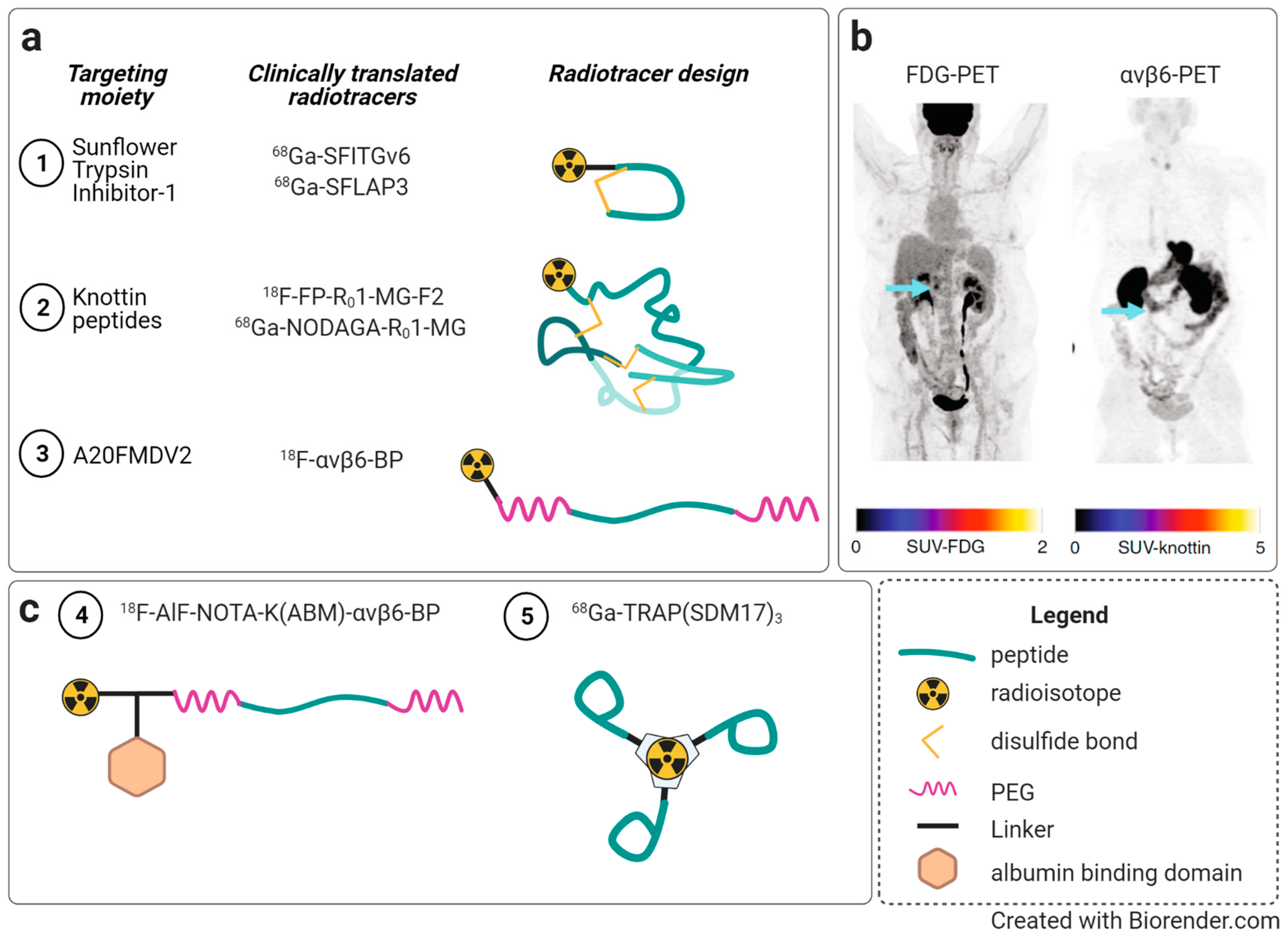
A disulfide-bridged 16-amino acid peptide, SFITGv6 (amino acid sequence: GRCRFRGDLMQLCYPD, ligand 11, Figure 7), containing the integrin targeting amino acid sequence FRGDLMQL with high affinity and selectivity for αvβ6 was identified within a Sunflower Trypsin Inhibitor-1 (SFTI-1)-based phage-displayed library [353,354]. The peptide demonstrated a homogenous binding to αvβ6 in head and neck squamous cell carcinoma (HNSCC) as well as in breast and lung cancer-derived metastases, which harbor elevated levels of αvβ6 [353]. In a first study in which HNSCC and non-small cell lung cancer patients received 68Ga-DOTA-labeled SFITGv6, a specific accumulation in tumors had been observed [353]. SFITGv6 may represent a promising tracer for imaging and possibly endoradiotherapy of αvβ6-positive cancers [353]. In further studies, diverse RGD-based SFTI-1 derivatives were generated, leading to the identification of another potent αvβ6-targeting peptide, SFLAP3 (amino acid sequence: GRCTGRGDLGRLCYPD), which is a ligand based on LAP3 (ligand 12, Figure 7) [354]. In comparison to SFITGv6, SFLAP3 exhibited improved affinity for αvβ6 in HNSCC in vitro and in vivo (mouse and human), rendering it also a promising small molecule compound for cancer diagnosis as well as therapy [354].
Furthermore, the linear 20-amino acid peptide A20FMDV2 with the amino acid sequence NAVPNLRGDLQVLAQKVART (ligand 13, Figure 7) was described as a potent αvβ6-inhibitor in vivo [355]. The sequence of the peptide derived from an envelope protein of the foot-and-mouth disease virus (FMDV), which mediates virus infection via binding to αvβ6 [355,356,357]. Within the peptide, the sequence RGDLQVL is responsible for αvβ6 targeting. A20FMDV2 was conjugated to 18F-fluorobenzoic acid for Positron Emission Tomography (PET) imaging of αvβ6-positive cancer in vivo (see Section 5). Its use has also been investigated for cancer therapy by conjugating the DNA-binding pyrrolobenzodiazepine-based payload SG3249 (tesirine) to A20FMDV2 [127]. In vitro studies showed the αvβ6-dependent cytotoxicity of this peptide–drug conjugate, which is called SG3299. In vivo, SG3299 treatment strongly reduced tumor growth and increased life span in mice with αvβ6-expressing xenografts. The nonapeptide cyclo[FRGDLAFp(NMe)K(Ac)] (Ac = acetate, ligand 14, Figure 7) represents a highly selective and affine cyclic nonapeptide for targeting αvβ6 (IC50 αvβ6 = 0.3 nM) [313,358]. The cyclic pentapeptide SDM17 encompassing the sequence cyclo[RGD-Chg-E]-CONH2 (Chg = l-cyclohexylglycine, ligand 15, Figure 7) also represents a highly affine αvβ6 targeting integrin ligand (IC50 αvβ6 = 1.3 nM) [359]. These peptidic ligands, together with other ligand architectures, e.g., linear peptides and knottin peptides, were developed as molecular imaging agents, which we review in detail in Section 5.
Integrin ligands targeting αvβ8. The octapeptide cyclo[GLRGDLp(NMe)K(Ac)] (ligand 16, Figure 7, IC50 αvβ8 = 8 nM) represents a selective and highly affine αvβ8-targeting ligand, bearing the potential to be used as a drug delivery vehicle as well as an imaging agent [360]. This first cyclic αvβ8-binding peptide featured a strong discriminating power of at least two orders of magnitude against the other RGD-recognizing integrins [360]. The ligand has been used so far for tumor imaging, which is further described in Section 5.
Integrin ligands targeting αvβ1. The first and only peptidomimetic inhibitor for αvβ1, called “c8” (ligand 17, Figure 7, IC50 αvβ1 = 0.089 nM), reduced TGF-β activation and consequently chemically induced pulmonary and liver fibrosis in mice, hence representing a promising αvβ1-targeting integrin ligand [141,142].
Pan-integrin ligands. A pan-inhibitor is defined as a compound that does not only display an inhibitory effect on an individual but rather on multiple integrin subtypes. In general, pan inhibitors are supposed to generate a better therapeutic outcome by the simultaneous inhibition of several integrins, compared to those targeting exclusively one integrin subtype, since it better addresses the frequently observed heterogeneity of integrin expression in tumors [110,361]. Furthermore, cancer cells were able to modulate their integrin expression pattern as a consequence of cancer treatment, which might not affect the efficacy of pan-selective ligands but could induce resistance to highly subtype-selective integrin ligands [362]. The small molecule αv-inhibitor CWHM12 represents such a pan-integrin ligand targeting both liver and lung fibrosis, which is driven through αv-integrins. It is assumed that pharmacological targeting of all αv-integrins may lead to better therapeutic effects in anti-fibrosis therapy [363]. CWHM12 seems to inhibit TGF-β binding, which is a major profibrogenic cytokine [363]. The compound MK-0429, which was mentioned in Section 4.2, is as well a small molecule αv pan-integrin ligand.

5. Integrin Targeted Molecular Imaging and Radiotherapy

The improved definition of the role of integrins as predictive biomarkers for response, prognosis, and even as therapeutic targets is an important subject of ongoing investigations. Nevertheless, the consistent overexpression of integrins in a large number of various tumor types strongly supports their roles as targets for molecular imaging. Somewhat independent from the biological functions of distinct integrins, their high expression levels in tumors can be exploited to visualize tumors and metastasis by diagnostic imaging. In addition, integrin imaging agents could be used to stratify patients for targeted therapies, to assess treatment response, and to monitor tumor growth. In this regard, nuclear imaging, including Positron Emission Tomography (PET) and Single Photon Emission Computed Tomography (SPECT), are the most established modalities for integrin imaging. Moreover, integrin imaging has also been described for a wide range of other modalities, e.g., fluorescence-guided surgery, optoacoustic imaging, ultrasound imaging, magnetic resonance imaging, Raman spectroscopy, and multimodal combinations thereof. The abundance of recent publications suggests continuous and intense research activities into pre-clinical development and clinical translation of integrin imaging probes, as we outlined below.
The first integrin-targeted molecular imaging approaches were focused on αvβ3, with the goal to develop companion diagnostics to obtain quantitative information on the angiogenic activity within tumors and to stratify patients via PET or SPECT imaging for αvβ3-targeted therapy. Therefore, αvβ3-targeting RGD-based peptides have been radiolabeled, e.g., with Fluorine-18, Gallium-68, Copper-64, or Technetium-99m, and entered pre-clinical and clinical evaluation [11,364,365,366,367]. So far, the collective evaluation of data from an abundance of studies revealed that imaging of angiogenesis is possible, but the interpretation of αvβ3 imaging data is complicated by the fact that αvβ3 is not exclusively expressed on newly formed tumor vessels, but also on tumor cells and macrophages, as well as often in normal tissues [368,369]. Furthermore, it was shown that angiogenesis does not depend on the presence of αvβ3 [368].
An important lesson from these early studies on integrin-targeting molecular imaging was that focusing on the angiogenic activity of tumors with αvβ3-imaging does not do justice to the multiple roles of αvβ3 and other integrins in tumor development, progression, and prognosis, so its clinical merit remains to be fully defined. The ongoing optimization of the pharmacokinetic properties of ligands for αvβ3 and the availability of selective ligands for other integrins (e.g., α5β1, αvβ6, and αvβ8; as described in Section 4.2 of this article), in conjunction with a better understanding of their expression patterns and biological functions, has certainly contributed to advances in molecular imaging of integrins. Furthermore, novel diagnostic and theranostic concepts of integrin-coated nanomaterials have emerged, such as integrin-targeted NPs for PET, ultrasound, and Raman spectroscopy. In this chapter, we will discuss novel developments in the field of molecular imaging of integrins, with particular attention on, but not limited to, studies published between 2015 and 2020.

5.1. Nuclear Imaging (PET/SPECT)

An extensive review on PET imaging probes summarizes the pre-clinical development and progress of 8 RGD-based PET tracers for αvβ3 imaging in clinical studies until 2016 [370]. Here, we focus on new developments in PET and SPECT imaging, not only those targeting αvβ3, but also other integrin subtypes, which might prove clinically even more useful. We will consider how recent studies are addressing challenges encountered in earlier studies and analyze additional evidence for the clinical relevance of RGD-based integrin imaging.
Nuclear imaging of integrin αvβ3. The earliest PET and SPECT imaging ligands consisted of radiolabeled peptide monomers, e.g., the cyclic pentapeptide 18F-Galacto-RGD, which was the first candidate to enter in-human imaging and showed the proof-of-principle of in vivo integrin imaging [367]. The drawback of this compound was the complex time-consuming radiosynthesis, which led to the exploration of simpler and faster radiolabeling strategies for integrin ligands (see [371] and [372] for reviews on this topic). In addition, it was also discovered in the past decade that the multimerization of RGD-based ligands can improve their affinity and tumor uptake compared to their respective monomers in PET and SPECT imaging approaches [373,374]. In this regard, it is important to note that higher affinity and tumor uptake of a ligand does not necessarily improve its imaging performance (e.g., with respect to tumor contrast) [375]. In fact, upon ligand multimerization, also unfavorable pharmacokinetic properties had been observed, such as high non-specific binding or slow wash-out from blood or non-tumor tissues (i.e., background uptake in the liver complicating detection of liver metastasis). To this end, preclinical studies with radiolabeled ligand dimers repeatedly showed increased tumor uptake compared to monomers while maintaining suitable pharmacokinetics [376,377]. Several dimeric αvβ3-targeted constructs, based on the cyclic pentapetide cyclo[RGDxK] (x = f,y) with different PEG-linker modifications, namely 18F-Alfatide (18F-AlF(NOTA-2P-RGD2)) [378], 18F-Alfatide II (18F-AlF(NOTA-2P-RGD2)) [379], and 18F-FPPRGD2 [380] have since been clinically evaluated. They appeared to have higher sensitivity and specificity for the detection of primary and metastatic tumor lesions than monomers [372]. However, this is ultimately difficult to evaluate, since most studies were performed in small cohorts of patients afflicted with different tumor entities. In addition, data for the direct clinical comparison of the performance of a monomer versus a dimer in the same patient are not available. Clinical evaluation of αvβ3-targeting dimers, often in comparison to 18F-FDG in proof-of-concept studies, is ongoing. Recent studies include the imaging of cancer of the esophagus [381], breast [382], and lung [383,384,385,386,387], as well as skeletal [379], brain metastases [388], and radioiodine refractory thyroid cancer [389]. Several of these studies reported higher diagnostic accuracy of αvβ3-targeting dimers than 18F-FDG, while in others, comparable performance was reported. The database Clinicaltrials.gov does currently not list large-scale clinical studies that describe PET/CT of 18F-Alfatide, 18F-Alfatide II, or 18F-FPPRGD2.
Tetra- and octameric RGD-based peptides (based on cyclo[RGDyK]) also showed increased tumor uptake and retention. In addition, they displayed a delayed blood clearance, which means that they are more suitable for imaging with longer-lived isotopes, such as Cu-64, to be able to image at the time-point where the highest tumor-to-background ratios are achieved [366,390].
More recently, the development of suitable chelators also allowed the introduction of 68Ga-labeled cyclic RGD-based trimers, which resulted in a good compromise between increased tumor accumulation and suitable pharmacokinetics in preclinical studies. Recently, four αvβ3-targeting 68Ga-labelled multimeric cyclic RGDfK peptides (one dimer and three trimers [391,392,393,394]) were directly compared [394]. These tracers were based on different chelators (1,4,7,10-Tetraazacyclododecane-1,4,7,10-tetraacetic acid (DOTA), 1,4,7-triazacyclononane phosphinic acid (TRAP), fusarinine-C (FSC), and tris(hydroxypyridinone (THP)), resulting in distinct topological structural arrangements, but they were all neutrally charged at physiologic pH. The authors found a general suitability of all four compounds for the imaging of αvβ3-expressing xenografts, but noticeable differences with regard to total tumor uptake and different tumor-to-normal tissue ratios [394]. In particular, the highest tumor-to-background (tumor:blood, tumor:muscle) ratios were reported for 68Ga-TRAP(cyclo[RGDfK])3 [391], but the highest tumor uptake was reported for 68Ga-FSC(cyclo[RGDfK])3 [392]. To date, such systematic comparisons of the pharmacokinetic and pharmacodynamic properties of different multimers of the same peptide sequence remain scarce but provide important insights into options for optimizing integrin-targeted tracers for in vivo imaging. Importantly, the selection of the best tracer will also depend on the needs of the intended clinical application, e.g., if it is more important to achieve the highest possible contrast or the highest possible tumor uptake.
In addition to multimeric probes carrying multiple identical integrin ligands, heterodimeric probes combining integrin ligands with ligands directed to other target structures have been explored (e.g., reviewed in [395]). The rationale behind this strategy is to benefit from the increased affinity and tumor retention of peptide dimers and to tackle the heterogeneity of target expression in distinct tumor types by dual receptor targeting, enabling imaging tumors that express either or both targets [396]. A F-18 labeled construct targeting αvβ3 and NRP-1 via an RGD-ATWLPPR heterodimer showed higher uptake and tumor-to-background ratios in a glioblastoma model than the respective monomers [397]. Another heterodimeric approach, targeting the Gastrin-Releasing Peptide Receptor (GRPR) with a bombesin (BBN) analog ligand and αvβ3 (via cyclo[RGDyK]) [384,398], has recently undergone proof-of-concept clinical evaluation [387,399]. In a small study cohort of 22 breast cancer patients, 68Ga-BBN-cyclo[RGDyK] showed significant uptake in primary lesions, axillary lymph nodes, and distant metastases [399]. A direct comparison to monomeric 68Ga-BBN PET in 11 of these patients revealed that tumor uptake and lesion detection rates were significantly higher for the heterodimeric 68Ga-BBN-cyclo[RGDyK]. Simultaneous evaluation of the Somatostatin-2-Receptor (SSTR2), which was targeted with the peptide TATE, and αvβ3 expression using the heterodimeric PET probe 68Ga-NOTA-3P-TATE-cyclo[RGDyK] in 32 patients afflicted with lung cancer or neuroendocrine neoplasms resulted in significantly higher tumor-to-background ratios than the monomeric 68Ga-NOTA-TATE, 68Ga-NOTA-cyclo[RGDyK], and 18F-FDG, respectively [400]. These recent clinical evaluations showed a very good correlation between the PET signal and the expression of αvβ3 and the respective other target determined by immunohistochemistry on tumor cells. Additional heterodimeric/heterobivalent ligands at the preclinical evaluation stage, including PET and SPECT tracers targeting GRPR/αvβ3 or PSMA/αvβ3 [401], were recently reviewed [396,402].
Apart from cancer imaging, a growing body of literature suggests a clinical value of αvβ3-targeted PET imaging to assess post-myocardial infarction angiogenesis, using different agents, including 99mTc-IDA-D-(cyclo[RGDfK])2 [403], 68Ga-NODAGA-cyclo[RGDyK] [404], 18F-Galacto-cyclo[RGDfK] [405,406], and others (see [407] for a review on this topic).
Nuclear imaging of integrin αvβ6. Nuclear imaging of αvβ6 has seen important advances during the last years. The frequent and widespread overexpression of αvβ6 in many cancer types [10,121,122,123,124,125,126,127], most prominently in HNSCC and pancreatic cancer, and low to undetectable expression in healthy adult epithelia, renders it an exquisite target to achieve high imaging contrasts. Early progress of αvβ6-targeted imaging was reviewed in [10]. Several radiolabeled tracers have shown promise in preclinical PET and SPECT imaging studies, including linear peptides [355,408] (e.g., 18F-A20FMDV2, ligand 13, Figure 7), cystine knot peptides [409,410,411], and cyclic peptides [358,412,413,414] (e.g., 68Ga-Avebehexin (based on the nonapeptide cyclo[FRGDLAFp(NMe)K], ligand 14, Figure 7). Since 2017, four tracers have advanced to studies in humans (Figure 8). The first in-human imaging was reported using the disulfide-bridged peptide 68Ga-SFITGv6 (ligand 11, Figure 7) in two patients in a compassionate use setting [353], after having thoroughly characterized this promising tracer in pre-clinical studies, which can also be labeled with 177Lu for targeted radiotherapy. In these first clinical images, specific accumulation of 68Ga-SFITGv6 in tumors, but not in inflammatory lesions, had been observed. In a second study, PET/CT scans revealed a good visualization of malignant lesions in non-small cell lung cancer patients, but a benefit over 18F-FDG imaging was not observed [415]. The same group recently presented 68Ga/177Lu-DOTA-SFLAP3 (ligand 12, Figure 7), which exhibited improved affinity toward αvβ6 and increased tumor-specific accumulation in mice when compared to 68Ga-SFITGv6. Moreover, PET/CT imaging of one HNSCC patient was performed, allowing clear visualization of the primary tumor (standardized uptake value (SUV)max: 5.1) and of a lymph node metastasis (SUVmax: 4.1) [354]. Cystine knot PET tracers (“knottin”) were also translated to in-human imaging. Knottins are small (≈4 kDa) peptides characterized by three threaded disulfide bonds arranged in a topological knot, with solvent-exposed bioactive loops extending from the structure [416]. They are considered highly versatile, since their pharmacokinetic properties can be modified by changes in the backbone and the introduction of stabilizers [411]. The authors evaluated RGD-based knottin peptides labeled with 18F, 64Cu, or 68Ga in vitro and in vivo. 68Ga-NODAGA-R01-MG and 18F-FP-R01-MG-F2, respectively, were imaged in a small number of cervical, lung, and pancreatic cancer patients as well as in patients with idiopathic pulmonary fibrosis. Although the small number of patients did not allow the analysis of the diagnostic efficacy of knottin peptide imaging, PET/CT imaging revealed rapid and sustained accumulation in diseased tissues with relatively low background uptake in healthy organs [416]. The fourth αvβ6-binding PET tracer that was introduced into clinical imaging, 18F-αvβ6-BP, is based on the 20-amino acid peptide A20FMDV2 (ligand 13, Figure 7) and features several large polyethylene glycol (PEG) modifiers in order to mitigate the high lipophilicity-related liver uptake of the neat peptide. 18F-αvβ6-BP was preclinically characterized and subsequently utilized for PET/CT imaging in patients with breast, colon, lung, or pancreatic cancer [417]. Significant uptake of 18F-αvβ6-BP in both the primary lesion and metastases, correlated to αvβ6 expression and qualified this tracer as another promising candidate for further clinical evaluation of αvβ6 by PET/CT [417]. It will be important to see additional clinical data on the diagnostic accuracy of these αvβ6-targeted tracers. In addition, data on the metabolic stability of these vastly different tracer concepts are scarce. Overall, cyclic peptides are expected to provide a higher metabolic stability than linear peptide or disulfide-bridged peptides [313].
While these results are highly encouraging, there are also ongoing efforts to further optimize tracers, especially with regard to increasing and prolonging tumor uptake and improving pharmacokinetic and washout parameters to reduce background uptake (e.g., in the gut), to ultimately enhance the imaging contrast (Figure 8). One approach relies on the introduction of an albumin binding moiety to the tracer 18F-αvβ6-BP, which resulted in an increased blood half-life and tumor uptake, but also in an augmented uptake by other tissues [418]. Another approach builds on the concept of increased affinity and tumor retention of integrin multimers, presenting a 68Ga-labeled trimeric conjugate of the αvβ6-specific cyclic pentapeptide SDM17 (ligand 15, Figure 7) [359]. 68Ga-TRAP(SDM17)3 featured an affinity that increased from 7.4 to 0.26 nM, an improved selectivity for αvβ6, and a 3-fold higher tumor uptake compared to its monomer 68Ga-TRAP(SDM17) [419]. This indicates that multimerization is also a promising strategy in αvβ6 imaging with the potential to improve imaging characteristics over monomeric compounds. Hence, several probe design strategies are currently being preclinically and clinically pursued, with encouraging results for αvβ6-targeted imaging approaches in cancer detection.
Nuclear imaging of integrin α5β1. Integrin α5β1 has been suggested for tumor imaging but also as an alternative or complimentary marker to αvβ3 for imaging of the tumor neovasculature, since its expression is upregulated during angiogenesis [116,117,118,420]. As described above, the expression of αvβ3 is not limited to tumor endothelium, but it also occurs in tumor cells and a variety of normal tissues. Following the discovery of several specific and selective ligands for α5β1 [15,57,345], α5β1-targeting probes were recently evaluated in pre-clinical models, including antagonists [18,421], a trimeric peptide [422], and a peptidomimetic [423] tracer for PET imaging. In addition, monomeric and dimeric 99mTc-labeled tracers based on the cyclic heptapeptide (cyclo[phg-isoDGRk], ligand 10, Figure 7) for SPECT imaging [351,352] were reported. Encouraging early results in tracer development await further follow-up studies with a clear focus on defining the role and significance of α5β1 as a novel candidate for clinical nuclear imaging, especially in terms of imaging tumor angiogenesis but also for other indications, such as rheumatic diseases [424].
Nuclear imaging of integrin αvβ8. Integrin αvβ8 is also expressed in tumors, e.g., in HNSCC, non-small cell lung cancer, and prostate cancer [104,108,425]. It is a major activator of TGF-β and associated with pathogenic processes related to its dysregulation, e.g., in angiogenesis, tumor growth, invasion, and metastasis [426]. Very recently, the first PET tracers for selective αvβ8 imaging have been developed and validated in vivo, including monomeric and trimeric variants of cyclo[GLRGDLp(NMe)K] (ligand 16, Figure 7) labeled with Ga-68 via click chemistry [360,426]. In accordance with the advantageous properties of multimeric ligands found for other integrins, the tracer 68Ga-Triveoctin, a trimer based on cyclo[GLRGDLp(NMe)K], exhibited higher tumor uptake and tumor-to-background ratios in xenografts in mice. Furthermore, the imaging of this tracer in a healthy volunteer indicated a favorable biodistribution for clinical imaging.
The possibility of nuclear imaging of αvβ8 and α5β1 opens new avenues for potential diagnostic and therapeutic applications by targeting these integrin subtypes. However, the successful implementation of such approaches will depend on a better understanding of their role in tumors and their potential as a diagnostic, prognostic, or therapeutic target.

5.2. Targeted Radiotherapy (TRT)

The ultimate goal of nuclear medicine, of course, is to visualize and treat tumors. One approach to achieve this goal is targeted radiotherapy (TRT). In this approach, the integrin ligand is labeled with a therapeutic isotope (e.g., Lutetium-177, Actinium-225) instead of an imaging isotope, which delivers particulate radiation to cancer cells, inducing cell death. TRT allows the treatment of patients afflicted with metastatic disease, which is not possible with external beam radiotherapy. This concept can also integrate both imaging and therapy, which is then called “theranostics”, where the same ligand is first used for diagnostic imaging and subsequently for TRT. Such a theranostic approach has only been clinically approved so far for neuroendocrine tumor therapy and is in late-stage clinical trials for prostate cancer, using agents targeting the SSTR2 [427] and the Prostate Specific Membrane Antigen (PSMA) [428], respectively. For integrin targeted TRT, tracers labeled with different therapeutic isotopes have been suggested, e.g., 177Lu and 67Cu. In order to create a therapeutic window, TRT agents should display a suitable biodistribution. In particular, radiation damage in organs such as the kidney, liver, and bone marrow are to be avoided while delivering a sufficiently high radiation dose to the tumor in order to induce a growth delay or regression. Recent reports on TRT agents targeting integrins are sparse. One study reported an αvβ3-targeted 177Lu-labeled cyclo[RGDfk] (“177Lu-EB-RGD”), which was modified with an albumin-binding domain to increase blood half-life and tumor accumulation [429]. 177Lu-EB-RGD treatment led to decreased tumor growth, which could be further amplified by combining TRT with anti-PD-L1 antibody therapy. In non-small cell lung cancer PDX models, treatment with 177Lu-EB-RGD led to complete tumor eradication in an αvβ3-high expressing model and a significant growth delay in a αvβ3-low expressing model [430]. In addition to targeting αvβ3, a 67Cu-labeled RGD multimer (67Cu-cyclam-RAFT-cyclo[RGDfK]4 was used to treat U87MG glioblastoma xenografts, leading to a significantly delayed tumor growth compared to the control groups, while no major treatment associated toxicities were observed [431].
To date, there are no reports on TRT approaches for selective integrin ligands to other integrin subtypes. One study describes radiolabeling of the αvβ6-targeting nonapeptide cyclo[FRGDLAFp(NMe)K] (ligand 14, Figure 7) with Lu-177 [414]. Although the Lu-177-labeled tracers exhibited a high target affinity and selectivity, the authors drew attention to the need for optimizing their biodistribution prior to attempting in vivo radiotherapy. In particular, the high kidney uptake and the moderate tumor uptake deemed the TRT agents unsuitable for further evaluation.
It is important to be aware that the promising results from these early TRT studies are not easily transferable to humans, since mice have a much higher tolerance for radiation than humans before they present systemic toxicity. Furthermore, differences in the body size between mice and humans (and the size of the treated tumors) play an important role in TRT, since the therapeutic effect is mediated by DNA damaging particles that have certain path lengths, defining how far they can travel through tissue and induce their damage. Lastly, the ubiquitous expression of αvβ3 will render the translation of TRT agents more difficult, since all αvβ3-expressing organs will be targeted. Nevertheless, clinical translation of integrin-targeted TRT can be expected in the near future. In this regard, αvβ6 currently seems to be the most promising target, due to its overexpression in tumors and the availability of novel ligands with suitable pharmacokinetic profiles.

5.3. Non-Nuclear Imaging of Integrins

Non-nuclear imaging approaches may serve as interesting alternatives to whole-body nuclear imaging of integrins in the preclinical and clinical setting. They bear the advantage of omitting patient exposure to radiation but require compromises either with regard to sensitivity, quantification, and/or tissue penetration. Here, we will briefly describe recent advances in integrin-targeted fluorescence imaging, optoacoustic imaging, and Raman spectroscopy. We refer the reader to more extensive reviews on different modalities, including optical imaging [432], magnetic resonance imaging [433,434], and molecular ultrasound [435]. Here, we will present selected, recent highlights from the field of non-nuclear imaging of integrins.
Fluorescence imaging. Fluorescence-based approaches to target overexpressed integrins on tumor cells have been explored extensively and have resulted in a large amount of probe constructs and pre-clinical studies to visualize tumors. In the majority of such approaches, cyclic RGD-based peptides have either been directly labeled with a fluorophore or have been used as targeting moiety in fluorescently labeled nanomaterials (see [436,437] for recent reviews), which can also be used to deliver drugs into tumors.
To date, most of these approaches are confined to preclinical imaging. Nevertheless, fluorescence imaging of integrins will most likely also benefit from the novel, more selective integrins ligands for distinct integrin subtypes. For example, integrin targeted probes for fluorescence-guided surgery (FGS) are emerging [438]. In FGS, an overexpressed biomarker is used to visualize tumor cells to achieve safer and more accurate surgical resections. Large-scale clinical trials with fluorescently labeled antibodies, peptides, or small molecules have only begun in recent years [438].
Integrin αvβ3-targeted FGS agents have shown promising results in surgical interventions in pets, such as cats and dogs [439,440]. Since such studies can be conducted in a very similar fashion to clinical phase I studies, they can deliver important data on safety and feasibility for improved lesion visualization of new FGS agents and drive translation.
Integrin αvβ6 could also be an excellent target to guide surgical interventions in tumors, e.g., in HNSCC, where it is highly overexpressed at the tumor border, which is potentially associated with its role in EMT during tumor progression and invasion [441]. Achieving complete resections and reducing the rate of positive tumor margins in HNSCC could significantly decrease disease recurrence and increase patient survival. Fluorescence imaging of αvβ6 using a cyclic peptide [358] that was labeled with Cy5.5 has been suggested [442]. In this cytology-based study, the authors reported a sensitivity of 100% and specificity of 98.3% compared to the cytological finding for the evaluation of bony resection margins of patients with bone-infiltrating HNSCC [442]. A recent histological analysis of HNSCC patient samples, which was aimed at identifying promising targets for fluorescence-guided surgery, also identified αvβ6, together with the EGFR, as promising biomarkers [441]. A fluorescently-labeled knottin-peptide for αvβ6 has also been evaluated for its suitability in fluorescent-guided surgery in a genetically engineered pancreatic cancer model, where the authors found specific uptake and high tumor-to-background ratios [443]. Lastly, recently, the first optical probes based on the A20FMDV2 peptide were reported [444], emphasizing the intense research efforts to further advance nuclear and optical αvβ6-targeted imaging toward clinical translation.
A small number of integrin-targeting probes has already been translated to humans for fluorescence-guided surgery. The RGD-based pentapeptide cyclo[RGDyK] was conjugated to a near infrared dye to generate “cRGD-ZW800-1”. The probe presented suitable in vivo imaging properties in preclinical studies in various tumor models [445] and was translated to first-in-human imaging in colon carcinoma, where the investigators were able to visualize colorectal tumors over normal mucosa and reported no safety concerns of the procedure [446]. Although the authors describe their approach as αvβ6-targeted, we would like to point the reader to studies showing that cyclo[RGDyK] exhibits high affinity to αvβ3 (IC50: 3.8 nM), and lower affinity to αvβ6 (IC50: 86 nM) [318], suggesting that tumor uptake of cRGD-ZW800-1 cannot be primarily be mediated via αvβ6 integrin.
Integrin αvβ3-targeted peptides have also been used for the coating of ultrasmall (6 nm) core–shell silica NPs for FGS in melanoma [447]. These multimodal, αvβ3-targeted nanomaterials underwent clinical translation within a phase I study using cRGDY-Cy5.5-PEG-C′dots (NCT02106598) for FGS in head and neck melanoma and a phase I trial (NCT01266096) using dual modality NPs, which are labeled with a radioactive isotope (124I or 131I) and a fluorescent dye (Cy5) in metastatic melanoma patients.
Optoacoustic imaging. Optoacoustic imaging is a relatively new optical imaging technique. In optoacoustic imaging with externally administered agents, ligands are conjugated to so-called sonophores, which absorb light, causing molecular vibration and small pressure waves that ultimately generate a thermoelastic expansion. The resulting acoustic waves are detected by sensitive ultrasound transducers and then reconstructed into images using backprojection algorithms [448]. Optoacoustic imaging is able to visualize molecular structures with high resolution in vivo and is often used without contrast agents, to image hemoglobin, lipids, water, melanin, or collagen. Nevertheless, using highly absorbing sonophores could overcome some of the limitations of fluorescence imaging (less scattering, more penetration depths). Recent publications on optoacoustic imaging with integrin-targeting probes indicate growing interest in this field.
In one of the first studies in this field, cyclo[RGDfK] was conjugated to a specific type of sonophore, the so-called quencher, BHQ-1, yielding BHQ-1-cRGD, which was evaluated using a high-resolution optoacoustic imaging technique, termed raster-scan optoacoustic mesoscopy (RSOM) [449]. BHQ-1-cRGD was able to produce αvβ3-specific optoacoustic signals in vitro and in vivo, rendering it a promising candidate for further optoacoustic studies. Other groups have presented similar approaches with promising results. For example, the near infrared dye indocyanice green (ICG) was conjugated to a small cyclic αvβ3-targeted RGD-containing azabicycloalkane peptidomimetic (IC50: 53.7 nM) [450]. While the authors were able to detect uptake in U87-MG subcutaneous tumors, but not in A431 tumors, the αvβ3 specificity of the uptake was not determined [451]. Overall, studies on optoacoustic integrin-targeted imaging are in very early stages, and more data are needed to define the potential clinical value of this approach. Currently, these studies are limited to “traditional” integrin-targeting ligands, such as cyclo[RGDfK], but they have not yet expanded to selective peptidic or peptidomimetic ligands for other integrin subtypes.
Raman spectroscopy. Surface-Enhanced Resonance Raman Spectroscopy (SERRS) is another innovative modality in which integrin targeting has been explored. cyclo[RGDyK]-SERRS NPs were designed to enable the detection of an early microscopic cancer cell spread from the primary tumor as well as diffuse growth patterns of glioblastoma. This technology has a higher spatial resolution than PET or SPECT and a higher sensitivity than fluorescence imaging. In a pre-clinical study using a genetically engineered murine glioblastoma model, RGD-SERRS NPs showed highly specific, blockable tumor delineation and were able to identify even small clusters of isolated tumors cells [452]. The same group recently reported a novel technology, called Surface Enhanced Spatially Offset Resonance Raman Spectroscopy” (SESO(R)RS), with which they were able to image deep-seated glioblastoma multiforme tumors in vivo in mice through the intact skull [453].
Over time, the molecular imaging of integrins became almost synonymous with angiogenesis imaging and αvβ3-imaging. However, a better understanding of the role of αvβ3 in tumor development and progression and the emergence of imaging probes for other RGD-binding integrins, most prominently αvβ6, are slowly starting to change this perception and approximate a more complex picture of the clinical potential of molecular imaging of integrins. Future emerging probes and clinical studies are necessary to refine and re-define the value of diagnostic imaging of integrins, fully embracing the potential of the variety of integrins and modalities available. Beyond whole-body nuclear imaging for different integrins, targeted radiotherapy and a range of alternative approaches based on fluorescence, optoacoustic, or Raman spectroscopy yield promise for diagnostic and interventional procedures.

6. Integrin Targeting in Non-Cancer Diseases

6.1. Fatal Role of Integrins in Promoting Sepsis

The progredient state of sepsis is characterized by enhanced vascular permeability and leakiness and thus loss of the barrier function of vessels, allowing the extravasation of pathogenic microorganisms into the blood stream. Consequently, infectious events are disseminated across the whole body, resulting in organ failures especially in the lungs and the urinary tract, and also cause cardiac problems, ultimately provoking a septic shock. Moreover, edema form, which trigger hypoxia as a cause of hampered oxygen supply in tissue areas that are flooded with interstitial fluids [454]. These multiple events represent major challenges in the management of sepsis, together with the development of antibiotic-resistance of bacterial strains. For these reasons, treatment strategies are urgently demanded with a special focus on the improvement of vascular stability. In this context, endothelial cell-expressed integrins come into play, since microbes exploit these adhesion receptors for their early infectious contacts with the endothelium of the inner vessel lining. This is permitted by the presence of an RGD-motif contained within cell surface proteins of many bacteria, viruses, or fungi, which enables them to anchor to integrins and mediates their cellular entry. These events promote vascular dysfunction and ultimately the initiation of apoptotic cell death. This has inspired new therapeutic regimen in order to block the attachment of microbes to the endothelium, thereby preserving cell–cell contacts with undisturbed tight junctions [37,455,456].
One of the most important integrins involved in microbe/endothelial cell attachment and internalization is αvβ3. As described above, this integrin subtype, in concert with growth factors and their receptors, such as VEGFRs, and other molecules, is instrumental for the control of vessel stability. Thus, targeting the αvβ3 docking site for pathogenic microorganisms was conceived for the treatment of sepsis by applying the αvβ3-directed peptide Cilengitide in low doses to ex vivo human vascular endothelial cells under static as well as experimental shear stress conditions. Already the precursor of Cilengitide, the cyclic peptide cyclo[RGDfV] has been shown to have beneficial effects on ischemic acute renal failure in rats pointing to the vascular stabilization effect of the ligand for αvβ3 [457]. Much later, Cilengitide treatment turned out to effectively hinder the attachment of microbes, such as E. coli isolated from patients with sepsis [458]. Another study focused on the elucidation of the molecular mechanisms causing endothelial dysfunction after infection with Staphylococcus aureus. Herewith, it was shown that the anchoring of S. aureus was mediated by its major cell wall protein Clumping factor A (ClfA), which is capable of binding to endothelial cell-expressed αvβ3 by using blood plasma fibrinogen for bridging to αvβ3. This association was accompanied by Ca2+ mobilization, granule exocytosis, and the deposition of the von Willebrand factor (VWF) on endothelial cell surfaces. This provoked finally within 24 h of S. aureus attachment a loss of the endothelial barrier function. Concomitant with increased endothelial permeability, also reduced endothelial cell proliferation and increased apoptosis was noticed. Most interestingly, also here, blocking of αvβ3 with Cilengitide reduced endothelial permeability and stabilized vascular endotheial (VE)–cadherin contacts. In vivo, the binding of wild-type and ClfA-deficient S. aureus Newman to the endothelium was measured in the mesenteric circulation of C57Bl/6 mice and confirmed the in vitro data of the efficacy of low-dose Cilengitide [459].
In a more recent study, the mechanical strength of the trimeric interaction between ClfA, fibrinogen, and αvβ3 was measured on living S. aureus by performing single-molecule experiments. These measurements revealed a strong stability of this complex, which was able to withstand applied forces of up to ≈800 pN when compared to the strength of the interaction between integrins and the RGD-motif (≈100 pN). Indeed, the adhesion forces between single bacteria and αvβ3 could be inhibited by either an anti-αvβ3 antibody or by Cilengitide, indicating the involvement of the RGD recognition sequence. Based on these findings, the authors proposed a mechanism for this interaction depending on the elasticity of fibrinogen. If no mechanical stress was applied, the Arg572-Gly573-Asp574-motif contained within the Aα-fibrinogen chains mediates only weak binding to αvβ3. However, under high mechanical stress, other cryptic Aα chain Arg95-Gly96-Asp97 sequences are exposed, which confer strong integrin binding emphasizing a force-dependent binding mechanism between ClfA and αvβ3, which might also be exploited to attack staphylococcal bloodstream infections [460]. Moreover, in earlier studies, it had been shown that host cell adhesion may also involve fibronectin to form a bridge between cell-expressed α5β1 and Fibronectin-Binding Proteins (FnBP) of certain bacteria representing an important prerequisite for their cellular entry. Hereby, one FnBP binds to several fibronectin molecules, which instigates α5β1 clustering followed by the intracellular signaling needed for internalization involving FAK, Src, PI3K, and Akt [461,462,463,464].
In order to explore if Cilengitide would be useful in a post-bacterial attachment situation, clinical strains of S. aureus or E. coli were perfused over human endothelial cells in a real-time ex vivo model of sepsis. In case Cilengitide was kept present from the beginning of the perfusion, the attachment of S. aureus and E. coli to endothelial cells was totally abrogated. By adding Cilengitide at a later time point, it also caused a detachment of bound bacteria [465].
In summary, these data underline the efficacy of Cilengitide to serve as a potent antagonist for the binding of bacteria to endothelial cells via e.g., ClfA, thereby impairing the spread of infection culminating in multiorgan failure. Even more, Cilengitide treatment is conceivable as a prophylaxis in high-risk patients [459,465]. The novel pure antagonistic ligands, which we described above, have not yet been tested so far, but they could be even more effective in hindering microbe attachment in sepsis.

6.2. Integrins in Fibrosis

Fibrosis is a chronic disease, which finally leads to organ failures. So far, no efficient treatment regimen is available. TGF-β is majorly involved in the pathogenesis of fibrosis. In fibrotic lesions, the spatially restricted generation of bioactive TGF-β from latent stores is required. In addition to a number of proteases capable of activating TGF-β, integrins are well-documented activators of latent TGF-β (see Section 3.1) [466]. Already in earlier studies, it has been shown that αv-integrins trigger TGF-β activation in fibrotic tissues [467,468]. This triggers the exposure of active TGF-β to its receptors so that it can exert its biological functions [469]. In vivo, the mechanosensitive activation of TGF-β in lung fibrosis has been suggested to involve αv-integrins [470].
In fact, the implication of distinct αv-integrins in fibrosis depends on the diseased organ. Integrins αvβ3/αvβ5 are crucial in cardiac fibrosis [471] and αvβ6 is crucial in renal and pulmonary fibrosis [141,472,473,474]. Currently, among the anti-fibrotic αv-integrins, αvβ6 is the best characterized integrin in fibrosis. The αvβ6 targeting antibody BG00011 is currently evaluated in a phase IIa clinical study for idiopathic pulmonary fibrosis (NCT03573505) [110]. An αvβ6-targeting RGD-mimetic, GSK3008348, was recently published as novel, highly selective, and affine αvβ6 ligand with anti-fibrotic therapeutic potential [475,476,477]. GSK300834 reduced downstream pro-fibrotic TGF-β signaling to normal levels by rapid internalization and lysosomal degradation of αvβ6. In a bleomycin-induced murine lung fibrosis model, GSK300834 was used as an inhalable therapeutic and reduced lung collagen deposition and fibrosis progression [475]. The results of a small phase Ib study (eight patients) indicate that nebulized dosing with GSK300834 was safe and led to successful target engagement [478]. Future clinical studies with GSK300834 are eagerly awaited.
Moreover, there is evidence for a function of αvβ8 in small airway fibrosis associated with Chronic Obstructive Pulmonary Disease (COPD) [479], although it is not totally clear yet. Only recently, the first selective inhibitor has become available as experimental tool to study fibrosis (ligand 16, Figure 7) [360]. Most interestingly, cell types associated with fibrosis, such as lung fibroblasts, have been shown to harbor αvβ1 on their cell surface. Similar to other αv-family members, such as αvβ6 [467] and αvβ8 [132], αvβ1 is also capable of binding to the RGD-motif contained within the amino-terminal fragment LAP of TGF-β1 and TGF-β3, respectively. Interestingly, however, in that study, the deletion of either αvβ3, αvβ5, or αvβ8 in fibroblasts did not affect the symptoms of fibrosis. Proof that αvβ1 could here be a major player for TGF-β activation in fibrosis was obtained by using a specific and highly integrin subtype-selective small molecule inhibitor to αvβ1, which blocked TGF-β activation on fibroblasts from multiple organs. In animal models of pathologic tissue fibrosis, continuous subcutaneous administration of this inhibitor led to a significantly reduced fibrosis, encouraging a new integrin-targeted therapeutic intervention for the blockade of TGF-β activation in fibrosis by use of this inhibitor with expected low undesirable side effects [141].

6.3. Integrins and SARS-COV-2

In light of the currently ongoing pandemic of Coronavirus Disease-19 (COVID-19) caused by the Severe Acute Respiratory Syndrome Coronavirus (SARS-CoV-2), we reviewed the literature concerning connections between integrins and SARS-CoV-2. It is well established that RGD-motifs on viral proteins promote their infections by docking to integrins, such as αvβ1, αvβ3, αvβ5, αvβ6, αvβ8, α5β1, α8β1, and αIIbβ3, which are expressed on host cells [40]. If integrins should play a role in the infection process of SARS-CoV-2, integrin ligands could potentially be used in therapeutic approaches to prevent or treat infections.
However, none of the previously known coronaviruses possessed an RGD-binding domain within the spike protein on the virion surface, which is instrumental in the viral cell attachment [480]. For SARS-CoV-2, the Angiotensin Converting Enzyme 2 (ACE2) has been identified as the important host cell surface receptor through which this virus gains entry by interacting with the receptor binding domain (RBD: amino acids 437–508) located on the spike protein. Sequencing of the spike protein now revealed the presence of an RGD-motif within the RBD (403–405: Arg-Gly-Asp), suggesting the possibility of integrin involvement in the cellular docking of SARS-CoV-2.
Hence, several questions are evident, which remain mostly unanswered. The first and most important question relates to the accessibility of the RGD-motif for integrin binding. Should this be confirmed, a number of important follow-up questions immediately arise: (1) Is there a role of distinct integrins in entry of SARS-CoV-2? (2) Given the variety of RGD-binding integrins, what is the selectivity, specificity, and affinity to different integrin subtypes for SARS-CoV-2? (3) Are ACE2 and integrin binding events interconnected or independent mechanisms for viral entry? (4) In case integrins are majorly involved, do integrin-binding ligands (e.g., peptides, peptidomimetics, or antibodies) have the potential to act as therapeutic agents in preventing or treating SARS-CoV-2 infections?
Computational studies suggest that the RGD-motif is not per se accessible for integrin binding, but it only becomes exposed following proteolytic processing and the associated conformational changes within the spike protein after SARS-CoV-2 binding to ACE2 [480,481]. Interestingly, ACE2 also displays a conserved RGD-motif, potentially available for integrin binding and the enhancement of cell adhesion and thus increased infectivity [482]. However, previous studies already showed that this RGD-motif within ACE2 is inaccessible and would require either a conformational shift to be exposed or an RGD-independent binding mechanism. In fact, the spike protein also contains an LDI-binding motif, which is recognized by some of the non-RGD binding integrin subtypes, indicating an even broader range of possible integrin involvement in host-cell entry of SARS-CoV-2 [483]. The option of integrin binding to both ACE2 and/or the SARS-CoV-2 spike protein led to the hypothesis that integrins might act as competitors of ACE2-mediated SARS-CoV-2 binding, thereby affecting viral entry [484]. Alternatively, Dakal proposed that the interaction between integrins and the RGD-motif on the spike protein could be calcium-dependent [485], allowing the inhibition of this interaction by chelation with EDTA leading, to hampered integrin-mediated signaling and viral infection.
Even though several options of a contribution of integrins by an RGD-based host cell entry of SARS-CoV-2 are discussed in the current literature, peer-reviewed experimental data on this topic are still extremely rare.
One study identified binding events of both, SARS-CoV-2 spike protein and ACE2 to α5β1, indicating a role of this binding mechanisms for viral cell entry [486]. Consequently, the α5β1-binding peptide ATN-161 was capable of inhibiting this association, thereby preventing viral infection of cells in vitro. To our knowledge, this was the first study that identified spike protein binding to an integrin subtype and effective blocking of SARS-CoV-2 infection by a synthetic integrin ligand. Therefore, further studies with ATN-161 are urgently awaited. Data from another study, which focused on a myocardial involvement in SARS-CoV-2 infection, also strengthened the notion of a contributing role of several integrins, but particularly of α5β1, in viral docking and cell entry. The authors found an upregulation of the gene encoding α5, and since α5 only dimerizes with β1, α5β1 was suggested as a candidate for SARS-CoV-2-facilitated cell entry [487]. However, this study did not involve experimental testing of this hypothesis. A role of α4 has been implicated in a case study. A patient with multiple sclerosis received the therapeutic α4-targeted antibody Natalizumab, 48 h before the onset of COVID-19 symptoms. Despite suggestive symptoms and an opaque chest x-ray, the patient repeatedly tested negative for SARS-CoV-2. Eventually, only a single RT-PCR was positive (7 days after the onset of symptoms), and the patient recovered within a week. The authors hypothesize that integrin blockade might have inhibited the viral replication rate, causing the repeated negative results according to the PCR analysis for SARS-CoV-2, as well as a benign course of the disease [488]. Further investigations in this direction are also warranted.
Lastly, it is conceivable that molecular imaging of integrins might also be envisaged for SARS-CoV-2 diagnosis and monitoring. One clinical study is currently exploring the clinical value of PET/CT imaging of αvβ3 in COVID-19 patients, using a dimeric cyclic RGD peptide conjugated with DOTA (68Ga-DOTA-(RGD)2), but the exact peptide sequence is not disclosed (NCT04596943). Another study focuses on PET/CT imaging of αvβ6 by the probe 18F-αvβ6-BP (NCT04376593). In a case report, the authors suggest that αvβ6 imaging could be useful to monitor the persistence and progression of lung damage [489]. Nevertheless, more information is needed on the involvement of integrins and integrin ligands in any aspect of SARS-COV-2 infection, disease progression and recovery, before clinically relevant imaging or treatment approaches can be realized.

7. Conclusions

Decades of research into the biological functions of integrins have unraveled strikingly complex molecular mechanisms employed in (patho-)physiological settings. Therefore, targeting of integrins as therapeutic approach in e.g., cancer, fibrosis, thrombosis, sepsis, and viral infections has become a large research area with so far moderate success toward clinical translation. Although some integrin-targeted therapeutics have reached the clinic, a decisive breakthrough in cancer treatment has not been achieved yet. In this review, we dissected the two aspects most relevant for the successful development of integrin-targeted therapeutics, namely a better understanding of the involvement of integrin subtypes in pathophysiological processes and the development of peptidic and peptidomimetic ligands that can selectively and specifically target such subtypes.
As the principal requirement to target integrins, it is important to understand the complex receptor-pharmacology and cell surface accessibility, which depends on the cellular integrin internalization, trafficking, and sorting. All of them are fine-tuned mechanisms deciding over the intracellular integrin fate, i.e., recycling versus degradation. Changes of those integrin endocytic routes are thought to majorly contribute to cancer cell properties, such as the regulation of intracellular signaling, cell proliferation, survival, invasion and metastasis, as well as the reorganization of the tumor microenvironment. Thus, in addition to inhibiting integrin signaling with antagonistic compounds, the blocking of integrin trafficking pathways might evolve as an additional strategy to combat cancer. In this regard, endosomes already emerged as important signaling platforms; however, the mode of their regulation and thus the significance and consequences of inhibiting their functions is just currently evolving. In addition to cellular integrin expression, another aspect came into play by the discovery of integrin-carrying (tumor) exosomes, long after the initiation of most integrin anti-cancer trials. These small vesicles help to define the ECM deposited around primary tumors, and they also travel to distant sites to prepare a pre-metastatic niche and thus strongly affect disease progression and organ-specific metastastasis. However, the horizontal transfer of engineered exosomes may also be therapeutically exploited by their integrin-directed travel, uptake, and delivery of drug cargo into recipient cells.
Until recently, most therapeutic approaches involving peptides or peptidomimetics employed ligands that targeted either predominantly αvβ3 or had a rather broad affinity spectrum against several integrin subtypes (e.g., αvβ3, αvβ5, and α5β1). The most prominent example in this regard is Cilengitide, which entered clinical trials as an αvβ3/αvβ5 antagonist for anti-angiogenic therapy in glioblastoma but failed to meet its clinical endpoint. Later, we learned that low-dose Cilengitide can also promote vessel growth, opposite to its intended clinical effect. Subsequently, alternative treatment tactics have emerged, which are currently in preclinical development or clinical testing. Novel, highly selective and affine ligands for single RGD-binding integrin subtypes (especially αvβ3, αvβ5, α5β1, αvβ6, and αvβ8) have been developed by rational design, computational docking studies, structure–activity relationship surveys, and the synthesis of ligand libraries. Promising novel therapeutic approaches are based on improving the delivery of drugs, e.g., chemotherapeutics or checkpoint inhibitors via agonistic actions of integrin ligands. In this regard, the first clinical results of iRGD combination treatment strategies are awaited soon. The full therapeutic potential of other integrin subtype-targeting ligands remains largely to be explored, e.g., with respect to their inhibitory action on integrin signaling and thus disease progression or their successful implementation as selective, target-specific drug-delivery vehicles. In parallel, molecular imaging of integrins has become an important tool to visualize integrin expression in non-invasive whole-body imaging but also to evaluate the potential of integrin ligands for therapeutic approaches, e.g., for radioligand therapy. To date, the molecular imaging of integrins was mostly focused on αvβ3-imaging. A number of αvβ3-targeted tracers have been in clinical studies for several years, but their clinical value has not yet been clearly defined. Initially, αvβ3-tracers were thought to predominantly show tumor vascularization, but it has become apparent that the overexpression of αvβ3 also occurs on tumor cells. More recently, imaging probes for other RGD-binding integrin subtypes have emerged, most prominently for αvβ6, which is overexpressed in distinct tumor entities (e.g., HNSCC and pancreatic cancer). To this end, several tracers targeting αvβ6, which bears excellent prospects as target for cancer diagnosis and radiotherapy, have recently been studied in humans. In conclusion, integrins still maintain their outstanding potential as valid targets and biomarkers for the therapy of cancer and other diseases. Important lessons from past clinical trials and the increased availability of synthetic, subtype specific integrin ligands show great promise for individualized diagnostic and therapeutic approaches in the era of personalized medicine.

Author Contributions

All authors (B.S.L., H.K., S.K. and U.R.) conceptualized, wrote and revised the manuscript. All authors have read and agreed to the published version of the manuscript.

Funding

This research received no external funding.

Conflicts of Interest

The authors declare no conflict of interest.

References

  1. Ruoslahti, E.; Pierschbacher, M.D. New perspectives in cell adhesion: RGD and integrins. Science 1987, 238, 491. [Google Scholar] [CrossRef] [PubMed]
  2. Hynes, R.O. Integrins: Versatility, modulation, and signaling in cell adhesion. Cell 1992, 69, 11–25. [Google Scholar] [CrossRef]
  3. Travis, J. Biotech gets a grip on cell adhesion. Science 1993, 260, 906. [Google Scholar] [CrossRef] [PubMed]
  4. Temming, K.; Schiffelers, R.M.; Molema, G.; Kok, R.J. RGD-based strategies for selective delivery of therapeutics and imaging agents to the tumour vasculature. Drug Resist. Updates 2005, 8, 381–402. [Google Scholar] [CrossRef] [PubMed]
  5. Barczyk, M.; Carracedo, S.; Gullberg, D. Integrins. Cell Tissue Res. 2010, 339, 269–280. [Google Scholar] [CrossRef] [Green Version]
  6. Kechagia, J.Z.; Ivaska, J.; Roca-Cusachs, P. Integrins as biomechanical sensors of the microenvironment. Nat. Rev. Mol. Cell Biol. 2019, 20, 457–473. [Google Scholar] [CrossRef]
  7. Reynolds, L.E.; Wyder, L.; Lively, J.C.; Taverna, D.; Robinson, S.D.; Huang, X.Z.; Sheppard, D.; Hynes, O.; Hodivala-Dilke, K.M. Enhanced pathological angiogenesis in mice lacking β(3) integrin or β(3) and β(5) integrins. Nat. Med. 2002, 8, 27–34. [Google Scholar] [CrossRef]
  8. Takada, Y.; Ye, X.; Simon, S. The integrins. Genome Biol. 2007, 8, 215. [Google Scholar] [CrossRef] [Green Version]
  9. Ley, K.; Rivera-Nieves, J.; Sandborn, W.J.; Shattil, S. Integrin-based therapeutics: Biological basis, clinical use and new drugs. Nat. Rev. Drug Discov. 2016, 15, 173–183. [Google Scholar] [CrossRef] [PubMed] [Green Version]
  10. Nieberler, M.; Reuning, U.; Reichart, F.; Notni, J.; Wester, H.-J.; Schwaiger, M.; Weinmüller, M.; Räder, A.; Steiger, K.; Kessler, H. Exploring the Role of RGD-Recognizing Integrins in Cancer. Cancers 2017, 9, 116. [Google Scholar] [CrossRef]
  11. Beer, A.J.; Kessler, H.; Wester, H.-J.; Schwaiger, M. PET Imaging of Integrin αVβ3 Expression. Theranostics 2011, 1, 48–57. [Google Scholar] [CrossRef] [PubMed] [Green Version]
  12. Xiong, J.P.; Stehle, T.; Diefenbach, B.; Zhang, R.; Dunker, R.; Scott, D.L.; Joachimiak, A.; Goodman, S.L.; Arnaout, M.A. Crystal structure of the extracellular segment of integrin alpha Vbeta3. Science 2001, 294, 339–345. [Google Scholar] [CrossRef] [PubMed] [Green Version]
  13. Xiong, J.P.; Stehle, T.; Zhang, R.; Joachimiak, A.; Frech, M.; Goodman, S.L.; Arnaout, M.A. Crystal structure of the extracellular segment of integrin alpha Vbeta3 in complex with an Arg-Gly-Asp ligand. Science 2002, 296, 151–155. [Google Scholar] [CrossRef] [PubMed]
  14. Meyer, A.; Auernheimer, J.; Modlinger, A.; Kessler, H. Targeting RGD recognizing integrins: Drug development, biomaterial research, tumor imaging and targeting. Curr. Pharm. Des. 2006, 12, 2723–2747. [Google Scholar] [CrossRef] [PubMed]
  15. Heckmann, D.; Meyer, A.; Marinelli, L.; Zahn, G.; Stragies, R.; Kessler, H. Probing integrin selectivity: Rational design of highly active and selective ligands for the alpha5beta1 and alphavbeta3 integrin receptor. Angew. Chem. Int. Ed. Engl. 2007, 46, 3571–3574. [Google Scholar] [CrossRef] [PubMed]
  16. Schottelius, M.; Laufer, B.; Kessler, H.; Wester, H.-J. Ligands for mapping αvβ3-integrin expression in vivo. Acc. Chem. Res. 2009, 42, 969–980. [Google Scholar] [CrossRef]
  17. Nagae, M.; Re, S.; Mihara, E.; Nogi, T.; Sugita, Y.; Takagi, J. Crystal structure of α5β1 integrin ectodomain: Atomic details of the fibronectin receptor. J. Cell Biol. 2012, 197, 131–140. [Google Scholar] [CrossRef] [Green Version]
  18. Neubauer, S.; Rechenmacher, F.; Beer, A.J.; Curnis, F.; Pohle, K.; D’Alessandria, C.; Wester, H.-J.; Reuning, U.; Corti, A.; Schwaiger, M.; et al. Selective imaging of the angiogenic relevant integrins α5β1 and αvβ3. Angew. Chem. Int. Ed. Engl. 2013, 52, 11656–11659. [Google Scholar] [CrossRef]
  19. Arosio, D.; Casagrande, C. Advancement in integrin facilitated drug delivery. Adv. Drug Deliv. Rev. 2016, 97, 111–143. [Google Scholar] [CrossRef] [PubMed]
  20. Mas-Moruno, C.; Fraioli, R.; Rechenmacher, F.; Neubauer, S.; Kapp, T.G.; Kessler, H. αvβ3- or α5β1-Integrin-Selective Peptidomimetics for Surface Coating. Angew. Chem. Int. Ed. Engl. 2016, 55, 7048–7067. [Google Scholar] [CrossRef]
  21. Jarvelainen, H.; Botta, G.P.; Mendoza, T.H.D.; Ruoslahti, E. Abstract 1106: Co-administration of the iRGD tumor-penetrating peptide improves the tumor immunostimulatory effects of low-dose IL-2. Cancer Res. 2019, 79, 1106. [Google Scholar] [CrossRef]
  22. Zuo, H. iRGD: A Promising Peptide for Cancer Imaging and a Potential Therapeutic Agent for Various Cancers. J. Oncol. 2019, 2019, 9367845. [Google Scholar] [CrossRef]
  23. Pierschbacher, M.D.; Ruoslahti, E. Variants of the cell recognition site of fibronectin that retain attachment-promoting activity. Proc. Natl. Acad. Sci. USA 1984, 81, 5985. [Google Scholar] [CrossRef] [Green Version]
  24. Gottschalk, K.E.; Kessler, H. The structures of integrins and integrin-ligand complexes: Implications for drug design and signal transduction. Angew. Chem. Int. Ed. Engl. 2002, 41, 3767–3774. [Google Scholar] [CrossRef]
  25. Marinelli, L.; Meyer, A.; Heckmann, D.; Lavecchia, A.; Novellino, E.; Kessler, H. Ligand binding analysis for human α5β1 integrin: Strategies for designing new alpha5beta1 integrin antagonists. J. Med. Chem. 2005, 48, 4204–4207. [Google Scholar] [CrossRef] [PubMed]
  26. Rocha, L.A.; Learmonth, D.A.; Sousa, R.A.; Salgado, A.J. αvβ3 and α5β1 integrin-specific ligands: From tumor angiogenesis inhibitors to vascularization promoters in regenerative medicine? Biotechnol. Adv. 2018, 36, 208–227. [Google Scholar] [CrossRef]
  27. Wong, P.-P.; Demircioglu, F.; Ghazaly, E.; Alrawashdeh, W.; Stratford, M.R.L.; Scudamore, C.L.; Cereser, B.; Crnogorac-Jurcevic, T.; McDonald, S.; Elia, G.; et al. Dual-Action Combination Therapy Enhances Angiogenesis while Reducing Tumor Growth and Spread. Cancer Cell 2015, 27, 123–137. [Google Scholar] [CrossRef] [PubMed] [Green Version]
  28. Misra, A.; Sheikh, A.Q.; Kumar, A.; Luo, J.; Zhang, J.; Hinton, R.B.; Smoot, L.; Kaplan, P.; Urban, Z.; Qyang, Y.; et al. Integrin β3 inhibition is a therapeutic strategy for supravalvular aortic stenosis. J. Exp. Med. 2016, 213, 451–463. [Google Scholar] [CrossRef]
  29. Dukinfield, M.; Maniati, E.; Reynolds, L.E.; Aubdool, A.; Baliga, R.S.; D’Amico, G.; Maiques, O.; Wang, J.; Bedi, K.C.; Margulies, K.B.; et al. Repurposing an anti-cancer agent for the treatment of hypertrophic heart disease. J. Pathol. 2019, 249, 523–535. [Google Scholar] [CrossRef] [Green Version]
  30. Giancotti, F.G.; Ruoslahti, E. Integrin Signaling. Science 1999, 285, 1028. [Google Scholar] [CrossRef]
  31. Desgrosellier, J.S.; Cheresh, D.A. Integrins in cancer: Biological implications and therapeutic opportunities. Nat. Rev. Cancer 2010, 10, 9–22. [Google Scholar] [CrossRef] [PubMed] [Green Version]
  32. White, D.J.; Puranen, S.; Johnson, M.S.; Heino, J. The collagen receptor subfamily of the integrins. Int. J. Biochem. Cell Biol. 2004, 36, 1405–1410. [Google Scholar] [CrossRef] [PubMed]
  33. Harris, E.S.; McIntyre, T.M.; Prescott, S.M.; Zimmerman, G.A. The leukocyte integrins. J. Biol. Chem. 2000, 275, 23409–23412. [Google Scholar] [CrossRef] [PubMed] [Green Version]
  34. Koivisto, L.; Heino, J.; Hakkinen, L.; Larjava, H. Integrins in Wound Healing. Adv. Wound Care 2014, 3, 762–783. [Google Scholar] [CrossRef] [PubMed] [Green Version]
  35. Kishimoto, T.K.; Larson, R.S.; Corbi, A.L.; Dustin, M.L.; Staunton, D.E.; Springer, T.A. The Leukocyte Integrins. In Advances in Immunology; Dixon, F.J., Ed.; Academic Press: Cambridge, MA, USA, 1989; Volume 46, pp. 149–182. [Google Scholar]
  36. Wu, X.; Reddy, D.S. Integrins as receptor targets for neurological disorders. Pharmacol. Ther. 2012, 134, 68–81. [Google Scholar] [CrossRef] [PubMed] [Green Version]
  37. Nader, D.; Curley, G.F.; Kerrigan, S.W. A new perspective in sepsis treatment: Could RGD-dependent integrins be novel targets? Drug Discov. Today 2020, 25, 2317–2325. [Google Scholar] [CrossRef] [PubMed]
  38. Conroy, K.P.; Kitto, L.J.; Henderson, N.C. αv integrins: Key regulators of tissue fibrosis. Cell Tissue Res. 2016, 365, 511–519. [Google Scholar] [CrossRef] [Green Version]
  39. Chen, C.; Li, R.; Ross, R.S.; Manso, A.M. Integrins and integrin-related proteins in cardiac fibrosis. J. Mol. Cell Cardiol. 2016, 93, 162–174. [Google Scholar] [CrossRef] [Green Version]
  40. Hussein, H.A.; Walker, L.R.; Abdel-Raouf, U.M.; Desouky, S.A.; Montasser, A.K.; Akula, S.M. Beyond RGD: Virus interactions with integrins. Arch. Virol. 2015, 160, 2669–2681. [Google Scholar] [CrossRef]
  41. Belkin, A.M.; Stepp, M.A. Integrins as receptors for laminins. Microsc. Res. Tech. 2000, 51, 280–301. [Google Scholar] [CrossRef]
  42. Mitroulis, I.; Alexaki, V.I.; Kourtzelis, I.; Ziogas, A.; Hajishengallis, G.; Chavakis, T. Leukocyte integrins: Role in leukocyte recruitment and as therapeutic targets in inflammatory disease. Pharmacol. Ther. 2015, 147, 123–135. [Google Scholar] [CrossRef] [PubMed] [Green Version]
  43. Pierschbacher, M.D.; Ruoslahti, E. Cell attachment activity of fibronectin can be duplicated by small synthetic fragments of the molecule. Nature 1984, 309, 30–33. [Google Scholar] [CrossRef]
  44. Suzuki, S.; Oldberg, A.; Hayman, E.G.; Pierschbacher, M.D.; Ruoslahti, E. Complete amino acid sequence of human vitronectin deduced from cDNA. Similarity of cell attachment sites in vitronectin and fibronectin. Embo J. 1985, 4, 2519–2524. [Google Scholar] [CrossRef] [PubMed]
  45. Plow, E.F.; Pierschbacher, M.D.; Ruoslahti, E.; Marguerie, G.A.; Ginsberg, M.H. The effect of Arg-Gly-Asp-containing peptides on fibrinogen and von Willebrand factor binding to platelets. Proc. Natl. Acad. Sci. USA 1985, 82, 8057–8061. [Google Scholar] [CrossRef] [PubMed] [Green Version]
  46. Hantgan, R.R.; Stahle, M.C.; Lord, S.T. Dynamic regulation of fibrinogen: Integrin αIIbβ3 binding. Biochemistry 2010, 49, 9217–9225. [Google Scholar] [CrossRef] [Green Version]
  47. Oldberg, A.; Franzén, A.; Heinegård, D. Cloning and sequence analysis of rat bone sialoprotein (osteopontin) cDNA reveals an Arg-Gly-Asp cell-binding sequence. Proc. Natl. Acad. Sci. USA 1986, 83, 8819–8823. [Google Scholar] [CrossRef] [PubMed] [Green Version]
  48. Humphries, J.D.; Byron, A.; Humphries, M.J. Integrin ligands at a glance. J. Cell Sci. 2006, 119, 3901. [Google Scholar] [CrossRef] [Green Version]
  49. Schnittert, J.; Bansal, R.; Storm, G.; Prakash, J. Integrins in wound healing, fibrosis and tumor stroma: High potential targets for therapeutics and drug delivery. Adv. Drug Deliv. Rev. 2018, 129, 37–53. [Google Scholar] [CrossRef]
  50. Humphries, M.J. Integrin structure. Biochem. Soc. Trans. 2000, 28, 311–339. [Google Scholar] [CrossRef]
  51. Humphries, M.J.; McEwan, P.A.; Barton, S.J.; Buckley, P.A.; Bella, J.; Mould, A.P. Integrin structure: Heady advances in ligand binding, but activation still makes the knees wobble. Trends Biochem. Sci. 2003, 28, 313–320. [Google Scholar] [CrossRef]
  52. Berman, A.E.; Kozlova, N.I.; Morozevich, G.E. Integrins: Structure and signaling. Biochemistry 2003, 68, 1284–1299. [Google Scholar] [CrossRef] [PubMed]
  53. Gailit, J.; Ruoslahti, E. Regulation of the fibronectin receptor affinity by divalent cations. J. Biol. Chem. 1988, 263, 12927–12932. [Google Scholar] [CrossRef]
  54. D’Souza, S.E.; Haas, T.A.; Piotrowicz, R.S.; Byers-Ward, V.; McGrath, D.E.; Soule, H.R.; Cierniewski, C.; Plow, E.F.; Smith, J.W. Ligand and cation binding are dual functions of a discrete segment of the integrin beta 3 subunit: Cation displacement is involved in ligand binding. Cell 1994, 79, 659–667. [Google Scholar] [CrossRef]
  55. Smith, J.W.; Piotrowicz, R.S.; Mathis, D. A mechanism for divalent cation regulation of beta 3-integrins. J. Biol. Chem. 1994, 269, 960–967. [Google Scholar] [CrossRef]
  56. Smallridge, R. The MIDAS touch. Nat. Rev. Mol. Cell Biol. 2002, 3, 313. [Google Scholar] [CrossRef]
  57. Heckmann, D.; Laufer, B.; Marinelli, L.; Limongelli, V.; Novellino, E.; Zahn, G.; Stragies, R.; Kessler, H. Breaking the Dogma of the Metal-Coordinating Carboxylate Group in Integrin Ligands: Introducing Hydroxamic Acids to the MIDAS To Tune Potency and Selectivity. Angew. Chem. Int. Ed. 2009, 48, 4436–4440. [Google Scholar] [CrossRef]
  58. Bollinger, M.; Manzenrieder, F.; Kolb, R.; Bochen, A.; Neubauer, S.; Marinelli, L.; Limongelli, V.; Novellino, E.; Moessmer, G.; Pell, R.; et al. Tailoring of Integrin Ligands: Probing the Charge Capability of the Metal Ion-Dependent Adhesion Site. J. Med. Chem. 2012, 55, 871–882. [Google Scholar] [CrossRef]
  59. Zhang, K.; Chen, J. The regulation of integrin function by divalent cations. Cell Adh. Migr. 2012, 6, 20–29. [Google Scholar] [CrossRef] [Green Version]
  60. Shimaoka, M.; Springer, T.A. Therapeutic antagonists and conformational regulation of integrin function. Nat. Rev. Drug Discov. 2003, 2, 703–716. [Google Scholar] [CrossRef] [PubMed]
  61. Harburger, D.S.; Calderwood, D.A. Integrin signalling at a glance. J. Cell Sci. 2009, 122, 159. [Google Scholar] [CrossRef] [PubMed] [Green Version]
  62. Mas-Moruno, C.; Rechenmacher, F.; Kessler, H. Cilengitide: The first anti-angiogenic small molecule drug candidate design, synthesis and clinical evaluation. Anticancer Agents Med. Chem. 2010, 10, 753–768. [Google Scholar] [CrossRef] [PubMed] [Green Version]
  63. Zhu, J.; Zhu, J.; Springer, T.A. Complete integrin headpiece opening in eight steps. J. Cell Biol. 2013, 201, 1053–1068. [Google Scholar] [CrossRef] [Green Version]
  64. Luo, B.-H.; Springer, T.A. Integrin structures and conformational signaling. Curr. Opin. Cell Biol. 2006, 18, 579–586. [Google Scholar] [CrossRef] [PubMed] [Green Version]
  65. Beglova, N.; Blacklow, S.C.; Takagi, J.; Springer, T.A. Cysteine-rich module structure reveals a fulcrum for integrin rearrangement upon activation. Nat. Struct. Biol. 2002, 9, 282–287. [Google Scholar] [CrossRef] [PubMed]
  66. Takagi, J.; Petre, B.M.; Walz, T.; Springer, T.A. Global conformational rearrangements in integrin extracellular domains in outside-in and inside-out signaling. Cell 2002, 110, 599–611. [Google Scholar] [CrossRef] [Green Version]
  67. Kim, C.; Ye, F.; Ginsberg, M.H. Regulation of Integrin Activation. Annu. Rev. Cell Dev. Biol. 2011, 27, 321–345. [Google Scholar] [CrossRef] [PubMed]
  68. Mould, A.P.; Humphries, M.J. Cell biology: Adhesion articulated. Nature 2004, 432, 27–28. [Google Scholar] [CrossRef]
  69. Shattil, S.J.; Kim, C.; Ginsberg, M.H. The final steps of integrin activation: The end game. Nat. Rev. Mol. Cell Biol. 2010, 11, 288–300. [Google Scholar] [CrossRef] [Green Version]
  70. Campbell, I.D.; Humphries, M.J. Integrin structure, activation, and interactions. Cold Spring Harb. Perspect. Biol. 2011, 3. [Google Scholar] [CrossRef] [Green Version]
  71. Xiao, T.; Takagi, J.; Coller, B.S.; Wang, J.H.; Springer, T.A. Structural basis for allostery in integrins and binding to fibrinogen-mimetic therapeutics. Nature 2004, 432, 59–67. [Google Scholar] [CrossRef] [Green Version]
  72. Arnaout, M.A.; Goodman, S.L.; Xiong, J.P. Structure and mechanics of integrin-based cell adhesion. Curr. Opin. Cell Biol. 2007, 19, 495–507. [Google Scholar] [CrossRef] [PubMed] [Green Version]
  73. Gottschalk, K.E.; Adams, P.D.; Brunger, A.T.; Kessler, H. Transmembrane signal transduction of the alpha(IIb)beta(3) integrin. Protein Sci. 2002, 11, 1800–1812. [Google Scholar] [CrossRef]
  74. Gottschalk, K.E.; Kessler, H. Evidence for hetero-association of transmembrane helices of integrins. FEBS Lett. 2004, 557, 253–258. [Google Scholar] [CrossRef] [Green Version]
  75. Hoefling, M.; Kessler, H.; Gottschalk, K.E. The transmembrane structure of integrin alphaIIbbeta3: Significance for signal transduction. Angew. Chem. Int. Ed. Engl. 2009, 48, 6590–6593. [Google Scholar] [CrossRef]
  76. Müller, M.A.; Opfer, J.; Brunie, L.; Volkhardt, L.A.; Sinner, E.K.; Boettiger, D.; Bochen, A.; Kessler, H.; Gottschalk, K.E.; Reuning, U. The glycophorin A transmembrane sequence within integrin αvβ3 creates a non-signaling integrin with low basal affinity that is strongly adhesive under force. J. Mol. Biol. 2013, 425, 2988–3006. [Google Scholar] [CrossRef]
  77. Gottschalk, K.E.; Günther, R.; Kessler, H. A Three-State Mechanism of Integrin Activation and Signal Transduction for Integrin αvβ3. ChemBioChem 2002, 3, 470–473. [Google Scholar] [CrossRef]
  78. Tadokoro, S.; Shattil, S.J.; Eto, K.; Tai, V.; Liddington, R.C.; de Pereda, J.M.; Ginsberg, M.H.; Calderwood, D.A. Talin binding to integrin beta tails: A final common step in integrin activation. Science 2003, 302, 103–106. [Google Scholar] [CrossRef] [PubMed]
  79. Moser, M.; Legate, K.R.; Zent, R.; Fässler, R. The tail of integrins, talin, and kindlins. Science 2009, 324, 895–899. [Google Scholar] [CrossRef]
  80. Eilken, H.M.; Adams, R.H. Turning on the angiogenic microswitch. Nat. Med. 2010, 16, 853–854. [Google Scholar] [CrossRef]
  81. Müller, M.A.; Brunie, L.; Bächer, A.S.; Kessler, H.; Gottschalk, K.E.; Reuning, U. Cytoplasmic salt bridge formation in integrin αvß3 stabilizes its inactive state affecting integrin-mediated cell biological effects. Cell Signal. 2014, 26, 2493–2503. [Google Scholar] [CrossRef] [PubMed]
  82. Takagi, J.; Erickson, H.P.; Springer, T.A. C-terminal opening mimics ‘inside-out’ activation of integrin α5β1. Nat. Struct. Biol. 2001, 8, 412–416. [Google Scholar] [CrossRef]
  83. Xiong, Y.M.; Chen, J.; Zhang, L. Modulation of CD11b/CD18 adhesive activity by its extracellular, membrane-proximal regions. J. Immunol. 2003, 171, 1042–1050. [Google Scholar] [CrossRef] [PubMed] [Green Version]
  84. Luo, B.H.; Springer, T.A.; Takagi, J. A specific interface between integrin transmembrane helices and affinity for ligand. PLoS Biol. 2004, 2, e153. [Google Scholar] [CrossRef]
  85. Gottschalk, K.E.; Kessler, H. A computational model of transmembrane integrin clustering. Structure 2004, 12, 1109–1116. [Google Scholar] [CrossRef] [Green Version]
  86. Fong, K.P.; Zhu, H.; Span, L.M.; Moore, D.T.; Yoon, K.; Tamura, R.; Yin, H.; DeGrado, W.F.; Bennett, J.S. Directly Activating the Integrin αIIbβ3 Initiates Outside-In Signaling by Causing αIIbβ3 Clustering. J. Biol. Chem. 2016, 291, 11706–11716. [Google Scholar] [CrossRef] [PubMed] [Green Version]
  87. Schaller, M.D.; Parsons, J.T. Focal adhesion kinase and associated proteins. Curr. Opin Cell Biol. 1994, 6, 705–710. [Google Scholar] [CrossRef]
  88. Stupack, D.G.; Cheresh, D.A. Get a ligand, get a life: Integrins, signaling and cell survival. J. Cell Sci. 2002, 115, 3729–3738. [Google Scholar] [CrossRef] [Green Version]
  89. Henning Stumpf, B.; Ambriović-Ristov, A.; Radenovic, A.; Smith, A.-S. Recent Advances and Prospects in the Research of Nascent Adhesions. Front. Physiol. 2020, 11. [Google Scholar] [CrossRef]
  90. Shattil, S.J.; Kashiwagi, H.; Pampori, N. Integrin signaling: The platelet paradigm. Blood 1998, 91, 2645–2657. [Google Scholar] [CrossRef] [PubMed] [Green Version]
  91. Askari, J.A.; Buckley, P.A.; Mould, A.P.; Humphries, M.J. Linking integrin conformation to function. J. Cell Sci. 2009, 122, 165–170. [Google Scholar] [CrossRef] [Green Version]
  92. Arnold, M.; Cavalcanti-Adam, E.A.; Glass, R.; Blümmel, J.; Eck, W.; Kantlehner, M.; Kessler, H.; Spatz, J.P. Activation of integrin function by nanopatterned adhesive interfaces. Chemphyschem 2004, 5, 383–388. [Google Scholar] [CrossRef] [PubMed]
  93. Liu, Z.; Wang, F.; Chen, X. Integrin αvβ3-Targeted Cancer Therapy. Drug Dev. Res. 2008, 69, 329–339. [Google Scholar] [CrossRef] [Green Version]
  94. Avraamides, C.J.; Garmy-Susini, B.; Varner, J.A. Integrins in angiogenesis and lymphangiogenesis. Nat. Rev. Cancer 2008, 8, 604–617. [Google Scholar] [CrossRef] [PubMed] [Green Version]
  95. Demircioglu, F.; Hodivala-Dilke, K. αvβ3 Integrin and tumour blood vessels-learning from the past to shape the future. Curr. Opin. Cell Biol. 2016, 42, 121–127. [Google Scholar] [CrossRef] [PubMed]
  96. Kerr, J.S.; Slee, A.M.; Mousa, S.A. The alpha v integrin antagonists as novel anticancer agents: An update. Expert Opin. Investig. Drugs 2002, 11, 1765–1774. [Google Scholar] [CrossRef] [PubMed]
  97. Weis, S.M.; Cheresh, D.A. αV integrins in angiogenesis and cancer. Cold Spring Harb. Perspect. Med. 2011, 1, a006478. [Google Scholar] [CrossRef] [Green Version]
  98. Ria, R.; Vacca, A.; Ribatti, D.; Di Raimondo, F.; Merchionne, F.; Dammacco, F. αvβ3 integrin engagement enhances cell invasiveness in human multiple myeloma. Haematologica 2002, 87, 836–845. [Google Scholar]
  99. Felding-Habermann, B.; Toole, T.E.; Smith, J.W.; Fransvea, E.; Ruggeri, Z.M.; Ginsberg, M.H.; Hughes, P.E.; Pampori, N.; Shattil, S.J.; Saven, A.; et al. Integrin activation controls metastasis in human breast cancer. Proc. Natl. Acad. Sci. USA 2001, 98, 1853. [Google Scholar] [CrossRef] [Green Version]
  100. Böger, C.; Warneke, V.S.; Behrens, H.M.; Kalthoff, H.; Goodman, S.L.; Becker, T.; Röcken, C. Integrins αvβ3 and αvβ5 as prognostic, diagnostic, and therapeutic targets in gastric cancer. Gastric Cancer 2015, 18, 784–795. [Google Scholar] [CrossRef] [Green Version]
  101. Schnell, O.; Krebs, B.; Wagner, E.; Romagna, A.; Beer, A.J.; Grau, S.J.; Thon, N.; Goetz, C.; Kretzschmar, H.A.; Tonn, J.C.; et al. Expression of integrin αvβ3 in gliomas correlates with tumor grade and is not restricted to tumor vasculature. Brain Pathol. 2008, 18, 378–386. [Google Scholar] [CrossRef] [Green Version]
  102. Berghoff, A.S.; Kovanda, A.K.; Melchardt, T.; Bartsch, R.; Hainfellner, J.A.; Sipos, B.; Schittenhelm, J.; Zielinski, C.C.; Widhalm, G.; Dieckmann, K.; et al. αvβ3, αvβ5 and αvβ6 integrins in brain metastases of lung cancer. Clin. Exp. Metastasis 2014, 31, 841–851. [Google Scholar] [CrossRef] [PubMed]
  103. Cooper, C.R.; Chay, C.H.; Pienta, K.J. The role of αvβ3 in prostate cancer progression. Neoplasia 2002, 4, 191–194. [Google Scholar] [CrossRef] [PubMed] [Green Version]
  104. Heß, K.; Böger, C.; Behrens, H.M.; Röcken, C. Correlation between the expression of integrins in prostate cancer and clinical outcome in 1284 patients. Ann. Diagn Pathol. 2014, 18, 343–350. [Google Scholar] [CrossRef] [Green Version]
  105. Hosotani, R.; Kawaguchi, M.; Masui, T.; Koshiba, T.; Ida, J.; Fujimoto, K.; Wada, M.; Doi, R.; Imamura, M. Expression of integrin alphaVbeta3 in pancreatic carcinoma: Relation to MMP-2 activation and lymph node metastasis. Pancreas 2002, 25, e30–e35. [Google Scholar] [CrossRef] [PubMed]
  106. Fabricius, E.M.; Wildner, G.P.; Kruse-Boitschenko, U.; Hoffmeister, B.; Goodman, S.L.; Raguse, J.D. Immunohistochemical analysis of integrins αvβ3, αvβ5 and α5β1, and their ligands, fibrinogen, fibronectin, osteopontin and vitronectin, in frozen sections of human oral head and neck squamous cell carcinomas. Exp. Med. 2011, 2, 9–19. [Google Scholar] [CrossRef]
  107. Caccavari, F.; Valdembri, D.; Sandri, C.; Bussolino, F.; Serini, G. Integrin signaling and lung cancer. Cell Adh. Migr. 2010, 4, 124–129. [Google Scholar] [CrossRef] [Green Version]
  108. Böger, C.; Kalthoff, H.; Goodman, S.L.; Behrens, H.M.; Röcken, C. Integrins and their ligands are expressed in non-small cell lung cancer but not correlated with parameters of disease progression. Virchows Arch. 2014, 464, 69–78. [Google Scholar] [CrossRef]
  109. Ermolayev, V.; Mohajerani, P.; Ale, A.; Sarantopoulos, A.; Aichler, M.; Kayser, G.; Walch, A.; Ntziachristos, V. Early recognition of lung cancer by integrin targeted imaging in K-ras mouse model. Int. J. Cancer 2015, 137, 1107–1118. [Google Scholar] [CrossRef]
  110. Hatley, R.J.D.; Macdonald, S.J.F.; Slack, R.J.; Le, J.; Ludbrook, S.B.; Lukey, P.T. An αv-RGD Integrin Inhibitor Toolbox: Drug Discovery Insight, Challenges and Opportunities. Angew. Chem. Int. Ed. Engl. 2018, 57, 3298–3321. [Google Scholar] [CrossRef]
  111. Bai, S.Y.; Xu, N.; Chen, C.; Song, Y.-l.; Hu, J.; Bai, C.-X. Integrin αvβ5 as a biomarker for the assessment of non-small cell lung cancer metastasis and overall survival. Clin. Respir. J. 2015, 9, 457–467. [Google Scholar] [CrossRef]
  112. Klemke, R.L.; Yebra, M.; Bayna, E.M.; Cheresh, D.A. Receptor tyrosine kinase signaling required for integrin alpha v beta 5-directed cell motility but not adhesion on vitronectin. J. Cell Biol. 1994, 127, 859–866. [Google Scholar] [CrossRef] [PubMed] [Green Version]
  113. Ricono, J.M.; Huang, M.; Barnes, L.A.; Lau, S.K.; Weis, S.M.; Schlaepfer, D.D.; Hanks, S.K.; Cheresh, D.A. Specific cross-talk between epidermal growth factor receptor and integrin alphavbeta5 promotes carcinoma cell invasion and metastasis. Cancer Res. 2009, 69, 1383–1391. [Google Scholar] [CrossRef] [PubMed] [Green Version]
  114. Friedlander, M.; Brooks, P.C.; Shaffer, R.W.; Kincaid, C.M.; Varner, J.A.; Cheresh, D.A. Definition of two angiogenic pathways by distinct alpha v integrins. Science 1995, 270, 1500–1502. [Google Scholar] [CrossRef]
  115. Alavi, A.; Hood, J.D.; Frausto, R.; Stupack, D.G.; Cheresh, D.A. Role of Raf in vascular protection from distinct apoptotic stimuli. Science 2003, 301, 94–96. [Google Scholar] [CrossRef] [PubMed]
  116. Caswell, P.T.; Chan, M.; Lindsay, A.J.; McCaffrey, M.W.; Boettiger, D.; Norman, J.C. Rab-coupling protein coordinates recycling of alpha5beta1 integrin and EGFR1 to promote cell migration in 3D microenvironments. J. Cell Biol. 2008, 183, 143–155. [Google Scholar] [CrossRef] [Green Version]
  117. Sawada, K.; Mitra, A.K.; Radjabi, A.R.; Bhaskar, V.; Kistner, E.O.; Tretiakova, M.; Jagadeeswaran, S.; Montag, A.; Becker, A.; Kenny, H.A.; et al. Loss of E-cadherin promotes ovarian cancer metastasis via alpha 5-integrin, which is a therapeutic target. Cancer Res. 2008, 68, 2329–2339. [Google Scholar] [CrossRef] [Green Version]
  118. Mierke, C.T.; Frey, B.; Fellner, M.; Herrmann, M.; Fabry, B. Integrin α5β1 facilitates cancer cell invasion through enhanced contractile forces. J. Cell Sci. 2011, 124, 369–383. [Google Scholar] [CrossRef] [Green Version]
  119. Diaz, C.; Neubauer, S.; Rechenmacher, F.; Kessler, H.; Missirlis, D. Recruitment of αvβ3 integrin to α5β1 integrin-induced clusters enables focal adhesion maturation and cell spreading. J. Cell Sci. 2020, 133. [Google Scholar] [CrossRef]
  120. Morozevich, G.E.; Kozlova, N.I.; Ushakova, N.A.; Preobrazhenskaya, M.E.; Berman, A.E. Integrin α5β1 simultaneously controls EGFR-dependent proliferation and Akt-dependent pro-survival signaling in epidermoid carcinoma cells. Aging 2012, 4, 368–374. [Google Scholar] [CrossRef]
  121. Impola, U.; Uitto, V.J.; Hietanen, J.; Hakkinen, L.; Zhang, L.; Larjava, H.; Isaka, K.; Saarialho-Kere, U. Differential expression of matrilysin-1 (MMP-7), 92 kD gelatinase (MMP-9), and metalloelastase (MMP-12) in oral verrucous and squamous cell cancer. J. Pathol. 2004, 202, 14–22. [Google Scholar] [CrossRef]
  122. Moore, K.M.; Thomas, G.J.; Duffy, S.W.; Warwick, J.; Gabe, R.; Chou, P.; Ellis, I.O.; Green, A.R.; Haider, S.; Brouilette, K.; et al. Therapeutic targeting of integrin αvβ6 in breast cancer. J. Natl. Cancer Inst. 2014, 106. [Google Scholar] [CrossRef] [Green Version]
  123. Yang, G.Y.; Guo, S.; Dong, C.Y.; Wang, X.Q.; Hu, B.Y.; Liu, Y.F.; Chen, Y.W.; Niu, J.; Dong, J.H. Integrin αvβ6 sustains and promotes tumor invasive growth in colon cancer progression. World J. Gastroenterol. 2015, 21, 7457–7467. [Google Scholar] [CrossRef]
  124. Patsenker, E.; Wilkens, L.; Banz, V.; Osterreicher, C.H.; Weimann, R.; Eisele, S.; Keogh, A.; Stroka, D.; Zimmermann, A.; Stickel, F. The alphavbeta6 integrin is a highly specific immunohistochemical marker for cholangiocarcinoma. J. Hepatol. 2010, 52, 362–369. [Google Scholar] [CrossRef]
  125. Ahmed, N.; Riley, C.; Rice, G.E.; Quinn, M.A.; Baker, M.S. αvβ6 integrin-A marker for the malignant potential of epithelial ovarian cancer. J. Histochem. Cytochem. 2002, 50, 1371–1380. [Google Scholar] [CrossRef] [Green Version]
  126. Reader, C.S.; Vallath, S.; Steele, C.W.; Haider, S.; Brentnall, A.; Desai, A.; Moore, K.M.; Jamieson, N.B.; Chang, D.; Bailey, P.; et al. The integrin αvβ6 drives pancreatic cancer through diverse mechanisms and represents an effective target for therapy. J. Pathol. 2019, 249, 332–342. [Google Scholar] [CrossRef]
  127. Moore, K.M.; Desai, A.; Delgado, B.d.L.; Trabulo, S.M.D.; Reader, C.; Brown, N.F.; Murray, E.R.; Brentnall, A.; Howard, P.; Masterson, L.; et al. Integrin αvβ6-specific therapy for pancreatic cancer developed from foot-and-mouth-disease virus. Theranostics 2020, 10, 2930–2942. [Google Scholar] [CrossRef] [PubMed]
  128. Niu, J.; Li, Z. The roles of integrin αvβ6 in cancer. Cancer Lett. 2017, 403, 128–137. [Google Scholar] [CrossRef]
  129. Lawrence, D.A.; Pircher, R.; Krycevemartinerie, C.; Jullien, P. Normal Embryo Fibroblasts Release Transforming Growth-Factors in a Latent Form. J. Cell Physiol. 1984, 121, 184–188. [Google Scholar] [CrossRef] [PubMed]
  130. Liu, S.; Wang, J.; Niu, W.; Liu, E.; Wang, J.; Peng, C.; Lin, P.; Wang, B.; Khan, A.Q.; Gao, H.; et al. The β6-integrin-ERK/MAP kinase pathway contributes to chemo resistance in colon cancer. Cancer Lett. 2013, 328, 325–334. [Google Scholar] [CrossRef] [PubMed]
  131. Desnoyers, A.; González, C.; Pérez-Segura, P.; Pandiella, A.; Amir, E.; Ocaña, A. Integrin ανβ6 Protein Expression and Prognosis in Solid Tumors: A Meta-Analysis. Mol. Diagn. Ther. 2020, 24, 143–151. [Google Scholar] [CrossRef] [PubMed]
  132. Mu, D.; Cambier, S.; Fjellbirkeland, L.; Baron, J.L.; Munger, J.S.; Kawakatsu, H.; Sheppard, D.; Broaddus, V.C.; Nishimura, S.L. The integrin αvβ8 mediates epithelial homeostasis through MT1-MMP-dependent activation of TGF-beta1. J. Cell Biol. 2002, 157, 493–507. [Google Scholar] [CrossRef] [Green Version]
  133. Khan, Z.; Marshall, J.F. The role of integrins in TGFβ activation in the tumour stroma. Cell Tissue Res. 2016, 365, 657–673. [Google Scholar] [CrossRef] [Green Version]
  134. Robertson, I.B.; Rifkin, D.B. Regulation of the Bioavailability of TGF-β and TGF-β-Related Proteins. Cold Spring Harb. Perspect. Biol. 2016, 8, a021907. [Google Scholar] [CrossRef] [PubMed]
  135. Zargham, R.; Thibault, G. Alpha 8 integrin expression is required for maintenance of the smooth muscle cell differentiated phenotype. Cardiovasc. Res. 2006, 71, 170–178. [Google Scholar] [CrossRef]
  136. Zargham, R.; Wamhoff, B.R.; Thibault, G. RNA interference targeting alpha8 integrin attenuates smooth muscle cell growth. FEBS Lett. 2007, 581, 939–943. [Google Scholar] [CrossRef] [PubMed] [Green Version]
  137. Müller, U.; Wang, D.; Denda, S.; Meneses, J.J.; Pedersen, R.A.; Reichardt, L.F. Integrin alpha8beta1 is critically important for epithelial-mesenchymal interactions during kidney morphogenesis. Cell 1997, 88, 603–613. [Google Scholar] [CrossRef] [Green Version]
  138. Scherberich, A.; Tucker, R.P.; Degen, M.; Brown-Luedi, M.; Andres, A.-C.; Chiquet-Ehrismann, R. Tenascin-W is found in malignant mammary tumors, promotes alpha8 integrin-dependent motility and requires p38MAPK activity for BMP-2 and TNF-alpha induced expression in vitro. Oncogene 2005, 24, 1525–1532. [Google Scholar] [CrossRef] [PubMed] [Green Version]
  139. Bledzka, K.; Smyth, S.S.; Plow, E.F. Integrin αIIbβ3: From discovery to efficacious therapeutic target. Circ. Res. 2013, 112, 1189–1200. [Google Scholar] [CrossRef] [Green Version]
  140. Hodivala-Dilke, K.M.; McHugh, K.P.; Tsakiris, D.A.; Rayburn, H.; Crowley, D.; Ullman-Culleré, M.; Ross, F.P.; Coller, B.S.; Teitelbaum, S.; Hynes, R.O. Beta3-integrin-deficient mice are a model for Glanzmann thrombasthenia showing placental defects and reduced survival. J. Clin. Investig. 1999, 103, 229–238. [Google Scholar] [CrossRef] [Green Version]
  141. Reed, N.I.; Jo, H.; Chen, C.; Tsujino, K.; Arnold, T.D.; DeGrado, W.F.; Sheppard, D. The αvβ1 integrin plays a critical in vivo role in tissue fibrosis. Sci. Transl. Med. 2015, 7, 288ra279. [Google Scholar] [CrossRef] [Green Version]
  142. Wilkinson, A.L.; Barrett, J.W.; Slack, R.J. Pharmacological characterisation of a tool αvβ1 integrin small molecule RGD-mimetic inhibitor. Eur. J. Pharm. 2019, 842, 239–247. [Google Scholar] [CrossRef] [PubMed]
  143. Folkman, J. Tumor angiogenesis: Therapeutic implications. N. Engl. J. Med. 1971, 285, 1182–1186. [Google Scholar] [CrossRef]
  144. Carmeliet, P.; Jain, R.K. Angiogenesis in cancer and other diseases. Nature 2000, 407, 249–257. [Google Scholar] [CrossRef]
  145. Hanahan, D.; Weinberg, R.A. Hallmarks of cancer: The next generation. Cell 2011, 144, 646–674. [Google Scholar] [CrossRef] [PubMed] [Green Version]
  146. Hodivala-Dilke, K.M.; Reynolds, A.R.; Reynolds, L.E. Integrins in angiogenesis: Multitalented molecules in a balancing act. Cell Tissue Res. 2003, 314, 131–144. [Google Scholar] [CrossRef]
  147. Brooks, P.C.; Montgomery, A.M.; Rosenfeld, M.; Reisfeld, R.A.; Hu, T.; Klier, G.; Cheresh, D.A. Integrin αvβ3 antagonists promote tumor regression by inducing apoptosis of angiogenic blood vessels. Cell 1994, 79, 1157–1164. [Google Scholar] [CrossRef]
  148. Caswell, P.; Norman, J. Endocytic transport of integrins during cell migration and invasion. Trends Cell Biol. 2008, 18, 257–263. [Google Scholar] [CrossRef]
  149. Rüegg, C.; Alghisi, G.C. Vascular integrins: Therapeutic and imaging targets of tumor angiogenesis. Recent Results Cancer Res. 2010, 180, 83–101. [Google Scholar] [CrossRef]
  150. Mas-Moruno, C.; Beck, J.G.; Doedens, L.; Frank, A.O.; Marinelli, L.; Cosconati, S.; Novellino, E.; Kessler, H. Increasing αvβ3 selectivity of the anti-angiogenic drug cilengitide by N-methylation. Angew. Chem. (Int. Ed. Engl.) 2011, 50, 9496–9500. [Google Scholar] [CrossRef] [Green Version]
  151. Łasiñska, I.; Mackiewicz, J. Integrins as A New Target for Cancer Treatment. Anticancer Agents Med. Chem. 2019, 19, 580–586. [Google Scholar] [CrossRef]
  152. Lombardi, G.; Pambuku, A.; Bellu, L.; Farina, M.; Della Puppa, A.; Denaro, L.; Zagonel, V. Effectiveness of antiangiogenic drugs in glioblastoma patients: A systematic review and meta-analysis of randomized clinical trials. Crit. Rev. Oncol. Hematol. 2017, 111, 94–102. [Google Scholar] [CrossRef]
  153. Weller, M.; Nabors, L.B.; Gorlia, T.; Leske, H.; Rushing, E.; Bady, P.; Hicking, C.; Perry, J.; Hong, Y.K.; Roth, P.; et al. Cilengitide in newly diagnosed glioblastoma: Biomarker expression and outcome. Oncotarget 2016, 7, 15018–15032. [Google Scholar] [CrossRef] [PubMed]
  154. Nabors, L.B.; Fink, K.L.; Mikkelsen, T.; Grujicic, D.; Tarnawski, R.; Nam, D.H.; Mazurkiewicz, M.; Salacz, M.; Ashby, L.; Zagonel, V.; et al. Two cilengitide regimens in combination with standard treatment for patients with newly diagnosed glioblastoma and unmethylated MGMT gene promoter: Results of the open-label, controlled, randomized phase II CORE study. Neuro Oncol. 2015, 17, 708–717. [Google Scholar] [CrossRef]
  155. MacDonald, T.J.; Taga, T.; Shimada, H.; Tabrizi, P.; Zlokovic, B.V.; Cheresh, D.A.; Laug, W.E. Preferential susceptibility of brain tumors to the antiangiogenic effects of an alpha(v) integrin antagonist. Neurosurgery 2001, 48, 151–157. [Google Scholar] [CrossRef] [PubMed]
  156. Yamada, S.; Bu, X.Y.; Khankaldyyan, V.; Gonzales-Gomez, I.; McComb, J.G.; Laug, W.E. Effect of the angiogenesis inhibitor Cilengitide (EMD 121974) on glioblastoma growth in nude mice. Neurosurgery 2006, 59, 1304–1312; discussion 1312. [Google Scholar] [CrossRef] [PubMed]
  157. Reynolds, A.R.; Hart, I.R.; Watson, A.R.; Welti, J.C.; Silva, R.G.; Robinson, S.D.; Da Violante, G.; Gourlaouen, M.; Salih, M.; Jones, M.C.; et al. Stimulation of tumor growth and angiogenesis by low concentrations of RGD-mimetic integrin inhibitors. Nat. Med. 2009, 15, 392–400. [Google Scholar] [CrossRef]
  158. Mahabeleshwar, G.H.; Feng, W.Y.; Reddy, K.; Plow, E.F.; Byzova, T.V. Mechanisms of integrin-vascular endothelial growth factor receptor cross-activation in angiogenesis. Circ. Res. 2007, 101, 570–580. [Google Scholar] [CrossRef] [PubMed] [Green Version]
  159. Eskens, F.A.; Dumez, H.; Hoekstra, R.; Perschl, A.; Brindley, C.; Böttcher, S.; Wynendaele, W.; Drevs, J.; Verweij, J.; van Oosterom, A.T. Phase I and pharmacokinetic study of continuous twice weekly intravenous administration of Cilengitide (EMD 121974), a novel inhibitor of the integrins alphavbeta3 and alphavbeta5 in patients with advanced solid tumours. Eur. J. Cancer 2003, 39, 917–926. [Google Scholar] [CrossRef]
  160. Stupp, R.; Hegi, M.E.; Gorlia, T.; Erridge, S.C.; Perry, J.; Hong, Y.K.; Aldape, K.D.; Lhermitte, B.; Pietsch, T.; Grujicic, D.; et al. Cilengitide combined with standard treatment for patients with newly diagnosed glioblastoma with methylated MGMT promoter (CENTRIC EORTC 26071-22072 study): A multicentre, randomised, open-label, phase 3 trial. Lancet Oncol. 2014, 15, 1100–1108. [Google Scholar] [CrossRef] [Green Version]
  161. Davis, G.E. Affinity of integrins for damaged extracellular matrix: αvβ3 binds to denatured collagen type I through RGD sites. Biochem. Biophys. Res. Commun. 1992, 182, 1025–1031. [Google Scholar] [CrossRef]
  162. Tilki, D.; Kilic, N.; Sevinc, S.; Zywietz, F.; Stief, C.G.; Ergun, S. Zone-specific remodeling of tumor blood vessels affects tumor growth. Cancer 2007, 110, 2347–2362. [Google Scholar] [CrossRef]
  163. Gaengel, K.; Genové, G.; Armulik, A.; Betsholtz, C. Endothelial-mural cell signaling in vascular development and angiogenesis. Arter. Thromb. Vasc. Biol. 2009, 29, 630–638. [Google Scholar] [CrossRef] [PubMed] [Green Version]
  164. Rahbari, N.N.; Kedrin, D.; Incio, J.; Liu, H.; Ho, W.W.; Nia, H.T.; Edrich, C.M.; Jung, K.; Daubriac, J.; Chen, I.; et al. Anti-VEGF therapy induces ECM remodeling and mechanical barriers to therapy in colorectal cancer liver metastases. Sci. Transl. Med. 2016, 8, 360ra135. [Google Scholar] [CrossRef] [Green Version]
  165. Mai, J.; Virtue, A.; Shen, J.; Wang, H.; Yang, X.F. An evolving new paradigm: Endothelial cells--conditional innate immune cells. J. Hematol. Oncol. 2013, 6, 61. [Google Scholar] [CrossRef] [PubMed] [Green Version]
  166. Klein, D. Vascular Wall-Resident Multipotent Stem Cells of Mesenchymal Nature within the Process of Vascular Remodeling: Cellular Basis, Clinical Relevance, and Implications for Stem Cell Therapy. Stem Cells Int. 2016, 2016, 1905846. [Google Scholar] [CrossRef] [PubMed] [Green Version]
  167. Guipaud, O.; Jaillet, C.; Clément-Colmou, K.; François, A.; Supiot, S.; Milliat, F. The importance of the vascular endothelial barrier in the immune-inflammatory response induced by radiotherapy. Br. J. Radiol. 2018, 91, 20170762. [Google Scholar] [CrossRef] [PubMed]
  168. Maeda, H.; Bharate, G.Y.; Daruwalla, J. Polymeric drugs for efficient tumor-targeted drug delivery based on EPR-effect. Eur. J. Pharm. Biopharm. 2009, 71, 409–419. [Google Scholar] [CrossRef]
  169. Weis, S.M.; Cheresh, D.A. Tumor angiogenesis: Molecular pathways and therapeutic targets. Nat. Med. 2011, 17, 1359–1370. [Google Scholar] [CrossRef]
  170. Torsvik, A.; Bjerkvig, R. Mesenchymal stem cell signaling in cancer progression. Cancer Treat. Rev. 2013, 39, 180–188. [Google Scholar] [CrossRef]
  171. Fukumura, D.; Jain, R.K. Tumor microvasculature and microenvironment: Targets for anti-angiogenesis and normalization. Microvasc. Res. 2007, 74, 72–84. [Google Scholar] [CrossRef] [Green Version]
  172. Greenberg, J.I.; Shields, D.J.; Barillas, S.G.; Acevedo, L.M.; Murphy, E.; Huang, J.; Scheppke, L.; Stockmann, C.; Johnson, R.S.; Angle, N.; et al. A role for VEGF as a negative regulator of pericyte function and vessel maturation. Nature 2008, 456, 809–813. [Google Scholar] [CrossRef]
  173. Carmeliet, P.; Jain, R.K. Principles and mechanisms of vessel normalization for cancer and other angiogenic diseases. Nat. Rev. Drug Discov. 2011, 10, 417–427. [Google Scholar] [CrossRef] [PubMed]
  174. Van der Veldt, A.A.; Lubberink, M.; Bahce, I.; Walraven, M.; de Boer, M.P.; Greuter, H.N.; Hendrikse, N.H.; Eriksson, J.; Windhorst, A.D.; Postmus, P.E.; et al. Rapid decrease in delivery of chemotherapy to tumors after anti-VEGF therapy: Implications for scheduling of anti-angiogenic drugs. Cancer Cell 2012, 21, 82–91. [Google Scholar] [CrossRef] [PubMed] [Green Version]
  175. Jain, R.K. Antiangiogenesis strategies revisited: From starving tumors to alleviating hypoxia. Cancer Cell 2014, 26, 605–622. [Google Scholar] [CrossRef] [PubMed] [Green Version]
  176. Wang, Z.; Dabrosin, C.; Yin, X.; Fuster, M.M.; Arreola, A.; Rathmell, W.K.; Generali, D.; Nagaraju, G.P.; El-Rayes, B.; Ribatti, D.; et al. Broad targeting of angiogenesis for cancer prevention and therapy. Semin Cancer Biol. 2015, 35, S224–S243. [Google Scholar] [CrossRef] [PubMed]
  177. Ye, W. The Complexity of Translating Anti-angiogenesis Therapy from Basic Science to the Clinic. Dev. Cell 2016, 37, 114–125. [Google Scholar] [CrossRef] [PubMed] [Green Version]
  178. Huang, Y.; Yuan, J.; Righi, E.; Kamoun, W.S.; Ancukiewicz, M.; Nezivar, J.; Santosuosso, M.; Martin, J.D.; Martin, M.R.; Vianello, F.; et al. Vascular normalizing doses of antiangiogenic treatment reprogram the immunosuppressive tumor microenvironment and enhance immunotherapy. Proc. Natl. Acad. Sci. USA 2012, 109, 17561–17566. [Google Scholar] [CrossRef] [Green Version]
  179. Lambert, A.W.; Pattabiraman, D.R.; Weinberg, R.A. Emerging Biological Principles of Metastasis. Cell 2017, 168, 670–691. [Google Scholar] [CrossRef] [PubMed] [Green Version]
  180. Niland, S.; Eble, J.A. Neuropilins in the Context of Tumor Vasculature. Int. J. Mol. Sci. 2019, 20, 639. [Google Scholar] [CrossRef] [Green Version]
  181. Mecollari, V.; Nieuwenhuis, B.; Verhaagen, J. A perspective on the role of class III semaphorin signaling in central nervous system trauma. Front. Cell Neuro Sci. 2014, 8, 328. [Google Scholar] [CrossRef] [Green Version]
  182. Soker, S.; Miao, H.Q.; Nomi, M.; Takashima, S.; Klagsbrun, M. VEGF165 mediates formation of complexes containing VEGFR-2 and neuropilin-1 that enhance VEGF165-receptor binding. J. Cell Biochem. 2002, 85, 357–368. [Google Scholar] [CrossRef]
  183. Hamerlik, P.; Lathia, J.D.; Rasmussen, R.; Wu, Q.; Bartkova, J.; Lee, M.; Moudry, P.; Bartek, J., Jr.; Fischer, W.; Lukas, J.; et al. Autocrine VEGF-VEGFR2-Neuropilin-1 signaling promotes glioma stem-like cell viability and tumor growth. J. Exp. Med. 2012, 209, 507–520. [Google Scholar] [CrossRef] [Green Version]
  184. Bagci, T.; Wu, J.K.; Pfannl, R.; Ilag, L.L.; Jay, D.G. Autocrine semaphorin 3A signaling promotes glioblastoma dispersal. Oncogene 2009, 28, 3537–3550. [Google Scholar] [CrossRef] [PubMed] [Green Version]
  185. He, Z.; Tessier-Lavigne, M. Neuropilin is a receptor for the axonal chemorepellent Semaphorin III. Cell 1997, 90, 739–751. [Google Scholar] [CrossRef] [Green Version]
  186. Kolodkin, A.L.; Levengood, D.V.; Rowe, E.G.; Tai, Y.T.; Giger, R.J.; Ginty, D.D. Neuropilin is a semaphorin III receptor. Cell 1997, 90, 753–762. [Google Scholar] [CrossRef] [Green Version]
  187. Valdembri, D.; Caswell, P.T.; Anderson, K.I.; Schwarz, J.P.; König, I.; Astanina, E.; Caccavari, F.; Norman, J.C.; Humphries, M.J.; Bussolino, F.; et al. Neuropilin-1/GIPC1 signaling regulates alpha5beta1 integrin traffic and function in endothelial cells. PLoS Biol. 2009, 7, e25. [Google Scholar] [CrossRef]
  188. Robinson, S.D.; Reynolds, L.E.; Kostourou, V.; Reynolds, A.R.; da Silva, R.G.; Tavora, B.; Baker, M.; Marshall, J.F.; Hodivala-Dilke, K.M. αvβ3 integrin limits the contribution of neuropilin-1 to vascular endothelial growth factor-induced angiogenesis. J. Biol. Chem. 2009, 284, 33966–33981. [Google Scholar] [CrossRef] [PubMed] [Green Version]
  189. Fukasawa, M.; Matsushita, A.; Korc, M. Neuropilin-1 interacts with integrin beta1 and modulates pancreatic cancer cell growth, survival and invasion. Cancer Biol. 2007, 6, 1173–1180. [Google Scholar] [CrossRef] [Green Version]
  190. Byzova, T.V.; Goldman, C.K.; Pampori, N.; Thomas, K.A.; Bett, A.; Shattil, S.J.; Plow, E.F. A mechanism for modulation of cellular responses to VEGF: Activation of the integrins. Mol. Cell 2000, 6, 851–860. [Google Scholar] [CrossRef]
  191. Cao, Y.; Hoeppner, L.H.; Bach, S.E.G.; Guo, Y.; Wang, E.; Wu, J.; Cowley, M.J.; Chang, D.K.; Waddell, N.; Grimmond, S.M. Neuropilin-2 promotes extravasation and metastasis by interacting with endothelial α5 integrin. Cancer Res. 2013, 73, 4579–4590. [Google Scholar] [CrossRef] [Green Version]
  192. Ou, J.J.; Wei, X.; Peng, Y.; Zha, L.; Zhou, R.B.; Shi, H.; Zhou, Q.; Liang, H.J. Neuropilin-2 mediates lymphangiogenesis of colorectal carcinoma via a VEGFC/VEGFR3 independent signaling. Cancer Lett. 2015, 358, 200–209. [Google Scholar] [CrossRef] [PubMed]
  193. Koch, S.; Tugues, S.; Li, X.; Gualandi, L.; Claesson-Welsh, L. Signal transduction by vascular endothelial growth factor receptors. Biochem. J. 2011, 437, 169–183. [Google Scholar] [CrossRef] [PubMed] [Green Version]
  194. Perrot-Applanat, M.; Di Benedetto, M. Autocrine functions of VEGF in breast tumor cells: Adhesion, survival, migration and invasion. Cell Adh. Migr. 2012, 6, 547–553. [Google Scholar] [CrossRef] [Green Version]
  195. Goel, H.L.; Pursell, B.; Chang, C.; Shaw, L.M.; Mao, J.; Simin, K.; Kumar, P.; Vander Kooi, C.W.; Shultz, L.D.; Greiner, D.L.; et al. GLI1 regulates a novel neuropilin-2/α6β1 integrin based autocrine pathway that contributes to breast cancer initiation. Embo Mol. Med. 2013, 5, 488–508. [Google Scholar] [CrossRef] [PubMed]
  196. Goel, H.L.; Mercurio, A.M. Enhancing integrin function by VEGF/neuropilin signaling: Implications for tumor biology. Cell Adh. Migr. 2012, 6, 554–560. [Google Scholar] [CrossRef] [Green Version]
  197. Moreno-Layseca, P.; Icha, J.; Hamidi, H.; Ivaska, J. Integrin trafficking in cells and tissues. Nat. Cell Biol. 2019, 21, 122–132. [Google Scholar] [CrossRef]
  198. Kawauchi, T. Cell adhesion and its endocytic regulation in cell migration during neural development and cancer metastasis. Int. J. Mol. Sci. 2012, 13, 4564–4590. [Google Scholar] [CrossRef] [PubMed] [Green Version]
  199. Gu, Z.; Noss, E.H.; Hsu, V.W.; Brenner, M.B. Integrins traffic rapidly via circular dorsal ruffles and macropinocytosis during stimulated cell migration. J. Cell Biol. 2011, 193, 61–70. [Google Scholar] [CrossRef]
  200. Lakshminarayan, R.; Wunder, C.; Becken, U.; Howes, M.T.; Benzing, C.; Arumugam, S.; Sales, S.; Ariotti, N.; Chambon, V.; Lamaze, C.; et al. Galectin-3 drives glycosphingolipid-dependent biogenesis of clathrin-independent carriers. Nat. Cell Biol. 2014, 16, 595–606. [Google Scholar] [CrossRef]
  201. Arias-Salgado, E.G.; Lizano, S.; Sarkar, S.; Brugge, J.S.; Ginsberg, M.H.; Shattil, S.J. Src kinase activation by direct interaction with the integrin beta cytoplasmic domain. Proc. Natl. Acad. Sci. USA 2003, 100, 13298–13302. [Google Scholar] [CrossRef] [PubMed] [Green Version]
  202. Chao, W.T.; Kunz, J. Focal adhesion disassembly requires clathrin-dependent endocytosis of integrins. FEBS Lett. 2009, 583, 1337–1343. [Google Scholar] [CrossRef] [Green Version]
  203. Ezratty, E.J.; Bertaux, C.; Marcantonio, E.E.; Gundersen, G.G. Clathrin mediates integrin endocytosis for focal adhesion disassembly in migrating cells. J. Cell Biol. 2009, 187, 733–747. [Google Scholar] [CrossRef] [PubMed] [Green Version]
  204. Shi, F.; Sottile, J. Caveolin-1-dependent beta1 integrin endocytosis is a critical regulator of fibronectin turnover. J. Cell Sci. 2008, 121, 2360–2371. [Google Scholar] [CrossRef] [PubMed] [Green Version]
  205. Bridgewater, R.E.; Norman, J.C.; Caswell, P.T. Integrin trafficking at a glance. J. Cell Sci. 2012, 125, 3695–3701. [Google Scholar] [CrossRef] [PubMed] [Green Version]
  206. Dozynkiewicz, M.A.; Jamieson, N.B.; Macpherson, I.; Grindlay, J.; van den Berghe, P.V.; von Thun, A.; Morton, J.P.; Gourley, C.; Timpson, P.; Nixon, C.; et al. Rab25 and CLIC3 collaborate to promote integrin recycling from late endosomes/lysosomes and drive cancer progression. Dev. Cell 2012, 22, 131–145. [Google Scholar] [CrossRef] [Green Version]
  207. Tiwari, S.; Askari, J.A.; Humphries, M.J.; Bulleid, N.J. Divalent cations regulate the folding and activation status of integrins during their intracellular trafficking. J. Cell Sci. 2011, 124, 1672–1680. [Google Scholar] [CrossRef] [Green Version]
  208. Böttcher, R.T.; Stremmel, C.; Meves, A.; Meyer, H.; Widmaier, M.; Tseng, H.Y.; Fässler, R. Sorting nexin 17 prevents lysosomal degradation of β1 integrins by binding to the β1-integrin tail. Nat. Cell Biol. 2012, 14, 584–592. [Google Scholar] [CrossRef]
  209. Margadant, C.; Kreft, M.; de Groot, D.J.; Norman, J.C.; Sonnenberg, A. Distinct roles of talin and kindlin in regulating integrin α5β1 function and trafficking. Curr. Biol. 2012, 22, 1554–1563. [Google Scholar] [CrossRef] [Green Version]
  210. Steinberg, F.; Heesom, K.J.; Bass, M.D.; Cullen, P.J. SNX17 protects integrins from degradation by sorting between lysosomal and recycling pathways. J. Cell Biol. 2012, 197, 219–230. [Google Scholar] [CrossRef]
  211. Caswell, P.T.; Norman, J.C. Integrin trafficking and the control of cell migration. Traffic 2006, 7, 14–21. [Google Scholar] [CrossRef] [Green Version]
  212. Morgan, M.R.; Byron, A.; Humphries, M.J.; Bass, M.D. Giving off mixed signals--distinct functions of alpha5beta1 and alphavbeta3 integrins in regulating cell behaviour. IUBMB Life 2009, 61, 731–738. [Google Scholar] [CrossRef] [PubMed] [Green Version]
  213. Scita, G.; Di Fiore, P.P. The endocytic matrix. Nature 2010, 463, 464–473. [Google Scholar] [CrossRef]
  214. Roberts, M.; Barry, S.; Woods, A.; van der Sluijs, P.; Norman, J. PDGF-regulated rab4-dependent recycling of αvβ3 integrin from early endosomes is necessary for cell adhesion and spreading. Curr. Biol. 2001, 11, 1392–1402. [Google Scholar] [CrossRef] [Green Version]
  215. Powelka, A.M.; Sun, J.; Li, J.; Gao, M.; Shaw, L.M.; Sonnenberg, A.; Hsu, V.W. Stimulation-dependent recycling of integrin beta1 regulated by ARF6 and Rab11. Traffic 2004, 5, 20–36. [Google Scholar] [CrossRef] [PubMed]
  216. Jones, M.C.; Caswell, P.T.; Moran-Jones, K.; Roberts, M.; Barry, S.T.; Gampel, A.; Mellor, H.; Norman, J.C. VEGFR1 (Flt1) regulates Rab4 recycling to control fibronectin polymerization and endothelial vessel branching. Traffic 2009, 10, 754–766. [Google Scholar] [CrossRef] [PubMed]
  217. Grant, B.D.; Donaldson, J.G. Pathways and mechanisms of endocytic recycling. Nat. Rev. Mol. Cell Biol. 2009, 10, 597–608. [Google Scholar] [CrossRef] [PubMed] [Green Version]
  218. Roberts, M.S.; Woods, A.J.; Dale, T.C.; Van Der Sluijs, P.; Norman, J.C. Protein kinase B/Akt acts via glycogen synthase kinase 3 to regulate recycling of alpha v beta 3 and alpha 5 beta 1 integrins. Mol. Cell Biol. 2004, 24, 1505–1515. [Google Scholar] [CrossRef] [PubMed] [Green Version]
  219. Yoon, S.O.; Shin, S.; Mercurio, A.M. Hypoxia stimulates carcinoma invasion by stabilizing microtubules and promoting the Rab11 trafficking of the alpha6beta4 integrin. Cancer Res. 2005, 65, 2761–2769. [Google Scholar] [CrossRef] [Green Version]
  220. Li, J.; Ballif, B.A.; Powelka, A.M.; Dai, J.; Gygi, S.P.; Hsu, V.W. Phosphorylation of ACAP1 by Akt regulates the stimulation-dependent recycling of integrin beta1 to control cell migration. Dev. Cell 2005, 9, 663–673. [Google Scholar] [CrossRef] [PubMed] [Green Version]
  221. Li, J.; Peters, P.J.; Bai, M.; Dai, J.; Bos, E.; Kirchhausen, T.; Kandror, K.V.; Hsu, V.W. An ACAP1-containing clathrin coat complex for endocytic recycling. J. Cell Biol. 2007, 178, 453–464. [Google Scholar] [CrossRef]
  222. Fang, Z.; Takizawa, N.; Wilson, K.A.; Smith, T.C.; Delprato, A.; Davidson, M.W.; Lambright, D.G.; Luna, E.J. The membrane-associated protein, supervillin, accelerates F-actin-dependent rapid integrin recycling and cell motility. Traffic 2010, 11, 782–799. [Google Scholar] [CrossRef] [PubMed] [Green Version]
  223. White, D.P.; Caswell, P.T.; Norman, J.C. αvβ3 and α5β1 integrin recycling pathways dictate downstream Rho kinase signaling to regulate persistent cell migration. J. Cell Biol. 2007, 177, 515–525. [Google Scholar] [CrossRef] [PubMed] [Green Version]
  224. Danen, E.H.; van Rheenen, J.; Franken, W.; Huveneers, S.; Sonneveld, P.; Jalink, K.; Sonnenberg, A. Integrins control motile strategy through a Rho-cofilin pathway. J. Cell Biol. 2005, 169, 515–526. [Google Scholar] [CrossRef] [PubMed] [Green Version]
  225. Morgan, M.R.; Hamidi, H.; Bass, M.D.; Warwood, S.; Ballestrem, C.; Humphries, M.J. Syndecan-4 phosphorylation is a control point for integrin recycling. Dev. Cell 2013, 24, 472–485. [Google Scholar] [CrossRef] [PubMed] [Green Version]
  226. Jacquemet, G.; Green, D.M.; Bridgewater, R.E.; von Kriegsheim, A.; Humphries, M.J.; Norman, J.C.; Caswell, P.T. RCP-driven α5β1 recycling suppresses Rac and promotes RhoA activity via the RacGAP1-IQGAP1 complex. J. Cell Biol. 2013, 202, 917–935. [Google Scholar] [CrossRef] [Green Version]
  227. Rainero, E.; Caswell, P.T.; Muller, P.A.; Grindlay, J.; McCaffrey, M.W.; Zhang, Q.; Wakelam, M.J.; Vousden, K.H.; Graziani, A.; Norman, J.C. Diacylglycerol kinase α controls RCP-dependent integrin trafficking to promote invasive migration. J. Cell Biol. 2012, 196, 277–295. [Google Scholar] [CrossRef]
  228. Sottile, J.; Chandler, J. Fibronectin matrix turnover occurs through a caveolin-1-dependent process. Mol. Biol. Cell 2005, 16, 757–768. [Google Scholar] [CrossRef] [Green Version]
  229. Arjonen, A.; Alanko, J.; Veltel, S.; Ivaska, J. Distinct recycling of active and inactive β1 integrins. Traffic 2012, 13, 610–625. [Google Scholar] [CrossRef] [Green Version]
  230. Lobert, V.H.; Stenmark, H. The ESCRT machinery mediates polarization of fibroblasts through regulation of myosin light chain. J. Cell Sci. 2012, 125, 29–36. [Google Scholar] [CrossRef] [Green Version]
  231. Lobert, V.H.; Brech, A.; Pedersen, N.M.; Wesche, J.; Oppelt, A.; Malerød, L.; Stenmark, H. Ubiquitination of alpha 5 beta 1 integrin controls fibroblast migration through lysosomal degradation of fibronectin-integrin complexes. Dev. Cell 2010, 19, 148–159. [Google Scholar] [CrossRef] [Green Version]
  232. Zhou, X.; Rowe, R.G.; Hiraoka, N.; George, J.P.; Wirtz, D.; Mosher, D.F.; Virtanen, I.; Chernousov, M.A.; Weiss, S.J. Fibronectin fibrillogenesis regulates three-dimensional neovessel formation. Genes Dev. 2008, 22, 1231–1243. [Google Scholar] [CrossRef] [Green Version]
  233. Astrof, S.; Hynes, R.O. Fibronectins in vascular morphogenesis. Angiogenesis 2009, 12, 165–175. [Google Scholar] [CrossRef] [PubMed] [Green Version]
  234. Schwarzbauer, J.E.; DeSimone, D.W. Fibronectins, their fibrillogenesis, and in vivo functions. Cold Spring Harb. Perspect. Biol. 2011, 3. [Google Scholar] [CrossRef] [Green Version]
  235. George, E.L.; Baldwin, H.S.; Hynes, R.O. Fibronectins are essential for heart and blood vessel morphogenesis but are dispensable for initial specification of precursor cells. Blood 1997, 90, 3073–3081. [Google Scholar] [CrossRef] [PubMed]
  236. Shi, F.; Sottile, J. MT1-MMP regulates the turnover and endocytosis of extracellular matrix fibronectin. J. Cell Sci. 2011, 124, 4039–4050. [Google Scholar] [CrossRef] [Green Version]
  237. Novo, D.; Heath, N.; Mitchell, L.; Caligiuri, G.; MacFarlane, A.; Reijmer, D.; Charlton, L.; Knight, J.; Calka, M.; McGhee, E.; et al. Mutant p53s generate pro-invasive niches by influencing exosome podocalyxin levels. Nat. Commun. 2018, 9, 5069. [Google Scholar] [CrossRef] [PubMed] [Green Version]
  238. Caswell, P.T.; Vadrevu, S.; Norman, J.C. Integrins: Masters and slaves of endocytic transport. Nat. Rev. Mol. Cell Biol. 2009, 10, 843–853. [Google Scholar] [CrossRef]
  239. Chung, Y.C.; Wei, W.C.; Huang, S.H.; Shih, C.M.; Hsu, C.P.; Chang, K.J.; Chao, W.T. Rab11 regulates E-cadherin expression and induces cell transformation in colorectal carcinoma. BMC Cancer 2014, 14, 587. [Google Scholar] [CrossRef] [Green Version]
  240. Palmieri, D.; Bouadis, A.; Ronchetti, R.; Merino, M.J.; Steeg, P.S. Rab11a differentially modulates epidermal growth factor-induced proliferation and motility in immortal breast cells. Breast Cancer Res. Treat. 2006, 100, 127–137. [Google Scholar] [CrossRef]
  241. Pellinen, T.; Arjonen, A.; Vuoriluoto, K.; Kallio, K.; Fransen, J.A.; Ivaska, J. Small GTPase Rab21 regulates cell adhesion and controls endosomal traffic of beta1-integrins. J. Cell Biol. 2006, 173, 767–780. [Google Scholar] [CrossRef]
  242. Mai, A.; Veltel, S.; Pellinen, T.; Padzik, A.; Coffey, E.; Marjomäki, V.; Ivaska, J. Competitive binding of Rab21 and p120RasGAP to integrins regulates receptor traffic and migration. J. Cell Biol. 2011, 194, 291–306. [Google Scholar] [CrossRef] [PubMed] [Green Version]
  243. Wang, S.; Hu, C.; Wu, F.; He, S. Rab25 GTPase: Functional roles in cancer. Oncotarget 2017, 8, 64591–64599. [Google Scholar] [CrossRef] [PubMed] [Green Version]
  244. Caswell, P.T.; Spence, H.J.; Parsons, M.; White, D.P.; Clark, K.; Cheng, K.W.; Mills, G.B.; Humphries, M.J.; Messent, A.J.; Anderson, K.I.; et al. Rab25 associates with α5vβ1 integrin to promote invasive migration in 3D microenvironments. Dev. Cell 2007, 13, 496–510. [Google Scholar] [CrossRef] [PubMed]
  245. Rink, J.; Ghigo, E.; Kalaidzidis, Y.; Zerial, M. Rab conversion as a mechanism of progression from early to late endosomes. Cell 2005, 122, 735–749. [Google Scholar] [CrossRef] [PubMed] [Green Version]
  246. Chen, Z.Y.; Cai, L.; Zhu, J.; Chen, M.; Chen, J.; Li, Z.H.; Liu, X.D.; Wang, S.G.; Bie, P.; Jiang, P.; et al. Fyn requires HnRNPA2B1 and Sam68 to synergistically regulate apoptosis in pancreatic cancer. Carcinogenesis 2011, 32, 1419–1426. [Google Scholar] [CrossRef] [Green Version]
  247. Goldenring, J.R. Recycling endosomes. Curr. Opin. Cell Biol. 2015, 35, 117–122. [Google Scholar] [CrossRef] [PubMed] [Green Version]
  248. Das, L.; Anderson, T.A.; Gard, J.M.; Sroka, I.C.; Strautman, S.R.; Nagle, R.B.; Morrissey, C.; Knudsen, B.S.; Cress, A.E. Characterization of Laminin Binding Integrin Internalization in Prostate Cancer Cells. J. Cell Biochem. 2017, 118, 1038–1049. [Google Scholar] [CrossRef] [Green Version]
  249. Bretscher, M.S. Circulating integrins: α5β1, α6β4 and Mac-1, but not α3β1, α4β1 or LFA-1. Embo J. 1992, 11, 405–410. [Google Scholar] [CrossRef]
  250. Harryman, W.L.; Hinton, J.P.; Rubenstein, C.P.; Singh, P.; Nagle, R.B.; Parker, S.J.; Knudsen, B.S.; Cress, A.E. The Cohesive Metastasis Phenotype in Human Prostate Cancer. Biochim. Biophys. Acta 2016, 1866, 221–231. [Google Scholar] [CrossRef] [Green Version]
  251. Harryman, W.L.; Gard, J.M.C.; Pond, K.W.; Simpson, S.J.; Heppner, L.H.; Hernandez-Cortes, D.; Little, A.S.; Eschbacher, J.M.; Cress, A.E. Targeting the Cohesive Cluster Phenotype in Chordoma via β1 Integrin Increases Ionizing Radiation Efficacy. Neoplasia 2017, 19, 919–927. [Google Scholar] [CrossRef]
  252. Willis, S.; Villalobos, V.M.; Gevaert, O.; Abramovitz, M.; Williams, C.; Sikic, B.I.; Leyland-Jones, B. Single Gene Prognostic Biomarkers in Ovarian Cancer: A Meta-Analysis. PLoS ONE 2016, 11, e0149183. [Google Scholar] [CrossRef]
  253. Das, L.; Gard, J.M.C.; Prekeris, R.; Nagle, R.B.; Morrissey, C.; Knudsen, B.S.; Miranti, C.K.; Cress, A.E. Novel Regulation of Integrin Trafficking by Rab11-FIP5 in Aggressive Prostate Cancer. Mol. Cancer Res. 2018, 16, 1319–1331. [Google Scholar] [CrossRef] [PubMed] [Green Version]
  254. Howe, E.N.; Burnette, M.D.; Justice, M.E.; Schnepp, P.M.; Hedrick, V.; Clancy, J.W.; Guldner, I.H.; Lamere, A.T.; Li, J.; Aryal, U.K.; et al. Rab11b-mediated integrin recycling promotes brain metastatic adaptation and outgrowth. Nat. Commun. 2020, 11, 3017. [Google Scholar] [CrossRef] [PubMed]
  255. Mosesson, Y.; Mills, G.B.; Yarden, Y. Derailed endocytosis: An emerging feature of cancer. Nat. Rev. Cancer 2008, 8, 835–850. [Google Scholar] [CrossRef] [PubMed]
  256. Boman, A.L.; Zhang, C.; Zhu, X.; Kahn, R.A. A family of ADP-ribosylation factor effectors that can alter membrane transport through the trans-Golgi. Mol. Biol. Cell 2000, 11, 1241–1255. [Google Scholar] [CrossRef] [Green Version]
  257. Dell’Angelica, E.C.; Puertollano, R.; Mullins, C.; Aguilar, R.C.; Vargas, J.D.; Hartnell, L.M.; Bonifacino, J.S. GGAs: A family of ADP ribosylation factor-binding proteins related to adaptors and associated with the Golgi complex. J. Cell Biol. 2000, 149, 81–94. [Google Scholar] [CrossRef] [Green Version]
  258. Puertollano, R.; Randazzo, P.A.; Presley, J.F.; Hartnell, L.M.; Bonifacino, J.S. The GGAs promote ARF-dependent recruitment of clathrin to the TGN. Cell 2001, 105, 93–102. [Google Scholar] [CrossRef] [Green Version]
  259. Puertollano, R.; Bonifacino, J.S. Interactions of GGA3 with the ubiquitin sorting machinery. Nat. Cell Biol. 2004, 6, 244–251. [Google Scholar] [CrossRef]
  260. Sahgal, P.; Alanko, J.; Icha, J.; Paatero, I.; Hamidi, H.; Arjonen, A.; Pietilä, M.; Rokka, A.; Ivaska, J. GGA2 and RAB13 promote activity-dependent β1-integrin recycling. J. Cell Sci. 2019, 132. [Google Scholar] [CrossRef] [Green Version]
  261. McNally, K.E.; Faulkner, R.; Steinberg, F.; Gallon, M.; Ghai, R.; Pim, D.; Langton, P.; Pearson, N.; Danson, C.M.; Nägele, H.; et al. Retriever is a multiprotein complex for retromer-independent endosomal cargo recycling. Nat. Cell Biol. 2017, 19, 1214–1225. [Google Scholar] [CrossRef]
  262. Jonker, C.T.H.; Galmes, R.; Veenendaal, T.; Ten Brink, C.; van der Welle, R.E.N.; Liv, N.; de Rooij, J.; Peden, A.A.; van der Sluijs, P.; Margadant, C.; et al. Vps3 and Vps8 control integrin trafficking from early to recycling endosomes and regulate integrin-dependent functions. Nat. Commun. 2018, 9, 792. [Google Scholar] [CrossRef]
  263. Colombo, M.; Raposo, G.; Théry, C. Biogenesis, secretion, and intercellular interactions of exosomes and other extracellular vesicles. Annu. Rev. Cell Dev. Biol. 2014, 30, 255–289. [Google Scholar] [CrossRef] [PubMed]
  264. Théry, C.; Zitvogel, L.; Amigorena, S. Exosomes: Composition, biogenesis and function. Nat. Rev. Immunol. 2002, 2, 569–579. [Google Scholar] [CrossRef] [PubMed]
  265. Simons, M.; Raposo, G. Exosomes--vesicular carriers for intercellular communication. Curr. Opin. Cell Biol. 2009, 21, 575–581. [Google Scholar] [CrossRef] [PubMed]
  266. Peinado, H.; Alečković, M.; Lavotshkin, S.; Matei, I.; Costa-Silva, B.; Moreno-Bueno, G.; Hergueta-Redondo, M.; Williams, C.; García-Santos, G.; Ghajar, C.; et al. Melanoma exosomes educate bone marrow progenitor cells toward a pro-metastatic phenotype through MET. Nat. Med. 2012, 18, 883–891. [Google Scholar] [CrossRef] [Green Version]
  267. Skog, J.; Würdinger, T.; van Rijn, S.; Meijer, D.H.; Gainche, L.; Sena-Esteves, M.; Curry, W.T., Jr.; Carter, B.S.; Krichevsky, A.M.; Breakefield, X.O. Glioblastoma microvesicles transport RNA and proteins that promote tumour growth and provide diagnostic biomarkers. Nat. Cell Biol. 2008, 10, 1470–1476. [Google Scholar] [CrossRef] [PubMed]
  268. Valadi, H.; Ekström, K.; Bossios, A.; Sjöstrand, M.; Lee, J.J.; Lötvall, J.O. Exosome-mediated transfer of mRNAs and microRNAs is a novel mechanism of genetic exchange between cells. Nat. Cell Biol. 2007, 9, 654–659. [Google Scholar] [CrossRef] [Green Version]
  269. Kahlert, C.; Melo, S.A.; Protopopov, A.; Tang, J.; Seth, S.; Koch, M.; Zhang, J.; Weitz, J.; Chin, L.; Futreal, A.; et al. Identification of double-stranded genomic DNA spanning all chromosomes with mutated KRAS and p53 DNA in the serum exosomes of patients with pancreatic cancer. J. Biol. Chem. 2014, 289, 3869–3875. [Google Scholar] [CrossRef] [Green Version]
  270. Nilendu, P.; Sarode, S.C.; Jahagirdar, D.; Tandon, I.; Patil, S.; Sarode, G.S.; Pal, J.K.; Sharma, N.K. Mutual concessions and compromises between stromal cells and cancer cells: Driving tumor development and drug resistance. Cell Oncol. 2018, 41, 353–367. [Google Scholar] [CrossRef]
  271. Sun, W.; Luo, J.D.; Jiang, H.; Duan, D.D. Tumor exosomes: A double-edged sword in cancer therapy. Acta Pharm. Sin. 2018, 39, 534–541. [Google Scholar] [CrossRef] [Green Version]
  272. Qiu, X.; Li, Z.; Han, X.; Zhen, L.; Luo, C.; Liu, M.; Yu, K.; Ren, Y. Tumor-derived nanovesicles promote lung distribution of the therapeutic nanovector through repression of Kupffer cell-mediated phagocytosis. Theranostics 2019, 9, 2618–2636. [Google Scholar] [CrossRef]
  273. Mirzaei, H.; Sahebkar, A.; Jaafari, M.R.; Goodarzi, M.; Mirzaei, H.R. Diagnostic and Therapeutic Potential of Exosomes in Cancer: The Beginning of a New Tale? J. Cell Physiol. 2017, 232, 3251–3260. [Google Scholar] [CrossRef] [PubMed]
  274. Carney, R.P.; Hazari, S.; Rojalin, T.; Knudson, A.; Gao, T.; Tang, Y.; Liu, R.; Viitala, T.; Yliperttula, M.; Lam, K.S. Targeting Tumor-Associated Exosomes with Integrin-Binding Peptides. Adv. Biosyst. 2017, 1. [Google Scholar] [CrossRef] [PubMed] [Green Version]
  275. Wortzel, I.; Dror, S.; Kenific, C.M.; Lyden, D. Exosome-Mediated Metastasis: Communication from a Distance. Dev. Cell 2019, 49, 347–360. [Google Scholar] [CrossRef]
  276. Costa-Silva, B.; Aiello, N.M.; Ocean, A.J.; Singh, S.; Zhang, H.; Thakur, B.K.; Becker, A.; Hoshino, A.; Mark, M.T.; Molina, H.; et al. Pancreatic cancer exosomes initiate pre-metastatic niche formation in the liver. Nat. Cell Biol. 2015, 17, 816–826. [Google Scholar] [CrossRef]
  277. Tominaga, N.; Kosaka, N.; Ono, M.; Katsuda, T.; Yoshioka, Y.; Tamura, K.; Lötvall, J.; Nakagama, H.; Ochiya, T. Brain metastatic cancer cells release microRNA-181c-containing extracellular vesicles capable of destructing blood-brain barrier. Nat. Commun. 2015, 6, 6716. [Google Scholar] [CrossRef] [PubMed] [Green Version]
  278. Li, X.; Tang, M.; Zhu, Q.; Wang, X.; Lin, Y.; Wang, X. The exosomal integrin α5β1/AEP complex derived from epithelial ovarian cancer cells promotes peritoneal metastasis through regulating mesothelial cell proliferation and migration. Cell Oncol. 2020, 43, 263–277. [Google Scholar] [CrossRef] [PubMed] [Green Version]
  279. Hurwitz, S.N.; Meckes, D.G., Jr. Extracellular Vesicle Integrins Distinguish Unique Cancers. Proteomes 2019, 7, 14. [Google Scholar] [CrossRef] [PubMed] [Green Version]
  280. Lu, X.; Kang, Y. Organotropism of breast cancer metastasis. J. Mammary Gland Biol. Neoplasia 2007, 12, 153–162. [Google Scholar] [CrossRef]
  281. Singh, A.; Fedele, C.; Lu, H.; Nevalainen, M.T.; Keen, J.H.; Languino, L.R. Exosome-mediated Transfer of αvβ3 Integrin from Tumorigenic to Nontumorigenic Cells Promotes a Migratory Phenotype. Mol. Cancer Res. 2016, 14, 1136–1146. [Google Scholar] [CrossRef] [Green Version]
  282. Fedele, C.; Singh, A.; Zerlanko, B.J.; Iozzo, R.V.; Languino, L.R. The αvβ6 integrin is transferred intercellularly via exosomes. J. Biol. Chem. 2015, 290, 4545–4551. [Google Scholar] [CrossRef] [Green Version]
  283. Sun, D.; Zhuang, X.; Zhang, S.; Deng, Z.B.; Grizzle, W.; Miller, D.; Zhang, H.G. Exosomes are endogenous nanoparticles that can deliver biological information between cells. Adv. Drug Deliv. Rev. 2013, 65, 342–347. [Google Scholar] [CrossRef]
  284. Vader, P.; Mol, E.A.; Pasterkamp, G.; Schiffelers, R.M. Extracellular vesicles for drug delivery. Adv. Drug Deliv. Rev. 2016, 106, 148–156. [Google Scholar] [CrossRef] [PubMed]
  285. Pegtel, D.M.; Gould, S.J. Exosomes. Annu. Rev. Biochem. 2019, 88, 487–514. [Google Scholar] [CrossRef] [PubMed]
  286. Rayamajhi, S.; Nguyen, T.D.T.; Marasini, R.; Aryal, S. Macrophage-derived exosome-mimetic hybrid vesicles for tumor targeted drug delivery. Acta Biomater. 2019, 94, 482–494. [Google Scholar] [CrossRef]
  287. Sato, Y.T.; Umezaki, K.; Sawada, S.; Mukai, S.A.; Sasaki, Y.; Harada, N.; Shiku, H.; Akiyoshi, K. Engineering hybrid exosomes by membrane fusion with liposomes. Sci. Rep. 2016, 6, 21933. [Google Scholar] [CrossRef] [PubMed] [Green Version]
  288. Nogueira, E.; Gomes, A.C.; Preto, A.; Cavaco-Paulo, A. Design of liposomal formulations for cell targeting. Colloids Surf. B Biointerfaces 2015, 136, 514–526. [Google Scholar] [CrossRef] [PubMed] [Green Version]
  289. Tian, Y.; Li, S.; Song, J.; Ji, T.; Zhu, M.; Anderson, G.J.; Wei, J.; Nie, G. A doxorubicin delivery platform using engineered natural membrane vesicle exosomes for targeted tumor therapy. Biomaterials 2014, 35, 2383–2390. [Google Scholar] [CrossRef] [PubMed]
  290. Tian, T.; Zhang, H.X.; He, C.P.; Fan, S.; Zhu, Y.L.; Qi, C.; Huang, N.P.; Xiao, Z.D.; Lu, Z.H.; Tannous, B.A.; et al. Surface functionalized exosomes as targeted drug delivery vehicles for cerebral ischemia therapy. Biomaterials 2018, 150, 137–149. [Google Scholar] [CrossRef]
  291. Cheruiyot, C.; Pataki, Z.; Ramratnam, B.; Li, M. Proteomic Analysis of Exosomes and Its Application in HIV-1 Infection. Proteom. Clin. Appl. 2018, 12, e1700142. [Google Scholar] [CrossRef]
  292. Sesé, M.; Fuentes, P.; Esteve-Codina, A.; Béjar, E.; McGrail, K.; Thomas, G.; Aasen, T.; Ramón, Y.C.S. Hypoxia-mediated translational activation of ITGB3 in breast cancer cells enhances TGF-β signaling and malignant features in vitro and in vivo. Oncotarget 2017, 8, 114856–114876. [Google Scholar] [CrossRef] [PubMed] [Green Version]
  293. Fuentes, P.; Sesé, M.; Guijarro, P.J.; Emperador, M.; Sánchez-Redondo, S.; Peinado, H.; Hümmer, S.; Ramón, Y.C.S. ITGB3-mediated uptake of small extracellular vesicles facilitates intercellular communication in breast cancer cells. Nat. Commun. 2020, 11, 4261. [Google Scholar] [CrossRef] [PubMed]
  294. Afratis, N.A.; Nikitovic, D.; Multhaupt, H.A.; Theocharis, A.D.; Couchman, J.R.; Karamanos, N.K. Syndecans-key regulators of cell signaling and biological functions. FEBS J. 2017, 284, 27–41. [Google Scholar] [CrossRef] [Green Version]
  295. Haubner, R.; Finsinger, D.; Kessler, H. Stereoisomeric Peptide Libraries and Peptidomimetics for Designing Selective Inhibitors of the αvβ3 Integrin for a New Cancer Therapy. Angew. Chem. Int. Ed. Engl. 1997, 36, 1374–1389. [Google Scholar] [CrossRef]
  296. Brooks, P.C.; Strömblad, S.; Klemke, R.; Visscher, D.; Sarkar, F.H.; Cheresh, D.A. Antiintegrin αvβ3 blocks human breast cancer growth and angiogenesis in human skin. J. Clin. Investig. 1995, 96, 1815–1822. [Google Scholar] [CrossRef] [Green Version]
  297. Goodman, S.L.; Hölzemann, G.; Sulyok, G.A.; Kessler, H. Nanomolar small molecule inhibitors for alphav(beta)6, alphav(beta)5, and alphav(beta)3 integrins. J. Med. Chem. 2002, 45, 1045–1051. [Google Scholar] [CrossRef]
  298. Sulyok, G.A.G.; Gibson, C.; Goodman, S.L.; Hölzemann, G.; Wiesner, M.; Kessler, H. Solid-Phase Synthesis of a Nonpeptide RGD Mimetic Library:  New Selective αvβ3 Integrin Antagonists. J. Med. Chem. 2001, 44, 1938–1950. [Google Scholar] [CrossRef]
  299. Wu, H.; Beuerlein, G.; Nie, Y.; Smith, H.; Lee, B.A.; Hensler, M.; Huse, W.D.; Watkins, J.D. Stepwise in vitro affinity maturation of Vitaxin, an αvβ3-specific humanized mAb. Proc. Natl. Acad. Sci. USA 1998, 95, 6037–6042. [Google Scholar] [CrossRef] [Green Version]
  300. Ovadia, O.; Greenberg, S.; Chatterjee, J.; Laufer, B.; Opperer, F.; Kessler, H.; Gilon, C.; Hoffman, A. The effect of multiple N-methylation on intestinal permeability of cyclic hexapeptides. Mol. Pharm. 2011, 8, 479–487. [Google Scholar] [CrossRef]
  301. Kessler, H. Conformation and Biological Activity of Cyclic Peptides. Angew. Chem. Int. Ed. Engl. 1982, 21, 512–523. [Google Scholar] [CrossRef]
  302. Hruby, V.J. Conformational restrictions of biologically active peptides via amino acid side chain groups. Life Sci. 1982, 31, 189–199. [Google Scholar] [CrossRef]
  303. Gilon, C.; Halle, D.; Chorev, M.; Selinger, Z.; Byk, G. Backbone cyclization: A new method for conferring conformational constraint on peptides. Biopolymers 1991, 31, 745–750. [Google Scholar] [CrossRef]
  304. Chatterjee, J.; Ovadia, O.; Zahn, G.; Marinelli, L.; Hoffman, A.; Gilon, C.; Kessler, H. Multiple N-methylation by a designed approach enhances receptor selectivity. J. Med. Chem. 2007, 50, 5878–5881. [Google Scholar] [CrossRef]
  305. Chatterjee, J.; Gilon, C.; Hoffman, A.; Kessler, H. N-methylation of peptides: A new perspective in medicinal chemistry. Acc. Chem. Res. 2008, 41, 1331–1342. [Google Scholar] [CrossRef] [PubMed]
  306. Haviv, F.; Fitzpatrick, T.D.; Swenson, R.E.; Nichols, C.J.; Mort, N.A.; Bush, E.N.; Diaz, G.; Bammert, G.; Nguyen, A. Effect of N-methyl substitution of the peptide bonds in luteinizing hormone-releasing hormone agonists. J. Med. Chem. 1993, 36, 363–369. [Google Scholar] [CrossRef]
  307. Cody, W.L.; He, J.X.; Reily, M.D.; Haleen, S.J.; Walker, D.M.; Reyner, E.L.; Stewart, B.H.; Doherty, A.M. Design of a potent combined pseudopeptide endothelin-A/endothelin-B receptor antagonist, Ac-DBhg16-Leu-Asp-Ile-[NMe]Ile-Trp21 (PD 156252): Examination of its pharmacokinetic and spectral properties. J. Med. Chem. 1997, 40, 2228–2240. [Google Scholar] [CrossRef] [PubMed]
  308. Weinmüller, M.; Rechenmacher, F.; Kiran Marelli, U.; Reichart, F.; Kapp, T.G.; Räder, A.F.B.; Di Leva, F.S.; Marinelli, L.; Novellino, E.; Muñoz-Félix, J.M.; et al. Overcoming the Lack of Oral Availability of Cyclic Hexapeptides: Design of a Selective and Orally Available Ligand for the Integrin αvβ3. Angew. Chem. Int. Ed. Engl. 2017, 56, 16405–16409. [Google Scholar] [CrossRef]
  309. Armstrong, C.T.; Mason, P.E.; Anderson, J.L.R.; Dempsey, C.E. Arginine side chain interactions and the role of arginine as a gating charge carrier in voltage sensitive ion channels. Sci. Rep. UK 2016, 6, 21759. [Google Scholar] [CrossRef] [Green Version]
  310. Peterlin Masic, L. Arginine Mimetic Structures in Biologically Active Antagonists and Inhibitors. Curr. Med. Chem. 2006, 13, 3627–3648. [Google Scholar] [CrossRef]
  311. Duggan, M.E.; Duong, L.T.; Fisher, J.E.; Hamill, T.G.; Hoffman, W.F.; Huff, J.R.; Ihle, N.C.; Leu, C.T.; Nagy, R.M.; Perkins, J.J.; et al. Nonpeptide αvβ3 antagonists. 1. Transformation of a potent, integrin-selective αIIbβ3 antagonist into a potent αvβ3 antagonist. J. Med. Chem. 2000, 43, 3736–3745. [Google Scholar] [CrossRef]
  312. Ghosh, S.; Santulli, R.J.; Kinney, W.A.; DeCorte, B.L.; Liu, L.; Lewis, J.M.; Proost, J.C.; Leo, G.C.; Masucci, J.; Hageman, W.E.; et al. 1,2,3,4-Tetrahydroquinoline-containing αVβ3 integrin antagonists with enhanced oral bioavailability. Bioorganic Med. Chem. Lett. 2004, 14, 5937–5941. [Google Scholar] [CrossRef] [PubMed]
  313. Kapp, T.G.; Fottner, M.; Maltsev, O.V.; Kessler, H. Small Cause, Great Impact: Modification of the Guanidine Group in the RGD Motif Controls Integrin Subtype Selectivity. Angew. Chem. Int. Ed. 2016, 55, 1540–1543. [Google Scholar] [CrossRef]
  314. Bauer, W.; Briner, U.; Doepfner, W.; Haller, R.; Huguenin, R.; Marbach, P.; Petcher, T.J.; Pless, J. SMS 201-995: A very potent and selective octapeptide analogue of somatostatin with prolonged action. Life Sci. 1982, 31, 1133–1140. [Google Scholar] [CrossRef]
  315. Powell, M.F.; Stewart, T.; Jr Otvos, L.; Urge, L.; Gaeta, F.C.A.; Sette, A.; Arrhenius, T.; Thomson, D.; Soda, K.; Colon, S.M. Peptide Stability in Drug Development. II. Effect of Single Amino Acid Substitution and Glycosylation on Peptide Reactivity in Human Serum. Pharm. Res. 1993, 10, 1268–1273. [Google Scholar] [CrossRef] [PubMed]
  316. Werle, M.; Bernkop-Schnürch, A. Strategies to improve plasma half life time of peptide and protein drugs. Amino Acids 2006, 30, 351–367. [Google Scholar] [CrossRef] [PubMed]
  317. Bochen, A.; Marelli, U.K.; Otto, E.; Pallarola, D.; Mas-Moruno, C.; Di Leva, F.S.; Boehm, H.; Spatz, J.P.; Novellino, E.; Kessler, H.; et al. Biselectivity of isoDGR Peptides for Fibronectin Binding Integrin Subtypes α5β1 and αvβ6: Conformational Control through Flanking Amino Acids. J. Med. Chem. 2013, 56, 1509–1519. [Google Scholar] [CrossRef]
  318. Kapp, T.G.; Rechenmacher, F.; Neubauer, S.; Maltsev, O.V.; Cavalcanti-Adam, E.A.; Zarka, R.; Reuning, U.; Notni, J.; Wester, H.-J.; Mas-Moruno, C.; et al. A Comprehensive Evaluation of the Activity and Selectivity Profile of Ligands for RGD-binding Integrins. Sci. Rep. 2017, 7, 39805. [Google Scholar] [CrossRef] [Green Version]
  319. Murphy, M.G.; Cerchio, K.; Stoch, S.A.; Gottesdiener, K.; Wu, M.; Recker, R. Effect of L-000845704, an αvβ3 integrin antagonist, on markers of bone turnover and bone mineral density in postmenopausal osteoporotic women. J. Clin. Endocrinol. Metab. 2005, 90, 2022–2028. [Google Scholar] [CrossRef]
  320. Hutchinson, J.H.; Halczenko, W.; Brashear, K.M.; Breslin, M.J.; Coleman, P.J.; Duong, L.T.; Fernandez-Metzler, C.; Gentile, M.A.; Fisher, J.E.; Hartman, G.D.; et al. Nonpeptide αvβ3 antagonists. In vitro and in vivo evaluation of a potent alphavbeta3 antagonist for the prevention and treatment of osteoporosis. J. Med. Chem. 2003, 46, 4790–4798. [Google Scholar] [CrossRef]
  321. Raab-Westphal, S.; Marshall, J.F.; Goodman, S.L. Integrins as Therapeutic Targets: Successes and Cancers. Cancers 2017, 9, 110. [Google Scholar] [CrossRef]
  322. Alday-Parejo, B.; Stupp, R.; Ruegg, C. Are Integrins Still Practicable Targets for Anti-Cancer Therapy? Cancers 2019, 11, 978. [Google Scholar] [CrossRef] [Green Version]
  323. Sani, S.; Messe, M.; Fuchs, Q.; Pierrevelcin, M.; Laquerriere, P.; Entz-Werle, N.; Reita, D.; Etienne-Selloum, N.; Bruban, V.; Choulier, L.; et al. Biological relevance of RGD-integrin subtype-specific ligands in cancer. ChemBioChem 2020. [Google Scholar] [CrossRef] [PubMed]
  324. Reardon, D.A.; Fink, K.L.; Mikkelsen, T.; Cloughesy, T.F.; O’Neill, A.; Plotkin, S.; Glantz, M.; Ravin, P.; Raizer, J.J.; Rich, K.M.; et al. Randomized phase II study of cilengitide, an integrin-targeting arginine-glycine-aspartic acid peptide, in recurrent glioblastoma multiforme. J. Clin. Oncol. 2008, 26, 5610–5617. [Google Scholar] [CrossRef]
  325. Nabors, L.B.; Mikkelsen, T.; Hegi, M.E.; Ye, X.; Batchelor, T.; Lesser, G.; Peereboom, D.; Rosenfeld, M.R.; Olsen, J.; Brem, S.; et al. A safety run-in and randomized phase 2 study of cilengitide combined with chemoradiation for newly diagnosed glioblastoma (NABTT 0306). Cancer 2012, 118, 5601–5607. [Google Scholar] [CrossRef] [PubMed]
  326. Stupp, R.; Hegi, M.E.; Neyns, B.; Goldbrunner, R.; Schlegel, U.; Clement, P.M.; Grabenbauer, G.G.; Ochsenbein, A.F.; Simon, M.; Dietrich, P.Y.; et al. Phase I/IIa study of cilengitide and temozolomide with concomitant radiotherapy followed by cilengitide and temozolomide maintenance therapy in patients with newly diagnosed glioblastoma. J. Clin. Oncol. 2010, 28, 2712–2718. [Google Scholar] [CrossRef] [PubMed] [Green Version]
  327. Legler, D.F.; Wiedle, G.; Ross, F.P.; Imhof, B.A. Superactivation of integrin alphavbeta3 by low antagonist concentrations. J. Cell Sci. 2001, 114, 1545–1553. [Google Scholar]
  328. Li, J.; Fukase, Y.; Shang, Y.; Zou, W.; Muñoz-Félix, J.M.; Buitrago, L.; van Agthoven, J.; Zhang, Y.; Hara, R.; Tanaka, Y.; et al. Novel Pure αVβ3 Integrin Antagonists That Do Not Induce Receptor Extension, Prime the Receptor, or Enhance Angiogenesis at Low Concentrations. ACS Pharmacol. Transl. Sci. 2019, 2, 387–401. [Google Scholar] [CrossRef] [PubMed]
  329. O’Donnell, P.H.; Undevia, S.D.; Stadler, W.M.; Karrison, T.M.; Nicholas, M.K.; Janisch, L.; Ratain, M.J. A phase I study of continuous infusion cilengitide in patients with solid tumors. Investig. New Drugs 2012, 30, 604–610. [Google Scholar] [CrossRef]
  330. Wong, P.P.; Bodrug, N.; Hodivala-Dilke, K.M. Exploring Novel Methods for Modulating Tumor Blood Vessels in Cancer Treatment. Curr. Biol. 2016, 26, R1161–R1166. [Google Scholar] [CrossRef]
  331. Dechantsreiter, M.A.; Planker, E.; Mathä, B.; Lohof, E.; Hölzemann, G.; Jonczyk, A.; Goodman, S.L.; Kessler, H. N-Methylated cyclic RGD peptides as highly active and selective αvβ3 integrin antagonists. J. Med. Chem. 1999, 42, 3033–3040. [Google Scholar] [CrossRef]
  332. Pickarski, M.; Gleason, A.; Bednar, B.; Duong, L.T. Orally active αvβ3 integrin inhibitor MK-0429 reduces melanoma metastasis. Oncol. Rep. 2015, 33, 2737–2745. [Google Scholar] [CrossRef] [PubMed] [Green Version]
  333. Zhou, X.; Zhang, J.; Haimbach, R.; Zhu, W.; Mayer-Ezell, R.; Garcia-Calvo, M.; Smith, E.; Price, O.; Kan, Y.; Zycband, E.; et al. An integrin antagonist (MK-0429) decreases proteinuria and renal fibrosis in the ZSF1 rat diabetic nephropathy model. Pharmacol. Res. Perspect. 2017, 5, e00354. [Google Scholar] [CrossRef] [PubMed] [Green Version]
  334. Van Agthoven, J.F.; Xiong, J.P.; Alonso, J.L.; Rui, X.; Adair, B.D.; Goodman, S.L.; Arnaout, M.A. Structural basis for pure antagonism of integrin αVβ3 by a high-affinity form of fibronectin. Nat. Struct. Mol. Biol. 2014, 21, 383–388. [Google Scholar] [CrossRef] [PubMed] [Green Version]
  335. Kessler, H.; Gratias, R.; Hessler, G.; Gurrath, M.; Müller, G. Conformation of cyclic peptides. Principle concepts and the design of selectivity and superactivity in bioactive sequences by ‘spatial screening’. Pure Appl. Chem. 1996, 68, 1201–1205. [Google Scholar] [CrossRef]
  336. Räder, A.F.B.; Weinmüller, M.; Reichart, F.; Schumacher-Klinger, A.; Merzbach, S.; Gilon, C.; Hoffman, A.; Kessler, H. Orally Active Peptides: Is There a Magic Bullet? Angew. Chem. Int. Ed. 2018, 57, 14414–14438. [Google Scholar] [CrossRef]
  337. Harris, I.S.; Treloar, A.E.; Inoue, S.; Sasaki, M.; Gorrini, C.; Lee, K.C.; Yung, K.Y.; Brenner, D.; Knobbe-Thomsen, C.B.; Cox, M.A.; et al. Glutathione and thioredoxin antioxidant pathways synergize to drive cancer initiation and progression. Cancer Cell 2015, 27, 211–222. [Google Scholar] [CrossRef] [Green Version]
  338. Sugahara, K.N.; Teesalu, T.; Karmali, P.P.; Kotamraju, V.R.; Agemy, L.; Girard, O.M.; Hanahan, D.; Mattrey, R.F.; Ruoslahti, E. Tissue-penetrating delivery of compounds and nanoparticles into tumors. Cancer Cell 2009, 16, 510–520. [Google Scholar] [CrossRef] [Green Version]
  339. Kang, S.; Lee, S.; Park, S. iRGD Peptide as a Tumor-Penetrating Enhancer for Tumor-Targeted Drug Delivery. Polymers 2020, 12, 1906. [Google Scholar] [CrossRef]
  340. Sugahara, K.N.; Teesalu, T.; Karmali, P.P.; Kotamraju, V.R.; Agemy, L.; Greenwald, D.R.; Ruoslahti, E. Coadministration of a tumor-penetrating peptide enhances the efficacy of cancer drugs. Science 2010, 328, 1031–1035. [Google Scholar] [CrossRef] [Green Version]
  341. Liu, X.; Jiang, J.; Ji, Y.; Lu, J.; Chan, R.; Meng, H. Targeted drug delivery using iRGD peptide for solid cancer treatment. Mol. Syst. Des. Eng. 2017, 2, 370–379. [Google Scholar] [CrossRef]
  342. Liu, X.; Lin, P.; Perrett, I.; Lin, J.; Liao, Y.P.; Chang, C.H.; Jiang, J.; Wu, N.; Donahue, T.; Wainberg, Z.; et al. Tumor-penetrating peptide enhances transcytosis of silicasome-based chemotherapy for pancreatic cancer. J. Clin. Investig. 2017, 127, 2007–2018. [Google Scholar] [CrossRef] [PubMed] [Green Version]
  343. Marinelli, L.; Gottschalk, K.-E.; Meyer, A.; Novellino, E.; Kessler, H. Human Integrin αvβ5:  Homology Modeling and Ligand Binding. J. Med. Chem. 2004, 47, 4166–4177. [Google Scholar] [CrossRef] [PubMed]
  344. Lippa, R.A.; Barrett, J.; Pal, S.; Rowedder, J.E.; Murphy, J.A.; Barrett, T.N. Discovery of the first potent and selective αvβ5 integrin inhibitor based on an amide-containing core. Eur. J. Med. Chem. 2020, 208, 112719. [Google Scholar] [CrossRef] [PubMed]
  345. Rechenmacher, F.; Neubauer, S.; Polleux, J.; Mas-Moruno, C.; De Simone, M.; Cavalcanti-Adam, E.A.; Spatz, J.P.; Fässler, R.; Kessler, H. Functionalizing αvβ3- or α5β1-selective integrin antagonists for surface coating: A method to discriminate integrin subtypes in vitro. Angew. Chem. Int. Ed. Engl. 2013, 52, 1572–1575. [Google Scholar] [CrossRef] [PubMed] [Green Version]
  346. Höltke, C. isoDGR-Peptides for Integrin Targeting: Is the Time Up for RGD? J. Med. Chem. 2018, 61, 7471–7473. [Google Scholar] [CrossRef] [PubMed] [Green Version]
  347. Curnis, F.; Longhi, R.; Crippa, L.; Cattaneo, A.; Dondossola, E.; Bachi, A.; Corti, A. Spontaneous formation of L-isoaspartate and gain of function in fibronectin. J. Biol. Chem. 2006, 281, 36466–36476. [Google Scholar] [CrossRef] [Green Version]
  348. Takahashi, S.; Leiss, M.; Moser, M.; Ohashi, T.; Kitao, T.; Heckmann, D.; Pfeifer, A.; Kessler, H.; Takagi, J.; Erickson, H.P.; et al. The RGD motif in fibronectin is essential for development but dispensable for fibril assembly. J. Cell Biol. 2007, 178, 167–178. [Google Scholar] [CrossRef] [Green Version]
  349. Ghitti, M.; Spitaleri, A.; Valentinis, B.; Mari, S.; Asperti, C.; Traversari, C.; Rizzardi, G.P.; Musco, G. Molecular Dynamics Reveal that isoDGR-Containing Cyclopeptides Are True αvβ3 Antagonists Unable To Promote Integrin Allostery and Activation. Angew. Chem. Int. Ed. 2012, 51, 7702–7705. [Google Scholar] [CrossRef]
  350. Curnis, F.; Sacchi, A.; Longhi, R.; Colombo, B.; Gasparri, A.; Corti, A. IsoDGR-tagged albumin: A new αvβ3 selective carrier for nanodrug delivery to tumors. Small 2013, 9, 673–678. [Google Scholar] [CrossRef] [PubMed]
  351. Gao, H.; Luo, C.; Yang, G.; Du, S.; Li, X.; Zhao, H.; Shi, J.; Wang, F. Improved in Vivo Targeting Capability and Pharmacokinetics of 99mTc-Labeled isoDGR by Dimerization and Albumin-Binding for Glioma Imaging. Bioconjugate Chem. 2019, 30, 2038–2048. [Google Scholar] [CrossRef]
  352. Zhao, H.; Gao, H.; Zhai, L.; Liu, X.; Jia, B.; Shi, J.; Wang, F. 99mTc-HisoDGR as a Potential SPECT Probe for Orthotopic Glioma Detection via Targeting of Integrin α5β1. Bioconjugate Chem. 2016, 27, 1259–1266. [Google Scholar] [CrossRef]
  353. Altmann, A.; Sauter, M.; Roesch, S.; Mier, W.; Warta, R.; Debus, J.; Dyckhoff, G.; Herold-Mende, C.; Haberkorn, U. Identification of a Novel ITGαvβ6-Binding Peptide Using Protein Separation and Phage Display. Clin. Cancer Res. 2017, 23, 4170. [Google Scholar] [CrossRef] [Green Version]
  354. Roesch, S.; Lindner, T.; Sauter, M.; Loktev, A.; Flechsig, P.; Müller, M.; Mier, W.; Warta, R.; Dyckhoff, G.; Herold-Mende, C.; et al. Comparison of the RGD Motif-Containing αvβ6 Integrin-Binding Peptides SFLAP3 and SFITGv6 for Diagnostic Application in HNSCC. J. Nucl. Med. 2018, 59, 1679–1685. [Google Scholar] [CrossRef] [PubMed] [Green Version]
  355. Hausner, S.H.; DiCara, D.; Marik, J.; Marshall, J.F.; Sutcliffe, J.L. Use of a peptide derived from foot-and-mouth disease virus for the Noninvasive Imaging of human cancer: Generation and evaluation of 4-[F-18]fluorobenzoyl A20FMDV2 for in vivo imaging of integrin alpha(v)beta(6) expression with positron emission tomography. Cancer Res. 2007, 67, 7833–7840. [Google Scholar] [CrossRef] [PubMed] [Green Version]
  356. Jackson, T.; Sheppard, D.; Denyer, M.; Blakemore, W.; King, A.M. The epithelial integrin alphavbeta6 is a receptor for foot-and-mouth disease virus. J. Virol. 2000, 74, 4949–4956. [Google Scholar] [CrossRef] [PubMed]
  357. Burman, A.; Clark, S.; Abrescia, N.G.; Fry, E.E.; Stuart, D.I.; Jackson, T. Specificity of the VP1 GH loop of Foot-and-Mouth Disease virus for alphav integrins. J. Virol. 2006, 80, 9798–9810. [Google Scholar] [CrossRef] [PubMed] [Green Version]
  358. Maltsev, O.V.; Marelli, U.K.; Kapp, T.G.; Di Leva, F.S.; Di Maro, S.; Nieberler, M.; Reuning, U.; Schwaiger, M.; Novellino, E.; Marinelli, L.; et al. Stable Peptides Instead of Stapled Peptides: Highly Potent αvβ6-Selective Integrin Ligands. Angew. Chem. Int. Ed. 2016, 55, 1535–1539. [Google Scholar] [CrossRef] [PubMed]
  359. Di’Leva, F.S.; Tomassi, S.; Di Maro, S.; Reichart, F.; Notni, J.; Dangi, A.; Marelli, U.K.; Brancaccio, D.; Merlino, F.; Wester, H.-J.; et al. From a Helix to a Small Cycle: Metadynamics-Inspired αvβ6 Integrin Selective Ligands. Angew. Chem. Int. Ed. 2018, 57, 14645–14649. [Google Scholar] [CrossRef] [PubMed]
  360. Reichart, F.; Maltsev, O.V.; Kapp, T.G.; Räder, A.F.B.; Weinmüller, M.; Marelli, U.K.; Notni, J.; Wurzer, A.; Beck, R.; Wester, H.J.; et al. Selective Targeting of Integrin αvβ8 by a Highly Active Cyclic Peptide. J. Med. Chem. 2019, 62, 2024–2037. [Google Scholar] [CrossRef]
  361. Nadrah, K.; Dolenc, M.S. Dual Antagonists of Integrins. Curr. Med. Chem. 2005, 12, 1449–1466. [Google Scholar] [CrossRef]
  362. Sheldrake, H.M.; Patterson, L.H. Strategies To Inhibit Tumor Associated Integrin Receptors: Rationale for Dual and Multi-Antagonists. J. Med. Chem. 2014, 57, 6301–6315. [Google Scholar] [CrossRef] [PubMed]
  363. Henderson, N.C.; Arnold, T.D.; Katamura, Y.; Giacomini, M.M.; Rodriguez, J.D.; McCarty, J.H.; Pellicoro, A.; Raschperger, E.; Betsholtz, C.; Ruminski, P.G.; et al. Targeting of αv integrin identifies a core molecular pathway that regulates fibrosis in several organs. Nat. Med. 2013, 19, 1617–1624. [Google Scholar] [CrossRef] [PubMed] [Green Version]
  364. Eo, J.S.; Jeong, J.M. Angiogenesis Imaging Using Ga-68-RGD PET/CT: Therapeutic Implications. Semin Nucl. Med. 2016, 46, 419–427. [Google Scholar] [CrossRef]
  365. Jia, B.; Shi, J.Y.; Yang, Z.; Xu, B.; Liu, Z.F.; Zhao, H.Y.; Liu, S.; Wang, F. Tc-99m-labeled cyclic RGDfK dimer: Initial evaluation for SPECT imaging of glioma integrin alpha(v)beta(3) expression. Bioconjugate Chem. 2006, 17, 1069–1076. [Google Scholar] [CrossRef]
  366. Li, Z.B.; Cai, W.B.; Cao, Q.Z.; Chen, K.; Wu, Z.H.; He, L.N.; Chen, X.Y. 64Cu-Labeled “Tetrameric and octameric RGD peptides for small-animal PET of Tumor alpha(v)beta(3) integrin expression. J. Nucl. Med. 2007, 48, 1162–1171. [Google Scholar] [CrossRef] [PubMed]
  367. Haubner, R.; Weber, W.A.; Beer, A.J.; Vabuliene, E.; Reim, D.; Sarbia, M.; Becker, K.F.; Goebel, M.; Hein, R.; Wester, H.J.; et al. Noninvasive visualization of the activated alpha v beta 3 integrin in cancer patients by positron emission tomography and [F-18]Galacto-RGD. PLoS Med. 2005, 2, 244–252. [Google Scholar] [CrossRef] [Green Version]
  368. Atkinson, S.J.; Ellison, T.S.; Steri, V.; Gould, E.; Robinson, S.D. Redefining the role(s) of endothelial αvβ3-integrin in angiogenesis. Biochem. Soc. Trans. 2014, 42, 1590–1595. [Google Scholar] [CrossRef]
  369. Beer, A.J.; Haubner, R.; Sarbia, M.; Goebel, M.; Luderschmidt, S.; Grosu, A.L.; Schnell, O.; Niemeyer, M.; Kessler, H.; Wester, H.J.; et al. Positron emission tomography using [F-18]Galacto-RGD identifies the level of integrin alpha(v)beta(3) expression in man. Clin. Cancer Res. 2006, 12, 3942–3949. [Google Scholar] [CrossRef] [Green Version]
  370. Chen, H.; Niu, G.; Wu, H.; Chen, X. Clinical Application of Radiolabeled RGD Peptides for PET Imaging of Integrin αvβ3. Theranostics 2016, 6, 78–92. [Google Scholar] [CrossRef] [Green Version]
  371. Haubner, R.; Maschauer, S.; Prante, O. PET Radiopharmaceuticals for Imaging Integrin Expression: Tracers in Clinical Studies and Recent Developments. Biomed. Res. Int. 2014, 2014. [Google Scholar] [CrossRef] [Green Version]
  372. Debordeaux, F.; Chansel-Debordeaux, L.; Pinaquy, J.B.; Fernandez, P.; Schulz, J. What about alpha(v)beta(3) integrins in molecular imaging in oncology? Nucl. Med. Biol. 2018, 62–63, 31–46. [Google Scholar] [CrossRef] [PubMed]
  373. Dijkgraaf, I.; Kruijtzer, J.A.; Liu, S.; Soede, A.C.; Oyen, W.J.; Corstens, F.H.; Liskamp, R.M.; Boerman, O.C. Improved targeting of the αvβ3 integrin by multimerisation of RGD peptides. Eur. J. Nucl. Med. Mol. Imaging 2007, 34, 267–273. [Google Scholar] [CrossRef] [PubMed]
  374. Liu, S. Radiolabeled multimeric cyclic RGD peptides as integrin αvβ3-targeted radiotracers for tumor imaging. Mol. Pharm. 2006, 3, 472–487. [Google Scholar] [CrossRef] [PubMed]
  375. Maschauer, S.; Einsiedel, J.; Reich, D.; Hubner, H.; Gmeiner, P.; Wester, H.J.; Prante, O.; Notni, J. Theranostic Value of Multimers: Lessons Learned from Trimerization of Neurotensin Receptor Ligands and Other Targeting Vectors. Pharm. Base 2017, 10, 29. [Google Scholar] [CrossRef] [PubMed] [Green Version]
  376. Janssen, M.; Oyen, W.J.G.; Massuger, L.F.A.G.; Frielink, C.; Dijkgraaf, I.; Edwards, D.S.; Radjopadhye, M.; Corstens, F.H.M.; Boerman, O.C. Comparison of a monomeric and dimeric radiolabeled RGD-peptide for tumor targeting. Cancer Biother. Radiopharm. 2002, 17, 641–646. [Google Scholar] [CrossRef]
  377. Liu, Z.F.; Niu, G.; Shi, J.Y.; Liu, S.L.; Wang, F.; Liu, S.; Chen, X.Y. Ga-68-labeled cyclic RGD dimers with Gly(3) and PEG(4) linkers: Promising agents for tumor integrin alpha(v)beta(3) PET imaging. Eur. J. Nucl. Med. Mol. Imaging 2009, 36, 947–957. [Google Scholar] [CrossRef]
  378. Gao, S.; Wu, H.H.; Li, W.W.; Zhao, S.Q.; Teng, X.P.; Lu, H.; Hu, X.D.; Wang, S.Z.; Yu, J.M.; Yuan, S.H. A pilot study imaging integrin αvβ3 with RGD PET/CT in suspected lung cancer patients. Eur. J. Nucl. Med. Mol. Imaging 2015, 42, 2029–2037. [Google Scholar] [CrossRef]
  379. Mi, B.M.; Yu, C.J.; Pan, D.H.; Yang, M.; Wan, W.X.; Niu, G.; Chen, X.Y. Pilot Prospective Evaluation of F-18-Alfatide II for Detection of Skeletal Metastases. Theranostics 2015, 5, 1115–1121. [Google Scholar] [CrossRef] [Green Version]
  380. Mittra, E.S.; Goris, M.L.; Iagaru, A.H.; Kardan, A.; Burton, L.; Berganos, R.; Chang, E.; Liu, S.L.; Shen, B.; Chin, F.T.; et al. Pilot Pharmacokinetic and Dosimetric Studies of F-18-FPPRGD2: A PET Radiopharmaceutical Agent for Imaging alpha(v)beta(3) Integrin Levels. Radiology 2011, 260, 182–191. [Google Scholar] [CrossRef] [Green Version]
  381. Dong, Y.J.; Wei, Y.C.; Chen, G.X.; Huang, Y.; Song, P.P.; Liu, S.G.; Zheng, J.S.; Cheng, M.; Yuan, S.H. Relationship Between Clinicopathological Characteristics and PET/CT Uptake in Esophageal Squamous Cell Carcinoma: [F-18]Alfatide versus [F-18]FDG. Mol. Imaging Biol. 2019, 21, 175–182. [Google Scholar] [CrossRef]
  382. Wu, J.; Wang, S.H.; Zhang, X.Z.; Teng, Z.G.; Wang, J.J.; Yung, B.C.; Niu, G.; Zhu, H.; Lu, G.M.; Chen, X.Y. F-18-Alfatide II PET/CT for Identification of Breast Cancer: A Preliminary Clinical Study. J. Nucl. Med. 2018, 59, 1809–1816. [Google Scholar] [CrossRef] [Green Version]
  383. Li, L.; Zhao, W.; Sun, X.R.; Liu, N.; Zhou, Y.; Luan, X.H.; Gao, S.; Zhao, S.Q.; Yu, J.M.; Yuan, S.H. F-18-RGD PET/CT imaging reveals characteristics of angiogenesis in non-small cell lung cancer. Transl. Lung Cancer Res. 2020, 9, 1324–1332. [Google Scholar] [CrossRef] [PubMed]
  384. Kang, F.; Wang, Z.; Li, G.Q.; Wang, S.J.; Liu, D.L.; Zhang, M.R.; Zhao, M.X.; Yang, W.D.; Wang, J. Inter-heterogeneity and intra-heterogeneity of alpha(v)beta(3) in non-small cell lung cancer and small cell lung cancer patients as revealed by Ga-68-RGD2 PET imaging. Eur. J. Nucl. Med. Mol. Imaging 2017, 44, 1520–1528. [Google Scholar] [CrossRef] [PubMed]
  385. Kang, F.; Wang, S.J.; Tian, F.; Zhao, M.X.; Zhang, M.R.; Wang, Z.; Li, G.Q.; Liu, C.L.; Yang, W.D.; Li, X.F.; et al. Comparing the Diagnostic Potential of Ga-68-Alfatide II and F-18-FDG in Differentiating Between Non Small Cell Lung Cancer and Tuberculosis. J. Nucl. Med. 2016, 57, 672–677. [Google Scholar] [CrossRef] [Green Version]
  386. Wei, Y.C.; Qin, X.T.; Luan, X.H.; Zhou, Y.; Yuan, S.H.; Yu, J.M. F-18-alfatide PET/CT may predict the survival of patients with stage II-IV lung cancer. J. Nucl. Med. 2020, 61, 1334. [Google Scholar]
  387. Zhou, Y.; Gao, S.; Huang, Y.; Zheng, J.S.; Dong, Y.J.; Zhang, B.J.; Zhao, S.Q.; Lu, H.; Liu, Z.B.; Yu, J.M.; et al. A Pilot Study of F-18-Alfatide PET/CT Imaging for Detecting Lymph Node Metastases in Patients with Non-Small Cell Lung Cancer. Sci. Rep. UK 2017, 7. [Google Scholar] [CrossRef] [Green Version]
  388. Yu, C.J.; Pan, D.H.; Mi, B.M.; Xu, Y.P.; Lang, L.X.; Niu, G.; Yang, M.; Wan, W.X.; Chen, X.Y. F-18-Alfatide II PET/CT in healthy human volunteers and patients with brain metastases. Eur. J. Nucl. Med. Mol. Imaging 2015, 42, 2021–2028. [Google Scholar] [CrossRef] [Green Version]
  389. Parihar, A.S.; Mittal, B.R.; Kumar, R.; Shukla, J.; Bhattacharya, A. Ga-68-DOTA-RGD(2) Positron Emission Tomography/Computed Tomography in Radioiodine Refractory Thyroid Cancer: Prospective Comparison of Diagnostic Accuracy with F-18-FDG Positron Emission Tomography/Computed Tomography and Evaluation Toward Potential Theranostics. Thyroid 2020, 30, 557–567. [Google Scholar] [CrossRef]
  390. Liu, Z.F.; Liu, S.L.; Wang, F.; Liu, S.; Chen, X.Y. Noninvasive imaging of tumor integrin expression using F-18-labeled RGD dimer peptide with PEG(4) linkers. Eur. J. Nucl. Med. Mol. Imaging 2009, 36, 1296–1307. [Google Scholar] [CrossRef] [PubMed]
  391. Notni, J.; Pohle, K.; Wester, H.J. Be spoilt for choice with radiolabelled RGD peptides: Preclinical evaluation of Ga-68-TRAP(RGD)(3). Nucl. Med. Biol. 2013, 40, 33–41. [Google Scholar] [CrossRef]
  392. Zhai, C.Y.; Franssen, G.M.; Petrik, M.; Laverman, P.; Summer, D.; Rangger, C.; Haubner, R.; Haas, H.; Decristoforo, C. Comparison of Ga-68-Labeled Fusarinine C-Based Multivalent RGD Conjugates and [Ga-68]NODAGA-RGD-In Vivo Imaging Studies in Human Xenograft Tumors. Mol. Imaging Biol. 2016, 18, 758–767. [Google Scholar] [CrossRef] [PubMed] [Green Version]
  393. Imberti, C.; Terry, S.Y.A.; Cullinane, C.; Clarke, F.; Cornish, G.H.; Ramakrishnan, N.K.; Roselt, P.; Cope, A.P.; Hicks, R.J.; Blower, P.J.; et al. Enhancing PET Signal at Target Tissue in Vivo: Dendritic and Multimeric Tris(hydroxypyridinone) Conjugates for Molecular Imaging of alpha(v)beta(3) Integrin Expression with Gallium-68. Bioconjugate Chem. 2017, 28, 481–495. [Google Scholar] [CrossRef] [PubMed]
  394. Lobeek, D.; Franssen, G.M.; Ma, M.T.; Wester, H.J.; Decristoforo, C.; Oyen, W.J.G.; Boerman, O.C.; Terry, S.Y.A.; Rijpkema, M. In Vivo Characterization of 4 Ga-68-Labeled Multimeric RGD Peptides to Image alpha(v)beta(3) Integrin Expression in 2 Human Tumor Xenograft Mouse Models. J. Nucl. Med. 2018, 59, 1296–1301. [Google Scholar] [CrossRef] [Green Version]
  395. Yan, Y.J.; Chen, X.Y. Peptide heterodimers for molecular imaging. Amino Acids 2011, 41, 1081–1092. [Google Scholar] [CrossRef] [PubMed] [Green Version]
  396. Judmann, B.; Braun, D.; Wangler, B.; Schirrmacher, R.; Fricker, G.; Wangler, C. Current State of Radiolabeled Heterobivalent Peptidic Ligands in Tumor Imaging and Therapy. Pharm. Base 2020, 13, 173. [Google Scholar] [CrossRef] [PubMed]
  397. Wu, H.; Chen, H.J.; Pan, D.F.; Ma, Y.F.; Liang, S.; Wan, Y.; Fang, Y. Imaging Integrin αvβ3 and NRP-1 Positive Gliomas with a Novel Fluorine-18 Labeled RGD-ATWLPPR Heterodimeric Peptide Probe. Mol. Imaging Biol. 2014, 16, 781–792. [Google Scholar] [CrossRef] [PubMed]
  398. Zhang, J.J.; Mao, F.; Niu, G.; Peng, L.; Lang, L.X.; Li, F.; Ying, H.Y.; Wu, H.W.; Zhu, Z.H.; Chen, X.Y. Dual Gastrin-Releasing Peptide Receptor and Integrin alpha(v)beta(3) targeting PET/CT using Ga-68-BBN-RGD in Patients with Breast Cancer. J. Nucl. Med. 2018, 59, 55. [Google Scholar]
  399. Zhang, J.; Mao, F.; Niu, G.; Peng, L.; Lang, L.; Li, F.; Ying, H.; Wu, H.; Pan, B.; Zhu, Z.; et al. 68Ga-BBN-RGD PET/CT for GRPR and Integrin αvβ3 Imaging in Patients with Breast Cancer. Theranostics 2018, 8, 1121–1130. [Google Scholar] [CrossRef]
  400. Zheng, Y.; Wang, H.; Tan, H.; Cui, X.; Yao, S.; Zang, J.; Zhang, L.; Zhu, Z. Evaluation of Lung Cancer and Neuroendocrine Neoplasm in a Single Scan by Targeting Both Somatostatin Receptor and Integrin αvβ3. Clin. Nucl. Med. 2019, 44, 687–694. [Google Scholar] [CrossRef]
  401. Shallal, H.M.; Minn, I.; Banerjee, S.R.; Lisok, A.; Mease, R.C.; Pomper, M.G. Heterobivalent Agents Targeting PSMA and Integrin-αvβ3. Bioconjugate Chem. 2014, 25, 393–405. [Google Scholar] [CrossRef] [PubMed]
  402. Reubi, J.C.; Maecke, H.R. Approaches to Multireceptor Targeting: Hybrid Radioligands, Radioligand Cocktails, and Sequential Radioligand Applications. J. Nucl. Med. 2017, 58, 10s–16s. [Google Scholar] [CrossRef] [Green Version]
  403. Lee, M.S.; Park, H.S.; Lee, B.C.; Jung, J.H.; Yoo, J.S.; Kim, S.E. Identification of Angiogenesis Rich-Viable Myocardium using RGD Dimer based SPECT after Myocardial Infarction. Sci. Rep. UK 2016, 6. [Google Scholar] [CrossRef] [PubMed]
  404. Grönman, M.; Tarkia, M.; Kiviniemi, T.; Halonen, P.; Kuivanen, A.; Savunen, T.; Tolvanen, T.; Teuho, J.; Käkelä, M.; Metsälä, O.; et al. Imaging of αvβ3 integrin expression in experimental myocardial ischemia with [68Ga]NODAGA-RGD positron emission tomography. J. Transl. Med. 2017, 15. [Google Scholar] [CrossRef] [PubMed]
  405. Makowski, M.R.; Rischpler, C.; Ebersberger, U.; Keithahn, A.; Kasel, M.; Hoffmann, E.; Rassaf, T.; Kessler, H.; Wester, H.-J.; Nekolla, S.G.; et al. Multiparametric PET and MRI of myocardial damage after myocardial infarction: Correlation of integrin αvβ3 expression and myocardial blood flow. Eur. J. Nucl. Med. Mol. Imaging 2020. [Google Scholar] [CrossRef] [PubMed]
  406. Higuchi, T.; Bengel, F.M.; Seidl, S.; Watzlowik, P.; Kessler, H.; Hegenloh, R.; Reder, S.; Nekolla, S.G.; Wester, H.J.; Schwaiger, M. Assessment of alpha(v)beta(3) integrin expression after myocardial infarction by positron emission tomography. Cardiovasc. Res. 2008, 78, 395–403. [Google Scholar] [CrossRef] [PubMed] [Green Version]
  407. Hendrikx, G.; Vöö, S.; Bauwens, M.; Post, M.J.; Mottaghy, F.M. SPECT and PET imaging of angiogenesis and arteriogenesis in pre-clinical models of myocardial ischemia and peripheral vascular disease. Eur. J. Nucl. Med. Mol. Imaging 2016, 43, 2433–2447. [Google Scholar] [CrossRef] [Green Version]
  408. Liu, Z.F.; Liu, H.; Ma, T.; Sun, X.L.; Shi, J.Y.; Jia, B.; Sun, Y.; Zhan, J.; Zhang, H.Q.; Zhu, Z.H.; et al. Integrin αvβ6-Targeted SPECT Imaging for Pancreatic Cancer Detection. J. Nucl. Med. 2014, 55, 989–994. [Google Scholar] [CrossRef] [Green Version]
  409. Hackel, B.J.; Kimura, R.H.; Miao, Z.; Liu, H.G.; Sathirachinda, A.; Cheng, Z.; Chin, F.T.; Gambhir, S.S. F-18-Fluorobenzoate-Labeled Cystine Knot Peptides for PET Imaging of Integrin alpha(v)beta(6). J. Nucl. Med. 2013, 54, 1101–1105. [Google Scholar] [CrossRef] [Green Version]
  410. Kimura, R.H.; Cheng, Z.; Gambhir, S.S.; Cochran, J.R. Engineered Knottin Peptides: A New Class of Agents for Imaging Integrin Expression in Living Subjects. Cancer Res. 2009, 69, 2435–2442. [Google Scholar] [CrossRef] [Green Version]
  411. Kimura, R.H.; Teed, R.; Hackel, B.J.; Pysz, M.A.; Chuang, C.Z.; Sathirachinda, A.; Willmann, J.K.; Gambhir, S.S. Pharmacokinetically Stabilized Cystine Knot Peptides That Bind Alpha-v-Beta-6 Integrin with Single-Digit Nanomolar Affinities for Detection of Pancreatic Cancer. Clin. Cancer Res. 2012, 18, 839–849. [Google Scholar] [CrossRef] [Green Version]
  412. Notni, J.; Steiger, K.; Mendler, C.; Reich, D.; Maltsev, O.V.; Kapp, T.G.; Hoffmann, F.; Weichert, W.; Kessler, H.; Schwaiger, M.; et al. Preclinical Characterization of Ga-68-Avebehexin, a Specific Probe for the “Cancer Integrin” αvβ6. Eur. J. Nucl. Med. Mol. Imaging 2016, 43, S107. [Google Scholar]
  413. Notni, J.; Reich, D.; Maltsev, O.V.; Kapp, T.G.; Steiger, K.; Hoffmann, F.; Esposito, I.; Weichert, W.; Kessler, H.; Wester, H.J. In Vivo PET Imaging of the Cancer Integrin αvβ6 Using (68)Ga-Labeled Cyclic RGD Nonapeptides. J. Nucl. Med. 2017, 58, 671–677. [Google Scholar] [CrossRef] [Green Version]
  414. Färber, S.F.; Wurzer, A.; Reichart, F.; Beck, R.; Kessler, H.; Wester, H.-J.; Notni, J. Therapeutic Radiopharmaceuticals Targeting Integrin αvβ6. ACS Omega 2018, 3, 2428–2436. [Google Scholar] [CrossRef] [Green Version]
  415. Flechsig, P.; Lindner, T.; Loktev, A.; Roesch, S.; Mier, W.; Sauter, M.; Meister, M.; Herold-Mende, C.; Haberkorn, U.; Altmann, A. PET/CT Imaging of NSCLC with a alpha(v)beta(6) Integrin-Targeting Peptide. Mol. Imaging Biol. 2019, 21, 973–983. [Google Scholar] [CrossRef] [PubMed]
  416. Kimura, R.H.; Wang, L.; Shen, B.; Huo, L.; Tummers, W.; Filipp, F.V.; Guo, H.W.H.; Haywood, T.; Abou-Elkacem, L.; Baratto, L.; et al. Evaluation of integrin alpha v beta(6) cystine knot PET tracers to detect cancer and idiopathic pulmonary fibrosis. Nat. Commun. 2019, 10. [Google Scholar] [CrossRef] [Green Version]
  417. Hausner, S.H.; Bold, R.J.; Cheuy, L.Y.; Chew, H.K.; Daly, M.E.; Davis, R.A.; Foster, C.C.; Kim, E.J.; Sutcliffe, J.L. Preclinical Development and First-in-Human Imaging of the Integrin alphavbeta6 with [18F]alphavbeta6-Binding Peptide in Metastatic Carcinoma. Clin. Cancer Res. 2019, 25, 1206–1215. [Google Scholar] [CrossRef] [PubMed] [Green Version]
  418. Hausner, S.H.; Bauer, N.; Davis, R.A.; Ganguly, T.; Tang, S.Y.C.; Sutcliffe, J.L. The Effects of an Albumin Binding Moiety on the Targeting and Pharmacokinetics of an Integrin αvβ6-Selective Peptide Labeled with Aluminum [18F]Fluoride. Mol. Imaging Biol. 2020, 22, 1543–1552. [Google Scholar] [CrossRef]
  419. Quigley, N.G.; Tomassi, S.; Di Leva, F.S.; Di Maro, S.; Richter, F.; Steiger, K.; Kossatz, S.; Marinelli, L.; Notni, J. Click-Chemistry (CuAAC) Trimerization of an alpha(v)beta(6) Integrin Targeting Ga-68-Peptide: Enhanced Contrast for in-Vivo PET Imaging of Human Lung Adenocarcinoma Xenografts. ChemBiochem. 2020, 21, 2836–2843. [Google Scholar] [CrossRef] [PubMed]
  420. Kim, S.; Bell, K.; Mousa, S.A.; Varner, J.A. Regulation of angiogenesis in vivo by ligation of integrin alpha 5 beta 1 with the central cell-binding domain of fibronectin. Am. J. Pathol. 2000, 156, 1345–1362. [Google Scholar] [CrossRef]
  421. D’Alessandria, C.; Pohle, K.; Rechenmacher, F.; Neubauer, S.; Notni, J.; Wester, H.-J.; Schwaiger, M.; Kessler, H.; Beer, A.J. In vivo biokinetic and metabolic characterization of the 68Ga-labelled α5β1-selective peptidomimetic FR366. Eur. J. Nucl. Med. Mol. Imaging 2015, 43, 953–963. [Google Scholar] [CrossRef]
  422. Kapp, T.G.; Di Leva, F.S.; Notni, J.; Rader, A.F.B.; Fottner, M.; Reichart, F.; Reich, D.; Wurzer, A.; Steiger, K.; Novellino, E.; et al. N-Methylation of isoDGR Peptides: Discovery of a Selective alpha 5 beta 1-Integrin Ligand as a Potent Tumor Imaging Agent. J. Med. Chem. 2018, 61, 2490–2499. [Google Scholar] [CrossRef] [PubMed]
  423. Notni, J.; Steiger, K.; Hoffmann, F.; Reich, D.; Kapp, T.G.; Rechenmacher, F.; Neubauer, S.; Kessler, H.; Wester, H.J. Complementary, Selective PET Imaging of Integrin Subtypes α5β1 and αvβ3 Using 68Ga-Aquibeprin and 68Ga-Avebetrin. J. Nucl. Med. 2015, 57, 460–466. [Google Scholar] [CrossRef] [PubMed] [Green Version]
  424. Notni, J.; Gassert, F.T.; Steiger, K.; Sommer, P.; Weichert, W.; Rummeny, E.J.; Schwaiger, M.; Kessler, H.; Meier, R.; Kimm, M.A. In vivo imaging of early stages of rheumatoid arthritis by alpha 5 beta 1-integrin-targeted positron emission tomography (vol 9, 87, 2019). Ejnmmi Res. 2019, 9. [Google Scholar] [CrossRef]
  425. Hayashido, Y.; Kitano, H.; Sakaue, T.; Fujii, T.; Suematsu, M.; Sakurai, S.; Okamoto, T. Overexpression of integrin αv facilitates proliferation and invasion of oral squamous cell carcinoma cells via mek/erk signaling pathway that is activated by interaction of integrin αvβ8 with type I collagen. Int. J. Oncol. 2014, 45, 1875–1882. [Google Scholar] [CrossRef] [Green Version]
  426. Quigley, N.G.; Steiger, K.; Richter, F.; Weichert, W.; Hoberuck, S.; Kotzerke, J.; Notni, J. Tracking a TGF-beta activator in vivo: Sensitive PET imaging of alpha v beta 8-integrin with the Ga-68-labeled cyclic RGD octapeptide trimer Ga-68-Triveoctin. Ejnmmi Res. 2020, 10. [Google Scholar] [CrossRef]
  427. Hennrich, U.; Kopka, K. Lutathera (R): The First FDA- and EMA-Approved Radiopharmaceutical for Peptide Receptor Radionuclide Therapy. Pharm. Base 2019, 12, 114. [Google Scholar] [CrossRef] [Green Version]
  428. Jones, W.; Griffiths, K.; Barata, P.C.; Paller, C.J. PSMA Theranostics: Review of the Current Status of PSMA-Targeted Imaging and Radioligand Therapy. Cancers 2020, 12, 1367. [Google Scholar] [CrossRef]
  429. Chen, H.; Zhao, L.; Fu, K.; Lin, Q.; Wen, X.; Jacobson, O.; Sun, L.; Wu, H.; Zhang, X.; Guo, Z.; et al. Integrin αvβ3-targeted radionuclide therapy combined with immune checkpoint blockade immunotherapy synergistically enhances anti-tumor efficacy. Theranostics 2019, 9, 7948–7960. [Google Scholar] [CrossRef]
  430. Zhao, L.; Chen, H.; Guo, Z.; Fu, K.; Yao, L.; Fu, L.; Guo, W.; Wen, X.; Jacobson, O.; Zhang, X.; et al. Targeted Radionuclide Therapy in Patient-Derived Xenografts Using 177Lu-EB-RGD. Mol. Cancer Ther. 2020, 19, 2034–2043. [Google Scholar] [CrossRef]
  431. Jin, Z.H.; Furukawa, T.; Degardin, M.; Sugyo, A.; Tsuji, A.B.; Yamasaki, T.; Kawamura, K.; Fujibayashi, Y.; Zhang, M.R.; Boturyn, D.; et al. αVβ3 Integrin-Targeted Radionuclide Therapy with 64Cu-cyclam-RAFT-c(-RGDfK-)4. Mol. Cancer Ther. 2016, 15, 2076–2085. [Google Scholar] [CrossRef] [Green Version]
  432. Ye, Y.P.; Chen, X.Y. Integrin Targeting for Tumor Optical Imaging. Theranostics 2011, 1, 102–126. [Google Scholar] [CrossRef] [Green Version]
  433. Beer, A.J.; Schwaiger, M. Imaging of integrin αvβ3 expression. Cancer Metast Rev. 2008, 27, 631–644. [Google Scholar] [CrossRef] [PubMed]
  434. Liu, Y.J.; Yang, Y.; Zhang, C.F. A concise review of magnetic resonance molecular imaging of tumor angiogenesis by targeting integrin alpha v beta 3 with magnetic probes. Int. J. Nanomed. 2013, 8, 1083–1093. [Google Scholar] [CrossRef] [Green Version]
  435. Kiessling, F.; Gaetjens, J.; Palmowski, M. Application of Molecular Ultrasound for Imaging Integrin Expression. Theranostics 2011, 1, 127–134. [Google Scholar] [CrossRef] [PubMed] [Green Version]
  436. Martelli, C.; Lo Dico, A.; Diceglie, C.; Lucignani, G.; Ottobrini, L. Optical imaging probes in oncology. Oncotarget 2016, 7, 48753–48787. [Google Scholar] [CrossRef] [Green Version]
  437. Wu, P.H.; Opadele, A.E.; Onodera, Y.; Nam, J.M. Targeting Integrins in Cancer Nanomedicine: Applications in Cancer Diagnosis and Therapy. Cancers 2019, 11, 1783. [Google Scholar] [CrossRef] [PubMed] [Green Version]
  438. Hernot, S.; van Manen, L.; Debie, P.; Mieog, J.S.D.; Vahrmeijer, A.L. Latest developments in molecular tracers for fluorescence image-guided cancer surgery. Lancet Oncol. 2019, 20, E354–E367. [Google Scholar] [CrossRef]
  439. Favril, S.; Brioschi, C.; Vanderperren, K.; Abma, E.; Stock, E.; Devriendt, N.; Polis, I.; De Cock, H.; Cordaro, A.; Miragoli, L.; et al. Preliminary safety and imaging efficacy of the near-infrared fluorescent contrast agent DA364 during fluorescence-guided surgery in dogs with spontaneous superficial tumors. Oncotarget 2020, 11, 2310–2326. [Google Scholar] [CrossRef] [PubMed]
  440. Wenk, C.H.F.; Ponce, F.; Guillermet, S.; Tenaud, C.; Boturyn, D.; Dumy, P.; Watrelot-Virieux, D.; Carozzo, C.; Josserand, V.; Coll, J.L. Near-infrared optical guided surgery of highly infiltrative fibrosarcomas in cats using an anti-alpha(v)beta(3) integrin molecular probe. Cancer Lett. 2013, 334, 188–195. [Google Scholar] [CrossRef]
  441. Baart, V.M.; van Duijn, C.; van Egmond, S.L.; Dijckmeester, W.A.; Jansen, J.C.; Vahrmeijer, A.L.; Sier, C.F.M.; Cohen, D. EGFR and αvβ6 as Promising Targets for Molecular Imaging of Cutaneous and Mucosal Squamous Cell Carcinoma of the Head and Neck Region. Cancers 2020, 12, 1474. [Google Scholar] [CrossRef]
  442. Nieberler, M.; Reuning, U.; Kessler, H.; Reichart, F.; Weirich, G.; Wolff, K.D. Fluorescence imaging of invasive head and neck carcinoma cells with integrin αvβ6-targeting RGD-peptides: An approach to a fluorescence-assisted intraoperative cytological assessment of bony resection margins. Br. J. Oral Maxillofac. Surg. 2018, 56, 972–978. [Google Scholar] [CrossRef]
  443. Tummers, W.S.; Kimura, R.H.; Abou-Elkacem, L.; Beinat, C.; Vahrmeijer, A.L.; Swijnenburg, R.-J.; Willmann, J.K.; Gambhir, S.S. Development and Preclinical Validation of a Cysteine Knottin Peptide Targeting Integrin αvβ6 for Near-infrared Fluorescent-guided Surgery in Pancreatic Cancer. Clin. Cancer Res. 2018, 24, 1667–1676. [Google Scholar] [CrossRef] [PubMed] [Green Version]
  444. Ganguly, T.; Tang, S.Y.; Bauer, N.; Sutcliffe, J.L. Evaluation of Two Optical Probes for Imaging the Integrin αvβ6− In Vitro and In Vivo in Tumor-Bearing Mice. Mol. Imaging Biol. 2020, 22, 1170–1181. [Google Scholar] [CrossRef] [PubMed]
  445. Handgraaf, H.J.M.; Boonstra, M.C.; Prevoo, H.A.J.M.; Kuil, J.; Bordo, M.W.; Boogerd, L.S.F.; Mulder, B.G.S.; Sier, C.F.M.; Vinkenburg-van Slooten, M.L.; Valentijn, A.R.P.M.; et al. Real-time near-infrared fluorescence imaging using cRGDZW800-1 for intraoperative visualization of multiple cancer types. Oncotarget 2017, 8, 21054–21066. [Google Scholar] [CrossRef] [Green Version]
  446. de Valk, K.S.; Deken, M.M.; Handgraaf, H.J.M.; Bhairosingh, S.S.; Bijlstra, O.D.; van Esdonk, M.J.; Terwisscha van Scheltinga, A.G.T.; Valentijn, A.R.P.M.; March, T.L.; Vuijk, J.; et al. 1 First-in-Human Assessment of cRGD-ZW800-1, a Zwitterionic, Integrin-Targeted, Near-Infrared Fluorescent Peptide in Colon Carcinoma. Clin. Cancer Res. 2020, 26. [Google Scholar] [CrossRef]
  447. Chen, F.; Madajewski, B.; Ma, K.; Zanoni, D.K.; Stambuk, H.; Turker, M.Z.; Monette, S.; Zhang, L.; Yoo, B.; Chen, P.M.; et al. Molecular phenotyping and image-guided surgical treatment of melanoma using spectrally distinct ultrasmall core-shell silica nanoparticles. Sci. Adv. 2019, 5. [Google Scholar] [CrossRef] [Green Version]
  448. Pirovano, G.; Roberts, S.; Kossatz, S.; Reiner, T. Optical Imaging Modalities: Principles and Applications in Preclinical Research and Clinical Settings. J. Nucl. Med. 2020, 61, 1419–1427. [Google Scholar] [CrossRef]
  449. Haedicke, K.; Brand, C.; Omar, M.; Ntziachristos, V.; Reiner, T.; Grimm, J. Sonophore labeled RGD: A targeted contrast agent for optoacoustic imaging. Photoacoustics 2017, 6, 1–8. [Google Scholar] [CrossRef] [PubMed]
  450. Manzoni, L.; Belvisi, L.; Arosio, D.; Civera, M.; Pilkington-Miksa, M.; Potenza, D.; Caprini, A.; Araldi, E.M.V.; Monferini, E.; Mancino, M.; et al. Cyclic RGD-Containing Functionalized Azabicycloalkane Peptides as Potent Integrin Antagonists for Tumor Targeting. Chemmedchem 2009, 4, 615–632. [Google Scholar] [CrossRef] [PubMed]
  451. Capozza, M.; Blasi, F.; Valbusa, G.; Oliva, P.; Cabella, C.; Buonsanti, F.; Cordaro, A.; Pizzuto, L.; Maiocchi, A.; Poggi, L. Photoacoustic imaging of integrin-overexpressing tumors using a novel ICG-based contrast agent in mice. Photoacoustics 2018, 11, 36–45. [Google Scholar] [CrossRef]
  452. Huang, R.; Harmsen, S.; Samii, J.M.; Karabeber, H.; Pitter, K.L.; Holland, E.C.; Kircher, M.F. High Precision Imaging of Microscopic Spread of Glioblastoma with a Targeted Ultrasensitive SERRS Molecular Imaging Probe. Theranostics 2016, 6, 1075–1084. [Google Scholar] [CrossRef]
  453. Nicolson, F.; Andreiuk, B.; Andreou, C.; Hsu, H.-T.; Rudder, S.; Kircher, M.F. Non-invasive In Vivo Imaging of Cancer Using Surface-Enhanced Spatially Offset Raman Spectroscopy (SESORS). Theranostics 2019, 9, 5899–5913. [Google Scholar] [CrossRef] [PubMed]
  454. Peters, K.; Unger, R.E.; Brunner, J.; Kirkpatrick, C.J. Molecular basis of endothelial dysfunction in sepsis. Cardiovasc Res. 2003, 60, 49–57. [Google Scholar] [CrossRef] [Green Version]
  455. Kerrigan, S.W.; Devine, T.; Fitzpatrick, G.; Thachil, J.; Cox, D. Early Host Interactions That Drive the Dysregulated Response in Sepsis. Front. Immunol. 2019, 10, 1748. [Google Scholar] [CrossRef] [PubMed] [Green Version]
  456. Ince, C.; Mayeux, P.R.; Nguyen, T.; Gomez, H.; Kellum, J.A.; Ospina-Tascón, G.A.; Hernandez, G.; Murray, P.; De Backer, D. The endothelium in sepsis. Shock 2016, 45, 259–270. [Google Scholar] [CrossRef] [PubMed] [Green Version]
  457. Noiri, E.; Gailit, J.; Sheth, D.; Magazine, H.; Gurrath, M.; Muller, G.; Kessler, H.; Goligorsky, M.S. Cyclic RGD peptides ameliorate ischemic acute renal failure in rats. Kidney Int. 1994, 46, 1050–1058. [Google Scholar] [CrossRef] [PubMed] [Green Version]
  458. McHale, T.M.; Garciarena, C.D.; Fagan, R.P.; Smith, S.G.J.; Martin-Loches, I.; Curley, G.F.; Fitzpatrick, F.; Kerrigan, S.W. Inhibition of Vascular Endothelial Cell Leak Following Escherichia coli Attachment in an Experimental Model of Sepsis. Crit Care Med. 2018, 46, e805–e810. [Google Scholar] [CrossRef] [PubMed]
  459. McDonnell, C.J.; Garciarena, C.D.; Watkin, R.L.; McHale, T.M.; McLoughlin, A.; Claes, J.; Verhamme, P.; Cummins, P.M.; Kerrigan, S.W. Inhibition of major integrin αvβ3 reduces Staphylococcus aureus attachment to sheared human endothelial cells. J. Thromb. Haemost. 2016, 14, 2536–2547. [Google Scholar] [CrossRef]
  460. Viela, F.; Speziale, P.; Pietrocola, G.; Dufrêne, Y.F. Mechanostability of the Fibrinogen Bridge between Staphylococcal Surface Protein ClfA and Endothelial Cell Integrin αvβ3. Nano Lett. 2019, 19, 7400–7410. [Google Scholar] [CrossRef]
  461. Sinha, B.; François, P.P.; Nüsse, O.; Foti, M.; Hartford, O.M.; Vaudaux, P.; Foster, T.J.; Lew, D.P.; Herrmann, M.; Krause, K.H. Fibronectin-binding protein acts as Staphylococcus aureus invasin via fibronectin bridging to integrin alpha5beta1. Cell Microbiol. 1999, 1, 101–117. [Google Scholar] [CrossRef]
  462. Fowler, T.; Wann, E.R.; Joh, D.; Johansson, S.; Foster, T.J.; Höök, M. Cellular invasion by Staphylococcus aureus involves a fibronectin bridge between the bacterial fibronectin-binding MSCRAMMs and host cell beta1 integrins. Eur J. Cell Biol. 2000, 79, 672–679. [Google Scholar] [CrossRef]
  463. Grundmeier, M.; Hussain, M.; Becker, P.; Heilmann, C.; Peters, G.; Sinha, B. Truncation of fibronectin-binding proteins in Staphylococcus aureus strain Newman leads to deficient adherence and host cell invasion due to loss of the cell wall anchor function. Infect. Immun. 2004, 72, 7155–7163. [Google Scholar] [CrossRef] [Green Version]
  464. Bingham, R.J.; Rudiño-Piñera, E.; Meenan, N.A.; Schwarz-Linek, U.; Turkenburg, J.P.; Höök, M.; Garman, E.F.; Potts, J.R. Crystal structures of fibronectin-binding sites from Staphylococcus aureus FnBPA in complex with fibronectin domains. Proc. Natl. Acad. Sci. USA 2008, 105, 12254–12258. [Google Scholar] [CrossRef] [Green Version]
  465. Garciarena, C.D.; McHale, T.M.; Martin-Loeches, I.; Kerrigan, S.W. Pre-emptive and therapeutic value of blocking bacterial attachment to the endothelial alphaVbeta3 integrin with cilengitide in sepsis. Crit Care 2017, 21, 246. [Google Scholar] [CrossRef] [PubMed] [Green Version]
  466. Frangogiannis, N. Transforming growth factor-β in tissue fibrosis. J. Exp. Med. 2020, 217, e20190103. [Google Scholar] [CrossRef]
  467. Munger, J.S.; Huang, X.; Kawakatsu, H.; Griffiths, M.J.; Dalton, S.L.; Wu, J.; Pittet, J.F.; Kaminski, N.; Garat, C.; Matthay, M.A.; et al. The integrin alpha v beta 6 binds and activates latent TGF beta 1: A mechanism for regulating pulmonary inflammation and fibrosis. Cell 1999, 96, 319–328. [Google Scholar] [CrossRef] [Green Version]
  468. Häkkinen, L.; Koivisto, L.; Gardner, H.; Saarialho-Kere, U.; Carroll, J.M.; Lakso, M.; Rauvala, H.; Laato, M.; Heino, J.; Larjava, H. Increased expression of beta6-integrin in skin leads to spontaneous development of chronic wounds. Am. J. Pathol. 2004, 164, 229–242. [Google Scholar] [CrossRef]
  469. Shi, M.; Zhu, J.; Wang, R.; Chen, X.; Mi, L.; Walz, T.; Springer, T.A. Latent TGF-β structure and activation. Nature 2011, 474, 343–349. [Google Scholar] [CrossRef] [PubMed] [Green Version]
  470. Froese, A.R.; Shimbori, C.; Bellaye, P.S.; Inman, M.; Obex, S.; Fatima, S.; Jenkins, G.; Gauldie, J.; Ask, K.; Kolb, M. Stretch-induced Activation of Transforming Growth Factor-β1 in Pulmonary Fibrosis. Am. J. Respir Crit Care Med. 2016, 194, 84–96. [Google Scholar] [CrossRef]
  471. Sarrazy, V.; Koehler, A.; Chow, M.L.; Zimina, E.; Li, C.X.; Kato, H.; Caldarone, C.A.; Hinz, B. Integrins αvβ5 and αvβ3 promote latent TGF-β1 activation by human cardiac fibroblast contraction. Cardiovasc Res. 2014, 102, 407–417. [Google Scholar] [CrossRef] [PubMed] [Green Version]
  472. Ma, L.J.; Yang, H.; Gaspert, A.; Carlesso, G.; Barty, M.M.; Davidson, J.M.; Sheppard, D.; Fogo, A.B. Transforming growth factor-beta-dependent and -independent pathways of induction of tubulointerstitial fibrosis in beta6(-/-) mice. Am. J. Pathol. 2003, 163, 1261–1273. [Google Scholar] [CrossRef]
  473. Chang, Y.; Lau, W.L.; Jo, H.; Tsujino, K.; Gewin, L.; Reed, N.I.; Atakilit, A.; Nunes, A.C.F.; DeGrado, W.F.; Sheppard, D. Pharmacologic Blockade of αvβ1 Integrin Ameliorates Renal Failure and Fibrosis In Vivo. J. Am. Soc. Nephrol. 2017, 28, 1998–2005. [Google Scholar] [CrossRef] [PubMed] [Green Version]
  474. Horan, G.S.; Wood, S.; Ona, V.; Li, D.J.; Lukashev, M.E.; Weinreb, P.H.; Simon, K.J.; Hahm, K.; Allaire, N.E.; Rinaldi, N.J.; et al. Partial inhibition of integrin alpha(v)beta6 prevents pulmonary fibrosis without exacerbating inflammation. Am. J. Respir. Crit. Care Med. 2008, 177, 56–65. [Google Scholar] [CrossRef] [PubMed]
  475. John, A.E.; Graves, R.H.; Pun, K.T.; Vitulli, G.; Forty, E.J.; Mercer, P.F.; Morrell, J.L.; Barrett, J.W.; Rogers, R.F.; Hafeji, M.; et al. Translational pharmacology of an inhaled small molecule alpha v beta 6 integrin inhibitor for idiopathic pulmonary fibrosis. Nat. Commun. 2020, 11. [Google Scholar] [CrossRef]
  476. Anderson, N.A.; Campbell, I.B.; Fallon, B.J.; Lynn, S.M.; Macdonald, S.J.F.; Pritchard, J.M.; Procopiou, P.A.; Sollis, S.L.; Thorp, L.R. Synthesis and determination of absolute configuration of a non-peptidic alpha(v)beta(6) integrin antagonist for the treatment of idiopathic pulmonary fibrosis. Org. Biomol. Chem. 2016, 14, 5992–6009. [Google Scholar] [CrossRef]
  477. Procopiou, P.A.; Anderson, N.A.; Barrett, J.; Barrett, T.N.; Crawford, M.H.J.; Fallon, B.J.; Hancock, A.P.; Le, J.; Lemma, S.; Marshall, R.P.; et al. Discovery of (S)-3-(3-(3,5-Dimethyl-1H-pyrazol-1-yl)phenyl)-4-((R)-3(2-(5,6,7,8-tetrahydro-1,8-naphthyridin-2-yl)ethyl)pyrrolidin-1-yl)butanoic Acid, a Nonpeptidic alpha(v)beta(6) Integrin Inhibitor for the Inhaled Treatment of Idiopathic Pulmonary Fibrosis. J. Med. Chem. 2018, 61, 8417–8443. [Google Scholar] [CrossRef] [PubMed]
  478. Maher, T.M.; Simpson, J.K.; Porter, J.C.; Wilson, F.J.; Chan, R.B.; Eames, R.; Cui, Y.; Siederer, S.; Parry, S.; Kenny, J.; et al. A positron emission tomography imaging study to confirm target engagement in the lungs of patients with idiopathic pulmonary fibrosis following a single dose of a novel inhaled alpha v beta 6 integrin inhibitor. Resp Res. 2020, 21. [Google Scholar] [CrossRef]
  479. Minagawa, S.; Lou, J.; Seed, R.I.; Cormier, A.; Wu, S.; Cheng, Y.; Murray, L.; Tsui, P.; Connor, J.; Herbst, R.; et al. Selective targeting of TGF-β activation to treat fibroinflammatory airway disease. Sci. Transl. Med. 2014, 6, 241ra279. [Google Scholar] [CrossRef] [Green Version]
  480. Sigrist, C.J.A.; Bridge, A.; Le Mercier, P. A potential role for integrins in host cell entry by SARS-CoV-2. Antivir. Res. 2020, 177. [Google Scholar] [CrossRef]
  481. Wrapp, D.; Wang, N.S.; Corbett, K.S.; Goldsmith, J.A.; Hsieh, C.L.; Abiona, O.; Graham, B.S.; McLellan, J.S. Cryo-EM structure of the 2019-nCoV spike in the prefusion conformation. Science 2020, 367, 1260. [Google Scholar] [CrossRef] [Green Version]
  482. Pirone, L.; Del Gatto, A.; Di Gaetano, S.; Saviano, M.; Capasso, D.; Zaccaro, L.; Pedone, E. A Multi-Targeting Approach to Fight SARS-CoV-2 Attachment. Front. Mol. Biosci. 2020, 7, 186. [Google Scholar] [CrossRef] [PubMed]
  483. Tresoldi, I.; Sangiuolo, C.F.; Manzari, V.; Modesti, A. SARS-COV-2 and infectivity: Possible increase in infectivity associated to integrin motif expression. J. Med. Virol. 2020, 92, 1741–1742. [Google Scholar] [CrossRef] [PubMed] [Green Version]
  484. Luan, J.W.; Lu, Y.; Gao, S.; Zhang, L.L. A potential inhibitory role for integrin in the receptor targeting of SARS-CoV-2. J. Infect. 2020, 81, 337–339. [Google Scholar] [CrossRef] [PubMed]
  485. Dakal, T.C. SARS-CoV-2 attachment to host cells is possibly mediated via RGD-integrin interaction in a calcium-dependent manner and suggests pulmonary EDTA chelation therapy as a novel treatment for COVID 19. Immunobiology 2021, 226. [Google Scholar] [CrossRef] [PubMed]
  486. Beddingfield, B.J.; Iwanaga, N.; Chapagain, P.P.; Zheng, W.; Roy, C.J.; Hu, T.Y.; Kolls, J.K.; Bix, G.J. The Integrin Binding Peptide, ATN-161, as a Novel Therapy for SARS-CoV-2 Infection. Jacc Basic Transl. Sci. 2020. [Google Scholar] [CrossRef]
  487. Bristow, M.R.; Zisman, L.S.; Altman, N.L.; Gilbert, E.M.; Lowes, B.D.; Minobe, W.A.; Slavov, D.; Schwisow, J.A.; Rodriguez, E.M.; Carroll, I.A.; et al. Dynamic Regulation of SARS-Cov-2 Binding and Cell Entry Mechanisms in Remodeled Human Ventricular Myocardium. Jacc Basic Transl. Sci. 2020, 5, 871–883. [Google Scholar] [CrossRef] [PubMed]
  488. Aguirre, C.; Meca-Lallana, V.; Barrios-Blandino, A.; del Río, B.; Vivancos, J. Covid-19 in a patient with multiple sclerosis treated with natalizumab: May the blockade of integrins have a protective role? Mult. Scler. Relat. Disord. 2020, 44. [Google Scholar] [CrossRef] [PubMed]
  489. Foster, C.C.; Davis, R.A.; Hausner, S.H.; Sutcliffe, J.L. αvβ6-Targeted Molecular PET/CT Imaging of the Lungs After SARS-CoV-2 Infection. J. Nucl. Med. 2020, 61, 1717–1719. [Google Scholar] [CrossRef]
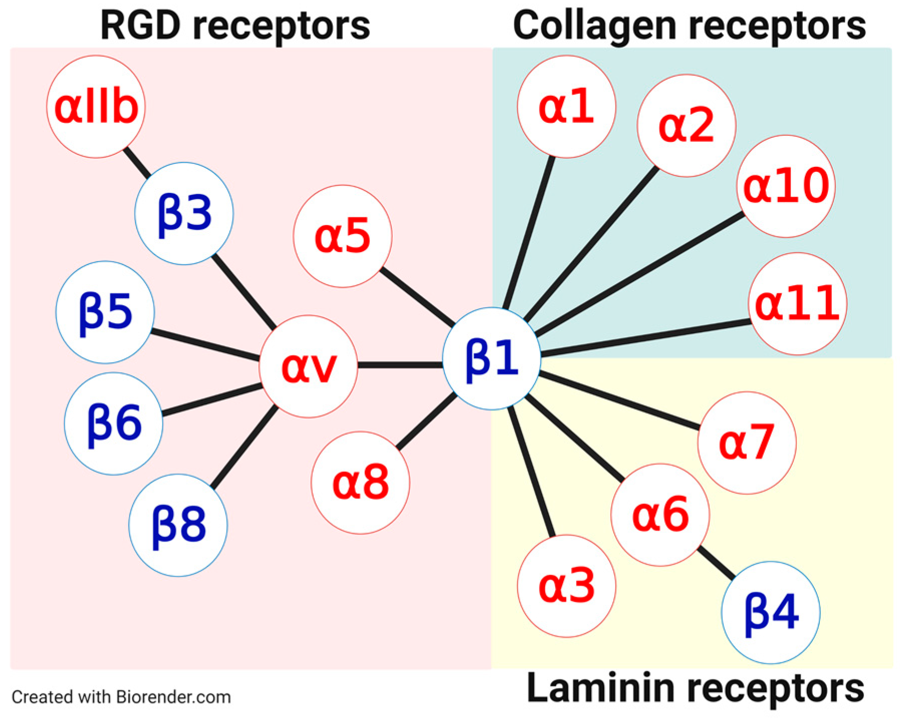
Figure 1. The extracellular matrix (ECM) protein binding integrin family and its classification into subfamilies. The classification is based on the binding specificity of these subfamilies toward ECM proteins. In higher vertebrates, 18 α and 8 β subunits were discovered, forming 24 different integrins through non-covalent interactions. Sixteen of these integrins bind ECM proteins. The leukocyte integrins, cell surface adhesion molecules, are excluded as they are not involved in ECM protein binding but in cell–cell interactions.
Cancers 13 01711 g001
Figure 2. Schematic illustration of integrin activation and the formation of cell adhesion structures. In the resting state, the integrins exhibit a bent conformation. Upon activation, an extended state is formed, the cytosolic salt bridge is disrupted, and the transmembrane helices dissociate. Now, they can homooligomerize to dimers (α-subunit) or trimers (β-subunit), which can further aggregate with other integrins or other proteins, finally forming the highly complex focal adhesions. Accompanied is the intracellular association with proteins such as talin, kindlin, and anchoring to the actin filament. Focal adhesions bind strongly in a multivalent manner to the ECM. RGD-containing ligands can bind to the initial homodimeric state but also to the binding sites of the focal adhesion–ECM complex. Preventing the dissociation of the heterodimers results in pure antagonism, as agonistic activity requires their dissociation of the heterodimeric state.
Cancers 13 01711 g002
Figure 3. Vascular promotion and vessel normalization provoked by anti-angiogenic therapy. In comparison to the quiescent normal vasculature consisting of mature vessels, the tumor vasculature is structurally and functionally immature. Due to those facts, no regular blood flow is established in tumor vessels, reinforcing a hypoxic environment within the tumors, the extravasation of detached tumor cells into the blood or lymphatic system, and the infiltration by immune regulatory cells. In response to anti-angiogenic therapy, the balance of anti-angiogenesis and pro-angiogenesis is brought back to near normal, achieving vascular normalization. These observations changed the paradigm in cancer therapy from anti-angiogenic strategies to vascular promotion/normalization therapy in order to enhance blood flow and, consequently, increase the delivery and intracellular uptake of chemotherapeutic drugs.
Cancers 13 01711 g003
Figure 4. Integrin internalization, sorting, and recycling. Integrin internalization occurs either clathrin-mediated or caveolar involving the dynamin GTPase to form vesicles. Rab GTPase family members and Arf GTPases are intimately involved in integrin trafficking, implicating ACAP1 in rapid integrin recycling from Arf6-positive endosomes. Integrin endocytosis to early endosomes is triggered by Rab5 and Rab21. Integrin recycling proceeds either through a short/rapid-loop by fusion to early endosomes implicating Rab4 signaling, or a long/slow-loop pathway, involving Rab11 signaling, delivering integrins back to the plasma membrane and into filopodia. This facilitates cell adhesion and enhances cell migration. Early endosomes mature to late endosomes/multivesicular bodies (MVBs) via the invagination of the endosomal membrane resulting in intraluminal vesicles (ILVs), which may be prone to degradation in lysosomes. Other receptors are recognized by specific coat proteins and sorted either to the plasma membrane, the trans-Golgi network, or in some cases to the endoplasmic reticulum (ER), thus evading degradation. Constitutive exocytosis of ILVs carrying integrins occurs upon their fusion with the cell membrane. Formed exosomes carrying integrins are taken up by recipient cells where they enrich the cell surface integrin density and thereby increase cell migration. Thus, the proportion between integrins depends on the balance between either their recycling or degradation exerting tremendous consequences for integrin signaling and the biological events arising thereof.
Cancers 13 01711 g004
Figure 5. Tumor exosome-mediated intercellular communication and the formation of a pre-metastatic niche. For the final colonization of circulating tumor cells, an appropriate niche at the metastatic site has to be created as well as EMT induced. The formation of such a pre-metastatic niche requires the communication of tumor cells with the local environment of a distant organ by the release of tumor-derived exosomes that deliver their pro-tumorigenic cargo to specifically targeted organs. Depicted is a scheme of a typical exosome that encloses some of the common exosomal components. In addition, integrins are expressed on the exosomal surface, which are capable of anchoring to distinct ECM proteins at the metastatic site.
Cancers 13 01711 g005
Figure 6. Exemplary peptidic modifications for improving the activity, stability, selectivity, and/or bioavailability of synthetic peptidic integrin ligands. The amino acids RGD coordinate to both integrin subunit: the basic amino acid Arg coordinates to the α-subunit, while Asp coordinates to the Metal Ion-Dependent Adhesion Site (MIDAS) region in the β-subunit. Starting from this linear RGD sequence contained within ECM proteins, various modifications were introduced for improving the integrin targeting properties. The cyclization of peptides and the incorporation of d-amino acids (e.g., cyclo[RGDfV] or Cilengitide) for example improved especially the stability and selectivity of the respective integrin ligands. N-methylation (e.g., cyclo[FRGDLAFp(NMe)K(Ac)] and the masking of charged residues (carboxylic and guanidinium group in 29P) contributed to a better bioavailability of the named compounds. Furthermore, N-methylation led to a higher stability and selectivity of the respective compounds.
Cancers 13 01711 g006
Figure 7. Overview of all described integrin targeting ligands. Within this review, only RGD targeting integrin ligands were described. Hereby, integrins αvβ3, αvβ5, α5β1, αvβ6, αvβ8, and αvβ1, respectively, represent the most important targets for cancer therapy and imaging. phg = d-phenylglycine, Chg = l-cyclohexylglycine, Ac = acetyl, NMe = N-methylated.
Cancers 13 01711 g007
Figure 8. Recent advances in translational αvβ6-targeted Positron Emission Tomography (PET) imaging. (a) Clinically translated αvβ6-targeted radiotracers are based on different design approaches, such as disulfide bridged peptides ➀, knottin peptides ➁, and linear peptides ➂. Further descriptions about these tracers are detailed in the text. (b) Comparison of [18F]FP-R01-MG-F2 PET and [18F]-FDG imaging in a patient with pancreatic cancer. The cyan arrow shows tracer accumulation at the head of the tumor (standardized uptake value (SUV)-knottin = 6.3). Several regions of relatively high accumulation were observed, including the kidneys, which are the main clearance route, and the stomach and small intestines, where integrin αvβ6 is expressed. Reprinted from Kimura et al. Nat Commun 10, 4673 (2019) under Creative Commons CC BY 4.0. (c) Design of recent preclinical probes to improve pharmacokinetics and increase tumor uptake. Introduction of an albumin binding domain increased circulation time and tumor uptake ➃ and trimerization of a peptide increased affinity and tumor uptake ➄.
Cancers 13 01711 g008
Publisher’s Note: MDPI stays neutral with regard to jurisdictional claims in published maps and institutional affiliations.