Review

The Roles of Diacylglycerol Kinase α in Cancer Cell Proliferation and Apoptosis

1
Department of Chemistry, Graduate School of Science, Chiba University, Chiba 263-8522, Japan
2
Department of Biosignaling and Radioisotope Experiment, Interdisciplinary Center for Science Research, Organization for Research and Academic Information, Shimane University, Izumo 693-8501, Japan
3
Department of Pharmaceutical Health Care and Sciences, Kyushu University, Fukuoka 812-8582, Japan
*
Author to whom correspondence should be addressed.
Cancers 2021, 13(20), 5190; https://doi.org/10.3390/cancers13205190
Submission received: 25 September 2021 / Revised: 14 October 2021 / Accepted: 14 October 2021 / Published: 16 October 2021
(This article belongs to the Special Issue Diacylglycerol Kinases in Cancer)

Abstract

:

Simple Summary

Diacylglycerol (DG) kinase (DGK) phosphorylates DG to generate phosphatidic acid (PA). DGKα is highly expressed in several refractory cancer cells, including melanoma, hepatocellular carcinoma, and glioblastoma cells, attenuates apoptosis, and promotes proliferation. In cancer cells, PA produced by DGKα plays an important role in proliferation/antiapoptosis. In addition to cancer cells, DGKα is highly abundant in T cells and induces a nonresponsive state (anergy), representing the main mechanism by which advanced cancers avoid immune action. In T cells, DGKα induces anergy through DG consumption. Therefore, a DGKα-specific inhibitor is expected to be a dual effective anticancer treatment that inhibits cancer cell proliferation and simultaneously activates T cell function. Moreover, the inhibition of DGKα synergistically enhances the anticancer effects of programmed cell death-1/programmed cell death ligand 1 blockade. Taken together, DGKα inhibition provides a promising new treatment strategy for refractory cancers.

Abstract

Diacylglycerol (DG) kinase (DGK) phosphorylates DG to generate phosphatidic acid (PA). The α isozyme is activated by Ca2+ through its EF-hand motifs and tyrosine phosphorylation. DGKα is highly expressed in several refractory cancer cells including melanoma, hepatocellular carcinoma, and glioblastoma cells. In melanoma cells, DGKα is an antiapoptotic factor that activates nuclear factor-κB (NF-κB) through the atypical protein kinase C (PKC) ζ-mediated phosphorylation of NF-κB. DGKα acts as an enhancer of proliferative activity through the Raf–MEK–ERK pathway and consequently exacerbates hepatocellular carcinoma progression. In glioblastoma and melanoma cells, DGKα attenuates apoptosis by enhancing the phosphodiesterase (PDE)-4A1–mammalian target of the rapamycin pathway. As PA activates PKCζ, Raf, and PDE, it is likely that PA generated by DGKα plays an important role in the proliferation/antiapoptosis of cancer cells. In addition to cancer cells, DGKα is highly abundant in T cells and induces a nonresponsive state (anergy), which represents the main mechanism by which advanced cancers escape immune action. In T cells, DGKα attenuates the activity of Ras-guanyl nucleotide-releasing protein, which is activated by DG and avoids anergy through DG consumption. Therefore, a DGKα-specific inhibitor is expected to be a dual effective anticancer treatment that inhibits cancer cell proliferation and simultaneously enhances T cell functions. Moreover, the inhibition of DGKα synergistically enhances the anticancer effects of programmed cell death-1/programmed cell death ligand 1 blockade. Taken together, DGKα inhibition provides a promising new treatment strategy for refractory cancers.

1. Introduction

Diacylglycerol kinase (DGK) phosphorylates diacylglycerol (DG, 1,2-diacyl-sn-glycerol) to generate phosphatidic acid (PA, 1,2-diacyl-sn-glycerol-3-phosphate) [1,2,3,4,5]. Mammalian DGK consists of ten isozymes, which are divided into five groups (type I (α, β, and γ), II (δ, η, and κ), III (ε), IV (ζ and ι) and V (θ)) according to their structural features. The α isozyme of DGK (one of the type I isozymes) was first cDNA-cloned approximately 30 years ago [6,7]. This isozyme characteristically has two tandem EF-hand motifs at its N terminus (Figure 1). DGKα is abundantly expressed in several cancer cell lines including hepatocellular carcinoma, melanoma, glioblastoma, colon adenocarcinoma, and breast adenocarcinoma cells, enhancing their progression and proliferation, and attenuating apoptosis [8,9,10,11]. Therefore, the inhibition of DGKα activity is expected to suppress the progression and proliferation of these cancers. In contrast, DGKα, which is also highly expressed in T lymphocytes, facilitates the nonresponsive state known as T cell clonal anergy [12,13]. Anergy induction in T lymphocytes is the primary mechanism by which progressive tumors evade immunity. Therefore, if a DGKα-specific inhibitor is developed, it would reversely and simultaneously inhibit tumor cell proliferation and enhance T cell function and consequently can be a dual effective drug. In this article, we review the properties of DGKα and its roles in cancer cell proliferation and apoptosis, and anticancer immunity.

2. Structure, Enzymatic Properties, and Activation Mechanisms of DGKα
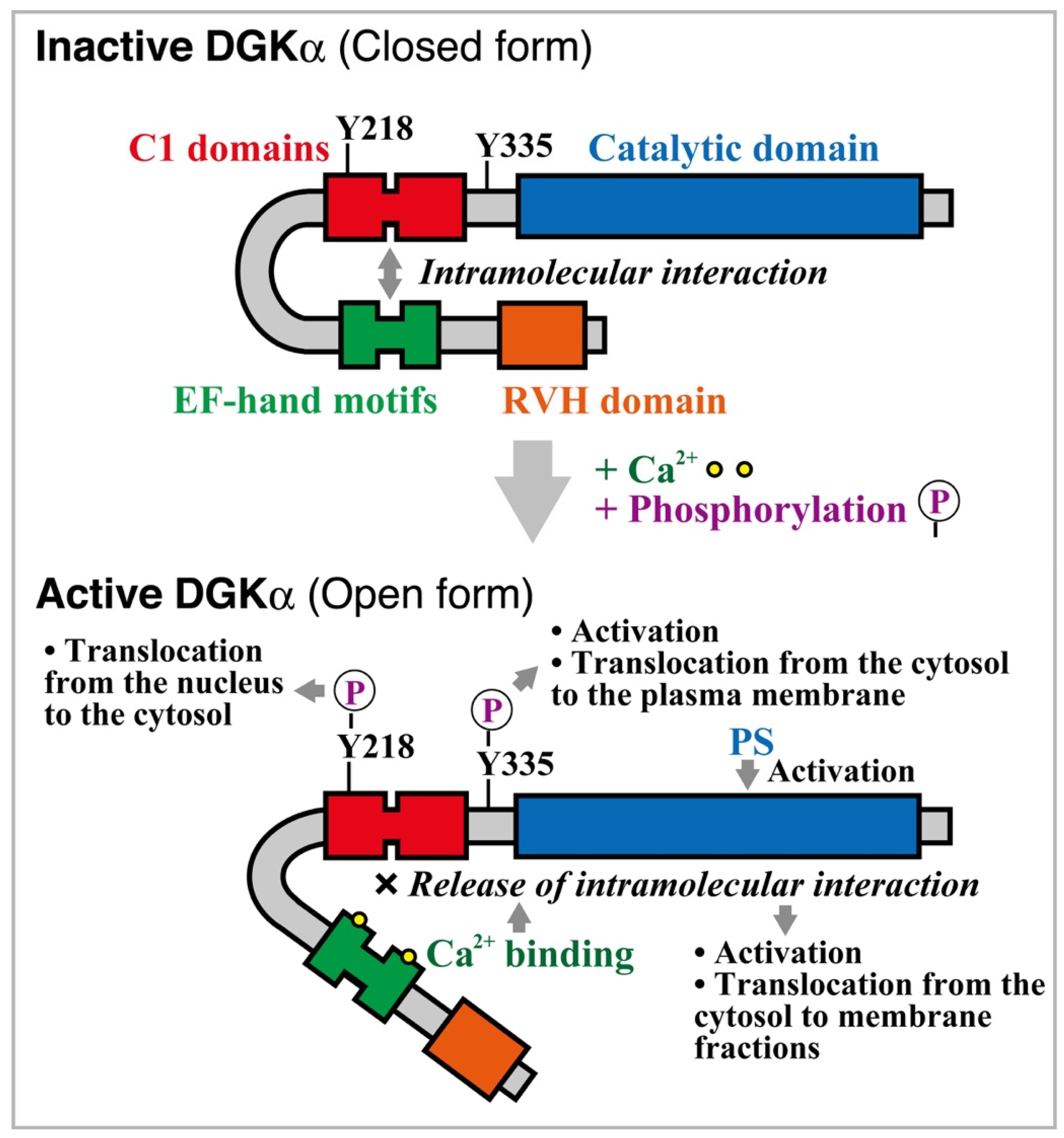
2.1. Structure

DGKα is a member of type I DGK [1,2,3,4,5]. This isozyme contains, from its N terminus, a recoverin homology (RVH) domain [14], two tandem EF-hand motifs, two tandem C1 domains, and a catalytic domain [15] (Figure 1). Indeed, two Ca2+ ions bind to two EF-hand motifs (one each) in DGKα and strongly activate it [6,16]. The apparent dissociation constant KD and ED50 value of DGKα for Ca2+ are approximately 0.3 μM [16,17]. In contrast to DGKα, DGKβ and DGKγ, which also have two tandem EF-hand motifs, are apparently active independent of Ca2+ [17]. The apparent KD values of DGKβ and DGKγ for Ca2+ are less than 0.01 μM [17]. Therefore, although DGKβ and DGKγ have a stronger affinity for Ca2+ than DGKα, they failed to show Ca2+-dependent activation, suggesting that pretreatment with a Ca2+ chelator, ethylene glycol tetraacetic acid (EGTA), cannot effectively release Ca2+ from these isozymes. The concentrations of Ca2+ in resting and stimulated cells are 0.05–0.1 nM and 1–10 μM, respectively [18,19]. Thus, it is likely that EF-hand motifs in DGKα associate with Ca2+, and the isozyme is activated by Ca2+ only after cell stimulation. In addition to activation, Ca2+ induces translocation of DGKα to membrane fractions [16].
The C1 domains are similar to those in protein kinase C (PKC). The domains in conventional and novel PKC isoforms bind to phorbol ester and DG [20,21]. However, the C1 domains in DGKα showed no phorbol ester-binding activity, and the catalytic domain alone, lacking the C1 domains, exhibited catalytic (DG recognition and phosphorylation) activity [15], indicating that the domains do not act as the catalytic domain.
Site-directed mutagenesis analyses showed that the N- and C-terminal regions of the catalytic domain bind to ATP and DG, respectively [22]. Miller et al. studied the 3D structure of Staphylococcus aureus DGK (DgkB), which is structurally similar to the catalytic domains of mammalian DGKs and its key active site residues [23]. However, 3D structures of full-length mammalian DGK isozymes have not yet been revealed. Nonetheless, the key active site residues and the components of the Asp–water–Mg2+ network are conserved in the catalytic cores of mammalian DGKs. There are remarkable similarities among ceramide kinase, sphingosine kinase, and DGK catalytic domains [24]. Therefore, it is possible that these enzymes utilize the same mechanism and have 3D structures similar to that of DgkB.

2.2. Enzymological Properties

DGKα generates PA using ATP and DG as substrates. The Km values for ATP and DG are 0.10–0.24 mM and 1.0–3.4 mol%, respectively, which are close to averages of DGK isozymes measured thus far [15,16,25,26].
Interestingly, in addition to DGK activity, DGKα has 2-monoacylglycerol (MG) kinase (MGK) activity (approximately 12% of DGK activity) but not 1-MGK activity [27]. Type I (α, β, and γ), II (δ, η, and κ) and III (ε) DGK isozymes also have 2-MGK activity (12–19% of DGK activity). However, type IV DGK isozymes (ζ and ι) do not show 1-MGK or 2-MGK activity (less than 1% of DGK activity). Alternatively, DGKθ (type V) has 1-MGK activity (6% of DGK activity) but not 2-MGK activity. DGK isozymes with 1-MGK or 2-MGK activity may produce lysoPA, utilizing 1-MG or 2-MG in cells. However, the physiological significance of their 1-MGK and 2-MGK activities is still unknown. Notably, intracellular lysoPA is involved in cancer cell migration [28].
We recently found, using liquid chromatography (LC)–tandem mass spectrometry (MS/MS), that the production of palmitic acid (16:0)- and/or palmitoleic acid (16:1)-containing PA species, such as 14:1/16:1-, 14:0/16:1-, 14:0/16:0-, 16:1/16:2-, 16:1/16:1-, 16:0/16:1-, 16:0/16:0-, 16:0/18:1- and 16:0/18:0-PA, were inhibited by CU-3, a DGKα-selective inhibitor [29] (see Section 5.1), in starved Jurkat T cells [30]. Moreover, LC–MS/MS revealed that the production of 16:0-containing PA species, such as 16:0/16:0- and 16:0/18:0-PA, was attenuated by CU-3 [29] in AKI melanoma cells under starved conditions [31]. These results strongly suggest that DGKα phosphorylates different DG species in cancer cells and T cells. DGKα does not exhibit DG species selectivity in vitro [25]. It is possible that DG supply enzymes, which provide distinct DG species upstream of DGKα, are different from each other in cancer and T cells. However, further studies are needed to identify the upstream enzymes.
DGK activity (conversion of DG to PA) is essential for phosphatidylinositol (PI) turnover (the PI 4,5-bisphosphate pathway) as the initial step for PI resynthesis [32,33]. As PI turnover exclusively generates 1-stearoyl-2-arachidonoyl-DG (18:0/20:4-DG), it has been generally considered that all DGK isozymes phosphorylate 18:0/20:4-DG species. However, our group recently revealed that DGK isoforms (DGKα, δ, η, and ζ) except for DGKε, phosphorylate a variety of DG molecular species, which are not coming from the PI 4,5-bisphosphate pathway [4,30,34,35,36,37,38,39,40].
Recently, Bozelli et al. reported that membrane morphology (membrane curvature) affects the substrate selectivity of DGKα [41]. On substantial membrane morphological changes, DGKα exhibits higher substrate acyl chain specificity for 16:0-containing DG molecular species, which are actually preferred by DGKα as substrates in cells [30,31,42]. It is possible that DGKα (and other DGK isozymes) metabolize specific molecular species of DG and, consequently, generate specific molecular species of PA in a membrane morphology-dependent manner.

2.3. Regulation of Activity and Subcellular Localization

Several studies have revealed calcium-dependent activation mechanisms of DGKα [43,44,45] (Figure 1). The N-terminal region containing the EF-hand motifs and RVH domain masks the catalytic domain and maintains the enzyme in an inactive state. Ca2+ induces the release of the intramolecular association between the C1 domains and the EF-hand motifs of DGKα (Figure 1). Therefore, dissociation between the EF-hand motifs and the C1 domains is the key event that activates DGKα (Figure 1). Experiments with 2-p-toluidinylnaphthalene 6-sulfonate, a probe for hydrophobic regions of proteins, showed that the binding of Ca2+ masked a hydrophobic region of DGKα EF-hand motifs [17]. We recently succeeded in the first crystal structure of Ca2+-bound DGKα EF-hand motifs and analyzed the structural changes on binding to Ca2+ [46]. The EF-hand motifs of DGKα adopt a canonical EF-hand fold but unpredictably possess a ligand mimic helix (an additional α-helix), which is packed into the hydrophobic core [46]. Conformational changes may contribute to the dissociation of intramolecular interactions between the EF-hand motifs and the C1 domains in DGKα and its activation (Figure 1).
It has been repeatedly reported that, similar to PKC [47,48], an acidic phospholipid, phosphatidylserine (PS), enhances DGKα activity [15,16,25,26]. Intriguingly, Ca2+ and PS stimulate DGKα enzyme activity via distinct mechanisms [22]. Although Ca2+ interacts with EF-hand motifs as described above, PS binds to the catalytic domain (Figure 1). Unlike DGKα, PKC interacts with PS through its C2 domain but not the catalytic domain [47,48].
In addition to PS, phosphoinositide 3-kinase lipid products, such as PI 3,4-bisphosphate and PI 3,4,5-trisphosphate, activate DGKα [49]. Interestingly, this activation occurs in a calcium-independent manner. In addition to acidic phospholipids, sphingosine, which is a basic lipid and forms a primary part of sphingolipids including sphingomyelin, activates DGKα in vitro and in cells [50,51,52].
Phosphorylation at Tyr-335 in DGKα (Tyr-335 in human DGKα (https://www.uniprot.org/uniprot/P23743 (accessed on 25 September 2021)); Tyr-334 in pig DGKα (https://www.uniprot.org/uniprot/P20192 (accessed on 25 September 2021)); Tyr-336 in mouse DGKα (https://www.uniprot.org/uniprot/O88673 (accessed on 25 September 2021)) is also involved in its activation and subcellular localization (Figure 1). In T cells, DGKα translocates from the cytosol to the plasma membrane in response to T cell receptor stimulation [53]. Tyr-335 phosphorylation induces the plasma membrane translocation of the enzyme in T cells [54]. In addition to T cell receptor, D-α-tocopherol (vitamin E) induces Src-dependent phosphorylation at Tyr-335 in DGKα, and consequently, the phosphorylation enhances translocation from the cytoplasm to the plasma membrane and activation of the enzyme in DDT1-MF2 cells (a smooth muscle cell line) [55] (Figure 1). Epigallocatechin gallate (a green tea polyphenol) also induces the Src-dependent tyrosine phosphorylation of DGKα and consequently causes its translocation and activation in DDT1-MF2 cells [56]. The effects of D-α-tocopherol and epigallocatechin gallate occur through a 67 kDa laminin receptor [57].
Tyr-218 in DGKα is phosphorylated by c-Abl [58] (Figure 1). Tyr-218 phosphorylation regulates serum-induced nuclear export of DGKα.
As described above, various signal transduction routes intricately regulate the activity and subcellular localization of DGKα activity.

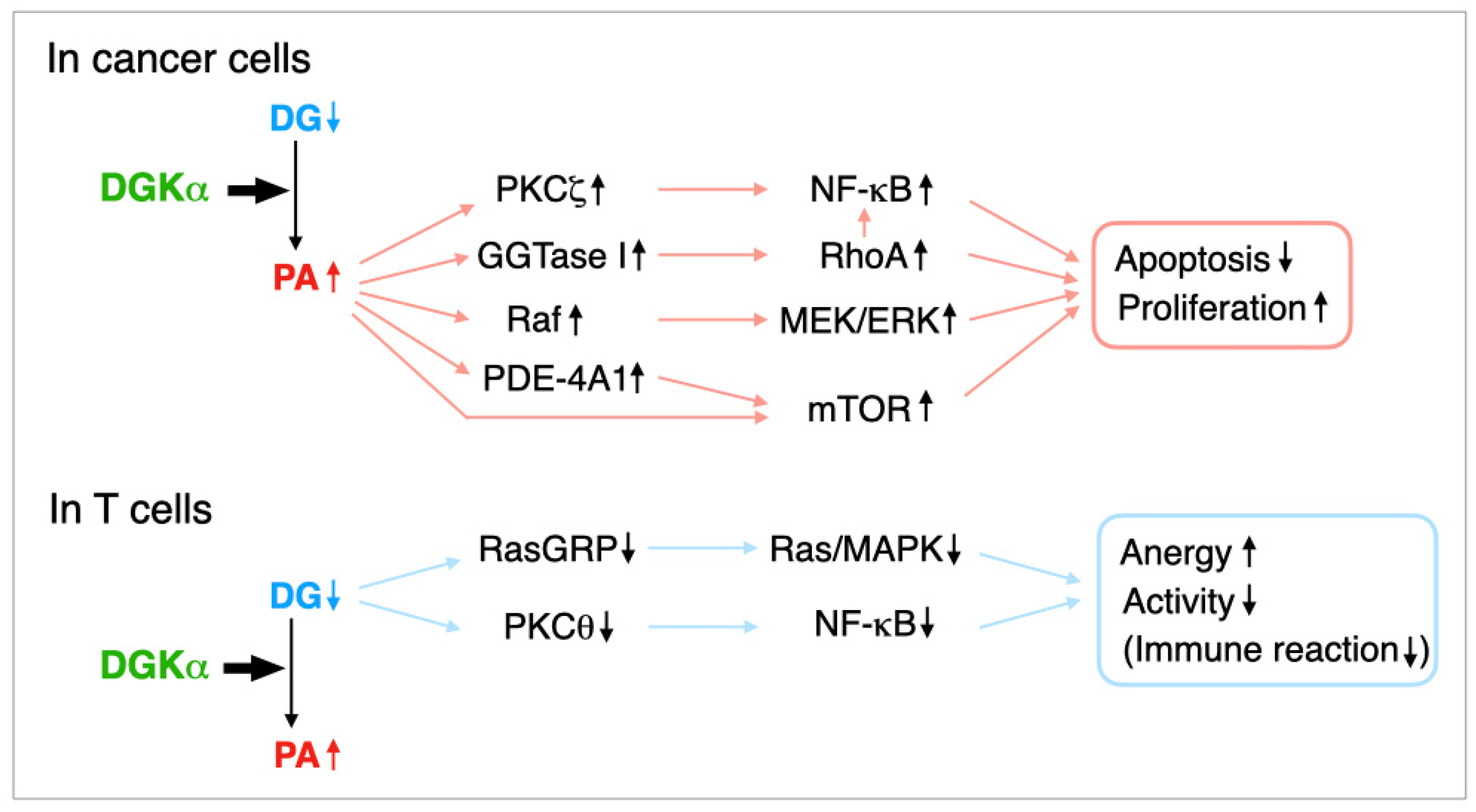
3. Regulation of Cancer Cell Proliferation and Apoptosis by DGKα

We demonstrated that DGKα is expressed in several human melanoma cell lines including AKI but not in noncancerous normal human epidermal melanocytes and that the isozyme suppresses tumor necrosis factor-α-induced apoptosis of AKI melanoma cells through the activation of nuclear factor-κB (NF-κB) [11], which is an antiapoptotic factor (Figure 2). DGKα activates NF-κB through PKCζ-dependent phosphorylation at Ser-311 of the p65/RelA subunit of NF-κB in AKI melanoma cells [59] (Figure 2).
In addition to melanoma cells, Takeishi et al. demonstrated that DGKα expression is correlated with hepatocellular carcinoma (HCC) progression [9]. Moreover, DGKα enhances HCC proliferation by activating extracellular signal-regulated kinase (ERK) [9], which is downstream of Ras–Raf–mitogen-activated protein kinase (MAPK)/ERK kinase (MEK) (Figure 2).
Dominguez et al. reported that DGKα inhibition using R59022 and R59949, which are commercially available semi-DGKα-selective inhibitors (see Section 5.1), and silencing induced apoptosis of several cancer cell lines including brain glioblastoma and melanoma cells [8] (Figure 2). In addition, they demonstrated that the apoptotic effects are mediated by the phosphodiesterase (PDE)-4A1–cAMP–mammalian target of rapamycin (mTOR) pathway [8] (Figure 2). In this case, DGKα inhibition/silencing decreased mTOR expression [8].
PA produced by DGKα activates the geranylgeranyltransferase (GGTase) I–RhoA and consequently GGTase I–RhoA–NF-κB pathways in glioblastoma cells [60] (Figure 2). GGTase I activated by PA geranylgeranylates RhoA and induces its membrane localization and activation. Geranylgeranylated RhoA and NF-κB activated by RhoA prevent cell death. Therefore, DGKα confers the mesenchymal phenotype, which is characterized by aggressiveness and treatment resistance, to glioblastoma cells.
The proliferation of colon and breast cancer cell lines was markedly suppressed by DGKα-siRNA and R59949 [10]. Moreover, DGKα interacts with Src and promotes Src activation [10]. Interestingly, Perez et al. recently demonstrated that the Src unique and SH3 domains bind to acidic phospholipids, including PA, suggesting the presence of a previously unrecognized additional regulatory mechanism of c-Src [61]. Therefore, in addition to conventional pathways, PA may activate Src in cancer cells and promote cancer cell proliferation.
In addition to the effects on apoptosis and proliferation, DGKα is reportedly essential for matrix invasion of breast carcinoma cells through the atypical PKC–β1 integrin signaling pathway [62].
DGKα is abundant in the nuclei of human erythroleukemia K562 cells and promotes cell proliferation by control of the retinoblastoma protein (pRb) phosphorylation and cell cycle progression through the G1/S checkpoint [63]. R59022, R59949, and DGKα knockdown significantly reduced K562 cell proliferation [63].
It is important to know how DGKα expression is regulated in cancer cells. Kefas et al. reported that microRNA-297 (miR-297) strongly reduced DGKα protein expression [64]. Moreover, miR-297 decreased glioblastoma cell survival, invasiveness, and tumorigenicity [64].
Taken together, inhibition of DGKα suppresses cancer cell proliferation, enhances cancer cell apoptosis, and attenuates cancer cell invasion. As PKCζ [65], GGTase I–RhoA(–NF-κB) [60], C-Raf (Raf-1) [66,67,68] and PDE-4A1 [69,70] are PA-binding proteins, and PA augments their activities, it is likely that PA produced by DGKα mediates the antiapoptosis and proproliferation effects of DGKα (Figure 2). PA interacts with and activates mTOR in addition to PDE-4A1 [71]. Therefore, PA may synergistically enhance the PDE-4A1–mTOR pathway. It is possible that PA simultaneously utilizes these three pathways, or, alternatively, may use a distinct pathway in different cancer cells.

4. Regulation of T Cell Receptor Signaling by DGKα

In addition to cancer cells, DGKα is abundantly expressed in T cells and the thymus [6,72]. DGKα serves as an immune checkpoint and induces T cell clonal anergy (the nonresponsive state) [12,13]. Anergy induction in T lymphocytes by progressive tumors is the primary machinery employed to escape immunological strike [73]. Particularly, DGKα reduces the antitumor immune reaction of tumor-infiltrating CD8+ T cells [74]. Therefore, the inhibition of DGKα activity is widely considered to enhance T cell proliferation/functions, which provide boosted immune surveillance and cancer immunity [75,76,77,78,79,80].
In addition to conventional and novel PKCs, DG activates Ras-guanyl nucleotide-releasing protein (RasGRP) [81], which drives Ras and the MAPK pathway (ERK and c-Jun N-terminal kinase (JNK), along with promoting the transcriptional activity of nuclear factor of activated T cells and the expression of interleukin-2 and CD25 (Figure 2). Indeed, DGKα reduces RasGRP1 activity through consumption of DG and, consequently, induces T cell anergy [13,53,82].
PKCθ (novel PKC), which is activated by DG [20,21], is expressed in T lymphocytes in a relatively selective manner and DGKα attenuates the PKCθ–NF-κB pathway, which is also important for preventing T cell anergy [83]. In this case, PKCθ activates IκB kinase β, leading to NF-κB activation [83].
What is the difference in DGKα functions between T lymphocytes and cancer cells? DGKα consumes DG and produces PA (Figure 2). It is likely that in T cells, DG, not PA, primarily attenuates anergy and stimulates functions and that, in contrast, PA, not DG, mainly promotes cell proliferation and suppresses apoptosis in cancer cells (Figure 2). Thus, it is speculated that DGKα-dependent consumption of DG and the production of PA dominantly affect T cells and cancer cells. As described above (see Section 2.2), DGKα consumes different DG molecular species in AKI melanoma cells (16:0/16:0- and 16:0/18:0-PA) [31] and Jurkat T cells (16:0- and/or 16:1-containing DG species) under starved conditions [30]. These results suggest that DG/PA molecular species having different fatty acid moieties in T cells and cancer cells may contribute to distinct functions of DGKα in these cells.
It is also important to know how DGKα expression is regulated in T cells. We analyzed the 5′-upstream region (3.4 kb) of the DGKα gene and revealed that the transcription of DGKα is differently regulated in human Jurkat T cells and human hepatocellular carcinoma HepG2 cells [84]. Moreover, Zheng et al. demonstrated that transcriptional regulator early growth response gene 2 (Egr2) up-regulates DGKα expression and is required for T cell anergy [85]. Therefore, it is likely that DGKα expression is differently regulated in immune and cancer cells.

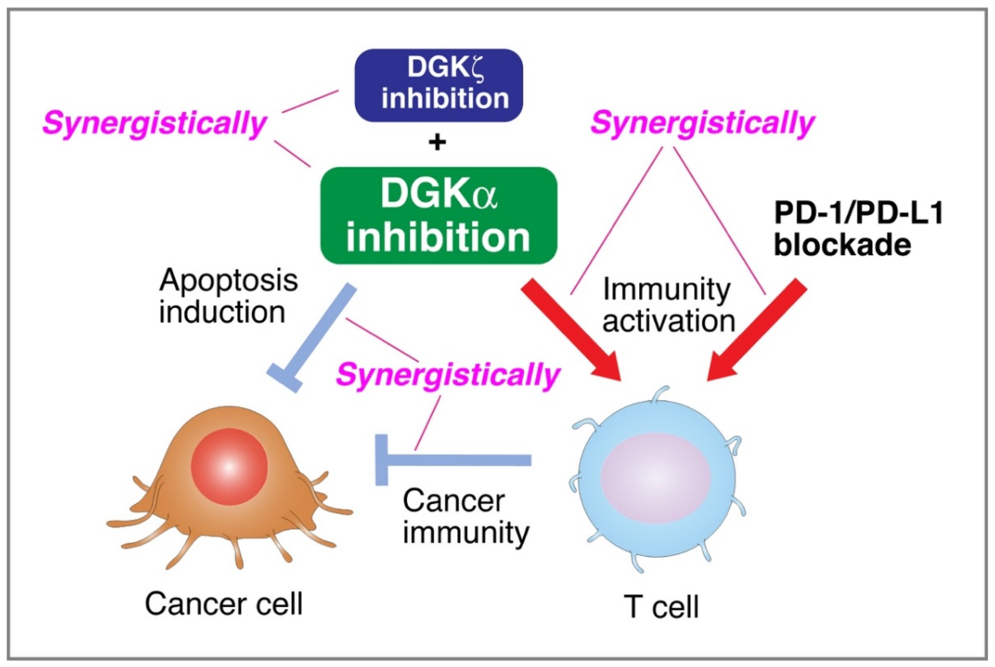
5. DGKα Inhibitors Simultaneously Attenuate Cancer Cell Proliferation and Activate T Cell Function

5.1. DGKα Inhibitors Simultaneously Attenuate Cancer Cell Proliferation and Activate T Cell Function

DGKα-selective inhibitors are expected to be dual effective drugs (i.e., ideal cancer therapy medicines) because, as described above (Section 3 and Section 4), they simultaneously suppress cancer cell proliferation and boost immune reactions, including anticancer immunity [79] (Figure 3). Several inhibitors of DGK isozymes, including DGKα, were identified as follows:
There are two DGK inhibitors, 6-{2-{4-[(4-fluorophenyl)phenylmethylene]-1-piperidinyl}ethyl}-7-methyl-5H-thiazolo-(3,2-a)pyrimidin-5-one (R59022) [86] and 3-{2-(4-[bis-(4-fluorophenyl)methylene]-1-piperidinyl)ethyl}-2,3-dihydro-2-thioxo-4(1H)quinazolinone (R59949) [87], which are commercially available. It was reported that they are selective for a calcium-dependent DGK isoform [88]. However, our comprehensive analyses of their effects on all DGK isoforms revealed that R59022 inhibited DGKα (type I), ε (type III) and θ (type V) and that R59949 inhibited DGKα (type I), γ (type I), δ (type II), and κ (type II) [89]. Therefore, these inhibitors semiselectively inhibit DGK isozymes. The half-maximal inhibitory concentration (IC50) values of R59022 and R59949 were 25 μM and 18 μM, respectively [89]. These values are considerably higher than those of newly found DGKα-selective inhibitors (see below).
We recently reported that high-throughput screening (HTS) of the core library (Drug Discovery Initiative, the University of Tokyo) that consists of 9600 compounds identified CU-3, 5-((2E)-3-(2-furyl)prop-2-enylidene)-3-[(phenylsulfonyl)amino]-2-thioxo-1,3-thiazolidin-4-one, as a DGKα-selective inhibitor with an IC50 value of 0.6 μM [29]. The IC50 values of CU-3 against nine other isozymes (β–κ) were 12- to 60-fold higher than those of DGKα [29]. As expected, CU-3 induced both apoptosis of several cancer-derived cell lines and T-cell activation [29,31] (Figure 3). Apoptosis induction of cancer cells (direct effect) and cancer immunity (indirect effect) induced by DGKα inhibition synergistically cause damage to cancer cells (Figure 3).
We obtained compound A, which was modified from a small molecule that was newly identified by HTS of chemical compound libraries in Ono Pharmaceutical Co., Ltd., and found that compound A also selectively inhibits type I DGK isozymes (DGKα, DGKβ, and DGKγ) but not type II–V isozymes, with IC50 values of 0.04, 0.02 and 0.01 μM, respectively [31]. The IC50 values for the other seven DGK isozymes (type II–V) were greater than 10 μM. Compound A also induced apoptosis of several cancer-derived cells and simultaneously activated T cells [31]. The DGKα-selective inhibitor DGKAI, which was obtained along with compound A (see above), also selectively inhibited DGKα, DGKβ and DGKγ with IC50 values of 0.01, 0.01, and <0.01 μM, respectively [90]. However, the IC50 values for the other seven DGK isozymes were greater than 10 μM. DGKAI was suggested to have dual effects on HCC proliferation (inhibition) and T cell immune response (activation) in vivo [90] (Figure 3). Unfortunately, structural information on these compounds is not currently available.
Other DGKα-selective inhibitors have been identified. Ritanserin, 6-[2-[4-[bis(4-fluorophenyl)methylidene]piperidin-1-yl]ethyl]-7-methyl-[1,3]thiazolo[2,3-b]pyrimidin-5-one, is a well-known serotonin receptor (5-HT2A/5-HT2c) antagonist with high structural similarity with both R59022 and R59949. Interestingly, Boroda et al. showed that ritanserin selectively inhibits DGKα (IC50 = 15.1 μM)) [60,91]. Moreover, Velnati et al. performed an in silico approach based on chemical homology with R59022 and R59949 and, as a result, found ritanserin and AMB639752 (1-(2,5-dimetil-1H-indol-3-yl)-2-(4-(furan-2-carbonil)piperazin-1-yl)etan-1-one), which selectively inhibit DGKα [92]. These compounds are also expected to be able to become ideal anticancer medicines.

5.2. Synergistic Effect of PD-1/PD-L1 Blockade

Immune checkpoint inhibitors, including anti-programmed cell death-1 (PD-1) and anti-PD-1 ligand (PD-L1) antibodies, have exhibited remarkable efficacy in some advanced cancers [93,94]. However, the clinical response rate to anti-PD-1/PD-L1 antibodies is still 10–40% in different advanced cancers [95,96], restricting their usefulness in cancer therapy [97] and suggesting the existence of different immunosuppressive mechanisms in cancer-bearing hosts. Notably, several reports have recently demonstrated that combined DGKα inhibition/silencing and PD-1/PD-L1 blockade synergistically enhance anticancer immunity (Figure 3). For example, Fu et al. recently reported that DGKα inhibition using R59022, R59949 and ritanserin enhanced the efficacy of anti-PD-1 therapy [98]. Moreover, Arranz-Nicolas et al. also showed the involvement of DGKα in CD3/CD28 and PD-1-mediated signal transduction pathways and the ability of DGKα inhibitors (R59949 and ritanserin) to cooperatively enhance immune checkpoint-targeted therapies in human T lymphocytes [99,100]. Furthermore, Okada et al. demonstrated that DGKα inhibition using DGKAI (see Section 5.1) and PD-L1 blockade synergistically suppressed the growth of HCC in vivo in an immune activity-dependent manner [90]. Therefore, the synergistic effect of DGKα inhibition and PD-1/PD-L1 blockade provides a promising strategy to strengthen the efficacy of immunotherapy in the treatment of cancer (Figure 3). The combination therapies with DGKα-specific inhibitors and anti-PD-1/PD-L1 antibodies, if established, will provide survival benefits for much greater numbers of advanced cancer patients.

5.3. Synergistic Effects of DGKα- and DGKζ-Inhibitions

In addition to DGKα, DGKζ reportedly restricts the intensity of T cell receptor signaling by metabolizing DG [12,76,78,80]. Moreover, DGKζ enhances the proliferation of human embryonic kidney 293 cells [101]. Therefore, we hypothesized that attenuation of DGKζ synergistically augments the enhancing effects of DGKα inhibition on cancer cell apoptosis and T cell function. Indeed, Takao et al. recently demonstrated that combined inhibition/silencing of DGKα and DGKζ synergistically provokes apoptosis of melanoma cells and interleukin-2 generation in T cells [102]. Therefore, a compound that inhibits both DGKα and DGKζ may be a promising anticancer treatment. Notably, Abdel-Magid reported that naphthyridineone compounds inhibit both DGKα and DGKζ and are useful as T cell function activators [103].

6. Conclusions

DGKα acts as an antiapoptosis/proproliferation factor in cancer cells. In contrast, DGKα attenuates the functions of T cells. Therefore, DGKα-selective inhibitors are expected to be ideal anticancer medicines because the inhibition of DGKα suppresses cancer cell proliferation and simultaneously activates T cell function (Figure 3). Moreover, synergistic effects of DGKα inhibition and PD-1/PD-L1 blockade would provide a promising new strategy for refractory cancer therapy (Figure 3). Furthermore, it is possible that the inhibition of DGKζ synergistically enhances the effects of DGKα inhibition (Figure 3). Immediate development of genuine DGKα-specific inhibitors (and DGKζ-specific inhibitors) is needed.

Author Contributions

Writing—original draft preparation, review and editing, F.S., F.H., M.E., H.S. and D.T. All authors have read and agreed to the published version of the manuscript.

Funding

This work was supported in part by grants from MEXT/JSPS (KAKENHI Grant Numbers: 26291017 (Grant-in-Aid for Scientific Research (B)) (F.S.), 15K14470 (Grant-in-Aid for Challenging Exploratory Research) (F.S.), 17H03650 (Grant-in-Aid for Scientific Research (B) (F.S.), 20H03205 (Grant-in-Aid for Scientific Research (B) (F.S.), 20K07049 (Grant-in-Aid for Scientific Research (C) (H.S.), 19K06527 (Grant-in-Aid for Scientific Research (C) (D.T.), and 20J21133 (Grant-in-Aid for JSPS Fellows) (F.H.)); the Japan Science and Technology Agency (Adaptable and Seamless Technology Transfer Program through Target-driven R&D: AS2621643Q and JPMJTM20CC) (F.S.); the Japan Food Chemical Research Foundation (F.S.); the SENSHIN Medical Research Foundation (F.S.); and the Uehara Memorial Foundation (F.S.); the Tojuro Iijima Foundation for Food Science and Technology (F.S.); and the Sugiyama Chemical and Industrial Laboratory (F.S.).

Conflicts of Interest

The authors declare no conflict of interest associated with the contents of this article.

References

  1. Goto, K.; Hozumi, Y.; Kondo, H. Diacylglycerol, phosphatidic acid, and the converting enzyme, diacylglycerol kinase, in the nucleus. Biochim. Biophys. Acta 2006, 1761, 535–541. [Google Scholar] [CrossRef] [PubMed]
  2. Merida, I.; Avila-Flores, A.; Merino, E. Diacylglycerol kinases: At the hub of cell signalling. Biochem. J. 2008, 409, 1–18. [Google Scholar] [CrossRef] [Green Version]
  3. Sakane, F.; Imai, S.; Kai, M.; Yasuda, S.; Kanoh, H. Diacylglycerol kinases: Why so many of them? Biochim. Biophys. Acta 2007, 1771, 793–806. [Google Scholar] [CrossRef]
  4. Sakane, F.; Mizuno, S.; Takahashi, D.; Sakai, H. Where do substrates of diacylglycerol kinases come from? Diacylglycerol kinases utilize diacylglycerol species supplied from phosphatidylinositol turnover-independent pathways. Adv. Biol. Regul. 2018, 67, 101–108. [Google Scholar] [CrossRef] [PubMed]
  5. Topham, M.K.; Epand, R.M. Mammalian diacylglycerol kinases: Molecular interactions and biological functions of selected isoforms. Biochim. Biophys. Acta 2009, 1790, 416–424. [Google Scholar] [CrossRef] [Green Version]
  6. Sakane, F.; Yamada, K.; Kanoh, H.; Yokoyama, C.; Tanabe, T. Porcine diacylglycerol kinase sequence has zinc finger and E-F hand motifs. Nature 1990, 344, 345–348. [Google Scholar] [CrossRef]
  7. Schaap, D.; de Widt, J.; van der Wal, J.; Vandekerckhove, J.; van Damme, J.; Gussow, D.; Ploegh, H.L.; van Blitterswijk, W.J.; van der Bend, R.L. Purification, cDNA-cloning and expression of human diacylglycerol kinase. FEBS Lett. 1990, 275, 151–158. [Google Scholar] [CrossRef] [Green Version]
  8. Dominguez, C.L.; Floyd, D.H.; Xiao, A.; Mullins, G.R.; Kefas, B.A.; Xin, W.; Yacur, M.N.; Abounader, R.; Lee, J.K.; Wilson, G.M.; et al. Diacylglycerol kinase α is a critical signaling node and novel therapeutic target in glioblastoma and other cancers. Cancer Discov. 2013, 3, 782–797. [Google Scholar] [CrossRef] [Green Version]
  9. Takeishi, K.; Taketomi, A.; Shirabe, K.; Toshima, T.; Motomura, T.; Ikegami, T.; Yoshizumi, T.; Sakane, F.; Maehara, Y. Diacylglycerol kinase alpha enhances hepatocellular carcinoma progression by activation of Ras-Raf-MEK-ERK pathway. J. Hepatol. 2012, 57, 77–83. [Google Scholar] [CrossRef] [PubMed]
  10. Torres-Ayuso, P.; Daza-Martin, M.; Martin-Perez, J.; Avila-Flores, A.; Merida, I. Diacylglycerol kinase α promotes 3D cancer cell growth and limits drug sensitivity through functional interaction with Src. Oncotarget 2014, 5, 9710–9726. [Google Scholar] [CrossRef] [Green Version]
  11. Yanagisawa, K.; Yasuda, S.; Kai, M.; Imai, S.; Yamada, K.; Yamashita, T.; Jimbow, K.; Kanoh, H.; Sakane, F. Diacylglycerol kinase α suppresses tumor necrosis factor-α-induced apoptosis of human melanoma cells through NF-κB activation. Biochim. Biophys. Acta 2007, 1771, 462–474. [Google Scholar] [CrossRef]
  12. Olenchock, B.A.; Guo, R.; Carpenter, J.H.; Jordan, M.; Topham, M.K.; Koretzky, G.A.; Zhong, X.P. Disruption of diacylglycerol metabolism impairs the induction of T cell anergy. Nat. Immunol. 2006, 7, 1174–1181. [Google Scholar] [CrossRef]
  13. Zha, Y.; Marks, R.; Ho, A.W.; Peterson, A.C.; Janardhan, S.; Brown, I.; Praveen, K.; Stang, S.; Stone, J.C.; Gajewski, T.F. T cell anergy is reversed by active Ras and is regulated by diacylglycerol kinase-α. Nat. Immunol. 2006, 7, 1166–1173. [Google Scholar] [CrossRef]
  14. Jiang, Y.; Qian, W.; Hawes, J.W.; Walsh, J.P. A domain with homology to neuronal calcium sensors is required for calcium-dependent activation of diacylglycerol kinase alpha. J. Biol. Chem. 2000, 275, 34092–34099. [Google Scholar] [CrossRef] [Green Version]
  15. Sakane, F.; Kai, M.; Wada, I.; Imai, S.; Kanoh, H. The C-terminal part of diacylglycerol kinase α lacking zinc fingers serves as a catalytic domain. Biochem. J. 1996, 318, 583–590. [Google Scholar] [CrossRef] [Green Version]
  16. Sakane, F.; Yamada, K.; Imai, S.; Kanoh, H. Porcine 80-kDa diacylglycerol kinase is a calcium-binding and calcium/phospholipid-dependent enzyme and undergoes calcium-dependent translocation. J. Biol. Chem. 1991, 266, 7096–7100. [Google Scholar] [CrossRef]
  17. Yamada, K.; Sakane, F.; Matsushima, N.; Kanoh, H. EF-hand motifs of α, β and γ isoforms of diacylglycerol kinase bind calcium with different affinities and conformational changes. Biochem. J. 1997, 321, 59–64. [Google Scholar] [CrossRef]
  18. Berridge, M.J.; Lipp, P.; Bootman, M.D. The versatility and universality of calcium signalling. Nat. Rev. Mol. Cell Biol. 2000, 1, 11–21. [Google Scholar] [CrossRef]
  19. Simons, T.J. Calcium and neuronal function. Neurosurg. Rev. 1988, 11, 119–129. [Google Scholar] [CrossRef]
  20. Kazanietz, M.G. Novel “nonkinase” phorbol ester receptors: The C1 domain connection. Mol. Pharmacol. 2002, 61, 759–767. [Google Scholar] [CrossRef] [Green Version]
  21. Nishizuka, Y. The role of protein kinase C in cell surface signal transduction and tumour promotion. Nature 1984, 308, 693–698. [Google Scholar] [CrossRef] [PubMed]
  22. Abe, T.; Lu, X.; Jiang, Y.; Boccone, C.E.; Qian, S.; Vattem, K.M.; Wek, R.C.; Walsh, J.P. Site-directed mutagenesis of the active site of diacylglycerol kinase alpha: Calcium and phosphatidylserine stimulate enzyme activity via distinct mechanisms. Biochem. J. 2003, 375, 673–680. [Google Scholar] [CrossRef] [Green Version]
  23. Miller, D.J.; Jerga, A.; Rock, C.O.; White, S.W. Analysis of the Staphylococcus aureus DgkB structure reveals a common catalytic mechanism for the soluble diacylglycerol kinases. Structure 2008, 16, 1036–1046. [Google Scholar] [CrossRef] [Green Version]
  24. Sugiura, M.; Kono, K.; Liu, H.; Shimizugawa, T.; Minekura, H.; Spiegel, S.; Kohama, T. Ceramide kinase, a novel lipid kinase. Molecular cloning and functional characterization. J. Biol. Chem. 2002, 277, 23294–23300. [Google Scholar] [CrossRef] [PubMed] [Green Version]
  25. Komenoi, S.; Takemura, F.; Sakai, H.; Sakane, F. Diacylglycerol kinase eta1 is a high affinity isozyme for diacylglycerol. FEBS Lett. 2015, 589, 1272–1277. [Google Scholar] [CrossRef] [Green Version]
  26. Takahashi, D.; Sakane, F. Expression and purification of human diacylglycerol kinase alpha from baculovirus-infected insect cells for structural studies. PeerJ 2018, 6, e5449. [Google Scholar] [CrossRef]
  27. Sato, Y.; Murakami, C.; Yamaki, A.; Mizuno, S.; Sakai, H.; Sakane, F. Distinct 1-monoacylglycerol and 2-monoacylglycerol kinase activities of diacylglycerol kinase isozymes. Biochim. Biophys. Acta 2016, 1864, 1170–1176. [Google Scholar] [CrossRef] [Green Version]
  28. Marchan, R.; Buttner, B.; Lambert, J.; Edlund, K.; Glaeser, I.; Blaszkewicz, M.; Leonhardt, G.; Marienhoff, L.; Kaszta, D.; Anft, M.; et al. Glycerol-3-phosphate Acyltransferase 1 Promotes Tumor Cell Migration and Poor Survival in Ovarian Carcinoma. Cancer Res. 2017, 77, 4589–4601. [Google Scholar] [CrossRef] [PubMed] [Green Version]
  29. Liu, K.; Kunii, N.; Sakuma, M.; Yamaki, A.; Mizuno, S.; Sato, M.; Sakai, H.; Kado, S.; Kumagai, K.; Kojima, H.; et al. A novel diacylglycerol kinase α-selective inhibitor, CU-3, induces cancer cell apoptosis and enhances immune response. J. Lipid Res. 2016, 57, 368–379. [Google Scholar] [CrossRef] [Green Version]
  30. Murakami, Y.; Murakami, C.; Hoshino, F.; Lu, Q.; Akiyama, R.; Yamaki, A.; Takahashi, D.; Sakane, F. Palmitic acid- and/or palmitoleic acid-containing phosphatidic acids are generated by diacylglycerol kinase α in starved Jurkat T cells. Biochem. Biophys. Res. Commun. 2020, 525, 1054–1060. [Google Scholar] [CrossRef]
  31. Yamaki, A.; Akiyama, R.; Murakami, C.; Takao, S.; Murakami, Y.; Mizuno, S.; Takahashi, D.; Kado, S.; Taketomi, A.; Shirai, Y.; et al. Diacylglycerol kinase alpha-selective inhibitors induce apoptosis and reduce viability of melanoma and several other cancer cell lines. J. Cell Biochem. 2019, 120, 10043–10056. [Google Scholar] [CrossRef]
  32. Berridge, M.J. The Inositol Trisphosphate/Calcium Signaling Pathway in Health and Disease. Physiol. Rev. 2016, 96, 1261–1296. [Google Scholar] [CrossRef] [Green Version]
  33. Macara, I.G. Oncogenes, ions, and phospholipids. Am. J. Physiol. 1985, 248, C3–C11. [Google Scholar] [CrossRef]
  34. Komenoi, S.; Suzuki, Y.; Asami, M.; Murakami, C.; Hoshino, F.; Chiba, S.; Takahashi, D.; Kado, S.; Sakane, F. Microarray analysis of gene expression in the diacylglycerol kinase η knockout mouse brain. Biochem. Biophys. Rep. 2019, 19, 100660. [Google Scholar] [CrossRef]
  35. Lu, Q.; Murakami, C.; Murakami, Y.; Hoshino, F.; Asami, M.; Usuki, T.; Sakai, H.; Sakane, F. 1-Stearoyl-2-docosahexaenoyl-phosphatidic acid interacts with and activates Praja-1, the E3 ubiquitin ligase acting on the serotonin transporter in the brain. FEBS Lett. 2020, 594, 1787–1796. [Google Scholar] [CrossRef]
  36. Mizuno, S.; Kado, S.; Goto, K.; Takahashi, D.; Sakane, F. Diacylglycerol kinase ζ generates dipalmitoyl-phosphatidic acid species during neuroblastoma cell differentiation. Biochem. Biophys. Rep. 2016, 8, 352–359. [Google Scholar]
  37. Murakami, C.; Hoshino, F.; Sakai, H.; Hayashi, Y.; Yamashita, A.; Sakane, F. Diacylglycerol kinase delta and sphingomyelin synthase-related protein functionally interact via their sterile alpha motif domains. J. Biol. Chem. 2020, 295, 2932–2947. [Google Scholar] [CrossRef]
  38. Murakami, C.; Sakane, F. Sphingomyelin synthase-related protein generates diacylglycerol via the hydrolysis of glycerophospholipids in the absence of ceramide. J. Biol. Chem. 2021, 296, 100454. [Google Scholar] [CrossRef]
  39. Sakai, H.; Kado, S.; Taketomi, A.; Sakane, F. Diacylglycerol kinase δ phosphorylates phosphatidylcholine-specific phospholipase C-dependent, palmitic acid-containing diacylglycerol species in response to high glucose levels. J. Biol. Chem. 2014, 289, 26607–26617. [Google Scholar] [CrossRef] [Green Version]
  40. Sakane, F.; Hoshino, F.; Murakami, C. New Era of Diacylglycerol Kinase, Phosphatidic Acid and Phosphatidic Acid-Binding Protein. Int. J. Mol. Sci. 2020, 21, 6794. [Google Scholar] [CrossRef]
  41. Bozelli, J.C., Jr.; Yune, J.; Takahashi, D.; Sakane, F.; Epand, R.M. Membrane morphology determines diacylglycerol kinase alpha substrate acyl chain specificity. FASEB J. 2021, 35, e21602. [Google Scholar] [CrossRef]
  42. Ware, T.B.; Franks, C.E.; Granade, M.E.; Zhang, M.; Kim, K.B.; Park, K.S.; Gahlmann, A.; Harris, T.E.; Hsu, K.L. Reprogramming fatty acyl specificity of lipid kinases via C1 domain engineering. Nat. Chem. Biol. 2020, 16, 170–178. [Google Scholar] [CrossRef]
  43. Sakane, F.; Imai, S.; Yamada, K.; Kanoh, H. The regulatory role of EF-hand motifs of pig 80K diacylglycerol kinase as assessed using truncation and deletion mutants. Biochem. Biophys. Res. Commun. 1991, 181, 1015–1021. [Google Scholar] [CrossRef]
  44. Takahashi, M.; Yamamoto, T.; Sakai, H.; Sakane, F. Calcium negatively regulates an intramolecular interaction between the N-terminal recoverin homology and EF-hand motif domains and the C-terminal C1 and catalytic domains of diacylglycerol kinase α. Biochem. Biophys. Res. Commun. 2012, 423, 571–576. [Google Scholar] [CrossRef] [Green Version]
  45. Yamamoto, T.; Sakai, H.; Sakane, F. EF-hand motifs of diacylglycerol kinase α interact intra-molecularly with its C1 domains. FEBS Open Bio. 2014, 4, 387–392. [Google Scholar] [CrossRef] [Green Version]
  46. Takahashi, D.; Suzuki, K.; Sakamoto, T.; Iwamoto, T.; Murata, T.; Sakane, F. Crystal structure and calcium-induced conformational changes of diacylglycerol kinase alpha EF-hand domains. Protein Sci. A Publ. Protein Soc. 2019, 28, 694–706. [Google Scholar] [CrossRef] [PubMed] [Green Version]
  47. Mellor, H.; Parker, P.J. The extended protein kinase C superfamily. Biochem. J. 1998, 332 Pt 2, 281–292. [Google Scholar] [CrossRef]
  48. Newton, A.C. Protein kinase C: Structure, function, and regulation. J. Biol. Chem. 1995, 270, 28495–28498. [Google Scholar] [CrossRef] [PubMed] [Green Version]
  49. Cipres, A.; Carrasco, S.; Merino, E.; Diaz, E.; Krishna, U.M.; Falck, J.R.; Martinez, A.C.; Merida, I. Regulation of diacylglycerol kinase α by phosphoinositide 3-kinase lipid products. J. Biol. Chem. 2003, 278, 35629–35635. [Google Scholar] [CrossRef] [PubMed] [Green Version]
  50. Sakane, F.; Yamada, K.; Kanoh, H. Different effects of sphingosine, R59022 and anionic amphiphiles on two diacylglycerol kinase isozymes purified from porcine thymus cytosol. FEBS Lett. 1989, 255, 409–413. [Google Scholar] [CrossRef] [Green Version]
  51. Yamada, K.; Sakane, F. The different effect of sphingosine on diacylglycerol kinase iszymes in Jurkat cells, a human T-cell line. Biochim. Biophys. Acta 1993, 1169, 211–216. [Google Scholar]
  52. Yamada, K.; Sakane, F.; Imai, S.; Takamura, H. Sphingosine activates cellular diacylglycerol kinase in intact Jurkat cells, a human T-cell line. Biochim. Biophys. Acta 1993, 1169, 217–224. [Google Scholar] [PubMed]
  53. Sanjuan, M.A.; Pradet-Balade, B.; Jones, D.R.; Martinez, A.C.; Stone, J.C.; Garcia-Sanz, J.A.; Merida, I. T cell activation in vivo targets diacylglycerol kinase alpha to the membrane: A novel mechanism for Ras attenuation. J. Immunol. 2003, 170, 2877–2883. [Google Scholar] [CrossRef] [PubMed]
  54. Merino, E.; Sanjuan, M.A.; Moraga, I.; Cipres, A.; Merida, I. Role of the diacylglycerol kinase alpha-conserved domains in membrane targeting in intact T cells. J. Biol. Chem. 2007, 282, 35396–35404. [Google Scholar] [CrossRef] [PubMed] [Green Version]
  55. Fukunaga-Takenaka, R.; Shirai, Y.; Yagi, K.; Adachi, N.; Sakai, N.; Merino, E.; Merida, I.; Saito, N. Importance of chroman ring and tyrosine phosphorylation in the subtype-specific translocation and activation of diacylglycerol kinase alpha by D-alpha-tocopherol. Genes Cells 2005, 10, 311–319. [Google Scholar] [CrossRef]
  56. Hayashi, D.; Wang, L.Q.; Ueda, S.; Yamanoue, M.; Ashida, H.; Shirai, Y. The mechanisms of ameliorating effect of a green tea polyphenol on diabetic nephropathy based on diacylglycerol kinase alpha. Sci. Rep. 2020, 10, 1–12. [Google Scholar] [CrossRef]
  57. Hayashi, D.; Ueda, S.; Yamanoue, M.; Saito, N.; Ashida, H.; Shirai, Y. Epigallocatechin-3-gallate activates diacylglycerol kinase alpha via a 67 kDa laminin receptor: A possibility of galloylated catechins as functional food to prevent and/or improve diabetic renal dysfunctions. J. Funct. Foods 2015, 15, 561–569. [Google Scholar] [CrossRef]
  58. Matsubara, T.; Ikeda, M.; Kiso, Y.; Sakuma, M.; Yoshino, K.; Sakane, F.; Merida, I.; Saito, N.; Shirai, Y. c-Abl Tyrosine Kinase Regulates Serum-induced Nuclear Export of Diacylglycerol Kinase α by Phosphorylation at Tyr-218. J. Biol. Chem. 2012, 287, 5507–5517. [Google Scholar] [CrossRef] [Green Version]
  59. Kai, M.; Yasuda, S.; Imai, S.; Toyota, M.; Kanoh, H.; Sakane, F. Diacylglycerol kinase α enhances protein kinase Cζ-dependent phosphorylation at Ser311 of p65/RelA subunit of nuclear factor-κB. FEBS Lett. 2009, 583, 3265–3268. [Google Scholar] [CrossRef] [Green Version]
  60. Olmez, I.; Love, S.; Xiao, A.; Manigat, L.; Randolph, P.; McKenna, B.D.; Neal, B.P.; Boroda, S.; Li, M.; Brenneman, B.; et al. Targeting the mesenchymal subtype in glioblastoma and other cancers via inhibition of diacylglycerol kinase alpha. Neuro Oncol. 2018, 20, 192–202. [Google Scholar] [CrossRef] [Green Version]
  61. Perez, Y.; Maffei, M.; Igea, A.; Amata, I.; Gairi, M.; Nebreda, A.R.; Bernado, P.; Pons, M. Lipid binding by the Unique and SH3 domains of c-Src suggests a new regulatory mechanism. Sci. Rep. 2013, 3, 1295. [Google Scholar] [CrossRef]
  62. Rainero, E.; Cianflone, C.; Porporato, P.E.; Chianale, F.; Malacarne, V.; Bettio, V.; Ruffo, E.; Ferrara, M.; Benecchia, F.; Capello, D.; et al. The diacylglycerol kinase alpha/atypical PKC/beta1 integrin pathway in SDF-1alpha mammary carcinoma invasiveness. PLoS ONE 2014, 9, e97144. [Google Scholar] [CrossRef]
  63. Poli, A.; Fiume, R.; Baldanzi, G.; Capello, D.; Ratti, S.; Gesi, M.; Manzoli, L.; Graziani, A.; Suh, P.G.; Cocco, L.; et al. Nuclear Localization of Diacylglycerol Kinase Alpha in K562 Cells Is Involved in Cell Cycle Progression. J. Cell. Physiol. 2017, 232, 2550–2557. [Google Scholar] [CrossRef]
  64. Kefas, B.; Floyd, D.H.; Comeau, L.; Frisbee, A.; Dominguez, C.; Dipierro, C.G.; Guessous, F.; Abounader, R.; Purow, B. A miR-297/hypoxia/DGK-alpha axis regulating glioblastoma survival. Neuro. Oncol. 2013, 15, 1652–1663. [Google Scholar] [CrossRef]
  65. Limatola, C.; Schaap, D.; Moolenaar, W.H.; van Blitterswijk, W.J. Phosphatidic acid activation of protein kinase C zeta overexpressed in COS cells: Comparison with other protein kinase C isotypes and other acidic lipids. Biochem. J. 1994, 304, 1001–1008. [Google Scholar] [CrossRef]
  66. Ghosh, S.; Moore, S.; Bell, R.M.; Dush, M. Functional analysis of a phosphatidic acid binding domain in human Raf-1 kinase: Mutations in the phosphatidate binding domain lead to tail and trunk abnormalities in developing zebrafish embryos. J. Biol. Chem. 2003, 278, 45690–45696. [Google Scholar] [CrossRef] [PubMed] [Green Version]
  67. Ghosh, S.; Strum, J.C.; Sciorra, V.A.; Daniel, L.; Bell, R.M. Raf-1 kinase possesses distinct binding domains for phosphatidylserine and phosphatidic acid. Phosphatidic acid regulates the translocation of Raf-1 in 12-O-tetradecanoylphorbol-13-acetate-stimulated Madin-Darby canine kidney cells. J. Biol. Chem. 1996, 271, 8472–8480. [Google Scholar] [CrossRef] [PubMed] [Green Version]
  68. Rizzo, M.A.; Shome, K.; Watkins, S.C.; Romero, G. The recruitment of Raf-1 to membranes is mediated by direct interaction with phosphatidic acid and is independent of association with Ras. J. Biol. Chem. 2000, 275, 23911–23918. [Google Scholar] [CrossRef] [Green Version]
  69. Baillie, G.S.; Huston, E.; Scotland, G.; Hodgkin, M.; Gall, I.; Peden, A.H.; MacKenzie, C.; Houslay, E.S.; Currie, R.; Pettitt, T.R.; et al. TAPAS-1, a novel microdomain within the unique N-terminal region of the PDE4A1 cAMP-specific phosphodiesterase that allows rapid, Ca2+-triggered membrane association with selectivity for interaction with phosphatidic acid. J. Biol. Chem. 2002, 277, 28298–28309. [Google Scholar] [CrossRef] [Green Version]
  70. Kassas, N.; Tanguy, E.; Thahouly, T.; Fouillen, L.; Heintz, D.; Chasserot-Golaz, S.; Bader, M.F.; Grant, N.J.; Vitale, N. Comparative Characterization of Phosphatidic Acid Sensors and Their Localization during Frustrated Phagocytosis. J. Biol. Chem. 2017, 292, 4266–4279. [Google Scholar] [CrossRef] [PubMed] [Green Version]
  71. Fang, Y.; Vilella-Bach, M.; Bachmann, R.; Flanigan, A.; Chen, J. Phosphatidic acid-mediated mitogenic activation of mTOR signaling. Science 2001, 294, 1942–1945. [Google Scholar] [CrossRef] [PubMed]
  72. Yamada, K.; Sakane, F.; Kanoh, H. Immunoquantitation of 80 kDa diacylglycerol kinase in pig and human lymphocytes and several other cells. FEBS Lett. 1989, 244, 402–406. [Google Scholar] [CrossRef] [Green Version]
  73. Foell, J.; Hewes, B.; Mittler, R.S. T cell costimulatory and inhibitory receptors as therapeutic targets for inducing anti-tumor immunity. Curr. Cancer Drug Targets 2007, 7, 55–70. [Google Scholar] [CrossRef]
  74. Prinz, P.U.; Mendler, A.N.; Masouris, I.; Durner, L.; Oberneder, R.; Noessner, E. High DGK-alpha and disabled MAPK pathways cause dysfunction of human tumor-infiltrating CD8+ T cells that is reversible by pharmacologic intervention. J. Immunol. 2012, 188, 5990–6000. [Google Scholar] [CrossRef] [Green Version]
  75. Baldanzi, G.; Ragnoli, B.; Malerba, M. Potential role of diacylglycerol kinases in immune-mediated diseases. Clin. Sci. 2020, 134, 1637–1658. [Google Scholar] [CrossRef]
  76. Merida, I.; Andrada, E.; Gharbi, S.I.; Avila-Flores, A. Redundant and specialized roles for diacylglycerol kinases alpha and zeta in the control of T cell functions. Sci. Signal. 2015, 8, re6. [Google Scholar] [CrossRef]
  77. Noessner, E. DGK-alpha: A Checkpoint in Cancer-Mediated Immuno-Inhibition and Target for Immunotherapy. Front. Cell. Dev. Biol. 2017, 5, 16. [Google Scholar] [CrossRef]
  78. Riese, M.J.; Moon, E.K.; Johnson, B.D.; Albelda, S.M. Diacylglycerol Kinases (DGKs): Novel Targets for Improving T Cell Activity in Cancer. Front. Cell. Dev. Biol. 2016, 4, 108. [Google Scholar] [CrossRef] [Green Version]
  79. Sakane, F.; Mizuno, S.; Komenoi, S. Diacylglycerol Kinases as Emerging Potential Drug Targets for a Variety of Diseases: An Update. Front. Cell. Dev. Biol. 2016, 4, 82. [Google Scholar] [CrossRef] [PubMed] [Green Version]
  80. Zhou, P.; Shaffer, D.R.; Alvarez Arias, D.A.; Nakazaki, Y.; Pos, W.; Torres, A.J.; Cremasco, V.; Dougan, S.K.; Cowley, G.S.; Elpek, K.; et al. In vivo discovery of immunotherapy targets in the tumour microenvironment. Nature 2014, 506, 52–57. [Google Scholar] [CrossRef] [PubMed] [Green Version]
  81. Ebinu, J.O.; Bottorff, D.A.; Chan, E.Y.; Stang, S.L.; Dunn, R.J.; Stone, J.C. RasGRP, a Ras guanyl nucleotide- releasing protein with calcium- and diacylglycerol-binding motifs. Science 1998, 280, 1082–1086. [Google Scholar] [CrossRef]
  82. Jones, D.R.; Sanjuan, M.A.; Stone, J.C.; Merida, I. Expression of a catalytically inactive form of diacylglycerol kinase alpha induces sustained signaling through RasGRP. FASEB J. 2002, 16, 595–597. [Google Scholar] [CrossRef] [PubMed]
  83. Diaz-Flores, E.; Siliceo, M.; Martinez, A.C.; Merida, I. Membrane translocation of protein kinase Ctheta during T lymphocyte activation requires phospholipase C-gamma-generated diacylglycerol. J. Biol. Chem. 2003, 278, 29208–29215. [Google Scholar] [CrossRef] [PubMed] [Green Version]
  84. Fujikawa, K.; Imai, S.; Sakane, F.; Kanoh, H. Isolation and characterization of the human diacylglycerol kinase gene. Biochem. J. 1993, 294, 443–449. [Google Scholar] [CrossRef] [Green Version]
  85. Zheng, Y.; Zha, Y.; Driessens, G.; Locke, F.; Gajewski, T.F. Transcriptional regulator early growth response gene 2 (Egr2) is required for T cell anergy in vitro and in vivo. J. Exp. Med. 2012, 209, 2157–21563. [Google Scholar] [CrossRef] [PubMed]
  86. de Chaffoy de Courcelles, D.C.; Roevens, P.; Van Belle, H. R 59 022, a diacylglycerol kinase inhibitor. Its effect on diacylglycerol and thrombin-induced C kinase activation in the intact platelet. J. Biol. Chem. 1985, 260, 15762–15770. [Google Scholar] [CrossRef]
  87. de Chaffoy de Courcelles, D.; Roevens, P.; Van Belle, H.; Kennis, L.; Somers, Y.; De Clerck, F. The role of endogenously formed diacylglycerol in the propagation and termination of platelet activation. A biochemical and functional analysis using the novel diacylglycerol kinase inhibitor, R 59 949. J. Biol. Chem. 1989, 264, 3274–3285. [Google Scholar] [CrossRef]
  88. Jiang, Y.; Sakane, F.; Kanoh, H.; Walsh, J.P. Selectivity of the diacylglycerol kinase inhibitor 3-2-(4-[bis-(4-fluorophenyl)methylene]-1-piperidinyl)ethyl-2, 3-dihydro-2-thioxo-4(1H)quinazolinone (R59949) among diacylglycerol kinase subtypes. Biochem. Pharmacol. 2000, 59, 763–772. [Google Scholar] [CrossRef]
  89. Sato, M.; Liu, K.; Sasaki, S.; Kunii, N.; Sakai, H.; Mizuno, H.; Saga, H.; Sakane, F. Evaluations of the selectivities of the diacylglycerol kinase inhibitors R59022 and R59949 among diacylglycerol kinase isozymes using a new non-radioactive assay method. Pharmacology 2013, 92, 99–107. [Google Scholar] [CrossRef]
  90. Okada, N.; Sugiyama, K.; Shichi, S.; Shirai, Y.; Goto, K.; Sakane, F.; Kitamura, H.; Taketomi, A. Combination therapy for hepatocellular carcinoma with diacylglycerol kinase alpha inhibition and anti-programmed cell death-1 ligand blockade. Cancer Immunol. Immunother. 2021, 1–15. [Google Scholar] [CrossRef]
  91. Boroda, S.; Niccum, M.; Raje, V.; Purow, B.W.; Harris, T.E. Dual activities of ritanserin and R59022 as DGKalpha inhibitors and serotonin receptor antagonists. Biochem. Pharmacol. 2017, 123, 29–39. [Google Scholar] [CrossRef] [Green Version]
  92. Velnati, S.; Massarotti, A.; Antona, A.; Talmon, M.; Fresu, L.G.; Galetto, A.S.; Capello, D.; Bertoni, A.; Mercalli, V.; Graziani, A.; et al. Structure activity relationship studies on Amb639752: Toward the identification of a common pharmacophoric structure for DGKalpha inhibitors. J. Enzyme Inhib. Med. Chem. 2020, 35, 96–108. [Google Scholar] [CrossRef] [Green Version]
  93. Larkin, J.; Chiarion-Sileni, V.; Gonzalez, R.; Grob, J.J.; Cowey, C.L.; Lao, C.D.; Schadendorf, D.; Dummer, R.; Smylie, M.; Rutkowski, P.; et al. Combined Nivolumab and Ipilimumab or Monotherapy in Untreated Melanoma. N. Engl. J. Med. 2015, 373, 23–34. [Google Scholar] [CrossRef] [Green Version]
  94. Topalian, S.L.; Drake, C.G.; Pardoll, D.M. Immune checkpoint blockade: A common denominator approach to cancer therapy. Cancer Cell 2015, 27, 450–461. [Google Scholar] [CrossRef] [Green Version]
  95. Kruger, S.; Ilmer, M.; Kobold, S.; Cadilha, B.L.; Endres, S.; Ormanns, S.; Schuebbe, G.; Renz, B.W.; D’Haese, J.G.; Schloesser, H.; et al. Advances in cancer immunotherapy 2019—Latest trends. J. Exp. Clin. Cancer Res. 2019, 38, 268. [Google Scholar] [CrossRef] [Green Version]
  96. Sharma, P.; Allison, J.P. The future of immune checkpoint therapy. Science 2015, 348, 56–61. [Google Scholar] [CrossRef]
  97. Cancer Communications. The 150 most important questions in cancer research and clinical oncology series: Questions 94-101: Edited by Cancer Communications. Cancer Commun. 2018, 38, 1–9. [Google Scholar]
  98. Fu, L.; Li, S.; Xiao, W.; Yu, K.; Li, S.; Yuan, S.; Shen, J.; Dong, X.; Fang, Z.; Zhang, J.; et al. DGKA Mediates Resistance to PD-1 Blockade. Cancer Immunol. Res. 2021, 9, 371–385. [Google Scholar] [CrossRef]
  99. Arranz-Nicolas, J.; Martin-Salgado, M.; Adan-Barrientos, I.; Liebana, R.; Del Carmen Moreno-Ortiz, M.; Leitner, J.; Steinberger, P.; Avila-Flores, A.; Merida, I. Diacylglycerol kinase alpha inhibition cooperates with PD-1-targeted therapies to restore the T cell activation program. Cancer Immunol. Immunother. 2021, 70, 3277–3289. [Google Scholar] [CrossRef]
  100. Arranz-Nicolas, J.; Ogando, J.; Soutar, D.; Arcos-Perez, R.; Meraviglia-Crivelli, D.; Manes, S.; Merida, I.; Avila-Flores, A. Diacylglycerol kinase alpha inactivation is an integral component of the costimulatory pathway that amplifies TCR signals. Cancer Immunol. Immunother. 2018, 67, 965–980. [Google Scholar] [CrossRef]
  101. Avila-Flores, A.; Santos, T.; Rincon, E.; Merida, I. Modulation of the mammalian target of rapamycin pathway by diacylglycerol kinase-produced phosphatidic acid. J. Biol. Chem. 2005, 280, 10091–10099. [Google Scholar] [CrossRef] [Green Version]
  102. Takao, S.; Akiyama, R.; Sakane, F. Combined inhibition/silencing of diacylglycerol kinase alpha and zeta simultaneously and synergistically enhances interleukin-2 production in T cells and induces cell death of melanoma cells. J. Cell Biochem. 2021, 122, 494–506. [Google Scholar] [CrossRef]
  103. Abdel-Magid, A.F. Cancer Immunotherapy through the Inhibition of Diacylglycerol Kinases Alpha and Zeta. ACS Med. Chem. Lett. 2020, 11, 1083–1085. [Google Scholar] [CrossRef] [Green Version]
Figure 1. Structure and activation mechanisms of DGKα. DGKα has a recoverin homology (RVH) domain, two Ca2+-binding EF-hand motifs, two C1 domains, and a catalytic domain. Ca2+-dependent detachment of the intramolecular interaction between the EF-hand motifs and the C1 domains is the crucial event that controls DGKα activity and subcellular localization (translocation from the cytosol to membrane fractions). Phosphatidylserine (PS) binds to the catalytic domain and enhances activity. Phosphorylation at Tyr-335 induces translocation from the cytosol to the plasma membrane and activation of DGKα. Phosphorylation at Tyr-218 induces translocation from the nucleus to the cytosol.
Cancers 13 05190 g001
Figure 2. Proposed pathways utilized by DGKα in cancer cells and T lymphocytes. In cancer cells, DGKα acts as an antiapoptosis/proproliferation factor via the PKCζ–NF-κB, GGTase I–RhoA(–NF-κB), Raf–MEK–ERK and PDE-4A1–mTOR pathways. As PKCζ, Raf, PDE-4A1, and mTOR are PA-binding proteins and PA activates them, it is likely that PA generated by DGKα mediates the antiapoptosis and proproliferation effects of the enzyme. In T cells, DGKα acts as an immune checkpoint and promotes the nonresponsive state known as clonal anergy through the inactivation of Ras-guanyl nucleotide-releasing protein (RasGRP), which is activated by DG and activates the Ras–MAPK pathway. In addition, DGKα attenuates the PKCθ–NF-κB pathway, which is also important for preventing T cell anergy.
Cancers 13 05190 g002
Figure 3. DGKα-selective inhibitors would be dual effective anticancer medicines that inhibit cancer cell proliferation and simultaneously activate T cell function. It is likely that apoptosis induction of cancer cells (direct effect) and cancer immunity (indirect effect) induced by DGKα inhibition synergistically cause damage to cancer cells. Moreover, the inhibition of DGKζ can synergistically enhance the effects of DGKα inhibition. Furthermore, the cooperative effect observed after PD-1/PD-L1 blockade and DGKα inhibition offers a promising strategy to improve the efficacy of immunotherapy in the treatment of cancer.
Cancers 13 05190 g003
Publisher’s Note: MDPI stays neutral with regard to jurisdictional claims in published maps and institutional affiliations.

