Review

Microenvironmental Reactive Oxygen Species in Colorectal Cancer: Involved Processes and Therapeutic Opportunities

1
Research Group of Cancer Biomarkers, Biomedical Research Institute of Lleida (IRBLleida), 25198 Lleida, Spain
2
Department of Medical Oncology, Arnau de Vilanova University Hospital (HUAV), 25198 Lleida, Spain
*
Author to whom correspondence should be addressed.
These authors contributed equally as first authors.
These authors contributed equally as last authors.
Cancers 2021, 13(20), 5037; https://doi.org/10.3390/cancers13205037
Submission received: 22 September 2021 / Revised: 6 October 2021 / Accepted: 7 October 2021 / Published: 9 October 2021

Abstract

:

Simple Summary

Colorectal cancer is a disease associated with a high mortality rate. During the tumorigenic process, several factors and signaling molecules produced by tumor cells and the cells that surround them (forming the tumor microenvironment) regulate and modify cancer proliferation and metastasis. These regulatory agents include reactive oxygen species (ROS), which are involved in different metabolic networks and in the maintenance of cell homeostasis. Their excess, however, can cause oxidative stress and be detrimental to the cell. In fact, oxidative stress has been linked to several processes related to colorectal cancer initiation and progression. The different activities where ROS are involved suggest that ROS level modulators could be used to benefit cancer patients.

Abstract

Colorectal cancer (CRC) is the fourth most common cause of cancer deaths worldwide. Although screening programs have reduced mortality rates, there is a need for research focused on finding the main factors that lead primary CRC to progress and metastasize. During tumor progression, malignant cells modify their habitat, corrupting or transforming cells of different origins and creating the tumor microenvironment (TME). Cells forming the TME like macrophages, neutrophils, and fibroblasts generate reactive oxygen species (ROS) that modify the cancer niche. The effects of ROS in cancer are very diverse: they promote cellular proliferation, epithelial-to-mesenchymal transition (EMT), evasion of cell death programs, migration, and angiogenesis. Due to the multifaceted role of ROS in cancer cell survival and function, ROS-modulating agents such as antioxidants or pro-oxidants could have therapeutic potential in cancer prevention and/or as a complement to systemic treatments. In this review, we will examine the main ROS producer cells and their effects on cancer progression and metastasis. Furthermore, we will enumerate the latest clinical trials where pro-oxidants and antioxidants have therapeutic uses in CRC.

Graphical Abstract

1. Introduction

Colorectal cancer (CRC) is one of the most frequently diagnosed cancers and is the fourth leading cause of cancer death in both genders worldwide [1]. Screening programs have promoted early detection and a reduction in mortality rates; however, an important number of patients are still diagnosed at advanced stages [2]. Therefore, a better understanding of factors that influence tumor progression is fundamental to prevent metastasis, reduce mortality, and improve prognosis.
Cancers are not just clonal masses of malignant cells but involve the intricate cooperation of many other cell types which are recruited and can be corrupted by the transformed ones, creating the tumor microenvironment (TME) [3]. Cells from the TME, like macrophages, neutrophils, and fibroblasts, are sources of growth factors, cytokines, proteases, and reactive oxygen species (ROS) that modify the cancer niche, acting on multiple cell types and matrix components [4].
ROS are endogenously generated through multiple mechanisms, with the mitochondria being their major source [5]. Excessive ROS levels cause oxidative damage to DNA, proteins, and lipids, and become detrimental to cells; therefore, the balance in ROS redox processes (which maintains physiological homeostasis) is critical, and its disruption affects several cellular processes associated with neoplastic transformation and aberrant growth [6]. The effects of ROS in cancers are very diverse: they promote cellular proliferation, evasion of apoptosis and anoikis, tissue invasion, and angiogenesis. ROS are also involved in one of the most accepted mechanisms leading to metastasis, the epithelial-to-mesenchymal transition (EMT) [7]. On the other hand, ROS play an important role in the antitumoral immune response through the activation of inflammatory and immune cells such as T lymphocytes and natural killer (NK) cells [8].
Herein, we present an overview of the origin and influence of ROS (with a focus on those produced by TME cells) on cancer progression and metastasis. Due to the dual role of ROS in both cell survival and cell death, there is therapeutic potential for the use of both pro-oxidants and antioxidants in CRC. In this regard, we have also reviewed the latest clinical trials where these interventions have been applied.

2. Oxidative Stress in Tumorigenesis

Cancer cells have high energy demands, and for this reason they require increased glycolytic activity to cover cell energy requirements and the production of anabolic intermediates. This dependence on glycolysis is independent of the presence of oxygen. Rapid glucose uptake will favor the conversion of pyruvate to lactate in the cytosol, although it is less efficient than oxidative phosphorylation in terms of ATP production is 10–100 times faster [9]. Moreover, this alternative glucose catalytic pathway will allow for the fueling of other non-mitochondrial pathways [10,11,12,13] responsible for intermediates and macromolecular synthesis, therefore ensuring tumor maintenance.
This increased glycolytic activity is known as the Warburg effect, where aerobic glycolysis predominates over mitochondrial oxidative phosphorylation [14]. Interestingly, the Warburg effect constitutes the basis for the detection of highly proliferative tumorigenic masses by measuring 2-deoxy-2-[18F]fluoro-D-glucose, a glucose analog, by positron emission tomography [15].
Cancer cells exhibit high levels of oxidative stress due to oncogenic transformations, including genetic and metabolic alterations and changes in the TME [16]. ROS are not only produced in the mitochondria but also in peroxisomes and the endoplasmic reticulum in both cancer cells and the microenvironment. The term “ROS” includes radical (superoxide, O2•−; hydroxyl, OH) and non-radical (hydrogen peroxide, H2O2) oxygen-containing molecules with different reactivity [17]. Besides, reactive nitrogen species (RNS) are derived from the nitric oxide (NO) produced by the activity of nitric oxide synthase (NOS). The reaction between NO and O2•− results in the production of peroxynitrite (ONOO), a highly reactive RNS that is especially harmful to proteins, particularly those containing transition metal centers [18].
Deleterious effects of ROS are counteracted by several ROS scavenging mechanisms. These are enzymatic-based systems (glutathione peroxidase, thioredoxin peroxidases, superoxide dismutases, peroxiredoxins, thioredoxins, glutaredoxins, cytochrome c oxidases, and catalases) and non-enzymatic antioxidants (coenzyme Q, ascorbic acid, tocopherol, vitamin E, and carotene) [19].
Oxidative stress is a master modulator of tumorigenesis, as reported in an excellent review by Gorrini et al. [20]. In fact, several cancer processes are redox-sensitive. These include cell cycle progression and proliferation, cell survival and apoptosis, energy metabolism, cell morphology, cell-to-cell adhesion, cell motility, angiogenesis, and tumor stemness [21]. Moreover, it has been widely described that oncogene activation triggers ROS accumulation in cancer cells [22,23]. However, it is still controversial as to whether ROS accumulation has oncogenic or tumor suppressive functions, as different studies have shown opposing results. The answer seems to be dependent on ROS levels, the stage of the cancer, and the reported differential effects triggered by ROS in cancer cells [7].
Low to moderate ROS levels act by promoting cell proliferation though the activation of survival cascades such as mitogen-activated protein kinase (MAPK)/extracellular signal-regulated kinase (ERK), phosphoinositide-3-kinase/protein kinase B (PI3K/Akt), cyclin D1 expression, and c-Jun N-terminal kinase (JNK) [24]. In addition, ROS trigger the reversible inactivation of phosphatase and tensin homolog (PTEN) [25]. Conversely, high ROS levels promote senescence [26], cell death [27,28,29], and cellular damage [30]. This cellular damage translates to oxidative modifications of proteins, lipids, and DNA. Interestingly, ROS trigger microsatellite instability through the induction of DNA damage [31,32].
Many mechanisms responsible for the development of CRC involve ROS. For instance, risk factors commonly associated with CRC pathogenesis, such as alcohol and smoking consumption, are inducers of ROS [33]. In addition, chronic gut inflammation is caused by a disruption of redox homeostasis. This is exemplified in inflammatory bowel disease and Cronh’s disease, where ROS production is exacerbated, leading to the activation of carcinogenesis-related genes and thus increasing the risk of CRC in these patients [34]. Moreover, the abnormal activation of KRAS, an oncogene mutated in approximately 35–45% of all CRC cases [35], impairs redox balance and is implicated in the activation of pro-oxidant programs [36]. In particular, the RAS proto-oncogene promotes superoxide production through the upregulation of NAPDH oxidase 1 (NOX1), a superoxide generating enzyme, through MAPK pathway activation [37].
Finally, cancer stem cells (CSCs), estimated to be tumor initiators due their self-renewal and differentiation capacities, possess a particular redox status. CSCs have low ROS levels, which could be due to their slower division rate compared to cancer cells [38]. These lower ROS levels make CSC less sensitive to ROS-generating therapies such as radiotherapy and chemotherapy, which is in turn believed to be responsible for cancer recurrence. Other authors suggest there is an upregulation of antioxidant mechanisms in CSC to overcome the deleterious effects of ROS from cancer cells and the TME [39].

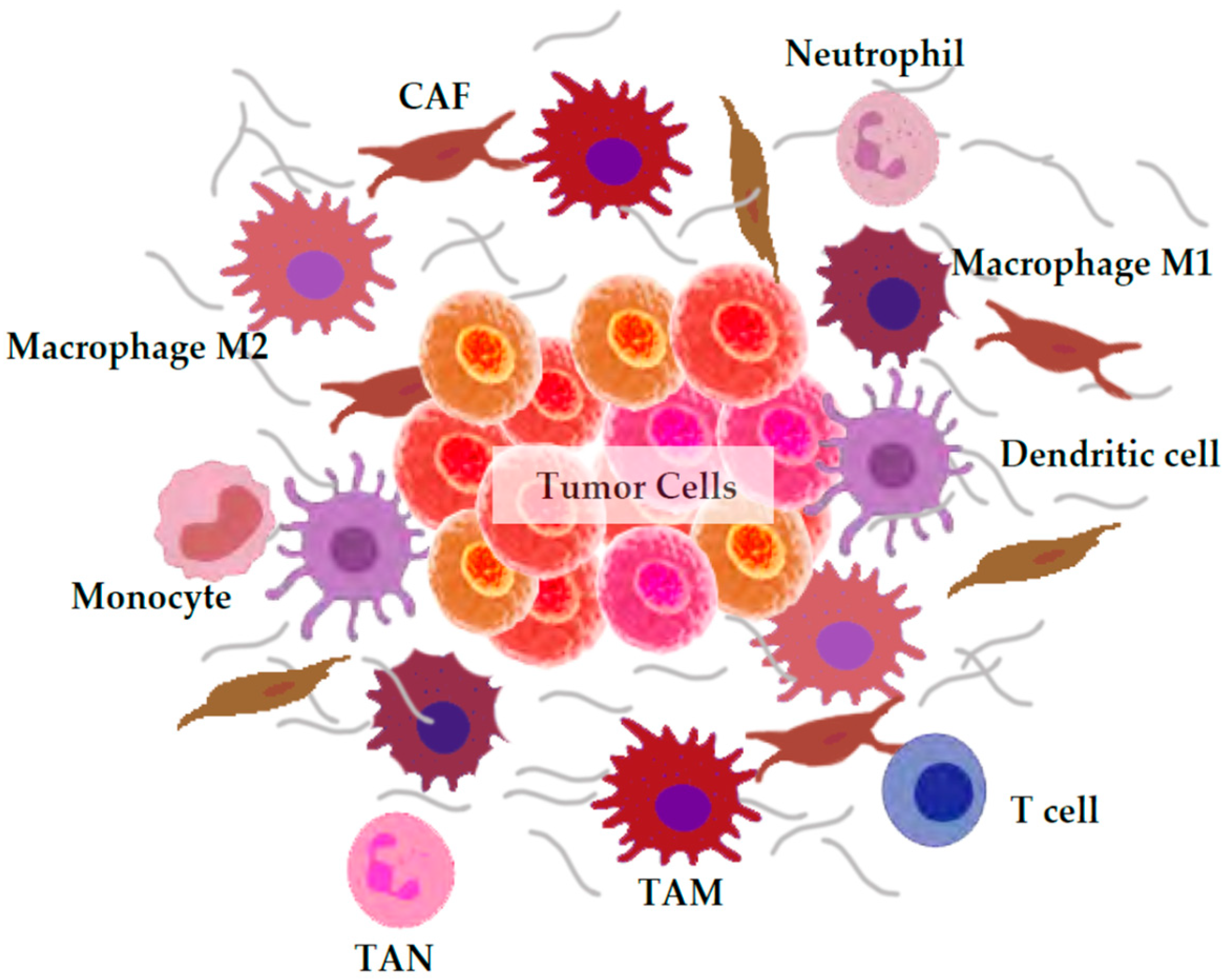
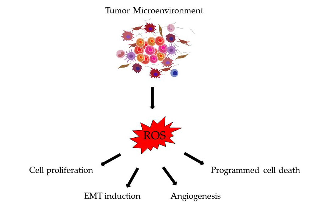
3. ROS Generation by the Tumor Microenvironment

The biological term “TME” encompasses the entity formed by cancer cells and the wide range of non-malignant cell types and components present in the tumor, such as the immune system, components of the blood and lymphatic vascular networks, fibroblasts, the extracellular matrix (ECM), and signaling molecules [3]. The interplay between all these components and the tumor cells is essential for cancer growth, development, metastasis, and treatment response. Among all these entities, tumor cells and immune cells such as macrophages and neutrophils are major ROS producers; however, there are other minor ROS sources worth consideration (Figure 1). Depending on the type of ROS-producing cell the consequences are different and can be either tumor-supportive or tumor-suppressive [4].

3.1. Macrophages

The production of ROS by the immune system cells is tightly linked to the defense response and phagocytosis. It is used by neutrophils and macrophages to destroy pathogens as wells as cancer cells. Besides, these ROS are also involved in the activation of T cells and NK cells [8].
Macrophages constitute the major component of the infiltrate of most tumors. They can be divided into two distinct types: M1 (or classically activated) and M2 (or alternatively activated). M1 macrophages are generally considered to be tumor-killing macrophages, while M2 macrophages promote tumor growth and metastasis and are associated with poor prognosis. ROS can stimulate activation statuses in tumor-associated macrophages (TAMs) [4]. Besides, the ROS scavengers N-acetylcysteine and the NADPH oxidase ROS inhibitor diphenyleneiodonium induce monocyte polarization toward M1-like macrophages and the repolarization of M2 macrophages into M1 phenotypes. This effect prevents M2 macrophage differentiation, and, more importantly, inhibits tumor progression and M2 macrophage infiltration in the TME of CRC cell models [40].
M1 macrophages eliminate pathogens and tumor cells by secreting agents such as tumor necrosis factor α (TNF-α), interleukin (IL)-12, RNS, and ROS [41], providing a pivotal contribution in the oxidative environment. It has also been shown that monocytes activated by contact with tumor cells produce very high levels of ROS [42,43]. Moreover, TAMs, similar in phenotype to M2, are corrupted by tumor cells to promote tumor immune escape, angiogenesis, tumor growth, and metastasis [44]. The infiltration of TAMs in subcutaneous colorectal tumors is inhibited by some ROS scavengers such as Oligo-Fucoidan [40]. However, TAM-produced ROS have pro- and anti-tumorigenic activities (depending on the context), and are affected by various factors including the tumor entity and stage, as well as pre- and co-treatments [40].

3.2. Neutrophils

As in the case of macrophages, the role of neutrophils in the tumor process has been associated with defensive responses. However, some populations of neutrophils, known as tumor-associated neutrophils (TANs), could be involved in tumor growth, invasion, and angiogenesis of cancer cells, as well as in the development of metastasis [45]. Under stimulation, neutrophils generate large amounts of superoxide by activating NADPH oxidase 2 (NOX2) and hydrogen peroxide, which can modify extracellular targets and affect neighboring cell functions [46]. In this regard, it has been demonstrated that cells of myeloid origin, such as macrophages and neutrophils, can initiate tumor growth in various organs (such as the intestine) by increasing ROS production [47]. However, the effect of neutrophils on CRC tumors is not yet clear. Rao et al. showed that the intratumoral increase in neutrophils was associated with malignant phenotypes and could predict an adverse prognosis in CRC [48]. On the other hand, another study analyzed the number of neutrophils in CRC tissues and demonstrated that high levels of TANs were associated with improved overall survival in patients with stage II CRC [49].

3.3. Cancer-Associated Fibroblasts (CAFs)

The most predominant cell type in the stroma is the fibroblast, the functions of which include the renewal of ECM, the regulation of epithelial differentiation, the regulation of inflammation, and the involvement in wound healing. Activated fibroblasts such as cancer-associated fibroblasts (CAFs) secrete ROS (among other several factors) [50], which modify the environment to favor tumor development, regulate the reorganization of the connective tissue, and also facilitate metastasis through the activation of tumor neo-angiogenesis [51]. Moreover, recent studies have demonstrated that cancer cells can induce ROS overproduction in CAFs [52], contributing to a pro-oxidative TME. ROS produced by CAFs in turn enhance ROS generation in cancer cells, increasing their tumor aggressiveness [53].

3.4. Others

Myeloid-derived suppressor cells (MDSCs) are immature myeloid cells that play an important role in promoting tumor progression because they are involved in the immune suppression of T and NK cells [54]. In fact, one of the most important mediators of T cell suppression by MDSCs is the ROS-dependent generation of peroxynitrite [55]. T lymphocytes or T cells are another main source of ROS. Indeed, peripheral blood T lymphocytes from cancer patients have shown to have increased ROS production compared to those from healthy subjects [56]. ROS are involved in various aspects of T cell biology, including activation, differentiation, apoptosis, and antigen recognition. T cell-intrinsic ROS also influence tumor progression. Increased ROS production in T lymphocytes promote their apoptosis and tumorigenesis, influencing their immunosuppressive capacity [4]. Dendritic cells (DCs), antigen-presenting cells acting during T cell-response process, are also involved in ROS activities. ROS produced by DCs influence the anti-tumoral immune response, as it is upregulated during cross-presentation to cytotoxic T cells [57].

4. Consequences of Oxidative Stress in Colorectal Cancer Progression and Metastasis

The roles of ROS in CRC initiation have classically been linked to inflammation and DNA damage [58]. However, oxidative stress is also involved in other processes related to cancer progression and metastasis, such as epithelial-to-mesenchymal transition (EMT), angiogenesis, and apoptosis. These processes are induced by molecules and regulating factors from the TME which affect tumor cell growth and the capacity to invade distant organs [59].

4.1. Cell Proliferation

One of the first events in tumorigenesis is increased cell proliferation, a feature influenced by ROS signaling molecules. For instance, the expression of NOX1 in NIH 3T3 fibroblasts increases the production of superoxide anion, and at the same time causes a 10-fold elevation in hydrogen peroxide levels. This results in the expression of cell cycle and cell growth genes [60]. The fact that cancer cells produce high amounts of hydrogen peroxide [61] adds more evidence to the positive correlation between ROS levels and proliferation. In contrast, the addition of an exogenous catalase, a hydrogen peroxide scavenger enzyme, has been shown to inhibit proliferation in a dose-dependent manner in several cancer cell lines [62]. Besides, a recent preclinical study showed that the combination of traditional chemotherapy with catalases has an additive antitumoral effect in lung adenocarcinoma cells [63]. Similarly, the overexpression of glutathione peroxidase 1 (GPx1), another hydrogen peroxide detoxifying enzyme, completely suppressed tumor cell growth in nude mice bearing v-Ha-ras-transformed rat kidney epithelial cells [64]. Moreover, the enforced expression of manganese superoxide dismutase (MnSOD), a mitochondrial superoxide anion detoxifier, reduces the growth rate of the rapid-growing pancreatic human MIA PaCa-2 cell line [65]. Similar results have been observed in colon cancer cell lines, where the inhibition of NOX1 supports proliferation by modulating ROS signaling [66]. Anti-proliferative effects, induction of apoptosis, and reduction of the Warburg effect triggered by an imbalance in the redox state were observed with the addition of the flavonoid morin in CRC cell lines [67]. Furthermore, herbal melanin promotes apoptosis and inhibits the MAPK pathway in HT29 and SW620 CRC cell lines [68]. Finally, a derivative of aminobenzenesulfonamide can hamper cell proliferation and migration, inducing apoptosis in CRC cells through ROS generation [69].

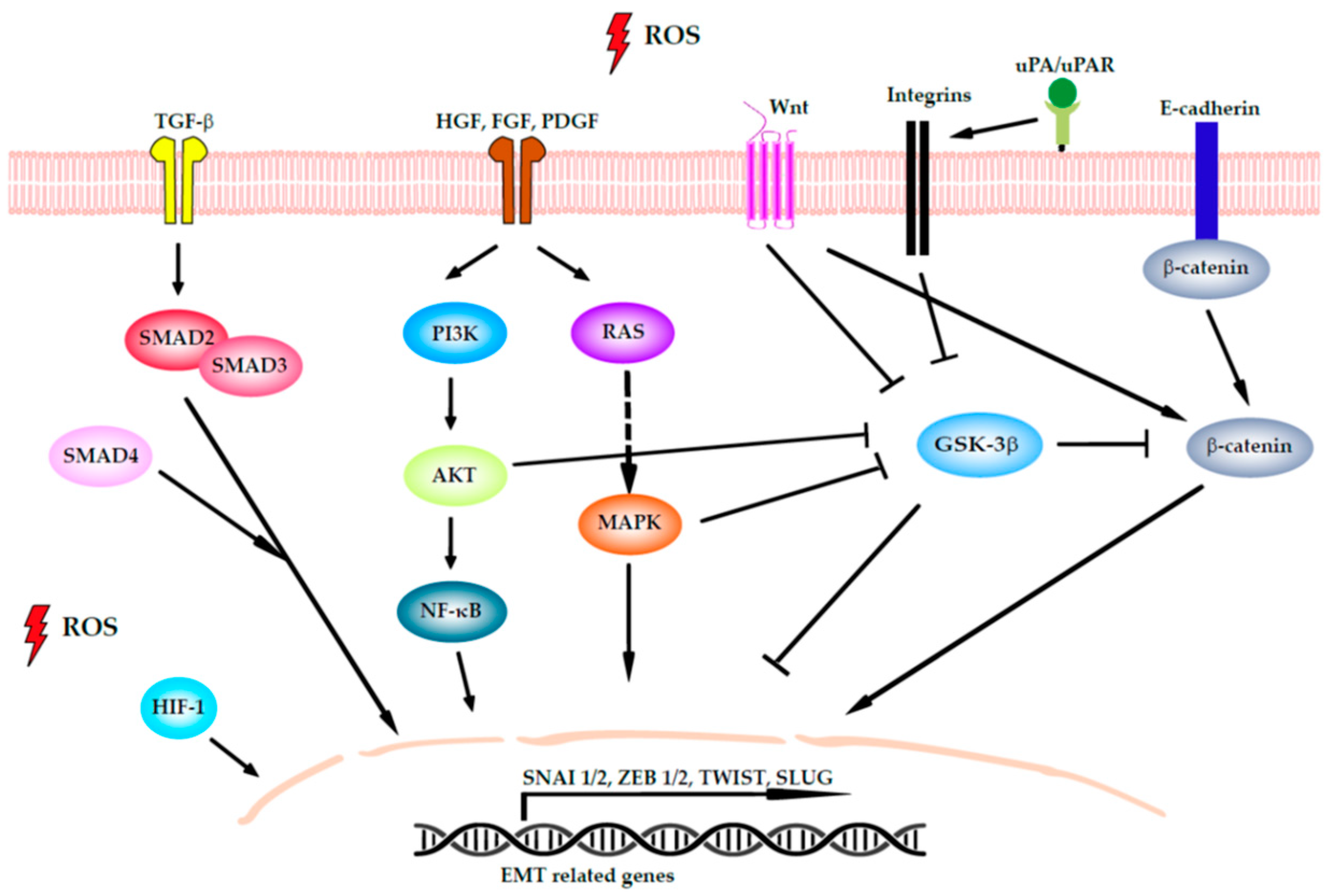
4.2. Induction of EMT

EMT is a well-defined process that is essential for the metastatic cascade in which tumor cells transition from an epithelial-like phenotype to a mesenchymal-like one, allowing them to escape from the basement membrane surrounding the primary tumor. During this process, cells with an epithelial phenotype lose cell–cell and cell-matrix adhesions and acquire properties of mesenchymal cells such as the ability to degrade ECM and to enhance their motility and migratory capability [70]. In CRC, as in many other cancers, the EMT process is highly regulated through some of the classic tumorigenic signaling pathways, such as the nuclear factor-κB (NF-κB), hypoxia-inducible factor 1 (HIF-1), and transforming growth factor β (TGF-β) pathways [71]. Oxidative stress plays a critical regulatory role in these pathways (Figure 2), for example by degrading their inhibitors or by inducing protein nuclear translocation and consequent transcription, as occurs with members of the NF-κB family. PI3K/Akt can facilitate protein synthesis and promote EMT, activating the NF-κB pathway. The PI3K/Akt pathway is also involved in the inhibition of glycogen synthase kinase-3 (GSK-3β), which confers stabilization to β-catenin to activate the transcript of Slug and vimentin [72]. All these inputs received by the cell lead to the activation of the EMT transcription factors (TFs) SNAI1/2, SLUG, TWIST, and ZEB1/2 [73] in colon tissues [74,75].
Aside from the role of ROS in EMT, ROS are also involved in other actions related to motility and migration. ROS mediates ECM remodeling through the arrangement of some integrins and the urokinase plasminogen activator (uPA) signaling pathways. Integrins are adhesion molecules present on the cell surface that bind the ECM with the intracellular actin cytoskeleton. The implications of ROS in regulating many integrin-mediated cellular activities are well established [76]. uPA is an extracellular serine protease for which activation by its specific receptor uPAR is required for ECM degradation and matrix metalloproteinase (MMP) activation. It has been reported that ROS induce the transcription of uPA and uPAR and stabilize their mRNA [77,78]. In this regard, Tochhawng et al. showed that gelsolin (an actin-binding protein) overexpression triggers the secretion of uPA, elevating intracellular superoxide levels in CRC cells [79]. For ECM remodeling the action of fibroblasts and CAFs in the TME is also essential. ROS levels modify CAF function by activating the TGF-β signaling pathway [80].
ROS also play an important role in actin polymerization, as they are involved in cytoskeleton remodeling and cell motility. The cytoskeleton is a dynamic network of microtubules and protein filaments where the two molecules actin and tubulin predominate and can modify their behavior under oxidation [72]. Cell motility is based on formation of actin stress fibers and actin rearrangements. Rho GTPases, which are involved in actin rearrangements, can be regulated by the focal adhesion kinase (FAK), Src, and PI3K/Akt signaling pathways, which are all modulated by ROS. Activation of the tyrosine kinase FAK leads to the recruitment of talin to nascent adhesions and the formation of focal adhesions and actin stress fibers [81]. Moreover, Src activation can enhance cell movement by promoting FC turnover and the detachment of tumor cells from the primary tumor, the latter action through downregulating E-cadherin and upregulating MMPs [82]. Besides, ROS regulate cell–cell junctions decreasing the expression of occluding, claudin, and E-cadherin, proteins repressed by the EMT master controller TF.

4.3. Angiogenesis

During tumor development, new blood vessels are formed to support tumor growth. This process is called angiogenesis, and like others it is highly dependent on ROS levels [83]. Pro-angiogenic factors are activated upon physical signals such as hypoxia, ischemia, and vasculature injury. Vascular endothelial growth factor (VEGF) is of great relevance and is the primary factor initiating the angiogenic cascade, promoting the extravasation of plasma proteins and forming a primitive scaffold for migrating endothelial cells [84]. VEGF is stimulated by exogenous ROS both in vitro and in vivo [85,86]. A major endogenous ROS source in endothelial cells is from NOX activity, which could be activated by growth factors including VEGF. Interestingly, the produced ROS activate VEGF receptor 2 autophopshorylation and are involved in the activation of TFs in angiogenesis [87].
Each step of angiogenesis is controlled by HIF-1, which is the master regulator of oxygen homeostasis and is activated by O2-dependent mechanisms [88]. Under hypoxic conditions, HIF-1 upregulates many growth factors and their receptors, including VEGF and their receptors (known as VEGFRs). Additionally, activation of the PI3K/AKT/mTOR pathway in tumor cells can also increase VEGF secretion by both HIF-1 dependent and independent mechanisms. This pathway modulates the expression of other angiogenic factors such as nitric oxide and angiopoietins [89]. The expression of several mutated p53 proteins in colorectal cancer cell lines (HCT116) increases intracellular ROS and raises the number of blood vessels in their respective xenografts [90]. The crosstalk between VEGF and other oncogenes such as EGFR has been described. EGFR regulates VEGF expression via the MAPK and PI3K signaling cascades and the expression of at least three different TFs: STAT3, Sp1, and HIF-1 [91].
In CRC, HIF-1, and VEGFA are highly expressed in tumor tissue [92]. VEGFA is the isoform that has the highest affinity for VEGFR2, which is mainly found in endothelial cells. However, VEGFR1 has been found to be expressed in CRC cells, and its activation induces tumor progression and metastasis features [93]. Interestingly, intracrine VEGF signaling by CRC cells has been involved in the acquisition of cell migration and invasion phenotypes in these cells [94].
Increasing knowledge about the VEGF/VEGFR axis has allowed the development of novel therapeutic approaches to target angiogenesis. Initially, monoclonal antibodies against VEGF or VEGFR were proved to be effective across different treatment lines in patients with metastatic CRC. The four current anti-angiogenic drugs approved by the Food and Drug Administration are bevacizumab (anti-VEGFA), aflibercept (anti-VEGFR1), ramucirumab, (anti-VEGFR2), and regorafenib (a multikinase inhibitor including VEGFR1 and VEGFR2). Nevertheless, the lack of validated predictive markers for the different anti-angiogenic treatments is emphasized [95].

4.4. Apoptosis, Autophagy, and Anoikis

The capacity to avoid cell death is one hallmark of cancer. Cell death programs, such as apoptosis, autophagy, and anoikis, serve as a natural barrier for cancer development and its activation is due to intra- and extracellular stresses that convey signals between regulators and effectors [96].
Apoptosis induced by ROS is triggered by the apoptosis signal-regulating kinase 1 (ASK1)/c-Jun N-terminal kinase (JNK) and ASK1/p38 signaling pathways in human cancer cells. When H2O2 oxidases thioredoxin 1, it dissociates from ASK1, activating the downstream MAP kinase kinase (MKK)4/MKK7/JNK and MKK3/MKK6/p38 pathways, leading to the suppression of anti-apoptotic factors [97]. ROS also activate the apoptotic pathway through death receptors and the initiator caspase 8, followed by the cleavage of downstream executor caspase 3 and Bcl-2, and finally by releasing and translocating cytochrome c [98]. Once cytochrome c is released from the mitochondria into the cytosol, it interacts with apoptotic protease-activating factor 1 (Apaf-1) to form the apoptosome, leading the activation of caspase-9 and the downstream caspase cascade [99].
In CRC, apoptosis induction via ROS in different types of cells has been demonstrated in several papers. For example, Chung et al. showed that hop proanthocyanidins or the condensed tannins found in some vegetables are cytotoxic to HT-29 colorectal adenocarcinoma cells through formation of ROS, leading to protein carbonylation and to cytoskeleton disorganization [100]. In this type of CRC cells, another set of experiments performed with resveratrol (a polyphenol found in grapes and wine) showed chemopreventive cancer properties, as this compound activated cell apoptosis through a ROS-dependent mitochondrial mechanism [101]. Resveratrol also triggered apoptosis via ROS in human CRC cells [102].
Autophagy is a cellular physiologic mechanism that can be strongly induced by certain cellular stresses. Cells break down cellular organelles, allowing the resulting catabolites to be used for biosynthesis and energy metabolism [96]. In this regard, it has been reported that H2O2 induces autophagic cell death in human CRC cells [103,104,105]. However, the role of the autophagy pathway in tumor progression is complex. Autophagy protects cells against the production of ROS through the elimination of the damaged mitochondria, leading to a reduction in ROS production and thereby limiting the tumor-promoting effect of ROS in DNA mutation [106].
Anoikis is a type of apoptosis induced upon cell detachment from the ECM, and is a critical mechanism for preventing adherent-independent cell growth and attachment. The deregulation of anoikis execution is an emerging hallmark of cancer cells and contributes to the formation of metastasis in distant organs [107]. ECM detachment causes a multitude of catastrophic metabolic alterations, including a robust increase in ROS [108]. It has been demonstrated that in CRC cells, anoikis can be regulated by β-catenin [109] and Src [110].

5. Pro-Oxidants and Antioxidants in Colorectal Cancer Therapeutics

There is plenty of evidence that oxidative stress and ROS are genotoxic, and an excess of these is deleterious for cells. As ROS are thought to be one of the major sources of endogenous DNA damage, it can be assumed that antioxidants could be beneficial as they minimize genotoxic ROS effects, acting as chemopreventive agents [111]. On the other hand, the use of pro-oxidants to elevate ROS levels in the TME could induce malignant cell death, being beneficial and able to supplement other cancer therapies.

5.1. Pro-Oxidants

The most promising pro-oxidant treatments are based on ROS-dependent cancer cell death induction through the alteration of several mechanisms, including the ubiquitin-proteasome pathway, tyrosine kinase cascades, glucose metabolism, glutathione reservoir, and thioredoxin activity, etc. Herein, we discuss the traditional treatments in CRC and their association with oxidative stress. Furthermore, we examine novel ROS-dependent therapeutic approaches in clinical trials (Table 1).

5.1.1. Oxaliplatin

Anti-neoplastic drugs can induce high levels of oxidative stress. This is the case for the most used and accepted treatment for advanced CRC or its metastatic form, which consists of a combination of folinic acid, 5-fluorouracil (5-FU), and oxaliplatin, namely FOLFOX6 [112]. Oxaliplatin, a platinum-based drug, in combination with 5-FU, improves its antitumor activity. Platinum-based drugs trigger a ROS generation burst, resulting in the loss of mitochondrial membrane potential [113] and a decrease in the glutathione level, leading malignant cells to die by apoptosis. Platinum is a transition metallic element able to lose electrons and positively charge ions, disturbing the normal electron flow of enzymes and substrates and ultimately affecting their catalytic activity [114]. Currently, clinical trials are focused on improving the anticancer effects of FOLFOX6 and lowering the severity of side effects with adjuvants or alternatives.

5.1.2. Arsenic Trioxide (AT)

AT is a potent oxidant which acts by reducing the intracellular redox buffering capacity and promoting apoptosis in malignant cells through JNK activation [115]. It is indicated for the treatment of a specific form of acute promyelocytic leukemia [116]. AT may sensitize CRC cells to 5-FU and leucovorin treatment. A phase I trial is studying the side-effects and best dose combination of AT with 5-FU and leucovorin in patients with stage IV colorectal cancer who have relapsed or did not respond to treatment. (NCT00449137).

5.1.3. Tyrosine Kinase Inhibitors (TKIs)

TKIs are the cornerstone treatment of many cancers. TKIs are small molecules that interfere with the autophosphorylation, dimerization, and activation of the kinase, acting as receptor antagonists. There are several TKI that differ in their pharmacological effects, side effects, and target kinases. The antitumor effects of TKI result in mitochondrial dysfunction and the uncoupling of electron transport chain proteins, increasing ROS levels [117]. Imatinib is a tyrosine kinase inhibitor which targets BCR-ABL, c-KIT, and PDGFR, and is used in a wide range of cancers [118]. The utility of imatinib as a first-line therapy in combination with XELOX and bevacizumab has been investigated in stage IV patients in a phase I/II trial (NCT00784446). Results showed tolerable toxicity and promising efficacy [119]. Erlotinib is another TKI which specifically inhibits EGFR signaling and has been found to induce metabolic oxidative stress through NOX4 activation [120]. A clinical trial is currently evaluating the efficacy and safety of erlotinib in combination with permetrexed in metastatic CRC refractory to standard chemotherapy (NCT02723578). Finally, vemurafenib, a TKI indicated for the treatment of melanoma harboring the mutation V600E in BRAF, has been shown to activate oxidative metabolism and promote ROS-dependent cell death [121]. At the clinical level, vermurafenib is under evaluation for its efficacy and safety with the administration of FOLFIRI (folinic acid, 5-FU and irinotecan) plus cetuximab in advanced CRC with the BRAF V600E mutation (NCT03727763).

5.1.4. Endoplasmic Reticulum (ER) Stress Inductors

ER stress inductors are part of the anti-cancer drug armamentarium. ER stress is a common event occurring when folding protein machinery is overloaded, resulting in the accumulation of damaged proteins in the ER. In turn, cells activate the unfolded protein response, which restores homeostasis or activates cell death [122]. Several ER stress inductors have been pharmacologically developed as a strategy to kill cancer cells. The proteasome inhibitor bortezomib binds to the active site of the subunit 20S of the proteasome, leading to the accumulation of unfolded and damaged proteins that, in its turn, induces ER stress and calcium and cytochrome C release, processes that lead to apoptosis [123]. Bortezomib has been evaluated in a phase II trial to study its effectiveness in metastatic or recurrent CRC (NCT00052507). Unfortunately, bortezomib was ineffective in controlling metastatic CRC disease, but a significant accumulation of HIF-1α was seen in tumor specimens and xenograft models, suggesting that proteasome inhibition could alter the response to tumor hypoxia [124]. Another drug that causes ER stress is celecoxib, a selective cyclooxygenase-2 inhibitor that can bind and inhibit the sarcoplasmic and the ER calcium ATPase, causing ER stress through calcium leakage into the cytosol and finally resulting in apoptosis [125]. Celecoxib is currently under investigation in a phase IV trial aiming to evaluate its anticancer effect as an adjuvant therapy with the FOLFIRI regimen in patients with metastatic CRC (NCT03645187)

5.1.5. Novel Anthracyclines: AVA6000

A first-in-human study has been initiated with AVA600 for patients with locally advanced and/or metastatic solid tumors including CRC (NCT04969835). This promising phase I study will evaluate the safety, tolerability, and pharmacokinetics of AVA6000, a modified pro-drug version of doxorubicin that remains inactive until it reaches the malignant microenvironment. There, once activated as doxorubicin, it attacks malignant cells, triggering multifactorial toxicity that involves oxidative stress by induction of O2•− and H2O2, DNA/RNA damage by binding and blocking topoisomerases, autophagy and apoptosis induction by calcium leakage and calcium channel dysregulation, and mitochondrial dysfunction through NO release [126].

5.1.6. Poly (ADP-Ribose) Polymerase (PARP) Inhibitors

PARPs are a family of enzymes responsible for the transfer of ADP-ribose to proteins in a reaction named ribosylation. PARPs play an important role in DNA repair, specifically in base and nucleotide excision repair, when DNA damage increases. Considering that cancer cells are usually defective in homologous recombination DNA repair pathways, it is thought that they greatly rely on PARP-mediated DNA repair for survival. PARP inhibitors (PARPi) affect DNA repair and act by provoking genomic instability and accumulation of damaged cells and consequently cell cycle arrest [127]. Some studies in ovarian cancer cell lines showed that PARP inhibition decreased their proliferation by increasing ROS levels and oxidative damage in all cancer lines analyzed [128]. At the clinical level, PARPi are the chosen treatment for certain breast and ovarian cancers, either as a single agent or in combination [129]. Two PARPi have been tested in CRC: olaparib and veliparib. Olaparib was tested in an interventional phase I trial to determine its safe dose in combination with irinotecan hydrochloride in patients with advanced or metastatic CRC (NCT00535353). Unfortunately, this combination was ineffective, dissuading researchers from further investigation [130]. Another PARPi, veliparib, was studied in combination with FOLFIRI +/− bevacizumab to evaluate its efficacy and tolerability in untreated CRC patients in a phase II trial (NCT02305758). Nevertheless, the addition of veliparib to the regimen did not provide superior efficacy [131].

5.2. Antioxidant Treatments or Interventions

Whether the enhancement or inhibition of antioxidants is beneficial or detrimental for cancer treatment is still a current controversial topic. The trend in favor of the beneficial effects of antioxidants hypothesizes that their supplementation to cancer patients can be beneficial in cancer prevention as it reduces the malignancy risk or provides an additive effect for the given chemotherapy. The other line of thought, which hypothesizes that antioxidants benefit malignant cells, considers that antioxidants promote the adaptation and survival of cancer cells in the hostile microenvironment.
Herein, we show interventions based on antioxidants which are under study in CRC patients in clinical trials (Table 2).

5.2.1. Dietary Supplementation of Vitamins

A phase II trial currently recruiting patients will evaluate the effect of Ocoxin®-Viusid® on the quality of life of patients with metastatic colorectal adenocarcinoma, with the aim of enhancing tolerance to chemotherapy (NCT03559543). Ocoxin®-Viusid® is an oral solution that includes several vitamins with anticancer activity such as vitamin B6, vitamin C, and cinnamic acid, among others [132,133,134]. Similarly, a phase II/III trial intends to compare the effects of vitamin B6, folic acid, and dietary supplementation with vitamin C on homocysteine status, oxidative stress markers, antioxidant enzymatic activities, and DNA methylation in a group of 500 randomized patients with histologically confirmed CRC (NCT01426490). Other vitamin sources are tocopherols, a group of fat-soluble compounds commonly found in vegetables (α, β, γ, and δ tocopherols), many of which have vitamin E activity. Due its structure, tocopherols have unmethylated carbons which can trap ROS, acting as antioxidants. Oral uptake of vitamin E has shown to exhibit an inhibitory growth effect against malignant cancers, including CRC, in different animal models of carcinogenesis [135,136]. The use of γ-tocopherol, the major form of vitamin E in US diet, has been tested in the clinic as a pre-operatory strategy (NCT00905918). In that trial, patients with confirmed CRC scheduled for surgery received oral supplementation with γ-tocopherol. The aim was to halt the development of cancer before surgery and to study the effects of vitamin E on plasma levels of oxidative and nitrosative biomarkers. Although the trial is completed the results are not yet available.

5.2.2. Trace Element Supplementation

Trace elements are minerals present in small amounts in our organism. In that group we can find zinc, an important cofactor of nucleic acid metabolism, replication, growth, and antioxidant activity [137]. Zinc supplementation was used within cycles of chemotherapy in patients with CRC, and changes in oxidative stress during chemotherapy after surgery were assessed (NCT02106806). Results derived from this trial showed an increase in superoxide dismutase (SOD) activity attributable to zinc supplementation, with an improvement in the patient quality of life. Nevertheless, no effect of zinc complementation was seen on oxidative stress markers in plasma such as vitamin C, vitamin E, malondialdehyde, and 8-isoprostane [138].

5.2.3. SOD Mimetics

Calmangofodipir is a manganese metabolite which possesses mitochondrial MnSOD mimetic activity. A phase I/II clinical trial was designed to analyze whether the pre-treatment with calmangafodipir could decrease the frequency and severity of side effects derived from FOLFOX6 administration in patients with metastatic CRC (NCT01619423). Results from this trial demonstrated that calmangafodipir prevents the development of oxaliplatin-induced peripheral neuropathy, but no influence was observed in tumor regression [139]. Interestingly, preclinical data indicate a differential effect of this compound administered with chemotherapy between normal and malignant cells, where apoptosis is induced because of an oxidative stress burst [140,141].

5.2.4. Polyphenols

Polyphenols are a family of organic compounds abundantly found in plants. Recent polyphenol studies have shown evidence that long-term consumption of rich diets in polyphenols may protect against cancer (among other diseases) [142]. One relevant polyphenol-containing fruit is pomegranate, which has strong antioxidant effects. The active principle is ellagitannin, which is converted into urolithin A in the human gut, increasing peroxiredoxin expression which regulates peroxide levels [143]. Some human studies are focused on the effect of urolilthin A. In this regard, an interventional phase I/II trial was performed to study the effect of two pomegranate extracts in CRC patients after diagnosis until surgery. The aims of this work were to evaluate phenolic disposition and urolithins in both tumor and non-tumor colon tissues, as well as to evaluate gene expression profiling to understand the anti-inflammatory and anti-cancer effects (NCT01916239). Authors demonstrated that oral intake of pomegranate extract was significantly associated with the expression of CD44, CTNNB1, CDKN1A, EGFR, and TYMs in colon samples. However, the authors admitted that these findings were not correlated with the individual capacity to produce specific urolithins or the levels or urolithins in the colon tissues [144]. Another important polyphenol is resveratrol, found in high concentrations in red grapes. Resveratrol has shown a plethora of therapeutic benefits, including anti-inflammatory, ROS-scavenging, immunomodulatory, and anti-carcinogenic properties, among others [145]. In a phase I trial, resveratrol was given to CRC patients before surgical resection of the tumor (NCT00256334). There, researchers sought a better understanding of the effects of resveratrol in Wnt signaling pathway, which is aberrantly activated in 85% of CRCs. Surprisingly, resveratrol administration exerted inhibitory effects on Wnt signaling only in normal colonic mucosa [146]. Another subclass of polyphenol compounds is that of catechins, present in a wide variety of foods including tea, apples, persimmons, cacaos, grapes, and berries. Catechins seem to have dual action in ROS, acting as antioxidants or pro-oxidants [147]. Catechins exert their antioxidant effect by reducing free radicals through the donation of one electron of their phenolic group. Moreover, they are capable of chelating metal ions involved in radical production. Indirectly, catechins upregulate the activity of antioxidant enzymes such as superoxide dismutase, catalase, and glutathione peroxidase, and can inhibit pro-oxidant enzymes [148]. At the clinical level, catechins are under study in a phase II trial (NCT01606124) to determine whether polyphenon E green tea extract administration can prevent or delay disease progression in patients with a high risk of recurrent CRC. Recently published results concluded that polyphenon E was well tolerated but did not significantly reduce the number of aberrant rectal crypt foci, considered as a surrogate endpoint biomarker of CRC [149]. Other polyphenol family members are quercetins, mostly found in flowers, vegetables, and fruits. It has been demonstrated that flavonoids such as quercetin have stronger antioxidant effects than vitamins due to their chemical structure [150]. For instance, in vivo studies using quercetin on ascites cells in Dalton’s lymphoma-bearing mice showed downregulation of total ROS levels and protein kinase C activity, improving the apoptotic potential due to an increase in caspase 4 and 9 and promoting death receptor-mediated apoptosis [151]. Quercetin supplementation was studied as a preventive measure in individuals at medium and high risk of developing CRC (NCT00003365). Unfortunately, the results have not been yet reported. Another interesting group of polyphenols are curcumoids, used worldwide for their multiple health benefits as well as their culinary and cosmetic properties. Curcumin consumption has been found to increase the serum activity of ROS-scavenging enzymes such as superoxide dismutase, catalase, and glutathione peroxidase, and reduce lipid peroxides [152]. A pilot study with curcumin is being tested in combination with 5-FU in chemorefractory metastatic CRC patients (NCT02724202). The aim of this study is to assess clinical safety and identify the clinical response rate of the combination treatment.

5.2.5. Organosulfur Compounds

Sulforaphane, another natural compound, is naturally derived from cruciferous vegetables like broccoli. Sulforaphane has been described as an antioxidant indirect molecule. In this regard, sulforaphane acts as an inducer of the Keap1/Nrf2/ARE pathway [153]. Nrf2 is a transcription factor and is considered the most important regulator of antioxidant gene expression, particularly the genes responsible for glutathione synthesis [20]. One study is currently assessing the benefit of the intake of cruciferous vegetables in volunteers scheduled for screening colonoscopy. Researchers will measure suforaphane levels in blood as well as histone deacetylase expression in tissue biopsies and peripheral blood mononuclear cells (PBMCs) (NCT01344330). Results are now under examination.

5.3. Future Perspectives

Recently, new emerging technologies such as drug conjugates, nanoparticles, and CRISPR technology have shown great potential for multiple applications. Targeted drugs and nanomedicine can improve precision therapy, drug delivery release, diagnosis, immunotherapy, and in vivo gene and epigene editing [154,155,156,157]. In CRC, various groups have designed nanoparticles to target cancer cell surface biomarkers such as carcinoembryonic antigen (CEA) or folate receptor-α to direct the conventional chemotherapeutic treatments to the tumor. Other approaches exploit the specific properties of the TME, such as the pH, to controllably release the content from the nanoparticle to the tumor [158]. Some of these studies have taken the first step towards clinical trials. One example is a phase I/II trial administering the C’Dot drug conjugate ELU001 (which targets the tumor-overexpressed folate receptor-α) in patients with advanced tumors including colon cancer (NCT05001282).
Several studies performed in cellular and animal models use nanoparticles containing antioxidant and pro-oxidant compounds to prevent tumor formation or promote tumor apoptotic cell death by ROS species, respectively [159,160].
As for CRISPR technology, genome-wide CRISPR screens have served for the identification of oxidative stress-responsive genes in CRC such as Galectin-2 (Gal2) which has a tumor-suppressive role in this cancer [161]. Moreover, House et al. engineered dCas9-Killer Red to generate oxidative stress at the desired genomic region that could be used to model more natural DNA damage [162].
Undoubtedly, there will be an increasing presence of these novel technologies in the near future to precisely tackle pro-oxidant and antioxidant genes and processes, favoring the development of more efficient and selective therapies for CRC.

6. Conclusions

The involvement of oxidative stress in the tumorigenic process is still a controversial topic. ROS produced either by tumor cells or by TME cells have very diverse and sometimes opposing effects on the evolution of cancer. Low to moderate ROS levels promote cell proliferation, EMT, and angiogenesis. Conversely, high ROS levels favor apoptosis, cell death, and cellular damage. Moreover, ROS influence the immune response against tumors, as they are used by macrophages and neutrophils to destroy cancer cells and activate T and NK cells. Overall, the effects seem to depend on ROS levels, the cancer stage, and the differential outcomes seen in tumor cells because of ROS exposure.
Such a duality of ROS effects in cancer cells and TME allows us to assume that their modulation can be exploited for cancer prevention and treatment. On the one hand, antioxidants could counteract the deleterious consequences of ROS. On the other hand, pro-oxidants could induce ROS-dependent cancer cell death. However, the results do not seem to be conclusive at this point in either case. Further research is needed to provide insights on the role of ROS modulators in the initiation and progression of CRC.

Author Contributions

M.A.S. and E.P. were responsible for conceptualization; M.A.S., I.H. and E.P. wrote the original draft of the review; E.P. developed the figures; A.S. (Antonieta Salud), R.M. and O.P. performed a critical review; A.S. (Anabel Sorolla) edited the manuscript. Finally, A.S. (Antonieta Salud) was responsible for project administration and funding. All authors have read and agreed to the published version of the manuscript.

Funding

This research was funded by Merck Salud Foundation project award (2019) given to A.S. (Antonieta Salud); by Merck Salud Foundation project award (2021) given to R.M.; by the University and Research Grants Management Agency (AGAUR) of the Catalonia Government (emergent research group recognition award (2017SRG1620) given to M.A.S.; and by AGAUR (2021FIB00025) given to I.H. The 2020 Miguel Servet fellowship from the Instituto de Salud Carlos III (Spanish National Institute of Health) under Grant CP20/00039 was given to A.S. (Antonieta Salud). The research was supported by the CERCA Program of the Generalitat de Catalunya and the IRBLleida (Fundació Pifarré).

Conflicts of Interest

The authors declare no conflict of interest. The funders had no role in the design of the study; in the collection, analyses, or interpretation of data; in the writing of the manuscript, or in the decision to publish the results.

References

  1. Ferlay, J.; Colombet, M.; Soerjomataram, I.; Parkin, D.M.; Piñeros, M.; Znaor, A.; Bray, F. Cancer statistics for the year 2020: An overview. Int. J. Cancer 2021, 149, 778–789. [Google Scholar] [CrossRef]
  2. Xi, Y.; Xu, P. Global colorectal cancer burden in 2020 and projections to 2040. Transl. Oncol. 2021, 14, 101174. [Google Scholar] [CrossRef]
  3. Balkwill, F.R.; Capasso, M.; Hagemann, T. The tumor microenvironment at a glance. J. Cell Sci. 2012, 125, 5591–5596. [Google Scholar] [CrossRef] [Green Version]
  4. Kennel, K.B.; Greten, F.R. Immune cell-produced ROS and their impact on tumor growth and metastasis. Redox Biol. 2021, 42, 101891. [Google Scholar] [CrossRef]
  5. Del Idelchik, M.P.S.; Begley, U.; Begley, T.J.; Melendez, J.A. Mitochondrial ROS control of cancer. Semin. Cancer Biol. 2017, 47, 57–66. [Google Scholar] [CrossRef]
  6. Saikolappan, S.; Kumar, B.; Shishodia, G.; Koul, S.; Koul, H.K. Reactive oxygen species and cancer: A complex interaction. Cancer Lett. 2019, 452, 132–143. [Google Scholar] [CrossRef]
  7. Aggarwal, V.; Tuli, H.S.; Varol, A.; Thakral, F.; Yerer, M.B.; Sak, K.; Varol, M.; Jain, A.; Khan, M.A.; Sethi, G. Role of reactive oxygen species in cancer progression: Molecular mechanisms and recent advancements. Biomolecules 2019, 9, 735. [Google Scholar] [CrossRef] [Green Version]
  8. Aboelella, N.S.; Brandle, C.; Kim, T.; Ding, Z.C.; Zhou, G. Oxidative stress in the tumor microenvironment and its relevance to cancer immunotherapy. Cancers 2021, 13, 986. [Google Scholar] [CrossRef]
  9. Liberti, M.V.; Locasale, J.W. The Warburg Effect: How Does it Benefit Cancer Cells? Trends Biochem. Sci. 2016, 41, 287. [Google Scholar] [CrossRef]
  10. Patra, K.C.; Hay, N. The pentose phosphate pathway and cancer. Bone 2013, 23, 1–7. [Google Scholar] [CrossRef] [Green Version]
  11. Akella, N.M.; Ciraku, L.; Reginato, M.J. Fueling the fire: Emerging role of the hexosamine biosynthetic pathway in cancer. BMC Biol. 2019, 17, 1–14. [Google Scholar] [CrossRef] [Green Version]
  12. Locasale, J.W. Serine, Glycine and the one-carbon cycle. Nat. Rev. Cancer 2013, 13, 572–583. [Google Scholar] [CrossRef] [Green Version]
  13. Baenke, F.; Peck, B.; Miess, H.; Schulze, A. Hooked on fat: The role of lipid synthesis in cancer metabolism and tumour development. DMM Dis. Model. Mech. 2013, 6, 1353–1363. [Google Scholar] [CrossRef] [Green Version]
  14. Vander Heiden, M.G.; Cantley, L.C.; Thompson, C.B. Understanding the Warburg Effect: The Metabolic Requirements of Cell Proliferation. Science 2008, 324, 1029–1033. [Google Scholar] [CrossRef] [PubMed] [Green Version]
  15. Shen, B.; Huang, T.; Sun, Y.; Jin, Z.; Li, X.F. Revisit 18F-fluorodeoxyglucose oncology positron emission tomography: “systems molecular imaging” of glucose metabolism. Oncotarget 2017, 8, 43536–43542. [Google Scholar] [CrossRef] [PubMed] [Green Version]
  16. Kumari, S.; Badana, A.K.; Murali Mohan, G.; Shailender, G.; Malla, R.R. Reactive Oxygen Species: A Key Constituent in Cancer Survival. Biomark. Insights 2018, 13. [Google Scholar] [CrossRef] [PubMed] [Green Version]
  17. Snezhkina, A.V.; Kudryavtseva, A.V.; Kardymon, O.L.; Savvateeva, M.V.; Melnikova, N.V.; Krasnov, G.S.; Dmitriev, A.A. ROS generation and antioxidant defense systems in normal and malignant cells. Oxid. Med. Cell. Longev. 2019. [Google Scholar] [CrossRef]
  18. Martínez, M.C.; Andriantsitohaina, R. Reactive nitrogen species: Molecular mechanisms and potential significance in health and disease. Antioxid. Redox Signal. 2009, 11, 669–702. [Google Scholar] [CrossRef] [PubMed]
  19. He, L.; He, T.; Farrar, S.; Ji, L.; Liu, T.; Ma, X. Antioxidants Maintain Cellular Redox Homeostasis by Elimination of Reactive Oxygen Species. Cell. Physiol. Biochem. 2017, 44, 532–553. [Google Scholar] [CrossRef] [PubMed]
  20. Gorrini, C.; Harris, I.S.; Mak, T.W. Modulation of oxidative stress as an anticancer strategy. Nat. Rev. Drug Discov. 2013, 12, 931–947. [Google Scholar] [CrossRef]
  21. Liou, G.Y.; Storz, P. Reactive Oxygen Species in Cancer. Free Radic Res 2010, 44, 479–496. [Google Scholar] [CrossRef] [Green Version]
  22. Irani, K.; Xia, Y.; Zweier, J.L.; Sollott, S.J.; Der, C.J.; Fearon, E.R.; Sundaresan, M.; Finkel, T.; Goldschmidt-clermont, P.J. Mitogenic signaling mediated by oxidants in Ras-transformed fibroblasts. Science 1997, 14, 1649–1652. [Google Scholar] [CrossRef]
  23. Lee, A.C.; Fenster, B.E.; Ito, H.; Takeda, K.; Bae, N.S.; Hirai, T.; Yu, Z.X.; Ferrans, V.J.; Howard, B.H.; Finkel, T. Ras proteins induce senescence by altering the intracellular levels of reactive oxygen species. J. Biol. Chem. 1999, 274, 7936–7940. [Google Scholar] [CrossRef] [Green Version]
  24. Son, Y.; Cheong, Y.-K.; Kim, N.-H.; Chung, H.-T.; Kang, D.G.; Pae, H.-O. Mitogen-Activated Protein Kinases and Reactive Oxygen Species: How Can ROS Activate MAPK Pathways? J. Signal Transduct. 2011, 2011, 1–6. [Google Scholar] [CrossRef]
  25. Leslie, N.R.; Bennett, D.; Lindsay, Y.E.; Stewart, H.; Gray, A.; Downes, C.P. Redox regulation of PI 3-kinase signalling via PTEN. EMBO J 2003, 22, 5501–5510. [Google Scholar] [CrossRef]
  26. Kodama, R.; Kato, M.; Furuta, S.; Ueno, S.; Zhang, Y.; Matsuno, K.; Yabe-Nishimura, C.; Tanaka, E.; Kamata, T. ROS-generating oxidases Nox1 and Nox4 contribute to oncogenic Ras-induced premature senescence. Genes Cells 2013, 18, 32–41. [Google Scholar] [CrossRef]
  27. Scherz-Shouval, R.; Elazar, Z. Regulation of autophagy by ROS: Physiology and pathology. Trends Biochem. Sci. 2011, 36, 30–38. [Google Scholar] [CrossRef]
  28. Chandra, J.; Samali, A.; Orrenius, S. Triggering and modulation of apoptosis by oxidative stress. Free Radic. Biol. Med. 2000, 29, 323–333. [Google Scholar] [CrossRef]
  29. Vandenabeele, P.; Galluzzi, L.; Vanden Berghe, T.; Kroemer, G. Molecular mechanisms of necroptosis: An ordered cellular explosion. Nat. Rev. Mol. Cell Biol. 2010, 11, 700–714. [Google Scholar] [CrossRef] [PubMed]
  30. Perillo, B.; Di Donato, M.; Pezone, A.; Di Zazzo, E.; Giovannelli, P.; Galasso, G.; Castoria, G.; Migliaccio, A. ROS in cancer therapy: The bright side of the moon. Exp. Mol. Med. 2020, 52, 192–203. [Google Scholar] [CrossRef] [PubMed]
  31. López-Lázaro, M. Dual role of hydrogen peroxide in cancer: Possible relevance to cancer chemoprevention and therapy. Cancer Lett. 2007, 252, 1–8. [Google Scholar] [CrossRef]
  32. Srinivas, U.S.; Tan, B.W.Q.; Vellayappan, B.A.; Jeyasekharan, A.D. ROS and the DNA damage response in cancer. Redox Biol. 2019, 25, 101084. [Google Scholar] [CrossRef] [PubMed]
  33. Jarosz, M.; Sekuła, W.; Rychlik, E. Trends in dietary patterns, alcohol intake, tobacco smoking, and colorectal cancer in polish population in 1960–2008. Biomed Res. Int. 2013, 2013, 183204. [Google Scholar] [CrossRef] [PubMed] [Green Version]
  34. Kappelman, M.D.; Farkas, D.K.; Long, M.D.; Erichsen, R.; Sandler, R.S.; Sørensen, H.T.; Baron, J.A. Risk of cancer in patients with inflammatory bowel diseases: A nationwide population-based cohort study with 30 years of follow-up evaluation. Clin. Gastroenterol. Hepatol. 2014, 176, 100–106. [Google Scholar] [CrossRef] [PubMed] [Green Version]
  35. Dinu, D.; Dobre, M.; Panaitescu, E.; Bîrlă, R.; Iosif, C.; Hoara, P.; Caragui, A.; Boeriu, M.; Constantinoiu, S.; Ardeleanu, C. Prognostic significance of KRAS gene mutations in colorectal cancer-preliminary study. J. Med. Life 2014, 7, 581–587. [Google Scholar]
  36. Lim, J.K.M.; Leprivier, G. The impact of oncogenic RAS on redox balance and implications for cancer development. Cell Death Dis. 2019, 10, 955. [Google Scholar] [CrossRef]
  37. Mitsushita, J.; Lambeth, J.D.; Kamata, T. The superoxide-generating oxidase Nox1 is functionally required for Ras oncogene transformation. Cancer Res. 2004, 64, 3580–3585. [Google Scholar] [CrossRef] [Green Version]
  38. Ding, S.; Li, C.; Cheng, N.; Cui, X.; Xu, X.; Zhou, G. Redox regulation in cancer stem cells. Oxid. Med. Cell. Longev. 2015, 2015, 750798. [Google Scholar] [CrossRef] [Green Version]
  39. Dando, I.; Cordani, M.; Pozza, E.D.; Biondani, G.; Donadelli, M.; Palmieri, M. Antioxidant mechanisms and ROS-related MicroRNAs in cancer stem cells. Oxid. Med. Cell. Longev. 2015, 2015, 425708. [Google Scholar] [CrossRef] [Green Version]
  40. Chen, L.; Tseng, H.; Chen, Y.; Tanzih, A.; Haq, A.; Hwang, P.; Hsu, H. Oligo-Fucoidan Prevents M2 Macrophage Differentiation and HCT116 Tumor Progression. Cancers 2020, 12, 421. [Google Scholar] [CrossRef] [Green Version]
  41. Mariani, F.; Sena, P.; Roncucci, L. Inflammatory pathways in the early steps of colorectal cancer development. World, J. Gastroenterol. 2014, 20, 9716–9731. [Google Scholar] [CrossRef]
  42. Nishikawa, M. Reactive oxygen species in tumor metastasis. Cancer Lett. 2008, 266, 53–59. [Google Scholar] [CrossRef] [PubMed]
  43. Mytar, B.; Woloszyn, M.; Macura-Biegun, A.; Hajto, B.; Ruggiero, I.; Piekarska, B.; Zembala, M. Involvement of pattern recognition receptors in the induction of cytokines and reactive oxygen intermediates production by human monocytes/macrophages stimulated with tumour cells. Anticancer Res. 2004, 24, 2287–2293. [Google Scholar] [PubMed]
  44. Yunna, C.; Mengru, H.; Lei, W.; Weidong, C. Macrophage M1/M2 polarization. Eur. J. Pharmacol. 2020, 877, 173090. [Google Scholar] [CrossRef] [PubMed]
  45. Mizuno, R.; Kawada, K.; Itatani, Y.; Ogawa, R.; Kiyasu, Y.; Sakai, Y. The role of tumor-associated neutrophils in colorectal cancer. Int. J. Mol. Sci. 2019, 20, 529. [Google Scholar] [CrossRef] [PubMed] [Green Version]
  46. Winterbourn, C.C.; Kettle, A.J.; Hampton, M.B. Reactive Oxygen Species and Neutrophil Function. Annu. Rev. Biochem. 2016, 85, 765–792. [Google Scholar] [CrossRef]
  47. Canli, Ö.; Nicolas, A.M.; Gupta, J.; Finkelmeier, F.; Goncharova, O.; Pesic, M.; Neumann, T.; Horst, D.; Löwer, M.; Sahin, U.; et al. Myeloid Cell-Derived Reactive Oxygen Species Induce Epithelial Mutagenesis. Cancer Cell 2017, 32, 869–883. [Google Scholar] [CrossRef] [Green Version]
  48. Rao, H.L.; Chen, J.W.; Li, M.; Xiao, Y.B.; Fu, J.; Zeng, Y.X.; Cai, M.Y.; Xie, D. Increased intratumoral neutrophil in colorectal carcinomas correlates closely with malignant phenotype and predicts patients’ adverse prognosis. PLoS ONE 2012, 7, e30806. [Google Scholar] [CrossRef] [Green Version]
  49. Berry, R.S.; Xiong, M.J.; Greenbaum, A.; Mortaji, P.; Nofchissey, R.A.; Schultz, F.; Martinez, C.; Luo, L.; Morris, K.T.; Hanson, J.A. High levels of tumor-associated neutrophils are associated with improved overall survival in patients with stage II colorectal cancer. PLoS ONE 2017, 12, e0188799. [Google Scholar] [CrossRef] [PubMed] [Green Version]
  50. Chan, J.S.K.; Tan, M.J.; Sng, M.K.; Teo, Z.; Phua, T.; Choo, C.C.; Li, L.; Zhu, P.; Tan, N.S. Cancer-associated fibroblasts enact field cancerization by promoting extratumoral oxidative stress. Cell Death Dis. 2017, 8, 1–12. [Google Scholar] [CrossRef]
  51. Onfroy-Roy, L.; Hamel, D.; Malaquin, L.; Ferrand, A. Colon fibroblasts and inflammation: Sparring partners in colorectal cancer initiation? Cancers 2021, 13, 1749. [Google Scholar] [CrossRef]
  52. Martinez-Outschoorn, U.E.; Balliet, R.M.; Rivadeneira, D.B.; Chiavarina, B.; Pavlides, S.; Wang, C.; Whitaker-Menezes, D.; Daumer, K.M.; Lin, Z.; Witkiewicz, A.K.; et al. Oxidative stress in cancer associated fibroblasts drives tumor-stroma co-evolution: A new paradigm for understanding tumor metabolism, the field effect and genomic instability in cancer cells. Cell Cycle 2010, 9, 3256–3276. [Google Scholar] [CrossRef] [Green Version]
  53. Policastro, L.L.; Ibañez, I.L.; Notcovich, C.; Duran, H.A.; Podhajcer, O.L. The tumor microenvironment: Characterization, redox considerations, and novel approaches for reactive oxygen species-targeted gene therapy. Antioxid. Redox Signal. 2013, 19, 854–895. [Google Scholar] [CrossRef]
  54. Qu, P.; Boelte, K.C.; Lin, P.C. Negative regulation of myeloid-derived suppressor cells in cancer. Immunol. Investig. 2012, 41, 562–580. [Google Scholar] [CrossRef]
  55. Talmadge, J.E. Pathways mediating the expansion and immunosuppressive activity of myeloid-derived suppressor cells and their relevance to cancer therapy. Clin. Cancer Res. 2007, 13, 5243–5248. [Google Scholar] [CrossRef] [Green Version]
  56. Toyokuni, S.; Okamoto, K.; Yodoi, J.; Hiai, H. Persistent oxidative stress in cancer. FEBS Lett. 1995, 358, 1–3. [Google Scholar] [CrossRef] [Green Version]
  57. Savina, A.; Jancic, C.; Hugues, S.; Guermonprez, P.; Vargas, P.; Moura, I.C.; Lennon-Duménil, A.M.; Seabra, M.C.; Raposo, G.; Amigorena, S. NOX2 Controls Phagosomal pH to Regulate Antigen Processing during Crosspresentation by Dendritic Cells. Cell 2006, 126, 205–218. [Google Scholar] [CrossRef] [Green Version]
  58. Aceto, G.M.; Catalano, T.; Curia, M.C.; Tong, Q. Molecular Aspects of Colorectal Adenomas: The Interplay among Microenvironment, Oxidative Stress, and Predisposition. Biomed. Res. Int. 2020, 2020, 1726309. [Google Scholar] [CrossRef] [PubMed]
  59. Vodicka, P.; Urbanova, M.; Makovicky, P.; Tomasova, K.; Kroupa, M.; Stetina, R.; Opattova, A.; Kostovcikova, K.; Siskova, A.; Schneiderova, M.; et al. Oxidative damage in sporadic colorectal cancer: Molecular mapping of base excision repair glycosylases in colorectal cancer patients. Int. J. Mol. Sci. 2020, 21, 2473. [Google Scholar] [CrossRef] [PubMed] [Green Version]
  60. Arnold, R.S.; Shi, J.; Murad, E.; Whalen, A.M.; Sun, C.Q.; Polavarapu, R.; Parthasarathy, S.; Petros, J.A.; Lambeth, J.D. Hydrogen peroxide mediates the cell growth and transformation caused by the mitogenic oxidase Nox1. Proc. Natl. Acad. Sci. USA 2001, 98, 5550–5555. [Google Scholar] [CrossRef] [PubMed] [Green Version]
  61. Szatrowski, T.P.; Nathan, C.F. Production of Large Amounts of Hydrogen Peroxide by Human Tumor Cells. Cancer Res. 1991, 51, 794–798. [Google Scholar]
  62. Policastro, L.; Molinari, B.; Larcher, F.; Blanco, P.; Podhajcer, O.L.; Costa, C.S.; Rojas, P.; Durán, H. Imbalance of Antioxidant Enzymes in Tumor Cells and Inhibition of Proliferation and Malignant Features by Scavenging Hydrogen Peroxide. Mol. Carcinog. 2004, 39, 103–113. [Google Scholar] [CrossRef]
  63. De Oliveira, V.A.; da Motta, L.L.; De Bastiani, M.A.; Lopes, F.M.; Müller, C.B.; Gabiatti, B.P.; França, F.S.; Castro, M.A.A.; Klamt, F. In vitro evaluation of antitumoral efficacy of catalase in combination with traditional chemotherapeutic drugs against human lung adenocarcinoma cells. Tumor Biol. 2016, 37, 10775–10784. [Google Scholar] [CrossRef]
  64. Yang, J.Q.; Buettner, G.R.; Domann, F.E.; Li, Q.; Engelhardt, J.F.; Weydert, C.D.; Oberley, L.W. v-Ha-ras mitogenic signaling through superoxide and derived reactive oxygen species. Mol. Carcinog. 2002, 33, 206–218. [Google Scholar] [CrossRef] [Green Version]
  65. Cullen, J.J.; Weydert, C.; Hinkhouse, M.M.; Ritchie, J.; Domann, F.E.; Spitz, D.; Oberley, L.W. The Role of Manganese Superoxide Dismutase in the Growth of Pancreatic Adenocarcinoma. Cancer Res. 2003, 63, 1297–1303. [Google Scholar] [CrossRef] [Green Version]
  66. Juhasz, A.; Markel, S.; Gaur, S.; Liu, H.; Lu, J.; Jiang, G.; Wu, X.; Antony, S.; Wu, Y.; Melillo, G.; et al. NADPH oxidase 1 supports proliferation of colon cancer cells by modulating reactive oxygen species-dependent signal transduction. J. Biol. Chem. 2017, 292, 7866–7887. [Google Scholar] [CrossRef] [Green Version]
  67. Sithara, T.; Arun, K.B.; Syama, H.P.; Reshmitha, T.R.; Nisha, P. Morin inhibits proliferation of SW480 colorectal cancer cells by inducing apoptosis mediated by reactive oxygen species formation and uncoupling of warburg effect. Front. Pharmacol. 2017, 8, 1–16. [Google Scholar] [CrossRef] [PubMed] [Green Version]
  68. Al-Obeed, O.; El-Obeid, A.S.; Matou-Nasri, S.; Vaali-Mohammed, M.A.; Alhaidan, Y.; Elwatidy, M.; Al Dosary, H.; Alehaideb, Z.; Alkhayal, K.; Haseeb, A.; et al. Herbal melanin inhibits colorectal cancer cell proliferation by altering redox balance, inducing apoptosis, and modulating MAPK signaling. Cancer Cell Int. 2020, 20, 1–17. [Google Scholar] [CrossRef] [Green Version]
  69. Al-Khayal, K.; Alafeefy, A.; Vaali-Mohammed, M.A.; Mahmood, A.; Zubaidi, A.; Al-Obeed, O.; Khan, Z.; Abdulla, M.; Ahmad, R. Novel derivative of aminobenzenesulfonamide (3c) induces apoptosis in colorectal cancer cells through ROS generation and inhibits cell migration. BMC Cancer 2017, 17, 1–12. [Google Scholar] [CrossRef] [PubMed] [Green Version]
  70. Bhatia, S.; Wang, P.; Toh, A.; Thompson, E.W. New Insights Into the Role of Phenotypic Plasticity and EMT in Driving Cancer Progression. Front. Mol. Biosci. 2020, 7, 1–18. [Google Scholar] [CrossRef] [PubMed]
  71. Basak, D.; Uddin, M.N.; Hancock, J. The Role of Oxidative Stress and Its Counteractive Utility in Colorectal Cancer (CRC). Cancers 2020, 12, 3336. [Google Scholar] [CrossRef] [PubMed]
  72. Jiang, J.; Wang, K.; Chen, Y.; Chen, H.; Nice, E.C.; Huang, C. Redox regulation in tumor cell epithelial–mesenchymal transition: Molecular basis and therapeutic strategy. Signal Transduct. Target. Ther. 2017, 2, 1–12. [Google Scholar] [CrossRef] [Green Version]
  73. Cannito, S.; Novo, E.; Di Bonzo, L.V.; Busletta, C.; Colombatto, S.; Parola, M. Epithelial-mesenchymal transition: From molecular mechanisms, redox regulation to implications in human health and disease. Antioxid. Redox Signal. 2010, 12, 1383–1430. [Google Scholar] [CrossRef] [PubMed]
  74. Parisi, E.; Sorolla, A.; Montal, R.; González-Resina, R.; Novell, A.; Salud, A.; Sorolla, M.A. Prognostic factors involved in the epithelial–mesenchymal transition process in colorectal cancer have a preponderant role in oxidative stress: A systematic review and meta-analysis. Cancers 2020, 12, 3330. [Google Scholar] [CrossRef] [PubMed]
  75. Cao, H.; Xu, E.; Liu, H.; Wan, L.; Lai, M. Epithelial-mesenchymal transition in colorectal cancer metastasis: A system review. Pathol. Res. Pract. 2015, 211, 557–569. [Google Scholar] [CrossRef] [PubMed]
  76. Goitre, L.; Pergolizzi, B.; Ferro, E.; Trabalzini, L.; Retta, S.F. Molecular Crosstalk between Integrins and Cadherins: Do Reactive Oxygen Species Set the Talk? J. Signal Transduct. 2012, 2012, 1–12. [Google Scholar] [CrossRef]
  77. Lee, K.H.; Kim, S.W.; Kim, J.R. Reactive oxygen species regulate urokinase plasminogen activator expression and cell invasion via mitogen-activated protein kinase pathways after treatment with hepatocyte growth factor in stomach cancer cells. J. Exp. Clin. Cancer Res. 2009, 28, 1–9. [Google Scholar] [CrossRef] [PubMed] [Green Version]
  78. Tran, H.; Maurer, F.; Nagamine, Y. Stabilization of Urokinase and Urokinase Receptor mRNAs by HuR Is Linked to Its Cytoplasmic Accumulation Induced by Activated Mitogen-Activated Protein Kinase-Activated Protein Kinase 2. Mol. Cell. Biol. 2003, 23, 7177–7188. [Google Scholar] [CrossRef] [Green Version]
  79. Tochhawng, L.; Deng, S.; Pugalenthi, G.; Kumar, A.P.; Lim, K.H.; Yang, H.; Hooi, S.C.; Goh, Y.C.; Maciver, S.K.; Pervaiz, S.; et al. Gelsolin-Cu/ZnSOD interaction alters intracellular reactive oxygen species levels to promote cancer cell invasion. Oncotarget 2016, 7, 52832–52848. [Google Scholar] [CrossRef]
  80. Weinberg, F.; Ramnath, N.; Nagrath, D. Reactive Oxygen Species in the Tumor Microenvironment: An Overview. Cancers 2019, 16, 1191. [Google Scholar] [CrossRef] [Green Version]
  81. Wang, Y.; McNiven, M.A. Invasive matrix degradation at focal adhesions occurs via protease recruitment by a FAK-p130Cas complex. J. Cell Biol. 2012, 196, 375–385. [Google Scholar] [CrossRef] [PubMed]
  82. Giannoni, E.; Chiarugi, P. Redox circuitries driving Src regulation. Antioxid. Redox Signal. 2014, 20, 2011–2025. [Google Scholar] [CrossRef] [PubMed]
  83. Ushio-Fukai, M. Redox signaling in angiogenesis: Role of NADPH oxidase. Cardiovasc. Res. 2006, 71, 226–235. [Google Scholar] [CrossRef] [Green Version]
  84. Zimna, A.; Kurpisz, M. Hypoxia-Inducible factor-1 in physiological and pathophysiological angiogenesis: Applications and therapies. Biomed Res. Int. 2015, 2015, 549412. [Google Scholar] [CrossRef] [PubMed] [Green Version]
  85. Chua, C.C.; Hamdy, R.C.; Chua, B.H.L. Upregulation of vascular endothelial growth factor by H2O2 in rat heart endothelial cells. Free Radic. Biol. Med. 1998, 25, 891–897. [Google Scholar] [CrossRef]
  86. Kuroki, M.; Voest, E.E.; Amano, S.; Beerepoot, L.V.; Takashima, S.; Tolentino, M.; Kim, R.Y.; Rohan, R.M.; Colby, K.A.; Yeo, K.T.; et al. Reactive oxygen intermediates increase vascular endothelial growth factor expression in vitro and in vivo. J. Clin. Investig. 1996, 98, 1667–1675. [Google Scholar] [CrossRef] [Green Version]
  87. Fukai, M.U.; Nakamura, Y. Reactive Oxygen Species and Angiogenesis NADPH Oxidase as. Cancer Lett. 2008, 266, 37–52. [Google Scholar] [CrossRef] [Green Version]
  88. Semenza, G.L. Targeting HIF-1 for cancer therapy. Nat. Rev. Cancer 2003, 3, 721–732. [Google Scholar] [CrossRef]
  89. Karar, J.; Maity, A. PI3K/AKT/mTOR Pathway in Angiogenesis. Front. Mol. Neurosci. 2011, 4, 1–8. [Google Scholar] [CrossRef] [Green Version]
  90. Khromova, N.V.; Kopnin, P.B.; Stepanova, E.V.; Agapova, L.S.; Kopnin, B.P. p53 hot-spot mutants increase tumor vascularization via ROS-mediated activation of the HIF1/VEGF-A pathway. Cancer Lett. 2009, 276, 143–151. [Google Scholar] [CrossRef]
  91. Larsen, A.K.; Ouaret, D.; El Ouadrani, K.; Petitprez, A. Targeting EGFR and VEGF(R) pathway cross-talk in tumor survival and angiogenesis. Pharmacol. Ther. 2011, 131, 80–90. [Google Scholar] [CrossRef]
  92. Cao, D.; Hou, M.; Guan, Y.S.; Jiang, M.; Yang, Y.; Gou, H.F. Expression of HIF-1alpha and VEGF in colorectal cancer: Association with clinical outcomes and prognostic implications. BMC Cancer 2009, 9, 1–9. [Google Scholar] [CrossRef] [Green Version]
  93. Fan, F.; Wey, J.S.; McCarty, M.F.; Belcheva, A.; Liu, W.; Bauer, T.W.; Somcio, R.J.; Wu, Y.; Hooper, A.; Hicklin, D.J.; et al. Expression and function of vascular endothelial growth factor receptor-1 on human colorectal cancer cells. Oncogene 2005, 24, 2647–2653. [Google Scholar] [CrossRef] [Green Version]
  94. Bhattacharya, R.; Fan, F.; Wang, R.; Ye, X.; Xia, L.; Boulbes, D.; Ellis, L.M. Intracrine VEGF signalling mediates colorectal cancer cell migration and invasion. Br. J. Cancer 2017, 117, 848–855. [Google Scholar] [CrossRef] [Green Version]
  95. Battaglin, F.; Puccini, A.; Intini, R.; Schirripa, M.; Ferro, A.; Bergamo, F.; Lonardi, S.; Zagonel, V.; Lenz, H.J.; Loupakis, F. The role of tumor angiogenesis as a therapeutic target in colorectal cancer. Expert Rev. Anticancer Ther. 2018, 18, 251–266. [Google Scholar] [CrossRef] [PubMed]
  96. Hanahan, D.; Weinberg, R.A. Hallmarks of cancer: The next generation. Cell 2011, 144, 646–674. [Google Scholar] [CrossRef] [PubMed] [Green Version]
  97. Arfin, S.; Jha, N.K.; Jha, S.K.; Kesari, K.K.; Ruokolainen, J.; Roychoudhury, S.; Rathi, B.; Kumar, D. Oxidative stress in cancer cell metabolism. Antioxidants 2021, 10, 642. [Google Scholar] [CrossRef]
  98. Fulda, S.; Debatin, K.M. Extrinsic versus intrinsic apoptosis pathways in anticancer chemotherapy. Oncogene 2006, 25, 4798–4811. [Google Scholar] [CrossRef] [PubMed] [Green Version]
  99. Green, D.R.; Reed, J.C. Mitochondria and apoptosis. Science 1998, 281, 1309–1312. [Google Scholar] [CrossRef]
  100. Chung, W.-G.; Mirandaa, C.L.; Stevensb, J.F.; Maiera, C.S. Hop proanthocyanidins induce apoptosis, protein carbonylation, and cytoskeleton disorganization in human colorectal adenocarcinoma cells via reactive oxygen species. Food Chem. Toxicol. 2009, 47, 827–836. [Google Scholar] [CrossRef] [Green Version]
  101. Juan, M.E.; Wenzel, U.; Daniel, H.; Planas, J.M. Resveratrol induces apoptosis through ROS-dependent mitochondria pathway in HT-29 human colorectal carcinoma cells. J. Agric. Food Chem. 2008, 56, 4813–4818. [Google Scholar] [CrossRef]
  102. Miki, H.; Uehara, N.; Kimura, A.; Sasaki, T.; Yuri, T.; Yoshizawa, K.; Tsubura, A. Resveratrol induces apoptosis via ROS-triggered autophagy in human colon cancer cells. Int. J. Oncol. 2012, 40, 1020–1028. [Google Scholar] [CrossRef] [Green Version]
  103. Lee, Y.J.; Kim, N.Y.; Suh, Y.A.; Lee, C.H. Involvement of ROS in curcumin-induced autophagic cell death. Korean J. Physiol. Pharmacol. 2011, 15, 1–7. [Google Scholar] [CrossRef] [Green Version]
  104. Kim, Y.; Kim, Y.S.; Kim, D.E.; Lee, J.S.; Song, J.H.; Kim, H.G.; Cho, D.H.; Jeong, S.Y.; Jin, D.H.; Jang, S.J.; et al. BIX-01294 induces autophagy-associated cell death via EHMT2/G9a dysfunction and intracellular reactive oxygen species production. Autophagy 2013, 9, 2126–2139. [Google Scholar] [CrossRef]
  105. Song, X.; Kim, S.Y.; Zhang, L.; Tang, D.; Bartlett, D.L.; Kwon, Y.T.; Lee, Y.J. Role of AMP-activated protein kinase in cross-talk between apoptosis and autophagy in human colon cancer. Cell Death Dis. 2014, 5, e1504–e1513. [Google Scholar] [CrossRef] [Green Version]
  106. Poillet-Perez, L.; Despouy, G.; Delage-Mourroux, R.; Boyer-Guittaut, M. Interplay between ROS and autophagy in cancer cells, from tumor initiation to cancer therapy. Redox Biol. 2015, 4, 184–192. [Google Scholar] [CrossRef] [PubMed] [Green Version]
  107. Paoli, P.; Giannoni, E.; Chiarugi, P. Anoikis molecular pathways and its role in cancer progression. Biochim. Biophys. Acta Mol. Cell Res. 2013, 1833, 3481–3498. [Google Scholar] [CrossRef] [Green Version]
  108. Hawk, M.A.; Schafer, Z.T. Mechanisms of redox metabolism and cancer cell survival during extracellular matrix detachment. J. Biol. Chem. 2018, 293, 7531–7537. [Google Scholar] [CrossRef] [PubMed] [Green Version]
  109. Yoo, B.H.; Masson, O.; Li, Y.; Khan, I.A.; Gowda, P.S.; Rosen, K.V. Anoikis of colon carcinoma cells triggered by β-catenin loss can be enhanced by tumor necrosis factor receptor 1 antagonists. Oncogene 2015, 34, 4939–4951. [Google Scholar] [CrossRef] [PubMed] [Green Version]
  110. Windham, T.C.; Parikh, N.U.; Siwak, D.R.; Summy, J.M.; McConkey, D.J.; Kraker, A.J.; Gallick, G.E. Src activation regulates anoikis in human colon tumor cell lines. Oncogene 2002, 21, 7797–7807. [Google Scholar] [CrossRef] [Green Version]
  111. Stone, W.L.; Krishnan, K.; Campbell, S.E.; Palau, V.E. The role of antioxidants and pro-oxidants in colon cancer. World J. Gastrointest. Oncol. 2014, 6, 55. [Google Scholar] [CrossRef]
  112. De Gramont, A.; Figer, A.; Seymour, M.; Homerin, M.; Hmissi, A.; Cassidy, J.; Boni, C.; Cortes-Funes, H.; Cervantes, A.; Freyer, G.; et al. Leucovorin and fluorouracil with or without oxaliplatin as first-line treatment in advanced colorectal cancer. J. Clin. Oncol. 2000, 18, 2938–2947. [Google Scholar] [CrossRef]
  113. Tolan, D.; Gandin, V.; Morrison, L.; El-Nahas, A.; Marzano, C.; Montagner, D.; Erxleben, A. Oxidative Stress Induced by Pt(IV) Pro-drugs Based on the Cisplatin Scaffold and Indole Carboxylic Acids in Axial Position. Sci. Rep. 2016, 6, 1–13. [Google Scholar] [CrossRef] [Green Version]
  114. Al-Fahdawi, M.Q.; Al-Doghachi, F.A.J.; Abdullah, Q.K.; Hammad, R.T.; Rasedee, A.; Ibrahim, W.N.; Alshwyeh, H.A.; Alosaimi, A.A.; Aldosary, S.K.; Eid, E.E.M.; et al. Oxidative stress cytotoxicity induced by platinum-doped magnesia nanoparticles in cancer cells. Biomed. Pharmacother. 2021, 138, 111483. [Google Scholar] [CrossRef]
  115. Davison, K.; Mann, K.K.; Miller, W.H. Arsenic trioxide: Mechanisms of action. Semin. Hematol. 2002, 39, 3–7. [Google Scholar] [CrossRef]
  116. Zhang, P. On arsenic trioxide in the clinical treatment of acute promyelocytic leukemia. Leuk. Res. Rep. 2017, 7, 29–32. [Google Scholar] [CrossRef] [PubMed]
  117. Rodríguez-Hernández, M.A.; de la Cruz-Ojeda, P.; López-Grueso, M.J.; Navarro-Villarán, E.; Requejo-Aguilar, R.; Castejón-Vega, B.; Negrete, M.; Gallego, P.; Vega-Ochoa, Á.; Victor, V.M.; et al. Integrated molecular signaling involving mitochondrial dysfunction and alteration of cell metabolism induced by tyrosine kinase inhibitors in cancer. Redox Biol. 2020, 36, 101510. [Google Scholar] [CrossRef] [PubMed]
  118. Kim, J.L.; Lee, D.H.; Jeong, S.; Kim, B.R.; Na, Y.J.; Park, S.H.; Jo, M.J.; Jeong, Y.A.; Oh, S.C. Imatinib-induced apoptosis of gastric cancer cells is mediated by endoplasmic reticulum stress. Oncol. Rep. 2019, 41, 1616–1626. [Google Scholar] [CrossRef] [PubMed]
  119. Hoehler, T.; Von Wichert, G.; Schimanski, C.; Kanzler, S.; Moehler, M.H.; Hinke, A.; Seufferlein, T.; Siebler, J.; Hochhaus, A.; Arnold, D.; et al. Phase I/II trial of capecitabine and oxaliplatin in combination with bevacizumab and imatinib in patients with metastatic colorectal cancer: AIO KRK 0205. Br. J. Cancer 2013, 109, 1408–1413. [Google Scholar] [CrossRef] [PubMed]
  120. Orcutt, K.P.; Parsons, A.D.; Sibenaller, Z.A.; Scarbrough, P.M.; Zhu, Y.; Sobhakumari, A.; Wilke, W.W.; Kalen, A.L.; Goswami, P.; Francis, J.; et al. Erlotinib-mediated Inhibition of EGFR Signaling Induces Metabolic Oxidative Stress through NOX4. Cancer Res. 2011, 71, 3932–3940. [Google Scholar] [CrossRef] [Green Version]
  121. Corazao-Rozas, P.; Guerreschi, P.; Jendoubi, M.; André, F.; Jonneaux, A.; Scalbert, C.; Garçon, G.; Malet-Martino, M.; Balayssac, S.; Rocchi, S.; et al. Mitochondrial oxidative stress is the achille’s heel of melanoma cells resistant to Braf-mutant inhibitor. Oncotarget 2013, 4, 1986–1998. [Google Scholar] [CrossRef] [PubMed] [Green Version]
  122. Jo, S.; Lee, Y.L.; Kim, S.; Lee, H.; Chung, H. PCGF2 negatively regulates arsenic trioxide-induced PML-RARA protein degradation via UBE2I inhibition in NB4 cells. Biochim. Biophys. Acta 2016, 1986, 1499–1509. [Google Scholar] [CrossRef]
  123. Takenokuchi, M.; Miyamoto, K.; Saigo, K.; Taniguchi, T. Bortezomib Causes ER Stress-related Death of Acute Promyelocytic Leukemia Cells Through Excessive Accumulation of PML–RARA. Anticancer Res. 2015, 35, 3307–3316. [Google Scholar] [PubMed]
  124. Mackay, H.; Hedley, D.; Major, P.; Townsley, C.; Mackenzie, M.; Vincent, M.; Degendorfer, P.; Tsao, M.S.; Nicklee, T.; Birle, D.; et al. A phase II trial with pharmacodynamic endpoints of the proteasome inhibitor bortezomib in patients with metastatic colorectal cancer. Clin. Cancer Res. 2005, 11, 5526–5533. [Google Scholar] [CrossRef] [Green Version]
  125. Gong, L.; Thorn, C.F.; Bertagnolli, M.M.; Grosser, T.; Altman, R.B.; Klein, T.E. Celecoxib pathways: Pharmacokinetics and pharmacodynamics. Pharmacogenet. Genom. 2012, 22, 310–318. [Google Scholar] [CrossRef] [Green Version]
  126. Songbo, M.; Lang, H.; Xinyong, C.; Bin, X.; Ping, Z.; Liang, S. Oxidative stress injury in doxorubicin-induced cardiotoxicity. Toxicol. Lett. 2019, 307, 41–48. [Google Scholar] [CrossRef]
  127. Morales, J.C.; Li, L.; Fattah, F.J.; Dong, Y.; Bey, E.A.; Patel, M.; Gao, J.; Boothman, D.A. Review of poly (ADP-ribose) polymerase (PARP) mechanisms of action and rationale for targeting in cancer and other diseases. Crit. Rev. Eukaryot. Gene Expr. 2014, 24, 15–28. [Google Scholar] [CrossRef] [Green Version]
  128. Hou, D.; Liu, Z.; Xu, X.; Liu, Q.; Zhang, X.; Kong, B.; Wei, J.J.; Gong, Y.; Shao, C. Increased oxidative stress mediates the antitumor effect of PARP inhibition in ovarian cancer. Redox Biol. 2018, 17, 99–111. [Google Scholar] [CrossRef]
  129. Zimmer, A.S.; Gillard, M.; Lipkowitz, S.; Lee, J.M. Update on PARP Inhibitors in Breast Cancer. Curr. Treat. Options Oncol. 2018, 19, 21. [Google Scholar] [CrossRef] [PubMed]
  130. Chen, E.X.; Jonker, D.J.; Siu, L.L.; McKeever, K.; Keller, D.; Wells, J.; Hagerman, L.; Seymour, L. A Phase I study of olaparib and irinotecan in patients with colorectal cancer: Canadian Cancer Trials Group IND 187. Investig. New Drugs 2016, 34, 450–457. [Google Scholar] [CrossRef] [PubMed]
  131. Gorbunova, V.; Beck, J.T.; Hofheinz, R.D.; Garcia-Alfonso, P.; Nechaeva, M.; Gracian, A.C.; Mangel, L.; Fernandez, E.E.; Deming, D.A.; Ramanathan, R.K.; et al. Correction: A phase 2 randomised study of veliparib plus FOLFIRI±bevacizumab versus placebo plus FOLFIRI±bevacizumab in metastatic colorectal cancer (British Journal of Cancer, (2019), 120, 2, (183-189), 10.1038/s41416-018-0343-z). Br. J. Cancer 2019, 121, 429–430. [Google Scholar] [CrossRef] [PubMed]
  132. Díaz-Rodríguez, E.; Hernández-García, S.; Sanz, E.; Pandiella, A. Antitumoral effect of Ocoxin on acute myeloid leukemia. Oncotarget 2016, 7, 6231–6242. [Google Scholar] [CrossRef] [PubMed] [Green Version]
  133. Hernández-García, S.; González, V.; Sanz, E.; Pandiella, A. Effect of Oncoxin Oral Solution in HER2-Overexpressing Breast Cancer. Nutr. Cancer 2015, 67, 1159–1169. [Google Scholar] [CrossRef] [PubMed]
  134. Díaz-Rodríguez, E.; Sanz, E.; Pandiella, A. Antitumoral effect of Ocoxin, a natural compound-containing nutritional supplement, in small cell lung cancer. Int. J. Oncol. 2018, 53, 113–123. [Google Scholar] [CrossRef]
  135. Das Gupta, S.; Suh, N. Tocopherols in cancer: An update. Mol. Nutr. Food Res. 2016, 60, 1354–1363. [Google Scholar] [CrossRef] [Green Version]
  136. Yang, C.S.; Suh, N.; Kong, A.N.T. Does vitamin E prevent or promote cancer? Cancer Prev. Res. 2012, 5, 701–705. [Google Scholar] [CrossRef] [Green Version]
  137. Roohani, N.; Hurrell, R.; Kelishadi, R.; Schulin, R. Zinc and its importance for human health: An integrative review. J. Res. Med. Sci. 2013, 18, 144–157. [Google Scholar]
  138. De Ribeiro, S.M.F.; Braga, C.B.M.; Peria, F.M.; Domenici, F.A.; Martinez, E.Z.; Feres, O.; da Rocha, J.J.R.; de Cunha, S.F. Effect of Zinc Supplementation on Antioxidant Defenses and Oxidative Stress Markers in Patients Undergoing Chemotherapy for Colorectal Cancer: A Placebo-Controlled, Prospective Randomized Trial. Biol. Trace Elem. Res. 2016, 169, 8–16. [Google Scholar] [CrossRef] [PubMed]
  139. Glimelius, B.; Manojlovic, N.; Pfeiffer, P.; Mosidze, B.; Kurteva, G.; Karlberg, M.; Mahalingam, D.; Buhl Jensen, P.; Kowalski, J.; Bengtson, M.; et al. Persistent prevention of oxaliplatin-induced peripheral neuropathy using calmangafodipir (PledOx®): A placebo-controlled randomised phase II study (PLIANT). Acta Oncol. 2018, 57, 393–402. [Google Scholar] [CrossRef] [Green Version]
  140. Karlsson, J.O.G.; Ignarro, L.J.; Lundström, I.; Jynge, P.; Almén, T. Calmangafodipir [Ca4Mn(DPDP)5], mangafodipir (MnDPDP) and MnPLED with special reference to their SOD mimetic and therapeutic properties. Drug Discov. Today 2015, 20, 411–421. [Google Scholar] [CrossRef] [Green Version]
  141. Kurz, T.; Grant, D.; Andersson, R.G.G.; Towart, R.; De Cesare, M.; Karlsson, J.O.G. Effects of mndpdp and icrf-187 on doxorubicin-induced cardiotoxicity and anticancer activity. Transl. Oncol. 2012, 5, 252–259. [Google Scholar] [CrossRef] [PubMed] [Green Version]
  142. Cory, H.; Passarelli, S.; Szeto, J.; Tamez, M.; Mattei, J. The Role of Polyphenols in Human Health and Food Systems: A Mini-Review. Front. Nutr. 2018, 5, 1–9. [Google Scholar] [CrossRef] [PubMed] [Green Version]
  143. Das, A.K.; Nanda, P.K.; Chowdhury, N.R.; Dandapat, P.; Gagaoua, M.; Chauhan, P.; Pateiro, M.; Lorenzo, J.M. Application of pomegranate by-products in muscle foods: Oxidative indices, colour stability, shelf life and health benefits. Molecules 2021, 26, 467. [Google Scholar] [CrossRef] [PubMed]
  144. Nuñez-Sánchez, M.A.; González-Sarrías, A.; García-Villalba, R.; Monedero-Saiz, T.; García-Talavera, N.V.; Gómez-Sánchez, M.B.; Sánchez-Álvarez, C.; García-Albert, A.M.; Rodríguez-Gil, F.J.; Ruiz-Marín, M.; et al. Gene expression changes in colon tissues from colorectal cancer patients following the intake of an ellagitannin-containing pomegranate extract: A randomized clinical trial. J. Nutr. Biochem. 2017, 42, 126–133. [Google Scholar] [CrossRef]
  145. Shaito, A.; Posadino, A.M.; Younes, N.; Hasan, H.; Halabi, S.; Alhababi, D.; Al-Mohannadi, A.; Abdel-Rahman, W.M.; Eid, A.H.; Nasrallah, G.K.; et al. Potential adverse effects of resveratrol: A literature review. Int. J. Mol. Sci. 2020, 21, 2084. [Google Scholar] [CrossRef] [PubMed] [Green Version]
  146. Nguyen, A.V.; Martinez, M.; Stamos, M.J.; Moyer, M.P.; Planutis, K.; Hope, C.; Holcombe, R.F. Results of a phase I pilot clinical trial examining the effect of plant-derived resveratrol and grape powder on Wnt pathway target gene expression in colonic mucosa and colon cancer. Cancer Manag. Res. 2009, I, 25–37. [Google Scholar] [CrossRef] [Green Version]
  147. Isemura, M. Catechin in human health and disease. Molecules 2019, 24, 528. [Google Scholar] [CrossRef] [Green Version]
  148. Bernatoniene, J.; Kopustinskiene, D.M. The Role of Catechins in Cellular Responses to Oxidative Stress. Molecules 2018, 23, 965. [Google Scholar] [CrossRef] [PubMed] [Green Version]
  149. Sinicrope, F.A.; Viggiano, T.R.; Buttar, N.S.; Wong Kee Song, L.M.; Schroeder, K.W.; Kraichely, R.E.; Larson, M.V.; Sedlack, R.E.; Kisiel, J.B.; Gostout, C.J.; et al. Randomized phase II trial of polyphenon E versus placebo in patients at high risk of recurrent colonic neoplasia. Cancer Prev. Res. 2021, 14, 573–580. [Google Scholar] [CrossRef]
  150. Salehi, B.; Machin, L.; Monzote, L.; Sharifi-Rad, J.; Ezzat, S.M.; Salem, M.A.; Merghany, R.M.; El Mahdy, N.M.; Klllç, C.S.; Sytar, O.; et al. Therapeutic Potential of Quercetin: New Insights and Perspectives for Human Health. ACS Omega 2020, 5, 11849–11872. [Google Scholar] [CrossRef]
  151. Maurya, A.K.; Vinayak, M. Modulation of PKC signaling and induction of apoptosis through suppression of reactive oxygen species and tumor necrosis factor receptor 1 (TNFR1): Key role of quercetin in cancer prevention. Tumor Biol. 2015, 36, 8913–8924. [Google Scholar] [CrossRef]
  152. Hewlings, S.J.; Kalman, D.S. Curcumin: A review of its effects on human health. Foods 2017, 6, 92. [Google Scholar] [CrossRef] [PubMed]
  153. Houghton, C.A. Sulforaphane: Its “Coming of Age” as a Clinically Relevant Nutraceutical in the Prevention and Treatment of Chronic Disease. Oxid. Med. Cell. Longev. 2019, 2019, 2716870. [Google Scholar] [CrossRef] [PubMed] [Green Version]
  154. Mitchell, M.J.; Billingsley, M.M.; Haley, R.M.; Wechsler, M.E.; Peppas, N.A.; Langer, R. Engineering precision nanoparticles for drug delivery. Nat. Rev. Drug Discov. 2021, 20, 101–124. [Google Scholar] [CrossRef]
  155. Sorolla, A.; Sorolla, M.A.; Wang, E.; Ceña, V. Peptides, proteins and nanotechnology: A promising synergy for breast cancer targeting and treatment. Expert Opin. Drug Deliv. 2020, 17, 1597–1613. [Google Scholar] [CrossRef] [PubMed]
  156. Kretzmann, J.A.; Evans, C.W.; Moses, C.; Sorolla, A.; Kretzmann, A.L.; Wang, E.; Ho, D.; Hackett, M.J.; Dessauvagie, B.F.; Smith, N.M.; et al. Tumour suppression by targeted intravenous non-viral CRISPRa using dendritic polymers. Chem. Sci. 2019, 10, 7718–7727. [Google Scholar] [CrossRef] [Green Version]
  157. Sorolla, A.; Wang, E.; Clemons, T.D.; Evans, C.W.; Plani-Lam, J.H.; Golden, E.; Dessauvagie, B.; Redfern, A.D.; Swaminathan-Iyer, K.; Blancafort, P. Triple-hit therapeutic approach for triple negative breast cancers using docetaxel nanoparticles, EN1-iPeps and RGD peptides. Nanomed. Nanotechnol. Biol. Med. 2019, 20, 102003. [Google Scholar] [CrossRef] [PubMed]
  158. Cisterna, B.A.; Kamaly, N.; Choi, W.I.L.; Tavakkoli, A.; Farokhzad, O.C.; Vilos, C. Targeted nanoparticles for colorectal cancer. Nanomedicine 2016, 11, 2443–2456. [Google Scholar] [CrossRef] [Green Version]
  159. Datta, A.; Mishra, S.; Manna, K.; Saha, K.D.; Mukherjee, S.; Roy, S. Pro-Oxidant Therapeutic Activities of Cerium Oxide Nanoparticles in Colorectal Carcinoma Cells. ACS Omega 2020, 5, 9714–9723. [Google Scholar] [CrossRef] [Green Version]
  160. Chaudhary, A.; Sutaria, D.; Huang, Y.; Wang, J.; Prabhu, S. Chemoprevention of Colon Cancer in a Rat Carcinogenesis Model Using a Novel Nanotechnology-based Combined Treatment System. Cancer Prev. Res. 2011, 4, 1655–1664. [Google Scholar] [CrossRef] [Green Version]
  161. Li, H.; Zhao, L.; Lau, Y.S.; Zhang, C.; Han, R. Genome-wide CRISPR screen identifies LGALS2 as an oxidative stress-responsive gene with an inhibitory function on colon tumor growth. Oncogene 2021, 40, 177–188. [Google Scholar] [CrossRef] [PubMed]
  162. House, N.C.M.; Parasuram, R.; Layer, J.V.; Price, B.D. Site-specific targeting of a light activated dCas9-KillerRed fusion protein generates transient, localized regions of oxidative DNA damage. PLoS ONE 2020, 15, e0237759. [Google Scholar] [CrossRef] [PubMed]
Figure 1. Main reactive oxygen species (ROS) producers in the tumor microenvironment (TME). The TME is a complex network of inflammatory and immune cells, fibroblasts, and stromal and epithelial cells, as well as soluble factors, signaling molecules, and extracellular matrix (ECM) components. Some of these components include ROS producers such as monocytes, macrophages (M1 and M2) and tumor-associated macrophages (TAMs), neutrophils and tumor associated neutrophils (TANs), dendritic and T cells, and cancer-associated fibroblasts (CAFs).
Cancers 13 05037 g001
Figure 2. Signaling networks that regulate epithelial-to-mesenchymal transition (EMT) in colorectal cancer (CRC). The signaling pathways can induce EMT by the activation of the transcription factors (TFs) SNAI1/2, ZEB1/2, TWIST, and SLUG. Transforming growth factor β (TGF-β) induces EMT by phosphorylation of Smad2 and Smad3, which localize to the nucleus with Smad4 to activate EMT TF. Wnt inhibits glycogen synthase kinase-3 (GSK3) to stabilize β-catenin. When β-catenin is active, it translocates to the nucleus to directly activate ZEB1 and SNAI1. Several growth factors that act through tyrosine kinase receptors, such as platelet-derived growth factor growth factor (PDGF), fibroblast growth factor (FGF), and hepatocyte growth factor (HGF), promote EMT through the RAS/mitogen-activated protein kinase (MAPK) signaling cascade, the phosphatidylinositol 3-kinase (PI3K)/protein kinase B (Akt) axis, and nuclear factor-κB (NF-κB). Both pathways inhibit GSK3 as well as integrin activation, thus cooperating with Wnt signaling. The urokinase plasminogen activator (uPA) bonds to its specific cellular receptor (uPAR) to concentrate proteolytic activity at the cell surface, with this being important for extracellular matrix remodeling. Finally, the E-cadherin present on cell surface binds to cadherins on adjacent cells, whereas its intracellular region contains binding sites to interact with catenins and other regulatory proteins. When the E-cadherin/β-catenin complex is disrupted, it not only affects epithelial integrity but also the Wnt-signaling pathway. Hypoxia-inducible factor 1 (HIF-1) is activated by intracellular ROS and modulates EMT TF activity.
Cancers 13 05037 g002
Table 1. Pro-oxidant compounds administered to colorectal cancer patients in clinical trials (https://clinicaltrials.gov/ accessed on 12 August 2021).
Drug or TreatmentMechanism of ActionInterventional ArmsInclusion CriteriaEndpointsPhaseNumber of SubjectsStatusResultsStart DateIdentifier
PicoplatinA platinum-based drug that increases intracellular ROS levelsArm 1: FOLPI: leucovorin + 5-FU + picoplatin
Arm 2: FOLFOX: leucovorin + 5-FU + oxaliplatin
Metastatic colorectal cancerPrimary: dose-limiting toxicity, maximum tolerated dose
Secondary: Safety and efficacy
Phase I/II43Not specifiedNo2007NCT00478946
Arsenic trioxidePotent oxidant and apoptosis inductorSingle Arm: Arsenic trioxide + 5-FU + leucovorin Refractory/relapsed metastatic colorectal cancerPrimary: maximum tolerated dose and thymidylate synthase expressionPhase I13CompletedYes PMID: 209501192007NCT00449137
ImatinibTK inhibitor of BCR-ABLSingle arm: XELOX (capecitabine + oxaliplatin) + bevacizumab + imatinibMetastatic colorectal cancer Primary: Dose-limiting toxicity
Secondary: ORR and PFS
Phase I/II51CompletedYes PMID: 239631392008NCT00784446
ErlotinibTK inhibitor of EGFRSingle arm: pemetrexed and erlotinibMetastatic and refractory colorectal cancer Primary: OS and PFS
Secondary: OS, disease control rate and treatment-related adverse events
Phase II50CompletedYes DOI: https://doi.org/10.1093/annonc/mdy281.1142016NCT02723578
VemurafenibTK inhibitor of BRAFSingle arm: FOLFIRI (leucovorin + 5-FU + irinotecan)
+ vemurafenib + cetuximab
BRAF V600E mutated advanced colorectal cancerPrimary: ORR Secondary: early tumor shrinkage and disease control ratePhase II30RecruitingNo2018NCT03727763
BortezomibProteasome inhibitor that promotes endoplasmic reticulum stressSingle arm: bortezomibMetastatic or recurrent colorectal cancerPrimary: efficacyPhase II19CompletedYes
PMID: 16061869
2003NCT00052507
CelecoxibCyclooxygenase 2 inhibitor that causes endoplasmic reticulum stressArm 1: FOLFIRI.
Arm 2: FOLFIRI + celecoxib
Metastatic colorectal cancerPrimary: number of patients with improved radiologyPhase IV50RecruitingNo2018NCT03645187
AVA6000 or
Pro-doxorubicin
Targets topoisomerase in DNA replication and promotes apoptosis by oxidative stressArm 1: AVA6000 standard 3 + 3 scheme
Arm 2: AVA6000 dose-expansion phase
Locally advanced and/or metastatic solid tumors including colorectal cancerPrimary: Dose limiting toxicities, maximum tolerated dose, adverse events Secondary: maximum drug concentration, elimination half-time, renal clearance, ORR, duration of response, PFS and OSPhase I80RecruitingNo2021NCT04969835
OlaparibPARP inhibitor; promotes DNA damage and oxidative stress Single arm: olaparib + irinotecan Locally advanced or metastatic incurable colorectal cancerPrimary: recommended phase II dose, safety, tolerability, dose-limiting toxicities and pharmacokinetic profile
Secondary: efficacy and pharmacodynamic outcomes
Phase I26CompletedYes
PMID: 27075016
2007NCT00535353
VeliparibPARP inhibitor; promotes DNA damage and oxidative stressArm 1: FOLFIRI ± bevacizumab + veliparib
Arm 2: FOLFIRI ± bevacizumab + placebo
Untreated metastatic colorectal cancerPrimary: PFS.
Secondary: OS and ORR
Phase II130CompletedYes
PMID: 30531832
2018NCT02305758
Abbreviators: EGFR: epidermal growth factor receptor; ORR: objective response rate; OS: overall survival; PARP: poly (ADP-ribose) polymerases; PFS: progression-free survival; ROS: reactive oxygen species, TK: tyrosine kinase.
Table 2. Interventions or supplementations diets with antioxidants in healthy volunteers or colorectal cancer patients in clinical trials (www.clinicaltrials.gov accessed on 12 August 2021).
Drug or TreatmentMechanism of ActionInterventional ArmsInclusion CriteriaEndpointsPhaseNumber of SubjectsStatusResultsStart DateIdentifier
Ocoxin®-Viusid® (vitamin B6, C, and cinnamic acid)Nutritional and vitamin supplement with anticancer and antioxidant activitySingle arm: Ocoxin-Viusid®Metastatic colorectal adenocarcinomaPrimary: quality of life, tolerance of chemotherapy, and nutritional statusPhase II40RecruitingNo2018NCT03559543
Vitamin C, B6, and folic acidVitamin supplement with anticancer and antioxidant activityControl group: vitamin C.
Arm 1: vitamin B6
Arm 2: folic acid
Arm 3: vitamin B6 + folic acid
Confirmed colorectal cancerPrimary: measurement of oxidative stress (TBARS), antioxidant activities, and DNA methylation statusPhase II/III300Not specifiedNo2011NCT01426490
Vitamin EVitamin supplement with anticancer and antioxidant activityArm 1: no intervention
Arm 2: high γ-tocopherol vitamin E mixture (1 week before surgery)
Arm 3: high γ-tocopherol vitamin E mixture (2 week before surgery)
Pre-surgical patients with colorectal cancerPrimary: measurement of plasma and urine levels of tocopherols and prostaglandin E2; measurement of plasma levels of F2-isoprostane, C-reactive protein, 3-NT, and urinary levels of 8-OHdG; and
measurement of tocopherols, cell proliferation and apoptosis indicators, β-catenin localization, RXR expression, cyclooxygenase-2, 8-OHdG, and 3-NT levels in colon tissue
Phase I14CompletedNo2009NCT00905918
Zinc Trace element cofactor of endogenous antioxidant enzymesExperimental: zinc + chemotherapy (CRC patients)
Placebo comparator: placebo + chemotherapy (CRC patients)
Zinc control: zinc (healthy volunteers).
Placebo control: placebo (healthy volunteers)
Stage II–IV colorectal patientsPrimary: oxidative stress markers (SOD, GPx, MDA, isoprostane, vitamin C and E; Secondary: FACIT-F and CTCAENot applicable55CompletedYes
PMID: 26066525
2014NCT02106806
CalmangafodipirMnSOD mimetic activityArm 1: FOLFOX6 + calmangafodipir 2 µmol/kg
Arm 2: FOLFOX6 + calmangafodipir 5 µmol/kg
Arm 3: FOLFOX6 + calmangafodipir 10 µmol/kg
Placebo arm: FOLFOX6 + 0.9% NaCl
Advanced metastatic colorectal cancerPrimary: number of patients with neuropathy grade 2 or higherPhase I/II186CompletedYes186NCT01619423
UrolithinA (pomegranate formulation)Anti-inflammatory and anti-cancer activityArm 1: standard pomegranate extract formulation
Arm 2: new pomegranate extract formulation-1
Arm 3: new pomegranate extract formulation-2
Pre-surgical colorectal cancer patientsPrimary: measurement of phenolics and their metabolites in colon tissues, plasma, and urine samples; analysis of the gene expression profile in colon tissue Secondary: evaluation of the number of patients with adverse events, measurement of circulating IGF-1 and CEA levels and microRNA expression in tumoral and colon tissuesPhase I/II60CompletedYes PMID: 281830472013NCT01916239
Resveratrol (grape extract) It is suggested that modulates Wnt signaling, with anti-oxidant and pro-apoptotic effectsSingle arm: resveratrolPre-surgical colorectal cancer patientsPrimary: evaluation of the modulation of Wnt signalling in vivo in colon cancer and normal colonic mucosaPhase I11CompletedYes PMID: 21188121 2005NCT00256334
Polyphenon E (green tea catechin extract)Reduces free radicals,
chelates metal ions,
upregulates antioxidant enzymes, and inhibits prooxidant enzymes
Arm I: polyphenon
Arm 2: placebo
Stage I–III high-risk colorectal cancer patientsPrimary: measurement of the percent change in rectal ACF before and after the intervention Secondary: study of the tolerability dose of catechin extractPhase II39TerminatedYes PMID: 336489402012NCT01606124
Sulindac + rutin + quercetin + curcuminAntioxidant effectArm 1: control diet
Arm 2: control diet + sulindac
Arms 3–5: control diet + rutin
Arms 5–7: control diet + quercetin Arm 8–10: control diet + curcumin
Individuals with average or above risk for development of colon cancerTo determine and compare the response of colon epithelium to different dietary treatments and to sulindac, and to identify the lowest optimal dose of the dietary supplementation to modulate biomarkers of colon epithelialNot applicable130TerminatedNo2004NCT00003365
CurcuminAntioxidant activitySingle arm: curcumin + 5-FU5-FU resistant metastatic colon cancer Early phase I13ActiveNo2016NCT02724202
Sulforaphane (cruciferous vegetables)Nrf2 enhancer which promotes antioxidant gene expressionSingle arm: cruciferous vegetable intakePatients scheduled for a screening colonoscopyPrimary: to determine the correlation of sulforaphane and indole-3-carbinol urinary levels with cruciferous intake Secondary: p21 and acetylated histone expression, and HDAC activity in PBMCs and colon tissueNot applicable108CompletedNo2011NCT01344330
Abbreviators: 3-NT: nitrotyrosine; 8-OHdG: 8-hydroxy-2’-deoxyguanosine; ACF: aberrant crypt foci; CEA: carcinoembriogenic antigen; CTCAE: common terminology criteria for adverse events; FACIT-F: functional assessment of cancer therapy-fatigue; GPx: glutathione peroxidase; HDAC: histone deacetylase; IGF-1: insulin like growth factor-1; MDA: malondialdehyde; PMBC: peripheral blood mononuclear cell; RXR: retinoic acid receptor; SOD: superoxide dismutase; TBARS: thiobarbituric acid reactive substances.
Publisher’s Note: MDPI stays neutral with regard to jurisdictional claims in published maps and institutional affiliations.

