
Pediatric Mixed-Phenotype Acute Leukemia: What’s New?

Simple Summary
Abstract
1. Introduction
2. Classification of MPAL
3. Molecular Studies in MPAL
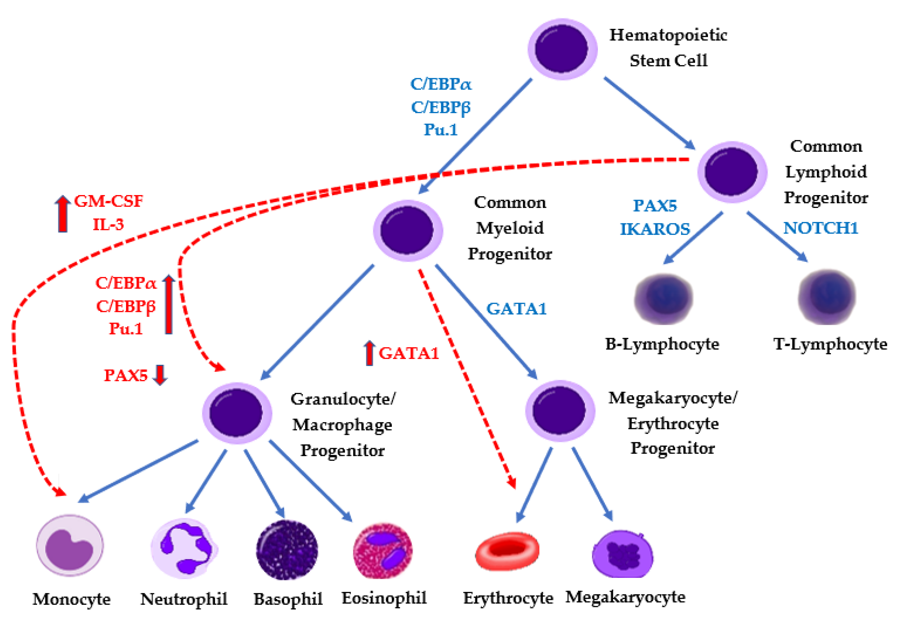
4. Lineage Switch in MPAL
5. Treatment of MPAL
6. Treatment of Ph+ MPAL
7. Role of Hematopoietic Stem Cell Transplantation
8. Assessing Response/Impact of MRD
9. Novel Approaches
10. Conclusions and Future Directions
Author Contributions
Funding
Conflicts of Interest
References
- Maruffi, M.; Sposto, R.; Oberley, M.J.; Kysh, L.; Orgel, E. Therapy for children and adults with mixed phenotype acute leukemia: A systematic review and meta-analysis. Leukemia 2018, 32, 1515–1528. [Google Scholar] [CrossRef]
- Weir, E.G.; Ansari-Lari, M.A.; Batista, D.A.S.; Griffin, C.A.; Fuller, S.; Smith, B.D.; Borowitz, M.J. Acute bilineal leukemia: A rare disease with poor outcome. Leukemia 2007, 21, 2264–2270. [Google Scholar] [CrossRef] [PubMed]
- Weinberg, O.K.; Arber, D.A. Mixed-phenotype acute leukemia: Historical overview and a new definition. Leukemia 2010, 24, 1844–1851. [Google Scholar] [CrossRef]
- Steensma, D.P. Oddballs: Acute Leukemias of Mixed Phenotype and Ambiguous Origin. Hematol. Oncol. Clin. 2011, 25, 1235–1253. [Google Scholar] [CrossRef] [PubMed]
- Orgel, E.; Alexander, T.B.; Wood, B.L.; Kahwash, S.; Devidas, M.; Dai, Y.; Alonzo, T.A.; Mullighan, C.G.; Inaba, H.; Hunger, S.P.; et al. Mixed-phenotype acute leukemia: A cohort and consensus research strategy from the Children’s Oncology Group Acute Leukemia of Ambiguous Lineage Task Force. Cancer 2020, 126, 593–601. [Google Scholar] [CrossRef]
- Arber, D.A.; Orazi, A.; Hasserjian, R.; Thiele, J.; Borowitz, M.J.; Le Beau, M.M.; Bloomfield, C.D.; Cazzola, M.; Vardiman, J.W. The 2016 revision to the World Health Organization classification of myeloid neoplasms and acute leukemia. Blood 2016, 127, 2391–2405. [Google Scholar] [CrossRef] [PubMed]
- Wolach, O.; Stone, R.M. Optimal therapeutic strategies for mixed phenotype acute leukemia. Curr. Opin. Hematol. 2020, 27, 95–102. [Google Scholar] [CrossRef]
- Matutes, E.; Pickl, W.F.; Van’t Veer, M.V.; Morilla, R.; Swansbury, J.; Strobl, H.; Attarbaschi, A.; Hopfinger, G.; Ashley, S.; Bene, M.C.; et al. Mixed-phenotype acute leukemia: Clinical and laboratory features and outcome in 100 patients defined according to the WHO 2008 classification. Blood 2011, 117, 3163–3171. [Google Scholar] [CrossRef]
- Catovsky, D.; Matutes, E.; Buccheri, V.; Shetty, V.; Hanslip, J.; Yoshida, N.; Morilla, R. A classification of acute leukaemia for the 1990s. Ann. Hematol. 1991, 62, 16–21. [Google Scholar] [CrossRef]
- Bene, M.C.; Castoldi, G.; Knapp, W.; Ludwig, W.D.; Matutes, E.; Orfao, A.; Van’t Veer, M.B. Proposals for the immunological classification of acute leukemias. European Group for the Immunological Characterization of Leukemias (EGIL). Leukemia 1995, 9, 1783–1786. [Google Scholar]
- Wolach, O.; Stone, R.M. How I treat mixed-phenotype acute leukemia. Blood 2015, 125, 2477–2485. [Google Scholar] [CrossRef]
- Alexander, T.B.; Gu, Z.; Iacobucci, I.; Dickerson, K.; Choi, J.K.; Xu, B.; Payne-Turner, D.; Yoshihara, H.; Loh, M.L.; Horan, J.; et al. The genetic basis and cell of origin of mixed phenotype acute leukaemia. Nature 2018, 562, 373–379. [Google Scholar] [CrossRef]
- Kotrova, M.; Musilova, A.; Stuchly, J.; Fiser, K.; Starkova, J.; Mejstrikova, E.; Stary, J.; Zuna, J.; Hrusak, O.; Trka, J.; et al. Distinct bilineal leukemia immunophenotypes are not genetically determined. Blood 2016, 128, 2263–2266. [Google Scholar] [CrossRef]
- Manola, K.N.; Panitsas, F.; Polychronopoulou, S.; Daraki, A.; Karakosta, M.; Stavropoulou, C.; Avgerinou, G.; Hatzipantelis, E.; Pantelias, G.; Sambani, C.; et al. Cytogenetic abnormalities and monosomal karyotypes in children and adolescents with acute myeloid leukemia: Correlations with clinical characteristics and outcome. Cancer Genet. 2013, 206, 63–72. [Google Scholar] [CrossRef]
- Rubnitz, J.E.; Onciu, M.; Pounds, S.; Shurtleff, S.; Cao, X.; Raimondi, S.C.; Behm, F.G.; Campana, D.; Razzouk, B.; Ribeiro, R.C.; et al. Acute mixed lineage leukemia in children: The experience of St Jude Children’s Research Hospital. Blood 2009, 113, 5083–5089. [Google Scholar] [CrossRef] [PubMed]
- Al-Seraihy, A.S.; Owaidah, T.M.; Ayas, M.; El-Solh, H.; Al-Mahr, M.; Al-Ahmari, A.; Belgaumi, A.F. Clinical characteristics and outcome of children with biphenotypic acute leukemia. Haematologica 2009, 94, 1682–1690. [Google Scholar] [CrossRef]
- Park, J.A.; Ghim, T.T.; Bae, K.W.; Im, H.J.; Jang, S.S.; Park, C.J.; Chi, H.S.; Seo, J.J. Stem cell transplant in the treatment of childhood biphenotypic acute leukemia. Pediatr. Blood Cancer 2009, 53, 444–452. [Google Scholar] [CrossRef] [PubMed]
- Xiao, W.; Bharadwaj, M.; Levine, M.; Farnhoud, N.; Pastore, F.; Getta, B.M.; Hultquist, A.; Famulare, C.; Medina-Martínez, J.S.; Patel, M.A.; et al. PHF6 and DNMT3A mutations are enriched in distinct subgroups of mixed phenotype acute leukemia with T-lineage differentiation. Blood Adv. 2018, 2, 3526–3539. [Google Scholar] [CrossRef] [PubMed]
- Takahashi, K.; Wang, F.; Morita, K.; Yan, Y.; Hu, P.; Zhao, P.; Zhar, A.A.; Wu, C.J.; Gumbs, C.; Little, L.; et al. Integrative genomic analysis of adult mixed phenotype acute leukemia delineates lineage associated molecular subtypes. Nat. Commun. 2018, 9, 1–12. [Google Scholar] [CrossRef]
- Hu, T.; Murdaugh, R.; Nakada, D. Transcriptional and Microenvironmental Regulation of Lineage Ambiguity in Leukemia. Front. Oncol. 2017, 7, 268. [Google Scholar] [CrossRef] [PubMed]
- Rayes, A.; McMasters, R.L.; O’Brien, M.M. Lineage Switch in MLL-Rearranged Infant Leukemia Following CD19-Directed Therapy. Pediatr. Blood Cancer 2016, 63, 1113–1115. [Google Scholar] [CrossRef]
- Fujisaki, H.; Hara, J.; Takai, K.; Nakanishi, K.; Matsuda, Y.; Ohta, H.; Osugi, Y.; Tokimasa, S.; Taniike, M.; Hosoi, G.; et al. Lineage switch in childhood leukemia with monosomy 7 and reverse of lineage switch in severe combined immunodeficient mice. Exp. Hematol. 1999, 27, 826–833. [Google Scholar] [CrossRef]
- Burda, P.; Laslo, P.; Stopka, T. The role of PU.1 and GATA-1 transcription factors during normal and leukemogenic hematopoiesis. Leukemia 2010, 24, 1249–1257. [Google Scholar] [CrossRef] [PubMed]
- Monma, F.; Nishii, K.; Ezuki, S.; Miyazaki, T.; Yamamori, S.; Usui, E.; Sugimoto, Y.; Lorenzo, V.F.; Katayama, N.; Shiku, H. Molecular and phenotypic analysis of Philadelphia chromosome-positive bilineage leukemia: Possibility of a lineage switch from T-lymphoid leukemic progenitor to myeloid cells. Cancer Genet. Cytogenet. 2006, 164, 118–121. [Google Scholar] [CrossRef]
- Yeh, J.-R.J.; Munson, K.M.; Chao, Y.L.; Peterson, Q.P.; MacRae, C.A.; Peterson, R.T. AML1-ETO reprograms hematopoietic cell fate by downregulating scl expression. Development 2008, 135, 401–410. [Google Scholar] [CrossRef] [PubMed]
- Jacoby, E.; Nguyen, S.M.; Fountaine, T.J.; Welp, K.; Gryder, B.; Qin, H.; Yang, Y.; Chien, C.D.; Seif, A.; Lei, H.; et al. CD19 CAR immune pressure induces B-precursor acute lymphoblastic leukaemia lineage switch exposing inherent leukaemic plasticity. Nat. Commun. 2016, 7, 12320. [Google Scholar] [CrossRef] [PubMed]
- McClellan, J.S.; Dove, C.; Gentles, A.J.; Ryan, C.; Majeti, R. Reprogramming of primary human Philadelphia chromosome-positive B cell acute lymphoblastic leukemia cells into nonleukemic macrophages. Proc. Natl. Acad. Sci. USA 2015, 112, 4074–4079. [Google Scholar] [CrossRef]
- Janz, M.; Dörken, B.; Mathas, S. Reprogramming of B Lymphoid Cells in Human Lymphoma Pathogenesis. Cell Cycle 2006, 5, 1057–1061. [Google Scholar] [CrossRef] [PubMed]
- Shain, K.H.; Dalton, W.S.; Tao, J. The tumor microenvironment shapes hallmarks of mature B-cell malignancies. Oncogene 2015, 34, 4673–4682. [Google Scholar] [CrossRef]
- Balducci, E.; Nivaggioni, V.; Boudjarane, J.; Bouriche, L.; Rahal, I.; Bernot, D.; Alazard, E.; Duployez, N.; Grardel, N.; Arnoux, I.; et al. Lineage switch from B acute lymphoblastic leukemia to acute monocytic leukemia with persistent t(4;11)(q21;q23) and cytogenetic evolution under CD19-targeted therapy. Ann. Hematol. 2017, 96, 1579–1581. [Google Scholar] [CrossRef]
- Gardner, R.; Wu, D.; Cherian, S.; Fang, M.; Hanafi, L.-A.; Finney, O.; Smithers, H.; Jensen, M.C.; Riddell, S.R.; Maloney, D.G.; et al. Acquisition of a CD19-negative myeloid phenotype allows immune escape of MLL-rearranged B-ALL from CD19 CAR-T-cell therapy. Blood 2016, 127, 2406–2410. [Google Scholar] [CrossRef]
- Berry, D.A.; Zhou, S.; Higley, H.; Mukundan, L.; Fu, S.; Reaman, G.H.; Wood, B.L.; Kelloff, G.J.; Jessup, J.M.; Radich, J.P. Association of Minimal Residual Disease With Clinical Outcome in Pediatric and Adult Acute Lymphoblastic Leukemia: A Meta-analysis. JAMA Oncol. 2017, 3, e170580. [Google Scholar] [CrossRef]
- Mejstrikova, E.; Volejnikova, J.; Fronkova, E.; Zdrahalova, K.; Kalina, T.; Sterba, J.; Jabali, Y.; Mihal, V.; Blazek, B.; Cerna, Z.; et al. Prognosis of children with mixed phenotype acute leukemia treated on the basis of consistent immunophenotypic criteria. Haematologica 2010, 95, 928–935. [Google Scholar] [CrossRef]
- Béné, M.C. Biphenotypic, bilineal, ambiguous or mixed lineage: Strange leukemias! Haematologica 2009, 94, 891–893. [Google Scholar] [CrossRef]
- Rasekh, E.O.; Osman, R.; Ibraheem, D.; Madney, Y.; Radwan, E.; Gameel, A.; Abdelhafiz, A.; Kamel, A.; Elfishawi, S. Acute lymphoblastic leukemia–like treatment regimen provides better response in mixed phenotype acute leukemia: A comparative study between adults and pediatric MPAL patients. Ann. Hematol. 2021, 100, 699–707. [Google Scholar] [CrossRef]
- Hrusak, O.; De Haas, V.; Stancikova, J.; Vakrmanova, B.; Janotova, I.; Mejstrikova, E.; Capek, V.; Trka, J.; Zaliova, M.; Luks, A.; et al. International cooperative study identifies treatment strategy in childhood ambiguous lineage leukemia. Blood 2018, 132, 264–276. [Google Scholar] [CrossRef]
- Brethon, B.; Lainey, E.; Caye-Eude, A.; Grain, A.; Fenneteau, O.; Yakouben, K.; Roupret-Serzec, J.; Le Mouel, L.; Cavé, H.; Baruchel, A. Case Report: Targeting 2 Antigens as a Promising Strategy in Mixed Phenotype Acute Leukemia: Combination of Blinatumomab With Gemtuzumab Ozogamicin in an Infant With a KMT2A-Rearranged Leukemia. Front. Oncol. 2021, 11, 637951. [Google Scholar] [CrossRef] [PubMed]
- Shi, R.; Munker, R. Survival of patients with mixed phenotype acute leukemias: A large population-based study. Leuk. Res. 2015, 39, 606–616. [Google Scholar] [CrossRef] [PubMed]
- Seetharam, S.; Thankamony, P.; Gopakumar, K.G.; Nair, R.A.; Jacob, P.M.; Krishna, K.M.J.; Rajeswari, B.; Nair, M.; Guruprasad, C.S.; Prasanth, V.R. Outcomes of pediatric mixed phenotype acute leukemia treated with lymphoid directed therapy: Analysis of an institutional series from India. Pediatr. Hematol. Oncol. 2021, 38, 358–366. [Google Scholar] [CrossRef]
- Oberley, M.J.; Raikar, S.; Wertheim, G.B.; Malvar, J.; Sposto, R.; Rabin, K.R.; Punia, J.N.; Seif, A.E.; Cahen, V.C.; Schore, R.J.; et al. Significance of minimal residual disease in pediatric mixed phenotype acute leukemia: A multicenter cohort study. Leuk. 2020, 34, 1741–1750. [Google Scholar] [CrossRef] [PubMed]
- Duong, V.H.; Begna, K.H.; Kashanian, S.; Sweet, K.; Wang, E.S.; Caddell, R.; Shafer, D.A.; Singh, Z.N.; Baer, M.R.; Al-Kali, A. Favorable outcomes of acute leukemias of ambiguous lineage treated with hyperCVAD: A multi-center retrospective study. Ann. Hematol. 2020, 99, 2119–2124. [Google Scholar] [CrossRef]
- Bachir, F.; Zerrouk, J.; Howard, S.C.; Graoui, O.; Lahjouji, A.; Hessissen, L.; Bennani, S.; Quessar, A.; El Aouad, R. Outcomes in Patients With Mixed Phenotype Acute Leukemia in Morocco. J. Pediatr. Hematol. 2014, 36, e392–e397. [Google Scholar] [CrossRef]
- Gerr, H.; Zimmermann, M.; Schrappe, M.; Dworzak, M.; Ludwig, W.-D.; Bradtke, J.; Moericke, A.; Schabath, R.; Creutzig, U.; Reinhardt, D. Acute leukaemias of ambiguous lineage in children: Characterization, prognosis and therapy recommendations. Br. J. Haematol. 2010, 149, 84–92. [Google Scholar] [CrossRef] [PubMed]
- Reid, J.H.; Perissinotti, A.J.; Benitez, L.L.; Boyer, D.; Lee, W.; Burke, P.W.; Pettit, K.; Bixby, D.L.; Marini, B.L. Hybrid chemotherapy regimen (FLAG-IDA-vincristine-prednisone) for acute leukemia with mixed-phenotype blasts. Leuk. Res. 2021, 103, 106539. [Google Scholar] [CrossRef] [PubMed]
- Lazarus, H.M.; Richards, S.M.; Chopra, R.; Litzow, M.R.; Burnett, A.K.; Wiernik, P.H.; Franklin, I.M.; Tallman, M.S.; Cook, L.; Buck, G.; et al. Central nervous system involvement in adult acute lymphoblastic leukemia at diagnosis: Results from the international ALL trial MRC UKALL XII/ECOG E2993. Blood 2006, 108, 465–472. [Google Scholar] [CrossRef] [PubMed]
- Raikar, S.S.; Park, S.I.; Leong, T.; Jaye, D.L.; Keller, F.G.; Horan, J.T.; Woods, W.G. Isolated myeloperoxidase expression in pediatric B/myeloid mixed phenotype acute leukemia is linked with better survival. Blood 2018, 131, 573–577. [Google Scholar] [CrossRef]
- Winters, A.C.; Bernt, K.M. MLL-Rearranged Leukemias—An Update on Science and Clinical Approaches. Front. Pediatr. 2017, 5, 4. [Google Scholar] [CrossRef] [PubMed]
- Young, K.; Loberg, M.A.; Eudy, E.; Schwartz, L.S.; Mujica, K.D.; Trowbridge, J.J. Heritable genetic background alters survival and phenotype of Mll-AF9-induced leukemias. Exp. Hematol. 2020, 89, 61–67.e3. [Google Scholar] [CrossRef]
- Qasrawi, A.; Ramlal, R.; Munker, R.; Hildebrandt, G.C. Prognostic impact of Philadelphia chromosome in mixed phenotype acute leukemia (MPAL): A cancer registry analysis on real-world outcome. Am. J. Hematol. 2020, 95, 1015–1021. [Google Scholar] [CrossRef] [PubMed]
- Shimizu, H.; Saitoh, T.; Machida, S.; Kako, S.; Doki, N.; Mori, T.; Sakura, T.; Kanda, Y.; Kanamori, H.; Miyawaki, S.; et al. Allogeneic hematopoietic stem cell transplantation for adult patients with mixed phenotype acute leukemia: Results of a matched-pair analysis. Eur. J. Haematol. 2015, 95, 455–460. [Google Scholar] [CrossRef]
- Tian, H.; Xu, Y.; Liu, L.; Yan, L.; Jin, Z.; Tang, X.; Han, Y.; Fu, Z.; Qiu, H.; Sun, A.; et al. Comparison of outcomes in mixed phenotype acute leukemia patients treated with chemotherapy and stem cell transplantation versus chemotherapy alone. Leuk. Res. 2016, 45, 40–46. [Google Scholar] [CrossRef]
- Issa, G.C.; Ravandi, F.; DiNardo, C.D.; Jabbour, E.; Kantarjian, H.M.; Andreeff, M. Therapeutic implications of menin inhibition in acute leukemias. Leukemia 2021, 35, 2482–2495. [Google Scholar] [CrossRef] [PubMed]
- Stein, E.M.; Garcia-Manero, G.; Rizzieri, D.A.; Tibes, R.; Berdeja, J.G.; Savona, M.R.; Jongen-Lavrenic, M.; Altman, J.K.; Thomson, B.; Blakemore, S.J.; et al. The DOT1L inhibitor pinometostat reduces H3K79 methylation and has modest clinical activity in adult acute leukemia. Blood 2018, 131, 2661–2669. [Google Scholar] [CrossRef] [PubMed]
- Loftus, J.P.; Yahiaoui, A.; Brown, P.A.; Niswander, L.; Bagashev, A.; Wang, M.; Schauf, A.; Tannheimer, S.; Tasian, S.K. Combinatorial efficacy of entospletinib and chemotherapy in patient-derived xenograft models of infant acute lymphoblastic leukemia. Haematologica 2020, 106, 1067–1078. [Google Scholar] [CrossRef]
- Milojkovic, D.; Ibrahim, A.; Reid, A.; Foroni, L.; Apperley, J.; Marin, D. Efficacy of combining dasatinib and FLAG-IDA for patients with chronic myeloid leukemia in blastic transformation. Haematologica 2011, 97, 473–474. [Google Scholar] [CrossRef] [PubMed]
- Labrador, J.; Hermida, G.J.; Alvarez, R.; Anso, V.; De Vicente, P.; Goñi, M.; Gonzalez-Lopez, T.J. Dasatinib and FLAG-IDA Is an Effective Therapy for Initial Myeloid Blast Crisis but Involves a High Risk of Opportunistic Infections. Case Rep. Hematol. 2020, 2020, 1–4. [Google Scholar] [CrossRef] [PubMed]
- Munker, R.; Brazauskas, R.; Wang, H.L.; de Lima, M.; Khoury, H.J.; Gale, R.P.; Maziarz, R.T.; Sandmaier, B.M.; Weisdorf, D.; Saber, W. Allogeneic Hematopoietic Cell Transplantation for Patients with Mixed Phenotype Acute Leukemia. Biol. Blood Marrow Transplant. 2016, 22, 1024–1029. [Google Scholar] [CrossRef]
- Getta, B.M.; Roshal, M.; Zheng, J.; Park, J.H.; Stein, E.M.; Levine, R.; Papadopoulos, E.B.; Jakubowski, A.A.; Kernan, N.A.; Steinherz, P.; et al. Allogeneic Hematopoietic Stem Cell Transplantation with Myeloablative Conditioning Is Associated with Favorable Outcomes in Mixed Phenotype Acute Leukemia. Biol. Blood Marrow Transplant. 2017, 23, 1879–1886. [Google Scholar] [CrossRef] [PubMed]
- Munker, R.; Labopin, M.; Esteve, J.; Schmid, C.; Mohty, M.; Nagler, A. Mixed phenotype acute leukemia: Outcomes with allogeneic stem cell transplantation. A retrospective study from the Acute Leukemia Working Party of the EBMT. Haematologica 2017, 102, 2134–2140. [Google Scholar] [CrossRef] [PubMed]
- Zając-Spychała, O.; Irga-Jaworska, N.; Drożyńska, E.; Muszyńska-Rosłan, K.; Krawczuk-Rybak, M.; Zawitkowska, J.; Kowalczyk, J.; Ćwiklińska, M.; Balwierz, W.; Mizia-Malarz, A.; et al. Mixed phenotype acute leukemia: Biological profile, clinical characteristic and treatment outcomes: Report of the population-based study. Eur. J. Haematol. 2020, 105, 85–93. [Google Scholar] [CrossRef]
- Wood, B.L. Principles of minimal residual disease detection for hematopoietic neoplasms by flow cytometry. Cytom. Part B Clin. Cytom. 2016, 90, 47–53. [Google Scholar] [CrossRef]
- Borowitz, M.J.; Wood, B.L.; Devidas, M.; Loh, M.L.; Raetz, E.A.; Salzer, W.L.; Nachman, J.B.; Carroll, A.J.; Heerema, N.A.; Gastier-Foster, J.M.; et al. Prognostic significance of minimal residual disease in high risk B-ALL: A report from Children’s Oncology Group study AALL0232. Blood 2015, 126, 964–971. [Google Scholar] [CrossRef] [PubMed]
- Choi, S.M.; Frederiksen, J.K.; Ross, C.W.; Bixby, D.L.; Shao, L. Philadelphia Chromosome–like Mixed-Phenotype Acute Leukemia Demonstrating P2RY8-CRLF2 Fusion and JAK1 Mutation. Am. J. Clin. Pathol. 2017, 148, 523–528. [Google Scholar] [CrossRef] [PubMed]
- Roberts, K.G.; Li, Y.; Payne-Turner, D.; Harvey, R.C.; Yang, Y.-L.; Pei, D.; McCastlain, K.; Ding, L.; Lu, C.; Song, G.; et al. Targetable Kinase-Activating Lesions in Ph-like Acute Lymphoblastic Leukemia. N. Engl. J. Med. 2014, 371, 1005–1015. [Google Scholar] [CrossRef]
- Shimizu, H.; Yokohama, A.; Hatsumi, N.; Takada, S.; Handa, H.; Sakura, T.; Nojima, Y. Philadelphia chromosome-positive mixed phenotype acute leukemia in the imatinib era. Eur. J. Haematol. 2014, 93, 297–301. [Google Scholar] [CrossRef] [PubMed]
- Hoehn, D.; Medeiros, L.J.; Chen, S.S.; Tian, M.T.; Jorgensen, J.L.; Ahmed, Y.; Lin, P. CD117 Expression Is a Sensitive but Nonspecific Predictor ofFLT3Mutation in T Acute Lymphoblastic Leukemia and T/Myeloid Acute Leukemia. Am. J. Clin. Pathol. 2012, 137, 213–219. [Google Scholar] [CrossRef]
- Manara, E.; Baron, E.; Tregnago, C.; Aveic, S.; Bisio, V.; Bresolin, S.; Masetti, R.; Locatelli, F.; Basso, G.; Pigazzi, M. MLL-AF6 fusion oncogene sequesters AF6 into the nucleus to trigger RAS activation in myeloid leukemia. Blood 2014, 124, 263–272. [Google Scholar] [CrossRef]
- Mansur, M.B.; Ford, A.M.; Emerenciano, M. The role of RAS mutations in MLL -rearranged leukaemia: A path to intervention? Biochim. Biophys. Acta (BBA) Bioenerg. 2017, 1868, 521–526. [Google Scholar] [CrossRef]
- Stam, R. (Ronald) Targeting FLT3 in primary MLL-gene-rearranged infant acute lymphoblastic leukemia. Blood 2005, 106, 2484–2490. [Google Scholar] [CrossRef] [PubMed]
- Wu, D.; Chen, W.; Chen, Z.; Li, Q. Venetoclax Combined with Hypomethylating Agents for Treatment-Naive B/Myeloid Mixed Phenotype Acute Leukemia. Case Rep. Hematol. 2021, 2021, 6661109. [Google Scholar] [CrossRef]
- Bacchiarri, F.; Sammartano, V.; Santoni, A.; Raspadori, D.; Zappone, E.; Defina, M.; Ciofini, S.; Sicuranza, A.; Bocchia, M.; Gozzetti, A. First reported case of secondary mixed phenotype acute leukemia after multiple myeloma. Am. J. Blood Res. 2021, 11, 123–131. [Google Scholar] [PubMed]
- Klocke, H.; Dong, Z.M.; O’Brien, C.; Burwick, N.; Richard, R.E.; Wu, D.Y.; Chauncey, T.R.; Graf, S.A. Venetoclax and Decitabine for T/Myeloid Mixed-Phenotype Acute Leukemia Not Otherwise Specified (MPAL NOS). Case Rep. Hematol. 2020, 2020, 8811673. [Google Scholar] [CrossRef]
- Baeuerle, P.A.; Reinhardt, C. Bispecific T-Cell Engaging Antibodies for Cancer Therapy. Cancer Res. 2009, 69, 4941–4944. [Google Scholar] [CrossRef] [PubMed]
- Goebeler, M.-E.; Bargou, R.C. T cell-engaging therapies—BiTEs and beyond. Nat. Rev. Clin. Oncol. 2020, 17, 418–434. [Google Scholar] [CrossRef]
- Maude, S.L.; Teachey, D.; Porter, D.L.; Grupp, S.A. CD19-targeted chimeric antigen receptor T-cell therapy for acute lymphoblastic leukemia. Blood 2015, 125, 4017–4023. [Google Scholar] [CrossRef]
- Park, J.H.; Geyer, M.B.; Brentjens, R.J. CD19-targeted CAR T-cell therapeutics for hematologic malignancies: Interpreting clinical outcomes to date. Blood 2016, 127, 3312–3320. [Google Scholar] [CrossRef]
- Durer, S.; Shafqat, M.; Comba, I.Y.; Malik, S.; Faridi, W.; Aslam, S.; Ijaz, A.; Tariq, M.J.; Fraz, M.A.; Usman, M.; et al. Concomitant use of blinatumomab and donor lymphocyte infusion for mixed-phenotype acute leukemia: A case report with literature review. Immunotherapy 2019, 11, 373–378. [Google Scholar] [CrossRef]
- El Chaer, F.; Ali, O.M.; Sausville, E.A.; Law, J.Y.; Lee, S.T.; Duong, V.H.; Baer, M.R.; Koka, R.; Singh, Z.N.; Wong, J.; et al. Treatment of CD19-positive mixed phenotype acute leukemia with blinatumomab. Am. J. Hematol. 2019, 94, E7–E8. [Google Scholar] [CrossRef]
- Kong, D.; Qu, C.; Dai, H.; Li, Z.; Yin, J.; Chen, S.; Kang, L.; Chen, G.; Zhu, M.; Yu, L.; et al. CAR-T therapy bridging to allogeneic HSCT provides durable molecular remission of Ph + mixed phenotype acute leukaemia with minimal residual disease. Br. J. Haematol. 2020, 191, e47–e49. [Google Scholar] [CrossRef] [PubMed]
- Li, M.-Y.; Lin, Z.-H.; Hu, M.-M.; Kang, L.-Q.; Wu, X.-X.; Chen, Q.-W.; Kong, X.; Zhang, J.; Qiu, H.-Y.; Wu, D.-P. Secondary donor-derived humanized CD19-modified CAR-T cells induce remission in relapsed/refractory mixed phenotype acute leukemia after allogeneic hematopoietic stem cell transplantation: A case report. Biomark. Res. 2020, 8, 1–6. [Google Scholar] [CrossRef]
- Bartram, J.; Balasch-Carulla, M.; Bhojaraja, S.; Adams, S.; Cheng, D.; Inglott, S.; Kulkarni, N.; Mahendrayogam, A.; O’Connor, O.; Pavasovic, V.; et al. Blinatumomab for paediatric mixed phenotype acute leukaemia. Br. J. Haematol. 2021. [Google Scholar] [CrossRef] [PubMed]
- Roskopf, C.C.; Braciak, T.A.; Fenn, N.C.; Kobold, S.; Fey, G.H.; Hopfner, K.-P.; Oduncu, F.S. Dual-targeting triplebody 33-3-19 mediates selective lysis of biphenotypic CD19+ CD33+ leukemia cells. Oncotarget 2016, 7, 22579–22589. [Google Scholar] [CrossRef] [PubMed]
- Hoseini, S.S.; Espinosa-Cotton, M.; Guo, H.-F.; Cheung, N.-K.V. Overcoming leukemia heterogeneity by combining T cell engaging bispecific antibodies. J. Immunother. Cancer 2020, 8, e001626. [Google Scholar] [CrossRef] [PubMed]


| Lineage Assignment Criteria |
| Myeloid Lineage |
| MPO+ (Flow cytometry, immunohistochemistry, or cytochemistry) or Monocytic differentiation (at least two of the following: nonspecific esterase cytochemistry, CD11c, CD14, CD64, lysozyme) |
| T-Lymphoid Lineage |
| Strong * cytoplasmic CD3 (with antibodies to CD3 ε chain) or Surface CD3 |
| B-Lymphoid Lineage |
| Strong * CD19 with at least 1 of the following strongly expressed: CD79a, cytoplasmic CD22, or CD10 or Weak CD19 with at least 2 of the following strongly expressed: CD79a, cytoplasmic CD22, or CD10 |
| Acute Undifferentiated Leukemia |
|---|
| Mixed-phenotype acute leukemia (MPAL) with t(9;22)(q34.1;q11.2); BCR-ABL1 |
| MPAL with t(v;11q23.3); KMT2A rearranged |
| MPAL, B/myeloid, NOS |
| MPAL, T/myeloid, NOS |
| Reference | MPAL Patients | Treatments | Outcomes |
|---|---|---|---|
| Bartram et al. [81] | Patient 1: 6-month-old female with B/Myeloid MPAL with KMT2A deletion Patient 2: 10-month-old male with B/Myeloid, KMT2A-USP2, and FLT3 ITD Patient 3: 72-month-old female with B/Myeloid MPAL and KMT2A-ARHGEF12 | ALL induction followed by FLA-IDA (myeloid regimen) with CD19+ MRD, and Blinatumomab followed by HSCT >10% disease after ALL induction (CD19+, CD19-, populations): Blinatumomab and FLA-IDA prior to HSCT ALL induction followed by Blinatumomab and high-dose Ara-C prior to HSCT | CR for 24 months post HSCT CR for 8 months post HSCT CR 5 months post HSCT |
| Li et al. [77] | 29-year-old male with B/myeloid CD19+ MPAL and SET-NUP14 fusion gene transcript | ALL induction followed by hybrid Consolidation (hyper-CVAD-B and hyper-CVAD-A) and HSCT For Relapse#1: received CD19 Directed CART therapy x2 (donor-derived) | Relapse#1 after 6 months Relapse#2: 24 months following first infusion of CAR T-Cells despite persistent CD19 CAR T-Cells at 8 months |
| Durer et al. [77] | 51-year-old female with B/myeloid CD19+ MPAL | ALL induction followed by HSCT but relapsed after 3 months. Received Blinatumomab and donor lymphocyte infusions (x 4 cycles) | Attained CR after one cycle and maintained CR for 15 months |
| El Chaer et al. [78] | Patient 1: 39-year-old male with Ph+ B/myeloid MPAL Patient 2: 55-year-old female with B/myeloid | Induction with Cytarabine, Daunorubicin, and Dasatinib followed by Blinatumomab with HSCT HyperCVAD (held due to toxicity), and later received 3 cycles of Blinatumomab followed by HSCT | CR at 6 months CR at 14 months |
| Brethon et al. [37] | 4-month-old female with B/myeloid MPAL with KMT2A-AFF1 | Interfant-06 protocol with refractory disease; received Blinatumomab and Gemtuzumab followed by HSCT, followed by CART for relapse | Relapsed 11 months post HSCT; then achieved CR for 12 months post CART |
Publisher’s Note: MDPI stays neutral with regard to jurisdictional claims in published maps and institutional affiliations. |
© 2021 by the authors. Licensee MDPI, Basel, Switzerland. This article is an open access article distributed under the terms and conditions of the Creative Commons Attribution (CC BY) license (https://creativecommons.org/licenses/by/4.0/).
Share and Cite
Batra, S.; Ross, A.J. Pediatric Mixed-Phenotype Acute Leukemia: What’s New? Cancers 2021, 13, 4658. https://doi.org/10.3390/cancers13184658
Batra S, Ross AJ. Pediatric Mixed-Phenotype Acute Leukemia: What’s New? Cancers. 2021; 13(18):4658. https://doi.org/10.3390/cancers13184658
Chicago/Turabian StyleBatra, Sandeep, and Anthony John Ross. 2021. "Pediatric Mixed-Phenotype Acute Leukemia: What’s New?" Cancers 13, no. 18: 4658. https://doi.org/10.3390/cancers13184658
APA StyleBatra, S., & Ross, A. J. (2021). Pediatric Mixed-Phenotype Acute Leukemia: What’s New? Cancers, 13(18), 4658. https://doi.org/10.3390/cancers13184658