Review

NK Cells in Myeloproliferative Neoplasms (MPN)

1
Internal Medicine V, Department of Haematology and Oncology, Medical University Innsbruck, Anichstr. 35, 6020 Innsbruck, Austria
2
Tyrolean Cancer Research Institute, Innrain 66, 6020 Innsbruck, Austria
*
Authors to whom correspondence should be addressed.
E.N. and J.S. contributed equally.
Cancers 2021, 13(17), 4400; https://doi.org/10.3390/cancers13174400
Submission received: 24 June 2021 / Revised: 23 August 2021 / Accepted: 24 August 2021 / Published: 31 August 2021
(This article belongs to the Special Issue Natural Killer Cells in Cancers)

Abstract

:

Simple Summary

NK cells are important innate immune effectors that contribute substantially to tumor control, however the role of NK cells in haematological cancers is not as well understood. The aim of this review is to highlight the importance of the role of NK cells in the management of Ph+ Myeloproliferative Neoplasms, and emphasize the need and possible benefits of a more in-depth investigation into their role in classical MPNs and show potential strategies to harness the anti-tumoral capacities of NK cells.

Abstract

Myeloproliferative neoplasms (MPNs) comprise a heterogenous group of hematologic neoplasms which are divided into Philadelphia positive (Ph+), and Philadelphia negative (Ph−) or classical MPNs. A variety of immunological factors including inflammatory, as well as immunomodulatory processes, closely interact with the disease phenotypes in MPNs. NK cells are important innate immune effectors and substantially contribute to tumor control. Changes to the absolute and proportionate numbers of NK cell, as well as phenotypical and functional alterations are seen in MPNs. In addition to the disease itself, a variety of therapeutic options in MPNs may modify NK cell characteristics. Reports of suppressive effects of MPN treatment strategies on NK cell activity have led to intensive investigations into the respective compounds, to elucidate the possible negative effects of MPN therapy on control of the leukemic clones. We hereby review the available literature on NK cells in Ph+ and Ph− MPNs and summarize today’s knowledge on disease-related alterations in this cell compartment with particular focus on known therapy-associated changes. Furthermore, we critically evaluate conflicting data with possible implications for future projects. We also aim to highlight the relevance of full NK cell functionality for disease control in MPNs and the importance of considering specific changes related to therapy in order to avoid suppressive effects on immune surveillance.

1. Introduction

1.1. Introduction to MPNs

The group of MPNs belong to the family of chronic myeloid neoplasms and comprise a variety of clonal hematological diseases, characterized by the increased proliferation of cells originating from the myeloid lineage [1,2,3,4,5]. Typically, the group of MPNs is subdivided into two major groups: the Philadelphia chromosome carrying CML and the classical or Ph− MPNs [6]. Summarized under the term classical MPNs are polycythemia vera (PV), essential thrombocythemia (ET), primary myelofibrosis (PMF), chronic neutrophilic leukemia (CNL) and chronic eosinophilic leukemia—not otherwise specified (CEL-NOS) [5]. Annual incidence rates of PV, ET, PMF and CML are around 1–2/100.000 whereas CNL and CEL-NOS are extremely rare conditions [6,7,8,9,10].
All MPN entities originate from a single mutated hematopoietic stem cell (HSC) with subsequent proliferation through clonal expansion [4]. Depending on disease entity and main disease-driving mutation, clonal expansion can affect single or multiple cell lineages [4]. The respective somatic driver mutations are then found among all myeloid lineages; however, they may also be detectable at lower frequencies within the lymphoid lineage, particularly in B and NK cells [4].

1.1.1. Disease Characteristics

MPNs include a variety of subtypes with numerous gene alterations involved, resulting in a heterogenous landscape of phenotypes. Accurate diagnosis of the respective disease entities therefore requires careful consideration of clinical and morphological features as well as the genetic aberrations involved [2,4,5,11,12].

1.1.2. Ph+ MPNs

The most prominent of the somatic alterations known in MPNs is the BCR-ABL1 gene rearrangement, constituting an exclusive feature of CML [13,14]. Due to the disease-defining reciprocal translocation t(9;22) (q34;q11), termed the Philadelphia chromosome, CML is considered a unique entity and model disease among the group of MPNs [13,15]. From a clinical perspective, CML often presents in chronic phase with symptoms ranging from mild and unspecific to asymptomatic [13,15]. Without accurate treatment, the disease naturally progresses over the accelerated phase into blast crisis in the course of 3–5 years [13,15]. This serious condition ultimately leads to patient death by means of thrombosis, anemia or infection [13]. However, with modern therapeutic standards and the introduction of tyrosine kinase inhibitors (TKIs) the life expectancy of CML patients has been significantly ameliorated and almost meets the general population’s [16].

1.1.3. Classical MPNs

Among the Ph− MPNs, the main clinical manifestations differ substantially and largely depend on the respective entity’s most prominent hematologic features. All MPNs can furthermore vary in disease severity and thus show a wide range of symptom intensity. PV patients may present with marked erythrocytosis and associated concomitant symptoms of hyperviscosity, microvascular symptoms and bleeding complications, whereas classical ET is characterized by an elevation in platelet count and history of thrombosis [1,17,18,19]. A typical sign of PMF is bone marrow fibrosis accompanied by marked anemia, cachexia and thrombohemorrhagic events [20,21]. A hallmark finding of CNL is an extensive bone marrow neutrophilic granulopoiesis, while CEL-NOS presents with typical hypereosinophilia with eosinophil clonality [9,10]. Frequently occurring mutated genes in PV, ET and PMF include JAK2, CALR and MPL. These mutations all ultimately result in increased downstream activation of the STAT and other disease-promoting pathways [4,18,19,20,22]. The mentioned classical genetic alterations are however absent in CNL and CEL-NOS [9,10].

2. Immunological Changes in MPNs

Immune evasion by tumor-associated immune dysregulation is a phenomenon that was detected in a variety of tumor entities [23]. Furthermore, the concept of oncoinflammation has given rise to the idea that a hyperinflammatory tumor microenvironment may also contribute to tumor development and progress [23,24].
Classical myeloproliferative diseases are also driven by inflammation and the excessive activation of inflammatory pathways has several immunomodulatory effects [25,26,27]. Increased pro-inflammatory cytokines and the accumulation of reactive oxygen species as well as the transcriptional deregulation of anti-oxidative stress genes were found in patients with JAK2V617F mutant MPN [28,29]. This up-regulation has been reported to stimulate the JAK/STAT signaling axis, resulting in an increased proliferation of various cell types, including an increased viability and survival of malignant cancer cell clones [30]. Studies in mice have shown that administering the anti-oxidant N-acetylcysteine (NAC), could reduce splenomegaly and the number of JAK2-mutant hematopoietic stem and progenitor cells in the spleen and BM, and normalize blood parameters [28].
With the discovery that CML patients can potentially stay in treatment free remission (TFR) even after treatment cessation, it became clear that the immune system must play a pivotal role in disease control of Ph+ MPNs [31,32]. Numerous findings have been reported, showing that immunological variables serve as predictors of molecular response and depth of remission, and thus support the hypothesis of a close interaction between the immune system and survival of the leukemic clone [33]. Current data suggest that the restoration of immune-effectors, particularly involving T and NK cells, are key-points to achievement of molecular response and durable TFR [33,34].

3. Natural Killer Cells

Natural killer (NK) cells are innate lymphocytes with effector functions, they play an important role in host defense and immune surveillance and are known for their ability to rapidly kill tumor- and virus-infected cells. NK cells also produce cytokines such as IFN-γ and TNF-α, which play an important role in the differentiation and maturation of both innate and adaptive immune cells [35]. Murine models have shown that NK cell development is complex and tightly regulated, and while human NK cells are generated and maintained by HSCs, the precise process is not yet fully elucidated [36,37]. The main site of NK cell generation is considered to be the immunological niche in the BM; however, whether the NK cell ontogenesis occurs exclusively in the BM niches remains debated, as evidence suggests that immature NK cells can migrate to secondary lymphoid tissue to mature [38]. IL-15 is produced by stromal cells in the BM and is important for the differentiation and survival of NK cells. As common lymphoid progenitors develop into NK progenitors, they down regulate CD34 and acquire the expression of the IL-15 receptor (IL-15R). Various other chemokines and receptors are expressed on the different subsets of NK cells, influencing their tissue localization and regulating their release from the BM [39]. Different anatomical sites and tissues possess different homeostatic mechanisms, which result in tissue specific NK cell development, and maintain the balance between immune tolerance and immune surveillance [36].
The diversity of the NK cell repertoire is determined by the large array of activating and inhibitory receptors expressed on the cell surface. Inhibitory receptor expression is intrinsic, encoded by the host germline, while activating receptors can be influenced by extrinsic factors such as epigenetics or chronic infection [40,41]. These activating and inhibitory markers form a delicate balance on the cell’s surface, determining whether the NK cell will be activated upon presentation of the target cell [42].
Unlike T cells, NK cells do not require any previous priming and they exert anti-metastatic functions via (1) the release of lytic granules containing perforin and granzymes, (2) IFN-γ secretion, and (3) the exposure of death receptor ligands, e.g., TRAIL and FASL, which when bound, can induce apoptosis [43].
Under homeostatic conditions, they remain in a resting state due to inhibitory markers such as inhibitory Killer Immunoglobulin-like receptors (iNKRs), C-type lectin receptor NKG2A, Ig-like transcripts (ILTs), and leukocyte Ig-like receptors (LIRs). These inhibitory receptors recognize a wide range of Human Leukocyte Antigens (HLAs) which allow autologous cells to be identified as “self” [36]. The dearth or downregulation of HLA-I alleles by infected or tumor cells boosts NK cell-mediated killing, engaging various NK cell activation receptors (aNKRs), a concept referred to as the “missing-self” hypothesis [44]. aNKRs include Natural Cytotoxicity Receptors (NCRs) such as NKp46, C-type lectin receptors such as NKG2D, DNAM-1 and Killer Ig-like Receptors (KIRs) [36,37]. aNKR ligands can also be upregulated by cellular stress or as a response to DNA damage, both of which are common in many different types of cancer [45]. The secretion of soluble NKG2D ligands (MICA and MICB) by cancer cells, has been postulated as a possible mechanism of NK cell killing evasion. MICA and MICB are able to downregulate NKG2D on the NK cell without triggering activation [46]. Indeed, sMICA levels were shown to be increased in CML patients and to return to normal levels with imatinib therapy [47].
The phenotype and density of the cell surface antigen expression is theorized to explain a functional distinction between the subsets [48]. The two main subsets of NK cells can be defined by their surface expression of CD56 and CD16. CD56, also known as neural cell adhesion molecule (NCAM), plays an important role in adhesion in bone marrow niches [49] and CD16 is the type III Fcγ receptor which binds to the Fc region of antibodies and triggers the release of cytotoxic granules [50]. CD56dimCD16+ cells are cytotoxic NK cells and make up the majority of circulating NK cells (~90%). CD56bright CD16 NK cells are defined as a regulatory subset, producing high levels of pro-inflammatory cytokines [36,51]. The vast array of NK cell surface receptors is complex and distinct within the different sub-populations, highlighting the functional heterogeneity of NK cells [48]. Environmental factors, including pathogen exposure or the microenvironment in which the cell develops and resides, may also influence receptor diversity [36].
NK cells are pivotal in the control of metastatic dissemination of solid tumors and an inverse correlation has been observed between circulating NK cells and metastases at clinical presentation in a range of different carcinomas, while improved NK cell cytotoxicity has previously been linked with good prognosis [52]. NK cells have been described as ‘critical’ to immunosurveillance and anti-tumor activity in B cell lymphomas [53], chronic myeloid leukemia (CML), acute myeloid leukemia (AML), and myelodysplastic syndromes (MDS) [54], highlighting the importance of further elucidating their role in hematological cancers.

4. NK Cells in MPN

4.1. CML

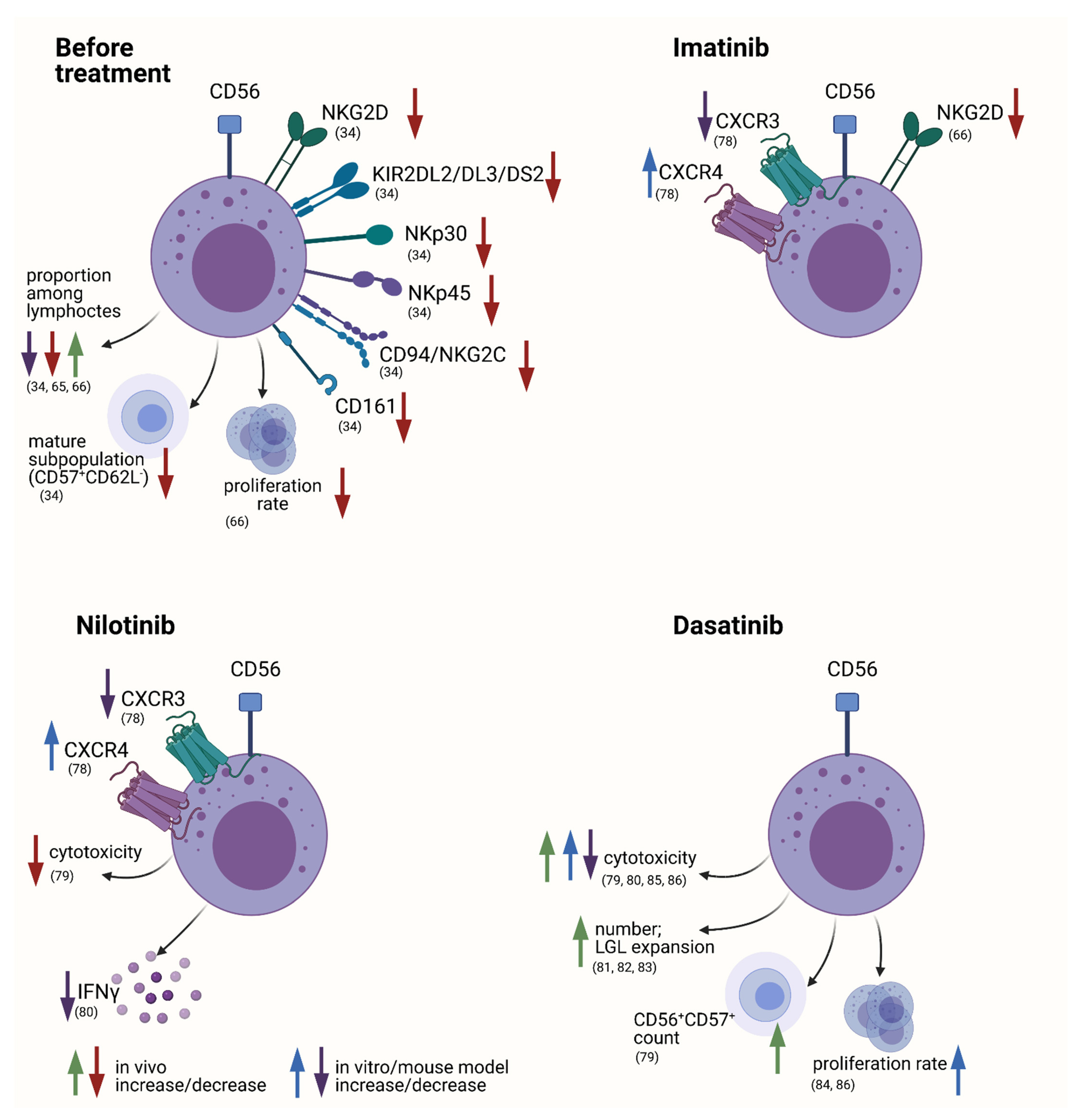
4.1.1. NK Cells in CML

A considerable amount of the immunological changes and influencing factors in CML concern phenotypic and functional alterations of the NK cell compartment which makes them an interesting topic for closer investigation [55] (Figure 1).
T/NK progenitors derived from untreated chronic phase CML patients, were shown to express the Philadelphia chromosome [56]. Interestingly, the vast proportion of mature NK cells were reportedly negative for BCR-ABL1 [56,57,58]. This discrepancy in Philadelphia positivity is explained by a lack of malignant precursors to differentiate into mature NK cells [59]. In advanced phases of CML, however, both CD56dim and CD56bright populations can become Ph+ and can thus contribute to the malignant clone [58,59]. Although, in chronic phases of CML, most peripheral NK cells are not directly part of the disease itself, they have, as important key players of immunity, been subjected to comprehensive investigation in order to obtain a better understanding of immunologic surveillance in CML.
The first findings demonstrating that NK cells are capable of killing leukemic blasts derived from CML patients, date back to 1983 [60]. Reports by Lotzová et al. confirmed the capability of NK cells to inhibit the growth of leukemic cells in CML [61]. Cervantes et al. furthermore demonstrated that NK cells derived from healthy donors, as well as from CML patients, exhibited cytotoxic activity towards the cell line K562 in vitro, and that this effect was mediated by direct cell-to-cell contact [62]. Cebo et al. supported this finding by showing that lysis of BCR-ABL1 expressing cell lines by NK cells was mediated by an interaction of NK-receptor NKG2D and its corresponding ligands [63]. Primitive, quiescent CD34+ progenitor cells derived from CML patients were the least susceptible to NK cell cytotoxicity in vitro [61,64].
NK cells therefore seem capable of killing the leukemic clone in CML and may play a crucial role in disease control. Various findings have demonstrated a decrease in quantity, as well as functional deficits, of NK cells in CML [54]. However, these assumptions were followed by contradictory findings, indicating a more complex situation [54,65].

General Alterations in the NK Cell Compartment

Several studies reported reduced relative amounts of NK cells within the lymphocytic compartment in newly diagnosed CML patients compared to healthy controls [34,66]. The decrease of NK cell proportions remained detectable even under imatinib treatment with stable remission in a study by Chen et al. [66]. Opposing data, however, demonstrated a significant increase of NK cell numbers with consequent TKI therapy, in particular with dasatinib treatment [34,67].
Chen et al. investigated the finding of NK cell reduction in newly diagnosed CML using a transgenic mouse model with reversible BCR-ABL1 expression [66]. They found that the relative proportions of murine NK1.1+ were significantly reduced with BCR-ABL1 induction and remained low even after BCR-ABL1 reversion [66]. Upon functional exploration, the group demonstrated that the murine NK cells isolated from BCR-ABL1+ mice did not show any difference in proliferative activity, whereas the degranulation capacity was significantly decreased when the mutation was expressed [66]. However, in contrast to findings in human studies, it should be highlighted that in this experimental setting, NK cells expressed the BCR-ABL1 transcript [66].
We have previously demonstrated that assumptions regarding NK cell numbers in CML must be interpreted cautiously [65]. When quantifying the lymphocytic compartment in this entity it is of utmost importance to take into account the high number of basophil and hematopoietic progenitors that rapidly normalizes with TKI administration. As lymphocytes, basophils and progenitors all have similar SSC and FSC characteristics in flow cytometry, relative numbers of lymphocytes such as NK cells will be underestimated at time of diagnosis. It is therefore crucial to first exclude the abundant cell types of basophils and progenitors. We have thus defined lymphocytes in whole blood via flow cytometry using the different levels of CD45 for exclusion of the aforementioned cell populations among the FSC and SSC defined lymphocyte gate. While other groups performed stainings on PBMCs, we used whole blood to obtain more accurate absolute numbers. With these improvements, we found, in contrast to previous reports, a significantly higher number of NK cells (including CD56dim and CD56bright) at time of diagnosis compared to months 6 and 12 after treatment initiation with nilotinib [65]. This was true for both percentages of lymphocytes, as well as for absolute numbers [65].
In addition to the changes in quantity, phenotypic alterations and changes in the composition of the NK cell compartment have been reported. Hughes et al. demonstrated a significant decrease, in proportions of lymphocytes, of the cytokine-producing CD56bright cells, as well as the CD56dim group at diagnosis compared with patients in molecular remission [34]. They also found a significant reduction of a mature NK subpopulation (defined as CD57+CD62L) at CML diagnosis [34]. Chen et al. showed that, even with the total NK cell proportions among lymphocytes being reduced in CML at diagnosis, concerning the CD56dim and CD56bright subpopulations, there was no difference in composition between CML patients and healthy individuals [66].
Further reports have described a loss of activating NK cell receptors at time of diagnosis, such as NKG2D, CD94/NKG2C, CD161, NKp30, NKp46 or KIR2DL2/DL3/DS2 [32,34,68]. Additionally, expression of the activating KIR receptor, KIR2DS1, was shown to be increased in CML patients undergoing TKI therapy compared with healthy individuals [69]. The KIR-ligand combination KIR2DS2/KIR2DL2 absent/HLA-C1 present was also significantly reduced in CML patients [69].
The aforementioned decrease of NK cell activating receptors may consequently cause an imbalance between activating and inactivating receptors and confer suppression of NK cell activity. Downregulation of receptor NKG2D or loss of the correlating ligands, is thought to contribute to immune escape of the leukemic clone by decreasing the NK cell mediated recognition of BCR-ABL1 positive cells [32,63,70]. In another report, however, NKG2D positive NK cell proportions were similar between healthy individuals and CML patients before treatment initiation [66].
In addition to the phenotypic changes mentioned—or resulting from them—NK cells in Ph+ MPN were described to display functional deficits, which may inhibit sufficient control of the leukemic clone. These alterations were discovered not only at baseline but intriguingly also during therapy.
Chen et al. demonstrated NK cell dysfunction in vitro [66]. Incubation of NK cells derived from newly diagnosed CML patients with the K562 cell line resulted in significantly lower degranulation, which persisted in patients undergoing imatinib therapy [66]. Furthermore, they showed that NK cells derived from CML patients, both before therapy initiation and under imatinib-mediated remission, had a significant reduction in proliferation rate using a 10-day expansion protocol with K562 and IL-2 incubation compared with healthy individuals [66]. However, this stands in contrast to findings of normal NK cell proliferative activity demonstrated in the group’s BCR-ABL expressing mouse model, as mentioned above [66]. It remains to be elucidated whether these discrepancies are due to the fact that NK cells derived from the mouse model expressed the BCR-ABL transcript, or can be explained by other experimental variables [66] (Figure 1).

Specific Changes during Course of Treatment with Possible Functional Consequences

Treatment of CML is primarily dictated by the characteristic BCR-ABL1 mutation and with the introduction of imatinib in 2000, mainly consists of single-agent TKI administration [16]. These TKIs target the BCR-ABL1 transcript itself and inhibit its tyrosine kinase function [71]. In the past years, 2nd generation TKIs, namely dasatinib, nilotinib and bosutinib, have proven higher efficacy with deeper molecular remissions in first- and further lines of treatment, compared to 1st generation TKI imatinib [72]. The 3rd generation TKI ponatinib may be indicated in case of resistance to prior lines and/or T314I mutation [72]. Later phases of CML, namely the advanced or blast phase, may even necessitate further treatment escalation and an allogeneic stem cell transplantation (allo-SCT) could be required as a final therapeutic option [72]. When deep remissions are achieved with TKI treatment, CML patients can ultimately become eligible for treatment cessation and experience treatment-free remission (TFR), meaning stable molecular remission without further specific therapy [72].
Single-agent treatment with IFN-α has disappeared from the therapeutic landscape of CML due to modern TKI regimes. Nevertheless, it is still under investigation as a possible add-on to current therapies and thus exploration of alterations in the NK cell compartment under IFN-α treatment are still of interest. In a study by Kreutzman et al. comparing patients under IFN-α monotherapy with patients after successful IFN-α discontinuation, NK cell proportions were considerably higher in the patient cohort after stopping IFN-α treatment compared with patients still under treatment and healthy individuals [73]. As the study had no long-term data available, it remained elusive to what extent the decrease of NK cell proportions was a consequence of IFN-α application or whether low NK cell counts were a negative prognostic factor for successful discontinuation. Ilander et al. similarly demonstrated that relative NK cell numbers among lymphocytes were significantly higher in patients after IFN-α monotherapy discontinuation compared to patients under treatment and healthy controls, however, this finding was not statistically significant concerning absolute NK cell numbers [74]. Mature NK cells (CD56dimCD62LlowCD27lowCD57+) were more abundant in patients after successful discontinuation of IFN-α therapy [74]. Furthermore, activated NK cells (CD56+HLA-DR+) were shown to be increased in patients with complete hematologic remission still undergoing IFN-α monotherapy [75]. Concurrently, in these patients NK cell cytotoxicity against cell line K562 was increased together with an increase in FasL expression on NK cells, which was suggested to help control the Fas expressing CD34+ stem cells [75]. The combination of TKIs with IFN-α led to an increase in the amount of CD56bright NK cells compared to TKI therapy alone [76].
Similarly, modern TKI therapy in CML patients has shown to exert various immunomodulatory effects, not overlooking the NK-cell compartment (Figure 1) [32]. The specific differences among the types of TKIs may be at least partly explained by the pattern of kinases and additional off-target effects that are inhibited in addition to BCR-ABL1 with each respective drug [77,78]. First reports of impairment of NK cell function in vitro, especially by dasatinib and nilotinib, led to extensive investigation of possible NK cell-modulating mechanisms [79].
In vitro experiments conducted by the group of Salih et al. showed that the three TKIs imatinib, nilotinib and dasatinib led to a significant reduction of MICA and MICB on the K562 cell line [80]. These cell surface proteins are ligands of NK cell receptor NKG2D and are considered important for the detection of CML cells [80]. The functional impact of this downregulation was confirmed in co-culture experiments of NK cells with 24h treated K652 cells [80]. The group was able to demonstrate that the loss of MICA and MICB led to reduced IFN-γ production, as well as to a decrease in NK cell cytotoxicity [80]. Confirmatory findings from Boissel et al. and Cebo et al. also demonstrate the downregulation of MICA/B expression on BCR-ABL1+ cells treated with imatinib in vitro [47,63]. They furthermore confirmed the reduced NKG2D dependent lytic capacity towards BCR-ABL1 expressing cell lines after imatinib application [47,63].
Salih et al. however, found no direct effect of in vitro imatinib administration on NK cell reactivity, as NK cell cytotoxic capacity and cytokine production were not affected by immediate addition of the substance without pre-incubation of K562 with imatinib [80]. There was also no effect of in vitro imatinib application concerning cytotoxicity or cytokine production when primary material derived from CML patients before treatment was used as the NK cell target [80]. Furthermore, in vivo treatment with imatinib did not lead to changes in the lytic capacity of CML-derived NK cells compared to healthy controls [79].
Chen et al. have also comprehensively characterized the NK cell compartment in CML patients under imatinib therapy and showed that the expression of activating NK cell receptor NKG2D was significantly decreased with imatinib therapy [66]. As NKG2D is known to be an activating NK cell receptor this finding would indicate that imatinib application could potentially decrease leukemic cell control [66]. Chen et al. however, were able to demonstrate, similarly to Salih et al., that addition of imatinib to degranulation assays in vitro had no functional consequences on either expanded NK cells derived from healthy individuals or from newly diagnosed CML patients [66].
Bellora et al. have further investigated phenotypic alterations of NK cells in vitro with imatinib and nilotinib administration [78]. With application of both substances respectively, they found a decrease in CXCR3 with CXCR4 upregulation [78]. It was hypothesized that this combination may inhibit successful recruitment of NK cells, as CXCR3 is important in chemotaxis whereas CXCR4 is involved in NK cell homing and the preservation of the cells in the bone marrow niche [78].
Upon investigation of the effect of nilotinib on functionality of polyclonal NK cells towards cell line K562 or primary PBMCs derived from CML patients, Salih et al. found no substantial decrease in cytotoxicity, but a decline in IFN-γ production [80]. Furthermore, upon incubation of NK cells derived from healthy donors with nilotinib, the group demonstrated an increase of dead cells in the CD56bright NK cell subset, with no effect on the cytotoxic CD56dim compartment [80]. As the CD56bright compartment is the major cytokine-producing NK cell population, this could partly explain the decrease in IFN-γ production [80]. In contrast to these in vitro results, Hayashi et al. found a decrease in cytotoxic activity of NK cells derived from patients under nilotinib treatment [79]. These discrepancies may be the result of different investigation methods, as Salih et al. investigated the direct effect of nilotinib administration as an in vitro effect on NK cells, whereas Hayashi et al. demonstrated the decrease in lytic capacity in nilotinib-treated patient samples. Furthermore, it may be possible that in Hayashi’s cohort, the lytic activity of NK cells was already diminished a priori, and it was not restored with nilotinib treatment.
During dasatinib treatment, a characteristic expansion of so-called large granular lymphocytes (LGL), comprising of CD8+ T-cells and NK cells, was previously described in vivo [32,81,82]. This finding is specific for dasatinib among the group of TKIs [81,82]. As cytotoxic T cells, as well as NK cells, might contribute to antitumor immunity in CML, the observed expansion of LGL may constitute a beneficial characteristic of dasatinib treatment [82].
Moreover, the D-first study, an open-label study on dasatinib, demonstrated that general lymphocytosis, defined by differential blood count, was a frequent event to occur in approximately 27% of patients by the 18 month mark [83]. Among these, NK cell counts were also found to increase with dasatinib treatment [83]. Hara et al. reported a dose-dependent augmentation of NK-cell numbers derived from healthy volunteers with in vitro dasatinib application [84].
Concerning the functional exploration of dasatinib on degranulation and cytotoxicity of NK cells, contradictory results have demonstrated the importance of a clear definition of the experimental setups comparing with- versus without- “wash-out” settings. This is especially necessary in vitro when the drug is added directly to the incubational assay. In a study conducted by Salih et al., dasatinib significantly reduced cytotoxicity and IFN-γ production in vitro [80]. Dasatinib administration also led to comparable short-term and reversible dose-dependent reduction of cytotoxicity, degranulation and cytokine secretion of NK cell lines in a study performed by Hassold et al. when no dasatinib wash-out was performed [85]. Simultaneously however, in the same study stimulatory effects were seen applying experimental settings with dasatinib pre-treatment and subsequent wash-out before functional investigation [85]. This finding suggests that the wash-out of the drug is crucial when clearly defining the long-term effects of dasatinib in vitro and more adequately mimicking in vivo conditions.
A confirmatory finding was provided in a study performed by Uchiyama et al. [86] Dasatinib administration to peripheral blood mononuclear cells (PBMCs) in vitro derived from healthy donors led to a dose-dependent increase in proliferation of NK cells (both in percentages and absolute counts) as well as to elevation of cytolytic activity using K562 target cells when dasatinib wash-out was performed [86].
Further investigations validating the stimulatory effect of dasatinib were provided by Hayashi et al. [79]. The group demonstrated that NK cells derived from patients under dasatinib treatment had an increase in cytotoxic reactivity [79]. Interestingly, they also found an increase in NK cell number with in vivo dasatinib treatment, especially the CD56+CD57+ compartment, which was simultaneously associated with higher NK cell-specific cytotoxic activity [79] (Figure 1).
With the current ongoing pandemic, it is interesting to note that the Italian Campus CML program has reported low incidences of COVID-19 infections in CML patients undergoing TKI treatment [87]. As NK cells are considered to be important players in infection control of COVID-19, it may be assumed that under successful TKI therapy NK cells are functional enough to exert antiviral characteristics [88].

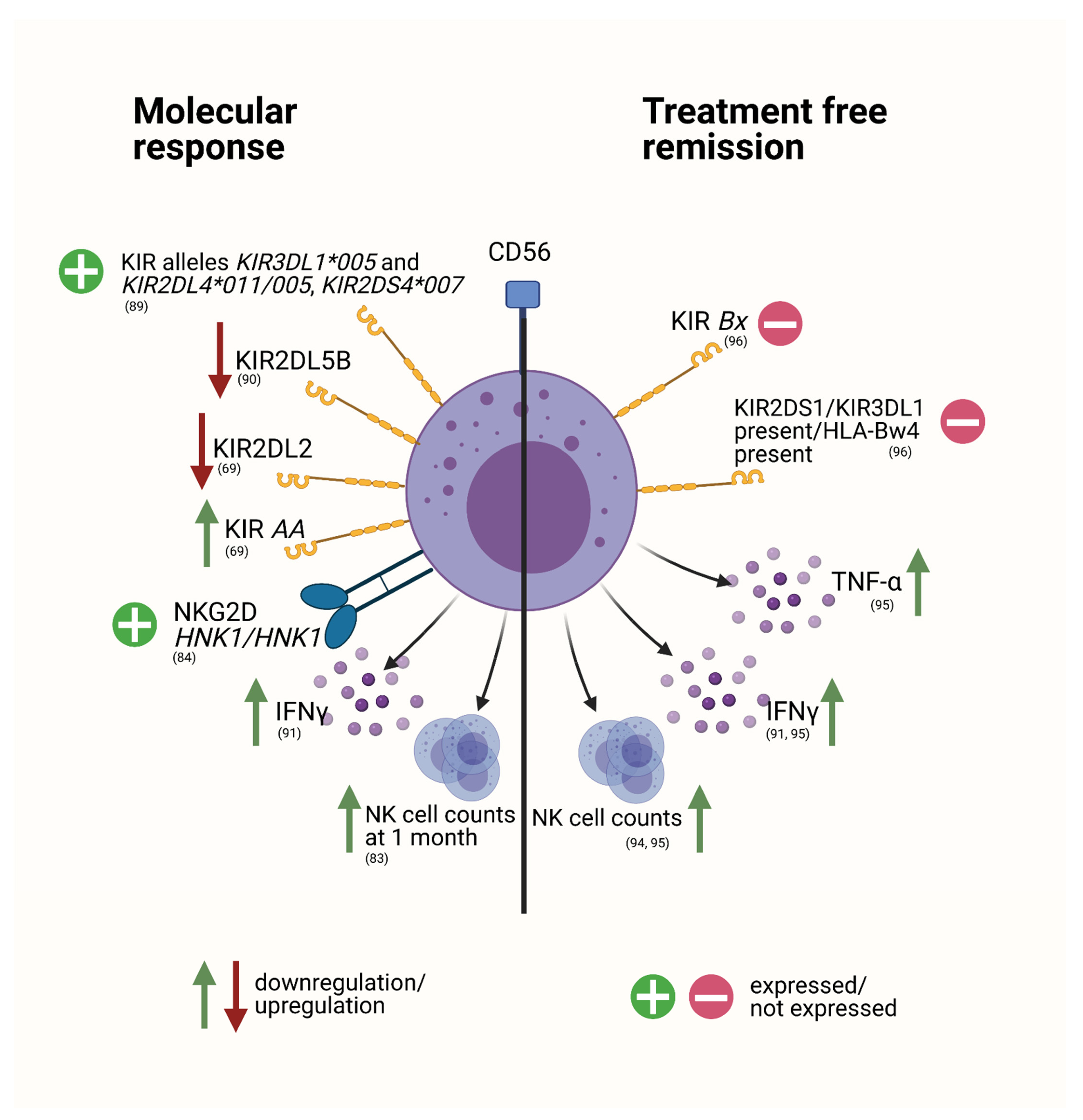
The Role of NK Cells to Disease Progression and Response to Therapy

In addition to the changes conferred by therapeutic interventions in CML, several studies assessing NK cell related parameters have also found prognostically relevant associations with response to TKI therapy (Figure 2).
A comprehensive investigation by Ureshino et al. on the predictive role of the inhibitory receptor KIR3DL1 and the associated HLA-B allelic polymorphisms has revealed that weakly interacting combinations of these two counterparts were associated with a superior response to TKI treatment [89]. Alleles KIR3DL1*005 and KIR2DL4*011/005 as well as KIR2DS4*007 also conferred favorable prognostic value [89]. A sub-study of the TIDEL-II study, a trial examining a risk-adapted scheme of imatinib and nilotinib combination, found the expression of KIR2DL5B to be an independent negative prognostic factor for achievement of MMR (major molecular remission) and was furthermore significantly linked to inferior achievement of MR4.5, event-free survival (EFS) and transformation-free survival (TFS) [90]. As KIR2DL5B is an inhibitory KIR receptor, it was consequently hypothesized that its expression could possibly suppress sufficient NK-cell mediated killing of leukemic cells [90]. Loss of the inhibitory receptor KIR2DL2 was associated with successful CMR (complete molecular remission) in a heterogeneously treated patient population under 1st and 2nd generation TKI therapy as reported by Nasa et al. [69]. In the same cohort, KIR genotype AA was linked to earlier CMR, with a higher probability of CMR achievement in comparison to genotype Bx [69]. KIR genotype AA thus appeared to be a strong predictive factor for successful CMR in this setting [69].
The amount of effector NK cells (defined as IFN-γ+ NK cells) has also been previously linked to successful and stable CMR under imatinib treatment [91]. The increase in this cell compartment may therefore constitute a marker for functioning immune surveillance [91]. In a patient population under dasatinib treatment, Iriyama et al. demonstrated that NK cell counts at 1 month after treatment initiation revealed to be a crucial predictive factor for DMR [83]. This suggests that a rapid increase in NK cell count, potentially caused by dasatinib itself, may reflect a successful immune response towards leukemic cells [83]. Additionally, a study by Hara et al. revealed that expression of NKG2D HNK1/HNK1 haplotype was linked to a higher likelihood of achieving MR4.5 in dasatinib treated patients [84] (Figure 2).
It is therefore likely, that NK cells have a potential role in control of the leukemic clone, even in patients with ongoing TKI treatment. Furthermore, various reports indicate that NK cells also seemingly play a pivotal role in the achievement and maintenance of successful TFR [92].

NK Cells in the Setting of Treatment Discontinuation

In the past years the concept of treatment discontinuation with consequent TFR has become a desirable and achievable goal in CML therapy [92]. However, TFR is not always successful. One potential reason for discrepancies in TFR achievement are differences in immune response (Figure 2) [92].
The IMMUNOSTIM trial group demonstrated that higher numbers of peripheral CD56dim NK cells at the time of treatment cessation were significantly and independently correlated to treatment-free remission after imatinib discontinuation [93]. In agreement with these results are the EUROSKI and DADI trials on imatinib and dasatinib treatment cessation respectively, also demonstrating the protective effect of NK cell increase in the setting of TFR [94,95].
The IMMUNOSTIM group furthermore found no differences in expression of an extensive panel of NK cell receptors comparing non-relapsing to relapsing patients, but demonstrated less NKp46 and DNAM-1 expressing NK cells (CD56dim and CD56bright) in all patients after discontinuation, compared with healthy individuals [93]. Degranulation capacity towards the K562 cell line was preserved in both relapsing and non-relapsing patients compared with healthy donors, while IFN-γ and TNF-α production after stimulation were diminished in relapsing patients [95]. When dichotomized into high versus low IFN-γ and TNF-α production, patients with increased amounts of cytokine production were significantly associated with higher molecular relapse-free survival [95]. Mizoguchi et al. in agreement with this, have described a sustained elevation of NK effector IFN-γ+ cells in successful TFR after imatinib treatment (STOP-IM trial), suggesting that this cell population is essential for control of the leukemic clone in this setting [91].
Furthermore, homozygosity for KIR A haplotype conferred a significantly higher likelihood of achieving successful TFR in patients after imatinib or nilotinib treatment cessation as reported by Caocci et al. [96]. Conversely, patients with Bx genotype and KIR-ligand combination KIR2DS1/KIR3DL1 present/HLA-Bw4 present had a higher risk of relapse [96].
Altogether, these results demonstrate the importance of NK cells in control of the leukemic clone in the setting of TFR. Therefore, it may be reasonable to consider a therapy that enhances NK cell function as well as investigating NK cell specific markers before discontinuation of treatment.

4.2. Classical MPNs

4.2.1. Immunological Changes in Classical MPNs

Typical untreated MPN presents with clonal myeloproliferation, decreased apoptosis of myeloid cells and in some cases progressive myelofibrosis [48]. MPN patients usually display an increased number of bone marrow nucleated-, terminally differentiated- and replicative mature cells and elevated peripheral blood cells. If left untreated, microvascular and major thrombotic events can occur [97]. The MPL mutation occurs in megakaryocytes, where it translates to a constantly activated MPL or thrombopoietin receptor [98], while the JAK2V617F mutation occurs in multipotent hematopoietic progenitor cells, but can be found in all myeloid lineages, as well as B cells, T cells and NK cells [4]. It is unsurprising that several blood parameters are seen to be altered in the various MPN phenotypes. While it is rare to find a case of MPN with normal blood parameters, it is not impossible. The presence of the JAK2V617F mutation with normal peripheral blood and T cell composition has previously been reported [97].

4.2.2. NK Cells in Classical MPNs

Baseline data for NK cells in untreated MPNs remains disputed. Some studies report low NK cell frequencies in untreated patients (excluding therapeutic phlebotomy), compared with healthy donors [48], while we reported similar NK cells frequencies in untreated patients when compared with healthy age-matched controls (Figure 3) [99]. The phenotype of NK cells, however, appears to be most affected by pharmacological treatments such as IFN-α or TKI’s [48,99].

4.2.3. The Effect of Treatment on NK Cells

Therapeutic phlebotomy can be used in Ph− MPNs to maintain hematocrit levels, thereby reducing risk of thrombosis, and cardiovascular related death [100]. Studies regarding the effect of phlebotomy in MPN are lacking; however, patients undergoing regular phlebotomy for hereditary hemochromatosis, as well as healthy controls, showed no changes in absolute numbers of NK cells, B cells, T cells, NKT cells or monocytes [101]. Anti-coagulants can be used for antithrombotic therapy and typical forms of management of bleeding or infection may be indicated [18,19,21,102]. For the prevention of venous thromboembolism administration of vitamin K antagonists (VKAs) or the use of direct oral anticoagulants (DOACs) is recommended. Low-dose acetylsalicylic acid (ASA) may be indicated in several MPN entities with concomitant thrombocytosis, such as PV or ET [17,103,104,105]. Another treatment option is the administration of interferon-alpha (IFN-α) in a variety of MPN entities, such as PV and ET [9,10,18,19,105,106,107]. Cytoreduction is frequently accomplished via treatment with hydroxyurea (HU), an inhibitor of ribonucleotide reductase, or busulfan, an alkylating agent [19,21,108,109]. Anagrelide, a phosphodiesterase inhibitor, is approved for the reduction of platelet count in ET [19,109]. With JAK2 mutations being the characteristic driver mutation in PV, ruxolitinib, a nonselective JAK1/2 inhibitor, is the option of choice in this entity, but can also constitute an alternative option in ET or PMF. [18,19,21,22,110] In situations where ruxolitinib has failed, or might not be appropriate, MF patients with anemia may be treated with immunomodulatory drugs (IMiDs) such as Thalidomide, Pomalidomide or Lenalidomide [111,112,113,114]. In high-risk PMF, CNL or CEL-NOS, certain patients may be even eligible for stem cell transplantation (SCT) [9,10,21].

Acetylsalicylic Acid (ASA)
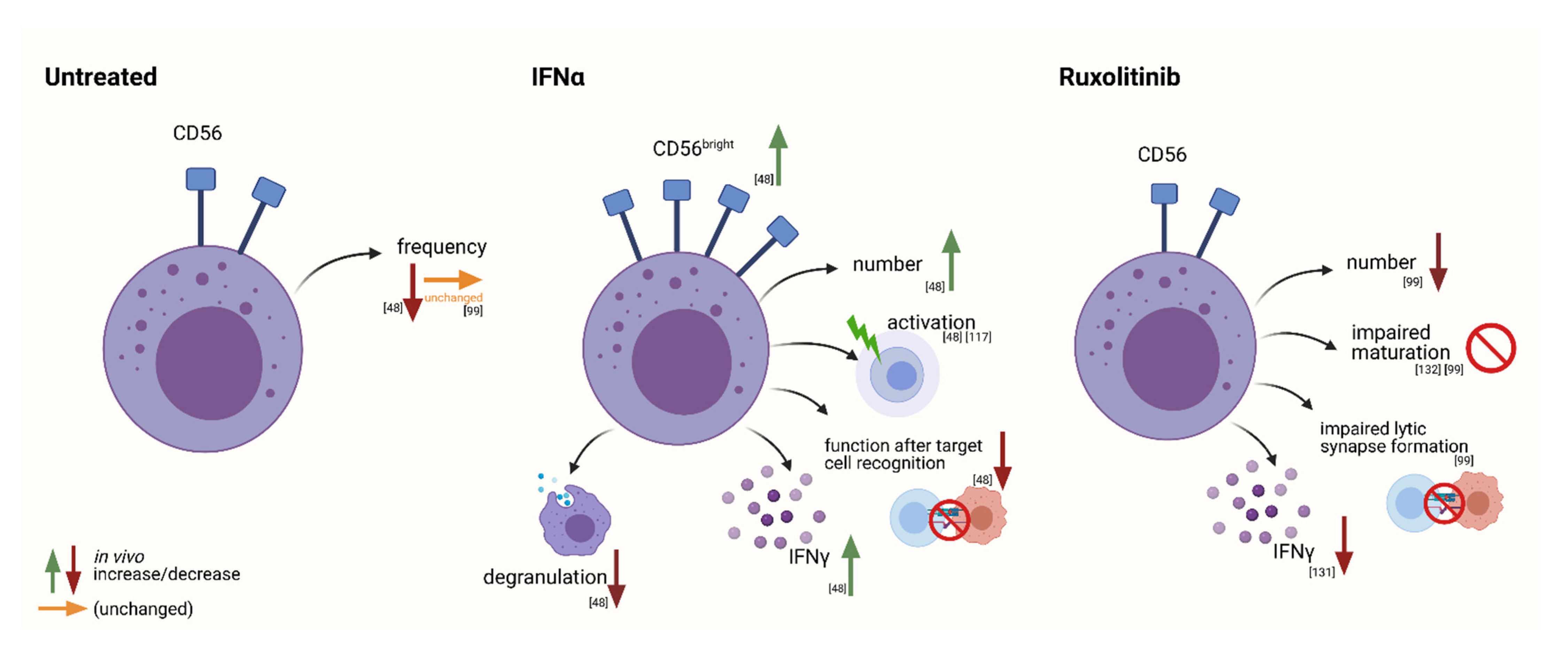
Low dose ASA therapy is recommended to many MPN patients, particularly in ET, to reduce the risk of thrombotic events. ASA irreversibly acetylates platelet cyclooxygenase (COX)-1 resulting in a long-lasting inhibition of TXA2 biosynthesis, a type of thromboxane which stimulates platelet activation, contributing to vasoconstriction and platelet aggregation [115,116]. Recent data describing the effect of ASA on NK cells is lacking, and the effects in MPN specifically have not been reported.

Interferon-Alpha2

INF-α is a type 1 interferon which stimulates the immune cells, providing anti-proliferative, immunomodulatory, and antiangiogenic effects [117]. Pegylated IFN-α has a much longer serum half-life than recombinant IFN-α, and allows for weekly, as opposed to daily, dosing [118]. Type 1 interferons act by activating the JAK/STAT pathway, resulting in the production of a complex named IFN-Stimulated Gene Factor 3 (ISGF3), which translocates to the nucleus and initiates the transcription of hundreds of IFN-stimulated genes (ISGs) [119]. ISGs can perform an array of functions, including inhibition of viral spread, the downregulation of telomerase activity in malignant and non-malignant hematopoietic cells and the induction of a direct pro-apoptotic effect on myeloid progenitors [117,120].
Chronic IFN-α treatment depletes HSCs and has severe immune-altering effects including the activation of dendritic cells, NK cells and T cells [48,117]. MPN patients undergoing therapy with IFN-α displayed a significantly increased frequency of circulating Tregs. This is hypothesized to be due to either the mobilization of Tregs to the periphery, decreasing their immunosuppressive action within the bone marrow, or as a counter response to immune activation [121]. IFN-α treatment of MPN patients also resulted in a decreased frequency of myeloid DCs (mDCs) and plasmacytoid DCs (pDCs) [121]. This marked immunomodulation however, does not correlate with molecular responses, and the duration of treatment is also proposed to be a contributing variable [121].
Riley et al. 2014, described the effects of IFN-α treatment on NK cells in detail (Figure 3, IFN-α), in a comparison with untreated patients, healthy donors, and patients undergoing treatment with hydroxyurea [48]. During IFN-α treatment, NK cells increased in number, and underwent a phenotype shift from cytotoxic cells highly capable of antibody-dependent cellular cytotoxicity, to a more immune-stimulatory profile with little cytotoxic importance [48]. NK cell numbers were the lowest in untreated patients compared with healthy donors, or patients treated with hydroxyurea or IFN-α. An expansion of CD56bright and subsequent decrease in CD56dim NK cells was observed in long-term (≥12 months) IFN-α treated patients, and data suggests an overall decrease in NK cell functionality upon target cell recognition, and a shift from a more cytotoxic phenotype, as previously mentioned, to an immune-stimulatory profile. The CD56dim subset, which is suggested to be a more mature subset, was seen to secrete an increased level of IFN-γ when stimulated with DC monokines (IL-12 and IL-15), however the overall production of IFN-γ was greater in CD56bright cells, highlighting the dependence of NK cell differentiation on DC cytokines [48]. A decrease in degranulation and CCL4 production was seen during treatment, and while the observed trends were not significant, they support the conjectured compromised functionality [48] (Figure 3).

Hydroxyurea

Hydroxyurea is an antimetabolite which scavenges tyrosol-free radicals, thereby inhibiting the enzyme ribonucleotide reductase and reducing deoxyribonucleotide production, arresting proliferating cancer cells in the S-phase [106,108]. In a large retrospective study of 3411 patients immunomodulatory effects were not evident, as no allergy or immunosuppression was detected, and little has been reported in studies so far [106]. The bone marrow suppression and antiproliferative effect of HU may result in neutropenia, anemia or thrombocytopenia [122]. Upon activation, murine cells in culture that were treated with HU, increased IL-2 secretion, while no effect was seen in untreated cells [122], and similar results were seen in humans undergoing HU treatment for HIV infection [123]. HU was shown to upregulate NKG2D ligand expression on myeloid leukemic cell lines [124]; however, in MPN patients no changes were observed between HU treated patients and healthy controls [125].

JAK Inhibitors

For the treatment of Ph− MPNs, JAK inhibitors such as ruxolitinib, have been designed to specifically inhibit the JAK/STAT pathway by binding to cytoplasmic JAK1 and JAK2 kinases and modulating intracellular cytokine signaling [126]. The inhibition of JAK2 leads to the desired myelosuppression, while the inhibition of JAK1 reduces the levels of pro-inflammatory cytokines produced, improving various symptoms including bone marrow fibrosis [127,128]. A high rate of infection has been observed in patients undergoing ruxolitinib therapy, and it is hypothesized that the inhibition of JAK1 could therefore be responsible for a certain level of immunosuppression. Cytokine action on the JAK/STAT pathway results in proliferation, differentiation, and activation of various immune cells, however the full extent of ruxolitinib-induced immunosuppression is not yet known [129]. In both the COMFORT I and COMFORT II studies, neutropenia was seen in ruxolitinib treated patients compared with the placebo group (7.1 vs. 2% and 8.9 vs. 6.3% respectively) [129]. Several studies have shown that ruxolitinib treatment impaired dendritic cell function, affecting cell differentiation, tissue migration and IL-12 production, which has been described to play an important role in NK cell differentiation [107,129,130]. Later studies confirmed that IL-12 was completely blocked by ruxolitinib, as well as IL-15 and the phosphorylation of STAT5, resulting in a functional impairment of IFN-γ production by NK cells (Figure 3, ruxolitinib) [131]. An inhibition of cytokine secretion by macrophages was also reported, possibly effecting NK cell recruitment, maturation, and killing activity [132]. The reported impairment of DC function also resulted in the impaired induction of antigen specific T cell responses [130]. Pharmacological inhibitors of the JAK/STAT pathway have detrimental effects on NK maturation and as a result many patients experience infectious complications during treatment [99]. Unsurprisingly, ruxolitinib lead to a reduced number of NK cells, most likely due to impaired maturation as a result of the lack of DC cytokines, displayed by an increased ratio of immature/mature NK cells [99]. Additionally, compromised lytic synapse formation with target cells lead to a reduction in killing activity [99]. However, these results were reversible, and when ruxolitinib treatment was discontinued, NK cell function was restored [99] (Figure 3).

Immunomodulatory Drugs (IMiDs)

IMiDs are anti-inflammatory, anti-angiogenic drugs that regulate cytokine response. Even though IMiDs only play a minimal role in the management of MPNs clinically, they have substantial immunomodulatory activity on NK cell function [111,112,113,114]. MF patients treated with IMiDs exhibited activated NKT cells, co-stimulation of T cells, and impaired proliferation and function of Tregs [133]. More detailed effects of IMiDs on the immune cells can be seen in patients with multiple myeloma. In addition to an increase in IL-2 production and resultant T cell proliferation, a subsequent increase in IFNγ was seen, resulting in an increased number and improved function of NK cells [134]. The cytokines which are upregulated with thalidomide administration are also associated with angiogenesis, however the precise role of thalidomide and NK cells in vascularization has not yet been defined [135].

5. Discussion

The concepts of immune evasion and excessive inflammation are two frequently occurring tumor promoting phenomena [23,136]. Properly functioning immune surveillance is crucial for the detection and elimination of altered cell populations that could hypothetically transform into malignancies [136]. On the other hand, chronic hyperinflammatory conditions may additionally support tumor pathogenesis and exert tumor promoting effects [24,136]. Immunologic changes may therefore substantially affect the development and course of neoplastic diseases and constitute useful biomarkers for better understanding and prediction of pathologic processes.
Changes in immunologic variables were also discovered in the group of MPNs [25,26,33,137]. These alterations also comprise the NK cell compartment [27,48,54,55]. Changes in amount, phenotype as well as function of NK cells were reported to be present at time of diagnosis, but also occur with therapeutic intervention (Figure 1 and Figure 3) [55,99]. It appears that these findings are not mere observations but may also have prognostic value, especially in the setting of CML (Figure 2) [95].
In CML, conflicting results on relative amounts of NK cells as proportions among lymphocytes were demonstrated [34,65,66,67]. We have, however, previously shown that assumptions concerning NK cell frequencies should be regarded critically, and we proposed a gating strategy using whole blood for more accurate quantification by flow cytometry [65]. Using this method we demonstrated increased frequencies of NK cells at time of diagnosis [65].
Similarly, also in classical MPN entities, ambiguous findings concerning NK cell frequencies at time of diagnosis were reported, with reports of decreasing as well as increasing proportions [48,99]. However, evidence of changes in the NK cell compartment in untreated patients remains scarce, and a more in-depth investigation of the disease-related effects is needed.
Additionally, phenotypic changes at time of diagnosis have been described in CML, mainly comprising a reduction of activating receptors [32,34,68]. These alterations may hypothetically lead to a more suppressed NK cell phenotype with a decrease in tumor cell recognition [32,63,70]. In agreement with this, a deficit in degranulation capacity of patient-derived NK cells at time of diagnosis was reported [66]. Additionally, the NK cells were shown to have a significantly reduced proliferative capacity, even though this finding could not be confirmed in the group’s mouse model [66].
The NK cell compartment may not only be altered by the disease itself, but the therapeutic agents used can also have immunomodulatory side effects (Figure 1 and Figure 3). Therapy of MPNs is heterogenous and the effects on NK cell functionality and phenotype vary substantially among the respective drugs. The effects of treatment on the NK cell compartment in Ph− MPNs is not easily summarized. One must consider the first line treatments for the various disease phenotypes, which, in many cases can be implemented throughout the course of disease until other factors such as age, mutational burden, or progression to fibrosis come into play.
Therapeutic strategies in CML are majorly comprised of the group of TKIs. A variety of effects on NK cells have been reported, however, strong discrepancies especially between in vivo or ex vivo, and in vitro models make it necessary to carefully evaluate the respective findings.
In vitro experiments on imatinib and nilotinib suggest negative effects on NK cell reactivity towards the leukemic clone [63,66,78,80]. Fortunately, however, the supposed decrease in functionality of NK cells conferred by imatinib as well as nilotinib application, was refuted in in vitro experiments using patient-derived material, suggesting that the suppressing effects of imatinib and the phenotypic alterations may not be of significant value for leukemic cell control [66,79,80]. When further investigating the in vivo effects of nilotinib therapy, on the other hand, Hayashi et al. found a decrease in cytotoxicity [79]. Even though this in vivo setting is probably depicting the situation more accurately, it would be of interest to investigate the demonstrated effects further by individually comparing patients pre-treatment to patients under treatment in order to reliably distinguish between disease-related and treatment-related effects. Another possible explanation for the ambiguous results is that nilotinib may not exert direct cytotoxicity-reducing effects, however it may indirectly modulate NK cells by changing the tumor environment.
Dasatinib is probably the most substantially studied TKI in terms of NK cell related effects in CML therapy. One of the observed drug-specific effects is the well-studied expansion of LGLs in vivo with simultaneous augmentation of NK cell counts [32,81,82,83]. This appears to be a direct effect of dasatinib application on NK cells, as in vitro use of dasatinib on healthy cells reportedly led to an increase in NK cell counts [84]. Concerning functional effects of dasatinib administration, it is important to clearly distinguish between direct and indirect effects. Direct administration of dasatinib led to a reduction of cytotoxicity, degranulation and cytokine secretion of NK cells in vitro [80,85]. However, when dasatinib was washed out after the application, the effects were contrary with an increase in proliferation as well as cytolytic capacity [85,86]. Most importantly, in vivo experiments examining patients under dasatinib treatment confirmed the increase in cytotoxic capacity of NK cells [79]. The stimulatory effects of dasatinib may therefore best reflect the actual effects. However so far, the in vitro results do not provide enough information to clearly distinguish between a direct effect of dasatinib application or indirect stimulation of NK cells.
Fully functioning NK cells are important players in leukemic immune surveillance in CML. This is also reflected by the prognostic value they exert for molecular response. An increase in NK effector cells, as well as weak linkage of inhibitory KIR/HLA combinations, were associated with superior outcome, as well as increasing NK cell counts [83,89,90,91,96]. These findings indicate that changes in the NK cell compartment under TKI therapy may actively influence sensitivity towards the treatment, making investigations into drug-specific alterations even more important.
The role of NK cells in CML becomes even more evident in investigations on patients after TKI treatment when the individual’s immune system takes control over of the leukemic clone. The amount of NK cells at the time of treatment cessation is an important prognostic factor in the setting of imatinib and dasatinib cessation [94,95]. Furthermore, preserved NK cell production of IFN-γ and TNF-α was associated with successful TFR after imatinib discontinuation, as well as an increase in the IFN-γ producing NK cell effector population [91,95].
Treatment with IFN-α is common in classical MPNs, and while it has vanished from the therapeutic landscape of CML treatment with the introduction of TKIs it is now being discussed as a possible add-on therapy. An expansion of the CD56bright population was seen in both classical MPNs as well as Ph+ MPNs with IFN-α [48,76]. This shift was suggested to constitute a maturation defect [48]. In CML patients, however, favorable effects of IFN-α, such as an increase in NK cell proportions, as well as increased numbers of mature and activated NK cells, and an upregulation of cytotoxicity were described [73,74,75,76].
The effect of HU on NK cells has not been thoroughly investigated and lacks in-depth analyses. However, currently available data suggest that there are no detectable changes in the NK cell compartment with HU treatment [125,138,139].
Ruxolitinib had drastic effects on the overall immune system, the JAK1 inhibition specifically, caused by ruxolitinib, is hypothesized to be responsible for a certain level of immunosuppression [129]. Ruxolitinib affected the NK cell compartment both directly and indirectly. Direct effects include a reduction in number of NK cells and a greater proportion of immature NK cells, as well as a reduction in killing activity due to the reduced ability to form lytic synapses with target cells [99]. Indirect effects include the impairment or complete blocking of dendritic cell IL-12 and IL-15 production, or the phosphorylation of STAT5, resulting in defective maturation and a diminished functional capacity of NK cells to produce IFN-γ [107,129,130,131]. The reduction in NK cell number is hypothesized to be a result of the impaired maturation. Indeed, we reported a 2.5 fold higher bright:dim ratio in untreated patients than in aged-matched healthy donors, indicative for a block in differentiation [99]. Many aspects of lymphoid cell development and homeostasis are controlled by cytokines, and as MPNs are diseases driven by inflammation [27], it is no surprise that this hyperinflammatory state can contribute to maturation defects and dysregulation of homeostasis [140]. Interestingly, studies investigating the effects of ruxolitinib treatment in patients with STAT1 gain-of-function mutations reported higher levels of STAT5 phosphorylation post-treatment, and the perforin expression appeared to be restored, contradictory to the results reported in MPN patients [141]. Vargas-Hernandez and colleagues suggested this could be a result of the degree of phosphorylation, with STAT1 levels elevated during IL-2 stimulation, while STAT5 was aberrantly phosphorylated, as both STAT1 and STAT5 were affected to the same degree of impairment [141]. The in-depth and specific effects of ruxolitinib on the NK cell phenoptype in Ph− MPNs, however, are hard to definitively conclude, as supporting literature and studies are limited.
Overall, the impact of NK cells in MPNs is well established and substantiated in CML. NK cells play a role in control of the leukemic clone which is not only reflected by their prognostic impact in the setting of TFR, but also important for the achievement of molecular remission under TKI therapy (Figure 2). The number of NK cells present, sufficient cytokine production, as well as expression of activating NK cell receptors with matching HLA-counterparts appear to be important prognostic features for molecular remission in CML. Interestingly, in contrast to CML, research on NK cells in classical MPNs still substantially lacks information at all stages of disease. The major questions to be addressed, concern alterations in the NK cell compartment at time of diagnosis, as well as most importantly, their prognostic impact. With the known suppressive effects of ruxolitinib administration on NK cell function, including impaired lytic synapse formation, and reduced recruitment, activation and killing activity (Figure 3), it would be of primary importance to thoroughly investigate this finding in terms of patient outcomes and whether patients may profit from additional NK-cell specific therapy.
NK cell activity may be influenced with the use of checkpoint-inhibitors, most of which so far have only been investigated in vitro [142]. One of the targets which could potentially influence NK cell reactivity and has been implemented in a variety of cancer entities, is PD-1 with its counterpart PDL-1 [142]. PD-1 blockade is a well-established mechanism of immune evasion and has already been discussed as a possible therapeutic target in CML [143,144]. Further research on these inhibitors in vitro and in vivo would thus certainly be of interest, especially in the field of CML, and could potentially ameliorate sensitivity towards TKI treatment or successful TFR through upregulation of NK cell activity.

6. Conclusions

Regardless of the amount of research currently documented on NK cells in MPNs, the story does not end here. In particular, the lack of data concerning classical MPNs and the NK cell population support the need for more in-depth studies. Furthermore, we believe that evidence of the great prognostic impact of NK cells in CML suggests that it is time to target NK function, hopefully improving control of the leukemic clone for a deeper molecular response and more sustainable treatment free remission.

Author Contributions

Conceptualization, S.S. and D.W.; writing—original draft preparation, E.N. and J.S.; writing—review and editing, E.N., J.S., S.S. and D.W.; visualization, E.N. and J.S.; supervision, S.S.; funding acquisition, D.W. All authors have read and agreed to the published cersion of the manuscript.

Funding

This work was funded by grant DJCLS 16 R/2018 from the Deutsche José Carreras Leukämie Stiftung and supported by Novartis through the European Treatment and Outcome Study (EUTOS) for CML.

Institutional Review Board Statement

Not applicable.

Informed Consent Statement

Not applicable.

Data Availability Statement

Data sharing not applicable. No new data were created or analyzed in this article.

Conflicts of Interest

The authors declare no conflict of interest.

References

  1. Grabek, J.; Straube, J.; Bywater, M.; Lane, S.W. MPN: The Molecular Drivers of Disease Initiation, Progression and Transformation and their Effect on Treatment. Cells 2020, 9, 1901. [Google Scholar] [CrossRef]
  2. Barbui, T.; Thiele, J.; Gisslinger, H.; Kvasnicka, H.M.; Vannucchi, A.M.; Guglielmelli, P.; Orazi, A.; Tefferi, A. The 2016 WHO classification and diagnostic criteria for myeloproliferative neoplasms: Document summary and in-depth discussion. Blood Cancer J. 2018, 8, 1–11. [Google Scholar] [CrossRef]
  3. Tefferi, A. Myeloproliferative neoplasms: A decade of discoveries and treatment advances. Am. J. Hematol. 2016, 91, 50–58. [Google Scholar] [CrossRef] [Green Version]
  4. Vainchenker, W.; Kralovics, R. Genetic basis and molecular pathophysiology of classical myeloproliferative neoplasms. Blood 2017, 129, 667–679. [Google Scholar] [CrossRef] [Green Version]
  5. Arber, D.A.; Orazi, A.; Hasserjian, R.; Thiele, J.; Borowitz, M.J.; Le Beau, M.M.; Bloomfield, C.D.; Cazzola, M.; Vardiman, J.W. The 2016 revision to the World Health Organization classification of myeloid neoplasms and acute leukemia. Blood 2016, 127, 2391–2405. [Google Scholar] [CrossRef]
  6. Shallis, R.M.; Wang, R.; Davidoff, A.; Ma, X.; Podoltsev, N.A.; Zeidan, A.M. Epidemiology of the classical myeloproliferative neoplasms: The four corners of an expansive and complex map. Blood Rev. 2020, 42, 100706. [Google Scholar] [CrossRef] [PubMed]
  7. Anderson, L.A.; McMullin, M.F. Epidemiology of MPN: What Do We Know? Curr. Hematol. Malign-Rep. 2014, 9, 340–349. [Google Scholar] [CrossRef] [PubMed]
  8. Titmarsh, G.J.; Duncombe, A.S.; McMullin, M.F.; O’Rorke, M.; Mesa, R.; De Vocht, F.; Horan, S.; Fritschi, L.; Clarke, M.; Anderson, L.A. How common are myeloproliferative neoplasms? A systematic review and meta-analysis. Am. J. Hematol. 2014, 89, 581–587. [Google Scholar] [CrossRef] [PubMed]
  9. Helbig, G.; Soja, A.; Bartkowska-Chrobok, A.; Kyrcz-Krzemień, S. Chronic eosinophilic leukemia-not otherwise specified has a poor prognosis with unresponsiveness to conventional treatment and high risk of acute transformation. Am. J. Hematol. 2012, 87, 643–645. [Google Scholar] [CrossRef]
  10. Szuber, N.; Elliott, M.; Tefferi, A. Chronic neutrophilic leukemia: 2020 update on diagnosis, molecular genetics, prognosis, and management. Am. J. Hematol. 2020, 95, 212–224. [Google Scholar] [CrossRef]
  11. James, C.; Ugo, V.; Le Couédic, J.-P.; Staerk, J.; Delhommeau, F.; Lacout, C.; Garçon, L.; Raslova, H.; Berger, R.; Griscelli, A.B.; et al. A unique clonal JAK2 mutation leading to constitutive signalling causes polycythaemia vera. Nat. Cell Biol. 2005, 434, 1144–1148. [Google Scholar] [CrossRef] [PubMed]
  12. Levine, R.L.; Wadleigh, M.; Cools, J.; Ebert, B.L.; Wernig, G.; Huntly, B.; Boggon, T.J.; Wlodarska, I.; Clark, J.J.; Moore, S.; et al. Activating mutation in the tyrosine kinase JAK2 in polycythemia vera, essential thrombocythemia, and myeloid metaplasia with myelofibrosis. Cancer Cell 2005, 7, 387–397. [Google Scholar] [CrossRef] [PubMed] [Green Version]
  13. Chereda, B.; Melo, J.V. Natural course and biology of CML. Ann. Hematol. 2015, 94, 107–121. [Google Scholar] [CrossRef] [PubMed]
  14. Quintás-Cardama, A.; Cortes, J. Molecular biology of bcr-abl1–positive chronic myeloid leukemia. Blood 2009, 113, 1619–1630. [Google Scholar] [CrossRef] [PubMed] [Green Version]
  15. Ren, R. Mechanisms of BCR–ABL in the pathogenesis of chronic myelogenous leukaemia. Nat. Rev. Cancer 2005, 5, 172–183. [Google Scholar] [CrossRef]
  16. Jabbour, E.; Kantarjian, H. Chronic myeloid leukemia: 2018 update on diagnosis, therapy and monitoring. Am. J. Hematol. 2018, 93, 442–459. [Google Scholar] [CrossRef] [Green Version]
  17. Tefferi, A.; Vannucchi, A.M.; Barbui, T. Polycythemia vera treatment algorithm. Blood Cancer J. 2018, 8, 1–7. [Google Scholar] [CrossRef]
  18. Spivak, J.L. Polycythemia vera. Curr. Treat. Options Oncol. 2018, 19, 1–14. [Google Scholar] [CrossRef]
  19. Tefferi, A.; Pardanani, A. Essential thrombocythemia. Engl. J. Med. 2019, 381, 2135–2144. [Google Scholar] [CrossRef]
  20. Tefferi, A. Primary myelofibrosis: 2021 update on diagnosis, risk-stratification and management. Am. J. Hematol. 2021, 96, 145–162. [Google Scholar] [CrossRef]
  21. Finazzi, G.; Vannucchi, A.M.; Barbui, T. Prefibrotic myelofibrosis: Treatment algorithm. Blood Cancer J. 2018, 8, 104. [Google Scholar] [CrossRef]
  22. Tefferi, A.; Barbui, T. Polycythemia vera and essential thrombocythemia: 2017 update on diagnosis, risk-stratification, and management. Am. J. Hematol. 2017, 92, 94–108. [Google Scholar] [CrossRef] [PubMed]
  23. Barosi, G. An Immune Dysregulation in MPN. Curr. Hematol. Malign-Rep. 2014, 9, 331–339. [Google Scholar] [CrossRef] [PubMed]
  24. Mantovani, A.; Allavena, P.; Sica, A.; Balkwill, F. Cancer-related inflammation. Nat. Cell Biol. 2008, 454, 436–444. [Google Scholar] [CrossRef] [PubMed]
  25. Wang, Y.; Zuo, X. Cytokines frequently implicated in myeloproliferative neoplasms. Cytokine X 2019, 1, 100005. [Google Scholar] [CrossRef] [PubMed]
  26. Longhitano, L.; Volti, G.L.; Giallongo, C.; Spampinato, M.; Barbagallo, I.; Di Rosa, M.; Romano, A.; Avola, R.; Tibullo, D.; Palumbo, G.A. The Role of Inflammation and Inflammasome in Myeloproliferative Disease. J. Clin. Med. 2020, 9, 2334. [Google Scholar] [CrossRef]
  27. Braun, L.M.; Zeiser, R. Immunotherapy in Myeloproliferative Diseases. Cells 2020, 9, 1559. [Google Scholar] [CrossRef]
  28. Marty, C.; Lacout, C.; Droin, N.; Le Couédic, J.-P.; Ribrag, V.; Solary, E.; Vainchenker, W.; Villeval, J.-L.; Plo, I. A role for reactive oxygen species in JAK2V617F myeloproliferative neoplasm progression. Leukemia 2013, 27, 2187–2195. [Google Scholar] [CrossRef] [PubMed] [Green Version]
  29. Hasselbalch, H.C.; Thomassen, M.; Riley, C.H.; Kjær, L.; Larsen, T.S.; Jensen, M.K.; Bjerrum, O.W.; Kruse, T.A.; Skov, V. Whole Blood Transcriptional Profiling Reveals Deregulation of Oxidative and Antioxidative Defence Genes in Myelofibrosis and Related Neoplasms. Potential Implications of Downregulation of Nrf2 for Genomic Instability and Disease Progression. PLoS ONE 2014, 9, e112786. [Google Scholar] [CrossRef]
  30. Bock, O.; Höftmann, J.; Theophile, K.; Hussein, K.; Wiese, B.; Schlué, J.; Kreipe, H. Bone Morphogenetic Proteins Are Overexpressed in the Bone Marrow of Primary Myelofibrosis and Are Apparently Induced by Fibrogenic Cytokines. Am. J. Pathol. 2008, 172, 951–960. [Google Scholar] [CrossRef] [Green Version]
  31. Murthy, G.S.G.; Atallah, E. Treatment-Free Remission in CML: The US Perspective. Curr. Hematol. Malign-Rep. 2019, 14, 56–61. [Google Scholar] [CrossRef]
  32. Hughes, A.; Yong, A.S.M. Immune Effector Recovery in Chronic Myeloid Leukemia and Treatment-Free Remission. Front. Immunol. 2017, 8, 469. [Google Scholar] [CrossRef] [Green Version]
  33. Ureshino, H.; Shindo, T.; Kimura, S. Role of cancer immunology in chronic myelogenous leukemia. Leuk. Res. 2020, 88, 106273. [Google Scholar] [CrossRef] [PubMed]
  34. Hughes, A.; Clarson, J.; Tang, C.; Vidovic, L.; White, D.; Hughes, T.; Yong, A. CML patients with deep molecular responses to TKI have restored immune effectors and decreased PD-1 and immune suppressors. Blood 2017, 129, 1166–1176. [Google Scholar] [CrossRef]
  35. Vitale, M.; Della Chiesa, M.; Carlomagno, S.; Pende, D.; Aricò, M.; Moretta, L.; Moretta, A. NK-dependent DC maturation is mediated by TNFα and IFNγ released upon engagement of the NKp30 triggering receptor. Blood 2005, 106, 566–571. [Google Scholar] [CrossRef] [PubMed]
  36. Di Vito, C.; Mikulak, J.; Mavilio, D. On the Way to Become a Natural Killer Cell. Front. Immunol. 2019, 10, 1812. [Google Scholar] [CrossRef]
  37. Del Zotto, G.; Marcenaro, E.; Vacca, P.; Sivori, S.; Pende, D.; Della Chiesa, M.; Moretta, F.; Ingegnere, T.; Mingari, M.C.; Moretta, A.; et al. Markers and function of human NK cells in normal and pathological conditions. Cytom. Part B Clin. Cytom. 2017, 92, 100–114. [Google Scholar] [CrossRef] [PubMed] [Green Version]
  38. Scoville, S.D.; Freud, A.G.; Caligiuri, M.A. Modeling Human Natural Killer Cell Development in the Era of Innate Lymphoid Cells. Front. Immunol. 2017, 8, 360. [Google Scholar] [CrossRef] [Green Version]
  39. Bonanni, V.; Sciume, G.; Santoni, A.; Bernardini, G. Bone Marrow NK Cells: Origin, Distinctive Features, and Requirements for Tissue Localization. Front. Immunol. 2019, 10, 1569. [Google Scholar] [CrossRef] [Green Version]
  40. Goh, W.; Huntington, N.D. Regulation of Murine Natural Killer Cell Development. Front. Immunol. 2017, 8, 130. [Google Scholar] [CrossRef] [Green Version]
  41. Horowitz, A.; Strauss-Albee, D.; Leipold, M.; Kubo, J.; Nemat-Gorgani, N.; Dogan, O.C.; Dekker, C.L.; Mackey, S.; Maecker, H.; Swan, G.E.; et al. Genetic and Environmental Determinants of Human NK Cell Diversity Revealed by Mass Cytometry. Sci. Transl. Med. 2013, 5, 208ra145. [Google Scholar] [CrossRef] [PubMed] [Green Version]
  42. Moretta, A.; Bottino, C.; Vitale, M.; Pende, D.; Biassoni, R.; Mingari, M.C.; Moretta, L. Receptors for Hla Class-I Molecules in Human Natural Killer Cells. Annu. Rev. Immunol. 1996, 14, 619–648. [Google Scholar] [CrossRef]
  43. Morvan, M.; Lanier, L.L. NK cells and cancer: You can teach innate cells new tricks. Nat. Rev. Cancer 2016, 16, 7–19. [Google Scholar] [CrossRef] [PubMed]
  44. Raulet, D.H. Missing self recognition and self tolerance of natural killer (NK) cells. Semin. Immunol. 2006, 18, 145–150. [Google Scholar] [CrossRef]
  45. Gasser, S.; Orsulic, S.; Brown, E.J.; Raulet, D.H. The DNA damage pathway regulates innate immune system ligands of the NKG2D receptor. Nat. Cell Biol. 2005, 436, 1186–1190. [Google Scholar] [CrossRef] [PubMed] [Green Version]
  46. Pardoll, D.M. Distinct Mechanisms of Tumor Resistance to NK Killing: Of Mice and Men. Immunity 2015, 42, 605–606. [Google Scholar] [CrossRef] [Green Version]
  47. Boissel, N.; Rea, D.; Tieng, V.; Dulphy, N.; Brun, M.; Cayuela, J.-M.; Rousselot, P.; Tamouza, R.; Le Bouteiller, P.; Mahon, F.-X.; et al. BCR/ABL Oncogene Directly Controls MHC Class I Chain-Related Molecule A Expression in Chronic Myelogenous Leukemia. J. Immunol. 2006, 176, 5108–5116. [Google Scholar] [CrossRef] [PubMed]
  48. Riley, C.H.; Hansen, M.; Brimnes, M.K.; Hasselbalch, H.C.; Bjerrum, O.W.; Straten, P.T.; Svane, I.M.; Jensen, M.K. Expansion of circulating CD56bright natural killer cells in patients with JAK2-positive chronic myeloproliferative neoplasms during treatment with interferon-α. Eur. J. Haematol. 2015, 94, 227–234. [Google Scholar] [CrossRef]
  49. Van Acker, H.H.; Capsomidis, A.; Smits, E.; Van Tendeloo, V.F. CD56 in the Immune System: More Than a Marker for Cytotoxicity? Front. Immunol. 2017, 8, 892. [Google Scholar] [CrossRef]
  50. Yeap, W.H.; Wong, K.L.; Shimasaki, N.; Teo, E.C.Y.; Quek, J.K.S.; Yong, H.X.; Diong, C.P.; Bertoletti, A.; Linn, Y.C.; Wong, S.C. CD16 is indispensable for antibody-dependent cellular cytotoxicity by human monocytes. Sci. Rep. 2016, 6, 34310. [Google Scholar] [CrossRef]
  51. Cooper, M.; Fehniger, T.; Caligiuri, M.A. The biology of human natural killer-cell subsets. Trends Immunol. 2001, 22, 633–640. [Google Scholar] [CrossRef]
  52. López-Soto, A.; Gonzalez, S.; Smyth, M.J.; Galluzzi, L. Control of Metastasis by NK Cells. Cancer Cell 2017, 32, 135–154. [Google Scholar] [CrossRef] [PubMed]
  53. Street, S.E.; Hayakawa, Y.; Zhan, Y.; Lew, A.; MacGregor, D.; Jamieson, A.; Diefenbach, A.; Yagita, H.; Godfrey, D.; Smyth, M.J. Innate Immune Surveillance of Spontaneous B Cell Lymphomas by Natural Killer Cells and γδ T Cells. J. Exp. Med. 2004, 199, 879–884. [Google Scholar] [CrossRef]
  54. Carlsten, M.; Järås, M. Natural Killer Cells in Myeloid Malignancies: Immune Surveillance, NK Cell Dysfunction, and Pharmacological Opportunities to Bolster the Endogenous NK Cells. Front. Immunol. 2019, 10, 2357. [Google Scholar] [CrossRef] [Green Version]
  55. Ilander, M.; Hekim, C.; Mustjoki, S. Immunology and Immunotherapy of Chronic Myeloid Leukemia. Curr. Hematol. Malign-Rep. 2014, 9, 17–23. [Google Scholar] [CrossRef] [PubMed]
  56. Takahashi, N.; Miura, I.; Saitoh, K.; Miura, A.B. Lineage involvement of stem cells bearing the philadelphia chromosome in chronic myeloid leukemia in the chronic phase as shown by a combination of fluorescence-activated cell sorting and fluorescence in situ hybridization. Blood 1998, 92, 4758–4763. [Google Scholar] [CrossRef]
  57. Min, C.-K.; Yang, I.H.; Kim, D.W.; Lee, J.W.; Han, C.W.; Min, W.S.; Kim, C.C. Detection of the BCR-ABL fusion gene in natural killer cells in patients with chronic myelogenous leukemia. Acta Haematol. 2000, 104, 135–138. [Google Scholar] [CrossRef]
  58. Cho, E.K.; Heo, D.S.; Seol, J.G.; Seo, E.J.; Chi, H.S.; Kim, E.S.; Lee, Y.Y.; Kim, B.K.; Kim, N.K. Ontogeny of natural killer cells and T cells by analysis of BCR–ABL rearrangement from patients with chronic myelogenous leukaemia. Br. J. Haematol. 2000, 11, 216–222. [Google Scholar]
  59. Nakajima, H.; Zhao, R.; Lund, T.C.; Ward, J.; Dolan, M.; Hirsch, B.; Miller, J.S. The BCR/ABL transgene causes abnormal NK cell differentiation and can be found in circulating NK cells of advanced phase chronic myelogenous leukemia patients. J. Immunol. 2002, 168, 643–650. [Google Scholar] [CrossRef] [Green Version]
  60. Pattengale, P.K.; Sundstrom, C.; Yu, A.; Levine, A. Lysis of fresh leukemic blasts by interferon-activated human natural killer cells. Nat. Immun. Cell Growth Regul. 1983, 3, 165–180. [Google Scholar]
  61. Lotzová, E.; Savary, C.A.; Herberman, R.B. Inhibition of clonogenic growth of fresh leukemia cells by unstimulated and IL-2 stimulated NK cells of normal donors. Leuk. Res. 1987, 11, 1059–1066. [Google Scholar] [CrossRef]
  62. Cervantes, F.; Pierson, B.A.; McGlave, P.B.; Verfaillie, C.; Miller, J.S. Autologous activated natural killer cells suppress primitive chronic myelogenous leukemia progenitors in long-term culture. Blood 1996, 87, 2476–2485. [Google Scholar] [CrossRef] [PubMed] [Green Version]
  63. Cebo, C.; Da Rocha, S.; Wittnebel, S.; Turhan, A.G.; Abdelali, J.; Caillat-Zucman, S.; Bourhis, J.H.; Chouaib, S.; Caignard, A. The Decreased Susceptibility of Bcr/Abl Targets to NK Cell-Mediated Lysis in Response to Imatinib Mesylate Involves Modulation of NKG2D Ligands, GM1 Expression, and Synapse Formation. J. Immunol. 2006, 176, 864–872. [Google Scholar] [CrossRef] [Green Version]
  64. Yong, A.S.M.; Keyvanfar, K.; Hensel, N.; Eniafe, R.; Savani, B.N.; Berg, M.; Lundqvist, A.; Adams, S.; Sloand, E.M.; Goldman, J.M.; et al. Primitive quiescent CD34+ cells in chronic myeloid leukemia are targeted by in vitro expanded natural killer cells, which are functionally enhanced by bortezomib. Blood 2009, 113, 875–882. [Google Scholar] [CrossRef]
  65. Sopper, S.; Mustjoki, S.; Gjertsen, B.T.; Giles, F.; Hochhaus, A.; Janssen, J.J.W.M.; Porkka, K.; Wolf, D. NK cell dynamics and association with molecular response in early chronic phase chronic myelogenous leukemia (CML-CP) patients treated with nilotinib. Leukemia 2017, 31, 2264–2267. [Google Scholar] [CrossRef] [PubMed]
  66. Chen, C.I.-U.; Koschmieder, S.; Kerstiens, L.; Schemionek, M.; Altvater, B.; Pscherer, S.; Gerss, J.; Maecker, H.T.; Berdel, W.E.; Juergens, H.; et al. NK cells are dysfunctional in human chronic myelogenous leukemia before and on imatinib treatment and in BCR–ABL-positive mice. Leukemia 2011, 26, 465–474. [Google Scholar] [CrossRef]
  67. Kumagai, T.; Matsuki, E.; Inokuchi, K.; Ohashi, K.; Shinagawa, A.; Takeuchi, J.; Yoshida, C.; Okamoto, S.; Wakita, H.; Kozai, Y.; et al. Relative increase in lymphocytes from as early as 1 month predicts improved response to dasatinib in chronic-phase chronic myelogenous leukemia. Int. J. Hematol. 2013, 99, 41–52. [Google Scholar] [CrossRef]
  68. Toubert, A.; Turhan, A.; Guerci-Bresler, A.; Dulphy, N.; Réa, D. Lymphocytes NK: Un rôle majeur dans le contrôle immunologique de la leucémie myéloïde chronique. J. Sci. 2018, 34, 540–546. [Google Scholar] [CrossRef]
  69. La Nasa, G.; Caocci, G.; Littera, R.; Atzeni, S.; Vacca, A.; Mulas, O.; Langiu, M.; Greco, M.; Orru, S.; Orrù, N.; et al. Homozygosity for killer immunoglobin-like receptor haplotype A predicts complete molecular response to treatment with tyrosine kinase inhibitors in chronic myeloid leukemia patients. Exp. Hematol. 2013, 41, 424–431. [Google Scholar] [CrossRef] [PubMed]
  70. Danier, A.C.A.; De Melo, R.P.; Napimoga, M.H.; Laguna-Abreu, M.T.C. The role of natural killer cells in chronic myeloid leukemia. Rev. Bras. Hematol. Hemoter. 2011, 33, 216–220. [Google Scholar] [CrossRef] [Green Version]
  71. Bumbea, H.; Vladareanu, A.-M.; Voican, I.; Cisleanu, D.; Barsan, L.; Onisai, M. Chronic myeloid leukemia therapy in the era of tyrosine kinase inhibitors. The first molecular targeted treatment. J. Med. Life 2010, 3, 162–166. [Google Scholar]
  72. Hochhaus, A.; Baccarani, M.; Silver, R.T.; Schiffer, C.; Apperley, J.F.; Cervantes, F.; Clark, R.E.; Cortes, J.E.; Deininger, M.W.; Guilhot, F.; et al. European LeukemiaNet 2020 recommendations for treating chronic myeloid leukemia. Leukemia 2020, 34, 966–984. [Google Scholar] [CrossRef] [Green Version]
  73. Kreutzman, A.; Rohon, P.; Faber, E.; Indrák, K.; Juvonen, V.; Kairisto, V.; Voglova, J.; Sinisalo, M.; Flochová, E.; Vakkila, J.; et al. Chronic Myeloid Leukemia Patients in Prolonged Remission following Interferon-α Monotherapy Have Distinct Cytokine and Oligoclonal Lymphocyte Profile. PLoS ONE 2011, 6, e23022. [Google Scholar] [CrossRef]
  74. Ilander, M.; Kreutzman, A.; Rohon, P.; Melo, T.; Faber, E.; Porkka, K.; Vakkila, J.; Mustjoki, S. Enlarged Memory T-Cell Pool and Enhanced Th1-Type Responses in Chronic Myeloid Leukemia Patients Who Have Successfully Discontinued IFN-α Monotherapy. PLoS ONE 2014, 9, e87794. [Google Scholar] [CrossRef] [PubMed] [Green Version]
  75. de Castro, F.A.; Palma, P.V.B.; Morais, F.R.; Simões, B.P.; Carvalho, P.V.B.; Ismael, S.J.; Voltarelli, C.P.; Morais, J.C. Immunological effects of interferon-alpha on chronic myelogenous leukemia. Leuk. Lymphoma 2003, 44, 2061–2067. [Google Scholar] [CrossRef] [PubMed]
  76. Alves, R.; McArdle, S.E.B.; Vadakekolathu, J.; Gonçalves, A.C.; Tavares, P.; Pereira, A.; Almeida, A.M.; Sarmento-Ribeiro, A.B.; Rutella, S. Flow cytometry and targeted immune transcriptomics identify distinct profiles in patients with chronic myeloid leukemia receiving tyrosine kinase inhibitors with or without interferon-α. J. Transl. Med. 2020, 18, 2–15. [Google Scholar] [CrossRef]
  77. Rix, U.; Hantschel, O.; Dürnberger, G.; Rix, L.L.R.; Planyavsky, M.; Fernbach, N.V.; Kaupe, I.; Bennett, K.L.; Valent, P.; Colinge, J.; et al. Chemical proteomic profiles of the BCR-ABL inhibitors imatinib, nilotinib, and dasatinib reveal novel kinase and nonkinase targets. Blood 2007, 110, 4055–4063. [Google Scholar] [CrossRef] [PubMed]
  78. Bellora, F.; Dondero, A.; Corrias, M.V.; Casu, B.; Regis, S.; Caliendo, F.; Moretta, A.; Cazzola, M.; Elena, C.; Vinti, L.; et al. Imatinib and Nilotinib Off-Target Effects on Human NK Cells, Monocytes, and M2 Macrophages. J. Immunol. 2017, 199, 1516–1525. [Google Scholar] [CrossRef] [Green Version]
  79. Hayashi, Y.; Nakamae, H.; Katayama, T.; Nakane, T.; Koh, H.; Nakamae, M.; Hirose, A.; Hagihara, K.; Terada, Y.; Nakao, Y.; et al. Different immunoprofiles in patients with chronic myeloid leukemia treated with imatinib, nilotinib or dasatinib. Leuk. Lymphoma 2012, 53, 1084–1089. [Google Scholar] [CrossRef] [PubMed]
  80. Salih, J.; Hilpert, J.; Placke, T.; Grünebach, F.; Steinle, A.; Salih, H.R.; Krusch, M. The BCR/ABL-inhibitors imatinib, nilotinib and dasatinib differentially affect NK cell reactivity. Int. J. Cancer 2010, 127, 2119–2128. [Google Scholar] [CrossRef]
  81. Qiu, Z.-Y.; Xu, W.; Li, J.-Y. Large granular lymphocytosis during dasatinib therapy. Cancer Biol. Ther. 2013, 15, 247–255. [Google Scholar] [CrossRef]
  82. Kreutzman, A.; Juvonen, V.; Kairisto, V.; Ekblom, M.; Stenke, L.; Seggewiss, R.; Porkka, K.; Mustjoki, S. Mono/oligoclonal T and NK cells are common in chronic myeloid leukemia patients at diagnosis and expand during dasatinib therapy. Blood 2010, 116, 772–782. [Google Scholar] [CrossRef] [Green Version]
  83. Iriyama, N.; Fujisawa, S.; Yoshida, C.; Wakita, H.; Chiba, S.; Okamoto, S.; Kawakami, K.; Takezako, N.; Kumagai, T.; Inokuchi, K.; et al. Early cytotoxic lymphocyte expansion contributes to a deep molecular response to dasatinib in patients with newly diagnosed chronic myeloid leukemia in the chronic phase: Results of the D-first study. Am. J. Hematol. 2015, 90, 819–824. [Google Scholar] [CrossRef]
  84. Hara, R.; Onizuka, M.; Matsusita, E.; Kikkawa, E.; Nakamura, Y.; Matsushita, H.; Ohgiya, D.; Murayama, H.; Machida, S.; Ohmachi, K.; et al. NKG2D gene polymorphisms are associated with disease control of chronic myeloid leukemia by dasatinib. Int. J. Hematol. 2017, 106, 666–674. [Google Scholar] [CrossRef]
  85. Hassold, N.; Seystahl, K.; Kempf, K.; Urlaub, D.; Zekl, M.; Einsele, H.; Watzl, C.; Wischhusen, J.; Seggewiss-Bernhardt, R. Enhancement of natural killer cell effector functions against selected lymphoma and leukemia cell lines by dasatinib. Int. J. Cancer 2012, 131, E916–E927. [Google Scholar] [CrossRef] [PubMed]
  86. Uchiyama, T.; Sato, N.; Narita, M.; Yamahira, A.; Iwabuchi, M.; Furukawa, T.; Sone, H.; Takahashi, M. Direct effect of dasatinib on proliferation and cytotoxicity of natural killer cells in in vitro study. Hematol. Oncol. 2013, 31, 156–163. [Google Scholar] [CrossRef]
  87. Breccia, M.; Abruzzese, E.; Bocchia, M.; Bonifacio, M.; Castagnetti, F.; Fava, C.; Galimberti, S.; Gozzini, A.; Gugliotta, G.; Iurlo, A.; et al. Chronic myeloid leukemia management at the time of the COVID-19 pandemic in Italy. A campus CML survey. Leukemia 2020, 34, 2260–2261. [Google Scholar] [CrossRef] [PubMed]
  88. Ahmed, F.; Jo, D.-H.; Lee, S.-H. Can Natural Killer Cells Be a Principal Player in Anti-SARS-CoV-2 Immunity? Front. Immunol. 2020, 11, 3246. [Google Scholar] [CrossRef]
  89. Ureshino, H.; Shindo, T.; Kojima, H.; Kusunoki, Y.; Miyazaki, Y.; Tanaka, H.; Saji, H.; Kawaguchi, A.; Kimura, S. Allelic Polymorphisms of KIRs and HLAs Predict Favorable Responses to Tyrosine Kinase Inhibitors in CML. Cancer Immunol. Res. 2018, 6, 745–754. [Google Scholar] [CrossRef] [PubMed] [Green Version]
  90. Yeung, D.T.; Tang, C.; Vidovic, L.; White, D.L.; Branford, S.; Hughes, T.P.; Yong, A.S. KIR2DL5B genotype predicts outcomes in CML patients treated with response-directed sequential imatinib/nilotinib strategy. Blood 2015, 126, 2720–2723. [Google Scholar] [CrossRef] [Green Version]
  91. Mizoguchi, I.; Yoshimoto, T.; Katagiri, S.; Mizuguchi, J.; Tauchi, T.; Kimura, Y.; Inokuchi, K.; Ohyashiki, J.H.; Ohyashiki, K. Sustained upregulation of effector natural killer cells in chronic myeloid leukemia after discontinuation of imatinib. Cancer Sci. 2013, 104, 1146–1153. [Google Scholar] [CrossRef]
  92. Molica, M.; Noguera, N.I.; Trawinska, M.M.; Martinelli, G.; Cerchione, C.; Abruzzese, E. Treatment-free remission in chronic myeloid leukemia: Lights and shadows. Hematol. Rep. 2020, 12 (Suppl. S1), 8950. [Google Scholar] [CrossRef]
  93. Rea, D.; Henry, G.; Khaznadar, Z.; Etienne, G.; Guilhot, F.; Nicolini, F.; Guilhot, J.; Rousselot, P.; Huguet, F.; Legros, L.; et al. Natural killer-cell counts are associated with molecular relapse-free survival after imatinib discontinuation in chronic myeloid leukemia: The IMMUNOSTIM study. Haematology 2017, 102, 1368–1377. [Google Scholar] [CrossRef] [PubMed]
  94. Okada, M.; Imagawa, J.; Tanaka, H.; Nakamae, H.; Hino, M.; Murai, K.; Ishida, Y.; Kumagai, T.; Sato, S.; Ohashi, K.; et al. Final 3-year Results of the Dasatinib Discontinuation Trial in Patients with Chronic Myeloid Leukemia Who Received Dasatinib as a Second-line Treatment. Clin. Lymphoma Myeloma Leuk. 2018, 18, 353–360.e1. [Google Scholar] [CrossRef] [PubMed] [Green Version]
  95. Ilander, M.; Olsson-Strömberg, U.; Schlums, H.; Guilhot, J.; Brück, O.; Lähteenmäki, H.; Kasanen, T.; Koskenvesa, P.; Söderlund, S.; Hoglund, M.; et al. Increased proportion of mature NK cells is associated with successful imatinib discontinuation in chronic myeloid leukemia. Leukemia 2017, 31, 1108–1116. [Google Scholar] [CrossRef]
  96. Caocci, G.; Martino, B.; Greco, M.; Abruzzese, E.; Trawinska, M.M.; Lai, S.; Ragatzu, P.; Galimberti, S.; Baratè, C.; Mulas, O.; et al. Killer immunoglobulin-like receptors can predict TKI treatment-free remission in chronic myeloid leukemia patients. Exp. Hematol. 2015, 43, 1015–1018.e1. [Google Scholar] [CrossRef]
  97. Ye, X.-P.; Bao, S.; Gao, H.-M.; Guo, Y.; Wei, Y.-P. A case of myeloproliferative neoplasm with a normal complete blood cell count: A novel problem of the JAK2 era. Oncol. Lett. 2016, 11, 2134–2136. [Google Scholar] [CrossRef] [Green Version]
  98. Ghanima, W.; Cooper, N.; Rodeghiero, F.; Godeau, B.; Bussel, J.B. Thrombopoietin receptor agonists: Ten years later. Haematology 2019, 104, 1112–1123. [Google Scholar] [CrossRef] [Green Version]
  99. Schönberg, K.; Rudolph, J.; Vonnahme, M.; Yajnanarayana, S.P.; Cornez, I.; Hejazi, M.; Manser, A.R.; Uhrberg, M.; Verbeek, W.; Koschmieder, S.; et al. JAK Inhibition Impairs NK Cell Function in Myeloproliferative Neoplasms. Cancer Res. 2015, 75, 2187–2199. [Google Scholar] [CrossRef] [Green Version]
  100. Marchioli, R.; Finazzi, G.; Specchia, G.; Cacciola, R.; Cavazzina, R.; Cilloni, D.; De Stefano, V.; Guglielmelli, P.; Iurlo, A.; Latagliata, R.; et al. Cardiovascular Events and Intensity of Treatment in Polycythemia Vera. New Engl. J. Med. 2013, 368, 22–33. [Google Scholar] [CrossRef] [PubMed] [Green Version]
  101. Bönnemann, V.; Claus, M.; Butzeck, B.; Collette, D.; Bröde, P.; Golka, K.; Watzl, C. Analysis of Natural Killer cell functions in patients with hereditary hemochromatosis. EXCLI J. 2020, 19, 430–441. [Google Scholar]
  102. De Stefano, V.; Finazzi, G.; Barbui, T. Antithrombotic therapy for venous thromboembolism in myeloproliferative neoplasms. Blood Cancer J. 2018, 8, 65. [Google Scholar] [CrossRef]
  103. Landolfi, R.; Marchioli, R.; Kutti, J.; Gisslinger, H.; Tognoni, G.; Patrono, C.; Barbui, T. Efficacy and Safety of Low-Dose Aspirin in Polycythemia Vera. N. Engl. J. Med. 2004, 350, 114–124. [Google Scholar] [CrossRef] [Green Version]
  104. Tefferi, A.; Vannucchi, A.M.; Barbui, T. Essential thrombocythemia treatment algorithm. Blood Cancer J. 2018, 8, 1–6. [Google Scholar] [CrossRef] [PubMed] [Green Version]
  105. Tefferi, A.; Barbui, T. Polycythemia vera and essential thrombocythemia: 2019 update on diagnosis, risk-stratification and management. Am. J. Hematol. 2019, 94, 133–143. [Google Scholar] [CrossRef] [PubMed] [Green Version]
  106. Antonioli, E.; Guglielmelli, P.; Pieri, L.; Finazzi, M.; Rumi, E.; Martinelli, V.; Vianelli, N.; Randi, M.L.; Bertozzi, I.; De Stefano, V.; et al. Hydroxyurea-related toxicity in 3411 patients with Ph’-negative MPN. Am. J. Hematol. 2012, 87, 552–554. [Google Scholar] [CrossRef] [PubMed]
  107. Michiels, J.J. Myeloproliferative and thrombotic burden and treatment outcome of thrombocythemia and polycythemia patients. World J. Crit. Care Med. 2015, 4, 230–239. [Google Scholar] [CrossRef]
  108. Singh, A.; Xu, Y.-J. The Cell Killing Mechanisms of Hydroxyurea. Genes 2016, 7, 99. [Google Scholar] [CrossRef] [Green Version]
  109. Freissmuth, M. Chemotherapie von Tumorerkrankungen; Springer Science and Business Media LLC: Berlin/Heidelberg, Germany, 2020; pp. 837–916. [Google Scholar]
  110. Economides, M.P.; Verstovsek, S.; Pemmaraju, N. Novel Therapies in Myeloproliferative Neoplasms (MPN): Beyond JAK Inhibitors. Curr. Hematol. Malign-Rep. 2019, 14, 460–468. [Google Scholar] [CrossRef]
  111. Daver, N.; Shastri, A.; Kadia, T.; Quintás-Cardama, A.; Jabbour, E.; Konopleva, M.; O’Brien, S.; Pierce, S.; Zhou, L.; Cortes, J.; et al. Modest activity of pomalidomide in patients with myelofibrosis and significant anemia. Leuk. Res. 2013, 37, 1440–1444. [Google Scholar] [CrossRef] [Green Version]
  112. Chihara, D.; Masarova, L.; Newberry, K.J.; Maeng, H.; Ravandi, F.; Garcia-Manero, G.; Ferrajoli, A.; Cortes, J.; Kantarjian, H.; Verstovsek, S. Long-term results of a phase II trial of lenalidomide plus prednisone therapy for patients with myelofibrosis. Leuk. Res. 2016, 48, 1–5. [Google Scholar] [CrossRef] [PubMed] [Green Version]
  113. Luo, X.; Xu, Z.; Li, B.; Qin, T.; Zhang, P.; Zhang, H.; Fang, L.; Pan, L.; Hu, N.; Qu, S.; et al. Thalidomide plus prednisone with or without danazol therapy in myelofibrosis: A retrospective analysis of incidence and durability of anemia response. Blood Cancer J. 2018, 8, 1–5. [Google Scholar] [CrossRef] [PubMed] [Green Version]
  114. Dredge, K.; Marriott, J.B.; Macdonald, C.D.; Man, H.-W.; Chen, R.; Muller, G.W.; Stirling, D.; Dalgleish, A.G. Novel thalidomide analogues display anti-angiogenic activity independently of immunomodulatory effects. Br. J. Cancer 2002, 87, 1166–1172. [Google Scholar] [CrossRef] [PubMed]
  115. De Stefano, V.; Rocca, B.; Tosetto, A.; Soldati, D.; Petrucci, G.; Beggiato, E.; Bertozzi, I.; Betti, S.; Carli, G.; Carpenedo, M.; et al. The Aspirin Regimens in Essential Thrombocythemia (ARES) phase II randomized trial design: Implementation of the serum thromboxane B2 assay as an evaluation tool of different aspirin dosing regimens in the clinical setting. Blood Cancer J. 2018, 8, 49. [Google Scholar] [CrossRef] [PubMed] [Green Version]
  116. Periayah, M.H.; Halim, A.S.; Saad, A.Z.M. Mechanism Action of Platelets and Crucial Blood Coagulation Pathways in Hemostasis. Int. J. Hematol. Oncol. Stem Cell Res. 2017, 11, 319–327. [Google Scholar]
  117. Hasselbalch, H.C.; Holmström, M.O. Perspectives on interferon-alpha in the treatment of polycythemia vera and related myeloproliferative neoplasms: Minimal residual disease and cure? Semin. Immunopathol. 2019, 41, 5–19. [Google Scholar] [CrossRef] [Green Version]
  118. Michallet, M.; Maloisel, F.; Delain, M.; Hellmann, A.; Rosas, A.; Silver, R.T.; Tendler, C. Pegylated recombinant interferon alpha-2b vs recombinant interferon alpha-2b for the initial treatment of chronic-phase chronic myelogenous leukemia: A phase III study. Leukemia 2003, 18, 309–315. [Google Scholar] [CrossRef] [Green Version]
  119. Schneider, W.M.; Chevillotte, M.D.; Rice, C.M. Interferon-Stimulated Genes: A Complex Web of Host Defenses. Annu. Rev. Immunol. 2014, 32, 513–545. [Google Scholar] [CrossRef] [Green Version]
  120. Kiladjian, J.-J.; Giraudier, S.; Cassinat, B. Interferon-alpha for the therapy of myeloproliferative neoplasms: Targeting the malignant clone. Leukemia 2015, 30, 776–781. [Google Scholar] [CrossRef]
  121. Riley, C.H.; Brimnes, M.K.; Hansen, M.; Jensen, M.K.; Hasselbalch, H.C.; Kjaer, L.; Straten, P.T.; Svane, I.M. Interferon-α induces marked alterations in circulating regulatory T cells, NK cell subsets, and dendritic cells in patients with JAK2V617F-positive essential thrombocythemia and polycythemia vera. Eur. J. Haematol. 2015, 97, 83–92. [Google Scholar] [CrossRef]
  122. Kuvibidila, S.; Warrier, R.P.; Haynes, J.; Baliga, S.B. Hydroxyurea and Zileuton Differentially Modulate Cell Proliferation and Interleukin-2 Secretion by Murine Spleen Cells: Possible Implication on the Immune Function and Risk of Pain Crisis in Patients with Sickle Cell Disease. Ochsner. J. 2015, 15, 241–247. [Google Scholar]
  123. Weinberg, A. In Vitro Hydroxyurea Decreases Th1 Cell-Mediated Immunity. Clin. Diagn. Lab. Immunol. 2001, 8, 702–705. [Google Scholar] [CrossRef] [PubMed] [Green Version]
  124. Lu, X.; Ohata, K.; Kondo, Y.; Espinoza, J.L.; Qi, Z.; Nakao, S. Hydroxyurea upregulates NKG2D ligand expression in myeloid leukemia cells synergistically with valproic acid and potentially enhances susceptibility of leukemic cells to natural killer cell-mediated cytolysis. Cancer Sci. 2010, 101, 609–615. [Google Scholar] [CrossRef]
  125. Kovacsovics-Bankowski, M.; Kelley, T.W.; Efimova, O.; Kim, S.J.; Wilson, A.; Swierczek, S.; Prchal, J. Changes in peripheral blood lymphocytes in polycythemia vera and essential thrombocythemia patients treated with pegylated-interferon alpha and correlation with JAK2 V617F allelic burden. Exp. Hematol. Oncol. 2015, 5, 28. [Google Scholar] [CrossRef] [PubMed] [Green Version]
  126. Linder, B.; Pinilla-Ibarz, J.; Sweet, K.; Corrales-Yepez, G.; Komrokji, R. Role of tyrosine-kinase inhibitors in myeloproliferative neoplasms: Comparative lessons learned. Onco. Targets Ther. 2016, 9, 4937–4957. [Google Scholar] [CrossRef] [PubMed] [Green Version]
  127. Verstovsek, S.; Mesa, R.A.; Gotlib, J.; Levy, R.S.; Gupta, V.; DiPersio, J.F.; Catalano, J.V.; Deininger, M.W.; Miller, C.B.; Silver, R.T.; et al. Efficacy, safety and survival with ruxolitinib in patients with myelofibrosis: Results of a median 2-year follow-up of COMFORT-I. Haematology 2013, 98, 1865–1871. [Google Scholar] [CrossRef]
  128. Kvasnicka, H.M.; Thiele, J.; Bueso-Ramos, C.E.; Sun, W.; Cortes, J.; Kantarjian, H.M.; Verstovsek, S. Long-term effects of ruxolitinib versus best available therapy on bone marrow fibrosis in patients with myelofibrosis. J. Hematol. Oncol. 2018, 11, 1–10. [Google Scholar] [CrossRef] [Green Version]
  129. Elli, E.M.; Baratè, C.; Mendicino, F.; Palandri, F.; Palumbo, G.A. Mechanisms Underlying the Anti-inflammatory and Immunosuppressive Activity of Ruxolitinib. Front. Oncol. 2019, 9, 1186. [Google Scholar] [CrossRef] [Green Version]
  130. Heine, A.; Held, S.A.E.; Daecke, S.N.; Wallner, S.; Yajnanarayana, S.P.; Kurts, C.; Wolf, D.; Brossart, P. The JAK-inhibitor ruxolitinib impairs dendritic cell function in vitro and in vivo. Blood 2013, 122, 1192–1202. [Google Scholar] [CrossRef]
  131. Curran, S.A.; Shyer, J.A.; Angelo, E.T.S.; Talbot, L.R.; Sharma, S.; Chung, D.J.; Heller, G.; Hsu, K.C.; Betts, B.C.; Young, J.W. Human dendritic cells mitigate NK-cell dysfunction mediated by nonselective JAK1/2 blockade. Cancer Immunol. Res. 2017, 5, 52–60. [Google Scholar] [CrossRef] [Green Version]
  132. Munegowda, M.A.; Hu, J. Transient blocking of NK cell function with small molecule inhibitors for helper dependant adenoviral vector-mediated gene delivery. Cell Biosci. 2015, 5, 1–10. [Google Scholar] [CrossRef] [Green Version]
  133. Hofmann, S.; Babiak, A.; Greiner, J. Immunotherapy for myeloproliferative neoplasms (MPN). Curr. Cancer Drug Targets 2011, 11, 72–84. [Google Scholar] [CrossRef]
  134. Chang, X.; Zhu, Y.; Shi, C.; Stewart, A.K. Mechanism of immunomodulatory drugs’ action in the treatment of multiple myeloma. Acta Biochim. Biophys. Sin. 2013, 46, 240–253. [Google Scholar] [CrossRef] [PubMed] [Green Version]
  135. Wang, X.; Shen, Y.; Li, S.; Lv, M.; Zhang, X.; Yang, J.; Wang, F.; Yang, J. Importance of the interaction between immune cells and tumor vasculature mediated by thalidomide in cancer treatment (Review). Int. J. Mol. Med. 2016, 38, 1021–1029. [Google Scholar] [CrossRef] [Green Version]
  136. Hanahan, D.; Weinberg, R.A. Hallmarks of cancer: The next generation. Cell 2011, 144, 646–674. [Google Scholar] [CrossRef] [Green Version]
  137. Wolf, D.; Sopper, S. Molecular response prediction in CML: Novel ideas? Oncotarget 2017, 8, 80105–80106. [Google Scholar] [CrossRef] [PubMed]
  138. Markasz, L.; Stuber, G.; Vanherberghen, B.; Flaberg, E.; Olah, E.; Carbone, E.; Eksborg, S.; Klein, E.; Skribek, H.; Szekely, L. Effect of frequently used chemotherapeutic drugs on the cytotoxic activity of human natural killer cells. Mol. Cancer Ther. 2007, 6, 644–654. [Google Scholar] [CrossRef] [Green Version]
  139. Abraham, A.A.; Lang, H.; Meier, E.R.; Nickel, R.S.; Dean, M.; Lawal, N.; Speller-Brown, B.; Wang, Y.; Kean, L.; Bollard, C.M. Characterization of natural killer cells expressing markers associated with maturity and cytotoxicity in children and young adults with sickle cell disease. Pediatr. Blood Cancer 2019, 66, e27601. [Google Scholar] [CrossRef] [PubMed]
  140. McLornan, D.P.; Khan, A.A.; Harrison, C.N. Immunological Consequences of JAK Inhibition: Friend or Foe? Curr. Hematol. Malign-Rep. 2015, 10, 370–379. [Google Scholar] [CrossRef]
  141. Vargas-Hernández, A.; Mace, E.; Zimmerman, O.; Zerbe, C.S.; Freeman, A.F.; Rosenzweig, S.; Leiding, J.W.; Torgerson, T.; Altman, M.C.; Schussler, E.; et al. Ruxolitinib partially reverses functional natural killer cell deficiency in patients with signal transducer and activator of transcription 1 (STAT1) gain-of-function mutations. J. Allergy Clin. Immunol. 2018, 141, 2142–2155. [Google Scholar] [CrossRef] [Green Version]
  142. Cao, Y.; Wang, X.; Jin, T.; Tian, Y.; Dai, C.; Widarma, C.; Song, R.; Xu, F. Immune checkpoint molecules in natural killer cells as potential targets for cancer immunotherapy. Signal. Transduct. Target. Ther. 2020, 5, 1–19. [Google Scholar] [CrossRef] [PubMed]
  143. Cayssials, E.; Guilhot, F. Chronic Myeloid Leukemia: Immunobiology and Novel Immunotherapeutic Approaches. BioDrugs 2017, 31, 143–149. [Google Scholar] [CrossRef] [PubMed]
  144. Swatler, J.; Turos-Korgul, L.; Kozlowska, E.; Piwocka, K. Immunosuppressive Cell Subsets and Factors in Myeloid Leukemias. Cancers 2021, 13, 1203. [Google Scholar] [CrossRef] [PubMed]
Figure 1. Phenotypic and functional alterations in the NK cell compartment of CML patients before therapy initiation, as well as TKI-specific changes. An in vivo increase or decrease is displayed with green or red arrows respectively, while blue and purple arrows indicate an observed increase or decrease in vitro or using a mouse model. References are given in brackets. Created with BioRender.com (accessed on 15 June 2021).
Cancers 13 04400 g001
Figure 2. NK-cell associated changes serving as prognostic factors for molecular response or treatment free remission in CML. Green arrows indicate a favorable effect of an increase in the respective marker, while red arrows demonstrate that a decrease is beneficial to achieving molecular remission or treatment free remission. A circled green cross or red minus indicate a positive prognostic value of either the expression or absence respectively, of a certain marker. References are shown in brackets. Created with BioRender.com (accessed on 15 June 2021). * refers to the genetic mutation.
Cancers 13 04400 g002
Figure 3. In vivo alterations to the NK cell compartment of Ph− MPNs before therapy initiation, as well as with IFNα or ruxolitinib therapy. Green arrows represent an increase in the frequency of specific properties observed, while red indicate a decrease or impairment, and yellow indicates no change. References are displayed in brackets. Created with BioRender.com (accessed on 15 June 2021).
Cancers 13 04400 g003
Publisher’s Note: MDPI stays neutral with regard to jurisdictional claims in published maps and institutional affiliations.

