Review

Histone Modifying Enzymes as Targets for Therapeutic Intervention in Oesophageal Adenocarcinoma

by
Oliver J. Pickering
,
Stella P. Breininger
,
Timothy J. Underwood
and
Zoë S. Walters
*
School of Cancer Sciences, Faculty of Medicine, University of Southampton, Southampton SO17 1BJ, UK
*
Author to whom correspondence should be addressed.
Cancers 2021, 13(16), 4084; https://doi.org/10.3390/cancers13164084
Submission received: 28 June 2021 / Revised: 3 August 2021 / Accepted: 10 August 2021 / Published: 13 August 2021
(This article belongs to the Special Issue Role of Epigenetic Modifications in Cancers)

Abstract

:

Simple Summary

Despite advances in both the surgical and medical management of oesophageal adenocarcinoma (OAC), overall prognosis remains poor, with less than 1 in 5 patients surviving more than 5 years from diagnosis. Patients suitable for treatment with curative intent receive pre-operative chemotherapy, but few (<20%) gain a clinically meaningful response. Epigenetic targets have been identified as key drivers in OAC. With therapies targeting histone-modifying enzymes (HME) already in clinical use in several cancer types, they may be a potential novel therapeutic in OAC. This review outlines the current literature surrounding the roles of HMEs in OAC tumorignesis and therapeutic efficacy.

Abstract

Oesophageal adenocarcinoma (OAC) has a dismal prognosis, where curable disease occurs in less than 40% of patients, and many of those with incurable disease survive for less than a year from diagnosis. Despite the widespread use of systematic chemotherapy in OAC treatment, many patients receive no benefit. New treatments are urgently needed for OAC patients. There is an emerging interest in epigenetic regulators in cancer pathogenesis, which are now translating into novel cancer therapeutic strategies. Histone-modifying enzymes (HMEs) are key epigenetic regulators responsible for dynamic covalent histone modifications that play roles in both normal and dysregulated cellular processes including tumorigenesis. Several HME inhibitors are in clinical use for haematological malignancies and sarcomas, with numerous on-going clinical trials for their use in solid tumours. This review discusses the current literature surrounding HMEs in OAC pathogenesis and their potential use in targeted therapies for this disease.

1. Introduction

1.1. Oesophageal Adenocarcinoma

Oesophageal cancer is the ninth most common malignancy worldwide, with an estimated global cancer burden of 572,034 cases and 508,585 deaths in 2018 [1]. It has a dismal prognosis with less than 20% of patients surviving 5 years after diagnosis [2,3,4]. Oesophageal cancer is comprised of two major, histologically distinct subtypes: oesophageal squamous cell carcinoma (OSCC) and adenocarcinoma (OAC). OSCC accounts for 70% of global oesophageal cancer cases but has seen a substantial fall in incidence in recent years, owing to improved living conditions and a decrease in the prevalence of tobacco smoking [5,6,7]. In contrast, OAC has seen a sustained rise in incidence in Western populations since the late 1980s, with incidence rates surpassing that of OSCC in the UK in 1985 [8,9]. OAC is highly aggressive and is usually diagnosed at a late stage, mainly due to the delayed nature of symptomatic presentation, with approximately 40% of patients presenting with stage 4 disease [10]. Despite advancements in both the medical and surgical management of OAC, less than 40% of patients are suitable for treatment with curative intent [1,10].
Gastro-oesophageal reflux is the most important risk factor for OAC with obesity, older age and smoking also significant factors [7]. OAC is believed to develop through a metaplasia-dysplasia-neoplasia sequence, with chronic inflammation secondary to reflux resulting in Barrett’s oesophagus, a precancerous condition in which the normal oesophageal squamous epithelium is replaced by metaplastic columnar epithelium, later progressing to adenocarcinoma [11]. Numerous molecular changes occur during the conversion of normal epithelium to OAC, with widespread genetic and epigenetic changes underlying this multi-step process [11].
Management of OAC is dependent on patient characteristics, as well as tumour stage and position. Surgery is the mainstay of curative treatment in combination with neoadjuvant chemotherapy or chemoradiotherapy. Despite its widespread use, less than 20% of OAC patients show a clinically significant response to chemotherapy, with the remaining patients undergoing neoadjuvant treatment with its many associated toxic side effects without benefit [12]. OAC remains a cancer of unmet need and a current research priority. An emerging area of interest is the role of epigenetic regulators in OAC pathogenesis, and these may provide potential novel avenues for future therapy, which we review here.

1.2. Epigenetics Overview

The term ‘epigenetics’ was first proposed in 1942 to describe changes in cellular phenotype without changes in genotype due to complex and poorly understood mechanisms [13]. The predominant epigenetic mechanisms regulating gene expression can be separated into four distinct categories: (1). DNA methylation; (2). covalent histone modifications; (3). non-covalent modifications including histone variants and nucleosome positioning; and (4). non-coding RNAs including microRNAs [14]. The interaction of these mechanisms, which are occurring simultaneously and are collectively referred to as the epigenome, influences the folded structure of the chromatin, regulating the availability of regions of DNA to act as a template for replication, recombination and transcription [15]. Epigenetic mechanisms allow the regulation of gene expression patterns through adapting chromatin, whilst leaving the underlying DNA sequence unchanged. These mechanisms, together with other mechanisms of transcriptional regulation, are considered key to both normal cellular function and abnormal disease processes [15]. Technological advances have enabled in-depth analysis of the epigenome, facilitating a rapid expansion of our understanding in this field, with epigenetic regulation in cancer of increasing interest [16,17]. Improvements in our understanding of epigenetic mechanisms at the centre of disease pathogenesis is facilitating research into developing biomarkers for patient stratification and epigenetic-based therapies [16].
The role of epigenetics in the development of Barrett’s oesophagus and progression to OAC has predominately focused on DNA methylation [11,18,19]. Genome-wide analysis has revealed that global hypomethylation is the dominant change during the formation of Barrett’s metaplasia, with selective promoter site hypermethylation at sites such as P16 and Secreted frizzled related protein (SFRP) tumour suppressor genes seen during the later stages of OAC pathogenesis [11]. Despite promising results for therapies targeting histone-modifying enzymes (HME) being seen in clinical trials, particularly in haematological malignancies where there are currently four US Food and Drug Administration (FDA) HME inhibitors, they have received little attention in OAC research and have not yet been reviewed [20,21,22,23,24,25,26]. This review focuses specifically on the role of HMEs in OAC and discusses their potential as novel therapies for this disease.

1.3. Histone Modifications

Histones are vulnerable to a variety of post-translational modifications through the enzymatic alteration of their amino (N)-terminal tails, which are accessible domains that protrude out of nucleosomes, allowing contact with adjacent nucleosomes. HMEs are responsible for covalent histone modifications including methylation, acetylation, phosphorylation and ubiquitination, playing fundamental roles in both normal and dysregulated biological processes [27,28]. These dynamic modifications do not occur in isolation, meaning transcription activation and repression are controlled by the interplay of numerous HMEs, together with other epigenetic mechanisms, to influence chromatin structure and therefore, gene expression [29,30]. Given the dysregulated activity of HMEs in cancer and their numerous histone and non-histone targets, HMEs are in the focus of cancer researchers [31,32]. To date, the limited work on HMEs in OAC has identified roles for histone methylation and acetylation in tumorigenesis, with few studies reporting on HME-targeted therapies for OAC (Table 1).

2. Histone-Modifying Enzymes in OAC

2.1. Histone Deacetylases in OAC Pathogenesis

During histone acetylation, an acetyl group is transferred from the co-factor acetyl CoA to the amino acids of histones. This occurs at lysine residues and is regulated by the opposing actions of histone acetyltransferases (HAT) and histone deacetylases (HDAC) [27]. Acetylation of the histone tails neutralises the lysine’s positive charge, which has the potential to weaken the interaction between the tail and the negatively charged nucleosomal DNA [30]. This results in an opening of the chromatin into the more transcriptionally active euchromatin state.
In order to examine the role of HDAC1 in OSCC and OAC, Miyashita et al. (2014) surgically induced chronic duodenal reflux oesophagitis in rats, allowing changes in histone acetylation to be investigated during OAC development [38,39,40,41]. A proportion of rats were sacrificed sequentially at 10-week intervals post-surgery and their resected oesophagi specimens were examined histologically. By 50 weeks post-surgery, all the sacrificed rats (n = 15) had areas of Barrett’s metaplasia, with 40% (6/15) also having histological evidence of OAC and 13% (2/15) having OSCC. When examining expression of HDAC1 and metastasis-associated gene 1 (MTA1), a component of the HDAC containing a nucleosome remodelling ‘NuRD complex’, there was no expression in either protein in normal oesophageal squamous epithelium. Contrarily, HDAC1 and MTA1 were expressed in all stages of squamous and adeno- carcinogenesis. These findings are consistent with a variety of cancer cell lines including breast, ovarian, gastric and pancreatic cells, where overexpression of MTA1 and NuRD complexes have been detected [42]. In both non-small cell lung cancer cells and human hepatoma cells, NuRD complexes have been shown to exert deacetylation activity against the tumour suppressor TP53, which results in an increase in its degradation by MDM2 and a reduction in TP53-mediated cell growth arrest and apoptosis [43,44]. Miyashita et al. (2014) conclude that their results suggest HDAC1/MTA1 complexes are involved in the pathogenesis of oesophageal cancer and may provide future targets for OAC treatments. However, given that TP53 loss of function mutations are seen in over 70% of OACs [45], it is unclear whether increased NuRD complex expression and its impact on TP53 mediated cell growth arrest is only relevant in TP53 wild-type OAC. Overexpression of MDM2, responsible for degradation of TP53 after deacetylation by NuRD complexes, has been shown to be mutually exclusive with TP53 mutations and so a similar relationship may exist with NuRD complexes and TP53, although this has not yet been demonstrated [45].
Langer et al. (2010) analysed HDAC1 and HDAC2 expression by immunohistochemistry (IHC) in 179 OAC patients, out of which 131 resection specimens were obtained from neoadjuvant treatment naïve OAC patients (oesophagectomy only), and in 48 patients with locally advanced OAC treated with conventional neoadjuvant chemotherapy followed by surgery [46]. Moderate or high HDAC1 expression was seen in 46% (80/179) of tumours. In contrast, 70% (106/179) of tumours had moderate or high HDAC2 expression. Higher HDAC2 expression correlated with lymphatic tumour spread (p = 0.046) and lower tumour differentiation grade (p = 0.002). Associations between HDAC1 or HDAC2 expression with neoadjuvant chemotherapy were evaluated by comparing HDAC1 or HDAC2 expression in pre-therapeutic tumour biopsies against histopathological tumour regression after treatment. In the available 37 specimens, high HDAC2 expression showed a non-significant trend with an improved response to chemotherapy, which may be limited by the studies’ small sample size. No significant association was seen between HDAC1 or HDAC2 expression and overall survival. The authors suggest that high HDAC expression may represent a marker for aggressive tumour behaviour in OAC but acknowledge the limited sample size of the study and the non-statistically significant findings. Our unpublished analysis of TCGA Research Network data on OAC (n = 80) revealed that high expression of HDAC1 (HR 2.22 (1.15–4.29), p = 0.015) and HDAC2 (HR 2.38 (1.25–4.54), p = 0.006) is associated with a worse prognosis in bulk transcriptome RNA datasets [47]. This is further supported by observations in prostate cancer, where high expression of HDAC1 is associated with lower tumour cell differentiation and increased cell proliferation [48]. Similarly, in breast cancer, higher HDAC1 expression correlated with more aggressive tumours [49]. In contrast, a correlation between advanced tumour stages and decreased HDAC1 expression has been observed in OSCC [50].
Genetic aberrations in the known proto-oncogene Myc has been shown to cause upregulation in HDAC expression in a number of malignancies, including colon cancer and neuroblastoma [51,52,53]. Whilst attempts to therapeutically target Myc throughout cancer have been largely unsuccessful, HME therapies have shown promise [54,55]. Both HDAC inhibitors (HDACi) and inhibitors of Bromodomain and Extra-Terminal motif (BET) protein, which are required for the recognition of acetylated lysine residues in histones, have been shown to have anti-tumour activities against numerous Myc-amplified cancers including neuroblastomas, acute myeloid leukemia, myeloma and prostate cancer [55,56,57,58]. Whilst a similar relationship between Myc and HDAC expression has not been reported in OAC, c-Myc is found upregulated in over 80% of OAC and so has the potential of being targeted through HME therapies [59,60].

2.2. HDAC Inhibitors for the Treatment of OAC

Much of the current research on OAC HME therapies has been reported on HDACi. There are currently over 20 HDACi in clinical trials for the treatment of cancer. The most extensively studied are non-selective HDACi [20]. Vorinostat (SAHA) was the first to be approved by the FDA in 2006 for the treatment of primary cutaneous T-cell lymphoma (CTCL) [23], which was followed by the FDA approval of belinostat and panobinostat for use in peripheral T-cell lymphoma (PTCL) [24] and multiple myeloma [25] respectively. There are also numerous selective HDACi in clinical trials, with most displaying selectivity for class I and class II HDACs. Romidepsin, an HDAC1 and HDAC2 inhibitor, was the first to be approved by the FDA for treatment of CTCL and PTCL [26] and there are promising results seen in pre-clinical trials for inhibitors of HDAC3 [61], 4 [62], 5 [63] and 8 [64] in various cancer types.
Lohse et al. (2018) evaluated the treatment response towards a panel of 215 FDA-approved anti-cancer drugs and 163 epigenetic compounds using established OAC lines (OE33, OE19, FLO1 and SKGT2) and two patient-derived OAC cell lines (EAC42 and EAC47) [33]. When focusing on epigenetic therapies, 10 of the 163 compounds tested displayed significant antitumour activity, classified as cell killing greater than 3 standard deviations above negative control, in all 6 included cell lines. With the exception of a polo-like kinase 1 (PLK1) inhibitor, the 9 remaining compounds were HDACi, providing the basis for further study focusing on HDACi-based treatments.
In order to allow direct chemo-sensitivity testing and screening of novel drugs including HDACi on OAC cells within the complex ecosystem of the tumour microenvironment, Saunders et al. (2017) derived 3D- tumour growth assays (3D-TGA) from pre-treatment tumour biopsy specimens [34]. Tumour growth assays with or without supporting mesenchymal cells showed sensitivity to achievable serum concentrations of panobinostat (HDACi) monotherapy, but the presence of mesenchymal cells led to a 1-fold increase in required panobinostat concentrations. Standard of care triplet chemotherapy with epiribucin, cisplatin and fluorouracil (ECF) were given in combination with panobinostat, resulting in increased treatment efficacy, with only 2/12 ECF-resistant samples remaining resistant after combination therapy.
Ahrens et al. (2015) assessed the effect of HDACi and/or DNA methyltransferase inhibitors (DNMTi) in OSCC and OAC [35]. In the two included OAC cell lines (OE33 and SKGT4), HDAC activity was increased compared to a non-neoplastic epithelial cell line (Het-1A), which reflected expression patterns observed in ten case-matched tissue specimens of normal oesophageal epithelium and OAC from neoadjuvant treatment naïve patients. OE33 cell lines were subject to treatment with one of three HDACi, the non-selective inhibitor vorinostat or the selective inhibitors MS-275 and FK228, or one of two DNMTi, azacytidine (AZA) or decitabine (DAC). After 72 h of treatment, both MS-275 and FK228 effectively reduced OAC cell viability, but no significant reduction was seen with vorinostat. AZA showed a significant effect on OE33 at the highest dose of 100μM only, whereas DAC treatment was infective at all doses (up to 100 μM).
When combining MS-275 and AZA, a marked reduction in OE33 cell viability was observed which was greater than either treatment alone. Interestingly, combination treatment did not affect control HET-1a cells, despite having greater HDAC expression, indicating that these agents selectively targeted OAC cell lines. Ahrens et al. (2015) demonstrated that combination treatment exerted anti-tumour effects by inducing DNA damage, cell viability loss and apoptosis, and by decreasing cell migration. Whilst further study is needed in this area, the synergistic effects of HDAC and DNMT inhibition in OAC looks promising, with identification of candidate genes providing a platform for detailed functional pathway analysis and investigation into biomarkers of treatment response.
The promising results seen with HDACi in OAC in vitro studies have been further supported by Feingold et al. (2018) who evaluated the role of Thioredoxin Interacting Protein (TXNIP), a hypothesised tumour suppressor in OAC, that has previously been shown to be regulated by histone acetylation [36,65]. Using several OAC cell lines, baseline TXNIP gene expression was analysed using qRT-PCR, immunoblot, and immunofluorescence techniques and was found to be significantly lower in OAC cells compared to normal oesophageal tissue. TXNIP overexpression through lentiviral cDNA expression in murine xenograft OAC models led to increased sensitivity of OAC to cisplatin mediated apoptosis in these models. To further explore this, OAC cells were treated with a combination of cisplatin and the HDACi entinostat, which led to an upregulation in the expression of TXNIP, confirmed by an increase in the H3K27Ac activation mark at the TXNIP promoter. The combination therapy acted synergistically to increase DNA damage and apoptosis of the OAC cells via the mitochondrial apoptotic pathway, providing evidence of the possible roles of HME inhibitors (HMEi) as combination therapies in OAC.
Kofonikolas et al. (2019) (conference abstract only) investigated the effects of BET or HDACi on OAC cell lines (SKGT4, JHesoAD, OACP4C, OACM5.1, FLO1, OE19 and ESO51) including a cell line with amplifications of the known oncogene Myc [37]. Myc-amplified OAC cells showed increased sensitivity to BET inhibition at similar concentrations used in melanoma and lymphoma cell lines, where clinical activity has been observed [37]. The OAC cell lines were less sensitive to HDACi, requiring much higher concentrations of vorinostat to reduce cell proliferation.
Despite the promising early results seen with HDACi in vitro they have been slow to progress to pre-clinical studies in OAC, which is likely due to numerous factors. We speculate that the overall sparsity of research focused on HME in OAC has limited progress with novel therapies in this area. As our knowledgebase improves, we suspect we will see an increase in pre-clinical and clinical research with epigenetic therapies. Secondly, whilst 3D OAC models exist, they are not easily reproducible and so 2D cell lines remain the predominant form of in vitro models. Without an attempt to mimic the impact of the TME, there is an increased risk of in vitro results not transferring to positive in vivo results. Finally, the lack of specific biomarkers for patient selection for HDACi treatment may also be limiting progression, although this has not been a limitation in other cancer types [66,67].

2.3. Histone Methyltransferases in OAC

During histone methylation, methyl groups are attached to amino acids of histones. Histone methylation mainly occurs on the side-chain nitrogen atoms of lysines and arginines, most frequently on histones H3 and H4 [68]. Lysine and arginine can both be mono- or di-methylated, whereas only lysine can undergo trimethylation [27]. The methylation process is regulated by histone methyltransferases (HMT) and histone demethylases (HDM). The action of histone methylation can either promote or inhibit gene expression depending on the specific site affected [29]. There are a number of well-characterised lysine methylation marks associated with transcriptional activation including H3K4, which has been shown to be in its tri-methylated state exclusively at active genes [69]. Notable examples of lysine methylation marks associated with transcriptional repression include H3K9 [70] and H3K27 [48]. The latter being methylated by Polycomb Repressive Complex 2 (PRC2), who’s enzymatic activity comes from the SET domain of the core subunit Enhancer of zeste homolog 2 (EZH2) together with the cofactor S-adenosyl-L-methionine (SAM), responsible for healthy embryonic development via epigenetic maintenance of genes with their primary role being in development and differentiation [71].
Whilst there are no reports of EZH2 involvement in OAC tumorigenesis, Frankell et al. (2019) predicted clinically significant sensitivity of OAC cells to EZH2 inhibition [45]. Using a cohort of 551 genomically characterized OACs with matched RNA sequencing data, they identified 77 OAC driver genes and 21 noncoding driver elements [45]. Using the Cancer Biomarkers database, they calculated the percentage of the included OAC cases that contained biomarkers of response to each drug class in the database. Several drugs with predicted sensitivity to >10% of OAC were reported, including EZH2 inhibitors (EZH2i), which are thought to be therapeutic candidates for OAC with SWItch/Sucrose Non-Fermentable (SWI/SNF) complex mutations. The SWI/SNF complex is a subfamily of ATP-dependent chromatin remodelling complexes that modulate transcription and are essential for cell differentiation, proliferation and DNA repair [72]. Mutations in the SWI/SNF complex are observed in 25% of all human cancers and 28% of OAC [73,74]. Functionally antagonistic roles of EZH2 and SWI/SNF have previously been demonstrated, leading to the hypothesis that targeting EZH2 in SWI/SNF mutant cancers could be effective due to synthetic lethality [75]. This has been investigated in ovarian clear cell carcinoma (OCCC) where EZH2i have shown efficacy in OCCC cells with ARID1A (the DNA binding component of the SWI/SNF complex) loss of function mutations [76].

2.4. Potential for EZH2 Inhibition in OAC

EZH2i have received much of the attention for histone methyltransferase inhibitors and are currently in over ten Phase 1 and 2 clinical trials for treatment of a variety of cancers including B cell lymphoma, metastatic prostate cancer and synovial sarcoma [77]. Six out of the ten EZH2i entered into clinical trials are SAM-competitive inhibitors and act on both the mutant and wild-type forms of EZH2, leading to upregulation of EZH2 target genes. Tazemetostat, an oral SAM-competitive inhibitor, was licensed by the FDA in 2020 for paediatric and adult locally advanced or metastatic epithelioid sarcoma [21], and has also shown promising anti-tumour results in a Phase 2 clinical trial in refractory metastatic mesothelioma [78]. Similarly, a Phase 2 trial in patients with refractory follicular lymphoma has demonstrated efficacy for tazemetostat with an observed treatment response seen in 74% of subjects with an activating EZH2 mutation and a complete response seen in 10% [79], leading to accelerated FDA approval in June 2020 [22]. In contrast, a Phase I trial of patients with haematological malignancies treated with GSK2816126, another SAM-competitive EZH2i, was terminated early due to lack of clinical activity despite anti-tumour effects seen in vitro and in vivo [80,81].
Kofonikolas et al. (2019) investigated the effects of EZH2i (Tazemetostat) on OAC cell lines [37]. Tazemetostat was ineffective in all cell lines, including a SWI/SNF mutant cell line harbouring a SMARCA4 deletion, despite the predicted OAC sensitivity to EZH2i [45]. However, SMARCA4 is a separate subunit of the SWI/SNF complex than ARID1A, with deletions previously shown to drive EZH2i resistance in OCCC cells [82]. Together with the extremely small sample size (n = 1), this may explain why no effect was seen.

3. Tumour Microenvironment and HMEs

The heterogenous collection of both infiltrating and host cells that surround tumour cells make up the complex ecosystem of the tumour microenvironment (TME). Whilst the constituents of the TME vary between cancer types, they typically include three broad categories, immune, stromal and cancer cells. Constituents include cancer-associated fibroblasts (CAFs) as the dominant stromal cell type, endothelial cells and extracellular matrix [83,84]. Through complex signalling networks comprising cytokines, growth factors and inflammatory mediators, the TME plays a crucial role in promoting tumorigenesis and drug resistance [85].

3.1. Tumour Microenivironment in OAC

Specifically in OAC, a chronic inflammatory state triggered by exposures to bile acid, smoking or alcohol promotes cellular proliferation and apoptotic resistance through cytokines and transcription factors such as interleukin-6, STAT3, Nuclear factor-kappa B and cyclooxygenase 2 [86,87,88]. Given the key role fibroblasts play in chronic inflammation, their presence within the TME is thought to be crucial to tumorigenesis [89]. The pro-tumorigenic phenotype of CAFs is thought to be induced by local secretion of growth factors such as transforming growth factor-β (TGF-β) from the tumour cells [89]. Most OACs contain a myofibroblastic CAF-rich stroma, which through interactions between CAF-secreted periostin and OAC-expressed integrins, cause PI3K-AKT signalling activation and tumour cell invasion [90]. Numerous immune cell types within the OAC TME are involved in promoting an immunosuppressed state that disrupts normal anti-tumour immunity [88]. Regulatory T-cells (Tregs) are seen in increased numbers in both peripheral blood and oesophageal mucosa in OAC and exert their immunosuppressive effects by an inhibition of effector T-cell function [88,91]. Similarly, expansion of the population of immature myeloid cells through pro-inflammatory and tumour-derived molecules such as vascular endothelial growth factor (VEGF) [92] and interleukin-1 [93] leads to Treg induction and subsequent inhibition of effector T-cells [94] and natural killer cells [95].

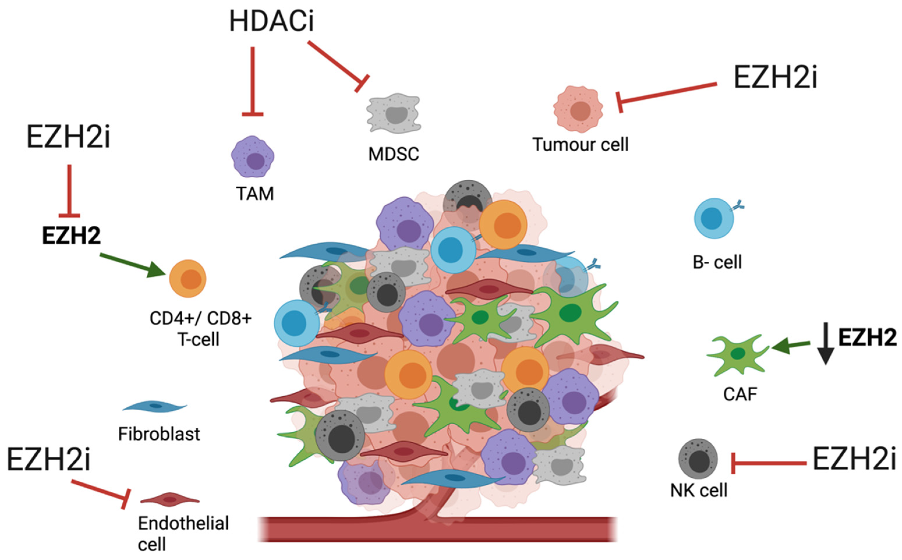
3.2. Impact of HME Therapies on the TME

Our increasing knowledge of the role of the TME in cancer has opened many possible therapeutic avenues. The recent focus in TME-targeted therapies in OAC has been on anti-angiogenic agents such as anti-vascular endothelial growth factor agents (anti-VEGF) [96], immune checkpoint inhibitors such as programmed cell death protein 1 (PD-1) inhibitors [97] and therapies to alter the CAF phenotype such as phosphodiesterase type 5 inhibitors (PDE5i) [88]. Whilst the impact of HME-targeted therapies on the OAC TME is unexplored, there are numerous theoretical targets which have been explored in other diseases (Figure 1).
The relationship between EZH2 and activity of TME constituents appears to be tissue-dependent with both pro- and anti-tumorigenic activity. In colorectal cancer mouse models, EZH2 expression was found upregulated in tumour infiltrating Tregs and was shown to be crucial in the Treg-mediated suppression of anti-cancer immunity [98]. Both pharmacological inhibition and gene knockout of EZH2 in these models led to Treg recruitment of CD8+ and CD4+ effector T-cells, resulting in increased anti-tumour activity [98]. Similarly, pharmacological or genetic disruption of EZH2 activity has been shown to enhance natural killer cell maturation and anti-tumour function, which are often repressed in cancer [99]. Tyan et al. (2012) reported that normal tissue-associated fibroblasts that were co-cultured with breast cancer cells gained persistent activity for promoting cancer cell invasion by fibroblast secretion of ADAM metallopeptidase with thrombospondin type 1 motif (ADAMTS1) [100]. This was thought to be due to an observed reduction in EZH2 binding and subsequent loss of the transcriptionally repressive Histone 3 lysine 27 (H3K27) methylation at the promoter region of ADAMTS1 [100].
Small molecule inhibition of HDAC (Trichostatin A) in melanoma, breast cancer and lung cancer mouse models led to a decreased tumour burden and increased overall survival [101]. When examining the role of T-cells in these observations, HDACi was shown to drive trafficking of T-cells to tumours, potentiating the anti-tumour response through CD4+ and CD8+ effector T-cells, whilst decreasing the presence of immunosuppressive immature myeloid cells [101]. When investigating the effects on tumour-associated macrophage (TAM) phenotypes, HDACi abrogated the pro-tumorigenic functions of TAMs in these mouse models [101]. Further to this, several studies have demonstrated that HDAC inhibition can increase the immunogenicity of tumour cells by up-regulating expression of major histocompatibility complex (MHC) proteins and co-stimulatory molecules such as CD40 on the surface of tumour cells, whilst downregulating the expression of anti-apoptotic proteins [102,103,104].
Given the observed role of HDACs in OAC tumorigenesis and the impact of HDACi on the TME in other cancers, targeted HDAC therapy in OAC may be successful in promoting an anti-tumour response within the OAC TME. Similarly, EZH2 inhibition in OAC may potentiate Treg recruitment of effector T-cells and NK cell maturation to increase this anti-tumour response as seen in other cancer types. Due to the impact of immune cell activity within the TME, combining HME therapies with immunotherapies may be key to improving OAC treatment and will be discussed below.

4. Combination Therapies

Much of the early success with HDACi has been seen in haematological malignancies, but clinical outcomes in solid tumours remain disappointing. The exact reasons for this observation have not been elicited, but many of the HDACi have short half-lives, which may limit their distribution to solid tumours, necessitating improvements in methods of drug delivery [20]. Sodji et al. (2015) demonstrated improved anti-proliferative effects with targeted HDACi therapy in oral and cervical cancer cells overexpressing folate receptors by conjugating HDACi with folic acid [105]. To further improve the efficacy of HDACi and limit toxicity seen in early clinical trials, combination treatments with chemotherapies or other epigenetic modifiers are being explored.
Combination treatment with HME agents and chemotherapies may be effective due to the impact of chemotherapy on the anti-tumour immune response. In immunocompetent mice implanted with subcutaneous Lewis lung cancer cells, GSK126 (EZH2i) treatment led to accumulation of myeloid-derived suppressor cells (MDSC) and decreased numbers of CD4+ T cells and CD8+ T cells, which masked the anti-tumour effects of EZH2i. Administration of either gemcitabine or 5-FU led to depletion of these MDSCs, recovering the activity of effector T-cells and potentiating the GSK126 anti-tumour response [106]. The effect of chemotherapies on depleting MDSCs is likely beneficial in OAC given their increased presence with the TME, which may disrupt the activity of an EZH2i.
The synergistic effects of EZH2i with chemotherapy has also been demonstrated in preclinical studies on non-Hodgkin lymphoma (NHL). Tazemetostat in combination with cyclophosphamide, doxorubicin, oncovin, and prednisolone (CHOP) showed significantly greater cytotoxicity than when given separately [107]. Similarly, combining cisplatin and the HDACi panobinostat decreased cell viability and increased apoptosis compared to monotherapy in hypoxia-induced cisplatin-resistant non-small cell lung cancer cells, indicating synergistic anti-proliferative activity [108].
Current standard of care (SOC) chemotherapy in OAC consists of docetaxel, oxaliplatin, leucovorin and 5-FU (FLOT) [109]. With so few patients (20%) demonstrating a clinically meaningful response to this regime, treatment resistance is a major factor in OAC survival rates [12]. In pre-clinical studies, EZH2 is found to be overexpressed in docetaxel-resistant prostate cancers cells [110], cisplatin-resistant breast cancer cells [111] and 5-FU resistant gastric cancer cells [112]. EZH2 knockdown in all three cancer types and small molecule inhibition in the docetaxel-resistant prostate cancer potentiated cancer cell sensitivity to the chemotherapy agents, suggesting that EZH2 inhibition in combination with FLOT chemotherapy could improve treatment sensitivity in OAC.
Aside from chemotherapies, co-administration of HME drugs in combination with novel agents has been investigated with promising early results. EZH2i and epidermal growth factor inhibitors have shown improved anti-tumour effects in colon cancer and gastric cancer cell lines [113,114]. Combination treatment with HMEi and PD-1 inhibitors may be of particular interest in OAC given the recent results of the CheckMate 577 phase 3 clinical trial in patients with oesophageal or gastroesophageal junction cancer. The trial demonstrated improved disease-free survival for patients receiving adjuvant nivolumab (PD-1 inhibitor) compared to placebo [97]. PD-1 receptors are present on activated T-cells and serve as an immunological checkpoint by downmodulating T-cell effector functions when bound to its ligands (PDL-1 or PDL-2) on antigen-presenting cells [115]. OAC tumour cells and tumour-inflitrating lymphocytes have been shown to highly express both PD-1, PD-L1 and PDL-2 [116]. By inhibiting their interaction, nivolumab has been shown to enhance T-cell effector activity and cytokine production [117].
Whilst HMEi and PD-1 inhibitor combination therapy has not been examined in OAC, evidence from other cancer types is encouraging. In prostate cancer, EZH2 inhibition together with PD-1 inhibition significantly enhanced the anti-tumour response with observed intratumoral trafficking of activated CD8+ T-cells and TAMs [118]. The HDACi vorinostat in combination with the PD-1 inhibitor pembrolizumab has demonstrated synergistic anti-tumour activity in metastatic non-small cell lung cancer [119]. Similarly, triple combination of vorinostat, anti-PD-1 and anti-cytotoxic T-lymphocyte-associated protein 4 (CTLA-4) agents in triple-negative breast cancer mouse models resulted in significant increases in anti-tumour activity and overall survival compared to monotherapy [120]. Given the increased presence of Tregs and reduced activity of effector T-cells typically seen in the OAC TME [88,91], these observations of HMEi and PD-1 inhibitor combination therapy highlight a potential avenue for future OAC research.

5. Future Considerations

The paucity of information surrounding HMEs in OAC reflects our current knowledge of OAC and underlines why it remains a cancer of unmet need. In order to expand our knowledge of HMEs it is essential we collect further omic data from OAC tumours, which should be compared to paired normal tissues, to gain a better understanding of the genetic and epigenetic pathways underlying the disease process. To further research the role of HMEs in the OAC TME and the impact of HME therapies we desperately need improved 3D cell models that can reproducibly recapitulate the TME seen in patients, mimicking the inner necrotic core and outer layer of proliferating cells within a tumour together with the nutrient, oxygen and growth factor gradients [121]. With HME therapies likely impacting the immune environment within the OAC TME, development of humanised mouse models will be key to evaluating the in vivo impact on immune cells surrounding the tumour.

6. Conclusions

Inhibitors of HMEs are already used in clinical practice for a variety of haematological malignancies, with encouraging results seen in numerous ongoing clinical trials. Evidence supports a role of HMEs in oesophageal cancer tumorigenesis; however, less is known about their role and mechanism in the cancer subtype OAC. Early research from in vitro studies on HME therapies in OAC, particularly HDACi, have been promising, with the identification of several novel candidate biomarkers of response. Broadening research to include other HME therapies such as methyltransferase inhibitors may be fruitful. Given observations of poor results seen with HMEi used in isolation in solid tumours, it is likely that the best therapeutic responses will be seen when using them in combination therapies.
Given the significant morbidity and mortality associated with OAC, new treatments are urgently needed. Further research to elicit the underlying epigenetic pathways and characterisation of the TME in OAC is essential to drive the search for epigenetic-based therapies.

Author Contributions

Conceptualization, O.J.P., S.P.B. and Z.S.W.; writing—original draft preparation, O.J.P.; writing—review and editing, S.P.B., Z.S.W. and T.J.U. All authors have read and agreed to the published version of the manuscript.

Funding

This research was funded by Cancer Research UK & Royal College of Surgeons of England Advanced Clinician Scientist Fellowship to T.J.U. “Cellular interplay in oesophageal cancer.” A23924.

Conflicts of Interest

The authors declare no conflict of interest.

Abbreviations

3D-TGA3 dimensional-tumour growth assay
ADAMTS1ADAM metallopeptidase with thrombospondin type 1 motif
ARID1AAT-Rich Interaction Domain 1A
AZAAzacytidine
BETBromodomain and Extra- Terminal motif protein
CAFCancer-associated fibroblast
CTCLPrimary cutaneous T-cell lymphoma
CTLA-4Cytotoxic T-lymphocyte-associated protein 4
DACDecitabine
DNMTDNA methyltransferase
DNMTiDNA methyltransferase inhibitors
ECFEpiribucin, cisplatin and fluorouracil
EZH2Enhancer of zeste homolog 2
EZH2iEZH2 inhibitor
FDAUS Food and Drug Administration
FLOTDocetaxel, oxaliplatin, leucovorin and 5-FU
H3K27Histone 3 lysine 27
HATHistone acetyltransferase
HDACHistone deacetylase
HDACiHistone deacetylase inhibitor
HDMHistone demethylases
HMEHistone modifying enzyme
HMEiHME inhibitors
HMTHistone methyltransferases
MTA1Metastasis-associated gene 1
MDSCMyeloid-derived suppressor cells
NK cell Natural killer cell
OACOesophageal adenocarcinoma
OCCCOvarian clear cell carcinoma
OSCCOesophageal squamous cell carcinoma
PD-1Programmed cell death protein 1
PDE5iPhosphodiesterase type 5 inhibitors
PRC2Polycomb Repressive Complex 2
PTCLPeripheral T-cell lymphoma
qRT-PCRReal-Time Quantitative Reverse Transcription PCR)
SAMS-adenosyl-L-methionine
SMARCA4SWI/SNF Related, Matrix Associated, Actin Dependent Regulator Of Chromatin, Subfamily A, Member 4
SOCStandard of care
SWI/SNFSWItch/Sucrose Non-Fermentable complex
TAMTumour-associated macrophages
TGF-βTransforming growth factor-β
TMETumour microenvironment
TregsRegulatory T-cells
TXNIPThioredoxin Interacting Protein
VEGFVascular endothelial growth factor

References

  1. Bray, F.; Ferlay, J.; Soerjomataram, I.; Siegel, R.L.; Torre, L.A.; Jemal, A. Global cancer statistics 2018: GLOBOCAN estimates of incidence and mortality worldwide for 36 cancers in 185 countries. CA Cancer J. Clin. 2018, 68, 394–424. [Google Scholar] [CrossRef] [PubMed] [Green Version]
  2. Dubecz, A.; Gall, I.; Solymosi, N.; Schweigert, M.; Peters, J.H.; Feith, M.; Stein, H.J. Temporal Trends in Long-Term Survival and Cure Rates in Esophageal Cancer: A SEER Database Analysis. J. Thorac. Oncol. 2012, 7, 443–447. [Google Scholar] [CrossRef] [Green Version]
  3. Gavin, A.T.; Francisci, S.; Foschi, R.; Donnelly, D.; Lemmens, V.; Brenner, H.; Anderson, L. Oesophageal cancer survival in Europe: A EUROCARE-4 study. Cancer Epidemiol. 2012, 36, 505–512. [Google Scholar] [CrossRef]
  4. Kauppila, J.H.; Mattsson, F.; Brusselaers, N.; Lagergren, J. Prognosis of oesophageal adenocarcinoma and squamous cell carcinoma following surgery and no surgery in a nationwide Swedish cohort study. BMJ Open 2018, 8, 21495. [Google Scholar] [CrossRef] [PubMed]
  5. Zhao, J.; He, Y.T.; Zheng, R.S.; Zhang, S.W.; Chen, W.Q. Analysis of esophageal cancer time trends in china, 1989–2008. Asian Pac. J. Cancer Prev. 2012, 13, 4613–4617. [Google Scholar] [CrossRef] [Green Version]
  6. Cook, M.B.; Chow, W.H.; Devesa, S.S. Oesophageal cancer incidence in the United States by race, sex, and histologic type, 1977–2005. Br. J. Cancer 2009, 101, 855–859. [Google Scholar] [CrossRef] [PubMed] [Green Version]
  7. Smyth, E.C.; Lagergren, J.; Fitzgerald, R.C.; Lordick, F.; Shah, M.A.; Lagergren, P.; Cunningham, D. Oesophageal cancer. Nat. Rev. Dis. Prim. 2017, 3, 17048. [Google Scholar] [CrossRef] [PubMed]
  8. Bosetti, C.; Levi, F.; Ferlay, J.; Garavello, W.; Lucchini, F.; Bertuccio, P.; Negri, E.; La Vecchia, C. Trends in oesophageal cancer incidence and mortality in Europe. Int. J. Cancer 2008, 122, 1118–1129. [Google Scholar] [CrossRef]
  9. Arnold, M.; Laversanne, M.; Brown, L.M.; Devesa, S.S.; Bray, F. Predicting the Future Burden of Esophageal Cancer by Histological Subtype: International Trends in Incidence up to 2030. Am. J. Gastroenterol. 2017, 112, 1247–1255. [Google Scholar] [CrossRef]
  10. Oesophago-Gastric Cancer Audit. Available online: https://digital.nhs.uk/data-and-information/clinical-audits-and-registries/national-oesophago-gastric-cancer-audit (accessed on 27 June 2021).
  11. Agarwal, A.; Polineni, R.; Hussein, Z.; Vigoda, I.; Bhagat, T.D.; Bhattacharyya, S.; Maitra, A.; Verma, A. Role of epigenetic alterations in the pathogenesis of Barrett’s esophagus and esophageal adenocarcinoma. Int. J. Clin. Exp. Pathol. 2012, 5, 382–396. [Google Scholar] [PubMed]
  12. Noble, F.; Lloyd, M.A.; Turkington, R.; Griffiths, E.; O’Donovan, M.; O’Neill, J.R.; Mercer, S.; Parsons, S.L.; Fitzgerald, R.C.; Underwood, T.J.; et al. Multicentre cohort study to define and validate pathological assessment of response to neoadjuvant therapy in oesophagogastric adenocarcinoma. Br. J. Surg. 2017, 104, 1816–1828. [Google Scholar] [CrossRef] [Green Version]
  13. Dupont, C.; Armant, D.R.; Brenner, C.A. Epigenetics: Definition, mechanisms and clinical perspective. Semin. Reprod. Med. 2009, 27, 351–357. [Google Scholar] [CrossRef] [Green Version]
  14. Sharma, S.; Kelly, T.K.; Jones, P.A. Epigenetics in cancer. Carcinogenesis 2009, 31, 27–36. [Google Scholar] [CrossRef] [PubMed]
  15. Margueron, R.; Trojer, P.; Reinberg, D. The key to development: Interpreting the histone code? Curr. Opin. Genet. Dev. 2005, 15, 163–176. [Google Scholar] [CrossRef] [PubMed]
  16. Nebbioso, A.; Tambaro, F.P.; Dell’Aversana, C.; Altucci, L. Cancer epigenetics: Moving forward. PLoS Genet. 2018, 14, e1007362. [Google Scholar] [CrossRef] [PubMed] [Green Version]
  17. Chen, Q.W.; Zhu, X.Y.; Li, Y.Y.; Meng, Z.Q. Epigenetic regulation and cancer (review). Oncol. Rep. 2014, 31, 523–532. [Google Scholar] [CrossRef] [Green Version]
  18. Salta, S.; Macedo-Silva, C.; Miranda-Gonçalves, V.; Lopes, N.; Gigliano, D.; Guimarães, R.; Farinha, M.; Sousa, O.; Henrique, R.; Jerónimo, C. A DNA methylation-based test for esophageal cancer detection. Biomark Res. 2020, 8, 1–9. [Google Scholar] [CrossRef]
  19. Wu, D.-L.; Sui, F.-Y.; Jiang, X.-M.; Jiang, X.-H. Methylation in esophageal carcinogenesis. World J. Gastroenterol. 2006, 12, 6933–6940. [Google Scholar] [CrossRef]
  20. Li, Y.; Seto, E. HDACs and HDAC inhibitors in cancer development and therapy. Cold Spring Harb. Perspect. Med. 2016, 6, a026831. [Google Scholar] [CrossRef] [PubMed] [Green Version]
  21. US Food and Drug Administration. FDA Approves First Treatment Option Specifically for Patients with Epithelioid Sarcoma, a Rare Soft Tissue Cancer. 2020. Available online: https://www.fda.gov/news-events/press-announcements/fda-approves-first-treatment-option-specifically-patients-epithelioid-sarcoma-rare-soft-tissue (accessed on 27 June 2021).
  22. US Food and Drug Administration. FDA Granted Accelerated Approval to Tazemetostat for Follicular Lymphoma|FDA. Available online: https://www.fda.gov/drugs/fda-granted-accelerated-approval-tazemetostat-follicular-lymphoma (accessed on 29 January 2021).
  23. Mann, B.S.; Johnson, J.R.; Cohen, M.H.; Justice, R.; Pazdur, R. FDA approval summary: Vorinostat for treatment of advanced primary cutaneous T-cell lymphoma. Oncologist 2007, 12, 1247–1252. [Google Scholar] [CrossRef]
  24. McDermott, J.; Jimeno, A. Belinostat for the treatment of peripheral T-cell lymphomas. Drugs Today 2014, 50, 337–345. [Google Scholar] [CrossRef]
  25. Richardson, P.G.; Laubach, J.P.; Lonial, S.; Moreau, P.; Yoon, S.-S.; Hungria, V.T.; Dimopoulos, M.; Beksac, M.; Alsina, M.; San-Miguel, J.F. Panobinostat: A novel pan-deacetylase inhibitor for the treatment of relapsed or relapsed and refractory multiple myeloma. Expert Rev. Anticancer. Ther. 2015, 15, 737–748. [Google Scholar] [CrossRef] [PubMed]
  26. Frye, R.; Myers, M.; Axelrod, K.C.; Ness, E.A.; Piekarz, R.L.; Bates, S.E.; Booher, S. Romidepsin: A new drug for the treatment of cutaneous T-cell lymphoma. Clin. J. Oncol. Nurs. 2012, 16, 195–204. [Google Scholar] [CrossRef] [PubMed] [Green Version]
  27. Bannister, A.J.; Kouzarides, T. Regulation of chromatin by histone modifications. Cell Res. 2011, 21, 381–395. [Google Scholar] [CrossRef]
  28. Vaquero, A.; Loyola, A.; Reinberg, D. The constantly changing face of chromatin. Sci. Aging Knowl. Environ. 2003, 2003, 4. [Google Scholar] [CrossRef] [Green Version]
  29. Cheng, Y.; He, C.; Wang, M.; Ma, X.; Mo, F.; Yang, S.; Han, J.; Wei, X. Targeting epigenetic regulators for cancer therapy: Mechanisms and advances in clinical trials. Signal. Transduct Target. Ther. 2019, 4, 62. [Google Scholar] [CrossRef] [PubMed] [Green Version]
  30. Zhao, Z.; Shilatifard, A. Epigenetic modifications of histones in cancer. Genome Biol. 2019, 20, 1–16. [Google Scholar] [CrossRef] [PubMed]
  31. Tessarz, P.; Kouzarides, T. Histone core modifications regulating nucleosome structure and dynamics. Nat. Rev. Mol. Cell Biol. 2014, 15, 703–708. [Google Scholar] [CrossRef] [PubMed]
  32. Polo, S.E.; Almouzni, G. Histone metabolic pathways and chromatin assembly factors as proliferation markers. Cancer Lett. 2005, 220, 1–9. [Google Scholar] [CrossRef] [PubMed]
  33. Lohse, I.; Al-Ali, H.; Volmar, C.-H.; Trotta, A.D.A.; Brothers, S.P.; Capobianco, A.J.; Wahlestedt, C. Ex vivo drug sensitivity testing as a means for drug repurposing in esophageal adenocarcinoma. PLoS ONE 2018, 13, e0203173. [Google Scholar] [CrossRef] [PubMed]
  34. Saunders, J.H.; Onion, D.; Collier, P.; Dorrington, M.S.; Argent, R.H.H.; Clarke, P.; Reece-Smith, A.M.; Parsons, S.L.; Grabowska, A.M. Individual patient oesophageal cancer 3D models for tailored treatment. Oncotarget 2017, 8, 24224–24236. [Google Scholar] [CrossRef] [PubMed] [Green Version]
  35. Ahrens, T.D.; Timme, S.; Hoeppner, J.; Ostendorp, J.; Hembach, S.; Follo, M.; Hopt, U.T.; Werner, M.; Busch, H.; Boerries, M.; et al. Selective inhibition of esophageal cancer cells by combination of HDAC inhibitors and Azacytidine. Epigenetics 2015, 10, 431–445. [Google Scholar] [CrossRef] [PubMed] [Green Version]
  36. Feingold, P.L.; Surman, D.R.; Brown, K.; Xu, Y.; McDuffie, L.A.; Shukla, V.; Reardon, E.S.; Crooks, D.R.; Trepel, J.B.; Lee, S.; et al. Induction of Thioredoxin-Interacting Protein by a Histone Deacetylase Inhibitor, Entinostat, Is Associated with DNA Damage and Apoptosis in Esophageal Adenocarcinoma. Mol. Cancer Ther. 2018, 17, 2013–2023. [Google Scholar] [CrossRef] [PubMed] [Green Version]
  37. Kofonikolas, K.; Frankell, A.M.; Smyth, E.C.; Fitzgerald, R.C. Examining the effects of BET, HDAC2, and EZH2 inhibition on esophageal adenocarcinoma (EAC) cell line proliferation. J. Clin. Oncol. 2019, 37 (Suppl. S4), 68. [Google Scholar] [CrossRef]
  38. Miwa, K.; Segawa, M.; Takano, Y.; Matsumoto, H.; Sahara, H.; Yagi, M.; Miyazaki, I.; Hattori, T. Induction of oesophageal and forestomach carcinomas in rats by reflux of duodenal contents. Br. J. Cancer 1994, 70, 185–189. [Google Scholar] [CrossRef] [PubMed] [Green Version]
  39. Miwa, K.; Sahara, H.; Segawa, M.; Kinami, S.; Sato, T.; Miyazaki, I.; Hattori, T. Reflux of duodenal or gastro-duodenal contents induces esophageal carcinoma in rats. Int J. Cancer 1996, 67, 269–274. [Google Scholar] [CrossRef]
  40. Miyashita, T.; Ohta, T.; Fujimura, T.; Ninomiya, I.; Fushida, S.; Hattori, T.; Miwa, K. Duodenal juice stimulates oesophageal stem cells to induce Barrett’s oesophagus and oesophageal adenocarcinoma in rats. Oncol. Rep. 2006, 15, 1469–1475. [Google Scholar] [CrossRef] [PubMed] [Green Version]
  41. Miyashita, T.; Tajima, H.; Munemoto, M.; Shah, F.A.; Harmon, J.W.; Watanabe, T.; Shoji, M.; Okamoto, K.; Nakanuma, S.; Sakai, S.; et al. Impact of histone deacetylase 1 and metastasis-associated gene 1 expression in esophageal carcinogenesis. Oncol. Lett. 2014, 8, 758–764. [Google Scholar] [CrossRef]
  42. Nicolson, G.L.; Nawa, A.; Toh, Y.; Taniguchi, S.; Nishimori, K.; Moustafa, A. Tumor metastasis-associated human MTA1 gene and its MTA1 protein product: Role in epithelial cancer cell invasion, proliferation and nuclear regulation. Clin. Exp. Metastasis 2003, 20, 19–24. [Google Scholar] [CrossRef]
  43. Moon, H.-E.; Cheon, H.; Lee, M.-S. Metastasis-associated protein 1 inhibits p53-induced apoptosis. Oncol. Rep. 2007, 18, 1311–1314. [Google Scholar] [CrossRef] [PubMed] [Green Version]
  44. Luo, J.; Su, F.; Chen, D.; Shiloh, A.; Gu, W. Deacetylation of p53 modulates its effect on cell growth and apoptosis. Nature 2000, 408, 377–381. [Google Scholar] [CrossRef]
  45. Frankell, A.M.; Jammula, S.; Li, X.; Contino, G.; Killcoyne, S.; Abbas, S.; Perner, J.; Perner, J.; Bower, L.; Devonshire, G.; et al. The landscape of selection in 551 esophageal adenocarcinomas defines genomic biomarkers for the clinic. Nat. Genet. 2019, 51, 506–516. [Google Scholar] [CrossRef]
  46. Langer, R.; Mutze, K.; Becker, K.; Feith, M.; Ott, K.; Höfler, H.; Keller, G. Expression of class I histone deacetylases (HDAC1 and HDAC2) in oesophageal adenocarcinomas: An immunohistochemical study. J. Clin. Pathol. 2010, 63, 994–998. [Google Scholar] [CrossRef] [PubMed] [Green Version]
  47. Nagy, Á.; Munkácsy, G.; Győrffy, B. Pancancer survival analysis of cancer hallmark genes. Sci. Rep. 2021, 11, 6047. [Google Scholar] [CrossRef] [PubMed]
  48. Cao, R.; Wang, L.; Wang, H.; Xia, L.; Erdjument-Bromage, H.; Tempst, P.; Jones, R.S.; Zhang, Y. Role of Histone H3 Lysine 27 Methylation in Polycomb-Group Silencing. Science 2002, 298, 1039–1043. [Google Scholar] [CrossRef] [PubMed] [Green Version]
  49. Suzuki, J.; Chen, Y.-Y.; Scott, G.K.; Devries, S.; Chin, K.; Benz, C.C.; Waldman, F.M.; Hwang, E.S. Protein Acetylation and Histone Deacetylase Expression Associated with Malignant Breast Cancer Progression. Clin. Cancer Res. 2009, 15, 3163–3171. [Google Scholar] [CrossRef] [PubMed] [Green Version]
  50. Toh, Y.; Yamamoto, M.; Endo, K.; Ikeda, Y.; Baba, H.; Kohnoe, S.; Yonemasu, H.; Hachitanda, Y.; Okamura, T.; Sugimachi, K. Histone H4 acetylation and histone deacetylase 1 expression in esophageal squamous cell carcinoma. Oncol. Rep. 2003, 10, 333–338. [Google Scholar] [CrossRef] [PubMed]
  51. Wang, J.; Elahi, A.; Ajidahun, A.; Clark, W.; Hernandez, J.; Achille, A.; Hao, J.-H.; Seto, E.; Shibata, D. The interplay between histone deacetylases and c-Myc in the transcriptional suppression of HPP1 in colon cancer. Cancer Biol. Ther. 2014, 15, 1198–1207. [Google Scholar] [CrossRef] [Green Version]
  52. Zhu, P.; Martin, E.; Mengwasser, J.; Schlag, P.; Janssen, K.-P.; Göttlicher, M. Induction of HDAC2 expression upon loss of APC in colorectal tumorigenesis. Cancer Cell 2004, 5, 455–463. [Google Scholar] [CrossRef] [Green Version]
  53. Raetz, E.A.; Kim, M.K.H.; Moos, P.; Carlson, M.; Bruggers, C.; Hooper, D.K.; Foot, L.; Liu, T.; Seeger, R.; Carroll, W.L. Identification of genes that are regulated transcriptionally by Myc in childhood tumors. Cancer 2003, 98, 841–853. [Google Scholar] [CrossRef]
  54. Meyer, N.; Penn, L. Reflecting on 25 years with MYC. Nat. Rev. Cancer 2008, 8, 976–990. [Google Scholar] [CrossRef] [PubMed]
  55. Doroshow, D.B.; Eder, J.P.; LoRusso, P.M. BET inhibitors: A novel epigenetic approach. Ann. Oncol. 2017, 28, 1776–1787. [Google Scholar] [CrossRef]
  56. Mertz, J.A.; Conery, A.R.; Bryant, B.; Sandy, P.; Balasubramanian, S.; Mele, D.A.; Bergeron, L.; Sims, R.J. Targeting MYC dependence in cancer by inhibiting BET bromodomains. Proc. Natl. Acad. Sci. USA 2011, 108, 16669–16674. [Google Scholar] [CrossRef] [PubMed] [Green Version]
  57. Delmore, J.E.; Issa, G.C.; Lemieux, M.; Rahl, P.B.; Shi, J.; Jacobs, H.M.; Kastritis, E.; Gilpatrick, T.; Paranal, R.M.; Qi, J.; et al. BET Bromodomain Inhibition as a Therapeutic Strategy to Target c-Myc. Cell 2011, 146, 904–917. [Google Scholar] [CrossRef] [Green Version]
  58. Nebbioso, A.; Carafa, V.; Conte, M.; Tambaro, F.P.; Abbondanza, C.; Martens, J.; Nees, M.; Benedetti, R.; Pallavicini, I.; Minucci, S.; et al. c-Myc Modulation and Acetylation Is a Key HDAC Inhibitor Target in Cancer. Clin. Cancer Res. 2017, 23, 2542–2555. [Google Scholar] [CrossRef] [Green Version]
  59. Von Rahden, B.H.A.; Stein, H.J.; Pühringer-Oppermann, F.; Sarbia, M. c-myc Amplification Is Frequent in Esophageal Adenocarcinoma and Correlated with the Upregulation of VEGF-A Expression. Neoplasia 2006, 8, 702–707. [Google Scholar] [CrossRef] [Green Version]
  60. Tselepis, C.; Morris, C.D.; Wakelin, D.; Hardy, R.; Perry, I.; Luong, Q.T.; Harper, E.; Harrison, R.; Attwood, S.E.A.; Jankowski, J. Upregulation of the oncogene c-myc in Barrett’s adenocarcinoma: Induction of c-myc by acidified bile acid in vitro. Gut 2003, 52, 174–180. [Google Scholar] [CrossRef] [PubMed]
  61. Minami, J.; Suzuki, R.; Mazitschek, R.; Gorgun, G.; Ghosh, B.; Cirstea, D.; Hu, Y.; Mimura, N.; Ohguchi, H.; Cottini, F.; et al. Histone deacetylase 3 as a novel therapeutic target in multiple myeloma. Leukemia 2013, 28, 680–689. [Google Scholar] [CrossRef] [PubMed] [Green Version]
  62. Stronach, E.A.; Alfraidi, A.; Rama, N.; Datler, C.; Studd, J.B.; Agarwal, R.; Guney, T.G.; Gourley, C.; Hennessy, B.T.; Mills, G.B.; et al. HDAC4-Regulated STAT1 Activation Mediates Platinum Resistance in Ovarian Cancer. Cancer Res. 2011, 71, 4412–4422. [Google Scholar] [CrossRef] [PubMed] [Green Version]
  63. Marek, L.; Hamacher, A.; Hansen, F.K.; Kuna, K.; Gohlke, H.; Kassack, M.U.; Kurz, T. Histone Deacetylase (HDAC) Inhibitors with a Novel Connecting Unit Linker Region Reveal a Selectivity Profile for HDAC4 and HDAC5 with Improved Activity against Chemoresistant Cancer Cells. J. Med. Chem. 2013, 56, 427–436. [Google Scholar] [CrossRef] [PubMed]
  64. Balasubramanian, S.; Ramos, J.; Luo, W.; Sirisawad, M.; Verner, E.; Buggy, J.J. A novel histone deacetylase 8 (HDAC8)-specific inhibitor PCI-34051 induces apoptosis in T-cell lymphomas. Leukemia 2008, 22, 1026–1034. [Google Scholar] [CrossRef]
  65. Kaadige, M.R.; Looper, R.E.; Kamalanaadhan, S.; Ayer, D.E. Glutamine-dependent anapleurosis dictates glucose uptake and cell growth by regulating MondoA transcriptional activity. Proc. Natl. Acad. Sci. USA 2009, 106, 14878–14883. [Google Scholar] [CrossRef] [PubMed] [Green Version]
  66. Chen, R.; Zhang, M.; Zhou, Y.; Guo, W.; Yi, M.; Zhang, Z.; Ding, Y.; Wang, Y. The application of histone deacetylases inhibitors in glioblastoma. J. Exp. Clin. Cancer Res. 2020, 39, 1–18. [Google Scholar] [CrossRef] [PubMed]
  67. Eckschlager, T.; Plch, J.; Stiborova, M.; Hrabeta, J. Histone Deacetylase Inhibitors as Anticancer Drugs. Int. J. Mol. Sci. 2017, 18, 1414. [Google Scholar] [CrossRef] [PubMed]
  68. Herz, H.-M.; Garruss, A.; Shilatifard, A. SET for life: Biochemical activities and biological functions of SET domain-containing proteins. Trends Biochem. Sci. 2013, 38, 621–639. [Google Scholar] [CrossRef] [Green Version]
  69. Santos-Rosa, H.; Schneider, R.; Bannister, A.; Sherriff, J.; Bernstein, B.E.; Emre, T.; Schreiber, S.L.; Mellor, J.; Kouzarides, T. Active genes are tri-methylated at K4 of histone H3. Nature 2002, 419, 407–411. [Google Scholar] [CrossRef] [PubMed]
  70. Lehnertz, B.; Ueda, Y.; Derijck, A.A.; Braunschweig, U.; Perez-Burgos, L.; Kubicek, S.; Chen, T.; Li, E.; Jenuwein, T.; Peters, A.H. Suv39h-Mediated Histone H3 Lysine 9 Methylation Directs DNA Methylation to Major Satellite Repeats at Pericentric Heterochromatin. Curr. Biol. 2003, 13, 1192–1200. [Google Scholar] [CrossRef] [Green Version]
  71. Bernstein, B.E.; Mikkelsen, T.S.; Xie, X.; Kamal, M.; Huebert, D.J.; Cuff, J.; Fry, B.; Meissner, A.; Wernig, M.; Plath, K.; et al. A Bivalent Chromatin Structure Marks Key Developmental Genes in Embryonic Stem Cells. Cell 2006, 125, 315–326. [Google Scholar] [CrossRef] [PubMed] [Green Version]
  72. Tang, L.; Nogales, E.; Ciferri, C. Structure and function of SWI/SNF chromatin remodeling complexes and mechanistic implications for transcription. Prog. Biophys. Mol. Biol. 2010, 102, 122–128. [Google Scholar] [CrossRef] [PubMed] [Green Version]
  73. Kadoch, C.; Hargreaves, D.C.; Hodges, C.; Elias, L.; Ho, L.; Ranish, J.; Crabtree, G.R. Proteomic and bioinformatic analysis of mammalian SWI/SNF complexes identifies extensive roles in human malignancy. Nat. Genet. 2013, 45, 592–601. [Google Scholar] [CrossRef]
  74. Mittal, P.; Roberts, C.W.M. The SWI/SNF complex in cancer—Biology, biomarkers and therapy. Nat. Rev. Clin. Oncol. 2020, 17, 435–448. [Google Scholar] [CrossRef]
  75. Eich, M.-L.; Athar, M.; Ferguson, J.E.; Varambally, S. EZH2-Targeted Therapies in Cancer: Hype or a Reality. Cancer Res. 2020, 80, 5449–5458. [Google Scholar] [CrossRef] [PubMed]
  76. Bitler, B.G.; Aird, K.M.; Garipov, A.; Li, H.; Amatangelo, M.; Kossenkov, A.V.; Schultz, D.C.; Liu, Q.; Shih, I.-M.; Conejo-Garcia, J.; et al. Synthetic lethality by targeting EZH2 methyltransferase activity in ARID1A-mutated cancers. Nat. Med. 2015, 21, 231–238. [Google Scholar] [CrossRef] [PubMed] [Green Version]
  77. Rugo, H.S.; Jacobs, I.; Sharma, S.; Scappaticci, F.; Paul, T.A.; Jensen-Pergakes, K.; Malouf, G.G. The Promise for Histone Methyltransferase Inhibitors for Epigenetic Therapy in Clinical Oncology: A Narrative Review. Adv. Ther. 2020, 37, 3059–3082. [Google Scholar] [CrossRef] [PubMed]
  78. Zauderer, M.G.; Szlosarek, P.; Le Moulec, S.; Popat, S.; Taylor, P.; Planchard, D.; Scherpereel, A.; Jahan, T.; Koczywas, M.; Forster, M.; et al. Phase 2, multicenter study of the EZH2 inhibitor tazemetostat as monotherapy in adults with relapsed or refractory (R/R) malignant mesothelioma (MM) with BAP1 inactivation. J. Clin. Oncol. 2018, 36 (Suppl. S15), 8515. [Google Scholar] [CrossRef]
  79. Morschhauser, F.; Tilly, H.; Chaidos, A.; Phillips, T.; Ribrag, V.; Campbell, P.; Laurent, D.G.; Jurczak, W.; McKay, P.; Opat, S.; et al. Interim Update From A Phase 2 Multicenter Study Of Tazemetostat, an Ezh2 Inhibitor, In Patients with Relapsed or Refractory Follicular Lymphoma. Hematol. Oncol. 2019, 37, 154–156. [Google Scholar] [CrossRef] [Green Version]
  80. Yap, T.A.; Johnson, P.W.M.; Winter, J.; Leonard, J.; Giulino-Roth, L.; Horner, T.; Radswillas, K.; Carver, J.; Dhar, A. A phase I, open-label study of GSK2816126, an enhancer of zeste homolog 2 (EZH2) inhibitor, in patients with relapsed/refractory diffuse large B-cell lymphoma (DLBCL), transformed follicular lymphoma (tFL), other non-Hodgkin’s lymphomas (NHL), multiple myeloma (MM) and solid tumor. J. Clin. Oncol. 2016, 34 (Suppl. S15), TPS2595. [Google Scholar] [CrossRef]
  81. McCabe, M.T.; Ott, H.M.; Ganji, G.; Korenchuk, S.; Thompson, C.; Van Aller, G.S.; Liu, Y.; Graves, A.P.; Iii, A.D.P.; Diaz, E.; et al. EZH2 inhibition as a therapeutic strategy for lymphoma with EZH2-activating mutations. Nature 2012, 492, 108–112. [Google Scholar] [CrossRef] [PubMed]
  82. Wu, S.; Fatkhutdinov, N.; Fukumoto, T.; Bitler, B.; Park, P.H.; Kossenkov, A.V.; Trizzino, M.; Tang, H.-Y.; Zhang, L.; Gardini, A.; et al. SWI/SNF catalytic subunits’ switch drives resistance to EZH2 inhibitors in ARID1A-mutated cells. Nat. Commun. 2018, 9, 4116. [Google Scholar] [CrossRef] [Green Version]
  83. Baghban, R.; Roshangar, L.; Jahanban-Esfahlan, R.; Seidi, K.; Ebrahimi-Kalan, A.; Jaymand, M.; Kolahian, S.; Javaheri, T.; Zare, P. Tumor microenvironment complexity and therapeutic implications at a glance. Cell Commun. Signal. 2020, 18, 1–19. [Google Scholar] [CrossRef] [Green Version]
  84. Anderson, N.M.; Simon, M.C. The tumor microenvironment. Curr. Biol. 2020, 30, R921–R925. [Google Scholar] [CrossRef]
  85. Hanahan, D.; Coussens, L.M. Accessories to the crime: Functions of cells recruited to the tumor microenvironment. Cancer Cell 2012, 21, 309–322. [Google Scholar] [CrossRef] [PubMed] [Green Version]
  86. Dvorak, K.; Dvorak, B. Role of interleukin-6 in Barrett’s esophagus pathogenesis. World J. Gastroenterol. 2013, 19, 2307–2312. [Google Scholar] [CrossRef]
  87. Dvorak, K.; Chavarria, M.; Payne, C.M.; Ramsey, L.; Crowley-Weber, C.; Dvorakova, B.; Dvorak, B.; Bernstein, H.; Holubec, H.; Sampliner, R.E.; et al. Activation of the Interleukin-6/STAT3 Antiapoptotic Pathway in Esophageal Cells by Bile Acids and Low pH: Relevance to Barrett’s Esophagus. Clin. Cancer Res. 2007, 13 Pt 1, 5305–5313. [Google Scholar] [CrossRef] [PubMed] [Green Version]
  88. Lin, E.W.; Karakasheva, T.A.; Hicks, P.D.; Bass, A.J.; Rustgi, A.K. The tumor microenvironment in esophageal cancer. Oncogene 2016, 35, 5337–5349. [Google Scholar] [CrossRef] [PubMed]
  89. Kalluri, R.; Zeisberg, M. Fibroblasts in cancer. Nat. Rev. Cancer 2006, 6, 392–401. [Google Scholar] [CrossRef]
  90. Underwood, T.J.; Hayden, A.L.; Derouet, M.; Garcia, E.; Noble, F.; White, M.; Thirdborough, S.; Mead, A.; Clemons, N.; Mellone, M.; et al. Cancer-associated fibroblasts predict poor outcome and promote periostin-dependent invasion in oesophageal adenocarcinoma. J. Pathol. 2015, 235, 466–477. [Google Scholar] [CrossRef]
  91. Ichihara, F.; Kono, K.; Takahashi, A.; Kawaida, H.; Sugai, H.; Fujii, H. Increased populations of regulatory T cells in peripheral blood and tumor-infiltrating lymphocytes in patients with gastric and esophageal cancers. Clin. Cancer Res. 2003, 9, 4404–4408. [Google Scholar] [PubMed]
  92. Jayaraman, P.; Parikh, F.; Lopez-Rivera, E.; Hailemichael, Y.; Clark, A.; Ma, G.; Cannan, D.; Ramacher, M.; Kato, M.; Overwijk, W.W.; et al. Tumor-Expressed Inducible Nitric Oxide Synthase Controls Induction of Functional Myeloid-Derived Suppressor Cells through Modulation of Vascular Endothelial Growth Factor Release. J. Immunol. 2012, 188, 5365–5376. [Google Scholar] [CrossRef]
  93. Ostrand-Rosenberg, S.; Sinha, P. Myeloid-Derived Suppressor Cells: Linking Inflammation and Cancer. J. Immunol. 2009, 182, 4499–4506. [Google Scholar] [CrossRef]
  94. Mazzoni, A.; Bronte, V.; Visintin, A.; Spitzer, J.H.; Apolloni, E.; Serafini, P.; Zanovello, P.; Segal, D.M. Myeloid Suppressor Lines Inhibit T Cell Responses by an NO-Dependent Mechanism. J. Immunol. 2002, 168, 689–695. [Google Scholar] [CrossRef] [PubMed]
  95. Liu, C.; Yu, S.; Kappes, J.; Wang, J.; Grizzle, W.E.; Zinn, K.; Zhang, H.-G. Expansion of spleen myeloid suppressor cells represses NK cell cytotoxicity in tumor-bearing host. Blood 2007, 109, 4336–4342. [Google Scholar] [CrossRef] [PubMed]
  96. Butters, O.; Young, K.; Cunningham, D.; Chau, I.; Starling, N. Targeting Vascular Endothelial Growth Factor in Oesophagogastric Cancer: A Review of Progress to Date and Immunotherapy Combination Strategies. Front. Oncol. 2019, 9, 618. [Google Scholar] [CrossRef]
  97. Kelly, R.J.; Ajani, J.A.; Kuzdzal, J.; Zander, T.; Van Cutsem, E.; Piessen, G.; Mendez, G.; Feliciano, J.; Motoyama, S.; Lièvre, A.; et al. Adjuvant Nivolumab in Resected Esophageal or Gastroesophageal Junction Cancer. N. Engl. J. Med. 2021, 384, 1191–1203. [Google Scholar] [CrossRef]
  98. Wang, D.; Quiros, J.; Mahuron, K.; Pai, C.-C.; Ranzani, V.; Young, A.; Silveria, S.; Harwin, T.; Abnousian, A.; Pagani, M.; et al. Targeting EZH2 Reprograms Intratumoral Regulatory T Cells to Enhance Cancer Immunity. Cell Rep. 2018, 23, 3262–3274. [Google Scholar] [CrossRef] [PubMed]
  99. Yin, J.; Leavenworth, J.W.; Li, Y.; Luo, Q.; Xie, H.; Liu, X.; Huang, S.; Yan, H.; Fu, Z.; Zhang, L.Y.; et al. Ezh2 regulates differentiation and function of natural killer cells through histone methyltransferase activity. Proc. Natl. Acad. Sci. USA 2015, 112, 15988–15993. [Google Scholar] [CrossRef] [PubMed] [Green Version]
  100. Tyan, S.-W.; Hsu, C.-H.; Peng, K.-L.; Chen, C.-C.; Kuo, W.-H.; Lee, E.Y.-H.P.; Shew, J.-Y.; Chang, K.-J.; Juan, L.-J.; Lee, W.-H. Breast Cancer Cells Induce Stromal Fibroblasts to Secrete ADAMTS1 for Cancer Invasion through an Epigenetic Change. PLoS ONE 2012, 7, e35128. [Google Scholar] [CrossRef]
  101. Li, X.; Su, X.; Liu, R.; Pan, Y.; Fang, J.; Cao, L.; Feng, C.; Shang, Q.; Chen, Y.; Shao, C.; et al. HDAC inhibition potentiates anti-tumor activity of macrophages and enhances anti-PD-L1-mediated tumor suppression. Oncogene 2021, 40, 1836–1850. [Google Scholar] [CrossRef] [PubMed]
  102. Conte, M.; De Palma, R.; Altucci, L. HDAC inhibitors as epigenetic regulators for cancer immunotherapy. Int. J. Biochem. Cell Biol. 2018, 98, 65–74. [Google Scholar] [CrossRef]
  103. Bolden, J.E.; Peart, M.J.; Johnstone, R.W. Anticancer activities of histone deacetylase inhibitors. Nat. Rev. Drug Discov. 2006, 5, 769–784. [Google Scholar] [CrossRef] [PubMed]
  104. Stagg, J.; Johnstone, R.W.; Smyth, M.J. From cancer immunosurveillance to cancer immunotherapy. Immunol. Rev. 2007, 220, 82–101. [Google Scholar] [CrossRef]
  105. Sodji, Q.H.; Kornacki, J.R.; McDonald, J.F.; Mrksich, M.; Oyelere, A.K. Design and structure activity relationship of tumor-homing histone deacetylase inhibitors conjugated to folic and pteroic acids. Eur. J. Med. Chem. 2015, 96, 340–359. [Google Scholar] [CrossRef] [Green Version]
  106. Huang, S.; Wang, Z.; Zhou, J.; Huang, J.; Zhou, L.; Luo, J.; Wan, Y.Y.; Long, H.; Zhu, B. Tumor Biology and Immunology EZH2 Inhibitor GSK126 Suppresses Antitumor Immunity by Driving Production of Myeloid-Derived Suppressor Cells. Cancer Res. 2019, 79, 2009–2020. [Google Scholar] [CrossRef] [PubMed]
  107. Knutson, S.K.; Warholic, N.M.; Johnston, L.D.; Klaus, C.R.; Wigle, T.J.; Iwanowicz, D.; Littlefield, B.A.; Porter-Scott, M.; Smith, J.J.; Moyer, M.P.; et al. Synergistic Anti-Tumor Activity of EZH2 Inhibitors and Glucocorticoid Receptor Agonists in Models of Germinal Center Non-Hodgkin Lymphomas. PLoS ONE 2014, 9, e111840. [Google Scholar] [CrossRef] [PubMed]
  108. Fischer, C.; Leithner, K.; Wohlkoenig, C.; Quehenberger, F.; Bertsch, A.; Olschewski, A.; Olschewski, H.; Hrzenjak, A. Panobinostat reduces hypoxia-induced cisplatin resistance of non-small cell lung carcinoma cells via HIF-1α destabilization. Mol. Cancer 2015, 14, 4. [Google Scholar] [CrossRef] [PubMed]
  109. Bose, K.; Franck, C.; Müller, M.N.; Canbay, A.; Link, A.; Venerito, M. Perioperative Therapy of Oesophagogastric Adenocarcinoma: Mainstay and Future Directions. Gastroenterol. Res. Pract. 2017, 2017, 1–6. [Google Scholar] [CrossRef] [Green Version]
  110. Qiu, X.; Wang, W.; Li, B.; Cheng, B.; Lin, K.; Bai, J.; Li, H.; Yang, G. Targeting Ezh2 could overcome docetaxel resistance in prostate cancer cells 11 Medical and Health Sciences 1112 Oncology and Carcinogenesis. BMC Cancer 2019, 19, 27. [Google Scholar] [CrossRef]
  111. Dou, D.; Ge, X.; Wang, X.; Xu, X.; Zhang, Z.; Seng, J.; Cao, Z.; Gu, Y.; Han, M. EZH2 Contributes To Cisplatin Resistance In Breast Cancer By Epigenetically Suppressing miR-381 Expression. OncoTargets Ther. 2019, 12, 9627–9637. [Google Scholar] [CrossRef] [PubMed] [Green Version]
  112. Wang, C.; Li, X.; Zhang, J.; Ge, Z.; Chen, H.; Hu, J. EZH2 contributes to 5-FU resistance in gastric cancer by epigenetically suppressing FBXO32 expression. OncoTargets Ther. 2018, 11, 7853–7864. [Google Scholar] [CrossRef] [Green Version]
  113. Yang, Y.; Zhu, F.; Wang, Q.; Ding, Y.; Rongbiao, Y.; Zeng, L. Inhibition of EZH2 and EGFR produces a synergistic effect on cell apoptosis by increasing autophagy in gastric cancer cells. OncoTargets Ther. 2018, 11, 8455–8463. [Google Scholar] [CrossRef] [PubMed] [Green Version]
  114. Katona, B.W.; Liu, Y.; Ma, A.; Jin, J.; Hua, X. EZH2 inhibition enhances the efficacy of an EGFR inhibitor in suppressing colon cancer cells. Cancer Biol. Ther. 2014, 15, 1677–1687. [Google Scholar] [CrossRef] [Green Version]
  115. Korman, A.J.; Peggs, K.S.; Allison, J.P. Checkpoint Blockade in Cancer Immunotherapy. Adv. Immunol. 2006, 90, 297–339. [Google Scholar] [CrossRef] [PubMed] [Green Version]
  116. Kollmann, D.; Ignatova, D.; Jedamzik, J.; Chang, Y.-T.; Jomrich, G.; Baierl, A.; Kazakov, D.; Michal, M.; French, L.E.; Hoetzenecker, W.; et al. PD-L1 expression is an independent predictor of favorable outcome in patients with localized esophageal adenocarcinoma. Oncol. Immunol. 2018, 7, e1435226. [Google Scholar] [CrossRef] [PubMed]
  117. Wang, C.; Thudium, K.B.; Han, M.; Wang, X.-T.; Huang, H.; Feingersh, D.; Garcia, C.; Wu, Y.; Kuhne, M.; Srinivasan, M.; et al. In Vitro Characterization of the Anti-PD-1 Antibody Nivolumab, BMS-936558, and In Vivo Toxicology in Non-Human Primates. Cancer Immunol. Res. 2014, 2, 846–856. [Google Scholar] [CrossRef] [PubMed] [Green Version]
  118. Sheahan, A.V.; Morel, K.L.; Burkhart, D.L.; Baca, S.C.; Labbé, D.P.; Roehle, K.; Heckler, M.; Calagua, C.; Ye, H.; Galbo, P.; et al. Targeting EZH2 Increases Therapeutic Efficacy of Check-Point Blockade in Models of Prostate Cancer. BioRxiv 2019, 730135. [Google Scholar] [CrossRef]
  119. Gray, J.E.; Saltos, A.; Tanvetyanon, T.; Haura, E.B.; Creelan, B.; Antonia, S.J.; Shafique, M.; Zheng, H.; Dai, W.; Saller, J.J.; et al. Phase I/Ib Study of Pembrolizumab Plus Vorinostat in Advanced/Metastatic Non–Small Cell Lung Cancer. Clin. Cancer Res. 2019, 25, 6623–6632. [Google Scholar] [CrossRef] [PubMed]
  120. Terranova-Barberio, M.; Thomas, S.; Ali, N.; Pawlowska, N.; Park, J.; Krings, G.; Rosenblum, M.D.; Budillon, A.; Munster, P.N. HDAC inhibition potentiates immunotherapy in triple negative breast cancer. Oncotarget 2017, 8, 114156–114172. [Google Scholar] [CrossRef] [Green Version]
  121. Lin, R.-Z.; Chang, H.-Y. Recent advances in three-dimensional multicellular spheroid culture for biomedical research. Biotechnol. J. 2008, 3, 1172–1184. [Google Scholar] [CrossRef] [PubMed]
Figure 1. Potential effects of HMEi in the tumour microenvironment. The TME is comprised of numerous cell types which are crucial in promoting tumorigenesis and drug resistance but may be uniquely susceptible to HMEi. EZH2: Enhancer of zeste homolog 2, HDAC: histone deacetylase, TAM: Tumour associated macrophage, CAF: Cancer associated fibroblast, MDSC: myeloid-derived suppressor cells, NK cell: Natural Killer cell. Red arrow: downregulation/inhibition, Green arrow: upregulation. (Created with BioRender.com, accessed on 27 June 2021).
Cancers 13 04084 g001
Table 1. Key findings of pre-clinical studies using HME therapies in oesophageal adenocarcinoma.
Author Epigenetic TargetDrugModelMain Findings
Lohse et al., 2018 [33]Epigenetic drug panel163 compoundsOAC cell linesHDACi displayed significant anti-tumour activity.
Saunders et al., 2017 [34]HDACPabinostatOAC 3D-TGAPabinostat and standard of care agents improved chemosensitivity of the 3D cell models.
Ahrens et al.,
2015 [35]
HDAC
DNMT
Vorinostat, MS-275, FK228 AZA, DACOAC cell linesMS-275, FK228 and AZA showed anti-proliferative effects on OAC cell lines.
Feingold et al., 2018 [36]HDACEntinostatOAC cell linesCombination treatment with entinostat and cisplatin led to cancer cell apoptosis.
Kofonikolas et al., 2019 * [37]HDAC
EZH2
BET
Vorinostat
Tazemetostat
JQ1
OAC cell linesMyc-amplified OAC cells showed increased sensitivity to BET inhibition.
HDAC: histone deacetylase, DNMT: DNA methyltransferase, 3D-TGA: 3 dimensional-tumour growth assay, EZH2: Enhancer of zeste homolog 2, BET: Bromodomain and Extra-Terminal motif protein. * Conference abstract only.
Publisher’s Note: MDPI stays neutral with regard to jurisdictional claims in published maps and institutional affiliations.

