
Superficially Invasive Vulvar Squamous Cell Carcinoma: A 37-Year-Long Experience of a Tertiary Referral Center

 ,
,  , ,
, ,  , ,
, ,
Abstract
Simple Summary
Abstract
1. Introduction
2. Methods
3. Results
4. Discussion
5. Conclusions
Author Contributions
Funding
Institutional Review Board Statement
Informed Consent Statement
Data Availability Statement
Acknowledgments
Conflicts of Interest
References
- Surveillance, Epidemiology, and End Results (SEER) Program Cancer Stat Facts: Vulvar Cancer. Available online: https://seer.cancer.gov/statfacts/html/vulva.html (accessed on 26 June 2021).
- World Health Organization International Agency for Research on Cancer (IARC). Globocan 2020: Vulva Fact Sheet. Available online: https://gco.iarc.fr/today/data/factsheets/cancers/21-Vulva-fact-sheet.pdf (accessed on 26 June 2021).
- Judson, P.L.; Habermann, E.B.; Baxter, N.N.; Durham, S.B.; Virnig, B.A. Trends in the Incidence of Invasive and In Situ Vulvar Carcinoma. Obstet. Gynecol. 2006, 107, 1018–1022. [Google Scholar] [CrossRef]
- Bodelon, C.; Madeleine, M.M.; Voigt, L.F.; Weiss, N.S. Is the incidence of invasive vulvar cancer increasing in the United States? Cancer Causes Control 2009, 20, 1779–1782. [Google Scholar] [CrossRef] [PubMed]
- Joura, E.A.; Losch, A.; Haider-Angeler, M.G.; Breitenecker, G.; Leodolter, S. Trends in vulvar neoplasia: Increasing incidence of vulvar intraepitheliai neoplasia and squamous cell carcinoma of the vulva in young women. J. Reprod. Med. Obstet. Gynecol. 2000, 45, 613–615. [Google Scholar] [CrossRef]
- Mancini, S.; Bucchi, L.; Baldacchini, F.; Giuliani, O.; Ravaioli, A.; Vattiato, R.; Preti, M.; Tumino, R.; Ferretti, S.; Biggeri, A.; et al. Incidence trends of vulvar squamous cell carcinoma in Italy from 1990 to 2015. Gynecol. Oncol. 2020, 157, 656–663. [Google Scholar] [CrossRef] [PubMed]
- Hinten, F.; Molijn, A.; Eckhardt, L.; Massuger, L.F.A.G.; Quint, W.; Bult, P.; Bulten, J.; Melchers, W.J.G.; de Hullu, J.A. Vulvar cancer: Two pathways with different localization and prognosis. Gynecol. Oncol. 2018, 149, 310–317. [Google Scholar] [CrossRef]
- Cohen, P.A.; Anderson, L.; Eva, L.; Scurry, J. Clinical and molecular classification of vulvar squamous pre-cancers. Int. J. Gynecol. Cancer 2019, 29, 821–828. [Google Scholar] [CrossRef] [PubMed]
- Kortekaas, K.E.; Bastiaannet, E.; van Doorn, H.C.; de Vos van Steenwijk, P.J.; Ewing-Graham, P.C.; Creutzberg, C.L.; Akdeniz, K.; Nooij, L.S.; van der Burg, S.H.; Bosse, T.; et al. Vulvar cancer subclassification by HPV and p53 status results in three clinically distinct subtypes. Gynecol. Oncol. 2020, 159, 649–656. [Google Scholar] [CrossRef] [PubMed]
- Franklin, E.W.; Rutledge, F.D. Prognostic factors in epidermoid carcinoma of the vulva. Obstet. Gynecol. 1971, 37, 892–901. [Google Scholar]
- Kneale, B.L.G. Microinvasive cancer of the vulva: Report of the International Society for the Study of Vulvar Disease Task Force: Proceedings of the 7th World Congress of the ISSVD. J. Reprod. Med. 1983, 29, 454–456. [Google Scholar]
- Pecorelli, S. Revised FIGO staging for carcinoma of the vulva, cervix, and endometrium. Int. J. Gynecol. Obstet. 2009, 105, 103–104. [Google Scholar] [CrossRef]
- Byrd, D.R.; Carducci, M.A.; Compton, C.C.; Fritz, A.G.; Greene, F. AJCC Cancer Staging Manual, 8th ed.; Springer: New York, NY, USA, 2016. [Google Scholar]
- Darragh, T.M.; Colgan, T.J.; Cox, J.T.; Heller, D.S.; Henry, M.R.; Luff, R.D.; McCalmont, T.; Nayar, R.; Palefsky, J.M.; Stoler, M.H.; et al. The Lower Anogenital Squamous Terminology Standardization Project for HPV-Associated Lesions: Background and consensus recommendations from the College of American Pathologists and the American Society for Colposcopy and Cervical Pathology. J. Low. Genit. Tract Dis. 2012, 16, 205–242. [Google Scholar] [CrossRef]
- Grimm, D.; Prieske, K.; Mathey, S.; Kuerti, S.; Burandt, E.; Schmalfeldt, B.; Woelber, L. Superficially invasive stage IA vulvar squamous cell carcinoma-therapy and prognosis. Int. J. Gynecol. Cancer 2019, 29, 466–473. [Google Scholar] [CrossRef]
- Magrina, J.F.; Gonzalez-Bosquet, J.; Weaver, A.L.; Gaffey, T.A.; Leslie, K.O.; Webb, M.J.; Podratz, K.C. Squamous cell carcinoma of the vulva stage IA: Long-term results. Gynecol. Oncol. 2000, 76, 24–27. [Google Scholar] [CrossRef]
- Micheletti, L.; Haefner, H.; Zalewski, K.; MacLean, A.; Gomez Cherey, F.; Pereira, C.; Sluga, C.; Solé-Sedeno, J.M.; Vargas-Hernandez, V.M.; Preti, M. The International Society for the Study of Vulvovaginal Disease Surgical Oncological Procedure Definitions Committee “Surgical Terminology for Vulvar Cancer Treatment”. J. Low. Genit. Tract Dis. 2020, 24, 62–68. [Google Scholar] [CrossRef] [PubMed]
- Wilkinson, E.J. Superficial Invasive Carcinoma of the Vulva. Clin. Obstet. Gynecol. 1985, 28, 188–195. [Google Scholar] [CrossRef]
- Liebig, C.; Ayala, G.; Wilks, J.A.; Berger, D.H.; Albo, D. Perineural invasion in cancer: A review of the literature. Cancer 2009, 115, 3379–3391. [Google Scholar] [CrossRef]
- Matsuo, K.; Sheridan, T.B.; Yoshino, K.; Miyake, T.; Hew, K.E.; Im, D.D.; Rosenshein, N.B.; Mabuchi, S.; Enomoto, T.; Kimura, T.; et al. Significance of lymphovascular space invasion in epithelial ovarian cancer. Cancer Med. 2012, 1, 156–164. [Google Scholar] [CrossRef] [PubMed]
- te Grootenhuis, N.C.; Pouwer, A.F.W.; de Bock, G.H.; Hollema, H.; Bulten, J.; van der Zee, A.G.J.; de Hullu, J.A.; Oonk, M.H.M. Prognostic factors for local recurrence of squamous cell carcinoma of the vulva: A systematic review. Gynecol. Oncol. 2018, 148, 622–631. [Google Scholar] [CrossRef] [PubMed]
- Spekreijse, J.J.; Streng, B.M.M.; Vermeulen, R.F.M.; Voss, F.O.; Vermaat, H.; van Beurden, M. The risk of developing squamous cell carcinoma in patients with anogenital lichen sclerosis: A systematic review. Gynecol. Oncol. 2020, 157, 671–677. [Google Scholar] [CrossRef]
- Lee, A.; Bradford, J.; Fischer, G. Long-term management of adult vulvar lichen sclerosus: A prospective cohort study of 507 women. JAMA Dermatol. 2015, 151, 1061–1067. [Google Scholar] [CrossRef]
- Chin, S.; Scurry, J.; Bradford, J.; Lee, G.; Fischer, G. Association of Topical Corticosteroids With Reduced Vulvar Squamous Cell Carcinoma Recurrence in Patients With Vulvar Lichen Sclerosus. JAMA Dermatol. 2020, 156, 813–814. [Google Scholar] [CrossRef]
- Yap, J.K.W.; Fox, R.; Leonard, S.; Ganesan, R.; Kehoe, S.T.; Dawson, C.W.; Woodman, C.B.; Luesley, D.M. Adjacent Lichen Sclerosis predicts local recurrence and second field tumour in women with vulvar squamous cell carcinoma. Gynecol. Oncol. 2016, 142, 420–426. [Google Scholar] [CrossRef]
- Woolderink, J.M.; de Bock, G.H.; de Hullu, J.A.; Davy, M.J.; van der Zee, A.G.J.; Mourits, M.J.E. Patterns and frequency of recurrences of squamous cell carcinoma of the vulva. Gynecol. Oncol. 2006, 103, 293–299. [Google Scholar] [CrossRef] [PubMed]
- Sznurkowski, J.J.; Emerich, J. Characteristic features of recurrences of squamous cell carcinoma of the vulva. Ginekol. Pol. 2010, 81, 12–19. [Google Scholar]
- Chan, J.K.; Sugiyama, V.; Pham, H.; Gu, M.; Rutgers, J.; Osann, K.; Cheung, M.K.; Berman, M.L.; Disaia, P.J. Margin distance and other clinico-pathologic prognostic factors in vulvar carcinoma: A multivariate analysis. Gynecol. Oncol. 2007, 104, 636–641. [Google Scholar] [CrossRef] [PubMed]
- Bleeker, M.C.G.; Visser, P.J.; Overbeek, L.I.H.; van Beurden, M.; Berkhof, J. Lichen Sclerosus: Incidence and Risk of Vulvar Squamous Cell Carcinoma. Cancer Epidemiol. Biomark. Prev. 2016, 25, 1224–1230. [Google Scholar] [CrossRef]
- Yang, B.; Hart, W.R. Vulvar intraepithelial neoplasia of the simplex (differentiated) type: A clinicopathologic study including analysis of HPV and p53 expression. Am. J. Surg. Pathol. 2000, 24, 429–441. [Google Scholar] [CrossRef] [PubMed]
- Eva, L.J.; Ganesan, R.; Chan, K.K.; Honest, H.; Luesley, D.M. Differentiated-type vulval intraepithelial neoplasia has a high-risk association with vulval squamous cell carcinoma. Int. J. Gynecol. Cancer 2009, 19, 741–744. [Google Scholar] [CrossRef] [PubMed]
- Thuijs, N.B.; van Beurden, M.; Bruggink, A.H.; Steenbergen, R.D.M.; Berkhof, J.; Bleeker, M.C.G. Vulvar intraepithelial neoplasia: Incidence and long-term risk of vulvar squamous cell carcinoma. Int. J. Cancer 2021, 148, 90–98. [Google Scholar] [CrossRef]
- Rouzier, R.; Morice, P.; Haie-Meder, C.; Lhomme, C.; Avril, M.F.; Duvillard, P.; Castaigne, D. Prognostic significance of epithelial disorders adjacent to invasive vulvar carcinomas. Gynecol. Oncol. 2001, 81, 414–419. [Google Scholar] [CrossRef]
- Thuijs, N.B.; Berkhof, J.; Özer, M.; Duin, S.; van Splunter, A.P.; Snoek, B.C.; Heideman, D.A.M.; van Beurden, M.; Steenbergen, R.D.M.; Bleeker, M.C.G. DNA methylation markers for cancer risk prediction of vulvar intraepithelial neoplasia. Int. J. Cancer 2021. [Google Scholar] [CrossRef] [PubMed]
- Preti, M.; Micheletti, L.; Privitera, S.; Radici, G.; Gallio, N.; Benedetto, C.; Bucchi, L. Vulvar Lichen Planus: A Risk Factor for Vulvar High-Grade Squamous Intraepithelial Lesion Recurrence? J. Low. Genit. Tract Dis. 2018, 22. [Google Scholar] [CrossRef]
- Regauer, S.; Reich, O.; Eberz, B. Vulvar cancers in women with vulvar lichen planus: A clinicopathological study. J. Am. Acad. Dermatol. 2014, 71, 698–707. [Google Scholar] [CrossRef]
- Day, T.; Otton, G.; Jaaback, K.; Weigner, J.; Scurry, J. Is Vulvovaginal Lichen Planus Associated With Squamous Cell Carcinoma? J. Low. Genit. Tract Dis. 2018, 22, 159–165. [Google Scholar] [CrossRef] [PubMed]
- Raimond, E.; Delorme, C.; Ouldamer, L.; Carcopino, X.; Bendifallah, S.; Touboul, C.; Daraï, E.; Ballester, M.; Graesslin, O.; Research group FRANCOGYN. Surgical treatment of vulvar cancer: Impact of tumor-free margin distance on recurrence and survival. A multicentre cohort analysis from the francogyn study group. Eur. J. Surg. Oncol. 2019, 45, 2109–2114. [Google Scholar] [CrossRef]
- Micheletti, L.; Preti, M.; Cintolesi, V.; Corvetto, E.; Privitera, S.; Palmese, E.; Benedetto, C. Prognostic impact of reduced tumor-free margin distance on long-term survival in FIGO stage IB/II vulvar squamous cell carcinoma. J. Gynecol. Oncol. 2018, 29, e61. [Google Scholar] [CrossRef] [PubMed]
- Barlow, E.L.; Jackson, M.; Hacker, N.F. The Prognostic Role of the Surgical Margins in Squamous Vulvar Cancer: A Retrospective Australian Study. Cancers 2020, 12, 3375. [Google Scholar] [CrossRef] [PubMed]
- Preti, M.; Ronco, G.; Ghiringhello, B.; Micheletti, L. Recurrent squamous cell carcinoma of the vulva: Clinicopathologic determinants identifying low risk patients. Cancer 2000, 88, 1869–1876. [Google Scholar] [CrossRef]
- Nooij, L.S.; van der Slot, M.A.; Dekkers, O.M.; Stijnen, T.; Gaarenstroom, K.N.; Creutzberg, C.L.; Smit, V.T.H.B.M.; Bosse, T.; van Poelgeest, M.I.E. Tumour-free margins in vulvar squamous cell carcinoma: Does distance really matter? Eur. J. Cancer 2016, 65, 139–149. [Google Scholar] [CrossRef]
- Atamdede, F.; Hoogerland, D. Regional lymph node recurrence following local excision for microinvasive vulvar carcinoma. Gynecol. Oncol. 1989, 34, 125–128. [Google Scholar] [CrossRef]
- Van Der Velden, J.; Kooyman, C.D.; Van Lindert, A.C.M.; Heintz, A.P.M. A stage Ia vulvar carcinoma with an inguinal lymph node recurrence after local excision. A case report and literature review. Int. J. Gynecol. Cancer 1992, 2, 157–159. [Google Scholar] [CrossRef]
- Hicks, M.L.; Hempling, R.E.; Piver, M.S. Vulvar carcinoma with 0.5 mm of invasion and associated inguinal lymph node metastasis. J. Surg. Oncol. 1993, 54, 271–273. [Google Scholar] [CrossRef]
- Volgger, B.; Marth, C.; Zeimet, A.; Müller-Holzner, E.; Ruth, N.; Dapunt, O. Fulminant course of a microinvasive vulvar carcinoma in an immunosuppressed woman. Gynecol. Oncol. 1997, 65, 177–179. [Google Scholar] [CrossRef] [PubMed]
- Thangavelu, A.; Andrew, A.; Buxton, E.J. Groin recurrence following Stage 1A squamous cell carcinoma of the vulva. Gynecol. Oncol. 2006, 101, 172–174. [Google Scholar] [CrossRef] [PubMed]
- Sidor, J.; Diallo-Danebrock, R.; Eltze, E.; Lellé, R.J. Challenging the concept of microinvasive carcinoma of the vulva: Report of a case with regional lymph node recurrence and review of the literature. BMC Cancer 2006, 6, 1–5. [Google Scholar] [CrossRef][Green Version]
- Vernooij, F.; Sie-Go, D.M.D.S.; Heintz, A.P.M. Lymph node recurrence following stage IA vulvar carcinoma: Two cases and a short overview of literature. Int. J. Gynecol. Cancer 2007, 17, 517–520. [Google Scholar] [CrossRef] [PubMed]
- Schausberger, C.; Six, L.; Horvat, R.; Joura, E.A. Groin metastasis after extensive microinvasive vulvar cancer: A case report. J. Reprod. Med. 2007, 52, 78–80. [Google Scholar]
- Iyibozkurt, A.C.; Dural, O.C.; Topuz, S.; Berkman, S.; Bengisu, E. Groin recurrence following stage IA squamous cell carcinoma of the vulva with negative nodes on superficial inguinal lymphadenectomy. Eur. J. Gynaecol. Oncol. 2010, 31, 354–356. [Google Scholar] [PubMed]
- Dvoretsky, P.M.; Bonfiglio, T.A.; Helmkamp, B.F.; Ramsey, G.; Chuang, C.; Beecham, J.B. The pathology of superficially invasive, thin vulvar squamous cell carcinoma. Int. J. Gynecol. Pathol. 1984, 3, 331–342. [Google Scholar] [CrossRef]
- Gadducci, A.; Pistolesi, S.; Cosio, S.; Naccarato, A.G. Is Perineural Invasion a Novel Prognostic Factor Useful to Tailor Adjuvant Treatment in Patients Treated With Primary Surgery for Cervical and Vulvar Carcinoma? Anticancer Res. 2020, 40, 3031–3037. [Google Scholar] [CrossRef] [PubMed]
- Ferrari, F.; Forte, S.; Ardighieri, L.; Bonetti, E.; Fernando, B.; Sartori, E.; Odicino, F. Multivariate analysis of prognostic factors in primary squamous cell vulvar cancer: The role of perineural invasion in recurrence and survival. Eur. J. Surg. Oncol. 2019, 45, 2115–2119. [Google Scholar] [CrossRef] [PubMed]


| Category | All Patients n = 48 | No Recurrence n = 38 | Loco-Regional Recurrence n = 10 | p-Value | |
|---|---|---|---|---|---|
| Mean age at diagnosis (range) | 69 (42–92) | 70 (43–70) | 63 (42–80) | 0.09 | |
| Age ≤69 years | Yes | 21 (44%) | 15 (39%) | 6 (60%) | 0.16 |
| No | 27 (56%) | 23 (61%) | 4 (40%) | ||
| Mean tumor size (mm) (range) | 12 (6–20) | 12 (6–20) | 10 (4–19) | 0.28 | |
| Tumor size ≤12 mm | Yes | 29 (60%) | 20 (53%) | 9 (90%) | 0.03 |
| No | 19 (40%) | 18 (47%) | 1 (10%) | ||
| Site of SISSCA Involvement | |||||
| Labia majora | Yes | 12 (25%) | 10 (26%) | 2 (20%) | 0.17 |
| No | 36 (75%) | 28 (74%) | 8 (80%) | ||
| Labia minora | Yes | 18 (38%) | 14 (37%) | 4 (40%) | 0.85 |
| No | 30 (62%) | 24 (63%) | 6 (60%) | ||
| Clitoris | Yes | 12 (25%) | 10 (26%) | 2 (20%) | 0.16 |
| No | 36 (75%) | 28 (74%) | 8 (80%) | ||
| Fourchette | Yes | 6 (13%) | 4 (11%) | 2 (33%) | 0.64 |
| No | 42 (87%) | 34 (89%) | 8 (77%) | ||
| Surgical Treatment | |||||
| Excisional biopsy | 5 (10%) | 3 (8%) | 2 (20%) | 0.53 | |
| Wide local excision | 33 (69%) | 27 (71%) | 6 (60%) | ||
| Vulvectomy | 10 (21%) | 8 (21%) | 2 (20%) | ||
| Surgical Margin Status | |||||
| Positive | 12 (25%) | 10 (26%) | 2 (20%) | 0.51 | |
| Negative | 36 (75%) | 28 (74%) | 8 (80%) | ||
| Histological Features | |||||
| Mean depth of invasion (mm) (range) | 0.6 (0.01–1) | 0.6 (0.01–1) | 0.5 (0.2–1) | 0.41 | |
| Depth of invasion ≤0.6 mm | Yes | 27 (56%) | 20 (53%) | 7 (70%) | 0.27 |
| No | 21 (44%) | 18 (47%) | 3 (30%) | ||
| Lichen sclerosus | Yes | 25 (52%) | 21 (55%) | 4 (40%) | 0.30 |
| No | 23 (48%) | 17 (45%) | 6 (60%) | ||
| Lichen planus | Yes | 5 (10%) | 4 (11%) | 1 (10%) | 0.96 |
| No | 43 (90%) | 34 (89%) | 9 (90%) | ||
| dVIN | Yes | 16 (33%) | 14 (37%) | 2 (20%) | 0.46 |
| No | 32 (77%) | 24 (63%) | 8 (80%) | ||
| VHSIL | Yes | 23 (48%) | 17 (45%) | 6 (60%) | 0.69 |
| No | 25 (52%) | 21 (55%) | 4 (40%) | ||
| Lichen sclerosus + dVIN or HSIL | Yes | 12 (25%) | 10 (26%) | 2 (20%) | 0.68 |
| No | 36 (75%) | 28 (74%) | 8 (80%) | ||
| LVI | Yes | 0 (0%) | 0 (0%) | 0 (%) | NP |
| No | 48 (100%) | 38 (100%) | 10 (100%) | ||
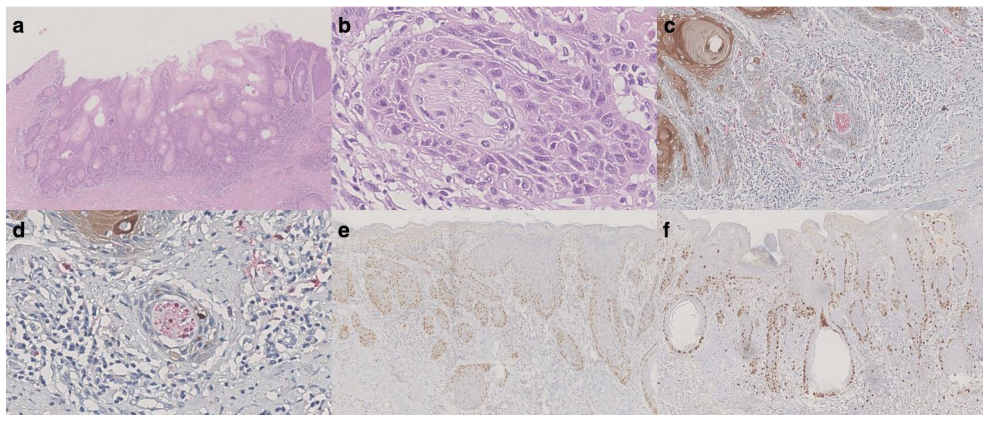
| PNI | Yes | 1 (2%) | 0 (0%) | 1 (10%) | NP |
| No | 47 (98%) | 38 (100% | 9 (90%) | ||
| N. Case | Age at Diagnosis (years) | Tumor Size (mm) | Surgical Treatment | Margin Status (mm) | Depth of Invasion (mm) | Associated Lesions | LVI | PNI | Type of Recurrence | Time to Recurrence (Months) |
|---|---|---|---|---|---|---|---|---|---|---|
| 1 | 60 | 10 | Excisional biopsy | Negative (8 mm) | 0.5 | LS | No | No | SISCCA | 8 |
| 2 | 73 | 10 | Wide local excision | Negative (9 mm) | 1 | LS | No | No | Invasive VSCC | 147 |
| 3 | 74 | 10 | Vulvectomy | Negative (6 mm) | 0.6 | LS | No | Si | SISCCA | 8 |
| 4 | 46 | 20 | Wide local excision | Positive | 0.3 | H-SIL | No | No | SISCCA + inguinal lymphnode involvement | 111 |
| 5 | 58 | 5 | Wide local excision | Negative (6 mm) | 0.5 | LS + H-SIL | No | No | Invasive VSCC | 249 |
| 6 | 78 | 10 | Wide local excision | Positive | 0.8 | H-SIL | No | No | SISCCA | 5 |
| 7 | 80 | 10 | Excisional biopsy | Negative (11 mm) | 0.2 | LS + dVIN | No | No | SISCCA | 15 |
| 8 | 42 | 6 | Vulvectomy | Negative (20 mm) | 0.2 | H-SIL | No | No | SISCCA | 145 |
| 9 | 66 | 10 | Wide local excision | Negative (10 mm) | 1 | LS | No | No | Invasive VSCC | 86 |
| 10 | 48 | 10 | Wide local excision | Negative (10 mm) | 0.2 | LS + dVIN | No | No | SISCCA | 61 |
| Author | Age at FS (years) | Depth of Invasion (mm) | Size of Lesion (cm) | Associated Diseases | Surgical Treatment | Time to Recurrence (Months) | Treatment of Recurrence | Positive LNs | Survival after FS (Months) | LVI | PNI |
|---|---|---|---|---|---|---|---|---|---|---|---|
| Atamdede et al., 1989 [43] | 75 | 0.72 | 1 | Diffuse vulvitis, dystrophic changes of the vulva | WLE | 14 | Bilateral inguinal and deep pelvic LAE + RT | All left-sided LNs | - | NO | - |
| Van Der Velden et al., 1992 [44] | 84 | 0.3 | 0.8 | - | Excisional biopsy and two LN biopsy | 18 | Left inguinal LAE + RT | Left LNs (ECG) | 24 | NO | - |
| Hicks et al., 1993 [45] | 66 | 0.5 | - | LS | Radical WLE | 12 | Left inguinal femoral LAE + RT | Superficial inguinal LNs | - | NO | - |
| Volgger et al., 1997 [46] | 39 | <1 | 0.3 | Immunosuppressive treatment and VIN 3 | Simple vulvectomy | 4 | Bilateral inguinal-femoral LAE | 2 right LNs (one with ECG) | 20 | NO | - |
| Thangavelu et al., 2006 [47] | 64 | <1 | - | VIN 1, LS | Vulvectomy with clitoral sparing | 3 | Right superficial and deep LAE + RT | 7 right LNs (ECG) | 4 | NO | - |
| Thangavelu et al., 2006 [47] | 51 | 0.9 | - | VIN 3, LS | Vulvectomy | 36 | Block dissection right groin LAE + RT | 1 right groin LN | 44 | NO | - |
| Sidor et al., 2006 [48] | 80 | 0.08 | 1.7 × 1.5 | - | Anterior radical vulvectomy | 21 | Bilateral inguinal LAE + RT | 1 left inguinal LN | 98 | NO | - |
| Vernooij, 2007 [49] | 45 | 1 (biopsy only) | - | VIN 3, LS | Local radical excision | 24 | Left inguinal LAE + RT | 1 left LN (ECG) | 168 | NO | NO |
| Vernooij, 2007 [49] | 80 | 1 | 1 × 0.7 | VIN 2, LS | Partial vulvectomy | 13 | Left inguinal LAE + RT | 2 left LN (ECG) | - | NO | - |
| Schausberger et al., 2007 [50] | 53 | <1 | - | VIN 3, CIN 3 | Excision and laser therapy | 24 | Right node excision | 1 right LN | 36 | - | - |
| Iyibozkurt et al., 2010 [51] | 62 | 1 (biopsy only) | - | - | Local radical excision + bilateral inguinal LAE | 27 | Biopsy right groin LN | 1 right LN | 31 | - | - |
| Grimm et al., 2019 [15] | 44 | 0.9 | 0.3 | VIN 3, LS | Anterior vulvectomy | 17 | Bilateral inguino femoral LAE | 1 right LN | 29 | - | - |
| Present study, 2021 | 73 | 0.6 | 0.5 | dVIN, LS | Wide local excision | 8 | Left inguinal LAE + WLE + RT | 1 left LN | 67 | NO | YES |
Publisher’s Note: MDPI stays neutral with regard to jurisdictional claims in published maps and institutional affiliations. |
© 2021 by the authors. Licensee MDPI, Basel, Switzerland. This article is an open access article distributed under the terms and conditions of the Creative Commons Attribution (CC BY) license (https://creativecommons.org/licenses/by/4.0/).
Share and Cite
Preti, M.; Borella, F.; Gallio, N.; Bertero, L.; Heller, D.S.; Vieira-Baptista, P.; Cosma, S.; Bevilacqua, F.; Privitera, S.; Micheletti, L.; et al. Superficially Invasive Vulvar Squamous Cell Carcinoma: A 37-Year-Long Experience of a Tertiary Referral Center. Cancers 2021, 13, 3859. https://doi.org/10.3390/cancers13153859
Preti M, Borella F, Gallio N, Bertero L, Heller DS, Vieira-Baptista P, Cosma S, Bevilacqua F, Privitera S, Micheletti L, et al. Superficially Invasive Vulvar Squamous Cell Carcinoma: A 37-Year-Long Experience of a Tertiary Referral Center. Cancers. 2021; 13(15):3859. https://doi.org/10.3390/cancers13153859
Chicago/Turabian StylePreti, Mario, Fulvio Borella, Niccolò Gallio, Luca Bertero, Debra Sandra Heller, Pedro Vieira-Baptista, Stefano Cosma, Federica Bevilacqua, Sebastiana Privitera, Leonardo Micheletti, and et al. 2021. "Superficially Invasive Vulvar Squamous Cell Carcinoma: A 37-Year-Long Experience of a Tertiary Referral Center" Cancers 13, no. 15: 3859. https://doi.org/10.3390/cancers13153859
APA StylePreti, M., Borella, F., Gallio, N., Bertero, L., Heller, D. S., Vieira-Baptista, P., Cosma, S., Bevilacqua, F., Privitera, S., Micheletti, L., & Benedetto, C. (2021). Superficially Invasive Vulvar Squamous Cell Carcinoma: A 37-Year-Long Experience of a Tertiary Referral Center. Cancers, 13(15), 3859. https://doi.org/10.3390/cancers13153859