
Human Papillomavirus Same Genotype Persistence and Risk of Cervical Intraepithelial Neoplasia2+ Recurrence

 ,
,  ,
, 
Abstract
:Simple Summary
Abstract
1. Introduction
2. Materials and Methods
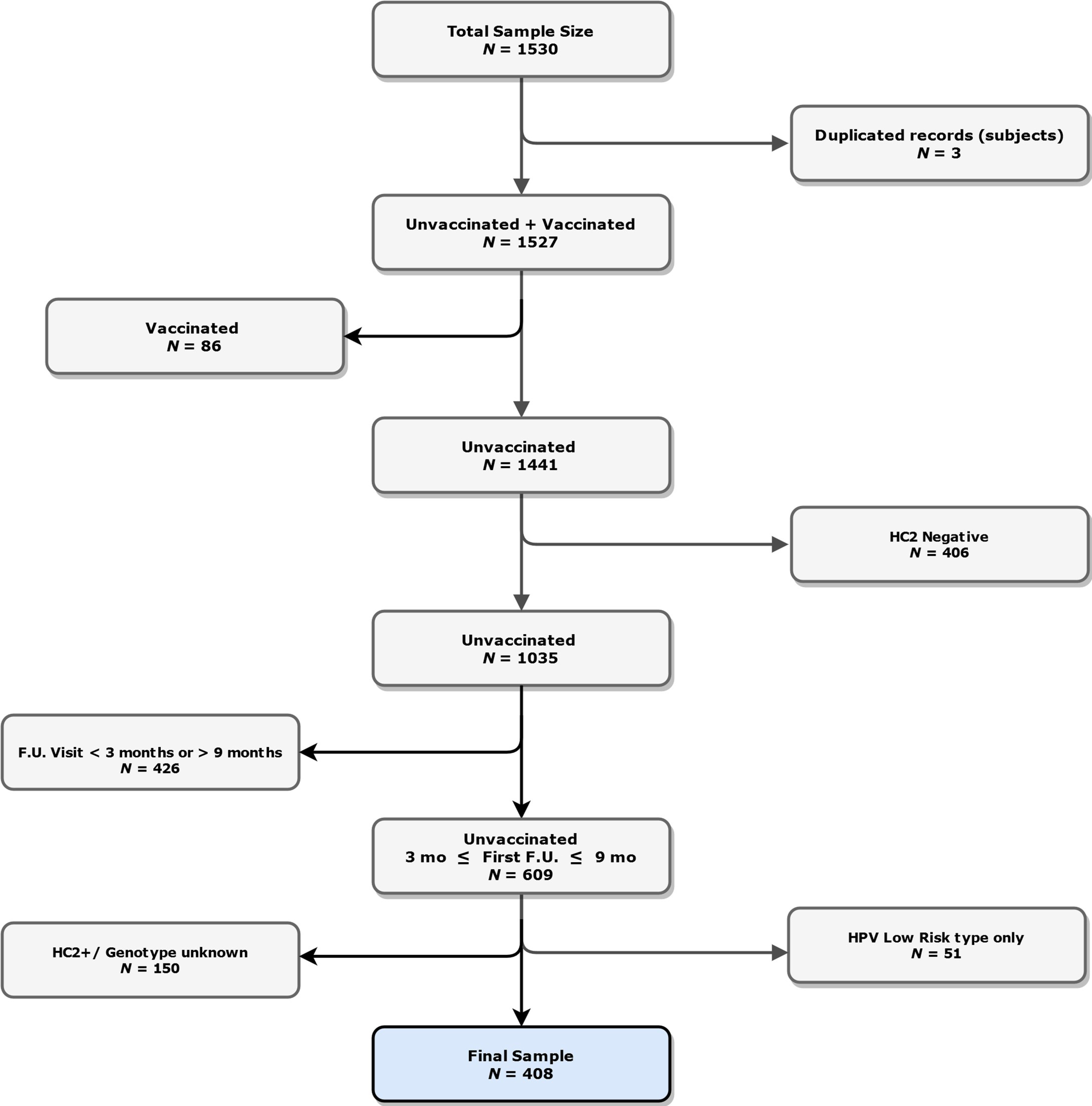
2.1. Population
2.2. Human Papillomavirus DNA Detection and Genotyping
2.3. Statistical Methods
3. Results
4. Discussion
5. Conclusions
Supplementary Materials
Author Contributions
Funding
Institutional Review Board Statement
Informed Consent Statement
Data Availability Statement
Acknowledgments
Conflicts of Interest
References
- Ghaem-Maghami, S.; Sagi, S.; Majeed, G.; Soutter, W.P. Incomplete excision of cervical intraepithelial neoplasia and risk of treatment failure: A meta-analysis. Lancet Oncol. 2007, 8, 985–993. [Google Scholar] [CrossRef]
- Del Mistro, A.; Matteucci, M.; Insacco, E.A.; Onnis, G.; Da Re, F.; Baboci, L.; Zorzi, M.; Minucci, D. Long-Term clinical outcome after treatment for high-grade cervical lesions: A retrospective monoinstitutional cohort study. Biomed. Res. Int. 2015, 2015, 984528. [Google Scholar] [CrossRef] [Green Version]
- Kocken, M.; Helmerhorst, T.J.; Berkhof, J.; Louwers, J.A.; Nobbenhuis, M.A.; Bais, A.G.; Hogewoning, C.J.A.; Zaal, A.; Verheijen, R.H.M.; Snijders, P.J.F.; et al. Risk of recurrent high-grade cervical intraepithelial neoplasia after successful treatment: A long-term multi-cohort study. Lancet Oncol. 2011, 12, 441–450. [Google Scholar] [CrossRef]
- Serati, M.; Siesto, G.; Carollo, S.; Formenti, G.; Riva, C.; Cromi, A.; Ghezzi, F. Risk factors for cervical intraepithelial neoplasia recurrence after conization: A 10-year study. Eur. J. Obstet. Gynecol. Reprod. Biol. 2012, 165, 86–90. [Google Scholar] [CrossRef]
- Arbyn, M.; Redman, C.W.E.; Verdoodt, F.; Kyrgiou, M.; Tzafetas, M.; Ghaem-Maghami, S.; Petry, K.U.; Leeson, S.; Bergeron, C.; Nieminen, P.; et al. Incomplete excision of cervical precancer as a predictor of treatment failure: A systematic review and meta-analysis. Lancet Oncol. 2017, 18, 1665–1679. [Google Scholar] [CrossRef] [Green Version]
- Zur Hausen, H. Papillomaviruses in the causation of human cancers—a brief historical account. Virology 2009, 384, 260–265. [Google Scholar] [CrossRef] [Green Version]
- Kjær, S.K.; Frederiksen, K.; Munk, C.; Iftner, T. Long-term absolute risk of cervical intraepithelial neoplasia grade 3 or worse following human papillomavirus infection: Role of persistence. J. Natl. Cancer Inst. 2010, 102, 1478–1488. [Google Scholar] [CrossRef] [Green Version]
- Elfgren, K.; Jacobs, M.; Walboomers, J.M.; Meijer, C.J.; Dillner, J. Rate of human papillomavirus clearance after treatment of cervical intraepithelial neoplasia. Obstet. Gynecol. 2002, 100, 965–971. [Google Scholar] [CrossRef]
- Kim, Y.T.; Lee, J.M.; Hur, S.Y.; Cho, C.H.; Kim, Y.T.; Kim, S.C.; Kang, S.B. Clearance of human papillomavirus infection after successful conization in patients with cervical intraepithelial neoplasia. Int. J. Cancer. 2010, 126, 1903–1909. [Google Scholar] [CrossRef]
- Carozzi, F.M.; Iossa, A.; Scalisi, A.; Sideri, M.; Andersson, K.L.; Confortini, M.; Del Mistro, A.; Maina, G.; Ronco, G.; Raggi, P.; et al. hr-HPV testing in the management of women with ASC-US+ and in the follow-up of women with cytological abnormalities and negative colposcopy. Recommendations of the Italian group for cervical cancer screening (GISCi). Epidemiol. Prev. 2015, 39, 84–90. [Google Scholar]
- Clarke, M.A.; Unger, E.R.; Zuna, R.; Nelson, E.; Darragh, T.M.; Cremer, M.; Stockdale, C.K.; Einstein, M.H.; Wentzensen, N. A Systematic Review of Tests for Postcolposcopy and Posttreatment Surveillance. J. Low. Genit. Tract. Dis. 2020, 24, 148–156. [Google Scholar] [CrossRef] [PubMed]
- Bowker, A.H. A Test for Symmetry in Contingency Tables. J. Am. Stat. Assoc. 1948, 43, 572–574. [Google Scholar] [CrossRef]
- Krauth, J. Niehtparametrische Ansätze zur Auswertung von Verlaufskurven. Biometrische. Z. 1973, 15, 557–566. [Google Scholar] [CrossRef]
- Byun, J.M.; Jeong, D.H.; Kim, Y.N.; Jung, E.J.; Lee, K.B.; Sung, M.S.; Kim, K.T. Persistent HPV-16 infection leads to recurrence of high-grade cervical intraepithelial neoplasia. Medicine 2018, 97, e13606. [Google Scholar] [CrossRef]
- Fernández-Montolí, M.E.; Tous, S.; Medina, G.; Castellarnau, M.; García-Tejedor, A.; de Sanjosé, S. Long-term predictors of residual or recurrent cervical intraepithelial neoplasia 2-3 after treatment with a large loop excision of the transformation zone: A retrospective study. BJOG 2020, 127, 377–387. [Google Scholar] [CrossRef]
- Bruno, M.T.; Cassaro, N.; Garofalo, S.; Boemi, S. HPV16 persistent infection and recurrent disease after LEEP. Virol. J. 2019, 16, 148. [Google Scholar] [CrossRef]
- Zhang, Q.; Dong, B.; Chen, L.; Lin, T.; Tong, Y.; Lin, W.; Lin, H.; Gao, Y.; Lin, F.; Sun, P. Evaluation of PCR-Reverse Dot Blot Human Papillomavirus Genotyping Test in Predicting Residual/Recurrent CIN 2+ in Posttreatment Patients in China. Cancer Manag. Res. 2020, 12, 2369–2379. [Google Scholar] [CrossRef] [Green Version]
- Nagai, Y.; Maehama, T.; Asato, T.; Kanazawa, K. Persistence of human papillomavirus infection after therapeutic conization for CIN 3: Is it an alarm for disease recurrence? Gynecol. Oncol. 2000, 79, 294–299. [Google Scholar] [CrossRef]
- Rositch, A.F.; Soeters, H.M.; Offutt-Powell, T.N.; Wheeler, B.S.; Taylor, S.M.; Smith, J.S. The incidence of human papillomavirus infection following treatment for cervical neoplasia: A systematic review. Gynecol. Oncol. 2014, 132, 767–779. [Google Scholar] [CrossRef] [Green Version]
- Kang, W.D.; Oh, M.J.; Kim, S.M.; Nam, J.H.; Park, C.S.; Choi, H.S. Significance of human papillomavirus genotyping with high-grade cervical intraepithelial neoplasia treated by a loop electrosurgical excision procedure. Am. J. Obstet. Gynecol. 2010, 203, 72.e1-6. [Google Scholar] [CrossRef] [PubMed]
- Bottari, F.; Iacobone, A.D.; Boveri, S.; Preti, E.P.; Franchi, D.; Mariani, L.; Preti, M.; Landoni, F.; Passerini, R.; Sandri, M.T. Onclarity Human Papillomavirus Extended Genotyping in the Management of Cervical Intraepithelial Neoplasia 2+ Lesions. J. Low. Genit. Tract. Dis. 2019, 23, 39–42. [Google Scholar] [CrossRef] [PubMed]
- Cricca, M.; Venturoli, S.; Morselli-Labate, A.M.; Costa, S.; Santini, D.; Ambretti, S.; Musiani, M.; Zerbini, M. HPV DNA patterns and disease implications in the follow-up of patients treated for HPV16 high-grade carcinoma in situ. J. Med. Virol. 2006, 78, 494–500. [Google Scholar] [CrossRef] [PubMed]
- Eltoum, I.A.; Chhieng, D.C.; Crowe, D.R.; Roberson, J.; Jin, G.; Broker, T.R. Significance and possible causes of false-negative results of reflex human Papillomavirus infection testing. Cancer 2017, 111, 154–159. [Google Scholar] [CrossRef]
- Kyrgiou, M.; Athanasiou, A.; Paraskevaidi, M.; Mitra, A.; Kalliala, I.; Martin-Hirsch, P.; Arbyn, M.; Bennett, P.; Paraskevaidis, E. Adverse obstetric outcomes after local treatment for cervical preinvasive and early invasive disease according to cone depth: Systematic review and meta-analysis. BMJ 2016, 354, i3633. [Google Scholar] [CrossRef] [Green Version]
- Bottari, F.; Iacobone, A.D.; Passerini, R.; Preti, E.P.; Sandri, M.T.; Cocuzza, C.E.; Gary, D.S.; Andrews, J.C. Human Papillomavirus Genotyping Compared With a Qualitative High-Risk Human Papillomavirus Test After Treatment of High-Grade Cervical Intraepithelial Neoplasia: A Systematic Review. Obstet. Gynecol. 2019, 134, 452–462. [Google Scholar] [CrossRef] [PubMed] [Green Version]
- Mariani, L.; Sandri, M.T.; Preti, M.; Origoni, M.; Costa, S.; Cristoforoni, P.; Bottari, F.; Sideri, M. HPV-Testing in Follow-up of Patients Treated for CIN2+ Lesions. J. Cancer 2016, 7, 107–114. [Google Scholar] [CrossRef] [PubMed] [Green Version]
- Iacobone, A.D.; Bottari, F.; Radice, D.; Preti, E.P.; Franchi, D.; Vidal Urbinati, A.M.; Boveri, S.; Passerini, R.; Sandri, M.T. Distribution of High-Risk Human Papillomavirus Genotypes and Multiple Infections in Preneoplastic and Neoplastic Cervical Lesions of Unvaccinated Women: A Cross-sectional Study. J. Low. Genit. Tract. Dis. 2019, 23, 259–264. [Google Scholar] [CrossRef]
- Pista, A.; de Oliveira, C.F.; Lopes, C.; Cunha, M.J.; CLEOPATRE Portugal Study Groupa. Human papillomavirus type distribution in cervical intraepithelial neoplasia grade 2/3 and cervical cancer in Portugal: A CLEOPATRE II Study. Int. J. Gynecol. Cancer 2013, 23, 500–506. [Google Scholar] [CrossRef]


| Characteristic | Level | N (Col %) | HPV Persistence, N (Row %) | p-Value | |
|---|---|---|---|---|---|
| All Patients N = 408 | Not-Persistent N = 312 | Persistent N = 96 | |||
| Age, years | 40 (35–46) a | 39 (35–46) a | 40 (35–46) a | 0.53 | |
| Histology | CIN1 | 72 (17.7) | 59 (81.9) | 13 (18.1) | |
| CIN2/3/AIS | 313 (76.7) | 232 (74.1) | 81 (25.9) | ||
| ICC | 23 (5.6) | 21 (91.3) | 2 (8.7) | 0.09 | |
| Infection status | Single infection | 296 (72.6) | 236 (79.7) | 60 (20.3) | |
| Multiple (≥2) infection | 112 (27.5) | 76 (67.9) | 36 (32.1) | 0.01 | |
| No. of infections b | 1 | 296 (72.6) | 236 (79.7) | 60 (20.3) | |
| 2 | 83 (20.3) | 60 (72.3) | 23 (27.7) | ||
| ≥3 | 29 (7.1) | 16 (55.2) | 13 (44.8) | 0.009 | |
| Ectocervical margin c | Negative | 392 (97.3) | 299 (76.3) | 93 (23.7) | |
| Positive | 11 (2.7) | 9 (81.8) | 2 (18.2) | 1.00 | |
| Endocervical margin c | Negative | 386 (95.8) | 298 (77.2) | 88 (22.8) | |
| Positive | 17 (4.2) | 10 (58.8) | 7 (41.2) | 0.14 | |
| Surgical margins | Negative | 376 (92.2) | 290 (77.1) | 86 (22.9) | |
| Positive | 32 (7.8) | 22 (68.8) | 10 (31.2) | 0.28 | |
| Glandular crypts involvement | No | 192 (47.1) | 149 (77.6) | 43 (22.4) | |
| Yes | 216 (52.9) | 163 (75.5) | 54 (24.5) | 0.64 | |
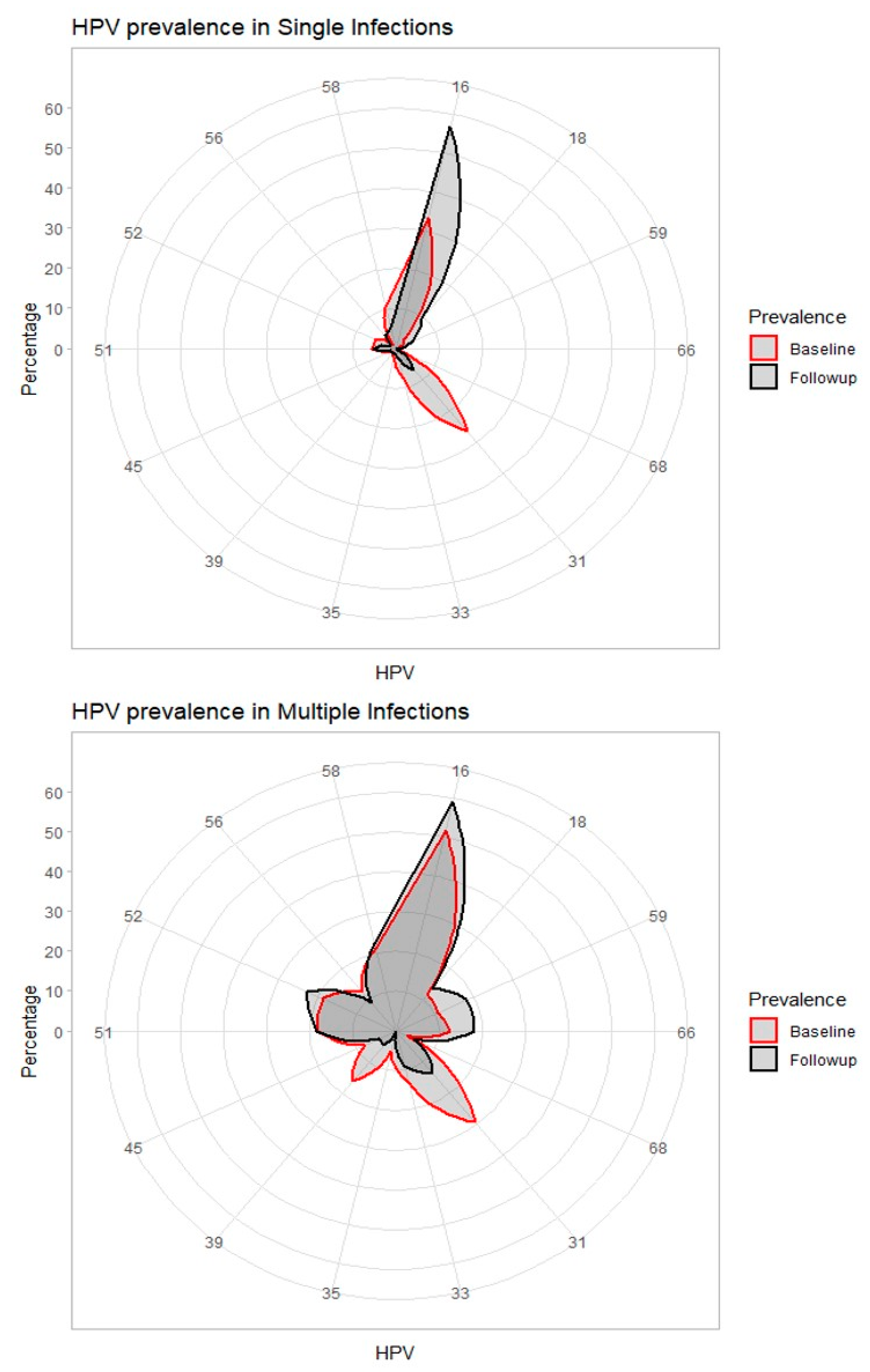
| Genotype ID | Single N (%) | Multiple a N (%) | ||
|---|---|---|---|---|
| Baseline N = 296 | Follow-Up b N = 74 | Baseline N = 112 | Follow-Up b N = 22 | |
| 16 | 99 (33.5) | 42 (56.8) | 58 (51.8) | 13 (59.1) |
| 18 | 9 (3.0) | 7 (9.5) | 13 (11.6) | 3 (13.6) |
| 31 | 78 (26.4) | 5 (6.8) | 33 (29.5) | 3 (13.6) |
| 33 | 22 (7.4) | 2 (2.7) | 15 (13.4) | 2 (9.1) |
| 35 | 6 (2.0) | 1 (1.4) | 6 (5.4) | 0 |
| 39 | 3 (1.0) | 1 (1.4) | 18 (16.1) | 1 (4.6) |
| 45 | 8 (2.7) | 1 (1.4) | 9 (8.0) | 1 (4.6) |
| 51 | 17 (5.7) | 4 (5.4) | 20 (17.9) | 4 (18.2) |
| 52 | 15 (5.1) | 1 (1.4) | 21 (18.8) | 5 (22.7) |
| 56 | 3 (1.0) | 3 (4.1) | 14 (12.5) | 2 (9.1) |
| 58 | 31 (10.5) | 4 (5.4) | 24 (21.4) | 5 (22.7) |
| 59 | 5 (1.7) | 3 (4.1) | 12 (10.7) | 4 (18.2) |
| 66 | 0 | 1 (1.4) | 14 (12.5) | 4 (18.2) |
| 68 | 0 | 0 | 3 (2.7) | 1 (4.6) |
| Characteristic | Level | Events/At Risk | CIF (95% CI) | p-Value a |
|---|---|---|---|---|
| Overall | - | 21/408 | 8.6 (5.2–13.2) | - |
| Histology | CIN1 | 1/72 | 4.2 (0.3–18.0) | |
| CIN2/3/AIS | 19/313 | 9.8 (5.7–15.2) | ||
| ICC | 1/23 | 5.9 (0.3–24.2) | 0.21 | |
| Persistence | Not-persistent | 2/312 | 1.7 (0.3–5.8) | |
| Persistent | 19/96 | 27.6 (16.2–40.2) | <0.001 | |
| No. of infections | Negative | 2/312 | 1.7 (0.3–5.8) | |
| Single | 14/74 | 26.2 (14.1–40.2) | ||
| Multiple | 5/22 | 27.2 (7.3–52.3) | <0.001 b | |
| Genotype 16/18 c | Negative | 2/312 | 1.7 (0.3–5.8) | |
| 16+ or 18+/Other HR | 16/64 | 32.7 (17.9–48.3) | ||
| 16− and 18−/Other HR | 3/32 | 16.1 (3.8–36.2) | <0.001 d | |
| Glandular crypts | No | 7/192 | 6.1 (2.3–12.6) | |
| Yes | 14/216 | 11.1 (5.9–18.2) | 0.15 | |
| Ectocervical margin e | Negative | 19/392 | 8.5 (4.9–13.3) | |
| Positive | 1/11 | 11.1 (4.7–40.6) | 0.78 | |
| Endocervical margin e | Negative | 18/386 | 8.0 (4.6–12.6) | |
| Positive | 2/17 | 24.7 (1.5–62.8) | 0.20 | |
| Surgical margins | Negative | 17/376 | 7.9 (4.4–12.6) | |
| Positive | 4/32 | 18.2 (4.6–38.8) | 0.09 |
Publisher’s Note: MDPI stays neutral with regard to jurisdictional claims in published maps and institutional affiliations. |
© 2021 by the authors. Licensee MDPI, Basel, Switzerland. This article is an open access article distributed under the terms and conditions of the Creative Commons Attribution (CC BY) license (https://creativecommons.org/licenses/by/4.0/).
Share and Cite
Iacobone, A.D.; Radice, D.; Sandri, M.T.; Preti, E.P.; Guerrieri, M.E.; Vidal Urbinati, A.M.; Pino, I.; Franchi, D.; Passerini, R.; Bottari, F. Human Papillomavirus Same Genotype Persistence and Risk of Cervical Intraepithelial Neoplasia2+ Recurrence. Cancers 2021, 13, 3664. https://doi.org/10.3390/cancers13153664
Iacobone AD, Radice D, Sandri MT, Preti EP, Guerrieri ME, Vidal Urbinati AM, Pino I, Franchi D, Passerini R, Bottari F. Human Papillomavirus Same Genotype Persistence and Risk of Cervical Intraepithelial Neoplasia2+ Recurrence. Cancers. 2021; 13(15):3664. https://doi.org/10.3390/cancers13153664
Chicago/Turabian StyleIacobone, Anna Daniela, Davide Radice, Maria Teresa Sandri, Eleonora Petra Preti, Maria Elena Guerrieri, Ailyn Mariela Vidal Urbinati, Ida Pino, Dorella Franchi, Rita Passerini, and Fabio Bottari. 2021. "Human Papillomavirus Same Genotype Persistence and Risk of Cervical Intraepithelial Neoplasia2+ Recurrence" Cancers 13, no. 15: 3664. https://doi.org/10.3390/cancers13153664
APA StyleIacobone, A. D., Radice, D., Sandri, M. T., Preti, E. P., Guerrieri, M. E., Vidal Urbinati, A. M., Pino, I., Franchi, D., Passerini, R., & Bottari, F. (2021). Human Papillomavirus Same Genotype Persistence and Risk of Cervical Intraepithelial Neoplasia2+ Recurrence. Cancers, 13(15), 3664. https://doi.org/10.3390/cancers13153664