Immunomodulatory Drugs in Acute Myeloid Leukemia Treatment


Abstract
:Simple Summary
Abstract
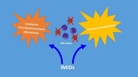
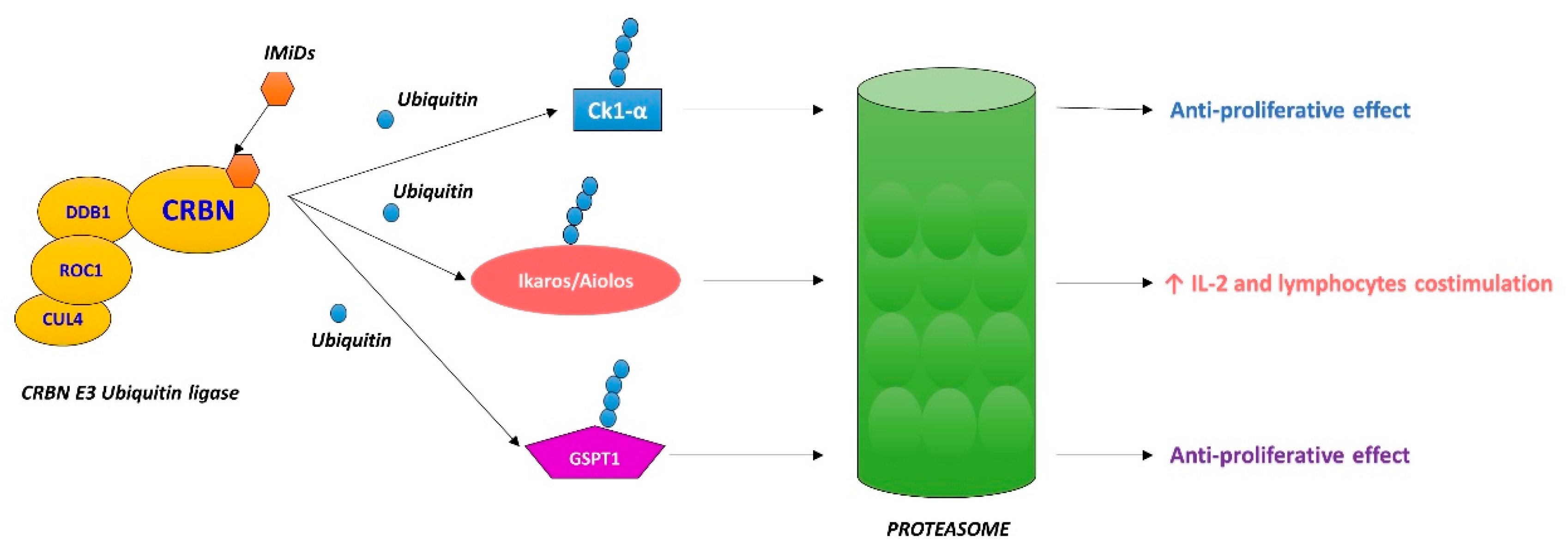
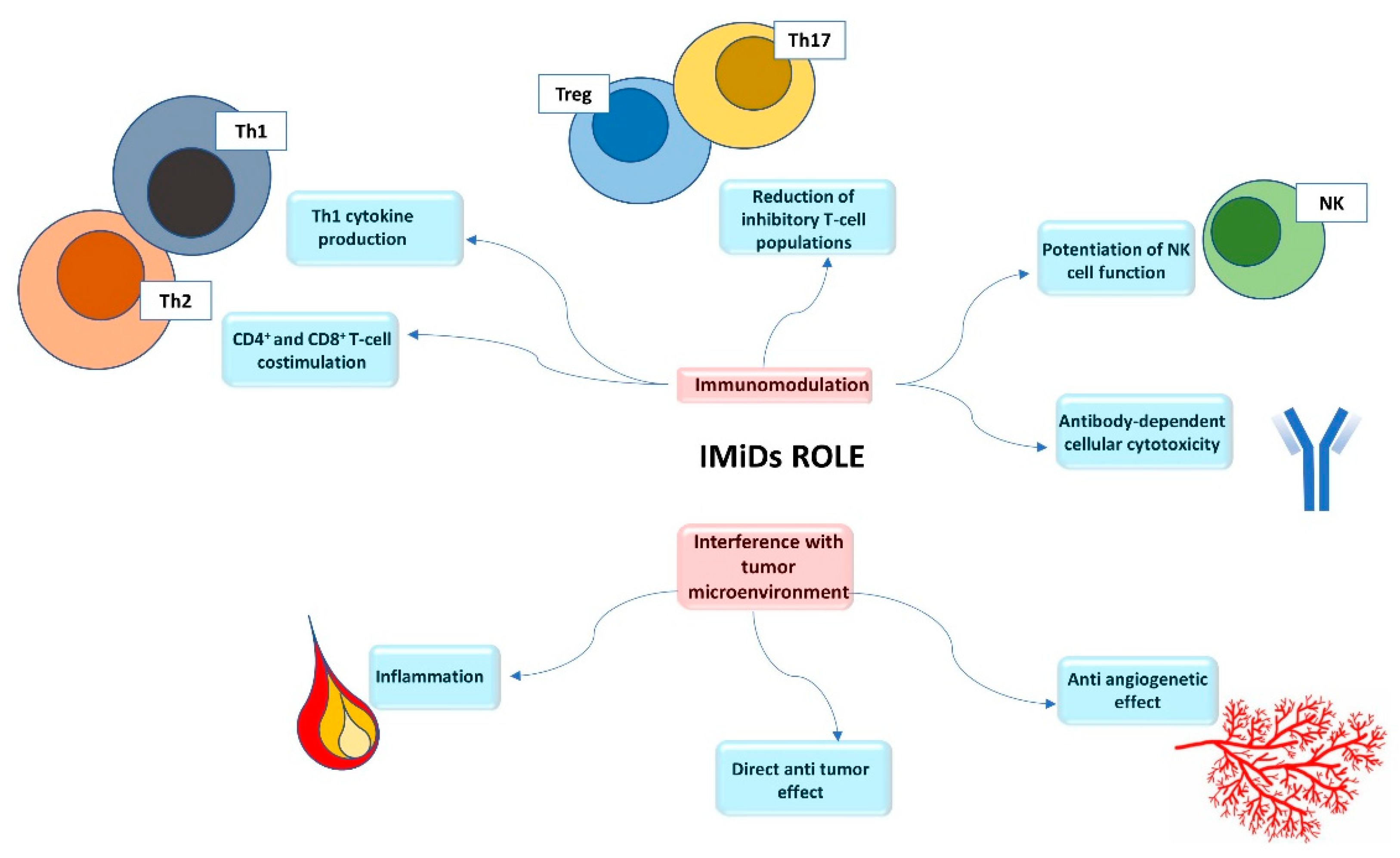
1. Introduction
2. Thalidomide
3. Lenalidomide
3.1. Lenalidomide as a Single Agent
3.2. Lenalidomide and Chemotherapy
3.3. Lenalidomide and 5-AZA
3.4. Lenalidomide and Allogeneic Stem Cell Transplantation
4. Pomalidomide
5. New Molecules
6. Conclusions
Author Contributions
Funding
Acknowledgments
Conflicts of Interest
References
- Knight, R. IMiDs: A Novel Class of Immunomodulators. Semin. Oncol. 2005, 32, 24–30. [Google Scholar] [CrossRef]
- Fuchs, O. Treatment of Lymphoid and Myeloid Malignancies by Immunomodulatory Drugs. Cardiovasc. Hematol. Disord. Targets 2018, 19, 51–78. [Google Scholar] [CrossRef] [PubMed]
- Ito, T.; Handa, H. Cereblon and its downstream substrates as molecular targets of immunomodulatory drugs. Int. J. Hematol. 2016, 104, 293–299. [Google Scholar] [CrossRef] [PubMed] [Green Version]
- Fischer, E.S.; Böhm, K.; Lydeard, J.R.; Yang, H.; Stadler, M.B.; Cavadini, S.; Nagel, J.; Serluca, F.; Acker, V.; Lingaraju, G.M.; et al. Structure of the DDB1–CRBN E3 ubiquitin ligase in complex with thalidomide. Nature 2014, 512, 49–53. [Google Scholar] [CrossRef] [PubMed] [Green Version]
- Krönke, J.; Udeshi, N.D.; Narla, A.; Grauman, P.; Hurst, S.N.; McConkey, M.; Svinkina, T.; Heckl, D.; Comer, E.; Li, X.; et al. Lenalidomide Causes Selective Degradation of IKZF1 and IKZF3 in Multiple Myeloma Cells. Science 2014, 343, 301–305. [Google Scholar] [CrossRef] [Green Version]
- Eichner, R.; Heider, M.; Fernández-Sáiz, V.; van Bebber, F.; Garz, A.-K.; Lemeer, S.; Rudelius, M.; Targosz, B.-S.; Jacobs, L.; Knorn, A.-M.; et al. Immunomodulatory drugs disrupt the cereblon–CD147–MCT1 axis to exert antitumor activity and teratogenicity. Nat. Med. 2016, 22, 735–743. [Google Scholar] [CrossRef]
- Shi, Q.; Chen, L. Cereblon: A Protein Crucial to the Multiple Functions of Immunomodulatory Drugs as well as Cell Metabolism and Disease Generation. J. Immunol. Res. 2017, 2017, 1–8. [Google Scholar] [CrossRef] [Green Version]
- Gbolahan, O.B.; Zeidan, A.M.; Stahl, M.; Zaid, M.A.; Farag, S.; Paczesny, S.; Konig, H. Immunotherapeutic concepts to target acute myeloid leukemia: Focusing on the role of monoclonal antibodies, hypomethylating agents and the leukemic microenvironment. Int. J. Mol. Sci. 2017, 18, 1660. [Google Scholar] [CrossRef] [Green Version]
- Vallet, S.; Witzens-Harig, M.; Jaeger, D.; Podar, K. Update on immunomodulatory drugs (IMiDs) in hematologic and solid malignancies. Expert Opin. Pharmacother. 2012, 13, 473–494. [Google Scholar] [CrossRef]
- Castelli, R.; Cassin, R.; Cannavò, A.; Cugno, M. Immunomodulatory drugs: New options for the treatment of myelodysplastic syndromes. Clin. Lymphoma Myeloma Leuk. 2013, 13, 1–7. [Google Scholar] [CrossRef]
- Ljunggren, H.G.; Kärre, K. In search of the “missing self”: MHC molecules and NK cell recognition. Immunol. Today 1990, 11, 237–244. [Google Scholar] [CrossRef]
- Le Roy, A.; Prébet, T.; Castellano, R.; Goubard, A.; Riccardi, F.; Fauriat, C.; Granjeaud, S.; Benyamine, A.; Castanier, C.; Orlanducci, F.; et al. Immunomodulatory drugs exert anti-leukemia effects in acute myeloid leukemia by direct and immunostimulatory activities. Front. Immunol. 2018, 9. [Google Scholar] [CrossRef] [PubMed]
- Khaznadar, Z.; Henry, G.; Setterblad, N.; Agaugue, S.; Raffoux, E.; Boissel, N.; Dombret, H.; Toubert, A.; Dulphy, N. Acute myeloid leukemia impairs natural killer cells through the formation of a deficient cytotoxic immunological synapse. Eur. J. Immunol. 2014, 44, 3068–3080. [Google Scholar] [CrossRef]
- Chretien, A.S.; Fauriat, C.; Orlanducci, F.; Galseran, C.; Rey, J.; Borg, G.B.; Gautherot, E.; Granjeaud, S.; Hamel-Broza, J.F.; Demerle, C.; et al. Natural killer defective maturation is associated with adverse clinical outcome in patients with acute myeloid leukemia. Front. Immunol. 2017, 8. [Google Scholar] [CrossRef]
- Carlsten, M.; Järås, M. Natural Killer Cells in Myeloid Malignancies: Immune Surveillance, NK Cell Dysfunction and Pharmacological Opportunities to Bolster the Endogenous NK Cells. Front. Immunol. 2019, 10, 2357. [Google Scholar] [CrossRef] [PubMed] [Green Version]
- Medinger, M.; Mross, K. Clinical trials with anti-angiogenic agents in hematological malignancies. J. Angiogenes Res. 2010, 2, 10. [Google Scholar] [CrossRef] [Green Version]
- Baer, M.R.; Gojo, I. Novel agents for the treatment of acute myeloid leukemia in the older patient. JNCCN J. Natl. Compr. Cancer Netw. 2011, 9, 331–335. [Google Scholar] [CrossRef] [Green Version]
- Kreuter, M.; Steins, M.; Woelke, K.; Buechner, T.; Berdel, W.E.; Mesters, R.M. Downregulation of neuropilin-1 in patients with acute myeloid leukemia treated with thalidomide. Eur. J. Haematol. 2007, 79, 392–397. [Google Scholar] [CrossRef]
- Aguayo, A.; Estey, E.; Kantarjian, H.; Mansouri, T.; Gidel, C.; Keating, M.; Giles, F.; Estrov, Z.; Barlogie, B.; Albitar, M. Cellular vascular endothelial growth factor is a predictor of outcome in patients with acute myeloid leukemia. Blood 1999, 94, 3717–3721. [Google Scholar] [CrossRef]
- Corral, L.G.; Haslett, P.A.J.; Muller, G.W.; Chen, R.; Wong, L.M.; Ocampo, C.J.; Patterson, R.T.; Stirling, D.I.; Kaplan, G. Differential cytokine modulation and T cell activation by two distinct classes of thalidomide analogues that are potent inhibitors of TNF-alpha. J. Immunol. 1999, 67, 501. [Google Scholar]
- Teo, S.K. Properties of thalidomide and its analogues: Implications for anticancer therapy. AAPS J. 2005, 7. [Google Scholar] [CrossRef] [PubMed] [Green Version]
- Steins, M.B.; Padró, T.; Bieker, R.; Ruiz, S.; Kropff, M.; Kienast, J.; Kessler, T.; Buechner, T.; Berdel, W.E.; Mesters, R.M. Efficacy and safety of thalidomide in patients with acute myeloid leukemia. Blood 2002, 99, 834–839. [Google Scholar] [CrossRef] [PubMed] [Green Version]
- Thomas, D.A.; Estey, E.; Giles, F.J.; Faderl, S.; Cortes, J.; Keating, M.; O’Brien, S.; Albitar, M.; Kantarjian, H. Single agent thalidomide in patients with relapsed or refractory acute myeloid leukaemia. Br. J. Haematol. 2003, 123, 436–441. [Google Scholar] [CrossRef] [PubMed]
- Cortes, J.; Kantarjian, H.; Albitar, M.; Thomas, D.; Faderl, S.; Koller, C.; Garcia-Manero, G.; Giles, F.; Andreeff, M.; O’Brien, S.; et al. A randomized trial of liposomal daunorubicin and cytarabine versus liposomal daunorubicin and topotecan with or without thalidomide as initial therapy for patients with poor prognosis acute myelogenous leukemia or myelodysplastic syndrome. Cancer 2003, 97, 1234–1241. [Google Scholar] [CrossRef] [PubMed]
- Barr, P.; Fu, P.; Lazarus, H.; Kane, D.; Meyerson, H.; Hartman, P.; Reyes, R.; Creger, R.; Stear, K.; Laughlin, M.; et al. Antiangiogenic activity of thalidomide in combination with fludarabine, carboplatin and topotecan for high-risk acute myelogenous leukemia. Leuk. Lymphoma 2007, 48, 1940–1949. [Google Scholar] [CrossRef]
- Chen, C.; Yang, J.; Xu, W. Thalidomide in Combination with Chemotherapy in Treating Elderly Patients with Acute Myeloid Leukemia. Oncol. Res. Treat. 2018, 41, 461–465. [Google Scholar] [CrossRef]
- Raza, A.; Mehdi, M.; Mumtaz, M.; Ali, F.; Lascher, S.; Galili, N. Combination of 5-azacytidine and thalidomide for the treatment of myelodysplastic syndromes and acute myeloid leukemia. Cancer 2008, 113, 1596–1604. [Google Scholar] [CrossRef]
- Kenealy, M.; Patton, N.; Filshie, R.; Nicol, A.; Ho, S.J.; Hertzberg, M.; Mills, T.; Prosser, I.; Link, E.; Cowan, L.; et al. Results of a phase II study of thalidomide and azacitidine in patients with clinically advanced myelodysplastic syndromes (MDS), chronic myelomonocytic leukemia (CMML) and low blast count acute myeloid leukemia (AML). Leuk. Lymphoma 2017, 58, 298–307. [Google Scholar] [CrossRef]
- Kian, M.M.; Mohammadi, S.; Tavallaei, M.; Chahardouli, B.; Rostami, S.; Zahedpanah, M.; Ghavamzadeh, A.; Nikbakht, M. Inhibitory effects of arsenic trioxide and thalidomide on angiogenesis and vascular endothelial growth factor expression in leukemia cells. Asian Pac. J. Cancer Prev. 2018, 19, 1127–1134. [Google Scholar] [CrossRef]
- Galustian, C.; Dalgleish, A. Lenalidomide: A novel anticancer drug with multiple modalities. Expert Opin. Pharmacother. 2009, 10, 125–133. [Google Scholar] [CrossRef]
- Xie, C.H.; Wei, M.; Yang, F.Y.; Wu, F.Z.; Chen, L.; Wang, J.K.; Liu, Q.; Huang, J.X. Efficacy and safety of lenalidomide for thtreatment of acute myeloid leukemia: A systematic review and meta-analysis. Cancer Manag. Res. 2018, 10, 3637–3648. [Google Scholar] [CrossRef] [PubMed] [Green Version]
- Fehniger, T.A.; Uy, G.L.; Trinkaus, K.; Nelson, A.D.; Demland, J.; Abboud, C.N.; Cashen, A.F.; Stockerl-Goldstein, K.E.; Westervelt, P.; DiPersio, J.F.; et al. A phase 2 study of high-dose lenalidomide as initial therapy for older patients with acute myeloid leukemia. Blood 2011, 117, 1828–1833. [Google Scholar] [CrossRef]
- Blum, W.; Klisovic, R.B.; Becker, H.; Yang, X.; Rozewski, D.M.; Phelps, M.A.; Garzon, R.; Walker, A.; Chandler, J.C.; Whitman, S.P.; et al. Dose escalation of lenalidomide in relapsed or refractory acute leukemias. J. Clin. Oncol. 2010, 28, 4919–4925. [Google Scholar] [CrossRef] [PubMed] [Green Version]
- Fehniger, T.A.; Byrd, J.C.; Marcucci, G.; Abboud, C.N.; Kefauver, C.; Payton, J.E.; Vij, R.; Blum, W. Single-agent lenalidomide induces complete remission of acute myeloid leukemia in patients with isolated trisomy 13. Blood 2009, 113, 1002–1005. [Google Scholar] [CrossRef] [PubMed]
- Lancet, J.E.; List, A.F.; Moscinski, L.C. Treatment of deletion 5q acute myeloid leukemia with lenalidomide. Leukemia 2007, 21, 586–588. [Google Scholar] [CrossRef] [PubMed] [Green Version]
- Chen, Y.; Kantarjian, H.; Estrov, Z.; Faderl, S.; Ravandi, F.; Rey, K.; Cortes, J.; Borthakur, G. A phase II study of lenalidomide alone in relapsed/refractory acute myeloid leukemia or high-risk myelodysplastic syndromes with chromosome 5 abnormalities. Clin. Lymphoma Myeloma Leuk. 2012, 12, 341–344. [Google Scholar] [CrossRef] [Green Version]
- De Angelo, D.J.; Brunner, A.M.; Werner, L.; Avigan, D.; Fathi, A.T.; Sperling, A.S.; Washington, A.; Stroopinsky, D.; Rosenblatt, J.; McMasters, M.; et al. A phase I study of lenalidomide plus chemotherapy with mitoxantrone, etoposide and cytarabine for the reinduction of patients with acute myeloid leukemia. Am. J. Hematol. 2018, 93, 254–261. [Google Scholar] [CrossRef] [Green Version]
- Price, S.L.; Lancet, J.E.; George, T.J.; Wetzstein, G.A.; List, A.F.; Ho, V.Q.; Fernandez, H.F.; Pinilla-Ibarz, J.; Kharfan-Dabaja, M.A.; Komrokji, R.S. Salvage chemotherapy regimens for acute myeloid leukemia: Is one better? Efficacy comparison between CLAG and MEC regimens. Leuk. Res. 2011, 35, 301–304. [Google Scholar] [CrossRef]
- Kohrt, H.E.; Patel, S.; Ho, M.; Owen, T.; Pollyea, D.A.; Majeti, R.; Gotlib, J.; Coutre, S.; Liedtke, M.; Berube, C.; et al. Second-line mitoxantrone, etoposide and cytarabine for acute myeloid leukemia: A single-center experience. Am. J. Hematol. 2010, 85, 877–881. [Google Scholar] [CrossRef]
- Jain, P.; Klotz, J.; Dunavin, N.; Lu, K.; Koklanaris, E.; Draper, D.; Superata, J.; Chinian, F.; Yu, Q.; Keyvanfar, K.; et al. Cellular immune profiling after sequential clofarabine and lenalidomide for high risk myelodysplastic syndromes and acute myeloid leukemia. Leuk. Res. Rep. 2017, 7, 40–44. [Google Scholar] [CrossRef]
- Ades, L.; Prebet, T.; Stamatoullas, A.; Recher, C.; Guieze, R.; Raffoux, E.; Bouabdallah, K.; Hunault, M.; Wattel, E.; Stalnikiewicz, L.; et al. Lenalidomide combined with intensive chemotherapy in acute myeloid leukemia and higher-risk myelodysplastic syndrome with 5q deletion. Results of a phase II study by the Groupe Francophone Des Myélodysplasies. Haematologica 2017, 102, 728–735. [Google Scholar] [CrossRef] [PubMed] [Green Version]
- Ossenkoppele, G.J.; for the Dutch-Belgian Hemato-Oncology Cooperative Group (HOVON) and Swiss Group for Clinical Cancer Research (SAKK); Breems, D.A.; Stuessi, G.; Van Norden, Y.; Bargetzi, M.; Biemond, B.J.; Borne, P.A.V.D.; Chalandon, Y.; Cloos, J.; et al. Lenalidomide added to standard intensive treatment for older patients with AML and high-risk MDS. Leukemia 2020, 34, 1751–1759. [Google Scholar] [CrossRef] [PubMed]
- Griffiths, E.A.; Brady, W.E.; Tan, W.; Vigil, C.E.; Thompson, J.E.; Ford, L.A.; Dickey, N.M.; Bashaw, H.L.; Sperrazza, J.; Wetzler, M.; et al. A phase I study of intermediate dose cytarabine in combination with lenalidomide in relapsed/refractory acute myeloid leukemia. Leuk. Res. 2016, 43, 44–48. [Google Scholar] [CrossRef]
- Visani, G.; Ferrara, F.; Di Raimondo, F.; Loscocco, F.; Fuligni, F.; Paolini, S.; Zammit, V.; Spina, E.; Rocchi, M.; Visani, A.; et al. Low-dose lenalidomide plus cytarabine in very elderly, unfit acute myeloid leukemia patients: Final result of a phase II study. Leuk. Res. 2017, 62, 77–83. [Google Scholar] [CrossRef] [PubMed]
- Visani, G.; Ferrara, F.; Di Raimondo, F.; Loscocco, F.; Sparaventi, G.; Paolini, S.; Fuligni, F.; Gazzola, A.; Rossi, M.; Laginestra, M.A.; et al. Low-dose lenalidomide plus cytarabine induce complete remission that can be predicted by genetic profiling in elderly acute myeloid leukemia patients. Leukemia 2014, 28, 967–970. [Google Scholar] [CrossRef] [Green Version]
- Pollyea, D.A.; Zehnder, J.; Coutre, S.; Gotlib, J.R.; Gallegos, L.; Abdel-Wahab, O.; Greenberg, P.; Zhang, B.; Liedtke, M.; Berube, C.; et al. Sequential azacitidine plus lenalidomide combination for elderly patients with untreated acute myeloid leukemia. Haematologica 2013, 98, 591–596. [Google Scholar] [CrossRef] [Green Version]
- Ramsingh, G.; Westervelt, P.; Cashen, A.F.; Uy, G.L.; Stockerl-Goldstein, K.; Abboud, C.N.; Bernabe, N.; Monahan, R.; Dipersio, J.F.; Vij, R. A phase 1 study of concomitant high-dose lenalidomide and 5-azacitidine induction in the treatment of AML. Leukemia 2013, 27, 725–728. [Google Scholar] [CrossRef] [Green Version]
- Kunacheewa, C.; Thongthang, P.; Ungprasert, P.; Utchariyaprasit, E.; Owattanapanich, W. A systematic review and meta-analysis of the efficacy and adverse events of azacitidine-plus-lenalidomide treatment for patients with acute myeloid leukemia, myelodysplastic syndromes and chronic myelomonocytic leukemia1. Hematology 2019, 24, 498–506. [Google Scholar] [CrossRef] [Green Version]
- Sockel, K.; Bornhaeuser, M.; Mischak-Weissinger, E.; Trenschel, R.; Wermke, M.; Unzicker, C.; Kobbe, G.; Finke, J.; Germing, U.; Mohr, B.; et al. Lenalidomide maintenance after allogeneic HSCT seems to trigger acute graft-versus-host disease in patients with high-risk myelodysplastic syndromes or acute myeloid leukemia and del(5q): Results of the LENAMAINT trial. Haematologica 2012, 97, e34–e35. [Google Scholar] [CrossRef] [Green Version]
- Craddock, C.; Slade, D.; De Santo, C.; Wheat, R.; Ferguson, P.; Hodgkinson, A.; Brock, K.; Cavenagh, J.; Ingram, W.; Dennis, M.; et al. Combination lenalidomide and azacitidine: A novel salvage therapy in patients who relapse after allogeneic stem-cell transplantation for acute myeloid leukemia. J. Clin. Oncol. 2019, 37, 580–588. [Google Scholar] [CrossRef]
- Choi, J.; Ritchey, J.; Prior, J.L.; Holt, M.; Shannon, W.D.; Deych, E.; Piwnica-Worms, D.R.; DiPersio, J.F. In vivo administration of hypomethylating agents mitigate graft-versus-host disease without sacrificing graft-versus-leukemia. Blood 2010, 116, 129–139. [Google Scholar] [CrossRef] [PubMed]
- Gandhi, A.K.; Kang, J.; Havens, C.G.; Conklin, T.; Ning, Y.; Wu, L.; Ito, T.; Ando, H.; Waldman, M.F.; Thakurta, A.; et al. Immunomodulatory agents lenalidomide and pomalidomide co-stimulate T cells by inducing degradation of T cell repressors Ikaros and Aiolos via modulation of the E3 ubiquitin ligase complex CRL4CRBN. Br. J. Haematol. 2014, 164, 811–821. [Google Scholar] [CrossRef] [PubMed] [Green Version]
- Zeidner, J.F.; Knaus, H.A.; Zeidan, A.M.; Blackford, A.L.; Montiel-Esparza, R.; Hackl, H.; Prince, G.T.; Gondek, L.P.; Ghiaur, G.; Showel, M.M.; et al. Immunomodulation with pomalidomide at early lymphocyte recovery after induction chemotherapy in newly diagnosed AML and high-risk MDS. Leukemia 2020, 34, 1563–1576. [Google Scholar] [CrossRef] [PubMed]
- Gao, S.; Wang, S.; Song, Y. Novel immunomodulatory drugs and neo-substrates. Biomark. Res. 2020, 8, 1–8. [Google Scholar] [CrossRef]
- Matyskiela, M.E.; Lu, G.; Ito, T.; Pagarigan, B.; Lu, C.C.; Miller, K.; Fang, W.; Wang, N.Y.; Nguyen, D.; Houston, J.; et al. A novel cereblon modulator recruits GSPT1 to the CRL4 CRBN ubiquitin ligase. Nature 2016, 535, 252–257. [Google Scholar] [CrossRef] [PubMed]
- Uy, G.L.; Minden, M.D.; Montesinos, P.; DeAngelo, D.J.; Altman, J.K.; Koprivnikar, J.; Vyas, P.; Fløisand, Y.; Belén Vidriales, M.; Gjertsen, B.T.; et al. Clinical Activity of CC-90009, a Cereblon E3 Ligase Modulator and First-in-Class GSPT1 Degrader, As a Single Agent in Patients with Relapsed or Refractory Acute Myeloid Leukemia (R/R AML): First Results from a Phase I Dose-Finding Study. Blood 2019, 134, 232. [Google Scholar] [CrossRef]
- Asatsuma-Okumura, T.; Ito, T.; Handa, H. Molecular mechanisms of cereblon-based drugs. Pharmacol. Ther. 2019, 202, 132–139. [Google Scholar] [CrossRef] [PubMed]
- Roe, J.S.; Vakoc, C.R. The essential transcriptional function of BRD4 in acute myeloid leukemia. Cold Spring Harb. Symp. Quant. Biol. 2016, 81, 61–66. [Google Scholar] [CrossRef]
- Saenz, D.T.; Fiskus, W.; Qian, Y.; Manshouri, T.; Rajapakshe, K.; Raina, K.; Coleman, K.G.; Crew, A.P.; Shen, A.; Mill, C.P.; et al. Novel BET protein proteolysis-Targeting chimera exerts superior lethal activity than bromodomain inhibitor (BETi) against post-myeloproliferative neoplasm secondary (s) AML cells. Leukemia 2017, 31, 1951–1961. [Google Scholar] [CrossRef] [Green Version]
- Kanakry, C.G.; Hess, A.D.; Gocke, C.D.; Thoburn, C.; Kos, F.; Meyer, C.; Briel, J.; Luznik, L.; Smith, B.D.; Levitsky, H.; et al. Early lymphocyte recovery after intensive timed sequential chemotherapy for acute myelogenous leukemia: Peripheral oligoclonal expansion of regulatory T cells. Blood 2011, 117, 608–617. [Google Scholar] [CrossRef] [Green Version]
- Smith, A.E.; Kulasekararaj, A.G.; Jiang, J.; Mian, S.; Mohamedali, A.; Gaken, J.; Ireland, R.; Czepulkowski, B.; Best, S.; Mufti, G.J. CSNK1A1 mutations and isolated del(5q) abnormality in myelodysplastic syndrome: A retrospective mutational analysis. Lancet Haematol. 2015, 2, e212–e221. [Google Scholar] [CrossRef]
- Järås, M.; Miller, P.G.; Chu, L.P.; Puram, R.V.; Fink, E.C.; Schneider, R.K.; Al-Shahrour, F.; Peña, P.; Breyfogle, L.J.; Hartwell, K.A.; et al. Csnk1a1 inhibition has p53-dependent therapeutic efficacy in acute myeloid leukemia. J. Exp. Med. 2014, 211, 605–612. [Google Scholar] [CrossRef] [PubMed]
- Short, N.J.; Konopleva, M.; Kadia, T.M.; Borthakur, G.; Ravandi, F.; DiNardo, C.D.; Daver, N. Advances in the treatment of acute myeloid leukemia: New drugs and new challenges. Cancer Discov. 2020, 10, 506–525. [Google Scholar] [CrossRef] [PubMed] [Green Version]

| Clinical Study. | AML Patients (n) | Treatment | Conclusion |
|---|---|---|---|
| Fehniger et al. [32] | Untreated AML (33) | HD lenalidomide | The overall CR/Cri was 30% |
| Blum et al. [33] | R/R AML (31) | HD lenalidomide | The overall CR/Cri was 16% |
| Chen et al. [36] | R/R AML (18) | Lenalidomide (standard dose) | Clinical activity of lenalidomide as a single agent in AML with chromosome 5 abnormalities appeared to be limited to patients with noncomplex cytogenetics |
| De Angelo et al. [37] | R/R AML (35) | MEC + lenalidomide | The combination of lenalidomide and MEC chemotherapy was well-tolerated and efficacy, even among patients with highly resistant disease |
| Ades et al. [41] | Untreated AML with del(5q) (62) | “3+7” plus lenalidomide vs. “3+7” | Compared with standard “3+7”, the treatment including lenalidomide produced higher hematologic CR rates in patients with a very poor cytogenetics risk but the response duration was short |
| Ossenkoppele et al. [42] | Untreated AML (200) | “3+7” plus lenalidomide vs. “3+7” | The addition of lenalidomide to standard induction does not improve the therapeutic outcome of older AML patients |
| Griffiths et al. [43] | R/R AML (32) | Lenalidomide + cytarabine | The combination did not appear to result in improved CR over single-agent cytarabine for R/R AML |
| Visani et al. [44] | Untreated AML (66) | Lenalidomide + cytarabine | The CR rate was 36% |
| Visani et al. [45] | Untreated AML (31) | Lenalidomide + cytarabine | The CR rate was 33% |
| Pollyea et al. [46] | Untreated AML (42) | 5-AZA + lenalidomide | The results indicated that sequential administration of lenalidomide and 5-AZA as a treatment option is preferable to a single-drug treatment because there is a higher ORR |
| Ramsingh et al. [47] | Untreated AML (9) R/R AML (10) | HD lenalidomide + 5-AZA | This combination was well tolerated |
| Sockel et al. [49] | AML with del(5q) in CR after allogeneic HSCT (9) | Lenalidomide maintenance after allogeneic HSCT | Lenalidomide maintenance to prevent relapse following allogeneic HSCT is not feasible, mainly due to the induction of severe aGvHD |
| Craddock et al. [50] | Relapsed AML after allo-SCT (24) | 5-AZA + lenalidomide | This combination may have a potentially important role as salvage therapy in patients with relapsed AML post-allograft |
| Zeidner et al. [53] | Untreated AML (46) | Cytarabine + daunorubicin + etoposide induction therapy followed by pomalidomide | The CR/Cri rate was 86% |
| Uy et al. [56] | R/R AML (45) | CC-9009 | In this phase 1 study of CC-90009 a promising antileukemic activity was observed |
© 2020 by the authors. Licensee MDPI, Basel, Switzerland. This article is an open access article distributed under the terms and conditions of the Creative Commons Attribution (CC BY) license (http://creativecommons.org/licenses/by/4.0/).
Share and Cite
Piccolomo, A.; Schifone, C.P.; Strafella, V.; Specchia, G.; Musto, P.; Albano, F. Immunomodulatory Drugs in Acute Myeloid Leukemia Treatment. Cancers 2020, 12, 2528. https://doi.org/10.3390/cancers12092528
Piccolomo A, Schifone CP, Strafella V, Specchia G, Musto P, Albano F. Immunomodulatory Drugs in Acute Myeloid Leukemia Treatment. Cancers. 2020; 12(9):2528. https://doi.org/10.3390/cancers12092528
Chicago/Turabian StylePiccolomo, Antonio, Claudia Pia Schifone, Vanda Strafella, Giorgina Specchia, Pellegrino Musto, and Francesco Albano. 2020. "Immunomodulatory Drugs in Acute Myeloid Leukemia Treatment" Cancers 12, no. 9: 2528. https://doi.org/10.3390/cancers12092528
APA StylePiccolomo, A., Schifone, C. P., Strafella, V., Specchia, G., Musto, P., & Albano, F. (2020). Immunomodulatory Drugs in Acute Myeloid Leukemia Treatment. Cancers, 12(9), 2528. https://doi.org/10.3390/cancers12092528