Targeting Epidermal Growth Factor Receptor (EGFR) in Pediatric Colorectal Cancer

 , , , , and
Abstract
1. Introduction
2. Discussion
3. Conclusions
Author Contributions
Funding
Conflicts of Interest
Informed Consent
References
- Pisani, P.; Buzzoni, C.; Crocetti, E.; Maso, L.D.; Rondelli, R.; Alessi, D.; Aricò, M.; Bidoli, E.; Ferrari, A.; Fusco, M.; et al. Italian cancer figures, report 2012: Cancer in children and adolescents. Epidemiol. Prev. 2013, 37, 1–225. [Google Scholar]
- Jasperson, K.W.; Tuohy, T.M.; Neklason, D.W.; Burt, R.W. Hereditary and familial colon cancer. Gastroenterology 2010, 138, 2044–2058. [Google Scholar] [CrossRef]
- Sandhu, J.; Lavingia, V.; Fakih, M. Systemic treatment for metastatic colorectal cancer in the era of precision medicine. J. Surg. Oncol. 2019, 119, 564–582. [Google Scholar] [CrossRef] [PubMed]
- Price, T.J.; Tang, M.; Gibbs, P.; Haller, D.G.; Peeters, M.; Arnold, D.; Segelov, E.; Roy, A.; Tebbutt, N.; Pavlakis, N.; et al. Targeted therapy for metastatic colorectal cancer. Expert Rev. Anticancer Ther. 2018, 18, 991–1006. [Google Scholar] [CrossRef] [PubMed]
- Kaplan, M.A.; Ozaydin, S.; Yerlikaya, H.; Karaagac, M.; Gumus, M.; Cil, T.; Yalcintas Arslan, U.; Ozdemir, N.; Sakin, A.; Bilici, M.; et al. Clinicopathologic and Prognostic Differences between Three Different Age Groups (Child/Adolescent, Young Adults, and Adults) of Colorectal Cancer Patients: A Multicentre Study. Oncol. Res. Treat. 2019, 42, 516–522. [Google Scholar] [CrossRef] [PubMed]
- Indini, A.; Bisogno, G.; Cecchetto, G.; Vitellaro, M.; Signoroni, S.; Massimino, M.; Riccipetitoni, G.; Zecca, M.; Dall’Igna, P.; De Pasquale, M.D.; et al. Gastrointestinal tract carcinoma in pediatric and adolescent age: The Italian TREP project experience. Pediatr. Blood Cancer 2017, 64. [Google Scholar] [CrossRef]
- Sultan, I.; Rodriguez-Galindo, C.; El-Taani, H.; Pastore, G.; Casanova, M.; Gallino, G.; Ferrari, A. Distinct features of colorectal cancer in children and adolescents: A population-based study of 159 cases. Cancer 2010, 116, 758–765. [Google Scholar] [CrossRef]
- Chang, M.T.; Asthana, S.; Gao, S.P.; Lee, B.H.; Chapman, J.S.; Kandoth, C.; Gao, J.; Socci, N.D.; Solit, D.B.; Olshen, A.B.; et al. Identifying recurrent mutations in cancer reveals widespread lineage diversity and mutational specificity. Nat. Biotechnol. 2016, 34, 155–163. [Google Scholar] [CrossRef]
- Richards, S.; Aziz, N.; Bale, S.; Bick, D.; Das, S.; Gastier-Foster, J.; Grody, W.W.; Hegde, M.; Lyon, E.; Spector, E.; et al. Standards and guidelines for the interpretation of sequence variants: a joint consensus recommendation of the American College of Medical Genetics and Genomics and the Association for Molecular Pathology. Genet. Med. 2015, 17, 405–424. [Google Scholar] [CrossRef]
- Da Costa Vieira, R.A.; Tramonte, M.S.; Lopes, L.F. Colorectal carcinoma in the first decade of life: a systematic review. Int. J. Colorectal. Dis. 2015, 30, 1001–1006. [Google Scholar] [CrossRef]
- Bleyer, A.; O’Leary, M.; Barr, R.; Ries, L.A.G. (Eds.) Cancer Epidemiology in Older Adolescents and Young Adults 15 to 29 Years of Age, Including SEER Incidence and Survival:1975–2000; NIH Pub. No. 06–5767; National Cancer Institute: Bethesda, MD, USA, 30 December 2006. Available online: http://seer.cancer.gov/publications/aya/ (accessed on 30 December 2019).
- Saab, R.; Furman, W.L. Epidemiology and management options for colorectal cancer in children. Paediatr. Drugs 2008, 10, 177–192. [Google Scholar] [CrossRef] [PubMed]
- Kaplan, M.A.; Isikdogan, A.; Gumus, M.; Arslan, U.Y.; Geredeli, C.; Ozdemir, N.; Koca, D.; Dane, F.; Suner, A.; Elkiran, E.T.; et al. Childhood, adolescents, and young adults (≤25 y) colorectal cancer: Study of Anatolian Society of Medical Oncology. J. Pediatr. Hematol. Oncol. 2013, 35, 83–89. [Google Scholar] [CrossRef] [PubMed]
- Kim, G.; Baik, S.H.; Lee, K.Y.; Hur, H.; Min, B.S.; Lyu, C.J.; Kim, N.K. Colon carcinoma in childhood: review of the literature with four case reports. Int. J. Colorectal. Dis. 2013, 28, 157–164. [Google Scholar] [CrossRef] [PubMed]
- Hill, D.A.; Furman, W.L.; Billups, C.A.; Riedley, S.E.; Cain, A.M.; Rao, B.N.; Pratt, C.B.; Spunt, S.L. Colorectal carcinoma in childhood and adolescence: a clinicopathologic review. J. Clin. Oncol. 2007, 25, 5808–5814. [Google Scholar] [CrossRef]
- Postgate, A.; Hyer, W.; Phillips, R.; Gupta, A.; Burling, D.; Bartram, C.; Marshall, M.; Taylor, S.; Brown, G.; Schofield, G.; et al. Feasibility of video capsule endoscopy in the management of children with Peutz-Jeghers syndrome: A blinded comparison with barium enterography for the detection of small bowel polyps. J. Pediatr. Gastroenterol. Nutr. 2009, 49, 417–423. [Google Scholar] [CrossRef]
- Ferrari, A.; Rognone, A.; Casanova, M.; Zaffignani, E.; Piva, L.; Collini, P.; Bertario, L.; Sala, P.; Leo, E.; Belli, F.; et al. Colorectal carcinoma in children and adolescents: The experience of the Istituto Nazionale Tumori of Milan, Italy. Pediatr. Blood Cancer 2008, 50, 588–593. [Google Scholar] [CrossRef]
- Poles, G.C.; Clark, D.E.; Mayo, S.W.; Beierle, E.A.; Goldfarb, M.; Gow, K.W.; Goldin, A.; Doski, J.J.; Nuchtern, J.G.; Vasudevan, S.A.; et al. Colorectal carcinoma in pediatric patients: A comparison with adult tumors, treatment and outcomes from the National Cancer Database. J. Pediatr. Surg. 2016, 51, 1061–1066. [Google Scholar] [CrossRef]
- Kauffman, W.M.; Jenkins, J.J., 3rd; Helton, K.; Rao, B.N.; Winer-Muram, H.T.; Pratt, C.B. Imaging features of ovarian metastases from colonic adenocarcinoma in adolescents. Pediatr. Radiol. 1995, 25, 286–288. [Google Scholar] [CrossRef]
- Choi, H.J.; Lee, J.H.; Seo, S.S.; Lee, S.; Kim, S.K.; Kim, J.Y.; Lee, J.S.; Park, S.Y.; Kim, Y.H. Computed tomography findings of ovarian metastases from colon cancer: Comparison with primary malignant ovarian tumors. J. Comput. Assist. Tomogr. 2005, 29, 69–73. [Google Scholar] [CrossRef]
- Tricoli, J.V.; Seibel, N.L.; Blair, D.G.; Albritton, K.; Hayes-Lattin, B. Unique characteristics of adolescent and young adult acute lymphoblastic leukemia, breast cancer, and colon cancer. J. Natl. Cancer Inst. 2011, 103, 628–635. [Google Scholar] [CrossRef]
- Khan, S.A.; Morris, M.; Idrees, K.; Gimbel, M.I.; Rosenberg, S.; Zeng, Z.; Li, F.; Gan, G.; Shia, J.; LaQuaglia, M.P.; et al. Colorectal cancer in the very young: A comparative study of tumor markers, pathology and survival in early onset and adult onset patients. J. Pediatr. Surg. 2016, 51, 1812–1817. [Google Scholar] [CrossRef] [PubMed]
- Chantada, G.L.; Perelli, V.B.; Lombardi, M.G.; Amaral, D.; Cascallar, D.; Scopinaro, M.; Deza, E.G.; Gercovich, F.G. Colorectal carcinoma in children, adolescents, and young adults. J. Pediatr. Hematol. Oncol. 2005, 27, 39–41. [Google Scholar] [CrossRef] [PubMed]
- Gatalica, Z.; Torlakovic, E. Pathology of the hereditary colorectal carcinoma. Fam. Cancer 2008, 7, 15–26. [Google Scholar] [CrossRef] [PubMed]
- Briggs, S.; Tomlinson, I. Germline and somatic polymerase epsilon and delta mutations define a new class of hypermutated colorectal and endometrial cancers. J. Pathol. 2013, 230, 148–153. [Google Scholar] [CrossRef] [PubMed]
- Broderick, P.; Dobbins, S.E.; Chubb, D.; Kinnersley, B.; Dunlop, M.G.; Tomlinson, I.; Houlston, R.S. Validation of Recently Proposed Colorectal Cancer Susceptibility Gene Variants in an Analysis of Families and Patients-a Systematic Review. Gastroenterology 2017, 152, 75–77. [Google Scholar] [CrossRef]
- O’Connell, J.B.; Maggard, M.A.; Livingston, E.H.; Yo, C.K. Colorectal cancer in the young. Am. J. Surg. 2004, 187, 343–348. [Google Scholar] [CrossRef]
- Weber, M.L.; Schneider, D.T.; Offenmuller, S.; Kaatsch, P.; Einsiedel, H.G.; Benesch, M.; Claviez, A.; Ebinger, M.; Kramm, C.; Kratz, C.; et al. Pediatric Colorectal Carcinoma is Associated With Excellent Outcome in the Context of Cancer Predisposition Syndromes. Pediatr. Blood Cancer 2016, 63, 611–617. [Google Scholar] [CrossRef]
- Bleyer, A.; Barr, R.; Hayes-Lattin, B.; Thomas, D.; Ellis, C.; Anderson, B. The distinctive biology of cancer in adolescents and young adults. Nat. Rev. Cancer 2008, 8, 288–298. [Google Scholar] [CrossRef]
- Dekker, E.; Tanis, P.J.; Vleugels, J.L.A.; Kasi, P.M.; Wallace, M.B. Colorectal cancer. Lancet 2019, 394, 1467–1480. [Google Scholar] [CrossRef]
- Saltz, L.B.; Clarke, S.; Diaz-Rubio, E.; Scheithauer, W.; Figer, A.; Wong, R.; Koski, S.; Lichinitser, M.; Yang, T.S.; Rivera, F.; et al. Bevacizumab in combination with oxaliplatin-based chemotherapy as first-line therapy in metastatic colorectal cancer: A randomized phase III study. J. Clin. Oncol. 2008, 26, 2013–2019. [Google Scholar] [CrossRef]
- Grothey, A.; Van Cutsem, E.; Sobrero, A.; Siena, S.; Falcone, A.; Ychou, M.; Humblet, Y.; Bouche, O.; Mineur, L.; Barone, C.; et al. Regorafenib monotherapy for previously treated metastatic colorectal cancer (CORRECT): An international, multicentre, randomised, placebo-controlled, phase 3 trial. Lancet 2013, 381, 303–312. [Google Scholar] [CrossRef]
- Van Cutsem, E.; Tabernero, J.; Lakomy, R.; Prenen, H.; Prausova, J.; Macarulla, T.; Ruff, P.; van Hazel, G.A.; Moiseyenko, V.; Ferry, D.; et al. Addition of aflibercept to fluorouracil, leucovorin, and irinotecan improves survival in a phase III randomized trial in patients with metastatic colorectal cancer previously treated with an oxaliplatin-based regimen. J. Clin. Oncol. 2012, 30, 3499–3506. [Google Scholar] [CrossRef]
- Pratt, C.B.; Rao, B.N.; Merchant, T.E.; Shah, A.; Avery, L.; Cain, A.; Bodner, S.; Poquette, C.; Tan, M. Treatment of colorectal carcinoma in adolescents and young adults with surgery, 5-fluorouracil/leucovorin/interferon-alpha 2a and radiation therapy. Med. Pediatr. Oncol. 1999, 32, 459–460. [Google Scholar] [CrossRef]
- Heinemann, V.; von Weikersthal, L.F.; Decker, T.; Kiani, A.; Vehling-Kaiser, U.; Al-Batran, S.E.; Heintges, T.; Lerchenmuller, C.; Kahl, C.; Seipelt, G.; et al. FOLFIRI plus cetuximab versus FOLFIRI plus bevacizumab as first-line treatment for patients with metastatic colorectal cancer (FIRE-3): A randomised, open-label, phase 3 trial. Lancet Oncol. 2014, 15, 1065–1075. [Google Scholar] [CrossRef]
- Dubois, E.A.; Cohen, A.F. Panitumumab. Br. J. Clin. Pharmacol. 2009, 68, 482–483. [Google Scholar] [CrossRef] [PubMed]
- Venook, A.P.; Niedzwiecki, D.; Lenz, H.J.; Innocenti, F.; Fruth, B.; Meyerhardt, J.A.; Schrag, D.; Greene, C.; O’Neil, B.H.; Atkins, J.N.; et al. Effect of First-Line Chemotherapy Combined With Cetuximab or Bevacizumab on Overall Survival in Patients With KRAS Wild-Type Advanced or Metastatic Colorectal Cancer: A Randomized Clinical Trial. Jama 2017, 317, 2392–2401. [Google Scholar] [CrossRef] [PubMed]
- Blanke, C.D.; Bot, B.M.; Thomas, D.M.; Bleyer, A.; Kohne, C.H.; Seymour, M.T.; de Gramont, A.; Goldberg, R.M.; Sargent, D.J. Impact of young age on treatment efficacy and safety in advanced colorectal cancer: a pooled analysis of patients from nine first-line phase III chemotherapy trials. J. Clin. Oncol. 2011, 29, 2781–2786. [Google Scholar] [CrossRef] [PubMed]
- Izquierdo, E.; Yuan, L.; George, S.; Hubank, M.; Jones, C.; Proszek, P.; Shipley, J.; Gatz, S.A.; Stinson, C.; Moore, A.S.; et al. Development of a targeted sequencing approach to identify prognostic, predictive and diagnostic markers in paediatric solid tumours. Oncotarget 2017, 8, 112036–112050. [Google Scholar] [CrossRef] [PubMed]
- Federman, N.; McDermott, R. Larotrectinib, a highly selective tropomyosin receptor kinase (TRK) inhibitor for the treatment of TRK fusion cancer. Expert Rev. Clin. Pharmacol. 2019, 12, 931–939. [Google Scholar] [CrossRef]


| Genomic and Genetic DATA | |||||
|---|---|---|---|---|---|
| GENE | REFSEQ | HGVS | DBSNP | ACMG | Location |
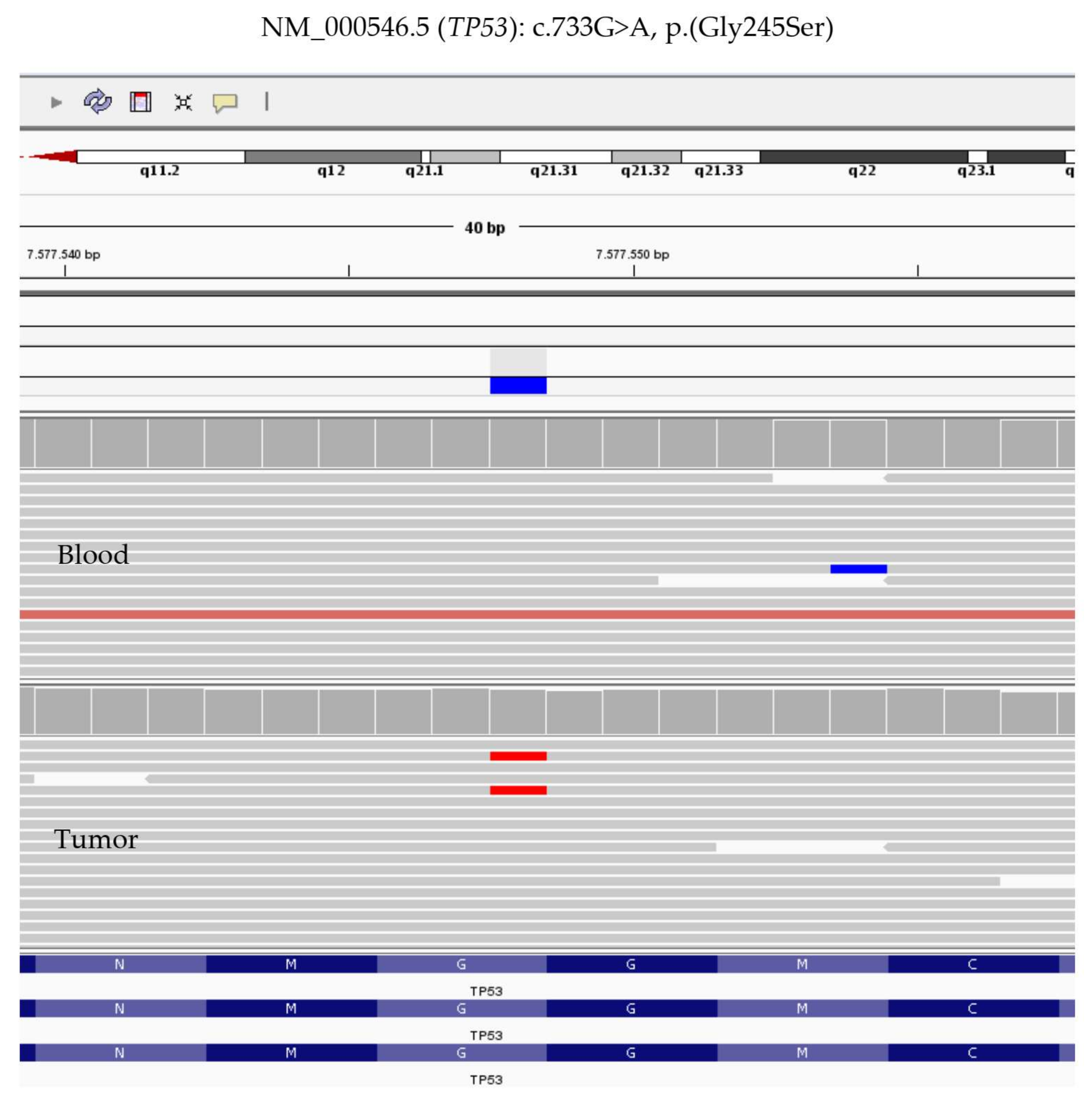
| EPCAM | NM_002354.2 | c.-40C > T | rs747979626 | VUS | 2:47596605 |
| MSH6 | NM_000179.2 | c.116G > A p.Gly39Glu | rs1042821 | BEN | 2:48010488 |
| TGFBR2 | NM_001024847.2 | c.118G > A p.Asp40Asn | rs61732532 | VUS | 3:30664714 |
| TGFBR2 | NM_001024847.2 | c.455-4T > A | rs11466512 | BEN | 3:30713126 |
| MSH3 | NM_002439.4 | c.154_171del p.Ala52_Ala57del | rs201874762 | BEN | 5:79950699 |
| MSH3 | NM_002439.4 | c.196_204del p.Pro66_Ala68del | rs879531814 | BEN | 5:79950741 |
| MSH3 | NM_002439.4 | c.359-7G > A | rs1382543 | BEN | 5:79960955 |
| APC | NM_000038.5 | c.5465T > A p.Val1822Asp | rs459552 | BEN | 5:112176756 |
| PMS2 | NM_000535.6 | c.2007-4G > A | rs1805326 | BEN | 7:6022626 |
| EGFR | NM_005228.3 | c.1562G > A p.Arg521Lys | rs2227983 | BEN | 7:55229255 |
| MLH3 | NM_014381.2 | c.2531C > T p.Pro844Leu | rs175080 | BEN | 14:75513828 |
| MLH3 | NM_014381.2 | c.1870G > C p.Glu624Gln | rs28756986 | BEN | 14:75514489 |
| NRAS | NM_002524 | no variants found | |||
| KRAS | NM_033360 | no variants found | |||
| BRAF | NM_004333 | no variants found | |||
| APC | NM_000038 | no variants found | |||
| MLH1 | NM_000249 | no variants found | |||
© 2020 by the authors. Licensee MDPI, Basel, Switzerland. This article is an open access article distributed under the terms and conditions of the Creative Commons Attribution (CC BY) license (http://creativecommons.org/licenses/by/4.0/).
Share and Cite
De Pasquale, M.D.; Crocoli, A.; Caldaro, T.; Rinelli, M.; Spinelli, G.P.; Francalanci, P.; Cozza, R.; Inserra, A.; Miele, E. Targeting Epidermal Growth Factor Receptor (EGFR) in Pediatric Colorectal Cancer. Cancers 2020, 12, 414. https://doi.org/10.3390/cancers12020414
De Pasquale MD, Crocoli A, Caldaro T, Rinelli M, Spinelli GP, Francalanci P, Cozza R, Inserra A, Miele E. Targeting Epidermal Growth Factor Receptor (EGFR) in Pediatric Colorectal Cancer. Cancers. 2020; 12(2):414. https://doi.org/10.3390/cancers12020414
Chicago/Turabian StyleDe Pasquale, Maria Debora, Alessandro Crocoli, Tamara Caldaro, Martina Rinelli, Gian Paolo Spinelli, Paola Francalanci, Raffaele Cozza, Alessandro Inserra, and Evelina Miele. 2020. "Targeting Epidermal Growth Factor Receptor (EGFR) in Pediatric Colorectal Cancer" Cancers 12, no. 2: 414. https://doi.org/10.3390/cancers12020414
APA StyleDe Pasquale, M. D., Crocoli, A., Caldaro, T., Rinelli, M., Spinelli, G. P., Francalanci, P., Cozza, R., Inserra, A., & Miele, E. (2020). Targeting Epidermal Growth Factor Receptor (EGFR) in Pediatric Colorectal Cancer. Cancers, 12(2), 414. https://doi.org/10.3390/cancers12020414