Pheochromocytomas and Paragangliomas: New Developments with Regard to Classification, Genetics, and Cell of Origin

1
Martini Hospital, 9728 NT Groningen, The Netherlands
2
Department of Pathology, Isala Hospital, 8025AB Zwolle, The Netherlands
3
Department of Pathology, University Medical Center Utrecht, 3584 CX Utrecht, The Netherlands
4
Princess Maxima Center for Pediatric Oncology, 3584 CS Utrecht, The Netherlands
*
Author to whom correspondence should be addressed.
Cancers 2019, 11(8), 1070; https://doi.org/10.3390/cancers11081070
Submission received: 19 June 2019
/
Revised: 22 July 2019
/
Accepted: 23 July 2019
/
Published: 29 July 2019
Abstract
:Pheochromocytomas (PCC) and paragangliomas (PGL) are rare neuroendocrine tumors that arise in the adrenal medulla and in extra-adrenal locations, such as the head, neck, thorax, abdomen, and pelvis. Classification of these tumors into those with or without metastatic potential on the basis of gross or microscopic features is challenging. Recent insights and scoring systems have attempted to develop solutions for this, as described in the latest World Health Organization (WHO) edition on endocrine tumor pathology. PCC and PGL are amongst the tumors most frequently accompanied by germline mutations. More than 20 genes are responsible for a hereditary background in up to 40% of these tumors; somatic mutations in the same and several additional genes form the basis for another 30%. However, this does not allow for a complete understanding of the pathogenesis or targeted treatment of PCC and PGL, for which surgery is the primary treatment and for which metastasis is associated with poor outcome. This review describes recent insights into the cell of origin of these tumors, the latest developments with regard to the genetic background, and the current status of tumor classification including proposed scoring systems.
1. Introduction
Pheochromocytomas (PCC) are rare neuroendocrine tumors that, by definition, arise in the adrenal medulla; however, the term paraganglioma (PGL) is reserved for tumors that arise from sympathetic ganglia in the thorax, abdomen, and pelvis as well as from parasympathetic paraganglia in the head and neck area. Although these tumors are rare, with an incidence of approximately five per million, they are relevant from various perspectives, including pathophysiology and genetic background. Such tumors also have a relationship with various other tumors, such as neuroblastomas, that also originate from the adrenal medulla and extra-adrenal sympathetic tissues as well as tumors co-occurring in various hereditary tumor syndromes, for instance, medullary thyroid carcinoma in MEN2, renal cell carcinoma in VHL disease, and gastrointestinal stromal tumors (GIST) in the context of succinate dehydrogenase subunit gene (collectively called SDHx) mutations.
PCC and PGL are composed of chromaffin cells, deriving from the adrenal medulla and sympathetic ganglia [1]. Previously, these chromaffin cells were thought to develop from a common neural crest precursor cell that migrated to the dorsal aortic area and also produced sympathetic neurons [2,3]. However, several recent studies have challenged the notion of the common ancestry of chromaffin cells and sympathetic neurons. These studies have shown that the progenitor cells have partly overlapping but distinct expression profiles [4,5,6,7]. Chromaffin cells, which form the chief component of the adrenal medulla, have been shown to derive from peripheral glial stem cells, known as Schwann cell precursors. These have a limited capability for expansion and migrate along preganglionic nerves to reach their adrenal medulla destination. These new findings are supported by genetic ablation experiments and further supported by distinct gene expression programs, such as detection by single cell RNA sequencing experiments. However, it has been shown recently that divergence is not absolute and that Schwann cell precursors appear to be responsible for a small but relevant fraction of sympathetic neurons; additionally, a proportion of chromaffin cells may derive from migrating neural crest cells [8]. It will be challenging to relate these findings to the development of PCC and PGL as well as to relate this work to the origin of neuroblastomas, the infantile and pediatric tumor originating from the same locations.
In this review, two major issues that have dominated the PCC and PGL field with regard to patient management and research will be discussed, with an emphasis on the latest developments. One is the genetic background of PCC and PGL, which have been shown to be among the tumors with the highest rate of hereditary predisposition. Currently, up to 35–40% of PCC and PGL carry germline mutations, divided amongst more than 20 different genes but functionally clustered into two major clusters [9]. We will focus on the more recently discovered genes and on SDHx genes, which are relevant for their association with metastatic potential. The other issue concerns the histopathological classification, in which the distinction between benign (non-metastatic) and potentially metastatic tumors on the basis of one or multiple criteria with sufficient sensitivity and specificity to be clinically useful has been virtually impossible. Currently, as discussed in the most recent version of the WHO volume on Endocrine Tumors, all PCC and PGL are considered to have some metastatic potential [10]. The current need is to recognize those tumors that have a sufficiently high metastatic potential to warrant close follow-up or treatment. Two recent histopathological scoring systems, correlating with metastatic potential, hold some promise for this purpose, including the Pheochromocytoma of the Adrenal gland Scaled Score (PASS) and the Grading system for Pheochromocytoma and Paraganglioma (GAPP) [11,12], both of which will be discussed.
2. Genetics
Knowledge of molecular abnormalities of PCC and PGL has rapidly grown over the past decades. Germline and somatic mutations in at least 20 susceptibility genes have been reported in hereditary and sporadic tumors. Currently, up to 40% of PCC/PGL have a hereditary origin and can be identified in another 30% somatic mutations. It has been shown that germline as well as somatic mutations are largely mutually exclusive; however, several examples of concurring mutations have been reported [9,10,13,14].
On the basis of their transcriptional profile, PCC and PGL have been divided into two clusters. Cluster 1 is characterized by the pseudohypoxia pathway and includes, among others, PCC/PGL with VHL and SDHx mutations. Cluster 2 is characterized by activation of kinase signaling pathways and includes, among others, PCC/PGL with RET and NF1 gene mutations [15,16,17,18,19,20,21]. With the identification of new somatic drivers, including gene fusions, a new cluster 3 has been recognized, representing tumors with Wnt signaling activation. This cluster includes PCC/PGL with MAML3 fusions, most commonly UBTF-MAML3, and somatic mutations in CSDE1 and may be associated with more aggressive disease [17,22,23,24]. A fourth cluster with tumors with adrenocortical features has also been suggested, but this cluster is less well defined and lacks consistent mutations [14,22]. In addition, somatic mutations in more common cancer-related genes, such as TP53 and BRAF, have been associated with PCC/PGL [10,15]. Furthermore, “disease modifying genes” have been recognized, including somatic mutations in ATRX, which mostly co-exist with mutations in SDHB or IDH1/2, with a suggested synergistic effect on tumorigenesis and tumor progression [15,24,25,26]. A discussion of all the gene mutations, proposed mechanisms, and associated conditions is beyond the scope of this review. We will focus on the driver mutations in Clusters 1 and 2, discuss the most common members with special attention to the SDHx family, and highlight some recently identified gene mutations, as seen in Table 1.
2.1. Cluster 1
This cluster represents germline or somatic gene mutations resulting in a dysfunctional hypoxic (pseudo-hypoxic) response with a central role for hypoxia inducible factor (HIF)1 alpha and HIF2 alpha, which are the main components of the response to low oxygen levels. Under normal conditions, HIF1 alpha and HIF2 alpha are in an inactivated state. Under hypoxic conditions or in the context of a dysfunctional system, HIFs are stabilized and activate target genes inducing angiogenesis, metabolism, apoptosis, and proliferation. The cluster can be divided into two subgroups. The VHL/EPAS1-related subgroup includes mutations in genes directly involved in activation of the HIF signaling pathway such as VHL germline (g), somatic (s), EPAS1/HIF2 alpha (g,s) and EGLN1/2 (g). The TCA-cycle-related subgroup includes mutations in genes encoding energy metabolism enzymes such as SDHx (g), FH (g), MDH2 (g) and IDH1/2 (s). Inactivating mutations lead to an accumulation of TCA metabolites causing epigenetic changes in HIF stability and are associated with a hypermethylated phenotype [13,16,17,20,21,23,24,25,27].
2.1.1. VHL/EPAS1-Related
The VHL gene encodes two proteins, pVHL30 and pVHL19. pVHL plays a critical role in degradation of HIF alpha, and pVHL dysfunction results in a pseudohypoxic response. The VHL gene is known to cause von Hippel-Lindau (VHL) disease, an autosomal dominant familial tumor syndrome characterized by multiple benign and malignant tumors including retinal and central nervous system hemangioblastomas, renal cysts, clear cell renal cell carcinomas (RCC), pancreatic cysts, pancreatic neuroendocrine tumors, endolymphatic sac tumors, and epididymal cystadenomas. VHL disease shows a genotype–phenotype correlation; VHL type 2 is characterized by missense mutations and is linked to PCC, which is observed in 10–26% of VHL patients. Germline mutations in VHL account for 5–10% of hereditary PCC/PGL, and somatic VHL mutations have been found in about 10% of sporadic PCC/PGL [10,28,29].
The EPAS1/HIF2 alpha gene encodes for HIF2 alpha and mutations lead to its reduced degradation and stabilization. Somatic EPAS1/HIF2 alpha mutations have been found in 5–10% of PCC/PGL. In addition, germline mutations have been identified, but they comprise <1% of PCC/PGL. EPAS1/HIF2 alpha mutations are also linked to polycythemia and somatostinoma [30,31,32].
2.1.2. Tricarboxylic Acid TCA Cycle-Related
Mutations in the SDHx family are currently the most common germline mutations found in hereditary PCC/PGL and account for about 20–30% of hereditary cases. Somatic mutations have rarely been reported [35]. SDHx-related PCC/PGL are caused by mutations in five genes of the SDHx family, SDHA, SDHB, SDHC, SDHD, and SDHAF2, which encode the corresponding proteins. The SDH complex, composed of the four subunits SDHA–D, is a key component of the TCA cycle and the electron transport chain and is located at the inner mitochondrial membrane. SDHAF2 is essential in the assembly of the complex. The complex catalyzes the oxidation of succinate to fumarate. Loss of SDH function results in accumulation of succinate, which inhibits the prolyl hydroxylation of HIF1 alpha, resulting in a hypoxic response [36,37]. Mutations in the SDHx family are predominantly linked to extra-adrenal PGLs. SDHB mutations are associated with abdominal and thoracic PGL and show a 30–70% risk of metastasis. SDHD and SDHA mutations are mostly found in abdominal, thoracic, head, and neck PGLs. SDHC and SDHAF2 mutations are associated with head and neck PGLs [10]. Mutations in the SDHD and SDHAF2 genes, both located on chromosome 11, show a parent-of-origin-dependent inheritance effect in which mutations almost exclusively cause disease after paternal transmission. Somatic loss of maternal chromosome 11 plays an important role in this effect according to the Hensen model [38,39]. Mutations in the SDHx family have also been associated with other tumors such as SDH-deficient GIST, SDH-deficient RCC, and SDH-deficient pituitary adenoma [10].
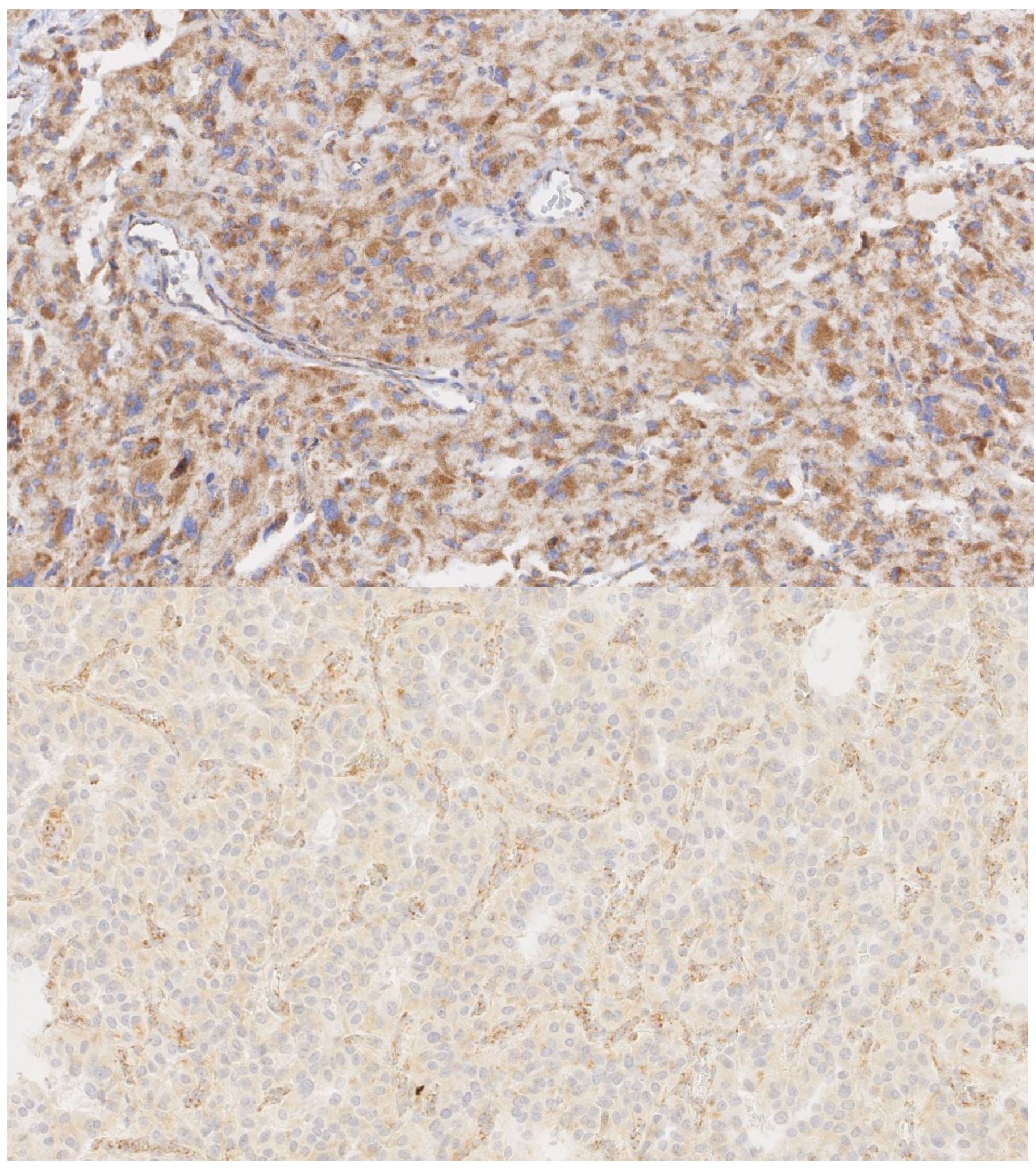
If the SDH complex becomes instable by dysfunction of any of the components, the SDHB subunit is degraded in the cytoplasm. The loss of SDHB expression can be detected by SDHB protein immunohistochemistry (IHC); this can be used to detect tumors with mutations in one of the five genes. In addition, loss of SDHA IHC can detect tumors with SDHA germline mutations [40,41,42,43]. The use of SDHB/SDHA IHC has been suggested as a supplementary tool in guiding molecular genetic testing. Correct interpretation of SDHB/SDHA IHC is important because false positive as well as false negative results can lead to incorrect interpretations or inappropriate genetic testing. SDHB IHC positivity comprises granular cytoplasmatic staining with the same intensity as the internal positive control cells (non-tumoral cells, e.g., lymphocytes, endothelial cells, fibroblasts). If the tumor cells show absent, less intense, or blush-like non-granular staining, SDHB IHC should be regarded as negative, as seen in Figure 1. Furthermore, interpretation of SDHB IHC in tumor areas with clear cytoplasm is not recommended because staining might appear negative. SDHB IHC negativity has also been shown in tumors with SDHC promoter methylation in the absence of SDHx germline mutations. In addition, SDHB negative PCC/PGL without known pathogenic SDHx mutations or SDHC promoter methylation have been described, suggesting that SDHB IHC might be of additional value in the assessment of SDHx genetic variants of unknown significance [42,43].
2.1.3. FH, MDH2, IDH1/IDH2
The FH gene encodes fumarate hydratase, a TCA enzyme which converts fumarate into malate. Mutant FH results in accumulation of fumarate leading to PHD inactivation and HIF stabilization. Mutations in this gene have classically been associated with hereditary cutaneous and uterine leiomyomatosis and a specific variant of RCC, which is regarded as a separate entity by the WHO [44]. Recently, germline mutations have been identified in PCC/PGL, accounting for <5% of PCC/PGL [45]. FH IHC can show loss of expression in FH-mutated PCC/PGL [46]. In addition, low tumor levels of 5-hmC (resembling those in SDHB-deficient tumors) and positive 2SC staining have been detected in tumors with FH mutations, suggestive of altered DNA methylation and protein succination, respectively [47]. The MDH2 gene encodes MDH2, another TCA enzyme that catalyzes the reversible oxidation of malate to oxaloacetate. MDH2 dysfunction leads to accumulation of metabolites and a hypermethylator phenotype. Germline mutations have been identified in less than 1% PCC/PGL and show undetectable 5-hmC staining. The isodehydrogenase (IDH) enzymes IDH1 and IDH2, encoded by IDH1 and IDH2, convert isocitrate to alpha-ketoglutarate in the TCA cycle. Mutations result in the production of 2-hydroxyglutarate instead of alpha-ketoglutarate, and its accumulation activates the hypoxic pathway. Somatic IDH1 and IDH2 mutations are typically present in low-grade and secondary high-grade gliomas and have rarely been found in PGLs [48,49].
2.1.4. Recently Identified PCC/PGL Genes
Germline mutations in other genes with a TCA-related function and/or a hypermethylated phenotype have been recently described in 1% or fewer of PCC/PGL and might be included in this cluster. The SLC25A11 gene encodes the mitochondrial 2-oxoglutarate/malate carrier and germline mutations have been identified in abdominal as well as head and neck PGLs and may be associated with metastases [50]. The IDH3B gene encodes IDHB3, an enzyme involved in the oxidation of isocitrate to alpha-ketoglutarate in the TCA cycle, and a germline mutation was identified in a jugular PGL [48]. GOT2 is another mitochondrial enzyme involved in amino acid metabolism as well as the urea and TCA cycle; additionally, a germline mutation in the encoding GOT2 gene has been found in metastatic abdominal and thoracic PGL with a high succinate/fumarate ratio in tumor cells [48]. The DNMT3A gene plays a role in DNA methylation during embryonic development and germline mutations have been found in multifocal PGLs [51]. The DLST gene encodes a mitochondrial protein that belongs to the 2-oxoacid dehydrogenase family. Germline mutations have recently been found in PCC/PGL with an expression and methylation profile suggestive of the pseudohypoxic pathway. In addition, positive DLST immunostaining was detected in tumors with TCA-cycle or EPAS1 mutations [52].
2.2. Cluster 2
The common denominator for Cluster 2 is the activation of kinase signaling pathways. It encompasses germline or somatic mutations in RET (g/s), NF1 (g/s), MAX (g/s), MEN1 (g), TMEM127 (g), H-RAS (s), and KIF1B (g/s). These mutations are associated with the activation of the RAS-RAF-ERK, PI3K-AKT-mTOR (RET, HRAS, NF1, TMEM127, KIF1B), and MYC-MAX (MAX) kinase signaling pathways. The activation or deregulation of these pathways leads to uncontrolled proliferation, growth, and cell survival [13,17,21,23,24,25].
The RET proto-oncogene encodes a transmembrane tyrosine kinase. When ligand binds to the RET receptor or if there is an activating mutation, a signaling cascade is triggered through the PI3 kinase pathway to regulate cell proliferation and apoptosis. Activating germline mutations in the RET gene cause the multiple endocrine neoplasia type 2 (MEN2) syndrome characterized by the development of PCC and medullary thyroid carcinoma. PCC occur in 40–50% of patients with MEN2 and the risk of PCC is associated with specific RET mutations. Clues to MEN2-related PCC include the presence of multiple, bilateral tumor nodules and adrenal medullary hyperplasia. Germline RET mutations have been detected in 5% of hereditary PCC/PGL and somatic mutations in about 5% of sporadic tumors [29,53,54].
The NF1 gene encodes neurofibromin 1, which is a negative regulator of the RAS intracellular signaling pathway. Inactivating mutations in the NF1 gene disrupt this inhibitory effect. NF1 is the most common somatically mutated gene in PCC/PGL, accounting for 20–40% of sporadic PCC/PGL. However, only approximately 6% of patients with the autosomal dominant NF1 syndrome, characterized by germline NF1 mutations, develop PCC, and germline mutations account for <5% of all PCC/PGL. Interestingly, up to 16% of NF1-related PCC show a composite histological phenotype of typical PCC intermixed with areas of ganglioneuroblastoma or ganglioneuroma [9,55].
TMEM 127 is a tumor suppressor gene linked with the mTOR kinase pathway, and germline mutations have been found in less than 5% of PCC/PGL. It encodes a transmembrane protein that functions as a negative regulator of mTOR. Mutant forms of the protein are nonfunctional and cause cytoplasmic localization of the protein. Notably, mutant alleles show relatively low penetrance [56,57].
MAX is a tumor suppressor gene with a regulatory role in the MYC-MAX-MD1 network. Truncated MAX mutations can cause dysregulation of the mTOR pathway. Germline mutations, which may exhibit a parent-of-origin effect with paternal transmission, as well as somatic mutations have been found in PCC/PGL, both in less than 5% of cases [58,59]. Although MAX IHC has been described in the literature, with the potential to detect MAX mutations in patients in case of absent immunostaining, comparable to negative SDHB staining, this has not found widespread application [59].
H-RAS is a proto-oncogene that encodes H-RAS protein, which can bind to GTP and activate the RAS/RAF/ERK signaling pathway leading to cell proliferation. Missense somatic mutations have been found in 5–10% of sporadic PCC/PGL [60,61].
The KIF1B gene encodes for two proteins isoforms, KIF1B alpha and KIF1B beta. KIF1B alpha is involved in the transport of mitochondria, and KIF1B beta in the transport of synaptic vesicle precursors. In addition to its transport function, KIF1B beta plays a role in apoptosis and dysfunctional KIF1B beta due to mutations in the KIF1B gene and can lead to tumorigenesis. The KIF1B gene might be one of the most frequently somatically mutated genes in PCC/PGL, with somatic mutations seen in up to 20% of cases, and also germline mutations have been described in fewer than 1%. KIF1B mutant PCC/PGL shows a similar transcription profile as RET and NF1 mutant PCC/PGL [62,63].
The MEN1 gene codes for the menin protein, which interacts with transcription factor JunD. JunD is a functional component of the AP1 transcription factor complex. It has been proposed to protect cells from p53-dependent senescence and apoptosis. Germline mutations in the MEN1 gene lead to MEN1 syndrome, characterized by various types of endocrine and non-endocrine tumors, including pituitary adenomas, hyperplastic parathyroids, and neuroendocrine pancreatic tumors, with a high penetrance of disease but PCC/PGL are rare and account for <1% of PCC/PGL [64,65,66].
3. Histopathological Classification
All PCC/PGL are believed to exhibit some metastatic potential, but only a subset of these tumors will actually metastasize. Until 2004, malignancy was defined by either metastasis or on the basis of extensive local invasion. The 2004 WHO classification defined malignant PCC by the development of metastasis. Due to incongruity, confusion was noted and the current WHO classification encourages the use of the terms “metastatic” and “non-metastatic”. Many histomorphological features and immunohistochemical markers have been studied to find biomarkers to differentiate between metastasizing and non-metastasizing PCC/PGL. However, no single histomorphological or immunohistochemical feature has indicated metastatic potential. Therefore, several algorithms, on the basis of multiple histologic properties, have been described to detect potential for aggressive behavior.
In 1990, Linnoila examined 120 PCC/PGL and developed a statistical model to predict malignancy. According to this model, there was a 95% probability that more than 70% of tumors could be classified correctly on the basis of four factors: extra-adrenal location, coarse nodularity, confluent tumor necrosis, and absence of hyaline globules. The majority of malignant PCC/PGL had two or three of these features (71%) while 89% of benign tumors had none or one [67]. Unfortunately, malignancy in this study was defined by regional or distant metastases and/or extensive local invasion.
3.1. Pheochromocytoma of the Adrenal Gland Scaled Score (PASS)
Thompson described the Pheochromocytoma of the Adrenal gland Scaled Score (PASS), which consists of twelve parameters: large nests or diffuse growth, central or confluent necrosis, high cellularity, cellular monotony, tumor cell spindling, >3 mitotic figures/10 high power fields, atypical mitotic figures, extension into adipose tissue, vascular invasion, capsular invasion, profound nuclear pleomorphism, and nuclear hyperchromasia. A maximum score of 20 points is obtained when all features are present. Tumors with a score <4 were considered to have no metastatic potential. Tumors with a score ≥4 were considered to have an increased metastatic potential, as seen in Table 2 [11].
Commonly agreed upon features are the presence of atypical mitosis, necrosis, capsular involvement, lymphovascular invasion, and extension into adipose tissue. However, significant interobserver and intraobserver variability is described in scoring the presence or absence of high cellularity, nuclear pleomorphism, and hyperchromasia [68]. A meta-analysis of the PASS algorithm found a sensitivity of 97% and a specificity of 68%. The positive predictive value (PPV) was 31%, and the negative predictive value (NPV) 99%. These findings suggest that a PASS score of <4 is highly indicative of a benign clinical course, but for tumors with PASS score of ≥4, the predictive value with regard to disease course is limited [69].
3.2. Grading of Adrenal Pheochromocytoma and Paraganglioma (GAPP)
Although PASS only applies to PCC, the Grading of Adrenal Pheochromocytoma and Paraganglioma (GAPP) is a tool for PCC as well as PGL. It is based on histological pattern, cellularity, comedo-type necrosis, capsular invasion, vascular invasion, Ki67 labelling index and catecholamine type. On the basis of a maximum score of 10 points, tumors are graded as well differentiated (0–2 points), moderately differentiated (3–6 points), and poorly differentiated (7–10 points), as seen in Table 2. The five-year survival rates in these groups are 100% for well differentiated, 66.8% for moderately differentiated and 22.4% for poorly differentiated tumors. Kimura also found that Ki67 was significantly different between the metastatic and non-metastatic group [12]. Using a 3% cutoff, Ki-67 shows a sensitivity, specificity, positive predictive value, and negative predictive value of 55.6%, 94.9%, 83.3%, and 82.2%, respectively [70].
A sensitivity of 50% and specificity of 80% has been described for PCC with GAPP. In addition, the PPV was 5% and NPV was 99%. For PGL, the sensitivity was 100% and the specificity was 68%, with a PPV of 29% and a NPV of 100% [69].
Both PASS and GAPP do not account for the effect of underlying somatic or germline mutations, whether these are “low risk driver mutations” or “high risk driver mutations”. For example, PCC in MEN 2A typically have a low risk of metastases, with an estimated prevalence of 3% [71]. However, in almost 50% of the tumors, PASS scores of ≥4, which are indicative of potential aggressive behavior, were found. In addition the same tumors also displayed an elevated GAPP and were scored as moderately differentiated. The most frequently reported histopathological characteristics in these tumors were the presence of large nests, diffuse growth, and high cellularity. Thirty percent of the MEN 2A-related PCC displayed a Ki67 labelling index of >3%, making it the most common GAPP criterion. These findings suggest that both algorithms are inadequate to determine metastatic potential in MEN2A-related PCC [72].
Recurrence and metastases are strongly associated with SDHB mutations; the presence of SDHB mutations should be considered a risk factor for metastases or recurrence [73]. As described above, SDHB IHC is a strong indication for tumors with SDHx mutations. Loss of SDHB staining is not seen either in prognostically unfavorable SDHB-mutated tumors or in all tumors with SDHx mutations; however, SDHB IHC negativity by itself has also been correlated with metastases [42,43]. In a GAPP follow-up study, SDHB IHC served as a surrogate marker for genetic testing. All tumors lacking SDHB staining were intermediate or high grade, and 10 out of 13 had metastasized [12]. Additionally, MAML3 fusion gene variants, leading to Wnt signaling upregulation, as seen in Cluster 3, described above, are associated with metastases. In the same study, Ki67 was found to be correlated with metastatic disease. Interestingly, the tumor with the highest Ki67 expression was MAML3 fusion-positive [22]. Another study assessed promoter methylation density of tumor suppressor genes with frequent hypermethylation in cancer. Fourteen PGL, six metastatic and eight non-metastatic, as well as two distant metastases were significantly methylated at one or more of the following gene promoters, RASSF1A, NORE1A, p16INK4A, RARB, DCR2, CDH1, and APC. Methylation of three or more promoters was found in five PGL, of which four were metastatic [74]. Despite the promising histomorphological, immunohistochemical, and molecular biomarkers described above, there are no criteria with sufficient sensitivity and specificity to accurately predict metastatic potential.
4. Discussion/Conclusions
Recently, PCC and PGL have proven to be enigmatic tumors with regard to the prediction of their clinical behavior; they are also exciting tumors with regard to their pathophysiology and genetic background. Major progress has been made in various fields, although the survival of metastatic PCC and PGL is still bleak. In the field of PCC and PGL genetics, the number of genes involved continues to increase, with a particular focus on enzymes and other proteins that are involved in the TCA cycle. With the advent of whole exome and whole genome sequencing, more such genes may be detected, all of which may potentially converge along the same pathophysiological pathway. Whole exome sequencing is expected to become the standard of genetic analysis in the next 5–10 years, allowing rapid detection of mutations in PCC and PGL patients and families. IHC, for instance, with SDHA and SDHB antibodies, may serve as a confirmatory technique with regard to the classification of variants of unknown significance. With regard to classification, the most important new developments can be found in the 2017 edition of the WHO volume on Endocrine Tumors, in which all PCC and PGL are now considered to have some metastatic potential and that the pathological grading system for PCC and PGL (GAPP), developed by Kimura, has been independently validated. In addition, a modified GAPP (M-GAPP) has been proposed, incorporating loss of SDHB staining as a criterion, which seems logical in light of the increased metastatic risk related to SDHB mutations [75]. However, the loss of SDHB staining occurs with all SDHx mutations, and increased risk has not been noted for SDHA, SDHC, and SDHD mutations. Both PASS and GAPP have a low PPV but a high NPV, suggesting that these models are good at ruling out but poor at predicting metatastatic potential.
Finally, although outside the scope of this review, the improved knowledge of the genetic background and the genes involved in the pathogenesis of PCC and PGL will allow for the development of targeted therapies for those tumors that cannot be resected surgically or that have metastasized, potentially improving prognosis for these patients.
Funding
This research received no external funding.
Acknowledgments
The authors would like to thank Lindsey Oudijk for the Figure and Marijn Vermeulen for English language editing.
Conflicts of Interest
The authors declare no conflict of interest.
References
- Lenders, J.W.M.; Duh, Q.; Eisenhofer, G.; Gimenez-Roqueplo, A.; Grebe, S.K.G.; Murad, M.H.; Naruse, M.; Pacak, K.; Young, W.F. Pheochromocytoma and Paraganglioma: An Endocrine Society Clinical Practice Guideline. J. Clin. Endocrinol. Metab. 2014, 99, 1915–1942. [Google Scholar] [CrossRef] [PubMed]
- Huber, K.; Kalcheim, C.; Unsicker, K. The Development of the Chromaffin Cell Lineage from the Neural Crest. Auton. Neurosci. 2009, 151, 10–16. [Google Scholar] [CrossRef] [PubMed]
- Saito, D.; Takase, Y.; Murai, H.; Takahashi, Y. The Dorsal Aorta Initiates a Molecular Cascade that Instructs Sympatho-Adrenal Specification. Science 2012, 336, 1578–1581. [Google Scholar] [CrossRef] [PubMed]
- Chan, W.H.; Gonsalvez, D.G.; Young, H.M.; Southard-Smith, E.M.; Cane, K.N.; Anderson, C.R. Differences in CART Expression and Cell Cycle Behavior Discriminate Sympathetic Neuroblast from Chromaffin Cell Lineages in Mouse Sympathoadrenal Cells. Dev. Neurobiol. 2016, 76, 137–149. [Google Scholar] [CrossRef] [PubMed]
- Ernsberger, U.; Esposito, L.; Partimo, S.; Huber, K.; Franke, A.; Bixby, J.L.; Kalcheim, C.; Unsicker, K. Expression of Neuronal Markers Suggests Heterogeneity of Chick Sympathoadrenal Cells Prior to Invasion of the Adrenal Anlagen. Cell Tissue Res. 2005, 319, 1–13. [Google Scholar] [CrossRef] [PubMed]
- Furlan, A.; Dyachuk, V.; Kastriti, M.E.; Calvo-Enrique, L.; Abdo, H.; Hadjab, S.; Chontorotzea, T.; Akkuratova, N.; Usoskin, D.; Kamenev, D.; et al. Multipotent Peripheral Glial Cells Generate Neuroendocrine Cells of the Adrenal Medulla. Science 2017, 357. [Google Scholar] [CrossRef]
- Lumb, R.; Tata, M.; Xu, X.; Joyce, A.; Marchant, C.; Harvey, N.; Ruhrberg, C.; Schwarz, Q. Neuropilins Guide Preganglionic Sympathetic Axons and Chromaffin Cell Precursors to Establish the Adrenal Medulla. Development 2018, 145. [Google Scholar] [CrossRef]
- Kastriti, M.E.; Kameneva, P.; Kamenev, D.; Dyachuk, V.; Furlan, A.; Hampl, M.; Memic, F.; Marklund, U.; Lallemend, F.; Hadjab, S.; et al. Schwann Cell Precursors Generate the Majority of Chromaffin Cells in Zuckerkandl Organ and some Sympathetic Neurons in Paraganglia. Front. Mol. Neurosci. 2019, 12, 6. [Google Scholar] [CrossRef]
- Fishbein, L.; Nathanson, K.L. Pheochromocytoma and Paraganglioma: Understanding the Complexities of the Genetic Background. Cancer Genet. 2012, 205, 1–11. [Google Scholar] [CrossRef]
- Loyd, R.V.; Osamura, R.Y.; Kloppel, G.; Rosai, J. WHO Classification of Tumours of Endocrine System; International Agency for Research on Cancer (IARC): Lyon, France, 2017. [Google Scholar]
- Thompson, L.D. Pheochromocytoma of the Adrenal Gland Scaled Score (PASS) to Separate Benign from Malignant Neoplasms: A Clinicopathologic and Immunophenotypic Study of 100 Cases. Am. J. Surg. Pathol. 2002, 26, 551–566. [Google Scholar] [CrossRef]
- Kimura, N.; Takayanagi, R.; Takizawa, N.; Itagaki, E.; Katabami, T.; Kakoi, N.; Rakugi, H.; Ikeda, Y.; Tanabe, A.; Nigawara, T.; et al. Pathological Grading for Predicting Metastasis in Phaeochromocytoma and Paraganglioma. Endocr. Relat. Cancer 2014, 21, 405–414. [Google Scholar] [CrossRef]
- Dahia, P.L. Pheochromocytoma and Paraganglioma Pathogenesis: Learning from Genetic Heterogeneity. Nat. Rev. Cancer 2014, 14, 108–119. [Google Scholar] [CrossRef]
- Dahia, P.L.M. Pheochromocytomas and Paragangliomas, Genetically Diverse and Minimalist, all at Once! Cancer Cell 2017, 31, 159–161. [Google Scholar] [CrossRef]
- Zhikrivetskaya, S.O.; Snezhkina, A.V.; Zaretsky, A.R.; Alekseev, B.Y.; Pokrovsky, A.V.; Golovyuk, A.L.; Melnikova, N.V.; Stepanov, O.A.; Kalinin, D.V.; Moskalev, A.A.; et al. Molecular Markers of Paragangliomas/Pheochromocytomas. Oncotarget 2017, 8, 25756–25782. [Google Scholar] [CrossRef]
- Jochmanová, I.; Yang, C.; Zhuang, Z.; Pacak, K. Hypoxia-Inducible Factor Signaling in Pheochromocytoma: Turning the Rudder in the Right Direction. J. Natl. Cancer Inst. 2013, 105, 1270–1283. [Google Scholar] [CrossRef]
- Jochmanova, I.; Pacak, K. Genomic Landscape of Pheochromocytoma and Paraganglioma. Trends Cancer 2018, 4, 6–9. [Google Scholar] [CrossRef]
- Burnichon, N.; Buffet, A.; Gimenez-Roqueplo, A. Pheochromocytoma and Paraganglioma: Molecular Testing and Personalized Medicine. Curr. Opin. Oncol. 2016, 28, 5–10. [Google Scholar] [CrossRef]
- Kavinga Gunawardane, P.T.; Grossman, A. The Clinical Genetics of Phaeochromocytoma and Paraganglioma. Arch. Endocrinol. Metab. 2017, 61, 490–500. [Google Scholar] [CrossRef]
- Amorim-Pires, D.; Peixoto, J.; Lima, J. Hypoxia Pathway Mutations in Pheochromocytomas and Paragangliomas. Cytogenet. Genome Res. 2016, 150, 227–241. [Google Scholar] [CrossRef]
- Vicha, A.; Musil, Z.; Pacak, K. Genetics of Pheochromocytoma and Paraganglioma Syndromes: New Advances and Future Treatment Options. Curr. Opin. Endocrinol. Diabetes Obes. 2013, 20, 186–191. [Google Scholar] [CrossRef]
- Fishbein, L.; Leshchiner, I.; Walter, V.; Danilova, L.; Robertson, A.G.; Johnson, A.R.; Lichtenberg, T.M.; Murray, B.A.; Ghayee, H.K.; Else, T.; et al. Comprehensive Molecular Characterization of Pheochromocytoma and Paraganglioma. Cancer Cell 2017, 31, 181–193. [Google Scholar] [CrossRef]
- Crona, J.; Taïeb, D.; Pacak, K. New Perspectives on Pheochromocytoma and Paraganglioma: Toward a Molecular Classification. Endocr. Rev. 2017, 38, 489–515. [Google Scholar] [CrossRef]
- Kantorovich, V.; Pacak, K. New Insights on the Pathogenesis of Paraganglioma and Pheochromocytoma. F1000Res 2018, 7. [Google Scholar] [CrossRef]
- Alrezk, R.; Suarez, A.; Tena, I.; Pacak, K. Update of Pheochromocytoma Syndromes: Genetics, Biochemical Evaluation, and Imaging. Front. Endocrinol. 2018, 9, 515. [Google Scholar] [CrossRef]
- Comino-Méndez, I.; Tejera, Á.; .M.;Currás-Freixes, M.; Remacha, L.; Gonzalvo, P.; Tonda, R.; Letón, R.; Blasco, M.A.; Robledo, M.; Cascón, A. ATRX Driver Mutation in a Composite Malignant Pheochromocytoma. Cancer Genet. 2016, 209, 272–277. [Google Scholar]
- Jochmanova, I.; Pacak, K. Pheochromocytoma: The First Metabolic Endocrine Cancer. Clin. Cancer Res. 2016, 22, 5001–5011. [Google Scholar] [CrossRef] [Green Version]
- Maher, E.R.; Neumann, H.P.; Richard, S. Von Hippel-Lindau Disease: A Clinical and Scientific Review. Eur. J. Hum. Genet. 2011, 19, 617–623. [Google Scholar] [CrossRef]
- Lyssikatos, C.; Faucs, F.R.; Stratakis, C.A. Familial Endocrine Tumor Syndromes; Cambridge University Press: Cambridge, UK, 2016. [Google Scholar]
- Zhuang, Z.; Yang, C.; Lorenzo, F.; Merino, M.; Fojo, T.; Kebebew, E.; Popovic, V.; Stratakis, C.A.; Prchal, J.T.; Pacak, K. Somatic HIF2A Gain-of-Function Mutations in Paraganglioma with Polycythemia. N. Engl. J. Med. 2012, 367, 922–930. [Google Scholar] [CrossRef]
- Lorenzo, F.R.; Yang, C.; Ng Tang Fui, M.; Vankayalapati, H.; Zhuang, Z.; Huynh, T.; Grossmann, M.; Pacak, K.; Prchal, J.T. A Novel EPAS1/HIF2A Germline Mutation in a Congenital Polycythemia with Paraganglioma. J. Mol. Med. 2013, 91, 507–512. [Google Scholar] [CrossRef]
- Oudijk, L.; Gaal, J.; Koopman, K.; de Krijger, R.R. An Update on the Histology of Pheochromocytomas: How does it Relate to Genetics? Horm. Metab. Res. 2018. [Google Scholar] [CrossRef]
- Ladroue, C.; Carcenac, R.; Leporrier, M.; Gad, S.; Le Hello, C.; Galateau-Salle, F.; Feunteun, J.; Pouysségur, J.; Richard, S.; Gardie, B. PHD2 Mutation and Congenital Erythrocytosis with Paraganglioma. N. Engl. J. Med. 2008, 359, 2685–2692. [Google Scholar] [CrossRef]
- Yang, C.; Zhuang, Z.; Fliedner, S.M.J.; Shankavaram, U.; Sun, M.G.; Bullova, P.; Zhu, R.; Elkahloun, A.G.; Kourlas, P.J.; Merino, M.; et al. Germ-Line PHD1 and PHD2 Mutations Detected in Patients with Pheochromocytoma/Paraganglioma-Polycythemia. J. Mol. Med. 2015, 93, 93–104. [Google Scholar] [CrossRef]
- van Nederveen, F.H.; Korpershoek, E.; Lenders, J.W.M.; de Krijger, R.R.; Dinjens, W.N.M. Somatic SDHB Mutation in an Extraadrenal Pheochromocytoma. N. Engl. J. Med. 2007, 357, 306–308. [Google Scholar] [CrossRef]
- Turchini, J.; Cheung, V.K.Y.; Tischler, A.S.; De Krijger, R.R.; Gill, A.J. Pathology and Genetics of Phaeochromocytoma and Paraganglioma. Histopathology 2018, 72, 97–105. [Google Scholar] [CrossRef]
- Gill, A.J. Succinate Dehydrogenase (SDH) and Mitochondrial Driven Neoplasia. Pathology 2012, 44, 285–292. [Google Scholar] [CrossRef]
- Hoekstra, A.S.; Hensen, E.F.; Jordanova, E.S.; Korpershoek, E.; van der Horst-Schrivers, A.N.; Cornelisse, C.; Corssmit, E.P.M.; Hes, F.J.; Jansen, J.C.; Kunst, H.P.M.; et al. Loss of Maternal Chromosome 11 is a Signature Event in SDHAF2, SDHD, and VHL-Related Paragangliomas, but Less Significant in SDHB-Related Paragangliomas. Oncotarget 2017, 8, 14525–14536. [Google Scholar] [CrossRef]
- Hensen, E.F.; Jordanova, E.S.; van Minderhout, I.J.H.M.; Hogendoorn, P.C.W.; Taschner, P.E.M.; van der Mey Andel, G.L.; Devilee, P.; Cornelisse, C.J. Somatic Loss of Maternal Chromosome 11 Causes Parent-of-Origin-Dependent Inheritance in SDHD-Linked Paraganglioma and Phaeochromocytoma Families. Oncogene 2004, 23, 4076–4083. [Google Scholar] [CrossRef]
- van Nederveen, F.H.; Gaal, J.; Favier, J.; Korpershoek, E.; Oldenburg, R.A.; de Bruyn, E.M.; Sleddens, H.F.; Derkx, P.; Riviere, J.; Dannenberg, H.; et al. An Immunohistochemical Procedure to Detect Patients with Paraganglioma and Phaeochromocytoma with Germline SDHB, SDHC, Or SDHD Gene Mutations: A Retrospective and Prospective Analysis. Lancet Oncol. 2009, 10, 764–771. [Google Scholar] [CrossRef]
- Korpershoek, E.; Favier, J.; Gaal, J.; Burnichon, N.; van Gessel, B.; Oudijk, L.; Badoual, C.; Gadessaud, N.; Venisse, A.; Bayley, J.; et al. SDHA Immunohistochemistry Detects Germline SDHA Gene Mutations in Apparently Sporadic Paragangliomas and Pheochromocytomas. J. Clin. Endocrinol. Metab. 2011, 96, 1472. [Google Scholar] [CrossRef]
- Papathomas, T.G.; Oudijk, L.; Persu, A.; Gill, A.J.; van Nederveen, F.; Tischler, A.S.; Tissier, F.; Volante, M.; Matias-Guiu, X.; Smid, M.; et al. SDHB/SDHA Immunohistochemistry in Pheochromocytomas and Paragangliomas: A Multicenter Interobserver Variation Analysis using Virtual Microscopy: A Multinational Study of the European Network for the Study of Adrenal Tumors (ENS@T). Mod. Pathol. 2015, 28, 807–821. [Google Scholar] [CrossRef]
- Oudijk, L.; Gaal, J.; de Krijger, R.R. The Role of Immunohistochemistry and Molecular Analysis of Succinate Dehydrogenase in the Diagnosis of Endocrine and Non-Endocrine Tumors and Related Syndromes. Endocr Pathol. 2018. [Google Scholar] [CrossRef] [PubMed]
- Tomlinson, I.P.M.; Alam, N.A.; Rowan, A.J.; Barclay, E.; Jaeger, E.E.M.; Kelsell, D.; Leigh, I.; Gorman, P.; Lamlum, H.; Rahman, S.; et al. Germline Mutations in FH Predispose to Dominantly Inherited Uterine Fibroids, Skin Leiomyomata and Papillary Renal Cell Cancer. Nat. Genet. 2002, 30, 406–410. [Google Scholar] [PubMed]
- Clark, G.R.; Sciacovelli, M.; Gaude, E.; Walsh, D.M.; Kirby, G.; Simpson, M.A.; Trembath, R.C.; Berg, J.N.; Woodward, E.R.; Kinning, E.; et al. Germline FH Mutations Presenting with Pheochromocytoma. J. Clin. Endocrinol. Metab. 2014, 99, 2046. [Google Scholar] [CrossRef] [PubMed]
- Cheung, V.K.Y.; Gill, A.J.; Chou, A. Old, New, and Emerging Immunohistochemical Markers in Pheochromocytoma and Paraganglioma. Endocr. Pathol. 2018, 29, 169–175. [Google Scholar] [CrossRef] [PubMed]
- Castro-Vega, L.J.; Buffet, A.; De Cubas, A.A.; Cascón, A.; Menara, M.; Khalifa, E.; Amar, L.; Azriel, S.; Bourdeau, I.; Chabre, O.; et al. Germline Mutations in FH Confer Predisposition to Malignant Pheochromocytomas and Paragangliomas. Hum. Mol. Genet. 2014, 23, 2440–2446. [Google Scholar] [CrossRef]
- Remacha, L.; Comino-Méndez, I.; Richter, S.; Contreras, L.; Currás-Freixes, M.; Pita, G.; Letón, R.; Galarreta, A.; Torres-Pérez, R.; Honrado, E.; et al. Targeted Exome Sequencing of Krebs Cycle Genes Reveals Candidate Cancer-Predisposing Mutations in Pheochromocytomas and Paragangliomas. Clin. Cancer Res. 2017, 23, 6315–6324. [Google Scholar] [CrossRef] [PubMed]
- Gaal, J.; Burnichon, N.; Korpershoek, E.; Roncelin, I.; Bertherat, J.; Plouin, P.; de Krijger, R.R.; Gimenez-Roqueplo, A.; Dinjens, W.N.M. Isocitrate Dehydrogenase Mutations are Rare in Pheochromocytomas and Paragangliomas. J. Clin. Endocrinol. Metab. 2010, 95, 1274–1278. [Google Scholar] [CrossRef]
- Buffet, A.; Morin, A.; Castro-Vega, L.J.; Habarou, F.; Lussey-Lepoutre, C.; Letouze, E.; Lefebvre, H.; Guilhem, I.; Haissaguerre, M.; Raingeard, I.; et al. Germline Mutations in the Mitochondrial 2-Oxoglutarate/Malate Carrier SLC25A11 Gene Confer a Predisposition to Metastatic Paragangliomas. Cancer Res. 2018, 78, 1914–1922. [Google Scholar] [CrossRef]
- Remacha, L.; Currás-Freixes, M.; Torres-Ruiz, R.; Schiavi, F.; Torres-Pérez, R.; Calsina, B.; Letón, R.; Comino-Méndez, I.; Roldán-Romero, J.M.; Montero-Conde, C.; et al. Gain-of-Function Mutations in DNMT3A in Patients with Paraganglioma. Genet. Med. 2018, 20, 1644–1651. [Google Scholar] [CrossRef]
- Remacha, L.; Pirman, D.; Mahoney, C.E.; Coloma, J.; Calsina, B.; Currás-Freixes, M.; Letón, R.; Torres-Pérez, R.; Richter, S.; Pita, G.; et al. Recurrent Germline DLST Mutations in Individuals with Multiple Pheochromocytomas and Paragangliomas. Am. J. Hum. Genet. 2019, 104, 1008–1010. [Google Scholar] [CrossRef]
- Raue, F.; Frank-Raue, K. Genotype-Phenotype Correlation in Multiple Endocrine Neoplasia Type 2. Clinics 2012, 67, 69–75. [Google Scholar] [CrossRef]
- Korpershoek, E.; Petri, B.; Post, E.; van Eijck, C.H.J.; Oldenburg, R.A.; Belt, E.J.T.; de Herder, W.W.; de Krijger, R.R.; Dinjens, W.N.M. Adrenal Medullary Hyperplasia is a Precursor Lesion for Pheochromocytoma in MEN2 Syndrome. Neoplasia 2014, 16, 868–873. [Google Scholar] [CrossRef]
- Kimura, N.; Watanabe, T.; Fukase, M.; Wakita, A.; Noshiro, T.; Kimura, I. Neurofibromin and NF1 Gene Analysis in Composite Pheochromocytoma and Tumors Associated with Von Recklinghausen’s Disease. Mod. Pathol. 2002, 15, 183–188. [Google Scholar] [CrossRef]
- Neumann, H.P.H.; Sullivan, M.; Winter, A.; Malinoc, A.; Hoffmann, M.M.; Boedeker, C.C.; Bertz, H.; Walz, M.K.; Moeller, L.C.; Schmid, K.W.; et al. Germline Mutations of the TMEM127 Gene in Patients with Paraganglioma of Head and Neck and Extraadrenal Abdominal Sites. J. Clin. Endocrinol. Metab. 2011, 96, 1279. [Google Scholar] [CrossRef]
- Yao, L.; Schiavi, F.; Cascon, A.; Qin, Y.; Inglada-Pérez, L.; King, E.E.; Toledo, R.A.; Ercolino, T.; Rapizzi, E.; Ricketts, C.J.; et al. Spectrum and Prevalence of FP/TMEM127 Gene Mutations in Pheochromocytomas and Paragangliomas. JAMA 2010, 304, 2611–2619. [Google Scholar] [CrossRef]
- Burnichon, N.; Cascon, A.; Schiavi, F.; Morales, N.P.; Comino-Mendez, I.; Abermil, N.; Inglada-Perez, L.; de Cubas, A.A.; Amar, L.; Barontini, M.; et al. MAX Mutations Cause Hereditary and Sporadic Pheochromocytoma and Paraganglioma. Clin. Cancer Res. 2012, 18, 2828–2837. [Google Scholar] [CrossRef]
- Comino-Méndez, I.; Gracia-Aznárez, F.J.; Schiavi, F.; Landa, I.; Leandro-García, L.J.; Letón, R.; Honrado, E.; Ramos-Medina, R.; Caronia, D.; Pita, G.; et al. Exome Sequencing Identifies MAX Mutations as a Cause of Hereditary Pheochromocytoma. Nat. Genet. 2011, 43, 663–667. [Google Scholar] [CrossRef]
- Crona, J.; Delgado Verdugo, A.; Maharjan, R.; Stålberg, P.; Granberg, D.; Hellman, P.; Björklund, P. Somatic Mutations in H-RAS in Sporadic Pheochromocytoma and Paraganglioma Identified by Exome Sequencing. J. Clin. Endocrinol. Metab. 2013, 98, 1266. [Google Scholar] [CrossRef]
- Oudijk, L.; de Krijger, R.R.; Rapa, I.; Beuschlein, F.; de Cubas, A.A.; Dei Tos, A.P.; Dinjens, W.N.M.; Korpershoek, E.; Mancikova, V.; Mannelli, M.; et al. H-RAS Mutations are Restricted to Sporadic Pheochromocytomas Lacking Specific Clinical Or Pathological Features: Data from a Multi-Institutional Series. J. Clin. Endocrinol. Metab. 2014, 99, 1376. [Google Scholar] [CrossRef]
- Evenepoel, L.; Helaers, R.; Vroonen, L.; Aydin, S.; Hamoir, M.; Maiter, D.; Vikkula, M.; Persu, A. KIF1B and NF1 are the most Frequently Mutated Genes in Paraganglioma and Pheochromocytoma Tumors. Endocr. Relat. Cancer 2017, 24, L61. [Google Scholar] [CrossRef]
- Yeh, I.; Lenci, R.E.; Qin, Y.; Buddavarapu, K.; Ligon, A.H.; Leteurtre, E.; Do Cao, C.; Cardot-Bauters, C.; Pigny, P.; Dahia, P.L.M. A Germline Mutation of the KIF1B Beta Gene on 1p36 in a Family with Neural and Nonneural Tumors. Hum. Genet. 2008, 124, 279–285. [Google Scholar] [CrossRef]
- Schussheim, D.H.; Skarulis, M.C.; Agarwal, S.K.; Simonds, W.F.; Burns, A.L.; Spiegel, A.M.; Marx, S.J. Multiple Endocrine Neoplasia Type 1: New Clinical and Basic Findings. Trends Endocrinol. Metab. 2001, 12, 173–178. [Google Scholar] [CrossRef]
- Carty, S.E.; Helm, A.K.; Amico, J.A.; Clarke, M.R.; Foley, T.P.; Watson, C.G.; Mulvihill, J.J. The Variable Penetrance and Spectrum of Manifestations of Multiple Endocrine Neoplasia Type 1. Surgery 1998, 124, 1114. [Google Scholar] [CrossRef]
- Khatami, F.; Tavangar, S.M. Multiple Endocrine Neoplasia Syndromes from Genetic and Epigenetic Perspectives. Biomark Insights 2018, 13. [Google Scholar] [CrossRef]
- Linnoila, R.I.; Keiser, H.R.; Steinberg, S.M.; Lack, E.E. Histopathology of Benign Versus Malignant Sympathoadrenal Paragangliomas: Clinicopathologic Study of 120 Cases Including Unusual Histologic Features. Hum. Pathol. 1990, 21, 1168–1180. [Google Scholar] [CrossRef]
- Wu, D.; Tischler, A.S.; Lloyd, R.V.; DeLellis, R.A.; de Krijger, R.; van Nederveen, F.; Nosé, V. Observer Variation in the Application of the Pheochromocytoma of the Adrenal Gland Scaled Score. Am. J. Surg. Pathol. 2009, 33, 599–608. [Google Scholar] [CrossRef]
- Stenman, A.; Zedenius, J.; Juhlin, C.C. The Value of Histological Algorithms to Predict the Malignancy Potential of Pheochromocytomas and Abdominal Paragangliomas-A Meta-Analysis and Systematic Review of the Literature. Cancers 2019, 11, 225. [Google Scholar] [CrossRef]
- Feng, C.; Li, H.; Yan, W.; Gao, J.; Xu, W.; Luo, Y.; Cao, J. [The Significance of Ki-67 Antigen Expression in the Distinction between Benign and Malignant Pheochromocytomas]. Zhonghua Wai Ke Za Zhi 2007, 45, 1697–1700. [Google Scholar]
- Welander, J.; Söderkvist, P.; Gimm, O. Genetics and Clinical Characteristics of Hereditary Pheochromocytomas and Paragangliomas. Endocr. Relat. Cancer 2011, 18, 253. [Google Scholar]
- Stenman, A.; Zedenius, J.; Juhlin, C.C. Over-Diagnosis of Potential Malignant Behavior in MEN 2A-Associated Pheochromocytomas using the PASS and GAPP Algorithms. Langenbecks Arch. Surg. 2018, 403, 785–790. [Google Scholar] [CrossRef]
- Gimenez-Roqueplo, A.; Favier, J.; Rustin, P.; Rieubland, C.; Crespin, M.; Nau, V.; Khau Van Kien, P.; Corvol, P.; Plouin, P.; Jeunemaitre, X. Mutations in the SDHB Gene are Associated with Extra-Adrenal and/Or Malignant Phaeochromocytomas. Cancer Res. 2003, 63, 5615–5621. [Google Scholar]
- Geli, J.; Kiss, N.; Karimi, M.; Lee, J.; Bäckdahl, M.; Ekström, T.J.; Larsson, C. Global and Regional CpG Methylation in Pheochromocytomas and Abdominal Paragangliomas: Association to Malignant Behavior. Clin. Cancer Res. 2008, 14, 2551–2559. [Google Scholar] [CrossRef]
- Koh, J.; Ahn, S.H.; Kim, H.; Kim, B.; Sung, T.; Kim, Y.H.; Hong, S.J.; Song, D.E.; Lee, S.H. Validation of Pathological Grading Systems for Predicting Metastatic Potential in Pheochromocytoma and Paraganglioma. PLoS ONE 2017, 12, e0187398. [Google Scholar] [CrossRef]
Figure 1.
SDHB immunohistochemistry of paraganglioma with normal staining pattern in upper panel, showing strong diffuse granular staining in the cytoplasm of all cells. Lower panel shows absent staining in tumor cells. Note the remaining cytoplasmic granular staining in pre-existent normal endothelial cells and fibroblasts. Magnification: 400×.

Table 1.
Clinicopathologic and genetic characteristics of PCC/PGL-related genes.
| GENE | Germline % | Somatic % | Tumor Type | Metastatic Risk % | Syndrome/Other Tumors |
|---|---|---|---|---|---|
| Cluster 1-VHL/EPAS1 | |||||
| VHL | 5–10 | 10 | PCC>>PGL | 5 | VHL syndrome |
| EPAS1/HIF2 alpha | <1 | 5–10 | PCC/ATPGL | 29 | Polycythemia, somatostatinoma |
| EGLN1/EGLN2 | <1 | - | PCC/ATPGL | ? | Polycythemia |
| Cluster 1-TCA | |||||
| SDHx | 20–30 | <1 | |||
| SDHA | <5 | PGL | Low | GIST, RCC, PA | |
| SDHB | 5–10 | ATPGL>>HNPGL/PCC | 30-70 | GIST, RCC, PA | |
| SDHC | <5 | HNPGL | Low | GIST, RCC | |
| SDHD | 5–10 | PGL>PCC | <5 | GIST, RCC, PA | |
| SDHAF2 | <1 | HNPGL | Low | ||
| FH | <5 | - | PCC/PGL | >50 | Leiomyoma, RCC |
| MDH2 | <1 | - | ATPGL | ? | |
| IDH1/IDH2 | - | <1 | PGL | ? | Low grade glioma |
| Recently identified genes | |||||
| SLC25A11 | <1 | - | PGL | High? | |
| IDH3B | <1 | - | HNPGL | ? | AML |
| GOT2 | <1 | - | ATPGL | High? | |
| DNMT3A | <1 | - | HNPGL | ? | AML |
| DLST | <1 | - | PCC/PGL | ? | |
| Cluster 2 | |||||
| RET | 5 | 5 | PCC | <5 | MEN2 syndrome |
| NF1 | <5 | 20–40 | PCC | 12 | NF1 syndrome |
| TMEM127 | <5 | - | PCC>PGL | Low | RCC |
| MAX | <5 | <5 | PCC/PGL | 10 | Renal oncocytoma |
| H-RAS | - | 5–10 | PCC | Low | |
| KIF1B | <1 | 20 | PCC | ? | Neuroblastoma |
| MEN1 | <1 | - | PCC/HNPGL | ? | MEN1 syndrome |
AML: acute myeloid leukemia; ATPGL: abdominal or thoracic paraganglioma; GIST: gastrointestinal stromal tumor; HNPGL: head and neck paraganglioma; MEN1: multiple endocrine neoplasia type 1; MEN2: MEN type 2; NF1 syndrome: neurofibromatosis type 1; PA: pituitary adenoma; PCC: pheochromocytoma; PGL: paraganglioma; RCC: renal cell carcinoma; VHL syndrome: von Hippel-Lindau syndrome.
Table 2.
Histopathological classification systems.
| PASS | GAPP | ||
|---|---|---|---|
| Pheochromocytoma | yes | Pheochromocytoma | yes |
| Paraganglioma | no | Paranganglioma | yes |
| Parameters | Score | Parameters | Score |
| Nuclear hyperchromasia | 1 | Histological Pattern | |
| Zellballen | 0 | ||
| Large and irregular cell nest | 1 | ||
| Pseudorosette (even focal) | 1 | ||
| Profound nuclear pleomorphism | 1 | Cellularity | |
| Low (less than 150 cells/U *) | 0 | ||
| Moderate (150–250 cells/U *) | 1 | ||
| High (more than 250 cells/U *) | 2 | ||
| Capsular invasion | 1 | Comedo Necrosis | |
| Absence | 0 | ||
| Presence | 2 | ||
| Vascular invasion | 1 | Vascular or Capsular Invasion | |
| Absence | 0 | ||
| Presence | 1 | ||
| Extension into periadrenal adipose tissue | 2 | Ki67 Labelling Index | |
| <1% | 0 | ||
| 1–3% | 1 | ||
| >3% | 2 | ||
| Atypical mitotic figures | 2 | Catecholamine Type | |
| Epinephrine type (E **, or E + NE ***) | 0 | ||
| Norepinephrine type (NE, or NE + D ****) | 1 | ||
| Non-functioning type | 1 | ||
| >3 mitotic figures/10 high-power field | 2 | Total | 10 |
| Tumour cell spindling | 2 | ||
| Cellular monotony | 2 | ||
| High cellularity | 2 | ||
| Central or confluent tumour necrosis | 2 | ||
| Large nests or diffuse growth (>10% of tumour volume) | 2 | ||
| Total | 20 |
A PASS score <4 or ≥ 4 suggest non-metastatic potential versus metastatic potential respectively. Using GAPP tumors are graded as well differentiated (0-2 points), moderately differentiated (3-6 points) and poorly differentiated (7-10 points). * U: numer of tumour cells in a square of 10 mm micrometer observed under high power magnification (×400). ** E: epinephrine. *** Norepinephrine. **** D: Dopamine.
© 2019 by the authors. Licensee MDPI, Basel, Switzerland. This article is an open access article distributed under the terms and conditions of the Creative Commons Attribution (CC BY) license (http://creativecommons.org/licenses/by/4.0/).
Share and Cite
MDPI and ACS Style
Koopman, K.; Gaal, J.; de Krijger, R.R. Pheochromocytomas and Paragangliomas: New Developments with Regard to Classification, Genetics, and Cell of Origin. Cancers 2019, 11, 1070. https://doi.org/10.3390/cancers11081070
AMA Style
Koopman K, Gaal J, de Krijger RR. Pheochromocytomas and Paragangliomas: New Developments with Regard to Classification, Genetics, and Cell of Origin. Cancers. 2019; 11(8):1070. https://doi.org/10.3390/cancers11081070
Chicago/Turabian StyleKoopman, Karen, Jose Gaal, and Ronald R. de Krijger. 2019. "Pheochromocytomas and Paragangliomas: New Developments with Regard to Classification, Genetics, and Cell of Origin" Cancers 11, no. 8: 1070. https://doi.org/10.3390/cancers11081070
Note that from the first issue of 2016, this journal uses article numbers instead of page numbers. See further details here.