Article

Frequent Occurrence of NRAS and BRAF Mutations in Human Acral Naevi

1
Department of Dermatology, Venerology and Allergology, University Hospital Essen, 45147 Essen, Germany
2
West German Cancer Center, University Hospital Essen, 45147 Essen, Germany
3
German Cancer Consortium (DKTK), 69120 Heidelberg, Germany
4
Dermatological Sciences, Institute of Cellular Medicine, Newcastle University, Newcastle upon Tyne NE2 4HH, UK
5
Department of Pathology, Memorial Sloan Kettering Cancer Center, New York, NY 10065, USA
6
Department of Dermatology, University of Cologne, 50937 Cologne, Germany
7
Department of Dermatology, University Hospital Heidelberg, 69120 Heidelberg, Germany
8
National Center for Tumor Diseases (NCT), German Cancer Research Center (DKFZ), Im Neuenheimer Feld 460, 69120 Heidelberg, Germany
9
Dermatopathologie bei Mainz, Bahnhofstraße 2 b, 55268 Nieder-Olm, Germany
*
Authors to whom correspondence should be addressed.
Cancers 2019, 11(4), 546; https://doi.org/10.3390/cancers11040546
Submission received: 23 March 2019 / Revised: 9 April 2019 / Accepted: 10 April 2019 / Published: 16 April 2019
(This article belongs to the Special Issue Application of Next-Generation Sequencing in Cancers)

Abstract

:
Acral naevi are benign melanocytic tumors occurring at acral sites. Occasionally they can progress to become malignant tumors (melanomas). The genetics of acral naevi have not been assessed in larger studies. In our study, a large cohort of 130 acral naevi was screened for gene mutations known to be important in other naevi and melanoma subtypes by targeted next-generation sequencing. Mutation status was correlated with clinicopathological parameters. Frequent mutations in genes activating the MAP kinase pathway were identified, including n = 87 (67%) BRAF, n = 24 (18%) NRAS, and one (1%) MAP2K1 mutations. BRAF mutations were almost exclusively V600E (n = 86, 99%) and primarily found in junctional and compound naevi. NRAS mutations were either Q61K or Q61R and frequently identified in dermal naevi. Recurrent non-V600E BRAF, KIT, NF1, and TERT promoter mutations, present in acral melanoma, were not identified. Our study identifies BRAF and NRAS mutations as the primary pathogenic event in acral naevi, however, distributed differently to those in non-acral naevi. The mutational profile of acral naevi is distinct from acral melanoma, which may be of diagnostic value in distinguishing these entities.

1. Introduction

Naevi are benign proliferations of melanocytes that usually appear in the first decades of life, but may also be present at birth (congenital naevi). The most frequent naevi arising on the skin are designated common acquired naevi. Naevi arising in acral sites can be located on the dorsal or volar aspect of the hands and feet. Acral naevi are generally acquired naevi. Whereas conventional naevi are more common in fair-skinned individuals and associated with UV exposure [1], acral naevi are more frequent in darker skinned individuals, the association with UV exposure less clear [2]. Both pigmented lesions, lentigines have a modest melanocyte hyperplasia, whereas nevi demonstrate melanocytic nest formation [3]. Naevi can progress to melanoma by acquiring additional gene alterations [4,5,6].
Histologically, naevi can be categorized into subtypes, depending on whether they are confined to the epidermis (junctional naevi), the dermis (dermal naevi), or show both epidermal and dermal components (compound naevi). Acral naevi differ somewhat histologically from their more common non-acral cutaneous counterparts, and may demonstrate factors such as asymmetry, irregular pigmentation, or nuclear hyperchromasia [7,8]. In particular, the presence of melanocytes above the basal layer is common in acral naevi and not necessarily associated with malignant behavior, as is the case in melanocytic tumors of other sites. Such factors can make it difficult to histologically distinguish benign from malignant melanocytic proliferations at acral sites, bearing the risk of misdiagnosis [7,8,9,10,11].
Most naevi are epidermal-derived common acquired naevi, harboring activating BRAF V600E mutations (80–90%) [12]. In contrast to acquired naevi, congenital naevi, arising in utero or shortly after birth, primarily harbor NRAS mutations (approximately 80% of cases [13]). Other less common naevi have signature mutation profiles. Spitz naevi frequently harbor translocations, i.e., in ALK, NTRK1, ROS1, RET, NTRK3, MET, and BRAF [14,15,16,17,18,19,20] or HRAS mutations and copy number gains [21]. Activating GNAQ, GNA11, and, less frequently, CYSLTR2 and PLCB4 mutations occur in blue naevi [22,23,24]. Blue naevi arising as part of Carney complex can harbor PRKAR1A mutations [25], which have also been related to morphologically related pigmented epithelioid melanocytomas [26]. Deep penetrating naevi frequently demonstrate CTNNB1 alterations in addition to MAPK-activating mutations [27]. In summary, many benign melanocytic proliferations have specific genetic alterations associated with a clinico-pathologic phenotype.
The genetics of melanoma have been well studied [28,29,30,31]. Cutaneous melanoma is genetically classified as BRAF-mutant (~50%), RAS-mutant (20–30%), NF1-mutant (10–15%), or triple wild-type (10–15%). BRAF V600 mutations are relevant therapeutically in melanoma, enabling treatment with BRAF and MEK inhibitors [32].
More recent genetic studies have focused on less frequent melanoma subtypes, such as acral melanoma [28,33,34]. They have recognized that acral melanoma has more frequent chromosomal alterations and a lower mutational burden than non-acral cutaneous melanoma [28]. In acral melanoma, BRAF and NRAS are still the most frequent mutations present (although less frequent than in non-acral cutaneous melanoma), followed by NF1 and KIT mutations. Two studies analyzing cohorts of 21 [33] and 24 [34] acral naevi, both reported frequent BRAF mutations.
Understanding the genetics of naevi enables a better understanding of the pathogenesis of these tumors and the genetic underpinnings of their progression to malignant tumors. This knowledge may help design ancillary assays or algorithms predicting the clinical behavior of tumors difficult to classify solely based on histomorphological evaluation.
The goal of our study was to investigate the frequency of activating mutations in a large cohort of acral naevi (130 samples). A custom targeted next-generation sequencing approach was used to assess various genes with high sensitivity, and associations of identified mutations with clinical pathologic parameters were explored.

2. Results

2.1. Sample Cohort

The study cohort consisted of 130 acral naevus samples from 123 patients (91 females and 32 males) with an average age of 41 years (range 10 to 79). The tumors included 20 (15.4%) junctional, 76 (58.4%) compound, 17 (13.1%) primarily dermal, and 17 (13.1%) dermal naevi. All samples were primary tumors. Available clinical data are listed in Table 1. Additionally, associations of histological type with size and volume of naevi are listed in Supplementary Table S1.

2.2. Mutation Analysis for Activating Oncogene Driver Mutations

Targeted amplicon sequencing showed that BRAF alterations were the most frequent mutations (n = 87, 67%), 86 (99%) being c.1799A>T, V600E, and 1 (1%) being a 1799_1801delTGA, V600E/K alteration. NRAS Q61 mutations were identified in 24 cases (18%), comprising 13 (54%) c.182A>G Q61R and 11 (46%) c.181C>A Q61K alterations. One MAP2K1 305_307delAGA, E102_I103V, c.309_311delCAA, K104del mutation was detected. Recurrent KIT, HRAS, KRAS, or TERT promoter mutations were not observed. All identified mutations were mutually exclusive of one another (Figure 1).

2.3. Associations of Clinical and Pathological Parameters with Oncogene Mutation Status

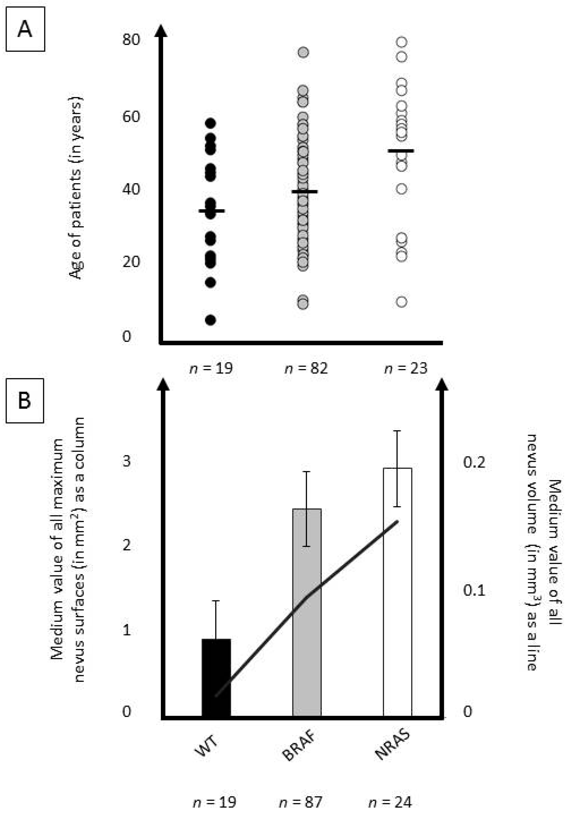
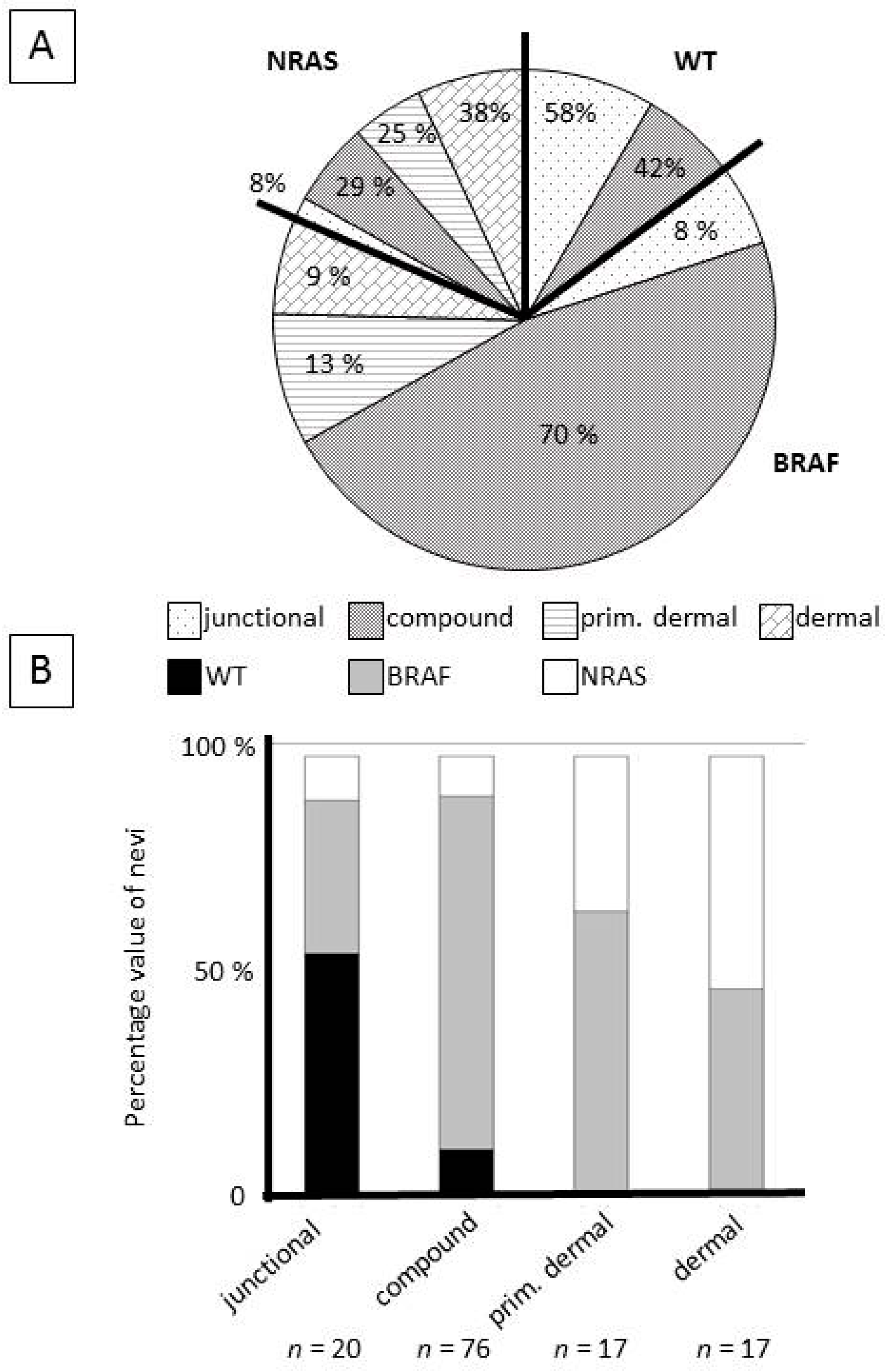
An analysis with available clinico-pathological data was performed. Statistically significant associations were found between oncogene mutation status and patient age, with BRAF-mutant (mean 40 years) and NRAS-mutant (mean 48 years) tumors occurring at older ages than wild-type tumors (mean 35 years; p = 0.04). Wild-type naevi were more often junctional than BRAF-/NRAS-mutant tumors, while BRAF-mutant tumors were often compound and NRAS-mutant tumors were often dermal (p < 0.0001). Complete details are presented in Table 1, and Figure 2 and Figure 3.

3. Discussion

Applying a targeted next generation sequencing approach, we analyzed the largest cohort of acral naevi reported to date, identifying highly recurrent BRAF and NRAS mutations.
The detected BRAF mutation frequency of 67% is lower than that described in non-acral cutaneous naevi (78–82%) [12,35]. V600E c.1799T>A alterations accounted for 99% of BRAF mutations, with only one exception, a single tumor with a 1799_1801delTGA, V600E/K alteration. This differs from melanoma, in which around 20% of BRAF V600 mutations are non-V600E (mostly V600K, but also V600R or others) [28,31,36]. While non-V600E BRAF mutations in melanoma were found to be more common in older patients, these mutations consistently represented between 10 and 20% of BRAF mutations in the 20–29, 30–39, 40–49, and 50–59 age groups [36].
The 18% NRAS mutation frequency we identified in acral naevi is considerably higher than ~6% NRAS mutations reported in cutaneous common acquired naevi, of which larger studies identified NRAS mutations in around 6% of samples [35,37]. The mutation frequency of Q61K (54%) and Q61R (46%) cannot be reliably compared to existing studies of conventional common naevi, as few NRAS mutations (<5 per study) were identified [35,37]. However, the distribution is similar to what has been described in larger melanoma cohorts [28,31,38], where the Q61K and Q61R mutations were the most frequent NRAS mutations and distributed relatively equally.
Sequencing the 19 tumors initially found to be NRAS- and BRAF-wild-type (15%) (applying the primary smaller 16 gene panel), with a larger panel covering 29 genes recurrently mutated in cutaneous or uveal melanoma [39], we only identified one MAP2K1 mutation (305_307delAGA, E102_I103V, c.309_311delCAA K104del), highly reminiscent of E102_I103del mutations reported to be activating with similar consequences to BRAF mutations [40]. In the genes analyzed in our study, no other mutations were identified, making BRAF and NRAS mutations the most common activating mutations detected in acral naevi.
Tumor suppressor genes, including CDKN2A and TP53, are not frequently altered in common naevi [41]. In our study, these genes were sequenced in the samples analyzed by the 29-gene panel and no alterations were identified. However, to reliably assess the presence of alterations in these genes, a comprehensive analysis of mutations and copy number alterations would be required.
There was a statistically significant association (p < 0.0001) of mutation status with naevi location (Table 1, Figure 2 and Figure 3). BRAF mutations were more frequent in superficial melanocytic naevi (junctional and compound). NRAS mutations were more frequent in deeper naevi (primarily dermal). In addition, the estimated naevus volume also varied, depending on mutation status (Figure 2). This is probably associated, at least partly, with naevus location, i.e., dermal naevi have a larger volume than junctional naevi (Supplementary Figure S1). Our study included more acral naevi excised from women. This is probably partially coincidental, however, a predominance for both acral naevi and melanoma in women of different ethnicities has been reported [2,5,42]. The association of increased patient age with the presence of BRAF or NRAS mutation will need to be confirmed in future studies.
Comparing the mutation profile identified in naevi to published data on acral and cutaneous melanoma, a few findings are intriguing. The BRAF mutation frequency of 67% in naevi is far higher than in acral melanoma (around 20%) [28,33,43]. However, the frequency of conventional c.1799C>A V600E mutations in naevi was higher than in melanoma (99% vs. ~80%, respectively [17,22,31]). NRAS mutation frequencies were relatively comparable between melanoma and naevi (both approximately 20% [17,22,31]). There are several mutations occurring in acral melanoma that we did not identify in acral naevi, including NF1, KIT, and TERT promoter alterations [28,31,33], recently reported in 15%, 12%, and 11% of acral melanomas, respectively [43]. The strong differences in gene mutations observed suggest that mutation type may be a diagnostic aid in distinguishing histopathologically difficult-to-classify naevi from melanoma. The identification of non-V600E BRAF, KIT, NF1, or TERT promoter mutations may warrant a higher level of suspicion for a potentially malignant acral melanocytic tumor.
All of the identified mutations are assumed to have arisen somatically. UV-exposure is expected to play a carcinogenic role, most likely more so in tumors arising on sun-exposed dorsal areas than palmar or plantar areas. However, other mechanisms, such as sporadic non-carcinogen-induced mutations, probably play a greater pathogenic role in these tumors than in non-acral cutaneous naevi.
Melanomas can arise with or without transformation from pre-existing naevi. Most naevi do not transform into melanoma, and many melanomas arise without knowledge of a preexisting naevus [8,44,45]. The genetic findings we have obtained do bring up some interesting aspects. Considering we detected no recurrent non-V600E BRAF, KIT, or NF1 mutations in naevi, this may imply that acral melanomas harboring these mutations arise primarily de novo, whereas BRAF V600E or NRAS Q61 mutant acral melanomas may more frequently arise from pre-existing naevi. Larger studies of acral melanoma with strong epidemiological data would be required to see if such an association does exist.
Our study has some caveats. We analyzed a maximum of 29 genes, and rarer mutations may have been missed. This also applies to translocations, which are rare in acral melanoma [28,43], but may occasionally occur in naevi [15]. Our sequencing panel does not allow copy number alterations to be reliably assessed; however, these have been shown to be very rare in naevi [46]. Strengths of our study are the large number of tumors included (130 naevi) and the use of a next-generation sequencing approach, simultaneously screening for many genes known to be important in melanocytic tumor pathogenesis. This differs from previous studies of naevi, in which Sanger-sequencing of individual gene mutations was performed [12,35,37].
In summary, our study presents the most comprehensive genetic analysis of a large group of acral naevi to date. BRAF V600E mutations were most frequent (66%), identified primarily in superficial (junctional and compound) naevi. However, NRAS Q61 mutations were also detected (18%), particularly in naevi with a more dermal location. The mutation profile identified in acral naevi differs considerably from acral melanoma, which could prove to be of diagnostic value.

4. Materials and Methods

4.1. Sample Selection

Samples of acral naevi were obtained from the databases of the Department of Dermatology University Hospital Essen and Dermatopathologie bei Mainz (n = 101), Germany. All cases were screened by at least one experienced board-certified dermatopathologist (KGG or EH). The study was done with the approval of the Ethics Committee of the University of Duisburg-Essen, under the IRB-number 18-8426-BO. The study was performed with patient informed consent and conducted in accordance with the Declaration of Helsinki.

4.2. Clinical and Histopathological Analysis

Available clinical information was taken from histology reports and patient records. Histological assessment included distinction between junctional, compound, primarily dermal, and dermal naevi. Primarily dermal was defined as naevi having primarily dermal melanocyte nests with up to three epidermal melanocyte nests. The ratio of epidermal to dermal melanocytes had to be >5 to be classified as primarily dermal. Tumors with a lower ratio were classified as compounds. Tumor thickness, tumor width, ratio of epidermal/dermal melanocytes, presence of superbasal intraepidermal melanocytes, signs of dysplasia, fibrosis, pigmentation, and presence of lymphocytes were assessed. The size of naevi was estimated by measuring the maximum width and height in mm and multiplying these to estimate surface area. Estimated naevus volume was calculated as 1/2 × 4/3 × Pi × r3.

4.3. DNA Isolation

DNA was isolated from 10 µm thick sections, cut from formalin-fixed, paraffin-embedded tumor tissues. The sections were deparaffinised and manually macrodissected. DNA isolation was performed with the QIAamp DNA Mini Kit (Qiagen, Hilden, Germany), according to the manufacturer’s instructions.

4.4. Targeted Sequencing

A custom amplicon-based sequencing panel covering 16 genes (Supplementary Table S2) was designed and prepared, applying the GeneRead Library Prep Kit from QIAGEN® (19300 Germantown Rd, Germantown, MD 20874, USA), according to the manufacturer’s instructions. Tumors not demonstrating a mutation in the 16 genes were further sequenced by a 29-gene panel (Supplementary Table S3) [47].
Adapter ligation and barcoding of individual samples were done, applying the NEBNext Ultra DNA Library Prep Mastermix Set and NEBNext Multiplex Oligos for Illumina from New England Biolabs. Up to 60 samples were sequenced in parallel on an Illumina MiSeq next-generation sequencer. Sequencing analysis was performed, applying the CLC Cancer Research Workbench from QIAGEN®, as described [47]. In brief, the following steps were applied. The workflow in CLC included adapter trimming and read pair merging before mapping to the human reference genome (hg19). Insertions and deletions, as well as single nucleotide variant detection, local realignment, and primer trimming, followed. Additional information was obtained regarding potential mutation type, known single nucleotide polymorphisms, and conservation scores by cross-referencing varying databases (COSMIC [48], ClinVar [49], dbSNP [50], 1000 Genomes Project [51] and HAPMAP [52]). The resulting CSV files were further analyzed manually. Mutations affecting the protein coding portion of the gene were considered if predicted to result in non-synonymous amino acid changes. Mutations were reported if the overall coverage of the mutation site was ≥30 reads, ≥5 reads reported the mutated variant, and the frequency of mutated reads was ≥1%. Access to raw sequencing data will be granted upon request.

4.5. Associations of Oncogene Mutation Status with Clinical and Pathologic Parameters

We investigated associations of mutation status with available clinical and pathological parameters, using chi-squared tests and Fisher exact tests, as appropriate. All statistical analyses were performed using IBM SPSS Statistics software (version 25.0; International Business Machines Corp., Armonk, NY, USA). A p-value of p ≤ 0.05 was considered statistically significant.

5. Conclusions

Acral naevi demonstrate frequent BRAF V600E mutations (66%), primarily in superficial (junctional and compound) locations, and frequent NRAS Q61 mutations (18%), particularly in more dermal localized tumors. Differences in mutation profile between acral naevi and melanoma may prove to be of diagnostic value.

Supplementary Materials

The following are available online at https://www.mdpi.com/2072-6694/11/4/546/s1; Figure S1: Associations of histological type with size and volume of naevi, Table S1: Associations of histopathological type of naevus with size (in mm2), Table S2: Genes covered in the applied sequencing panel, Table S3: Genes covered in the additional 29 gene sequencing panel (entire gene exons were sequenced).

Author Contributions

Conceptualization, K.G.G. and P.J.; Software and Methodology: P.J., K.G.G., R.M., I.C., and A.Z.; Validation, P.J., A.P., E.H., D.S., and K.G.G.; Formal Analysis, P.J., I.C., A.Z., C.F., T.J.B., E.H., and K.G.G.; Investigation, All Authors; Resources P.J., I.M., A.S., A.P., C.F., and K.G.G.; Data Curation, D.S., E.H., and K.G.G.; Writing—Original Draft Preparation, P.J., I.C., R.M., D.S., E.H., and K.G.G.; Writing—Review & Editing, all authors; Visualization, P.J., I.S., J.K., and K.G.G.; Supervision, E.H. and K.G.G.; Project Administration, P.J. and K.G.G.; Funding Acquisition, I.C., D.S., and K.G.G.

Funding

The research was supported by the Fleur-Hiege Stiftung. The funders had no role in study design, data collection, and analysis, decision to publish, or preparation of the manuscript.

Acknowledgments

We would like to thank Nadine Stadler and Birgit Real for their excellent technical support. We acknowledge support by the Open Access Publication Fund of the University of Duisburg-Essen.

Conflicts of Interest

Dirk Schadendorf is on the advisory board and has received honararia from Roche, Genetech, Novartis, Amgen, GSK, BMS, Boehringer Ingelheim, and Merck. All other authors declare no conflict of interest.

References

  1. Dodd, A.T.; Morelli, J.; Mokrohisky, S.T.; Asdigian, N.; Byers, T.E.; Crane, L.A. Melanocytic nevi and sun exposure in a cohort of colorado children: Anatomic distribution and site-specific sunburn. Cancer Epidemiol. Biomark. Prev. 2007, 16, 2136–2143. [Google Scholar] [CrossRef] [PubMed]
  2. Palicka, G.A.; Rhodes, A.R. Acral melanocytic nevi: Prevalence and distribution of gross morphologic features in white and black adults. Arch. Dermatol. 2010, 146, 1085–1094. [Google Scholar] [CrossRef]
  3. Weedon, D. Lentigines, nevi, and melanomas. In Weedon´s Skin Pathology, 3rd ed.; Elsevier: Amsterdam, The Netherlands, 2010; Volume 3, pp. 709–756. [Google Scholar]
  4. Shain, A.H.; Yeh, I.; Kovalyshyn, I.; Sriharan, A.; Talevich, E.; Gagnon, A.; Dummer, R.; North, J.; Pincus, L.; Ruben, B.; et al. The genetic evolution of melanoma from precursor lesions. N. Engl. J. Med. 2015, 373, 1926–1936. [Google Scholar] [CrossRef] [PubMed]
  5. Kuchelmeister, C.; Schaumburg-Lever, G.; Garbe, C. Acral cutaneous melanoma in caucasians: Clinical features, histopathology and prognosis in 112 patients. Br. J. Dermatol. 2000, 143, 275–280. [Google Scholar] [CrossRef]
  6. Teramoto, Y.; Keim, U.; Gesierich, A.; Schuler, G.; Fiedler, E.; Tuting, T.; Ulrich, C.; Wollina, U.; Hassel, J.C.; Gutzmer, R.; et al. Acral lentiginous melanoma: A skin cancer with unfavourable prognostic features. A study of the german central malignant melanoma registry (cmmr) in 2050 patients. Br. J. Dermatol. 2018, 178, 443–451. [Google Scholar] [CrossRef]
  7. Hosler, G.A.; Moresi, J.M.; Barrett, T.L. Nevi with site-related atypia: A review of melanocytic nevi with atypical histologic features based on anatomic site. J. Cutan. Pathol. 2008, 35, 889–898. [Google Scholar] [CrossRef]
  8. Elder, D.E. Precursors to melanoma and their mimics: Nevi of special sites. Mod. Pathol. 2006, 19 (Suppl. 2), S4–S20. [Google Scholar] [CrossRef]
  9. Massi, G.; Vellone, V.G.; Pagliarello, C.; Fabrizi, G. Plantar melanoma that mimics melanocytic nevi: A report of 4 cases with lymph node metastases and with review of positive and negative controls. Am. J. Dermatopathol. 2009, 31, 117–131. [Google Scholar] [CrossRef]
  10. Bronsnick, T.; Kazi, N.; Kirkorian, A.Y.; Rao, B.K. Outcomes of biopsies and excisions of dysplastic acral nevi: A study of 187 lesions. Dermatol. Surg. 2014, 40, 455–459. [Google Scholar] [CrossRef] [PubMed]
  11. Khalifeh, I.; Taraif, S.; Reed, J.A.; Lazar, A.F.; Diwan, A.H.; Prieto, V.G. A subgroup of melanocytic nevi on the distal lower extremity (ankle) shares features of acral nevi, dysplastic nevi, and melanoma in situ: A potential misdiagnosis of melanoma in situ. Am. J. Surg. Pathol. 2007, 31, 1130–1136. [Google Scholar] [PubMed]
  12. Pollock, P.M.; Harper, U.L.; Hansen, K.S.; Yudt, L.M.; Stark, M.; Robbins, C.M.; Moses, T.Y.; Hostetter, G.; Wagner, U.; Kakareka, J.; et al. High frequency of braf mutations in nevi. Nat. Genet. 2003, 33, 19–20. [Google Scholar] [CrossRef]
  13. Bauer, J.; Curtin, J.A.; Pinkel, D.; Bastian, B.C. Congenital melanocytic nevi frequently harbor nras mutations but no braf mutations. J. Investig. Dermatol. 2007, 127, 179–182. [Google Scholar] [CrossRef]
  14. Kiuru, M.; Jungbluth, A.; Kutzner, H.; Wiesner, T.; Busam, K.J. Spitz tumors: Comparison of histological features in relationship to immunohistochemical staining for alk and ntrk1. Int. J. Surg. Pathol. 2016, 24, 200–206. [Google Scholar] [CrossRef]
  15. Wiesner, T.; He, J.; Yelensky, R.; Esteve-Puig, R.; Botton, T.; Yeh, I.; Lipson, D.; Otto, G.; Brennan, K.; Murali, R.; et al. Kinase fusions are frequent in spitz tumours and spitzoid melanomas. Nat. Commun. 2014, 5, 3116. [Google Scholar] [CrossRef]
  16. Busam, K.J.; Kutzner, H.; Cerroni, L.; Wiesner, T. Clinical and pathologic findings of spitz nevi and atypical spitz tumors with alk fusions. Am. J. Surg. Pathol. 2014, 38, 925–933. [Google Scholar] [CrossRef]
  17. Wiesner, T.; Kutzner, H. Morphological and genetic aspects of spitz tumors. Pathologe 2015, 36, 37–45. [Google Scholar] [CrossRef]
  18. Yeh, I.; Botton, T.; Talevich, E.; Shain, A.H.; Sparatta, A.J.; de la Fouchardiere, A.; Mully, T.W.; North, J.P.; Garrido, M.C.; Gagnon, A.; et al. Activating met kinase rearrangements in melanoma and spitz tumours. Nat. Commun. 2015, 6, 7174. [Google Scholar] [CrossRef]
  19. Yeh, I.; Tee, M.K.; Botton, T.; Shain, A.H.; Sparatta, A.J.; Gagnon, A.; Vemula, S.S.; Garrido, M.C.; Nakamaru, K.; Isoyama, T.; et al. Ntrk3 kinase fusions in spitz tumours. J. Pathol. 2016, 240, 282–290. [Google Scholar] [CrossRef]
  20. VandenBoom, T.; Quan, V.L.; Zhang, B.; Garfield, E.M.; Kong, B.Y.; Isales, M.C.; Panah, E.; Igartua, C.; Taxter, T.; Beaubier, N.; et al. Genomic fusions in pigmented spindle cell nevus of reed. Am. J. Surg. Pathol. 2018, 42, 1042–1051. [Google Scholar] [CrossRef]
  21. Bastian, B.C.; LeBoit, P.E.; Pinkel, D. Mutations and copy number increase of hras in spitz nevi with distinctive histopathological features. Am. J. Pathol. 2000, 157, 967–972. [Google Scholar] [CrossRef]
  22. Van Raamsdonk, C.D.; Griewank, K.G.; Crosby, M.B.; Garrido, M.C.; Vemula, S.; Wiesner, T.; Obenauf, A.C.; Wackernagel, W.; Green, G.; Bouvier, N.; et al. Mutations in gna11 in uveal melanoma. N. Engl. J. Med. 2010, 363, 2191–2199. [Google Scholar] [CrossRef]
  23. Moller, I.; Murali, R.; Muller, H.; Wiesner, T.; Jackett, L.A.; Scholz, S.L.; Cosgarea, I.; van de Nes, J.A.; Sucker, A.; Hillen, U.; et al. Activating cysteinyl leukotriene receptor 2 (cysltr2) mutations in blue nevi. Mod. Pathol. 2017, 30, 350–356. [Google Scholar] [CrossRef]
  24. Griewank, K.G.; Muller, H.; Jackett, L.A.; Emberger, M.; Moller, I.; van de Nes, J.A.; Zimmer, L.; Livingstone, E.; Wiesner, T.; Scholz, S.L.; et al. Sf3b1 and bap1 mutations in blue nevus-like melanoma. Mod. Pathol. 2017, 30, 928–939. [Google Scholar] [CrossRef]
  25. Bastian, B.C. The molecular pathology of melanoma: An integrated taxonomy of melanocytic neoplasia. Annu. Rev. Pathol. 2014, 9, 239–271. [Google Scholar] [CrossRef]
  26. Cohen, J.N.; Joseph, N.M.; North, J.P.; Onodera, C.; Zembowicz, A.; LeBoit, P.E. Genomic analysis of pigmented epithelioid melanocytomas reveals recurrent alterations in prkar1a, and prkca genes. Am. J. Surg. Pathol. 2017, 41, 1333–1346. [Google Scholar] [CrossRef]
  27. Yeh, I.; Lang, U.E.; Durieux, E.; Tee, M.K.; Jorapur, A.; Shain, A.H.; Haddad, V.; Pissaloux, D.; Chen, X.; Cerroni, L.; et al. Combined activation of map kinase pathway and beta-catenin signaling cause deep penetrating nevi. Nat. Commun. 2017, 8, 644. [Google Scholar] [CrossRef]
  28. Hayward, N.K.; Wilmott, J.S.; Waddell, N.; Johansson, P.A.; Field, M.A.; Nones, K.; Patch, A.M.; Kakavand, H.; Alexandrov, L.B.; Burke, H.; et al. Whole-genome landscapes of major melanoma subtypes. Nature 2017, 545, 175–180. [Google Scholar] [CrossRef]
  29. Hodis, E.; Watson, I.R.; Kryukov, G.V.; Arold, S.T.; Imielinski, M.; Theurillat, J.P.; Nickerson, E.; Auclair, D.; Li, L.; Place, C.; et al. A landscape of driver mutations in melanoma. Cell 2012, 150, 251–263. [Google Scholar] [CrossRef]
  30. Krauthammer, M.; Kong, Y.; Ha, B.H.; Evans, P.; Bacchiocchi, A.; McCusker, J.P.; Cheng, E.; Davis, M.J.; Goh, G.; Choi, M.; et al. Exome sequencing identifies recurrent somatic rac1 mutations in melanoma. Nat. Genet. 2012, 44, 1006–1014. [Google Scholar] [CrossRef]
  31. Cancer Genome Atlas Network. Genomic classification of cutaneous melanoma. Cell 2015, 161, 1681–1696. [Google Scholar] [CrossRef]
  32. Dummer, R.; Ascierto, P.A.; Gogas, H.J.; Arance, A.; Mandala, M.; Liszkay, G.; Garbe, C.; Schadendorf, D.; Krajsova, I.; Gutzmer, R.; et al. Encorafenib plus binimetinib versus vemurafenib or encorafenib in patients with braf-mutant melanoma (columbus): A multicentre, open-label, randomised phase 3 trial. Lancet Oncol. 2018, 19, 603–615. [Google Scholar] [CrossRef]
  33. Moon, K.R.; Choi, Y.D.; Kim, J.M.; Jin, S.; Shin, M.H.; Shim, H.J.; Lee, J.B.; Yun, S.J. Genetic alterations in primary acral melanoma and acral melanocytic nevus in korea: Common mutated genes show distinct cytomorphological features. J. Investig. Dermatol. 2018, 138, 933–945. [Google Scholar] [CrossRef]
  34. Haugh, A.M.; Zhang, B.; Quan, V.L.; Garfield, E.M.; Bubley, J.A.; Kudalkar, E.; Verzi, A.E.; Walton, K.; VandenBoom, T.; Merkel, E.A.; et al. Distinct patterns of acral melanoma based on site and relative sun exposure. J. Investig. Dermatol. 2018, 138, 384–393. [Google Scholar] [CrossRef]
  35. Poynter, J.N.; Elder, J.T.; Fullen, D.R.; Nair, R.P.; Soengas, M.S.; Johnson, T.M.; Redman, B.; Thomas, N.E.; Gruber, S.B. Braf and nras mutations in melanoma and melanocytic nevi. Melanoma Res. 2006, 16, 267–273. [Google Scholar] [CrossRef]
  36. Menzies, A.M.; Haydu, L.E.; Visintin, L.; Carlino, M.S.; Howle, J.R.; Thompson, J.F.; Kefford, R.F.; Scolyer, R.A.; Long, G.V. Distinguishing clinicopathologic features of patients with v600e and v600k braf-mutant metastatic melanoma. Clin. Cancer Res. 2012, 18, 3242–3249. [Google Scholar] [CrossRef]
  37. Roh, M.R.; Eliades, P.; Gupta, S.; Tsao, H. Genetics of melanocytic nevi. Pigment Cell Melanoma Res. 2015, 28, 661–672. [Google Scholar] [CrossRef]
  38. Griewank, K.; Westekemper, H.; Murali, R.; Mach, M.; Schilling, B.; Wiesner, T.; Schimming, T.; Livingstone, E.; Sucker, A.; Grabellus, F.; et al. Conjunctival melanomas harbor braf and nras mutations and copy number changes similar to cutaneous and mucosal melanomas. Clin. Cancer Res. 2013, 19, 3143–3152. [Google Scholar] [CrossRef]
  39. Scholz, S.L.; Cosgarea, I.; Susskind, D.; Murali, R.; Moller, I.; Reis, H.; Leonardelli, S.; Schilling, B.; Schimming, T.; Hadaschik, E.; et al. Nf1 mutations in conjunctival melanoma. Br. J. Cancer 2018, 118, 1243–1247. [Google Scholar] [CrossRef]
  40. Chakraborty, R.; Hampton, O.A.; Shen, X.; Simko, S.J.; Shih, A.; Abhyankar, H.; Lim, K.P.; Covington, K.R.; Trevino, L.; Dewal, N.; et al. Mutually exclusive recurrent somatic mutations in map2k1 and braf support a central role for erk activation in lch pathogenesis. Blood 2014, 124, 3007–3015. [Google Scholar] [CrossRef]
  41. Bauer, J.; Bastian, B.C. Distinguishing melanocytic nevi from melanoma by DNA copy number changes: Comparative genomic hybridization as a research and diagnostic tool. Dermatol. Ther. 2006, 19, 40–49. [Google Scholar] [CrossRef]
  42. Kogushi-Nishi, H.; Kawasaki, J.; Kageshita, T.; Ishihara, T.; Ihn, H. The prevalence of melanocytic nevi on the soles in the japanese population. J. Am. Acad. Dermatol. 2009, 60, 767–771. [Google Scholar] [CrossRef]
  43. Yeh, I.; Jorgenson, E.; Shen, L.; Xu, M.; North, J.P.; Shain, A.H.; Reuss, D.; Wu, H.; Robinson, W.A.; Olshen, A.; et al. Targeted genomic profiling of acral melanoma. J. Natl. Cancer Inst. 2019. [Google Scholar] [CrossRef]
  44. Pampena, R.; Kyrgidis, A.; Lallas, A.; Moscarella, E.; Argenziano, G.; Longo, C. A meta-analysis of nevus-associated melanoma: Prevalence and practical implications. J. Am. Acad. Dermatol. 2017, 77, 938–945. [Google Scholar] [CrossRef] [PubMed]
  45. Lin, W.M.; Luo, S.; Muzikansky, A.; Lobo, A.Z.; Tanabe, K.K.; Sober, A.J.; Cosimi, A.B.; Tsao, H.; Duncan, L.M. Outcome of patients with de novo versus nevus-associated melanoma. J. Am. Acad. Dermatol. 2015, 72, 54–58. [Google Scholar] [CrossRef] [PubMed]
  46. Bastian, B.C.; Olshen, A.B.; LeBoit, P.E.; Pinkel, D. Classifying melanocytic tumors based on DNA copy number changes. Am. J. Pathol. 2003, 163, 1765–1770. [Google Scholar] [CrossRef]
  47. van de Nes, J.; Gessi, M.; Sucker, A.; Moller, I.; Stiller, M.; Horn, S.; Scholz, S.L.; Pischler, C.; Stadtler, N.; Schilling, B.; et al. Targeted next generation sequencing reveals unique mutation profile of primary melanocytic tumors of the central nervous system. J. Neuro-Oncol. 2016, 127, 435–444. [Google Scholar] [CrossRef] [Green Version]
  48. COSMIC. Available online: https://cancer.sanger.ac.uk/cosmic (accessed on 12 April 2019).
  49. ClinVar. Available online: https://www.ncbi.nlm.nih.gov/clinvar/ (accessed on 12 April 2019).
  50. dbSNP. Available online: https://www.ncbi.nlm.nih.gov/snp (accessed on 12 April 2019).
  51. 1000 Genomes Project. Available online: http://www.internationalgenome.org/ (accessed on 12 April 2019).
  52. HAPMAP. Available online: https://www.ncbi.nlm.nih.gov/probe/docs/projhapmap/ (accessed on 12 April 2019).
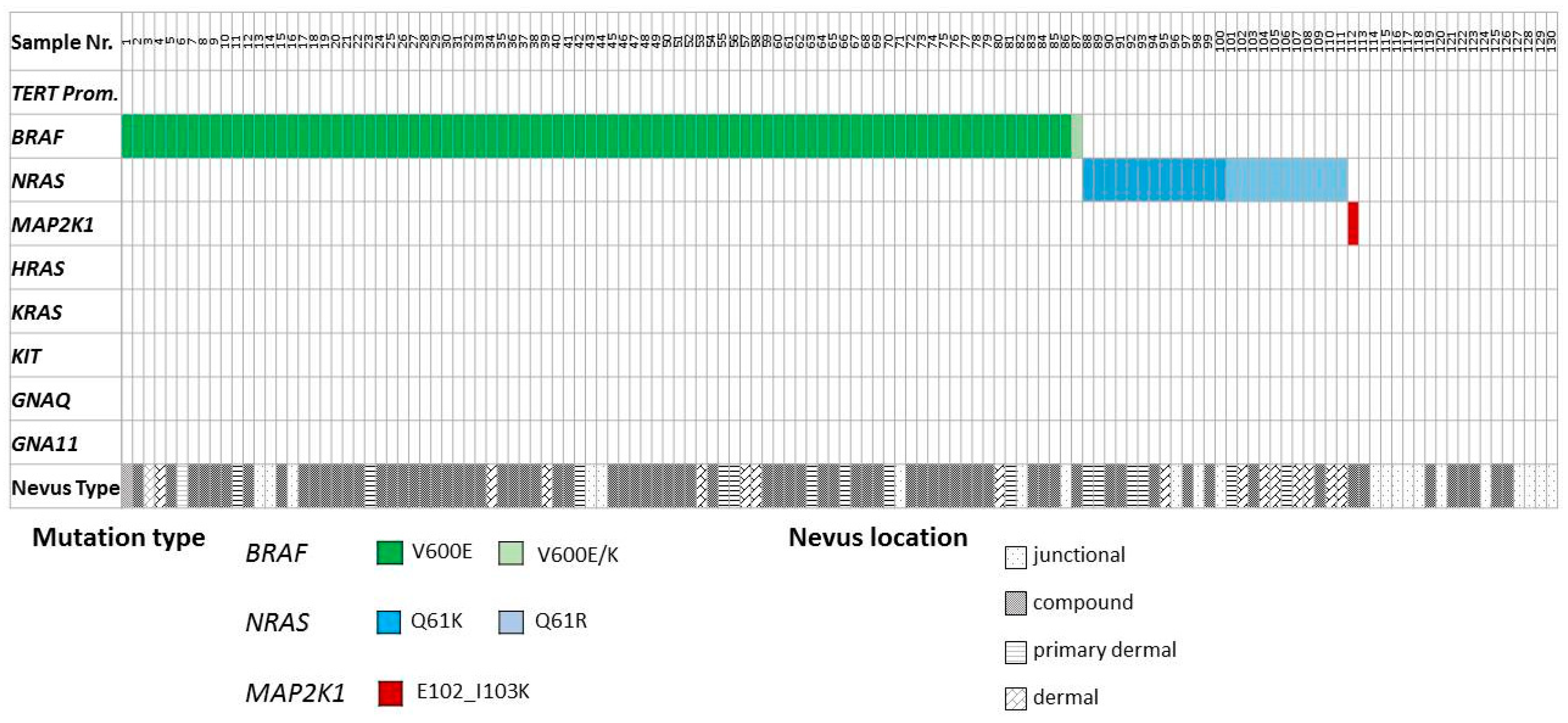
Figure 1. Distribution of activating mutations identified in acral naevi. Distribution of activating mutations identified in different oncogenes in the acral naevus cohort. The resulting amino acid changes as well as naevus location (junctional, compound, primarily dermal, or dermal) are color-coded according to the scheme underneath the illustration.
Cancers 11 00546 g001
Figure 2. Associations of patients’ age, size, and volume of naevi with oncogene mutation status. (A) Distribution of patients according to their age in wild-type cohort (black), BRAF-mutant cohort (grey) and NRAS-mutant cohort (white) (mean age of cohort indicated by black line). (B) Medium size of naevus surface (in mm2) depicted as columns for wild-type cohort (in black), BRAF-mutant cohort (in grey) and NRAS-mutant cohort (in white) with standard deviation. Medium volume of naevi for all three cohorts (WT, BRAF, NRAS) is depicted as a black line.
Cancers 11 00546 g002
Figure 3. Associations of histological type and oncogene mutation status of naevi. (A) Percentage value of different histological naevus types in wild-type (WT), BRAF-, and NRAS-mutant cohorts. (B) Percentage value of wild-type (WT), BRAF-, and NRAS-mutant naevi in the four different naevus locations (junctional, compound, primarily dermal, and dermal).
Cancers 11 00546 g003
Table 1. Associations of BRAF and NRAS mutation status with clinical and pathological parameters.
VariableSpecific VariableAll
n = 130
%WT
n = 19
%BRAF-Mutant
n = 87
%NRAS-Mutant
n = 24
%p-Value *
Mean Age (Years) 41 35 40 48 0.04
Sex +Female9174%14 73.7%6073.2 %1773.9%0.08
Male3226%526.3%2227.5%626.1%
Sites of Involvementfoot12193.1%16 84.2%8193.1%24100%0.34
hand75.4%210.5%55.7%00
LND21.5%15.3%11.2%00-
Sites of Involvementvolar4131.6%526.4%2933.3%729.2%0.11
dorsal2519.2%7 36.8%1618.4%28.3%
LND6449.2%736.8%4248.3%1562.5%-
Histotypejunctional2015.4%11 57.9%78.1%28.3%<0.0001
compound7658.4%852.1%6170.1%729.2%
prim. dermal1713.1%001112.6%625%
dermal1713.1%0089.2%937.5%
WT = wild-type for BRAF and NRAS; prim. = primarily; * age Kruskal–Wallis test; all others: Fisher exact test; LND: Localization not determined; One patient having a MAP2K1 mutation (was included in the wild-type group); + one woman had three BRAF-mutant naevi, one woman had two BRAF-mutant naevi, one woman showed two NRAS-mutant naevi, two men had two BRAF-mutant naevi, and one man showed one BRAF-mutant naevus and one WT naevus (this man is represented in the “BRAF-mutant” column and in the “WT” column but counted once in the “All” column).