Segmentation and Grade Prediction of Colon Cancer Digital Pathology Images Across Multiple Institutions
Abstract
1. Introduction
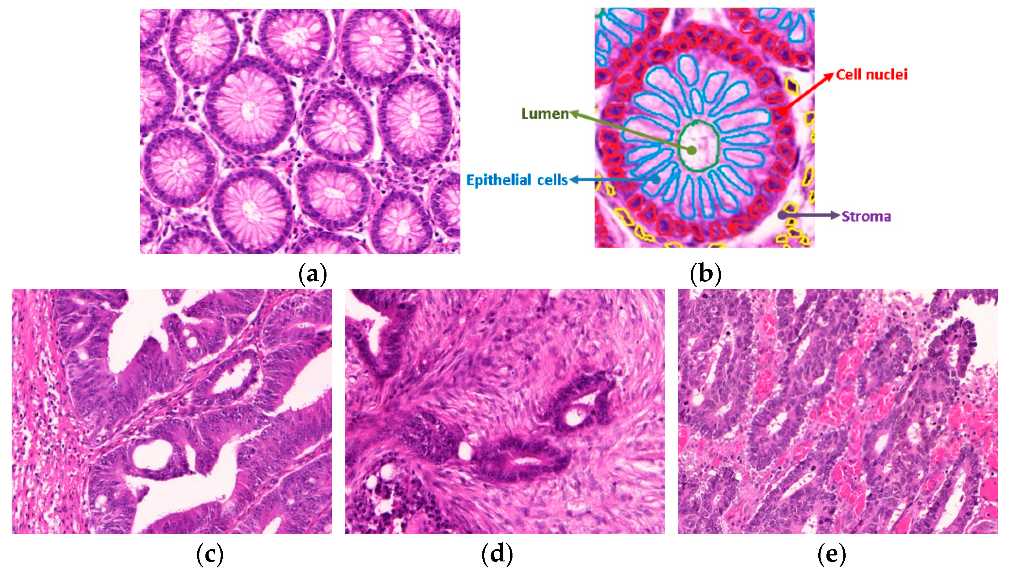
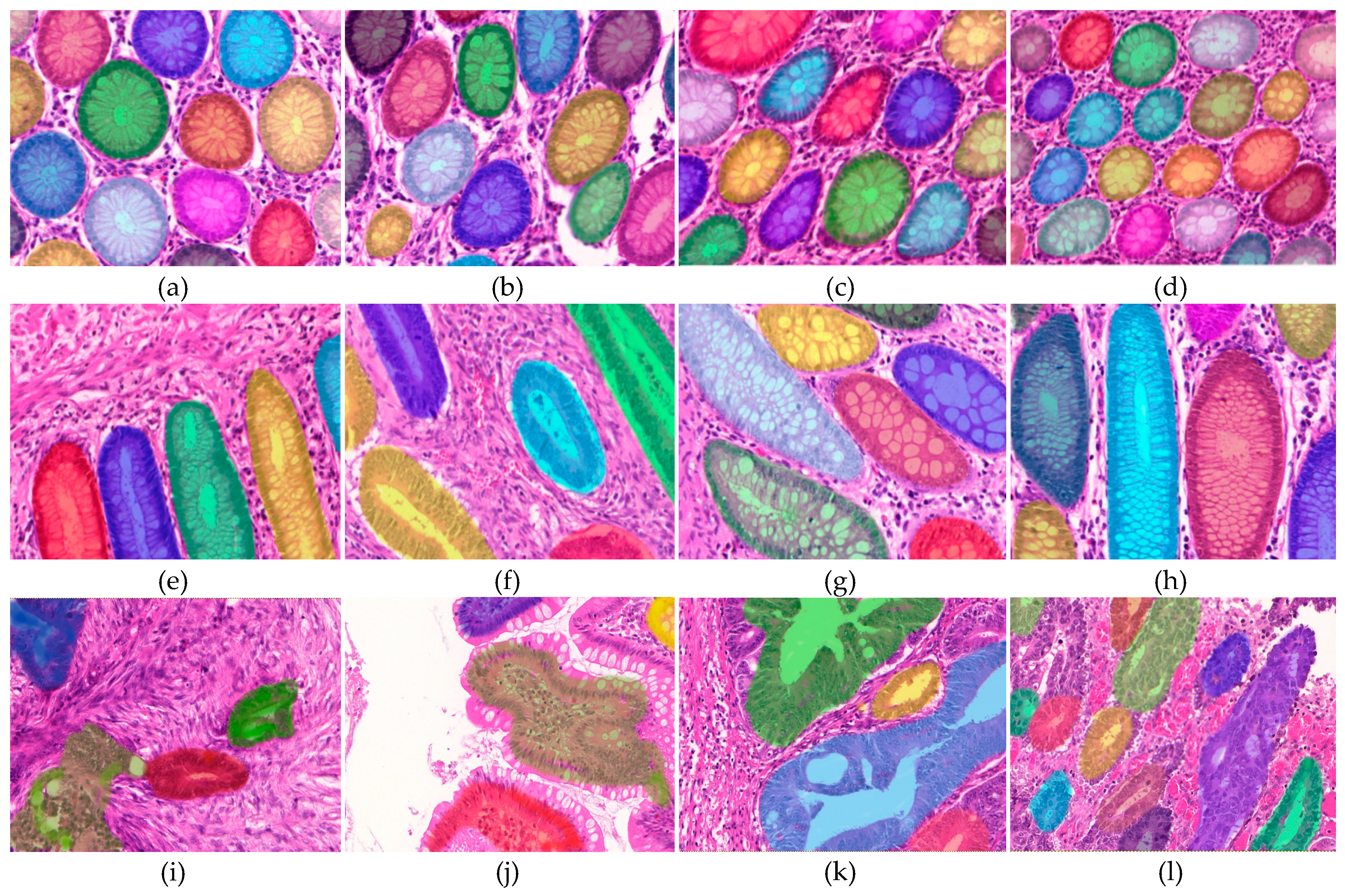
- A robust gland segmentation method that exploits the organizational appearance of colon glands to demarcate gland boundaries and to segment internal glandular structures, including epithelial cells, nuclei and lumen, and yields promising results for multiple datasets. The radiomic features extracted from the segmented structures were later used in the classification.
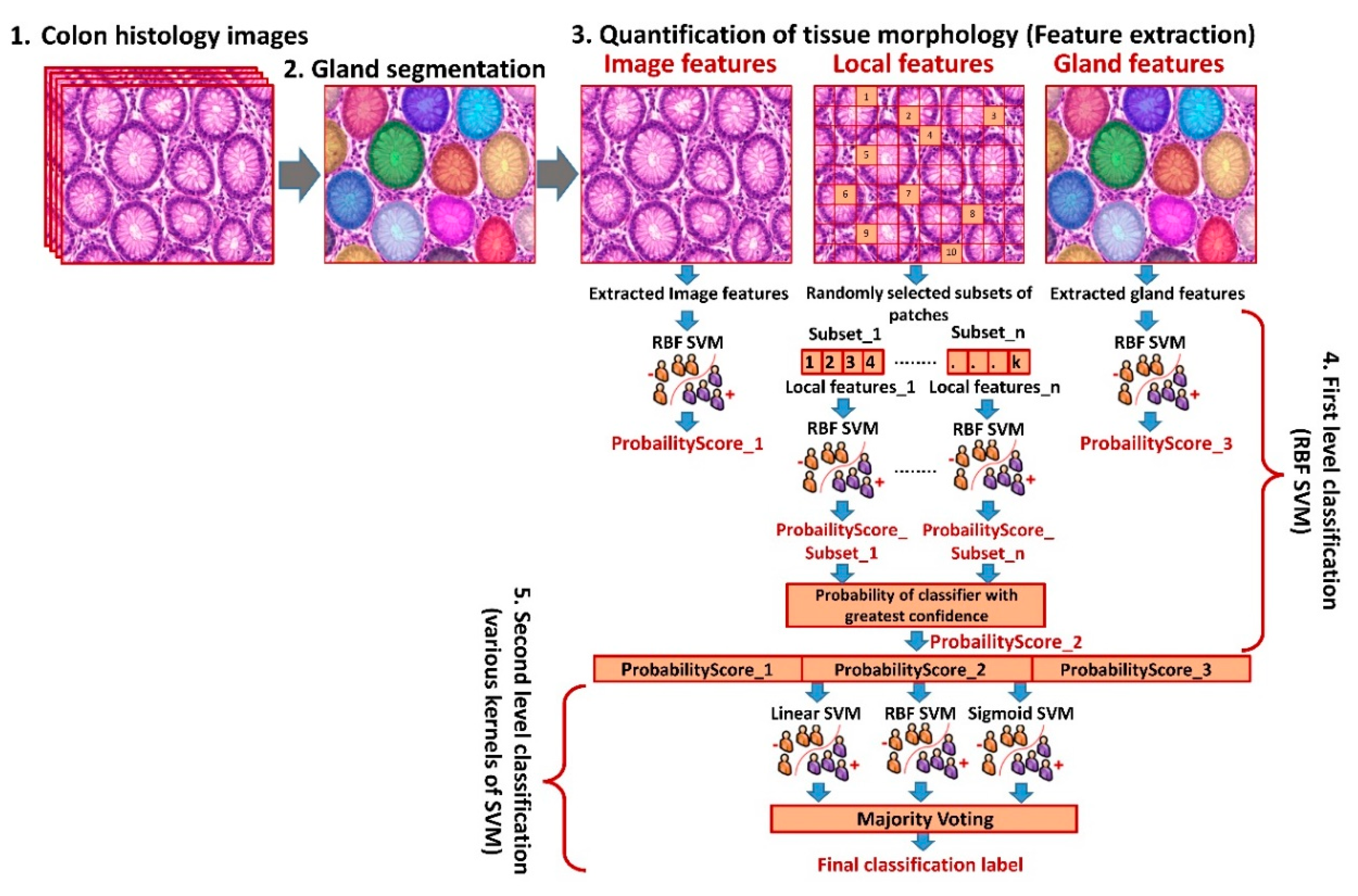
- A multi-scale feature extraction mechanism comprising: (i) Gland-based features: extracted from individual glands, which are segmented by the proposed gland segmentation method, (ii) local-patch-based features: computed from randomly-selected patches of each histologic colon image, and (iii) image-based features: calculated by considering whole image as one unit and capturing global details.
- Novel gland-based features, which encode morphometric properties of epithelial cells and lumen and combine them with the spatial distribution of the nuclei in relationship to stroma and lumen.
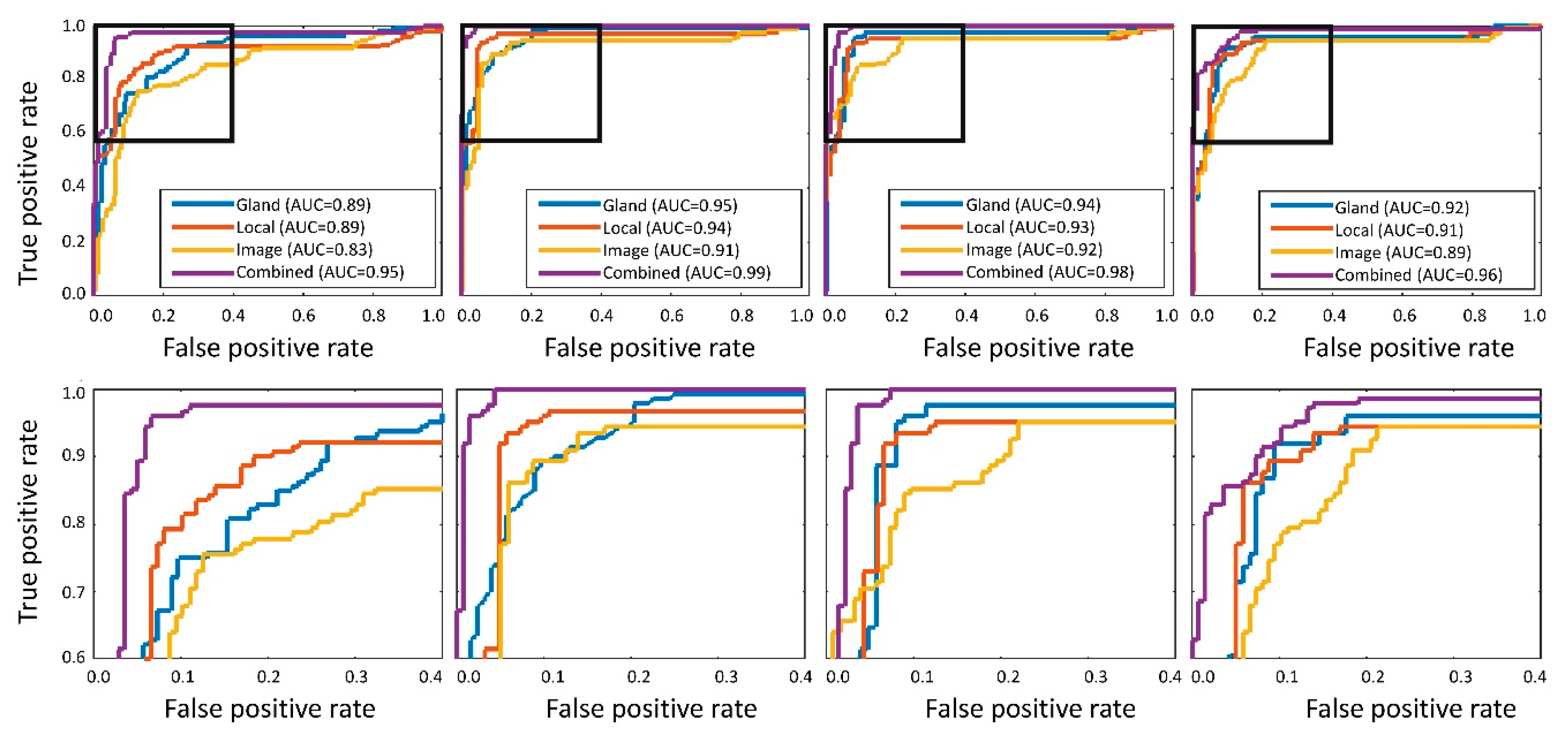
- A multi-level hierarchical classification methodology to divide large-scale classification problem into a set of small-scale and easier-to-solve problems. In the proposed framework, the first-level classification produces probability estimates based on each feature type (image-, gland-, and local-patch-based features), and the second-level is an ensemble of various support vector machine (SVM) classifiers trained on the probability estimates generated by the first-level.
- Demonstration of the generalizability of the proposed pipeline across multiple datasets.
1.1. Related Work
2. Results
2.1. Performance of The Proposed Gland Segmentation Method
2.2. Performance of The Proposed Cancer Detection and Grading Method
2.3. Performance of the Proposed Cancer Detection and Grading Method across Different Institutions
2.4. Interpretation of Features of Normal/Malignant Colon Tissues
- Lower entropy values in normal colon tissue images (p = 5.6 × 10⁻⁵), suggestive of more uniformity and proper organizational structure of tissue compared to malignant colon tissue that shows very high entropy, indicative of more randomness and lack of any organizational structure;
- Lower contrast values in normal colon tissue (p = 2.1 × 10⁻⁶), which again point towards coherent and properly organized tissue. On the other hand, the higher contrast values in malignant colon tissues show the lack of proper organizational structure;
- Lower values of standard deviation of distances of cell nuclei from the centroid of lumen for normal colon tissue (p = 8.6 × 10⁻⁴) are suggestive of the fact that cell nuclei are almost equally spaced from the centroid of the lumen, whereas the same is not true for malignant colon tissue where cell nuclei are at random distances from lumen;
- Higher values of eccentricity (p = 2.1 × 10⁻³) and compactness (p = 5.8 × 10⁻⁴) in normal colon tissues, consistent with the well-defined shape of normal colon tissue. On the contrary, the malignant colon tissues exhibit lower compactness and eccentricity, indicative of the migratory and deeply infiltrated tissue.
3. Discussion
3.1. Validation of the Proposed Method across Different Datasets
3.2. Importance of the Study
3.3. Limitations and Future Work
4. Materials and Methods
4.1. Datasets
4.2. The Gland Segmentation Algorithm
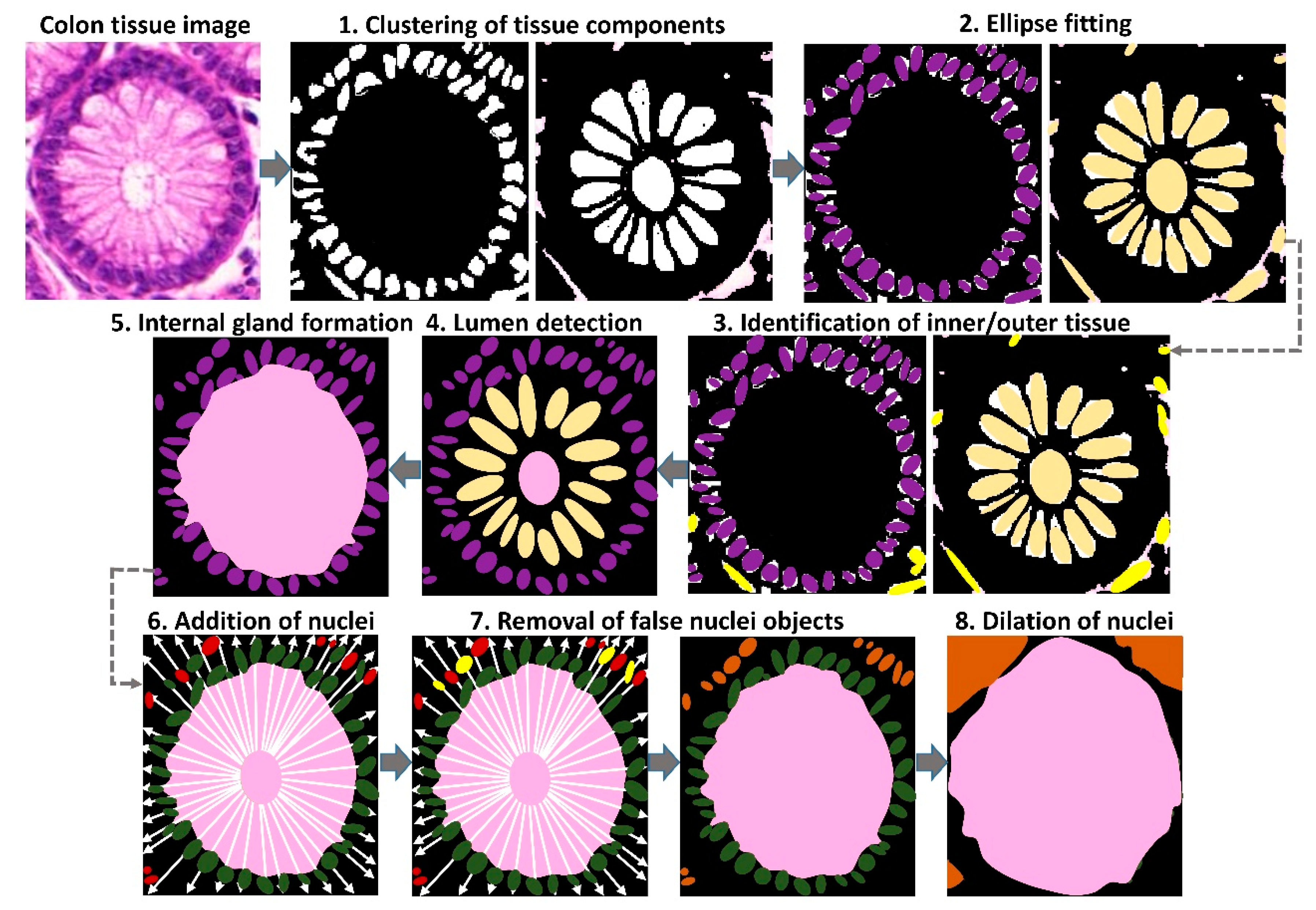
4.2.1. Clustering of Tissue Components
4.2.2. Ellipse Fitting
4.2.3. Discrimination of True and False Glandular Components
4.2.4. Lumen Detection
4.2.5. Formation of Internal Gland Regions
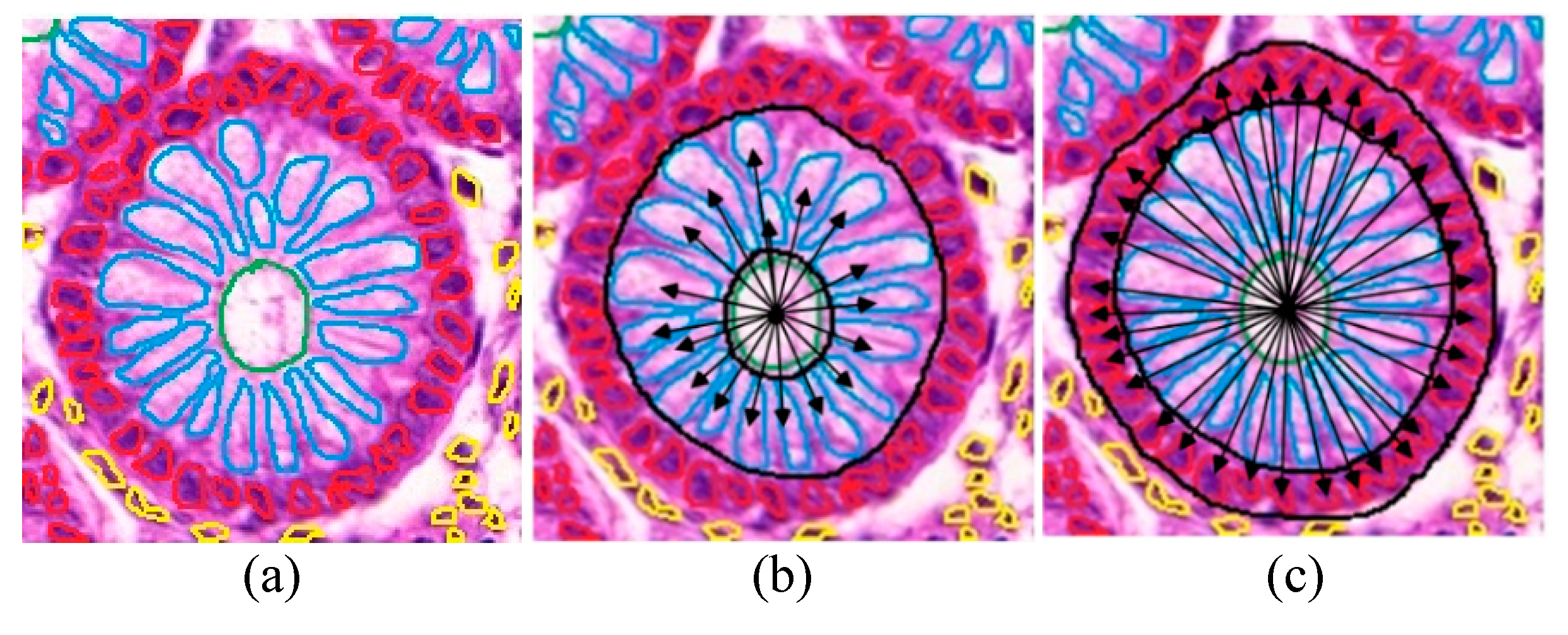
4.2.6. Addition of Nuclei to the Internal Glandular Region
4.2.7. Removal of False Nuclei
4.2.8. Dilation of Nuclei
4.3. Features for Colon Cancer Detection/grading
4.3.1. Image-Based Features
4.3.2. Gland-Based Features
4.3.3. Patch-Based Features
4.4. Hierarchical Classification Framework
5. Conclusions
Author Contributions
Funding
Conflicts of Interest
References
- Cross, S.E.; Jin, Y.S.; Rao, J.; Gimzewski, J.K. Nanomechanical analysis of cells from cancer patients. Nat. Nanotechnol. 2007, 2, 780–783. [Google Scholar] [CrossRef] [PubMed]
- Backes, Y.; Moons, L.M.; Novelli, M.R.; van Bergeijk, J.D.; Groen, J.N.; Seerden, T.C.; Schwartz, M.P.; de Vos Tot Nederveen Cappel, W.H.; Spanier, B.W.; Geesing, J.M.; et al. Diagnosis of T1 colorectal cancer in pedunculated polyps in daily clinical practice: A multicenter study. Mod. Pathol. 2017, 30, 104–112. [Google Scholar] [CrossRef] [PubMed]
- Rathore, S.; Iftikhar, M.A.; Hussain, M.; Jalil, A. Classification of colon biopsy images based on novel structural features. In Proceedings of the 2013 IEEE 9th International Conference on Emerging Technologies (ICET), Islamabad, Pakistan, 9–10 December 2013; pp. 1–6. [Google Scholar]
- Doyle, S.; Madabhushi, A.; Feldman, M.; Tomaszeweski, J. A boosting cascade for automated detection of prostate cancer from digitized histology. Med. Image Comput. Comput. Assist. Interv. 2006, 9 Pt 2, 504–511. [Google Scholar]
- Khan, A.M.; Rajpoot, N.; Treanor, D.; Magee, D. A nonlinear mapping approach to stain normalization in digital histopathology images using image-specific color deconvolution. IEEE Trans. Biomed. Eng. 2014, 61, 1729–1738. [Google Scholar] [CrossRef] [PubMed]
- Gurcan, M.N.; Boucheron, L.E.; Can, A.; Madabhushi, A.; Rajpoot, N.M.; Yener, B. Histopathological image analysis: A review. IEEE Rev. Biomed. Eng. 2009, 2, 147–171. [Google Scholar] [CrossRef]
- Rathore, S.; Hussain, M.; Ali, A.; Khan, A. A recent survey on colon cancer detection techniques. IEEE/ACM Trans. Comput. Biol. Bioinform. 2013, 10, 545–563. [Google Scholar] [CrossRef]
- Rathore, S.; Hussain, M.; Khan, A. Automated colon cancer detection using hybrid of novel geometric features and some traditional features. Comput. Biol. Med. 2015, 65, 279–296. [Google Scholar] [CrossRef]
- Altunbay, D.; Cigir, C.; Sokmensuer, C.; Gunduz-Demir, C. Color graphs for automated cancer diagnosis and grading. IEEE Trans. Biomed. Eng. 2010, 57, 665–674. [Google Scholar] [CrossRef]
- Ozdemir, E.; Gunduz-Demir, C. A hybrid classification model for digital pathology using structural and statistical pattern recognition. IEEE Trans. Med. Imaging 2013, 32, 474–483. [Google Scholar] [CrossRef]
- Rathore, S.; Hussain, M.; Iftikhar, M.A.; Jalil, A. Novel structural descriptors for automated colon cancer detection and grading. Comput. Methods Progr. Biomed. 2015, 121, 92–108. [Google Scholar] [CrossRef]
- Chaddad, A.; Daniel, P.; Niazi, T. Radiomics Evaluation of Histological Heterogeneity Using Multiscale Textures Derived From 3D Wavelet Transformation of Multispectral Images. Front. Oncol. 2018, 8, 96. [Google Scholar] [CrossRef] [PubMed]
- Haj-Hassan, H.; Chaddad, A.; Harkouss, Y.; Desrosiers, C.; Toews, M.; Tanougast, C. Classifications of Multispectral Colorectal Cancer Tissues Using Convolution Neural Network. J. Pathol. Inform. 2017, 8, 1. [Google Scholar] [PubMed]
- Chaddad, A.; Tanougast, C. Texture Analysis of Abnormal Cell Images for Predicting the Continuum of Colorectal Cancer. Anal. Cell Pathol. 2017, 2017, 8428102. [Google Scholar] [CrossRef] [PubMed]
- Ronneberger, O.; Fischer, P.; Brox, T. U-Net: Convolutional Networks for Biomedical Image Segmentation. arXiv 2015, arXiv:1505.04597. [Google Scholar]
- Gunduz-Demir, C.; Kandemir, M.; Tosun, A.B.; Sokmensuer, C. Automatic segmentation of colon glands using object-graphs. Med. Image Anal. 2010, 14, 1–12. [Google Scholar] [CrossRef] [PubMed]
- Graham, S.; Chen, H.; Gamper, J.; Dou, Q.; Heng, P.-A.; Snead, D.; Tsang, Y.W.; Rajpoot, N. MILD-Net: Minimal Information Loss Dilated Network for Gland Instance Segmentation in Colon Histology Images. Med. Image Anal. 2019, 52, 199–211. [Google Scholar] [CrossRef]
- Kainz, P.; Pfeiffer, M.; Urschler, M. Semantic Segmentation of Colon Glands with Deep Convolutional Neural Networks and Total Variation Segmentation. arXiv 2015, arXiv:1511.06919. [Google Scholar]
- Fu, H.; Qiu, G.; Shu, J.; Ilyas, M. A novel polar space random field model for the detection of glandular structures. IEEE Trans. Med. Imaging 2014, 33, 764–776. [Google Scholar] [CrossRef]
- Li, W.; Manivannan, S.; Akbar, S.; Zhang, J.; Trucco, E.; McKenna, S.J. Gland segmentation in colon histology images using hand-crafted features and convolutional neural networks. In Proceedings of the 2016 IEEE 13th International Symposium on Biomedical Imaging (ISBI), Prague, Czech Republic, 13–16 April 2016. [Google Scholar]
- Chen, H.; Qi, X.; Yu, L.; Dou, Q.; Qin, J.; Heng, P.A. DCAN: Deep contour-aware networks for object instance segmentation from histology images. Med. Image Anal. 2017, 36, 135–146. [Google Scholar] [CrossRef]
- Masood, K.; Rajpoot, N. Texture based classification of hyperspectral colon biopsy samples using CLBP. In Proceedings of the IEEE International Symposium on Biomedical Imaging: From Nano to Macro, Boston, MA, USA, 28 June–1 July 2009; pp. 1011–1014. [Google Scholar]
- Rathore, S.; Hussain, M.; Iftikhar, M.A.; Jalil, A. Ensemble classification of colon biopsy images based on information rich hybrid features. Comput. Biol. Med. 2014, 47, 76–92. [Google Scholar] [CrossRef]
- Awan, R.; Sirinukunwattana, K.; Epstein, D.; Jefferyes, S.; Qidwai, U.; Aftab, Z.; Mujeeb, I.; Snead, D.; Rajpoot, N. Glandular Morphometrics for Objective Grading of Colorectal Adenocarcinoma Histology Images. Sci. Rep. 2017, 7, 16852. [Google Scholar] [CrossRef] [PubMed]
- Chaddad, A.; Tanougast, C.; Golato, A.; Dandache, A. Carcinoma cell identification via optical microscopy and shape feature analysis. J. Biomed. Sci. Eng. 2013, 6, 1029–1033. [Google Scholar] [CrossRef]
- Rathore, S.; Hussain, M.; Khan, A. GECC: Gene Expression Based Ensemble Classification of Colon Samples. IEEE/ACM Trans. Comput. Biol. Bioinform. 2014, 11, 1131–1145. [Google Scholar] [CrossRef] [PubMed]
- Rathore, S.; Iftikhar, M.A. CBISC: A Novel Approach for Colon Biopsy Image Segmentation and Classification. Arab. J. Sci. Eng. 2016, 41, 5061–5076. [Google Scholar] [CrossRef]
- Tommelein, J.; Verset, L.; Boterberg, T.; Demetter, P.; Bracke, M.; de Wever, O. Cancer-associated fibroblasts connect metastasis-promoting communication in colorectal cancer. Front. Oncol. 2015, 5, 63. [Google Scholar] [CrossRef]
- Becht, E.; de Reynies, A.; Giraldo, N.A.; Pilati, C.; Buttard, B.; Lacroix, L.; Selves, J.; Sautes-Fridman, C.; Laurent-Puig, P.; Fridman, W.H. Immune and Stromal Classification of Colorectal Cancer Is Associated with Molecular Subtypes and Relevant for Precision Immunotherapy. Clin. Cancer Res. 2016, 22, 4057–4066. [Google Scholar] [CrossRef]
- Sirinukunwattana, K.; Pluim, J.P.; Chen, H.; Qi, X.; Heng, P.A.; Guo, Y.B.; Wang, L.Y.; Matuszewski, B.J.; Bruni, E.; Sanchez, U.; et al. Gland segmentation in colon histology images: The glas challenge contest. Med. Image Anal. 2017, 35, 489–502. [Google Scholar] [CrossRef]
- Sirinukunwattana, K.; Snead, D.R.; Rajpoot, N.M. A Stochastic Polygons Model for Glandular Structures in Colon Histology Images. IEEE Trans. Med. Imaging 2015, 34, 2366–2378. [Google Scholar] [CrossRef]
- Haralick, R.M.; Shanmugam, K.; Dinstein, I.H. Textural features for image classification. IEEE Trans. Syst. Man Cybern. 1973, 3, 610–621. [Google Scholar] [CrossRef]
- Liu, M.; Zhang, D.; Shen, D. Semble sparse classification of Alzheimer disease. Neuroimage 2012, 60, 1106–1116. [Google Scholar] [CrossRef]
- Rathore, S.; Iftikhar, M.A.; Hassan, M. Ensemble Sparse Classification of Colon Cancer. In Proceedings of the 2016 International Conference on Frontiers of Information Technology (FIT), Islamabad, Pakistan, 19–21 December 2016. [Google Scholar]
- Fujima, N.; Shimizu, Y.; Yoshida, D.; Kano, S.; Mizumachi, T.; Homma, A.; Yasuda, K.; Onimaru, R.; Sakai, O.; Kudo, K.; et al. Machine-Learning-Based Prediction of Treatment Outcomes Using MR Imaging-Derived Quantitative Tumor Information in Patients with Sinonasal Squamous Cell Carcinomas: A Preliminary Study. Cancers 2019, 11, 800. [Google Scholar] [CrossRef] [PubMed]
- Rehman, O.; Zhuang, H.; Ali, A.M.; Ibrahim, A.; Li, Z. Validation of miRNAs as Breast Cancer Biomarkers with a Machine Learning Approach. Cancers 2019, 11, 431. [Google Scholar] [CrossRef] [PubMed]
- Wilcoxon, F. Individual comparisons by ranking methods. Biom. Bull. 1945, 1, 80–83. [Google Scholar] [CrossRef]
- Dunn, O.J. Estimation of the Means for Dependent Variables. Ann. Math. Stat. 1958, 29, 1095–1111. [Google Scholar] [CrossRef]

| Performance Metrics | RMC-Dataset | GlaS-Dataset |
|---|---|---|
| Segmentation accuracy | 87.50 | 88.40 |
| Jaccard index | 0.86 | 0.89 |
| Dice similarity | 0.84 | 0.87 |
| Sensitivity | 0.90 | 0.92 |
| Specificity | 0.82 | 0.88 |
| F-Score | 0.88 | 0.89 |
| Gland-Based | Local-Patch-Based | Image-Based | ||||
|---|---|---|---|---|---|---|
| GlaS-Dataset | RMC-Dataset | GlaS-Dataset | RMC-Dataset | GlaS-Dataset | RMC-Dataset | |
| Cancer Detection | ||||||
| Accuracy | 93.7 | 92.1 | 93.1 | 91.5 | 92.5 | 90.9 |
| Sensitivity | 92.4 | 86.8 | 91.3 | 85.2 | 92.3 | 84.5 |
| Specificity | 95.1 | 98.6 | 93.9 | 97.4 | 92.7 | 95.9 |
| MCC | 87.4 | 85.0 | 86.2 | 83.7 | 85.0 | 82.3 |
| Cancer Grading | ||||||
| Accuracy | 90.5 | 92.5 | 89.5 | 91.5 | 89.7 | 90.7 |
| Sensitivity | 84.4 | 89.0 | 82.9 | 87.0 | 81.5 | 85.5 |
| Specificity | 93.1 | 94.9 | 92.0 | 92.5 | 91.6 | 92.4 |
| MCC | 79.0 | 83.9 | 76.0 | 80.6 | 74.7 | 78.6 |
| GlaS-Dataset | RMC-Dataset | |||||||
|---|---|---|---|---|---|---|---|---|
| Linear | RBF | Sigmoid | Ensemble | Linear | RBF | Sigmoid | Ensemble | |
| Cancer Detection | ||||||||
| Accuracy | 94.3 | 95.4 | 94.8 | 98.3 | 92.1 | 93.3 | 92.7 | 97.6 |
| Sensitivity | 91.3 | 92.4 | 92.4 | 97.8 | 96.7 | 98.9 | 98.9 | 98.9 |
| Specificity | 97.6 | 98.8 | 97.6 | 98.8 | 86.5 | 86.5 | 85.1 | 95.9 |
| MCC | 88.7 | 91.2 | 89.8 | 96.5 | 84.2 | 86.9 | 85.8 | 95.1 |
| Cancer Grading | ||||||||
| Accuracy | 94.2 | 94.5 | 93.7 | 98.6 | 91.3 | 93.6 | 93.5 | 98.6 |
| Sensitivity | 90.7 | 91.4 | 90.4 | 97.3 | 84.7 | 87.7 | 87.8 | 97.4 |
| Specificity | 95.7 | 95.9 | 95.0 | 99.0 | 93.7 | 95.7 | 95.4 | 99.0 |
| MCC | 86.4 | 87.3 | 85.4 | 96.4 | 78.4 | 83.3 | 83.2 | 96.4 |
| Train RMC-Dataset + Test GlaS-Dataset | Train GlaS-Dataset + Test RMC-Dataset | |||||||
|---|---|---|---|---|---|---|---|---|
| Linear | RBF | Sigmoid | Ensemble | Linear | RBF | Sigmoid | Ensemble | |
| Cancer Detection | ||||||||
| Accuracy | 89.7 | 91.5 | 90.9 | 94.5 | 89.7 | 90.2 | 87.9 | 93.7 |
| Sensitivity | 97.8 | 96.7 | 96.7 | 96.7 | 96.0 | 96.7 | 91.3 | 96.7 |
| Specificity | 79.7 | 85.1 | 83.8 | 91.9 | 81.7 | 82.9 | 84.1 | 90.2 |
| MCC | 79.9 | 83.1 | 81.9 | 89.0 | 79.9 | 80.9 | 75.8 | 87.4 |
| Cancer Grading | ||||||||
| Accuracy | 89.8 | 91.3 | 90.0 | 95.0 | 89.8 | 90.6 | 90.7 | 95.0 |
| Sensitivity | 82.2 | 84.5 | 81.6 | 91.6 | 84.1 | 84.7 | 86.0 | 92.8 |
| Specificity | 92.2 | 93.6 | 93.0 | 96.3 | 92.1 | 92.8 | 92.4 | 95.8 |
| MCC | 74.3 | 78.2 | 74.6 | 87.9 | 76.2 | 77.5 | 78.5 | 88.6 |
© 2019 by the authors. Licensee MDPI, Basel, Switzerland. This article is an open access article distributed under the terms and conditions of the Creative Commons Attribution (CC BY) license (http://creativecommons.org/licenses/by/4.0/).
Share and Cite
Rathore, S.; Iftikhar, M.A.; Chaddad, A.; Niazi, T.; Karasic, T.; Bilello, M. Segmentation and Grade Prediction of Colon Cancer Digital Pathology Images Across Multiple Institutions. Cancers 2019, 11, 1700. https://doi.org/10.3390/cancers11111700
Rathore S, Iftikhar MA, Chaddad A, Niazi T, Karasic T, Bilello M. Segmentation and Grade Prediction of Colon Cancer Digital Pathology Images Across Multiple Institutions. Cancers. 2019; 11(11):1700. https://doi.org/10.3390/cancers11111700
Chicago/Turabian StyleRathore, Saima, Muhammad Aksam Iftikhar, Ahmad Chaddad, Tamim Niazi, Thomas Karasic, and Michel Bilello. 2019. "Segmentation and Grade Prediction of Colon Cancer Digital Pathology Images Across Multiple Institutions" Cancers 11, no. 11: 1700. https://doi.org/10.3390/cancers11111700
APA StyleRathore, S., Iftikhar, M. A., Chaddad, A., Niazi, T., Karasic, T., & Bilello, M. (2019). Segmentation and Grade Prediction of Colon Cancer Digital Pathology Images Across Multiple Institutions. Cancers, 11(11), 1700. https://doi.org/10.3390/cancers11111700


